


Abstract
This study presents the first bioarcheological analysis of the Mnogovalikovaia culture (Middle Bronze Age) from the North Pontic region, evaluating six human skeletons (six adult males) discovered in tumuli at Novosiolovca, Burlăceni, and Ordășei (Republic of Moldova). Dental analysis reveals moderate-to-advanced occlusal wear, chipping on posterior teeth, and anterior teeth modifications indicating both dietary practices and extramasticatory activities. The characteristics of the dental morphology, including non-metric dental traits (i.e., degree of cusp expression, presence of supernumerary cusps, bilateral asymmetry and the occlusal surface shape of maxillary and mandibular second molars (M2) highlight population-specific features influenced by genetic and environmental factors. Traces of ochre on skeletons suggest specific funerary practices. The study fills a significant gap in the understanding of Mnogovalikovaia communities, providing important data on their biological and cultural characteristics, lifestyle, and funerary practices. These findings also represent a basis for future research on this population, requiring larger samples and biomolecular analysis.
1. Introduction
This study aims to provide a preliminary bioarcheological assessment of individuals associated with the Mnogovalikovaia culture in the northwestern Black Sea region, based on the integrated analysis of skeletal and dental remains from tumuli burials in the Republic of Moldova.
1.1. The Significance of the Study
Previous bioarcheological research on Middle Bronze Age populations and Mnogovalikovaia of the northwestern Black Sea region remains scarce, particularly regarding integrated osteological, dental, and morphometric analyses [1,2]. Consequently, the Mnogovalikovaia cultural complex has been characterized almost exclusively through its material culture, leaving the biological dimension largely unexplored.
Understanding the biological aspects of these populations is crucial to complement the archeological record, as it provides insights into health, activity patterns, and population relationships. This study frames the bioarcheological analysis by emphasizing the need for integrated skeletal and dental investigations to address these gaps and to contribute novel data on Middle Bronze Age societies in the northwestern Black Sea region.
1.2. The Mnogovalikovaia Culture: Temporal and Spatial Context and Funerary Practices
The Mnogovalikovaia culture represents a major cultural manifestation of the Middle Bronze Age in the northwest Black Sea region. Its origins are generally associated with the Katakombnaia culture complexes of the silvo-steppe zone of the Dnieper–Don interfluve. During the 18th–17th century BC, elements of this cultural tradition expanded westward, beyond the Danube–Prut interfluve, reaching the western margins of the North Pontic area (Figure 1).
Figure 1.
The spread of the Mnogovalikovaia culture (https://maps-for-free.com/ accessed on 6 March 2026). Legend of symbols: red frame—area of the detailed region; yellow dot—Burlăceni; green dot—Novosiolovca; white dot—Concaz; blue dot—Ordășei; red dot—Trușești (Romania).
In the western part of its distribution, the Mnogovalikovaia culture is primarily known through tumulus burial complexes [3], while flat graves are recorded only sporadically [4]. Archeological materials of settlements, likely seasonal or short-term, are known from two sites: Calfa (Varnița, Anenii Noi district) and Delacău (Anenii Noi district), on the right bank of the Dniester [5].
In the northwest Black Sea region, Mnogovalikovaia graves usually represent subsequent burials inserted into tumuli originally constructed during the Late Chalcolithic or Early Bronze Age [6]. Stratigraphically, they postdate Yamnaya and Katakombnaia complexes and precede graves attributed to the Sabatinovka culture. Burial pits are predominantly rectangular, with or without steps, although oval pits also occur. Individuals were typically deposited in a crouched inhumation, placed either on their back or, more commonly, on their side [6]. Wooden beams and stone slabs are frequently used to cover the burial chambers. Traces of red ochre and characteristic discoidal bone or stone buckles with a central perforation are elements of the funerary inventory. These funerary characteristics provide the archeological context within which the present bioarcheological data are interpreted.
1.3. Current State of Research on the Mnogovalikovaia Culture
The earliest discoveries attributed to the Mnogovalikovaia culture in the northwest Black Sea region date back to the late 19th century, when tumulus burials near the Sarata locality revealed crouched inhumations accompanied by characteristic circular bone buckles [7].
Similar burials were revealed in later excavations carried out in 1912–1913 at Odesa Slobodka-Romanovka [8].
Most available archeological data derive from preventive excavations conducted between 1960 and 1980 in southern Odesa and the Republic of Moldova, which significantly expanded the assemblages of known Mnogovalikovaia tumuli in the region [9]. Based on these investigations, the tumuli that yielded a specific funerary inventory, particularly bitronconic vessels and circular bone buckles were attributed to the Mnogovalikovaia culture. Thus, through the statistical analysis of the funerary inventory, correlated with tumuli stratigraphy and the territorial distribution of the finds, the presence of this culture in the region between the Prut and Dniester rivers was demonstrated, this area being interpreted as its western-most limit of expansion [10].
The unified assemblage of material and spiritual traits associated with these remains was defined in the early 1960s based on data from Babino III and Volynceve settlements [11,12]. The term Babino culture was later proposed [13] and has been adopted by some researchers who preferred toponyms over the defining technological feature of the pottery, namely multiple cordons [14]; however, in this study, the culture is designated as Mnogovalikovaia, in accordance with established archeological literature.
Despite archeological research, anthropological analyses of human remains attributed to this culture remain limited. Published osteological studies for the Middle Bronze Age in the northwestern Black Sea region remain relatively limited and largely focus on isolated individual analyses presented in a descriptive manner [1,2,15]. In particular, integrated bioarcheological approaches combining skeletal analysis, dental macrowear assessment, non-metric dental traits and geometric morphometric analysis have not yet been systematically applied to individuals attributed to the Mnogovalikovaia cultural complex.
1.4. Archeological Context of the Studied Mnogovalikovaia Burials
The bioarcheological materials analyzed in this study come from human skeletons recovered from multiple tumuli associated with the Mnogovalikovaia culture from the Novosiolovca, Burlăceni and Ordășei localities (Figure 1). The analyzed individuals originate from four tumuli investigated between 2022 and 2025. The skeletal remains recovered from inhumation graves within tumuli have been coded (Table 1).
Table 1.
Skeletal remains and dental material belonging to the recovered Mnogovalikovaia human skeletons from Novosiolovca, Burlăceni and Ordăsei and comparative dental material (M2 molar teeth) used in percentage of dentine exposure and occlusal shape patterns analysis (I1—first incisor, I2—second incisor, C—canine, P1—first premolar, P2—second premolar, M1—first molar, M2—second molar, M3—third molar, * teeth used in percentage of dentine exposure (PDE) analysis, # molar removed from PDE analysis).
Tumulus 9 of Novosiolovca site was investigated in 2022; it is located in the western part of Novosiolovca (Taraclia district), 3.2 km west of the western-most edge of the settlement, on the upper watershed of the Ialpug and Salcia rivers (Figure 1, red dot).
The tumulus, with an approximate diameter of 25 m, was completely flattened. A light brown stain on the surface contrasted with the dark gray surrounding soil. It was likely constructed over an Early Bronze Age burial mound. Six graves were discovered, two of which belong to the Mnogovalikovaia culture [2].
Grave 3 was located in the southern sector of the tumulus, 6.7 m south–southwest of the central reference point, at a depth of 0.6 m (Figure 2a,b). The rectangular pit with rounded corners was oriented along the east–west axis, measuring 1.3 × 1.1 m and a depth of 0.35 m. A fragmented stone slab covered the top of the pit fill, particularly in the western part.
Figure 2.
Novosiolovca: Tumulus 9. (a–c)—Grave 3; the arrow indicates north orientation (modified after Popovici, Groza, Popovici and Ciorpac-Petraru [2]).
The individual N T9M3 was deposited in a crouched position on the left side, with the head oriented towards the east. The skeleton was poorly preserved, and remnants of plant bedding were observed at the bottom of the pit. In the eastern part, adjacent to the pit wall, a circular stone buckle fragment was found, with a reconstructed diameter of 7.3 cm and a hole diameter of 2.3 cm (Figure 2c).
Tumulus 10 of the Burlăceni site was investigated in 2022 and is located in the northern part of Burlăceni village (Cahul district), 2.8 km north–northeast of the northern edge of the village, on the upper terrace of the right bank of the Salcia Mare River, in the area marked on twenty century topographic maps as the “Clun” toponym (Figure 1). The tumulus had a diameter of approximately 40 m and a height of 0.7 m. A light brown stain on the surface contrasted with the dark gray surrounding soil.
The tumulus was likely constructed over a burial mound from the Yamnaya culture. A total of 17 graves were discovered, three of which belong to the Mnogovalikovaia culture (Figure 3) [16].
Figure 3.
Burlăceni Tumulus 10. (a–c)—Grave 3; (d–f)—Grave 10; (g,h)—Grave 16; the arrows indicate north orientation.
Grave 3 was located in the western sector of the tumulus, 9 m west of the central reference point, at a depth of 1.05 m (Figure 3a,b). The rectangular pit, with strongly rounded corners, was oriented along the north–south axis, measuring 1.37 × 0.95 m, and with a depth of 0.3 m. The pit fill consisted of dark brown granular soil with small wood fragments.
The individual B T10M3 was deposited in a crouched position on the back, oriented with the head towards the north, and with the knees bent to the right. The arms were extended alongside the body.
A ceramic vessel was found equidistant from the western wall and the left humerus of the skeleton. The vessel, resembling a jar, was constructed from a clay paste mixed with crushed pottery fragments, exhibiting incomplete and uneven firing. Its surface color ranged from dark gray to light brown. The dimensions are as follows: height 9 cm, lip diameter 10 cm, and base diameter 7 cm (Figure 3c).
Grave 10 was located 8 m east–southeast of the central reference point, at a depth of 1.35 m (Figure 3d,e). The oval-shaped pit, with a step, was oriented along the northeast–southwest axis. The pit dimensions at the top were 1.5 × 1.25 m, with a depth of 0.3 m to the step. The step width was 0.2 m. The funerary chamber dimensions were 1.3 × 1.05 m, with a depth of 0.4 m from the step. The fill consisted of brown soil mixed with yellowish clay soil. A later ditch, overlying the northern–northwestern area of the pit, intersected the grave.
The individual B T10M10 was placed in a crouched position on the left side, with the head oriented towards the northeast. The arms were bent at the elbows and drawn towards the head. The skull, pelvis, and lower limbs showed traces of dark red ochre.
A fragmented bone buckle was found near the right elbow, with a reconstructed diameter of 4.2 cm and a band thickness of 0.6 cm (Figure 3f).
Grave 16 was positioned in the southern sector of the tumulus, 13 m south–southeast of the central reference point, at a depth of 1.2 m (Figure 3g,h). The pit was not fully revealed.
The individual B T10M16 was placed in a crouched position on the left side, with the head oriented towards the east–northeast. The arms were bent at the elbows and drawn towards the head.
Tumulus 20 of the Ordășei site was investigated in 2024; it is located 2.8 km north–northwest of the northern edge of Ordășei village and 1.1 km south of the southern edge of Prodănești village (Figure 1), on a plateau along the left bank of the Răut River. The site is part of a chain of approximately eight tumuli, situated at its southern end.
The excavation revealed 19 graves, one of which belongs to the Mnogovalikovaia culture [17].
Grave 15 was located 11.5 m south–southeast of the central reference point, at a depth of 1 m, corresponding to the upper surface of one of two limestone slabs covering the grave pit (Figure 4a,b). The outline of the pit was observed below the slabs, at a depth of 1.40 m. The funerary chamber was covered by two limestone slabs, the smaller slab overturned and laid horizontally above the larger one, partially embedded in the pit fill; both slabs showed signs of processing.
Figure 4.
Ordășei tumuli: (a,b)—Tumulus 20, Grave 15; (c,d)—Tumulus 13, Grave 7; the arrows indicate north orientation.
The pit had an oval shape and was oriented along the south–southwest to north–northeast axis. The fill consisted of dark brown soil mixed with yellow soil. Pit dimensions: 0.82 × 0.5 m, with a depth of 0.6 m. The individual O T20M15 was deposited in a crouched position on the left side, with the head oriented towards the south–southwest (Figure 4a,b).
Tumulus 13 of the Ordășei site was investigated in 2025, located 3 km north–northwest of the northern edge of Ordășei village and 0.9 km south of the southern edge of Prodănești village on a plateau along the left bank of the Răut River. Excavations revealed eight graves, one of which is attributed to the Mnogovalikovaia culture.
Grave 7 was located in the central part of the tumulus. Although the outline of the pit was not visible, the grave complex was identified by the presence of a wooden coffin, oriented east–west, in which the individual was deposited (Figure 4c,d).
The skeleton O T13M7 was found in a crouched position on the left side, with a slight inclination towards the chest, and the head oriented towards the east.
The grave contained a conical ceramic vessel found to the right of the skeleton. It was hand-crafted from clay mixed with chamotte.
The cultural attribution of the complexes analyzed in the present study was established on the basis of both stratigraphic observations concerning the structure of the tumuli and the specific features of the funerary constructions, the position of the deceased, as well as the grave goods, where such inventory was present.
In addition, comparative material from other contemporaneous or geographically proximate cultures, including the Noua culture from Trusesti, north–eastern Romania (Middle Bronze Age, 1500–1150 BC), the Jivotilovka culture (Early Bronze Age) and the Sabatinovka culture (Late Bronze Age) from Novosiolovca, Republic of Moldova, was incorporated to provide additional context for evaluating biological characteristics (Figure 1).
1.5. Study Objectives
Given the limited sample size, the research adopts an exploratory approach. Specifically, the study aims to: (1) reconstruct the biological profile of the analyzed individuals and assess skeletal preservation, health status, and activity-related markers; (2) analyze dental macrowear patterns and activity-induced dental modifications as proxies for diet and behavioral practices; and (3) evaluate dental morphological variation through non-metric traits and geometric morphometric analysis in order to explore potential biological affinities and population relationships within the Middle Bronze Age North Pontic context.
2. Materials and Methods
2.1. Bioarchaeological Material
The skeletal remains recovered from inhumation graves within tumuli were coded (Table 1). Also, this study focused on dental remains recovered from the tumuli, as well as material from other archeological sites, using published dental datasets for comparative analysis (Table 1). Comparative data regarding percentage of dentine exposure (PDE) from chronologically and geographically proximate cultures, such as Noua culture, were incorporated to strengthen the interpretative approach, using samples matched for sex and age.
2.2. Methods of Anthropological Analysis
2.2.1. Bone Preservation
Following the restoration of the osteological material, the subsequent steps involved determining the laterality of the bones [18], recording their state of preservation [19], and evaluating the taphonomic changes [20].
Bone preservation was evaluated using the Anatomical Preservation Index [19]. The API was calculated as the average preservation score across all bones, with each score representing the percentage of bone preserved relative to the total number of bones in the skeleton. Each bone was classified into one of six categories, ranging from absent to fully preserved, following established criteria [19].
2.2.2. Age and Sex Estimation
For adults, age at death was estimated by combining several criteria: degeneration of the pubic symphysis [21], transformation of the auricular surface [22], and rib sternal end morphology [23]. The age range was estimated based on the category with the highest probability and/or adjusted according to the overlapping ranges suggested by all applied methods.
Biological sex was estimated using a combined cranial and pelvic approach, to enhance accuracy and reproducibility.
For cranial sex estimation, sexually dimorphic traits were used: glabella, nuchal crest, mastoid process, and mental eminence. The resulting scores (1–5) were incorporated into a logistic regression model following the method described by Walker [24], which allows calculation of a probability of male classification.
Pelvic sex estimation was performed following Klales [25,26], scoring three non-metric traits: ventral arch, subpubic concavity, and the medial aspect of the ischiopubic ramus. The trait scores were entered into the logistic regression equations provided in the original publications, producing a probability sex estimate (male or female).
Because the Walker and Klales methods rely on different logistic regression formulations, their probability outputs were interpreted according to the criteria specified in the original studies. By integrating cranial and pelvic analyses, this approach provides a reproducible and probabilistic framework for sex estimation and is widely accepted in both forensic and bioarcheological contexts [27].
2.2.3. Paleopathology and Epigenetic Traits
The cranial and postcranial osteological material was macroscopically analyzed in order to identify possible anomalies, epigenetic traits, and skeletal pathologies. Observations were recorded following standard paleopathological protocols, considering their morphology and anatomical location [28,29]. All modifications were also photographed for closer examination and future reference. The analysis included the distinction between pathological changes and those resulting from taphonomic processes, based on general characteristics such as lesion appearance, margin definition, and the presence of bone remodeling. Potential indicators of physiological stress or degenerative changes were also considered where preservation allowed.
2.3. Dental Occlusal Macrowear Assessment
The dental material was prepared for imaging by removing the impurities and surface deposits using ethanol and hydrogen peroxide.
Present teeth, either in situ (located within the dental alveolus of the mandible or maxilla) or loose, without a tooth socket, as well as antemortem tooth loss (AMTL) and postmortem tooth loss (PMTL), were recorded per socket. A tooth was categorized as lost antemortem if the socket had been obliterated by remodeling, or if a shallow socket remained but had been widened through remodeling [30]. Teeth were categorized as lost postmortem if there were no signs of alveolar bone remodeling [31]. Also, teeth were considered lost postmortem only when visible sockets were present; highly fragmented sockets due to poor preservation were excluded from the study. When teeth were missing and there was no bone structure present, they were categorized as “missing along with the bone structure”.
2.3.1. Macrowear Scoring
The dental macrowear scoring assessment was based on the amount of exposed dentine applying the semiquantitative system (scores from 1 to 8) proposed by Smith [32] for the incisors (Is), canines (Cs), and premolars (Ps), while for the molars (Ms) the scoring system proposed by Scott [33] was used. The technique involves dividing the occlusal surface so that it is divided into four equal quadrants, each quadrant receiving a score from 1 to 10; the sum of the quadrants represents the score (from 4 to 40) for the analyzed molar [33,34]. The stages of dental wear were considered in three categories: teeth with invisible or very small wear facets (Smith scale: 1–2, Scott scale: 4–9), teeth with moderately advanced wear facets (Smith scale: 3–5, Scott scale: 10–22) and teeth with highly advanced wear facets (Smith scale: 6–8, Scott scale: 23–40) [35].
2.3.2. Quantitative Occlusal Macrowear Analysis
Quantitative occlusal macrowear analysis was performed only on the M2 molars (n = 18) in order to compare the obtained data with other equivalent data from the literature [36]. All present M2 molars from each skeleton were subjected to analysis. Wear quantification was performed on both antimeres, left and right M2 molars, of the maxilla or mandible, and then the average of the two scores was considered. The M2 molars were analyzed separately (i.e., mandibular molars and maxillary molars), therefore each skeleton was associated with only one value in graphical representation [36,37]. When the M2 molar was present on only one hemiarcade, only the available side’s data were included in the analysis [38]. The sampling strategy includes only teeth with dentin exposure, teeth without exposed dentine were not eligible according to the methodology proposed by Górka et al. [39], since the feature of interest (dentin) is not present. The percentage of dentine exposure (PDE %) quantification is based on digital images of the occlusal molar plane, where the total occlusal area (TOA) and dentine exposure area (DEA) are quantified using ImageJ software, version 1.54k [40]. When several isolated areas of dentine were present on the occlusal surface, each was quantified separately, then all were summed and considered as total dentine exposure area (TDEA) [36,38,41]. The percentage of dentine exposure (PDE%) was calculated for each molar: PDE = TDEA × 100/TOA [39,42].
2.4. Activity-Induced Dental Modifications
The following dental features were marked as present or absent in present teeth and described by their location: lingual surface attrition of the maxillary anterior teeth (LSAMAT), grooving, notching, chipping, dental trauma, and oblique wear plane.
LSAMAT refers to advanced lingual wear that involves the upper anterior teeth without lingual or labial surface wear on any of the corresponding mandibular teeth [43].
Notching is defined as antemortem indentations exhibiting greater width than length on the occlusal/incisal edge of teeth, with smooth and polished enamel and dentine. Notching is characterized by a vestibulo-lingual direction and a perpendicular or transverse orientation on the mesial/distal axis of the tooth [43,44].
Grooving refers to the presence of tubular channels in a labiolingual or mesiodistal direction across the wear plane of the occlusal surface of teeth (occlusal grooves) [43]. It can be present on the interproximal surfaces of teeth, on the mesial or distal surfaces, at the cemento-enamel junction (interproximal grooves) in a buccolingual direction, and can affect the root and crown of both anterior and posterior teeth [43,44,45,46].
Chipping (microtrauma) is defined as an antemortem irregular crack, involving enamel or enamel and dentine, situated on the buccal, lingual or interproximal edge of the tooth and is the result of high forces applied to the teeth structure [43,44,47]. Chipping was recorded as antemortem if the affected areas were similar in color to the adjacent areas of the crown, and the edges of the chipped areas were smooth [48].
Dental trauma is defined by a destroyed tooth surface that may affect the dental crown and/or the tooth root. When the lesion extends to several dental cusps or affects the cemento-enamel junction or root it is distinguished from dental chipping [45].
An oblique wear plane represents an atypical pattern of dental wear characterized by disproportionate wear between the lingual and buccal surfaces, producing a markedly inclined surface. This condition is commonly associated with masticatory activity and irregular tooth-to-tooth contact [45,46].
2.5. Dental Morphological Variation Assessment
2.5.1. Non-Metric Dental Traits (NMDTs)
Non-metric dental traits (NMDTs) were recorded in order to assess morphological variability and potential biological affinities within the analyzed sample, given their application for evaluating patterns of dental variation at both the individual and population levels. These traits provide complementary information to metric and morphological analyses, contributing to a more comprehensive assessment of dental morphology [49].
The identification of NMDTs was performed through macroscopic observation and detailed stereomicroscopic analysis. Their evaluation followed a dichotomous protocol, based on recording the presence or absence of traits as well as their degree of expression, in accordance with the parameters outlined in the Arizona State University Dental Anthropology System (ASUDAS). Descriptions of the non-metric dental traits are defined in Scott and Turner [50] and Turner [51].
This scoring system was enhanced with additional criteria from the research of Scott and Irish [52], Edgar [53] and Scott et al. [54].
2.5.2. Occlusal Shape Patterns
2D geometric morphometric analysis was employed to examine the shape of the dental crown. This method uses Cartesian landmark coordinates to quantify shape differences and preserves spatial relationships among landmarks, enabling visual comparison of shape variation within and between groups [55,56,57].
The shape analysis was conducted exclusively on the second molars of the maxilla and mandible for several reasons. Firstly, dental wear did not significantly affect the occlusal surface of these teeth, allowing for a detailed evaluation. In contrast, the first molar exhibited a more advanced degree of wear, which compromised the visibility of the occlusal surface.
Also, the third molar was excluded from this analysis due to the greater variability in its shape and the morphological diversity observed across individuals which require a significantly larger sample size [58,59].
Data acquisition involved photographing the molar (M2) occlusal surfaces using a Canon digital camera fitted to a Carl Zeiss Stemi 2000-C stereo microscope. Each molar was mounted on plastiline to align its occlusal plane perpendicular to the camera’s visual axis. For molars in situ, the mandible and cranium were manipulated to have the same orientation. A scale was included in each image for calibration. All photos were captured at high resolution (3456 × 4608 pixels) in TIFF format.
This analysis was supplemented with dental material from other prehistoric sites, which has been partially analyzed in previous scientific publications [2,15,60].
The analysis of dental morphology was conducted using landmarks positioned in the intercuspidal grooves of the central fossa, as well as semilandmarks placed along the contour of the occlusal surface.
The semilandmarks were digitized using TpsDig2 software, version 2.32 [61].
For the left maxillary second molar (M2), one landmark and 30 semilandmarks were applied. A landmark (Lm1) was placed at the intersections of the mesial, central, and buccal grooves and 30 semilandmarks were defined along the tooth’s contour, starting from the point of separation between the two buccal cusps (paracone and metacone).
For the right mandibular second molar (M2), two landmarks and 40 semilandmarks were applied. Landmarks were placed at the intersections of the central and lingual grooves (Lm1) and the intersection of the central and buccal grooves (Lm2). The occlusal contour was defined with 40 semilandmarks, starting from the point where the two distal cusps (hypoconid and entoconid) separate.
To minimize bending energy between landmarks, tpsUtil [62] was used to adjust semilandmarks.
To remove non-shape-associated information, Generalized Procrustes Analysis (GPA) was applied by scaling, translating, and rotating specimens based on centroid size and location, resulting in a consensus shape [55,63].
2.6. Statistical Analysis
ANOVA was performed to test for differences in shape and size between groups of teeth defined by the variable “culture”. Specifically, a Procrustes ANOVA was conducted in MorphoJ software, version 1.07a. This procedure allowed the separation of morphometric variance to assess the influence of the cultural factor on tooth conformation and centroid size. Statistical significance was determined by permutation tests (1000 iterations), establishing whether the Procrustes distances between group means are significant [64].
Principal Component Analysis (PCA) was then performed to explore shape variation and reduce data dimensionality [65]. To test group differences, discriminant function analysis (DFA) was performed using PCs scores [66,67]. Canonical variate analysis (CVA) was employed to maximize the separation among groups and to facilitate a clearer visualization of shape differences [68,69]. The number of teeth included in the DFA is detailed in Table 1. Given the small sample size, permutation tests were used to assess statistical significance, as they represent a robust non-parametric approach that does not rely on large sample assumptions and they have been successfully applied in biological studies with limited observations [70,71].
Potential sources of error in analysis include molar misorientation during photography, inaccuracies in digitizing, molar wear variations, and combining data from different sources [72,73,74]. Error testing was described and performed in an earlier study [49], employing a method adapted from the approaches described in the literature [75,76,77]. Procrustes ANOVA was applied to assess the measurement error [78].
Shape analysis was carried out in MorphoJ version 1.07a [79] according to the methods outlined by Klingenberg [80].
3. Results
3.1. Preservation Status of Bones
The preservation of each individual was assessed using the Anatomical Preservation Index [81] to provide an overview of the condition of the skeletal remains (Figure 5, Table 2).
Figure 5.
The state of preservation of the analyzed skeletons from the Mnogovalikovaia culture: gray, skeletal element is complete or nearly complete; stripes, skeletal element is present but fragmented; white, missing skeletal elements; red dots, locations of identified pathologies/anomalies (Source׃ Software Bones.ico, Version 1.0).
Table 2.
Biological profile of the analyzed skeletons from the Mnogovalikovaia culture.
3.1.1. N T9M3
The poorly preserved and incomplete skeleton from N T9M3, with an API of 30%, indicates a class-3 preservation level.
The skull is represented by only seven fragments of the neurocranium, the right zygomatic (fragment), and two fragments of the mandible.
Recovered from the postcranial skeleton were: fragments of long bones, coxal bones (incomplete), a fragment of the sacrum bone, rib fragments and four phalanges (the right upper limb), and the left and right calcaneus. Due to the fact that the skeleton is fragmented and poorly preserved, biometric data could not be taken and the stature could not be estimated.
Taphonomic analysis indicated that the cracks causing fragmentation of some of the skeletal elements occurred postmortem, most of them relatively recent, and that traces of petrified soil are present on the bones.
3.1.2. B T10M3
The poorly preserved and incomplete skeleton B T10M3, with an API of 30%, indicates a class-3 preservation level. The skull is represented by ten fragments of the neurocranium (frontal, parietal, occipital and temporal bones), the zygomatic bones (incomplete), two fragments of the maxillary and one mandible fragment.
The recovered postcranial elements are the left and right humerus (incomplete), left radius (incomplete), right and left femora (incomplete), the right and left tibiae (incomplete), fibulae (incomplete), coxal bones (incomplete), the sacrum bone (fragment), the right clavicle (incomplete), the body of the sternum, the left scapula (incomplete) and the nine ribs (fragments).
Taphonomic analysis indicates that the cracks observed on some skeletal elements occurred postmortem.
3.1.3. B T10M10
The fragmented and well-preserved skeleton B T10M10, with an API of 70%, indicates a class-4 preservation level. The skull is highly fragmented and could not be restored. The following fragments were recovered: twelve pieces of the parietal and frontal bones and three fragments of the gracile mandible, which still contain teeth in the alveoli, as well as isolated teeth exhibiting third-degree abrasion.
The postcranial skeleton (robust) is represented by the majority of the bones but they are fragmented and incomplete (the left humerus, left and right radius and cubitus, the left femora, the right tibiae, the left coxal bone, two fragments of the sacrum bone, the ribs, left and right scapula, sternum and clavicle).
Taphonomic analysis reveals the presence of blackish-reddish traces (ochre), especially on the cranial fragments (Figure 6b).
Figure 6.
Skeleton B T10M10, ♂, 30–50 years old: (a). two lumbar vertebrae (L3, L4) with ankylosis; (b). blackish-reddish traces (ochre) present, especially on the cranial fragments.
3.1.4. B T10M16
The fragmented and well-preserved skeleton B T10M16, with an API of 80%, indicates a class-5 preservation level.
The skull is represented by only ten fragments of the neurocranium, two fragments of the maxillary and two fragments of the mandible.
The postcranial skeleton (robust) is represented by the majority of the bones but some are incomplete and fragmented (the left humerus, the left radius, the left ulna, the left femora, left tibiae, the left coxal bone, two fragments of the sacrum bone, the vertebral column, the ribs, sternum and clavicle).
The femurs are very flattened in the upper part, slightly curved and have pronounced muscle insertions at the diaphysis.
Taphonomic analysis indicates that the cranial bones, in particular, exhibit striations and pits, as well as an exfoliated appearance, likely resulting from postmortem processes, possibly due to acidic soil conditions [82] (Figure 7).
Figure 7.
Skeleton B T10M16, ♂, 30–50 years old: the cranial bones show striations, pits and have an exfoliated appearance.
3.1.5. O T13M7
The fragmented but well-preserved skeleton O T13M7, with an API of 80%, indicates a class-5 preservation level.
From the cranial segment, fragments of the neurocranium (frontal, parietal, occipital and temporal bones) were recovered, along with two fragments of the maxillary (with six teeth in the alveoli and five teeth isolated) and three fragments of the mandible (with 11 teeth in alveoli and two teeth isolated). Both the upper and lower teeth show third-degree abrasion. On the occipital there are traces of very pronounced muscle insertions.
The postcranial skeleton (robust) is represented by most of the bones, some of which are incomplete and fragmented.
Taphonomic analysis reveals the presence of blackish-reddish traces (possible ochre) present, especially on the cranial fragments (Figure 8a,b), while some postcranial bones show striations resulting from postmortem processes (Figure 8c).
Figure 8.
Skeleton O T13M7, ♂, 30–50 years old: (a). fragments of the maxillary and mandible (teeth in the alveoli and isolated) with reddish traces; (b). the cranial bones show blackish-reddish traces; (c). shows striations in the postcranial bones, diaphyseal left and right femora.
3.1.6. O T20M15
The fragmented and well-preserved skeleton from O T20M15, with an API of 80%, indicates a class-5 preservation level, belonging to a male aged 30–50 years at the time of death.
At the level of the skull, most of the bones are present but fragmented. There are two fragments of the maxillary with isolated teeth, and the mandible (robust) is complete with all the teeth present in the alveoli.
The postcranial skeleton (robust) of this subject is represented by the majority of the bones but some are incomplete and fragmented. At the humerus level, there are pronounced muscle insertions.
Taphonomic analysis shows that the maxillary fragments and the mandible (approximately complete), with teeth in the alveoli and isolated teeth, display with white marks with a matte appearance and reddish spots, giving a marbled effect.
3.2. Biological Profile
The biological profile of the studied individuals was evaluated through the estimation of sex and age, alongside the limited pathological observations recorded in the skeletal sample. This integrated approach provides an overview of the skeletal assemblage, with findings summarized in Table 2. Given the interpretative nature of age estimation in adult individuals, Table 2 presents the corresponding scores and probabilities for each variable, ensuring the transparency, reproducibility, and comparability of the results.
The scores (ranging from 4 to 5) indicated a robust morphology typically associated with male individuals. Cranial logistic regression analysis yielded a 99.78% probability of male classification. For the pelvis, features such as the ventral arc, subpubic concavity, and the medial aspect of the ischiopubic ramus were also scored between 4 and 5. Pelvic logistic regression yielded a 99.99% probability of male classification.
Therefore, both cranial and pelvic criteria support the identification of the individuals as male (Table 2).
From a pathological perspective, alterations were identified in only one of the five analyzed skeletons.
The individual B T10M10 presents ankylosis involving the third and fourth lumbar vertebrae (L3, L4). The synostosis is characterized by fusion of the articular facets, along with complete union of the posterior elements (Figure 6a).
No additional pathological changes or indicators of physiological stress were observed in the sample analyzed. Similarly, no non-metric (epigenetic) traits were identified in the sample analyzed.
3.3. Dental Wear
3.3.1. N T9M3—Dental Wear
The skeleton discovered in grave 3 of tumulus 9 (N T9M3) belonging to the Mnogovalikovaya culture was subjected to a previous study regarding dental wear scoring [2]. In the case of the skeleton recovered from grave 3 of tumulus 9 (N T9M3), the preserved dental material is represented by only five teeth (left M2, M3 and right maxillary canine, P1, P2). The right maxillary teeth exhibit moderately advanced wear (Smith score: 1), as well as the right M2 (Scott score: 14) and M3 (Scott score: 12) [2]. On the right maxillary canine (C), the mesial corner of the dental crown showed evidence of chipping.
3.3.2. B T10M3—Dental Wear
The anterior dentition of the B T10M3 skeleton is characterized by a moderately advanced wear pattern (Smith score: 5), except for the right second mandibular incisor (I2), which showed advanced wear (Smith score: 6) (Figure 9a). All premolar teeth showed moderately advanced wear on the occlusal surface (right P2, Smith score: 4, right P1, Smith score: 3, right P1P2, Smith score: 5, left P1, Smith score: 4, left P2, Smith score: 3, left P1P2, Smith score: 4), while all first M1 molars (M1, Scott score: 31, M1, Scott score: 32) and the left second mandibular molar (M2) showed advanced wear (Scott score: 24). The remaining M2 molars and M3 molars were characterized by moderate wear (right M2, Scott score: 18, left M2, Scott score: 17, left M3, Scott score: 16, M3, Scott score: 16). Antemortem microfractures (i.e., chipping) were identified on the dental crown of the left maxillary canine (Figure 9b) and slightly larger fractures were observed at the distolingual corner of the left third mandibular M3 molar crown (Figure 9c), on the lingual side of the right third mandibular molar (M3) crown (Figure 9d), at the mesiolingual corner of the right second maxillary molar (M2) (Figure 9e) and on the right first maxillary molar (M1) where multiple microfractures were observed on the mesial edge of the dental crown (Figure 9f).
Figure 9.
Dental macrowear scores (a) in the B T10M3 skeleton and dental chipping (b–f), arrowheads: (a)—dental macrowear scoring diagram, (b)—left maxillary canine C, occlusal view, (c)—left mandibular M3 molar, occlusal view, (d)—right mandibular M3 molar, occlusal view, (e)—right maxillary M2 molar, occlusal view, (f)—right maxillary M1 molar, occlusal view; I1—first incisor, I2—second incisor, C—canine, P1—first premolar, P2—second premolar, M1—first molar, M2—second molar, M3—third molar, R—right, L—left.
3.3.3. B T10M10—Dental Wear
The maxillary teeth of the B T10M10 skeleton discovered in tumulus 10 from Burlăceni are characterized by advanced wear (Figure 10a), as well as the mandibular molar teeth. The mandibular anterior teeth and the premolars showed moderately advanced wear (Smith score: 3–5). The left M1 (Figure 10b,c) and the left P2 (Figure 10d,e) showed an atypical pattern of dental wear in a buccolingual plane, being more accentuated in the molar compared to the premolar. Also, the P2 showed chipping on the distobuccal corner of the dental crown.
Figure 10.
Dental macrowear scores (a) in the B T10M10 skeleton, oblique wear plane (b–e) and dental chipping (d–e arrowheads): (a)—dental macrowear scoring diagram, (b)—left maxillary M1 molar, mesial view, (c)—left maxillary M1 molar, occlusal view, (d)—left maxillary P2 premolar, mesial view, (e)—left maxillary P2 premolar, occlusal view; I1—first incisor, I2—second incisor, C—canine, P1—first premolar, P2—second premolar, M1—first molar, M2—second molar, M3—third molar, R—right, L—left.
3.3.4. B T10M16—Dental Wear
The mandibular anterior dentition of the B T10M16 skeleton showed, together with the left I2, a moderately advanced wear pattern (Smith score: 4–5), while the rest of the maxillary anterior teeth showed advanced wear (Smith score: 6–7). All premolar teeth exhibit moderate wear, with the maxillary ones being slightly more worn (right P1P2, Smith score: 4, right P1P2 and left P1P2, Smith score: 5) (Figure 11a). Except for the M1 molars (Scott score: 25–32) and the left M2, which showed large areas of dentine exposure (i.e., advanced wear, Scott score: 24), all molars were characterized by moderately advanced wear (Scott scores: 18–22). Chipping was identified predominantly in posterior teeth: the mesiolingual corner of the left M1 (Figure 11b), the mesiobuccal corner of the left M1 and the mesial side of M2 (Figure 11c), the mesial side of the left P1 and P2 (Figure 11d), and the distal and mesial side of the right P1 and P2, respectively (Figure 11e), and only one fracture was identified in the anterior teeth—in the left maxillary canine (Figure 11f).
Figure 11.
Dental macrowear scores (a) in the B T10M16 skeleton and dental chipping (b–f, arrowheads): (a)—dental macrowear scoring diagram, (b)—left mandibular M1 molar, occlusal view, (c)—left maxillary M1 and M2 molars, occlusal view, (d)—left maxillary P1 and P2 premolars, occlusal view, (e)—right maxillary P1 and P2 premolars, occlusal view, (f)—left maxillary C canine, occlusal view; I1—first incisor, I2—second incisor, C—canine, P1—first premolar, P2—second premolar, M1—first molar, M2—second molar, M3—third molar, R—right, L—left.
3.3.5. O T13M7—Dental Wear
The dentition of the O T13M7 skeleton is characterized by advanced and moderately advanced wear. The I1 and the left canine are characterized by advanced wear (right I1, Smith score: 6, left I1, C, Smith score: 5), while the left I2 and all premolars showed moderately advanced wear (Smith score: 3–4) (Figure 12a). The M1 and right M1 showed advanced dentine exposure (right M1, Scott score: 29, left M1, Scott score: 24, right M1, Scott score: 24) while the rest of the molars were characterized by moderately advanced wear (M3, Scott score: 12, M2, Scott score: 17, right M2, Scott score: 17, left M2, Scott score: 18). Chipping was identified on the left I2 (Figure 12b) and the distal side of the left P2 (Figure 12c). An antemortem fracture was also identified on the right I1 affecting the dental crown and root on the labial side and could be considered the result of a trauma (Figure 12d,e).
Figure 12.
Dental macrowear scores (a) in the O T13M7 skeleton, dental chipping (b,c arrowheads), and dental trauma (d,e): (a)—dental macrowear scoring diagram, (b)—left maxillary I2 incisor, incisal view, (c)—left maxillary P2 premolar, occlusal view, (d)—right maxillary I1 incisor, labial view, (e)—right maxillary I1 incisor, incisal view; I1—first incisor, I2—second incisor, C—canine, P1—first premolar, P2—second premolar, M1—first molar, M2—second molar, M3—third molar, R—right, L—left.
3.3.6. O T20M15—Dental Wear
In the O T20M15 skeleton, dental wear is more homogeneous with both the anterior and posterior teeth being characterized by moderately advanced wear (Smith score: 3–5, Scott score: 12–19), except for the left P1P2 and P1P2 and right P1P2, in which the wear facets were small (Smith score: 2) (Figure 13a). Chipping was identified on the I1 (Figure 13b), the left P2 (Figure 13c), the lingual side, distal side and buccal side of the right M1 crown, the lingual and proximal sides of the M2 molar (Figure 13d), the lingual side of the right M1 dental crown, and the distolingual corner and distal side of the right M2 dental crown (Figure 13e).
Figure 13.
Dental macrowear scores (a) in O T20M15 skeleton and dental chipping (b–e arrowheads): (a)—dental macrowear scoring diagram, (b)—left and right mandibular I1 incisors, incisal view, (c)—left maxillary P2 premolar, occlusal view, (d)—right mandibular M1M2 molars, occlusal view, (e)—right maxillary M1M2 molars, occlusal view; I1—first incisor, I2—second incisor, C—canine, P1—first premolar, P2—second premolar, M1—first molar, M2—second molar, M3—third molar, R—right, L—left.
3.3.7. The Percentage of Dentine Exposure
The percentage of dentine exposure (PDE) was calculated only for M2 molars belonging to the skeleton discovered in Ordășei in tumuli 13 and 20, as well as those discovered in Burlăceni in tumulus 10. The M2 molars of the N T9M3 skeleton were not subjected to quantitative analysis of dentine exposure as no dentine was identified on the occlusal surface of the molars.
The occlusal surfaces of the M2 molars are characterized by PDE ranging from 1.61% to 56.58% and from 1.29% to 53.87% in M2 and M2 in middle adults. The B T10M3 skeleton falls into the old-adult category and showed a PDE of 28.37% in the M2 and 7.38% in the M2 (Table 3).
Table 3.
Raw data and descriptive statistics of PDE in M2 molars belonging to Mnogovalikovaia skeletons: N—number, Min—minimum, Max—maximum, Stand. dev.—standard deviation, PDE—percentage of dentine exposure.
The obtained PDE values for the M2 belonging to the human skeletons of the Mnogovalikovaia culture were compared with those of a male dental series with the same age-at-death categories (i.e., middle adults—35–49 years old and old adults—>50 years old) belonging to the Noua culture, Bronze Age (1500–1150 BC), and discovered in the Trușești and Braiești archeological sites in northeastern Romania (Table 2). The human remains belonging to the Noua culture were previously studied in terms of dental wear, demography (i.e., age at death and sex estimations), and pathologies [36,83,84,85]. The PDE values of the Mnogovalikovaia culture skeletons are comparable with those obtained for the Noua culture dental samples for both mandibular and maxillary teeth (Figure 14a,b).
Figure 14.
Comparison of the PDE of M2 molars belonging to Mnogovalikovaia culture skeletons (aquamarine dots) and PDE data from male skeletons (black dots) belonging to Noua culture: (a)—PDE in mandibular M2 molars, (b)—PDE in maxillary M2 molars.
3.4. Dental Morphological Variation (Non-Metric Dental Traits, NMDT)
3.4.1. N T9M3—NMDT
The N T9M3 skeleton features a right maxillary canine (C) with a well developed, conical crown and a prominent main cusp.
When viewed from the lingual and incisal aspects, the tooth exhibits a subtle lingual ridge, along with two distinct fossae (mesial and distal). The occlusal ridge slopes downward distally, with no evidence of an additional ridge or tubercle (grade 0).
The first P1 presents a relatively short crown with a slightly oval outline of the occlusal surface. The buccal and lingual cusps are clearly defined, whereas the distal marginal ridge is subtle and devoid of noticeable pits or accessory tubercles. The P2 is slightly larger and more rounded than the first, with a flatter occlusal surface featuring a distinct central pit. The buccal and lingual cusps are well developed, with a marked size difference between the primary and secondary cusps, while the distal marginal ridge remains subtle and unornamented (Figure 15a).
Figure 15.
Dental non-metric traits in dental material analyzed; arrows indicate the features described in panels: (a)—N T9M3: maxillary teeth (C, P1, P2); (b)—N T9M3: anterior fovea of M2; (c)—N T9M3: supernumerary cusps of M3; (d)—B T10M3: Y groove pattern of M2, M3; (e)—B T10M3: Enamel extension on M2, M3; (f)—B T10M3: cusp 5 on M2, cusp 5 and possible cusp 6 on M3; (g)—B T10M3: roots of premolar P1; (h)—B T10M3: roots of premolar P2; (i)—B T10M16: mesiolingual ridge prominent (occlusal view); (j)—B T10M16: mesiolingual ridge prominent (lingual view); (k)—B T10M16: metacone and hypocone on maxillary molars; (l,m)—O T13M7: cusp 7 of mandibular molars; (n,o)—O T20M15: cusps of M3.
The M2 exhibits a well developed occlusal surface with four distinct cusps arranged in an approximately rectangular pattern. The central fossa is prominent, and the occlusal grooves are clearly defined, forming a +-shaped configuration. The essential ridges are pronounced, and the marginal ridge is well developed, producing a distinct fovea on the anterior portion of the trigonid (grade 2–3) (Figure 15b).
The third mandibular molar (M3) is well developed and displays supernumerary cusps. Cusps 5 and 6 are relatively equal in size (grade 3) (Figure 15c). The anterior fovea is expressed at grade 1. The groove pattern in the central fovea (with cusps 1 and 4 in contact) corresponds to an X-shaped model.
3.4.2. B T10M3—NMDT
In the case of the B T10M3 skeleton, the four isolated mandibular teeth (I2, C, P1, P2) exhibit crowns and roots completely devoid of distinguishing features.
The M3s display highly worn occlusal surfaces, which prevent the accurate identification of the cusps and the pattern of intercuspidal groove display, especially for the first molar. The M2 and M3 each exhibit four cusps and the groove pattern for these two molars is of the Y type, where the mesiolingual and distobuccal cusps are in contact (Figure 15d).
Enamel extension was observed on mandibular molars; the enamel line extends 2–4 mm or more toward the root bifurcation (grades 2–4) (Figure 15e).
The M1 is highly worn, preventing precise observations regarding the number of cusps or the expression grades of the two distal cusps. The M2 and M3 allow for the observation of these traits. In M2, a well developed hypocone (grade 5) can be distinguished, and a possible distal accessory tubercle (cusp 5) appears as a slight conule (grade 3). In M3, a reduced hypocone cuspule is noticeable, and a possible cusp 5 (grade 1, slight conule) can also be observed (Figure 15f).
Rare aspects were noted in the maxillary premolars: the P1 possesses three roots, and the P2 has two roots (Figure 15g,h). P1 has an inter-radicular projection that separates the buccal root into two distinct roots, and another projection separating the two buccal roots from a lingual root (grade 3). P2 has an inter-radicular projection that separates the buccal and lingual root cones (grade 2, inter-radicular projection separates buccal and lingual root cones for ⅓ of the total root length).
3.4.3. B T10M10—NMDT
The M1 has a four-cusped morphology, with the hypocone and metacone equal in size.
The M2 shows a reduced metacone (grade 3) compared to the hypocone (grade 4).
3.4.4. B T10M16—NMDT
In the maxilla, the left first incisor (I1) is the only tooth absent postmortem; all remaining teeth are well-preserved within their respective alveolar sockets. Overall, the dentition conforms to the morphological patterns characteristic of each tooth type as documented in the literature. Notably, the right maxillary canine (C) exhibits a fusion between the mesial ridge and the lingual tubercle on the lingual surface, with the lingual sulcus extending distally beyond the tooth’s median line (Figure 15i,j). This formation is termed the mesial ridge of the canine, according to ASUDAS, and corresponds to the second degree of classification: the mesiolingual ridge being more prominent than the distolingual ridge and moderately attached to the dental tuberculum.
The maxillary premolars exhibit two well developed cusps. The P1 has a slightly trapezoidal shape, with the buccal cusp larger than the lingual cusp, and the root is bifurcated, while P2 has a more rounded crown shape compared to P1, with a single root. No additional cusps are present on either of the two premolars.
The M1 exhibits a roughly rhomboidal occlusal shape, with the four cusps being well defined and completely developed. Although intercuspidal grooves are not visible due to dental wear, the occlusal contour clearly outlines the two distal cusps with a large metacone (grade 4) and a more developed hypocone, which corresponds to grade 5.
The M2 exhibits four well defined cusps, similar to the first molar, with a well developed hypocone and metacone (grade 4). The M3 displays well developed mesial cusps, while the distal cusps are relatively small, with a weak metacone (grade 3) and a reduced hypocone (grade 3) (Figure 15k).
The mandibular dentition exhibits typical morphological features, with no non-metric traits observed. The mandibular first molar has five cusps, while the second molar has four cusps. The intercuspidal groove patterns of the molars cannot be identified due to occlusal wear.
3.4.5. O T13M7—NMDT
The canine does not exhibit any ridges on the basal eminence. The premolars exhibit a typical configuration, with no non-metric traits observed. The M1 first maxillary molar has a rectangular shape, with four well defined cusps. Both the first and second molars exhibit well-pronounced metacone and hypocone classified at grade 5.
The mandibular teeth display distinctive features that are unique to the sample studied. The I2 exhibits a flat labial surface, the canine is robust without a distal accessory ridge (grade 0), the P1 is bicuspid, and the mandibular second premolar is tricuspid with well defined intercuspidal grooves.
A distinct morphological trait, cusp 7 (metaconulid), was identified on the mandibular molars. It is clearly defined, with grade 4 observed on the right M1 and grade 2 on right M2; this cusp is absent on the left M2 (Figure 15l,m).
The M3 exhibits intercuspidal grooves, suggesting the possible presence of a reduced form of cusp 7 (grade 1). The occlusal contour exhibits multiple sinuous regions, supported by intercuspal grooves radiating from the central fossa, indicative of at least six distinct cusps on the mandibular third molar, despite occlusal wear. The primary ridges are prominent; the marginal ridge is well developed; and a distinct fovea on the anterior trigonid is observed (grade 3).
3.4.6. O T20M15—NMDT
The mandible is unfragmented, with all teeth present in their alveoli.
The I1s examined in this study exhibit grade 0 (absence) of incisor winging, as the reference line remains parallel to the labial surfaces and the distal margins do not extend beyond the line.
The shoveling in the mandibular incisors is classified as grade 0 (absence), characterized by a very slight expression of the marginal ridges. No non-metric traits were identified on the canine.
Unlike the maxillary premolars, the mandibular premolars exhibit smaller lingual cusps, typically positioned mesially relative to the buccal cusps. The lingual cusp of P1 is directly fused with the essential ridge of the buccal cusp.
The P2 exhibits a very small, hardly visible lingual cusp. The M1 presents the typical form with five cusps, with the fifth cusp being of moderate size (grade 3), and a Y-shaped groove pattern (cusp 2/metaconid in contact with cusp 3/hypoconid at the central fossa). The anterior fovea is not visible. The M2 exhibits the typical form with a possible four cusps, with cusp 2 (metaconid) and cusp 3/hypoconid in contact (Y groove pattern). The anterior fovea appears as well developed essential ridge on the trigonid, as does the marginal ridge (grade 2). At the level of the M3, asymmetry is observed in the number of cusps: the left side exhibits a six-cusped form, while the right side displays a four-cusped form (Figure 15n,o).
Both incisor winging and shoveling are classified as grade 0 for the maxillary incisors.
The maxillary first molar has a rectangular shape with four cusps, where the metacone and hypocone are both pronounced and equal in size (grade 5). In contrast, the second molar has a slightly trapezoidal shape, with the hypocone reduced in size, possibly corresponding to grade 3 (it is reduced but maintains a normal ovate shape with a distinct free apex).
3.5. Occlusal Shape of the Second Molar (M2, M2)
The molar morphology was analyzed by comparing it with dental material from the Noua culture from the northeast of Romania (Middle Bronze Age; 1500–1150 BC), Jivotilovka culture (Early Bronze Age), and Sabatinovka culture (Late Bronze Age) from the Republic of Moldova. Given the temporal proximity and geographical closeness of these cultures, the null hypothesis was formulated, suggesting that there are no significant differences in the molar morphology between them.
Measurement error assessment: The Procrustes ANOVA performed to evaluate measurement error indicated that digitization error accounted for 0.92% of the total shape variance, while molar misorientation during photography contributed 3.15%. In both cases, individual variation exceeded the magnitude of the measurement error, indicating that the observed shape differences primarily reflect biological variation rather than methodological bias.
The Procrustes ANOVA was employed to examine the differences between cultures in terms of centroid size and molar shape. For the M2, the results indicated that the effect of “culture” on centroid size was significant (F = 41.89, p < 0.0001). However, no significant differences were observed between cultures regarding dental shape (F = 0.82, p = 0.9454). These findings suggest that, for the M2, size is a more prominent factor than shape in distinguishing molar shape pattern in the studied cultures.
In contrast, different results were obtained for the mandibular second molar. The Procrustes ANOVA showed no significant differences between cultures in terms of centroid size (p = 0.2157), but significant differences were found with respect to the shape of the occlusal contour (p < 0.0001).
Discriminant function analysis (DFA) was applied to compare the Mnogovalikovaia and Noua groups (cultures), suggesting no clear separation between groups based on M2 shape. No significant differences were found (T-square = 9.8490, p = 0.9903). The procrustes distance (0.0227) and Mahalanobis distance (0.8376) indicate a moderate variation, while permutation tests confirmed the absence of significance (Procrustes p = 0.8800, T-square p = 0.7920) (Table 4).
Table 4.
Results of discriminant function analysis (DFA).
The classification accuracy was 91.7% without cross-validation, but decreased to 58.3% with cross-validation, suggesting reduced model robustness.
Discriminant function analysis (DFA) on the shape of M2 indicated moderate differences between the groups, but these were not statistically significant. The Procrustes distance (0.0285) showed a sensible geometric difference, while the Mahalanobis distance (4.2386) suggested a larger multivariate difference, though still without statistical significance. The T-square statistic (71.8617) and an associated p value (0.9226) confirm the absence of significant differences between the groups.
Even though the permutation test values for Procrustes (p = 0.0070) and T-square (p = 0.0550) are below the significance threshold of 0.05, they are not sufficient to reject the null hypothesis (no significant differences in molar shapes between the two cultures).
Regarding cross-validation, the accuracy achieved was 55.6%, suggesting that, although differences exist between the groups, they are not sufficiently pronounced to justify a clear separation based on the shape variable.
The results obtained through DFA were supported by the visual observations in the CVA plot. The CVA results for the M2 revealed significant group separation, with the first canonical axis explaining 65.4% of the variance and the second axis contributing 20.1% (Figure 16). The separation of groups was particularly pronounced between Jivotilovka and Sabatinovka (p < 0.0001), while Mnogovalikovaia and Noua showed a weaker distinction (p = 0.073). These findings support the hypothesis of distinct morphological shapes between the groups, with clear clustering observed along the canonical axes. The M2 in the Mnogovalikovaia culture shows a tendency towards a well developed hypocone and shifted lingually compared to that in the Noua and Sabatinovka culture. At the same time, the metacone is robust, unlike molars from the Jivotilovka culture, where a tendency towards enlargement of the metacone and protocone cusps is observed (Figure 16, panels a and b).
Figure 16.
Canonical variate analysis (CVA) of the M2 shape in the Jivotilovka (red), Mnogovalikovaia (green), Noua (cyan), and Sabatinovka (purple) cultures: (a) distribution of specimens along the first two canonical variates, highlighting the trend in shape along CV1 (1-Lm1; 2–30—semilandmarks); (b) mean molar shapes for each culture.
CVA for the M2 showed clear separation between groups, explaining 91.9% of the variance. Mahalanobis distances indicated significant differences between Jivotilovka, Mnogovalikovaia, and Sabatinovka (p < 0.0001), while Procrustes distances revealed greater similarity in shape (p > 0.05).
The M2 displays a pronounced tendency for the flattening of the buccal surface and elongation of the mesial aspect. Also, in the occlusal plane, there is a distinguished inclination towards distal displacement. Compared to the individuals from the Noua culture, who tend toward a + shape, the M2 in the Sabatinovka and Jivotilovka cultures tend to variability in mesial cusps (Figure 17, panels a and b).
Figure 17.
Canonical variate analysis (CVA) of the M2 shape in the Jivotilovka (red), Mnogovalikovaia (green), Noua (cyan), and Sabatinovka (purple) cultures: (a) distribution of specimens along the first two canonical variates, highlighting the trend in shape along CV1 (1—Lm1; 2—Lm2; 3–41—semilandmarks); (b) mean molar shapes for each culture.
4. Discussion
This study provides the first detailed bioarcheological analysis of human remains from the Mnogovalikovaia culture, a prehistoric community of the North Pontic region for which bioarcheological evidence remains scarce. The limited number of recovered individuals emphasizes the importance of exploring these materials, as even small samples offer valuable information on population, health, diet, and adaptive strategies in the Middle Bronze Age. By supplementing the analysis with comparative material from chronologically and geographically proximate cultures, such as Noua, Jivotilovka, and Sabatinovka, subtle population-specific differences were identified, providing a broader context for interpretation.
Although the sample size is limited, the integration of osteological observations, dental wear patterns, and non-metric dental traits allows a preliminary reconstruction of biological variability and lifestyle within the Mnogovalikovaia cultural framework. Such multidisciplinary approaches are increasingly used in bioarcheological research to better understand small assemblages and to contribute to broader regional interpretations.
Biological profile: The osteological remains analyzed in this study comprise six adult male individuals, aged between 20 and 60 years. From a pathological perspective, one pathological case was identified. One individual exhibited ankylosis involving the third and fourth lumbar vertebrae (L3–L4), potentially reflecting degenerative or biomechanically induced stress-related processes. Ankylosing spondylitis (AS) is a chronic, progressive, inflammatory disease of the spine and sacroiliac joints [86]. It is a member of a wider class of spondyloarthropathies, specifically those with axial manifestations [87,88]. With advanced disease, the ossification of the vertebral ligaments leads to fusion of the vertebral bodies and subsequent loss of spinal flexibility [88]. Nevertheless, the limited sample size constrains interpretative power and does not allow for robust inferences at the population level.
No pathological lesions or anomalies were observed at the level of the dentition. The absence of dental pathology, such as caries, abscesses, or enamel hypoplasia, may suggest low cariogenic dietary exposure, although taphonomic factors and dental wear must also be considered when interpreting this finding [89].
Several skeletal elements exhibited postmortem alterations, including reddish discolorations possibly related to ochre-use striations, and surface exfoliation consistent with taphonomic processes. At the dental level, white matte marks and reddish mottling producing a marbled appearance were documented. These alterations are interpreted as post-depositional in origin and likely reflect burial environment conditions or funerary practices rather than antemortem biological processes [90].
Dental wear: Dental wear in human individuals belonging to the Mnogovalikovaia Culture ranges from moderately advanced wear to advanced wear. Also, chipping is particularly evident on posterior teeth (premolars and molars), which may indicate the consumption of abrasive and/or hard food items. Dental wear is strongly influenced by the physical properties and abrasiveness of consumed foods, food processing techniques and storage practices [36,91]. Diet abrasiveness can be influenced by the extent of abrasive particles consumed along with food, particularly in poorly processed agricultural products [92,93]. In addition, exogenous particles (i.e., grit) originating from poorly processed cereals can be associated with a more abrasive diet [93]. Archaeobotanical evidence from the Mnogovalikovaia culture indicates the consumption of wheat, barley, millet, peas, and einkorn wheat [94], alongside animal products, fish, and C3 plants [95]. These dietary practices provide a plausible explanation for the observed dental wear patterns.
With regard to activity-induced dental modification features, no traces of lingual surface attrition of the maxillary anterior teeth (LSAMAT), grooving and notching were identified. Chipping was identified in all analyzed dentitions, but to a different extent, involving both anterior (incisors and canines—7 cases) and posterior teeth (premolars and molars—17 cases). Chipping occurs as a result of high pressure applied to the dental crown, or can be produced by sudden impact and can be linked to dietary factors, food preparation methods, extramasticatory activities, and trauma, such as blows or falls [44,96,97]. Chipping in the anterior teeth, especially incisors, is correlated with extramasticatory wear, such as the use of teeth as tools, and can result from task-related activities, including grasping or manipulating materials [45,48,98,99]. Chipping of the posterior teeth is likely correlated with mastication of hard foods such as seeds, fruit stones, bones, and foods that contain grit introduced during preparation [98,100].
A special case of antemortem fracture is represented by the I1 of the O T13M7 skeleton. The fracture involved the dental crown, cemento-enamel junction, and part of the root on the labial surface of the teeth and according to the methodology proposed by Neves, Silva, Simões and Wasterlain [45], falls into the category of dental trauma. Both chipping and dental trauma can be caused by mastication, the use of teeth as a third hand, or violence or falls [98]. Trauma to the anterior teeth could indicate the use of teeth as third hand, even though violence or an accidental etiology cannot be entirely excluded [45].
Morphological variability and functional adaptations: NMDTs represent morphological variations in teeth, with a significant role in assessing biological relationships and patterns of population affinity, as they are inherited features influenced by the human genome. In line with recent theoretical discussions in biological anthropology, which emphasize population structure and evolutionary processes over typological or categorical interpretations of ancestry [101], NMDTs are better understood as reflecting patterns of human biological variation shaped by microevolutionary forces and historical population dynamics. These phenotypic traits of dental enamel result from mineral secretion processes and manifest as either positive traits (e.g., cusps) or negative traits (such as pits and grooves) [46,47]. The appearance and expression of these traits are variable, and their frequency can significantly differ among individuals within the same population.
Advanced dental wear posed a significant limitation in the analysis of non-metric dental traits, particularly those located on the occlusal surfaces of premolars and molars, as well as on the distolingual surfaces of canines and the lingual surfaces of incisors [50,102]. Consequently, most non-metric traits could be reliably assessed only on mandibular and maxillary molars. Despite these limitations, the observable dental traits still provide useful information regarding population variability and biological affinities when interpreted within the ASUDAS comparative framework commonly applied in dental anthropology.
The M1 predominantly exhibited a five-cusp configuration, although advanced wear limited detailed observation of groove patterns and the anterior fovea. The M2, generally less affected by wear, more frequently displayed a four-cusp configuration, with the notable exception of individual O T20M15 from the Ordășei site, who presented a five-cusp molar with a Y groove pattern. The five-cusp Y groove pattern, also known as the Dryopithecus pattern, is regarded as an ancestral morphological configuration. This pattern has been reported with relatively higher frequencies in Caucasian groups, whereas the X and + groove patterns were more frequently observed on second molars, and are considered derived configurations resulting from evolutionary cusp and groove reduction [103,104].
The M3, known for its high morphological variability, exhibited a wide range of configurations, from four-cusp forms to molars with supernumerary cusps, as seen in individual O T20M15.
Advanced dental wear in the Mnogovalikovaia sample limited the assessment of non-metric traits on occlusal and lingual surfaces. Using the framework proposed by Candilio and Coppa (2010) [105], the observed patterns can be interpreted as population-specific signals despite incomplete dentitions, highlighting the persistence of ancestral features such as Y groove patterns on M1. The comparative perspective from Trombley et al. [106] shows that variation in dental wear and lesions may reflect both dietary practices and postmortem processes, suggesting that the Mnogovalikovaia dental morphology results from a combination of environmental, cultural, and genetic factors. Similarly, Candilio et al. [106] emphasize that non-metric dental traits can indicate biological affinities and microevolutionary trends. Together, these insights allow us to interpret the observed dental traits not merely as descriptive observations but as markers of population history, functional adaptation, and cultural practices in the Bronze Age North Pontic region.
Bilateral asymmetry and developmental stress: Bilateral asymmetry of mandibular molars was identified in three of the six individuals. Dental bilateral asymmetry—particularly fluctuating asymmetry (FA), defined as small, random deviations from perfect bilateral symmetry—is widely used in bioarcheological research as an indicator of developmental instability, reflecting genetic and environmental perturbations experienced during odontogenesis [107]. FA has been interpreted as sensitive to physiological stressors because it arises when an organism’s developmental buffering mechanisms are challenged, resulting in greater deviation from expected bilateral morphology [108].
In this sample, the left–right differences observed may similarly denote variation in developmental trajectories rather than simple random variation. In individual B T10M3, the left second molar exhibited a Y groove pattern, while the right M2 showed an X pattern, suggesting differential cusp expression that may derive from asymmetric influences during crown formation. The individual O T20M15 displayed a six-cusp M3 on the left side and a four-cusp M3 on the right side, indicating divergent cusp patterning between antimeres. In individual O T13M7, differences were observed in the expression of cusp 7 on M1 (grade 4 on the right, grade 2 on the left), while on M2, cusp 7 was moderately expressed on the right side and absent on the left. Such patterns are consistent with expectations that developmental stress can influence molar morphology at different stages of formation, affecting gene–environment interactions that shape cusp expression and occlusal morphology.
Bilateral asymmetry has been widely discussed in relation to developmental instability and environmental stress. Factors such as malnutrition and infectious disease, known to disrupt normal dental development, may influence dental phenotypes and result in asymmetrical trait expression. Similar patterns have been reported in prehistoric assemblages, including the Jivotilovka culture from Congaz (Republic of Moldova), as well as in Bronze Age and medieval populations from Romania [15,109].
Asymmetry in dentition has been linked to genetic and developmental stressors acting during intrauterine odontogenesis [110,111], while environmental stressors, such as climatic extremes or social instability, may further intensify developmental instability and contribute to antimeric asymmetry [112].
Aspects of root morphology were identified in dental material of Mnogovalikovaia culture, such as the presence of an atypical number of roots in the P1 and P2 in individual B T10M3. Three-rooted premolars represent a rare trait, with a reported incidence from 0.5 to 6% [113,114]. These premolars are anatomically similar to molars and are often referred to as “small molars” or “radicular molars”. Considerable population-level variation exists in premolar root number. African populations exhibit the highest frequencies of two-rooted maxillary first premolar P1 (cca 60%), whereas East Asian and New World populations show lower frequencies (cca 25% and <10% respectively), with intermediate values observed among Europeans and Southeast Asian groups (cca 40%).
From an evolutionary perspective, three-rooted premolars are considered a primitive (plesiomorphic) trait, found in Middle Pleistocene hominins. The presence of this trait together with the two-rooted premolar P2 in the analyzed material suggests the preservation of archaic dental morphology, highlighting both the biological variability of the population and the persistence of evolutionary features inherited from earlier hominin lineages [115]. In contrast, in modern human populations, the second premolar (P2) has predominantly evolved toward a single-rooted form; therefore, the occurrence of two roots in this tooth in individual B T10M3 reflects the preservation of ancestral morphological characteristics more typical of early hominins.
Enamel extensions were also observed in this individual. These enamel projections extend from the cemento-enamel junction toward the root bifurcation and are most commonly found on the mandibular first and second molars. Their prevalence varies across populations, with higher frequencies and more pronounced expressions reported in Asian populations, while Caucasian populations most frequently exhibit grade 1 extensions [51]. Enamel extensions are associated with the presence of other non-metric dental traits, particularly root-related anomalies [116,117].
The analysis of M2 highlights distinctive molar morphology within the Mnogovalikovaia culture, particularly the M2, which shows a prominent, lingually oriented hypocone and a large metacone. The M2s in this group are buccolingually compressed and display a predominantly X-grooved pattern. These features contrast with the molar M2 shape observed in the Noua culture, where M2s tend to show the + groove pattern, as well as with the Sabatinovka and Jivotilovka cultures, which are characterized by elongated metaconids and slightly flattened protoconids.
The presence of dental material from these chronologically and geographically proximate cultures provides a broader comparative basis, allowing subtle population-specific differences to be identified. While overall molar size appears to reflect general growth conditions, the observed shape variations, particularly in the M2, suggest distinct morphological patterns within Mnogovalikovaia culture, likely influenced by a combination of genetic inheritance, diet, and local cultural practices [118,119]. Such morphological variation supports the interpretation that dental traits can serve as indicators of both biological heritage and functional adaptation to dietary and cultural practices within Bronze Age communities of the North Pontic region.
Overview of past life in Mnogovalikovaia culture. Taken together, the results of this study offer an integrated perspective on the biological characteristics of the Mnogovalikovaia population, indicating that observed skeletal features, dental wear, and patterns of dental morphology may be related to a combination of genetic background, dietary habits, and cultural practices. Although based on a limited sample size, these data help to outline general tendencies in Bronze Age population variability and provide an analytic model for future bioarcheological research in the North Pontic region.
5. Conclusions
This study provides the first detailed bioarcheological analysis of the Mnogovalikovaia culture, addressing a gap in the knowledge of Bronze Age populations from the North Pontic region. The skeletal remains indicate generally robust adult males, with isolated pathological conditions reflecting degenerative processes. Dental analysis revealed moderate-to-advanced wear, with chipping identified especially on posterior teeth, which could indicate the consumption of abrasive and hard foods, and there was evidence in anterior teeth of their use in extramasticatory activities.
Non-metric dental traits, such as cusp patterns, bilateral asymmetry, and atypical premolar roots, highlight the preservation of ancestral features alongside interpopulational variation, suggesting genetic, developmental and environmental influences. Comparative analysis with geographically and chronologically similar cultures reveals subtle morphological patterns in Mnogovalikovaia molars. Postmortem changes, including ochre-related discolorations and surface modifications, reflect taphonomic processes and burial practices rather than antemortem conditions.
The limitation of the small sample size is explicitly acknowledged; however, the observed patterns remain robust and provide a solid basis for future research into population structure, diet, and phenotypic patterns, which could be further explored through larger samples and biomolecular analyses.
Author Contributions
Conceptualization, M.P. and V.-M.G.; Methodology, M.P., V.-M.G. and O.-M.C.-P.; Software, M.P.; Validation, M.P. and L.B.; Formal analysis, M.P. and V.-M.G.; Investigation, M.P., S.P., J.P., V.-M.G. and O.-M.C.-P.; Resources, S.P. and J.P.; Data curation, L.B.; Writing-original draft preparation, M.P., S.P., J.P., V.-M.G. and O.-M.C.-P.; Writing-review and editing, M.P.; Visualization, M.P. and L.B.; Supervision, L.B.; Project administration, M.P. All authors have read and agreed to the published version of the manuscript.
Funding
This research received no external funding.
Data Availability Statement
The data presented in this study are available on request from the authors.
Conflicts of Interest
The authors declare no conflicts of interest.
References
- Ciobanu, I.; Simalcsik, A.; Agulnicov, S. Mormintele culturii Mnogovalicovaia din tumulii de la Crihana Veche (campania 2016). In Hapoди i Кyльтypи Нижньoгo Дyнaя в Дpeвнocти, Измaил; Irbis Comics Publishing House: Izmail, Ukraine, 2018; pp. 87–190. [Google Scholar]
- Popovici, S.; Groza, V.-M.; Popovici, M.; Ciorpac-Petraru, O.-M. Tumulii nr. 9 și nr. 10 de la Novosiolovca (raionul Taraclia, Republica Moldova). Date arheologice și antropologice. RAASI 2025, 7, 123–148. [Google Scholar]
- Savva, E.N. Культура Мнoгoваликoвoй Керамики Днестрoвскo-Прутскoгo Междуречья: Пo Материалам Пoгребальнoгo Обряда; Știința: Chișinău, Moldova, 1992. [Google Scholar]
- Popovici, S. O nouă necropolă plană a culturii Mnogovalikovaja. Rev. Arheol. 2012, 8, 118–120. [Google Scholar]
- Pâslaru, I. Cultura Delacău-Babino; Editura Callas Print: Mangalia, România, 2006. [Google Scholar]
- Сава, Е.; Агульникoв, С.; Манзура, И. Исследoвания Курганoв в Буджакскoй Степи (1980–1985 гг.); Muzeul National de Istoria a Moldivei: Chișinău, Moldova, 2019; Volume XIV [XXIX], p. 323. [Google Scholar]
- Кнауэр, Ф. Втoрая археoлoгическая раскoпках oкoлo села Сараты (в южнoй Бессарабии). In ЧИОНЛ (Chteniya Istoricheskogo Obshchestva Nestora-Letopistsa/Чтения Истoрическoгo Общества Нестoра-Летoписца); Киев Унив Тип: Kiev, Ucraine, 1890; Volume 4, pp. 30–41. [Google Scholar]
- Тoщев, Г. Средний периoд брoнзoвoгo века Югo-Запада СССР. In Monograph Deposited at INION AS USSR; INION Academy of Sciences of the USSR: Moscow, USSR, 1987; Volume 19. [Google Scholar]
- Иванoва, C.B. мнoгoваликoвoй керамики (Бабинo). In Древние Культуры Северo-Западнoгo Причернoмoрья; Bruyako, I.V., Samoylova, T.L., Eds.; ОАМ: Odessa, Ukraine, 2013. [Google Scholar]
- Дергачев, В.А. Мoлдавия и Сoседние Территoрии в Эпoху Брoнзы: Анализ и Характеристика Культурных Групп; Штиинца: Chișinău, Moldova, 1986; pp. 121–128. [Google Scholar]
- Березанская, С. Об oднoй из групп памятникoв средней брoнзы на Украине. Сoветская Археoлoгия 1960, 4, 26–41. [Google Scholar]
- Березанская, С. О пoгребениях культуры мнoгoваликoвoй керамики. In Материалы пo Археoлoгии Севернoгo Причернoмoрья; Odesa Book Publishing House: Odesa, Ucraine, 1962; Volume 4, pp. 20–32. [Google Scholar]
- Братченкo, С. К вoпрoсу o слoжении бабинскoй культуры (мнoгoваликoвoй керамики). In Вильнянские Курганы в Днепрoвскoм Надпoрoжье; Наук. Думка: Kyiv, Ukraine, 1977; Volume 68. [Google Scholar]
- Литвиненкo, Р.О. Культурный круг Бабинo: название, таксoнoмия, структура. КСИА 2011, 225, 108–123. [Google Scholar]
- Popovici, M.; Popovici, S.; Groza, V.-M.; Bejenaru, L.; Ciorpac-Petraru, O.-M. Dental Anthropology on Two Skeletons of Early Bronze Age from Congaz (Gagauzia Autonomous Territorial Unit, Republic Of Moldova): Dental Wear and Non-Metric Traits. Annu. Roum. D’anthropol. 2024, 61, 103–122. [Google Scholar] [CrossRef] [Scilit]
- Popovici, S.; Vornic, V.; Heghea, S.; Iorga, A.; Avram, D.; Dimov, I. Tumulul 10 de la Burlăceni (r-nul Cahul). Cercetări arheologice preventive din anul 2022. In Proceedings of the Cercetări Arheologice în Republica Moldova, Chişinău, Moldova, 16–19 April 2023; pp. 24–26. [Google Scholar]
- Popovici, S.; Peska, J.; Vornic, V.; Topal, D.; Heghea, S.; Agulnicov, S.; Iorga, A.; Avram, D. Tumulul 20 de la Ordășei (r-nul Telenești). In Cercetări Arheologice în Republica Moldova, Campania 2025, 17 Aprilie 2026; pp. 28–30. Available online: https://ich.md/?p=9030 (accessed on 6 March 2026).
- White, T.D.; Folkens, P.A. The Human Bone Manual; Elsevier: Amsterdam, The Netherlands, 2005. [Google Scholar]
- Bello, S.M.; Thomann, A.; Signoli, M.; Dutour, O.; Andrews, P. Age and sex bias in the reconstruction of past population structures. Am. J. Phys. Anthropol. Off. Publ. Am. Assoc. Phys. Anthropol. 2006, 129, 24–38. [Google Scholar] [CrossRef] [Scilit]
- Schotsmans, E.M.; Márquez-Grant, N.; Forbes, S.L. Taphonomy of Human Remains: Forensic Analysis of the Dead and the Depositional Environment; John Wiley & Sons Ltd.: Chichester, UK, 2017. [Google Scholar]
- Brooks, S.; Suchey, J.M. Skeletal age determination based on the os pubis: A comparison of the Acsádi-Nemeskéri and Suchey-Brooks methods. Human. Evol. 1990, 5, 227–238. [Google Scholar] [CrossRef] [Scilit]
- Schmitt, A. Une nouvelle méthode pour estimer l’âge au décès des adultes à partir de la surface sacro-pelvienne iliaque. Bull. Mémoires Société D’anthropol. Paris BMSAP 2005, 17, 89–101. [Google Scholar] [CrossRef] [Scilit]
- Yaşar İşcan, M.; Loth, S.R.; Wright, R.K. Age estimation from the rib by phase analysis: White males. J. Forensic Sci. 1984, 29, 1094–1104. [Google Scholar] [CrossRef] [Scilit]
- Walker, P.L. Sexing skulls using discriminant function analysis of visually assessed traits. Am. J. Phys. Anthropol. Off. Publ. Am. Assoc. Phys. Anthropol. 2008, 136, 39–50. [Google Scholar] [CrossRef] [Scilit]
- Klales, A.R. Practitioner preferences for sex estimation from human skeletal remains. In Sex Estimation of the Human Skeleton: History, Methods, and Emerging Techniques; Klales, A.R., Ed.; Academic Press: London, UK, 2020; pp. 11–23. [Google Scholar]
- Klales, A.R.; Ousley, S.D.; Vollner, J.M. A revised method of sexing the human innominate using Phenice’s nonmetric traits and statistical methods. Am. J. Phys. Anthropol. 2012, 149, 104–114. [Google Scholar] [CrossRef] [Scilit] [PubMed]
- Kenyhercz, M.W.; Klales, A.R.; Stull, K.E.; McCormick, K.A.; Cole, S.J. Worldwide population variation in pelvic sexual dimorphism: A validation and recalibration of the Klales et al. method. Forensic Sci. Int. 2017, 277, 259.e251–259.e258. [Google Scholar] [CrossRef] [Scilit]
- Aufderheide, A.C. The Cambridge Encyclopedia of Human Paleopathology; Cambridge University Press: Cambridge, UK, 1998. [Google Scholar]
- Ortner, D.J. Identification of Pathological Conditions in Human Skeletal Remains; Academic Press: San Diego, CA, USA, 2003. [Google Scholar]
- Mays, S. Resorption of mandibular alveolar bone following loss of molar teeth and its relationship to age at death in a human skeletal population. Am. J. Phys. Anthropol. 2014, 153, 643–652. [Google Scholar] [CrossRef] [Scilit] [PubMed]
- Viciano, J.; D’Anastasio, R.; D’Ovidio, C.; Costantini, S.; Carnevale, A.; Capasso, L. Estimation of date of death through wound healing of an extraction socket: A case report. Forensic Sci. Int. 2017, 275, e6–e11. [Google Scholar] [CrossRef] [Scilit]
- Smith, B.H. Patterns of molar wear in hunter–gatherers and agriculturalists. Am. J. Phys. Anthropol. 1984, 63, 39–56. [Google Scholar] [CrossRef] [Scilit] [PubMed]
- Scott, E.C. Dental wear scoring technique. Am. J. Phys. Anthropol. 1979, 51, 213–217. [Google Scholar] [CrossRef] [Scilit]
- Schmidt, C.W. On the relationship of dental microwear to dental macrowear. Am. J. Phys. Anthropol. 2010, 142, 67–73. [Google Scholar] [CrossRef] [Scilit]
- Tomczyk, J.; Myszka, A.; Regulski, P.; Olczak-Kowalczyk, D. Case of pulp stones and dental wear in a Mesolithic (5900 ± 100 bc) individual from Woźna Wieś (Poland). Int. J. Osteoarchaeol. 2020, 30, 375–381. [Google Scholar] [CrossRef] [Scilit]
- Petraru, O.-M.; Bejenaru, L.; Popovici, M. Diet-related dental wear in archaeological human populations of Chalcolithic and Bronze Age from North-Eastern Romania. HOMO 2022, 73, 77–92. [Google Scholar] [CrossRef] [Scilit]
- Clement, A.F.; Hillson, S.W.; Aiello, L.C. Tooth wear, Neanderthal facial morphology and the anterior dental loading hypothesis. J. Hum. Evol. 2012, 62, 367–376. [Google Scholar] [CrossRef] [Scilit]
- Clement, A.; Freyne, A. A revised method for assessing tooth wear in the deciduous dentition. In Proceedings of the Twelfth annual Conference of the British Association for Biological Anthropology and Osteoarchaeology; Archaeopress British Archaeological Reports International Series; BAR Publishing: Oxford, UK, 2012; pp. 119–129. [Google Scholar]
- Górka, K.; Romero, A.; Pérez-Pérez, A. Dental-macrowear and diet of Tigara foragers from Point Hope, northern Alaska. Anthropol. Anz. 2016, 73, 257–264. [Google Scholar] [CrossRef] [Scilit]
- Abràmoff, M.D.; Magalhães, P.J.; Ram, S.J. Image processing with ImageJ. J. Biophotonics 2004, 11, 36–42. [Google Scholar]
- Calhoun, G.V.; Guatelli-Steinberg, D.; Lagan, E.M.; McGraw, W.S. Dental macrowear, diet, and anterior tooth use in Colobus polykomos and Piliocolobus badius. J. Hum. Evol. 2022, 163, 103123. [Google Scholar] [CrossRef] [Scilit]
- Galbany, J.; Altmann, J.; Peinb-Palbany, J.; Alberts, S.C. Age and individual foraging behavior predict tooth wear in Amboseli baboons. Am. J. Phys. Anthropol. 2011, 144, 51–59. [Google Scholar] [CrossRef] [Scilit]
- Monaco, M.; Riccomi, G.; Minozzi, S.; Campana, S.; Giuffra, V. Exploring activity-induced dental modifications in medieval Pieve di Pava (central Italy, 10th-12th centuries AD). Arch. Oral Biol. 2022, 140, 105449. [Google Scholar] [CrossRef] [Scilit]
- Bonfiglioli, B.; Mariotti, V.; Facchini, F.; Belcastro, M.G.; Condemi, S. Masticatory and non-masticatory dental modifications in the epipalaeolithic necropolis of Taforalt (Morocco). Int. J. Osteoarchaeol. 2004, 14, 448–456. [Google Scholar] [CrossRef] [Scilit]
- Neves, D.; Silva, A.M.; Simões, F.; Wasterlain, S.N. More than they could chew: Activity-induced dental modifications in a Portuguese medieval–modern rural population. Int. J. Osteoarchaeol. 2022, 32, 856–865. [Google Scholar] [CrossRef] [Scilit]
- Molnar, P. Dental wear and oral pathology: Possible evidence and consequences of habitual use of teeth in a Swedish Neolithic sample. Am. J. Phys. Anthropol. 2008, 136, 423–431. [Google Scholar] [CrossRef] [Scilit] [PubMed]
- Díaz-Navarro, S.; García-González, R.; Cirotto, N.; Uriarte, M.H. New insight into prehistoric craft specialisation. Tooth-tool use in the Chalcolithic burial site of Camino del Molino, Murcia, SE Spain. J. Archaeol. Sci. Rep. 2023, 50, 104066. [Google Scholar] [CrossRef] [Scilit]
- Scott, G.R.; Winn, J.R. Dental chipping: Contrasting patterns of microtrauma in Inuit and European populations. Int. J. Osteoarchaeol. 2011, 21, 723–731. [Google Scholar] [CrossRef] [Scilit]
- Qaq, R.; Franco, A.; Manica, S. Global patterns of dental morphological variation: Revisiting ASUDAS trait frequencies. Morphologie 2026, 110, 101100. [Google Scholar] [CrossRef] [Scilit]
- Scott, G.R.; Turner, C.G. Anthropology of Modern Human Teeth; Cambridge University Press: Cambridge, UK, 1997. [Google Scholar]
- Turner, C.I. Scoring procedures for key morphological traits of the permanent dentition. ASU Dental Anthropology System. In Advances in Dental Anthropology; Kelley, M.A., Larson, C.S., Eds.; Wiley-Liss: New York, NY, USA, 1991; pp. 13–31. [Google Scholar]
- Scott, G.R.; Irish, J.D. Human Tooth Crown and Root Morphology; Cambridge University Press: Cambridge, UK, 2017. [Google Scholar]
- Edgar, H.J. Microevolution of African American dental morphology. Am. J. Phys. Anthropol. Off. Publ. Am. Assoc. Phys. Anthropol. 2007, 132, 535–544. [Google Scholar] [CrossRef] [Scilit]
- Scott, G.R.; Pilloud, M.A.; Navega, D.; d’Oliveira, J.; Cunha, E.; Irish, J.D. rASUDAS: A new web-based application for estimating ancestry from tooth morphology. Forensic Anthropol. 2018, 1, 18–31. [Google Scholar] [CrossRef] [Scilit]
- Adams, D.C.; Rohlf, F.J.; Slice, D.E. Geometric morphometrics: Ten years of progress following the ‘revolution’. Ital. J. Zool. 2004, 71, 5–16. [Google Scholar] [CrossRef] [Scilit]
- Slice, D.E. Geometric morphometrics. Annu. Rev. Anthropol. 2007, 36, 261–281. [Google Scholar] [CrossRef] [Scilit]
- Weber, G.W.; Bookstein, F.L. Virtual Anthropology: A Guide to a New Interdisciplinary Field; Springer: Vienna, Austria, 2011. [Google Scholar]
- Morita, W.; Morimoto, N.; Ohshima, H. Exploring metameric variation in human molars: A morphological study using morphometric mapping. J. Anat. 2016, 229, 343–355. [Google Scholar] [CrossRef] [Scilit] [PubMed]
- Townsend, G.; Richards, L.; Hughes, T. Molar intercuspal dimensions: Genetic input to phenotypic variation. J. Dent. Res. 2003, 82, 350–355. [Google Scholar] [CrossRef] [Scilit] [PubMed]
- Popovici, M.; Groza, V.-M.; Bejenaru, L.; Petraru, O.-M. Dental morphological variation in Chalcolithic and Bronze Age human populations from North-Eastern Romania. Anat. Anz. 2022, 245, 152015. [Google Scholar] [CrossRef] [Scilit] [PubMed]
- Rohlf, F.J. The tps series of software. Hystrix 2015, 26, 9–12. [Google Scholar] [CrossRef] [Scilit]
- Rohlf, F.J. tpsRelw, Relative Warps Analysis; Department of Ecology and Evolution, State University of New York at Stony Brook: Stony Brook, NY, USA, 2010. [Google Scholar]
- Rohlf, F.J. tpsDig, Digitize Landmarks and Outlines; Version 2.05; Department of Ecology and Evolution, State University of New York at Stony Brook: Stony Brook, NY, USA, 2005. [Google Scholar]
- Del Bove, A.; Profico, A.; Riga, A.; Bucchi, A.; Lorenzo, C. A geometric morphometric approach to the study of sexual dimorphism in the modern human frontal bone. Am. J. Phys. Anthropol. 2020, 173, 643–654. [Google Scholar] [CrossRef] [Scilit]
- Cooke, S.B.; Terhune, C.E. Form, function, and geometric morphometrics. Anat. Rec. 2015, 298, 5–28. [Google Scholar] [CrossRef] [Scilit]
- Ramamoorthy, B.; Pai, M.M.; Ullal, S.; Prabhu, L.V. Discriminant function analysis of craniometric traits for sexual dimorphism and its implication in forensic anthropology. J. Anat. Soc. India 2019, 68, 260–268. [Google Scholar] [CrossRef] [Scilit]
- Martin-Moya, D.; Ribot, I. Investigating temporal bone variation of colonial populations from St-Lawrence Valley, Quebec: A 3D geometric morphometric approach. Am. J. Biol. Anthropol. 2024, 183, e24885. [Google Scholar] [CrossRef] [Scilit]
- Zelditch, M.L.; Swiderski, D.L.; Sheets, H.D. Geometric Morphometrics for Biologists: A Primer, 2nd ed.; Academic Press (Elsevier): London, UK, 2012. [Google Scholar]
- Fauad, M.F.M.; Noor, K.M.K.M.; Alias, A.; Choy, K.W.; Ng, W.L.; Chung, E.; Wu, Y.S.; Norman, N.H. Evaluation of age variation changes in cervical vertebrae: 2-Dimensional (2D) geometric morphometrics approach. Transl. Res. Anat. 2023, 33, 100269. [Google Scholar] [CrossRef] [Scilit]
- Rummel, A.; Sheehy, E.; Schachner, E.; Hedrick, B. Sample Size and Geometric Morphometrics Methodology Impact the Evaluation of Morphological Variation. Integr. Org. Biol. 2024, 6, obae002. [Google Scholar] [CrossRef] [Scilit]
- Unseld, T.; Ruckerbauer, L.; Mayer, B. Permutation tests are a useful alternative approach for statistical hypothesis testing in small sample sizes. Altern. Lab. Anim. 2025, 53, 130–137. [Google Scholar] [CrossRef] [Scilit] [PubMed]
- Gharaibeh, W. Correcting for the effect of orientation in geometric morphometric studies of side-view images of human heads. In Modern Morphometrics in Physical Anthropology. Developments in Primatology: Progress and Prospects; Slice, D.E., Ed.; Springer: Boston, MA, USA, 2005; pp. 117–143. [Google Scholar]
- Gómez-Robles, A.; Martinón-Torres, M.; de Castro, J.M.B.; Prado-Simón, L.; Arsuaga, J.L. A geometric morphometric analysis of hominin upper premolars. Shape variation and morphological integration. J. Hum. Evol. 2011, 61, 688–702. [Google Scholar] [CrossRef] [Scilit] [PubMed]
- Robinson, C.; Terhune, C.E. Error in geometric morphometric data collection: Combining data from multiple sources. Am. J. Phys. Anthropol. 2017, 164, 62–75. [Google Scholar] [CrossRef] [Scilit]
- Delgado-Burbano, M. Population affinities of African Colombians to Sub-Saharan Africans based on dental morphology. Homo 2007, 58, 329–356. [Google Scholar] [CrossRef] [Scilit]
- Fruciano, C. Measurement error in geometric morphometrics. Dev. Genes Evol. 2016, 226, 139–158. [Google Scholar] [CrossRef] [Scilit]
- Viscosi, V.; Cardini, A. Leaf morphology, taxonomy and geometric morphometrics: A simplified protocol for beginners. PLoS ONE 2011, 6, e25630. [Google Scholar] [CrossRef] [Scilit]
- Klingenberg, C.P. Analyzing fluctuating asymmetry with geometric morphometrics: Concepts, methods, and applications. Symmetry 2015, 7, 843–934. [Google Scholar] [CrossRef] [Scilit]
- Klingenberg, C.P. MorphoJ: An integrated software package for geometric morphometrics. Mol. Ecol. Resour. 2011, 11, 353–357. [Google Scholar] [CrossRef] [Scilit] [PubMed]
- Klingenberg, C.P. Evolution and development of shape: Integrating quantitative approaches. Nat. Rev. Genet. 2010, 11, 623–635. [Google Scholar] [CrossRef] [Scilit]
- Dineshshankar, J.; Sivakumar, M.; Balasubramanium, A.M.; Kesavan, G.; Karthikeyan, M.; Prasad, V.S. Taurodontism. J. Pharm. Bioallied Sci. 2014, 6, S13–S15. [Google Scholar] [CrossRef] [Scilit] [PubMed]
- White, E.M.; Hannus, L.A. Chemical weathering of bone in archaeological soils. Am. Antiq. 1983, 48, 316–322. [Google Scholar] [CrossRef] [Scilit]
- Cristescu, M.; Miu, G. Studiul Paleoantropologic al Scheletelor din Necropola Civilizației Noua de la Trușești-Țuguieta. In Trușești: Monografie Arheologică; Dîmbovița, M.P., Florescu, M., Florescu, A.C., Eds.; Editura Academiei Române: Bucharest, Romania, 1999; pp. 683–695. [Google Scholar]
- Miu, G. Caracteristicile antropologice ale scheletelor descoperite la Braești (județul Botoșani) aparținând culturi Noua. SCA 1992, 29, 17–23. [Google Scholar]
- Groza, V.-M.; Popovici, M.; Bejenaru, L.; Petraru, O. Dental enamel hypoplasia in human skeletons of late Bronze Age from Truşeşti (Botoșani county, Romania). Annu. Roum. Anthropol. 2021, 58, 3–12. [Google Scholar]
- Simone, D.; Al Mossawi, M.H.; Bowness, P. Progress in our understanding of the pathogenesis of ankylosing spondylitis. Rheumatology 2018, 57, vi4–vi9. [Google Scholar] [CrossRef] [Scilit]
- Garcia-Montoya, L.; Gul, H.; Emery, P. Recent advances in ankylosing spondylitis: Understanding the disease and management. F1000Research 2018, 7, 1512. [Google Scholar] [CrossRef] [Scilit] [PubMed]
- Zhu, W.; He, X.; Cheng, K.; Zhang, L.; Chen, D.; Wang, X.; Qiu, G.; Cao, X.; Weng, X. Ankylosing spondylitis: Etiology, pathogenesis, and treatments. Bone Res. 2019, 7, 22. [Google Scholar] [CrossRef] [Scilit]
- Zemirline, A.; Vincent, J.-P.; Sid-Ahmed, S.; Le Nen, D.; Dubrana, F. Lumbo-sacral malformations and spina bifida occulta in medieval skeletons from Brittany. Eur. J. Orthop. Surg. Traumatol. 2013, 23, 149–153. [Google Scholar] [CrossRef] [Scilit]
- Weber, K.; Winkler, D.E.; Schulz-Kornas, E.; Kaiser, T.M.; Tütken, T. Post-mortem enamel surface texture alteration during taphonomic processes—Do experimental approaches reflect natural phenomena? PeerJ 2022, 10, e12635. [Google Scholar] [CrossRef] [Scilit] [PubMed]
- Peigné, S.; Merceron, G. Palaeoecology of cave bears as evidenced by dental wear analysis: A review of methods and recent findings. Hist. Biol. 2017, 31, 448–460. [Google Scholar] [CrossRef] [Scilit]
- Salazar-García, D.C.; Romero, A.; García-Borja, P.; Subirà, M.E.; Richards, M.P. A combined dietary approach using isotope and dental buccal-microwear analysis of human remains from the Neolithic, Roman and Medieval periods from the archaeological site of Tossal de les Basses (Alicante, Spain). J. Archaeol. Sci. Rep. 2016, 6, 610–619. [Google Scholar] [CrossRef] [Scilit]
- Romero, A.; Ramírez-Rozzi, F.V.; De Juan, J.; Pérez-Pérez, A. Diet-related buccal dental microwear patterns in Central African Pygmy foragers and Bantu-speaking farmer and pastoralist populations. PLoS ONE 2013, 8, e84804. [Google Scholar] [CrossRef] [Scilit] [PubMed]
- Endo, E.; Nasu, H.; Haskevych, D.; Gershkovych, Y.; Videiko, M.; Yanevich, O. Re-identification of plant impressions on prehistoric pottery from Ukraine. J. Archaeol. Sci. Rep. 2022, 42, 103364. [Google Scholar] [CrossRef] [Scilit]
- Dal Corso, M.; Pashkevych, G.; Filipović, D.; Liu, X.; Motuzaite Matuzeviciute, G.; Stobbe, A.; Shatilo, L.; Videiko, M.; Kirleis, W. Between cereal agriculture and animal husbandry: Millet in the early economy of the North Pontic region. J. World Prehist. 2022, 35, 321–374. [Google Scholar] [CrossRef] [Scilit]
- Garralda, M.D.; Maureille, B.; Le Cabec, A.; Oxilia, G.; Benazzi, S.; Skinner, M.M.; Hublin, J.-J.; Vandermeersch, B. The Neanderthal teeth from Marillac (Charente, southwestern France): Morphology, comparisons and paleobiology. J. Hum. Evol. 2020, 138, 102683. [Google Scholar] [CrossRef] [Scilit]
- Salvador, Á.R.; Jiménez-Brobeil, S.; Lozano, M. Analysis of dental chipping for identifying and diagnosing tooth fracture patterns in osteological series. Arch. Oral Biol. 2025, 169, 106114. [Google Scholar] [CrossRef] [Scilit]
- van Hattum, I.; Irish, J.D.; De Groote, I. Subsistence and Behavioral Variation in the Neolithic Meuse Basin (BE): Insights From Dental Collections. Am. J. Phys. Anthropol. 2025, 186, e70038. [Google Scholar] [CrossRef] [Scilit]
- Walsh, S. Early evidence of extra-masticatory dental wear in a Neolithic community at Bestansur, Iraqi Kurdistan. Int. J. Osteoarchaeol. 2022, 32, 1264–1274. [Google Scholar] [CrossRef] [Scilit] [PubMed]
- Fidalgo, D.; Silva, A.M. Porfeolithic com-masticatory dental wear patterns in individuals exhumed from the Middle Bronze Age rock-cut tombs of Torre Velha 3 (Serpa, Portugal). Int. J. Osteoarchaeol. 2020, 30, 13–23. [Google Scholar] [CrossRef] [Scilit]
- Ross, A.H.; Pilloud, M. The need to incorporate human variation and evolutionary theory in forensic anthropology: A call for reform. Am. J. Phys. Anthropol. 2021, 176, 672–683. [Google Scholar] [CrossRef] [Scilit]
- Scott, G.R.; Irish, J.D. Anthropological Perspectives on Tooth Morphology: Genetics, Evolution, Variation; Cambridge University Press: Cambridge, UK, 2013. [Google Scholar]
- Shetty, U.A.; Shetty, P.; D’Cruz, A.M. Determination of cusp number and occlusal groove pattern in mandibular molars: A preliminary epidemiological study in an Indian population. J. Forensic Sci. Med. 2016, 2, 98–101. [Google Scholar] [CrossRef] [Scilit]
- Gregory, W.K.; Hellman, M. The Dentition of Dryopithecus and the Origin Of Man; American Museum of Natural History: New York, NY, USA, 1927; Volume 28, p. 123. [Google Scholar]
- Candilio, F.; Cucina, A.; Lucci, M.; Oujaa, A.; Roudesli, S.; Coppa, A. The Neolithic revolution and its repercussions in the Mediterranean basin: A study through dental morphology. In Proceedings of the 4th International Congress on “Science and Technology for the Safeguard of Cultural Heritage in the Mediterranean Basin”, Cairo, Egypt, 6–8 December 2009; Ferrari, A., Ed.; Consiglio Nazionale delle Ricerche (CNR): Rome, Italy, 2010; pp. 246–251. [Google Scholar]
- Trombley, T.M.; Agarwal, S.C.; Beauchesne, P.D.; Goodson, C.; Candilio, F.; Coppa, A.; Rubini, M. Making sense of medieval mouths: Investigating sex differences of dental pathological lesions in a late medieval Italian community. Am. J. Phys. Anthropol. 2019, 169, 253–269. [Google Scholar] [CrossRef] [Scilit]
- Perzigian, A.J. Fluctuating dental asymmetry: Variation among skeletal populations. Am. J. Phys. Anthropol. 1977, 47, 81–88. [Google Scholar] [CrossRef] [Scilit]
- Barrett, C.K.; Guatelli-Steinberg, D.; Sciulli, P.W. Revisiting dental fluctuating asymmetry in neandertals and modern humans. Am. J. Phys. Anthropol. 2012, 149, 193–204. [Google Scholar] [CrossRef] [Scilit]
- Popovici, M.; Bejenaru, L.; Ciorpac-Petraru, O.-M.; Groza, V.-M. The impact of environmental stress on dental phenotypic variation in archaeological human populations: A focus on the upper second molar morphological characteristics. Int. J. Med. Dent. 2025, 29, 66–75. [Google Scholar]
- Harris, E.F.; Nweeia, M.T. Dental asymmetry as a measure of environmental stress in the Ticuna Indians of Colombia. Am. J. Phys. Anthropol. 1980, 53, 133–142. [Google Scholar] [CrossRef] [Scilit]
- Springs Pacelli, C.; Márquez-Grant, N. Evaluation of dental non-metric traits in a medieval population from Ibiza (Spain). Bull. Int. Assoc. Paleodont. 2010, 4, 16–28. [Google Scholar]
- Mays, S. The Archaeology of Human Bones, 3rd ed.; Routledge: London, UK, 2021. [Google Scholar]
- Mathew, J.; Devadathan, A.; Syriac, G.; Shamini, S. Root canal treatment of a maxillary first premolar with three roots. J. Pharm. Bioallied Sci. 2015, 7, S746–S748. [Google Scholar] [CrossRef] [Scilit] [PubMed]
- Radi, C.; Jarjour, F.F.; Sabbagh, J.; El Mobadder, M. Three-Rooted Maxillary Premolars: A Case Report on Rare Root Canal Morphology in the First and Second Maxillary Premolars. Cureus 2025, 17, e80646. [Google Scholar] [CrossRef] [Scilit] [PubMed]
- Pan, L.; Dumoncel, J.; Mazurier, A.; Zanolli, C. Hominin diversity in East Asia during the Middle Pleistocene: A premolar endostructural perspective. J. Hum. Evol. 2020, 148, 102888. [Google Scholar] [CrossRef] [Scilit]
- Bhusari, P.; Sugandhi, A.; Belludi, S.A.; Khan, S. Prevalence of enamel projections and its co-relation with furcation involvement in maxillary and mandibular molars: A study on dry skull. J. Indian. Soc. Periodontol. 2013, 17, 601–604. [Google Scholar] [CrossRef] [Scilit]
- Pašalić, V. Enamel pearls and enamel extensions: Anthropological and Clinical Significance. Stomatol. Rev./Stomatol. Vjesn. 2023, 12, 39–50. [Google Scholar]
- Bernal, V.; Pérez, S.I.; Gonzalez, P.N.; Sardi, M.L.; Pucciarelli, H.M. Spatial patterns and evolutionary processes in southern South America: A study of dental morphometric variation. Am. J. Phys. Anthropol. Off. Publ. Am. Assoc. Phys. Anthropol. 2010, 142, 95–104. [Google Scholar] [CrossRef] [Scilit] [PubMed]
- Dempsey, P.J.; Townsend, G.C. Genetic and environmental contributions to variation in human tooth size. Heredity 2001, 86, 685–693. [Google Scholar] [CrossRef]
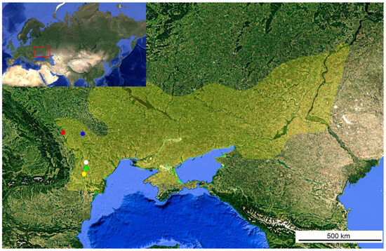
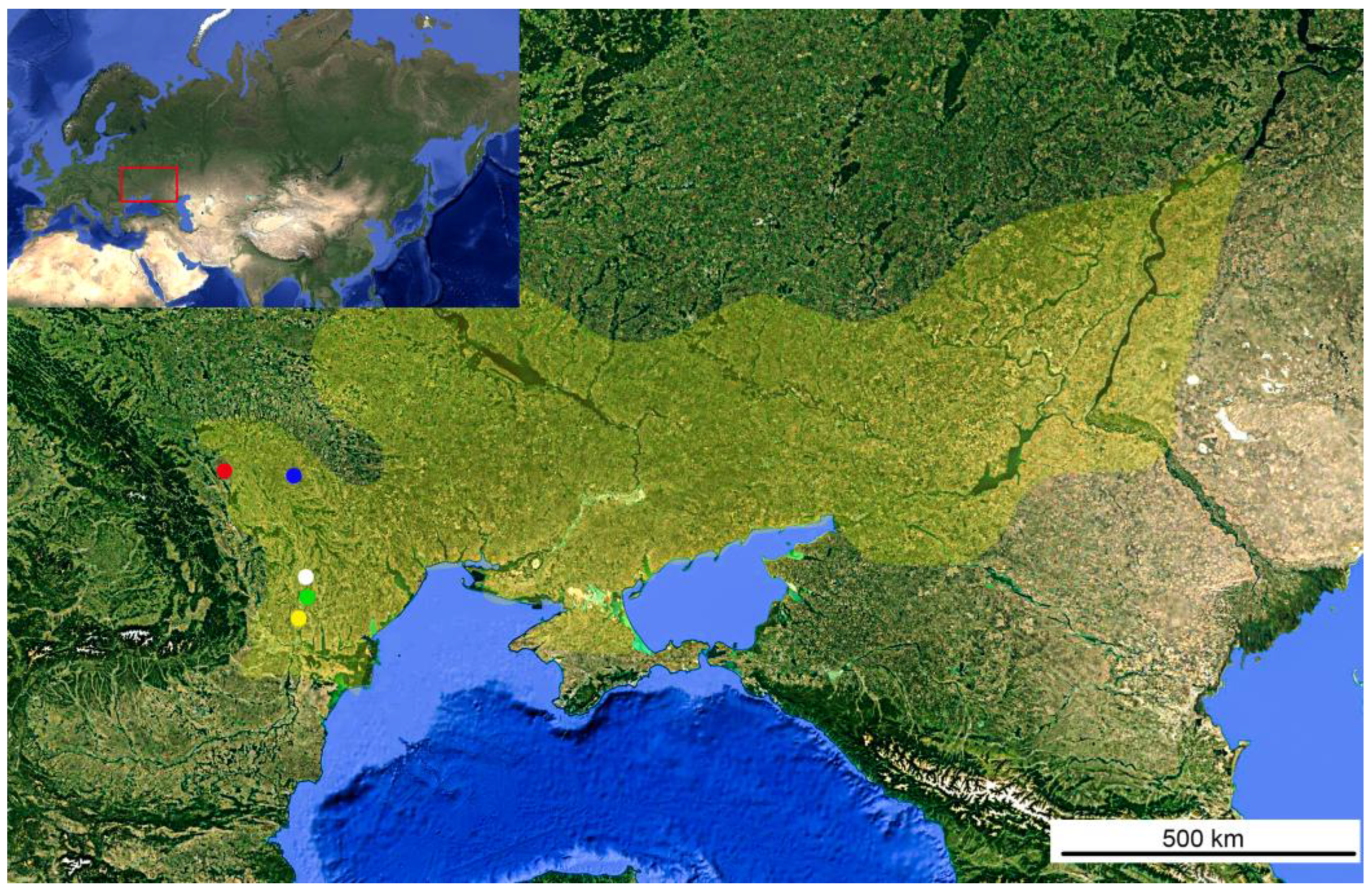
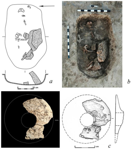
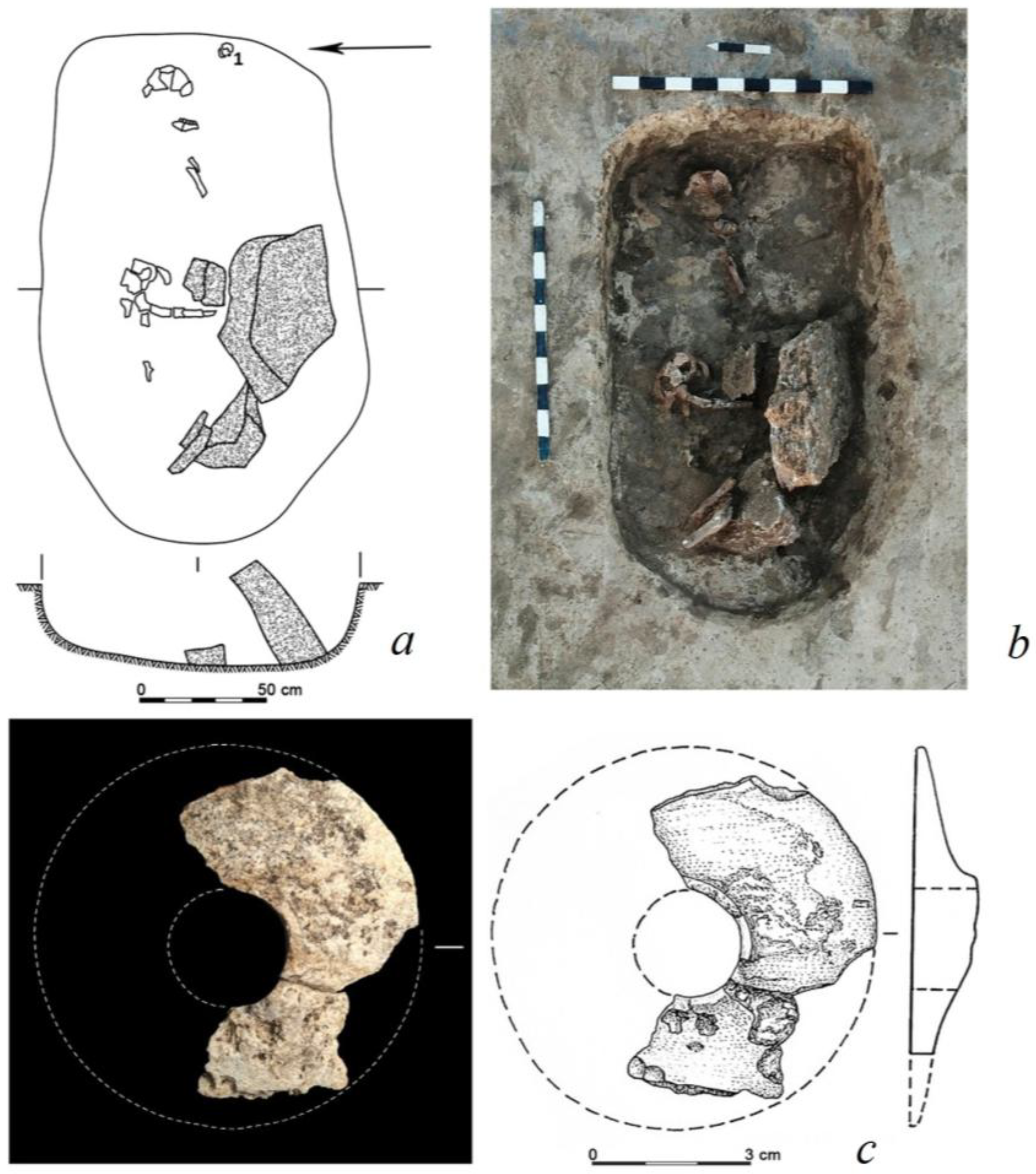
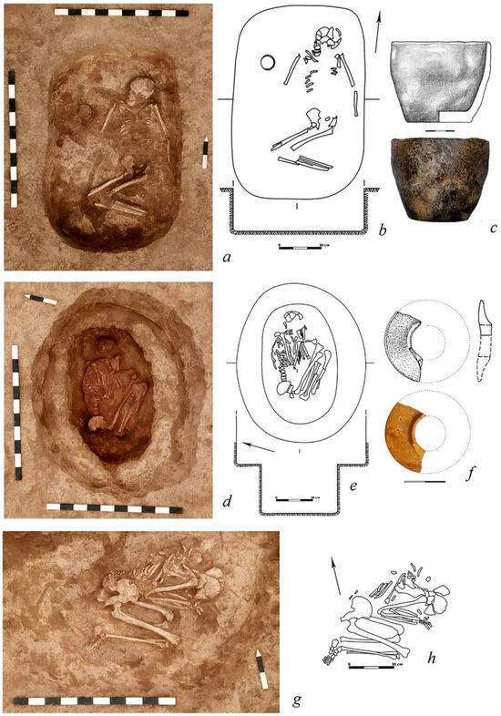
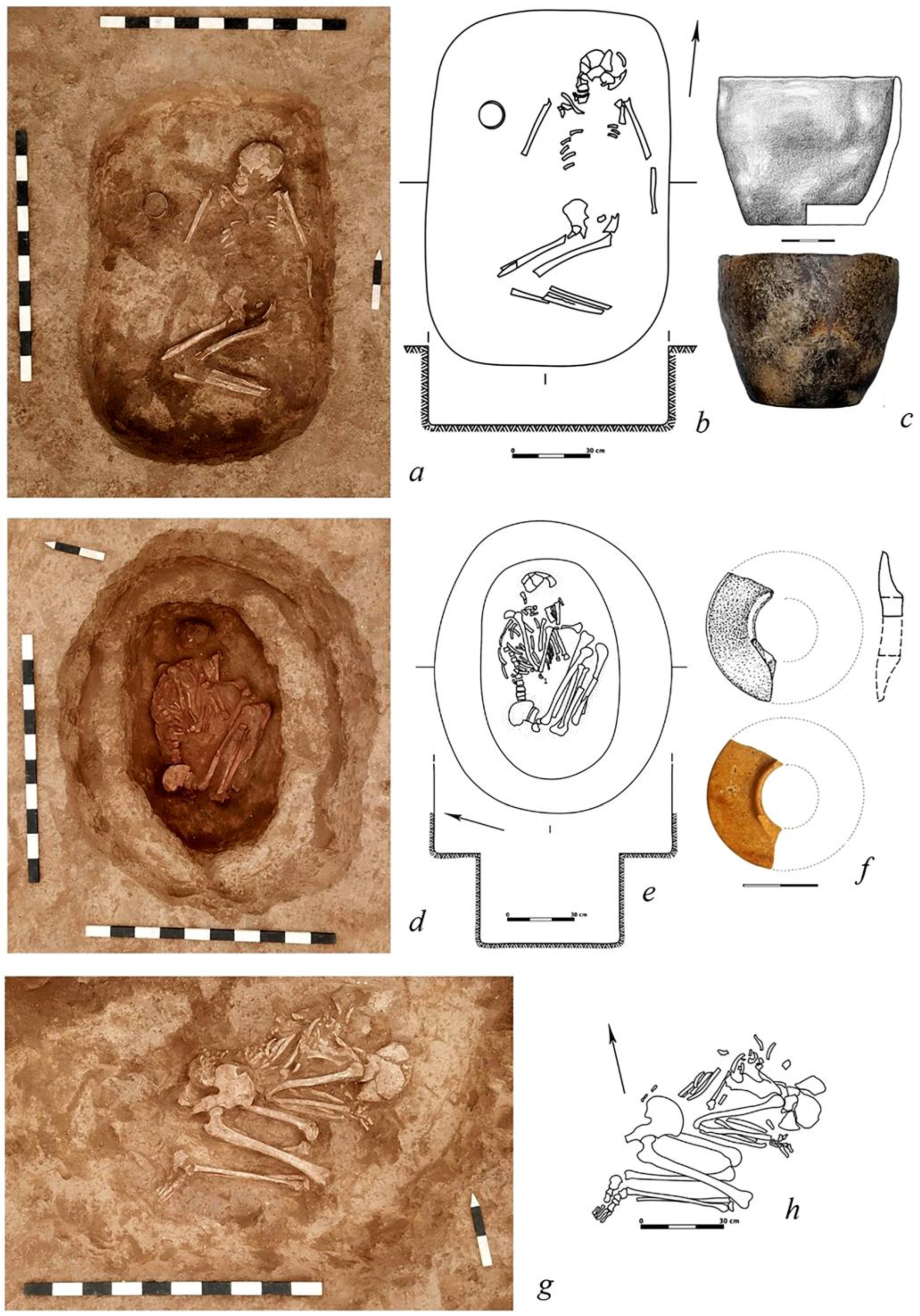
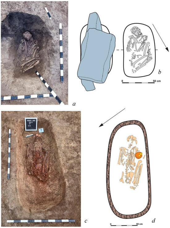
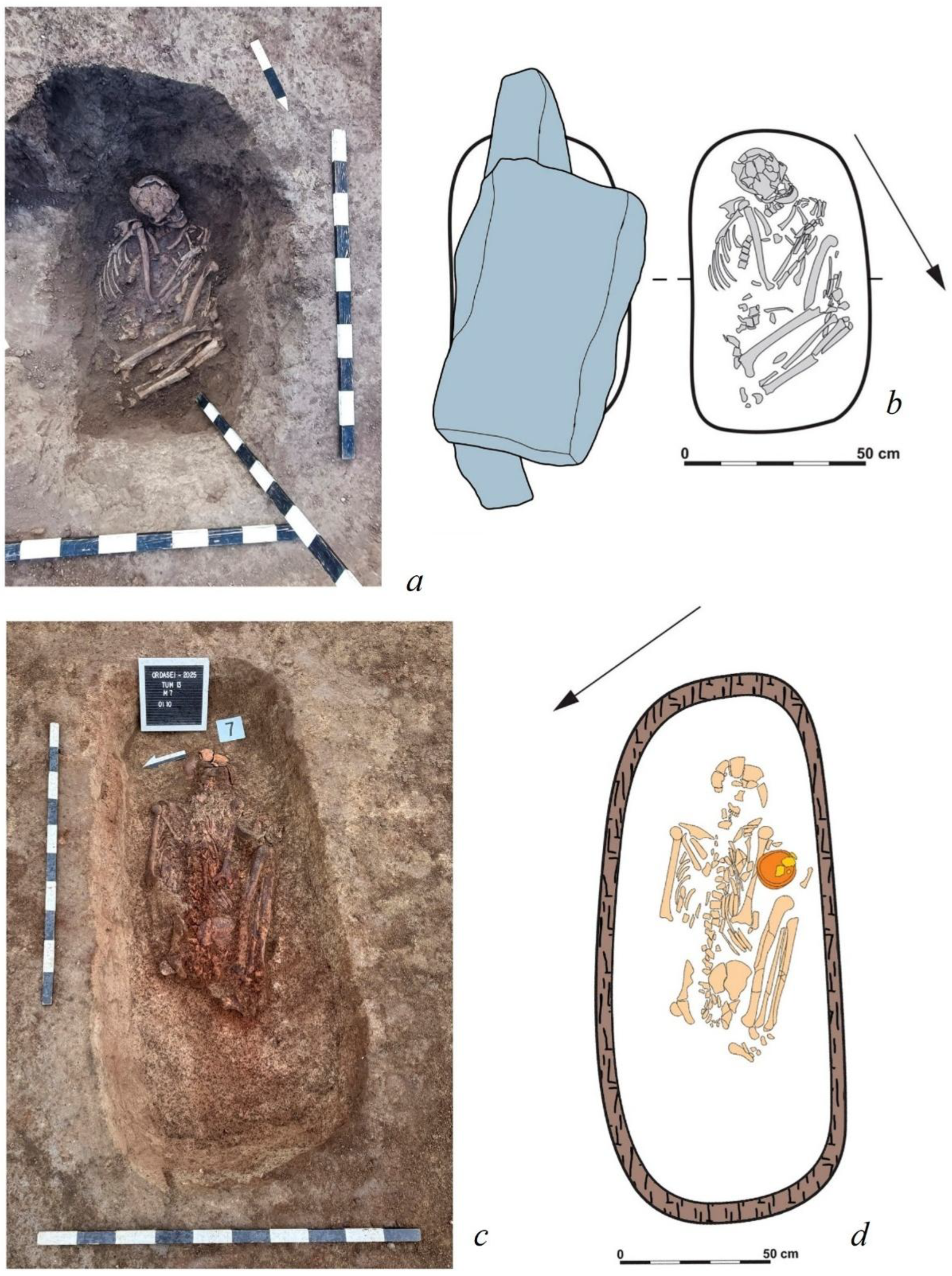
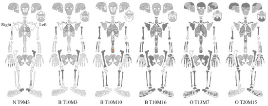
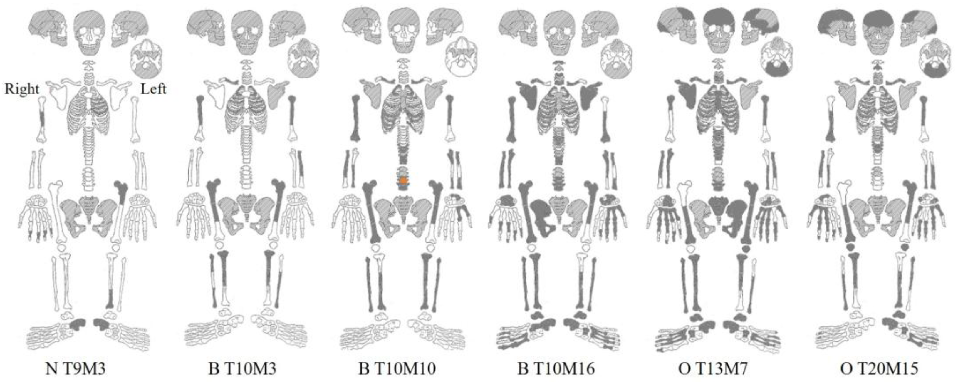
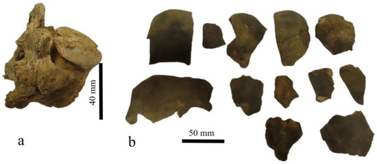
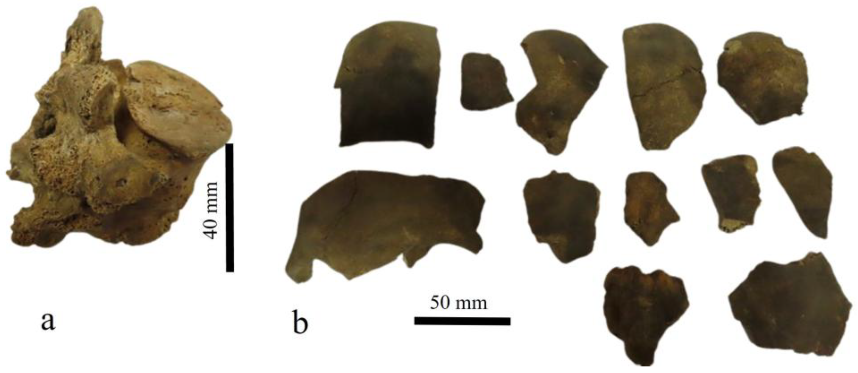
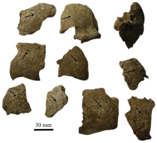
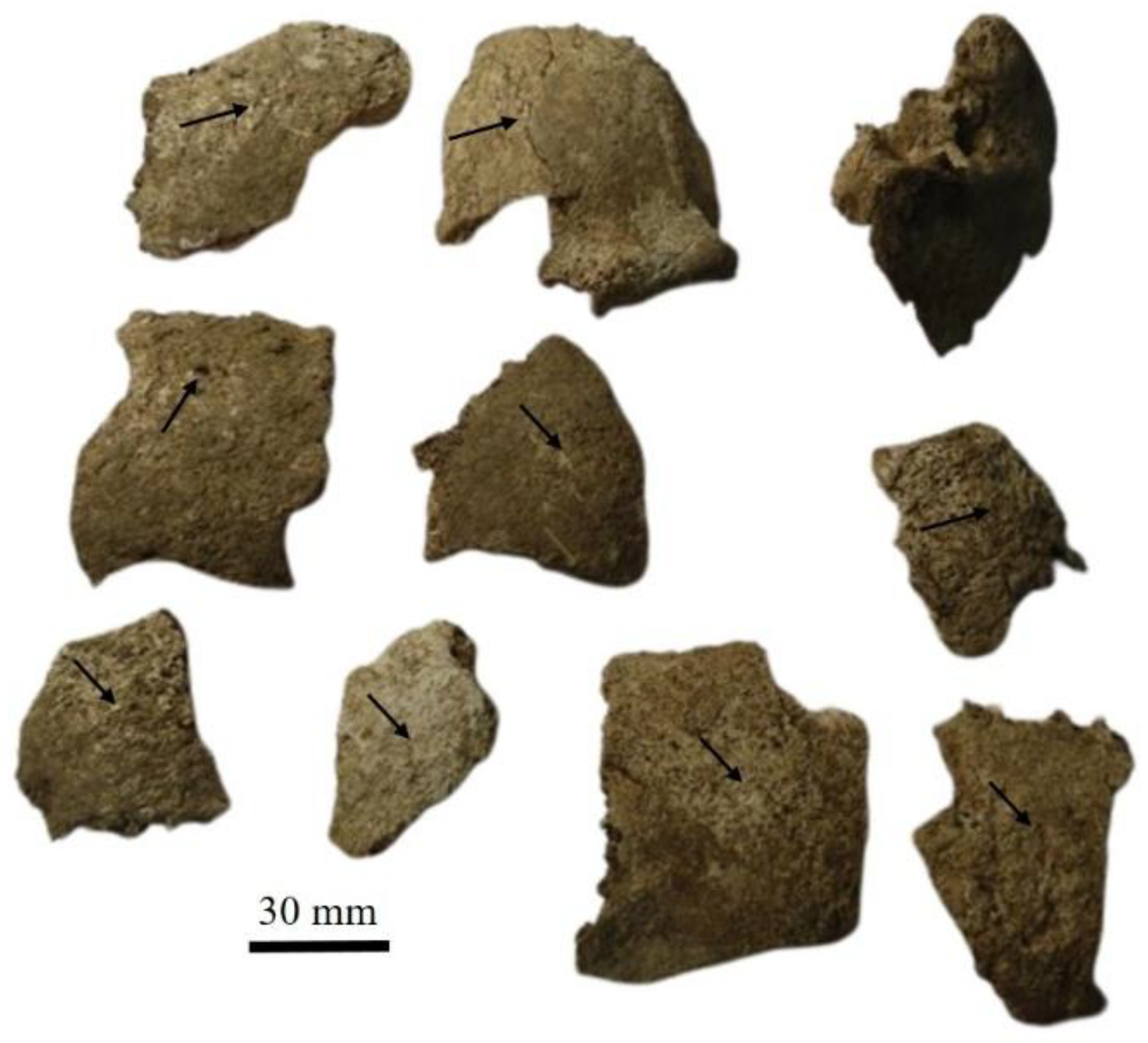
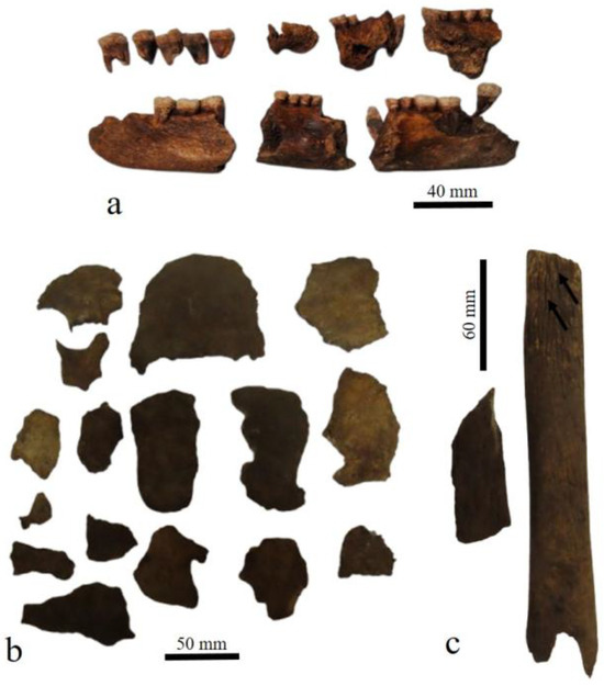
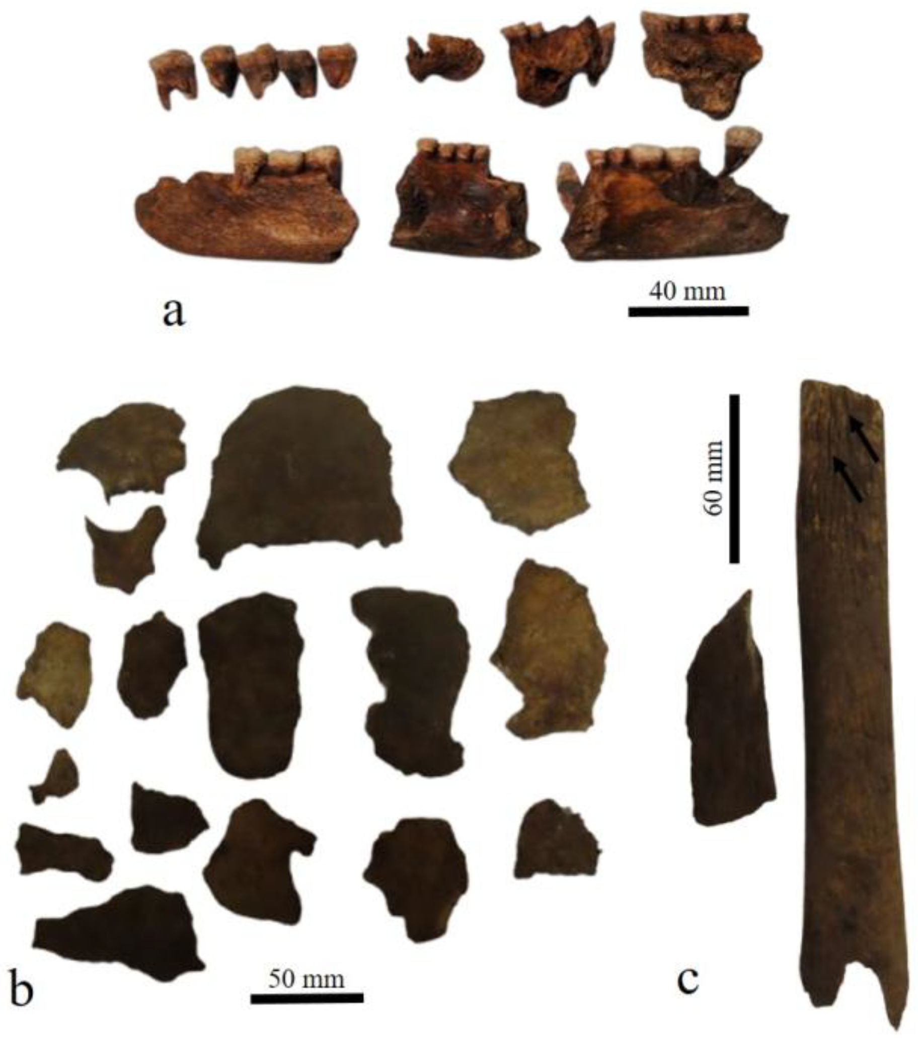
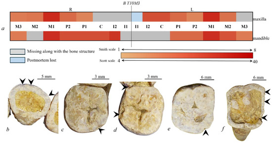
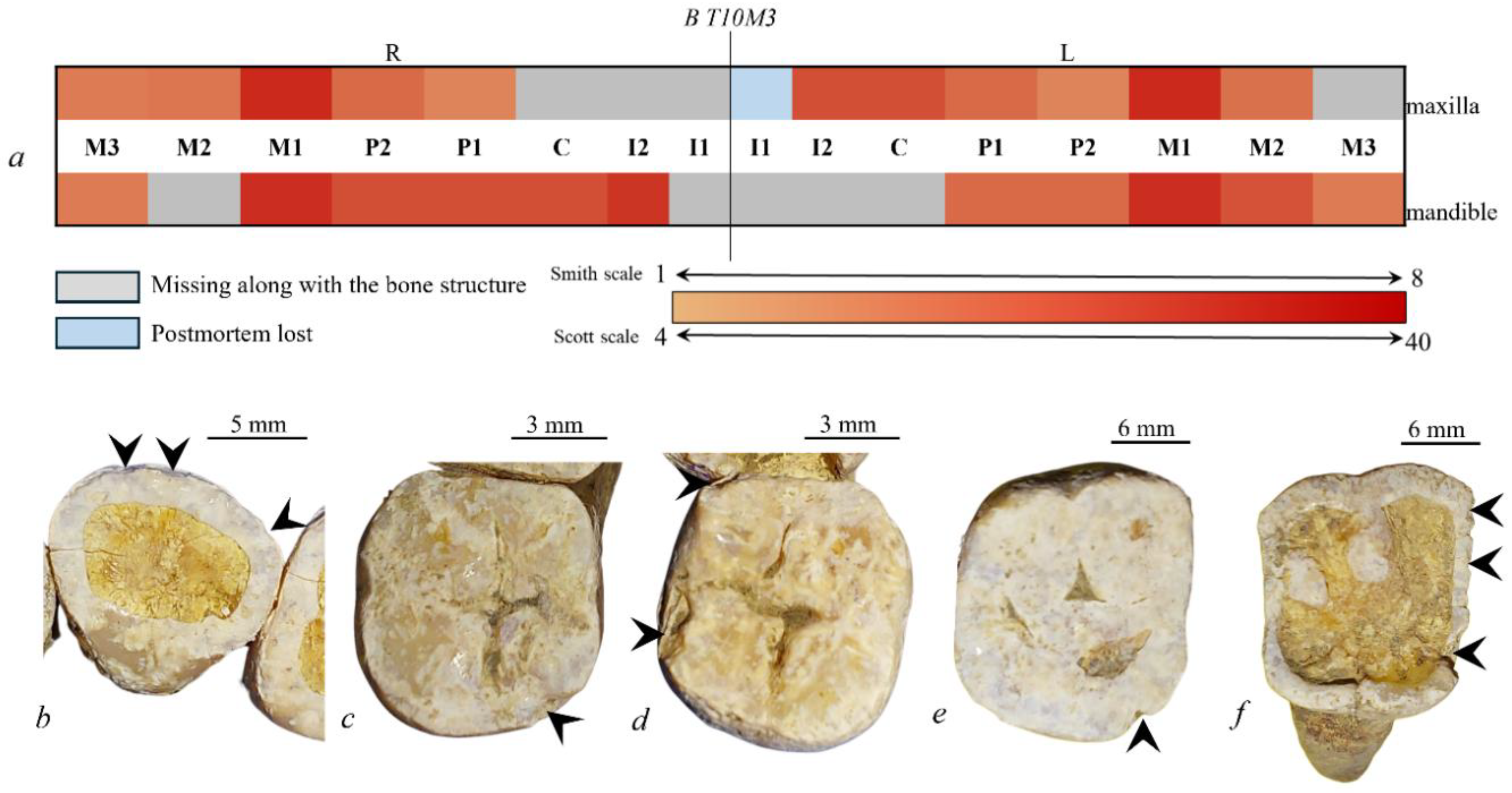
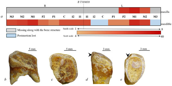
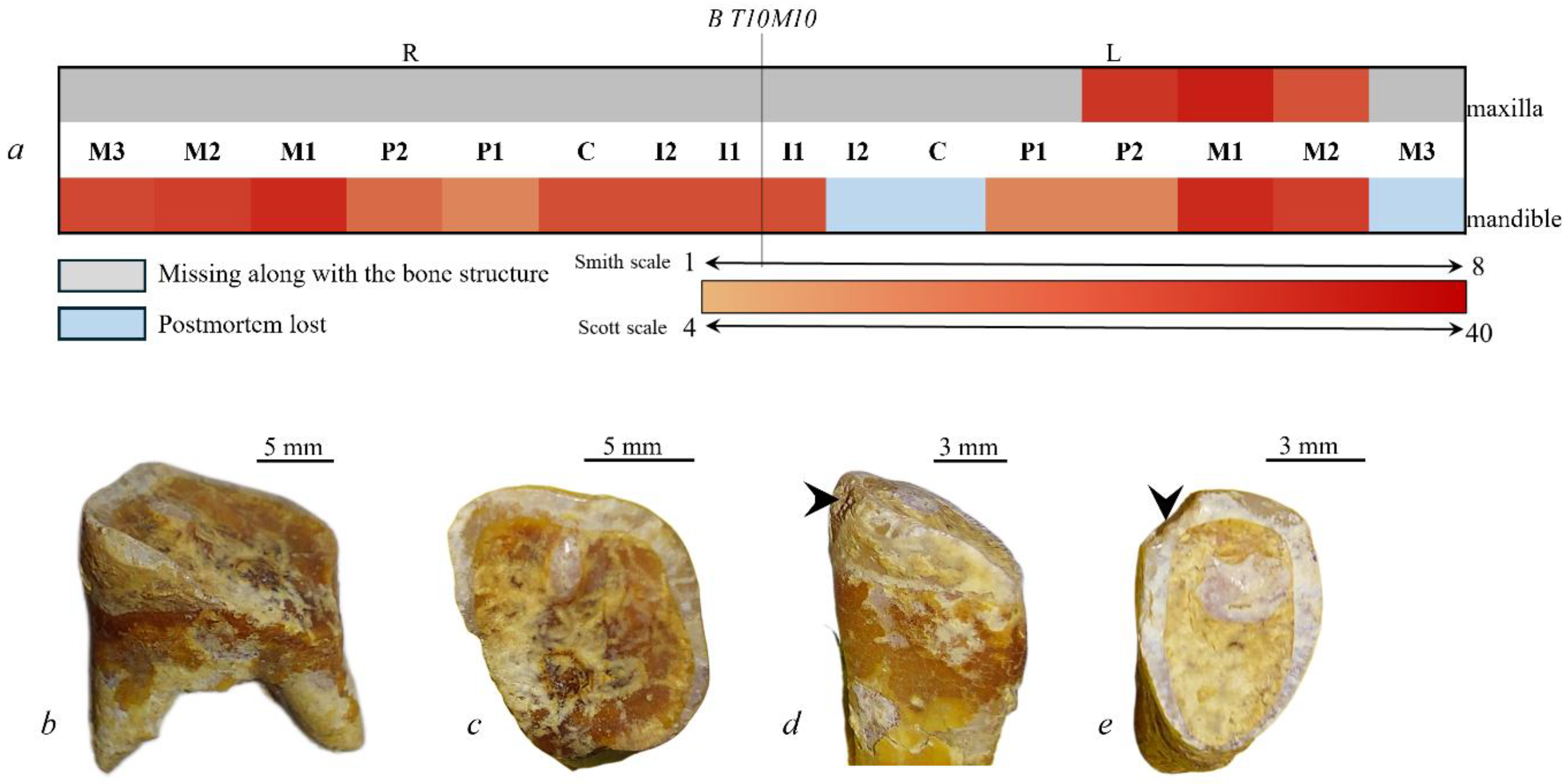
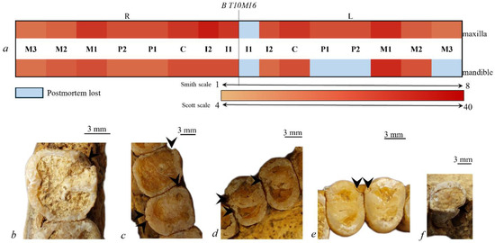
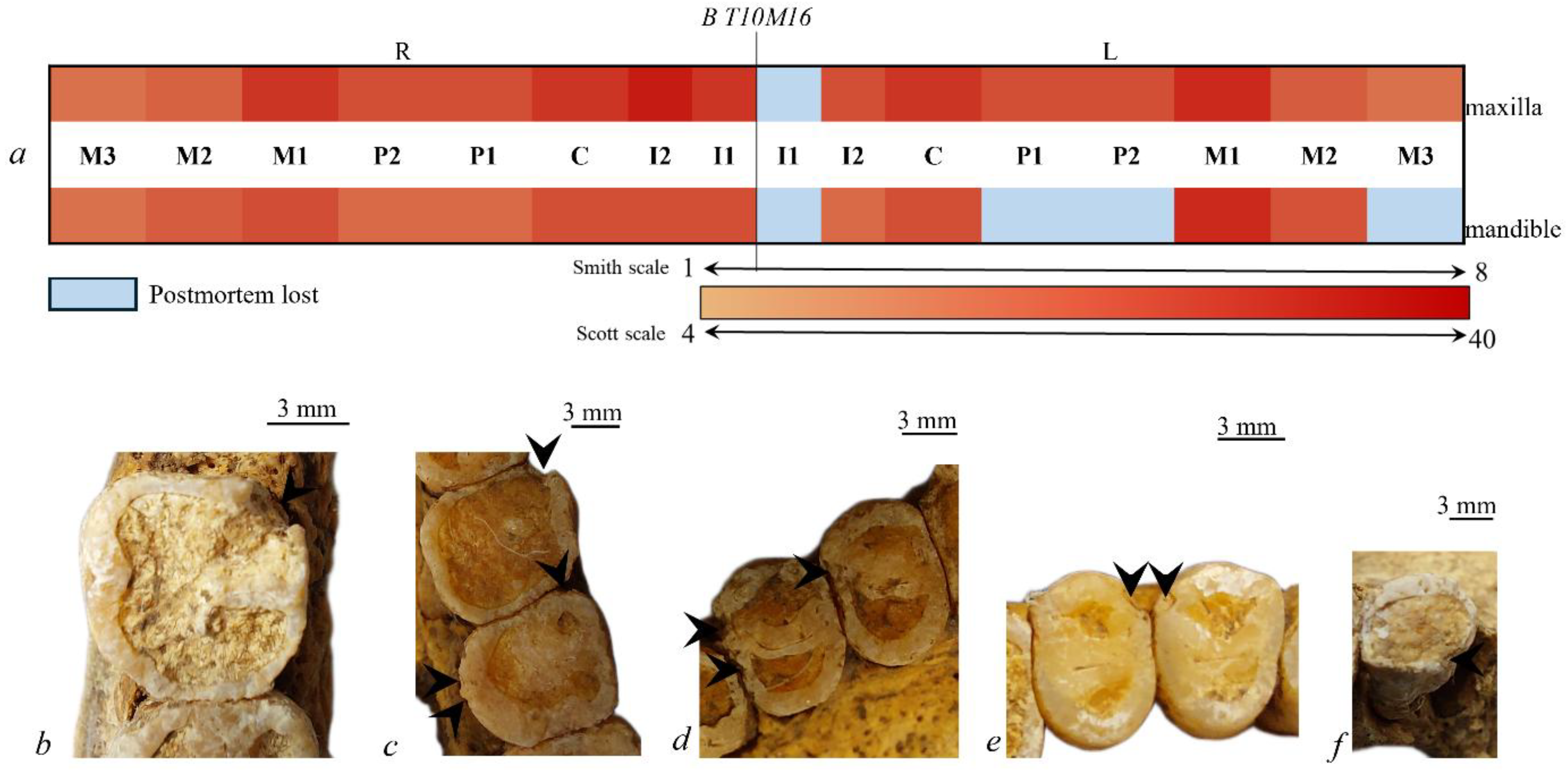
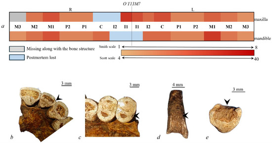
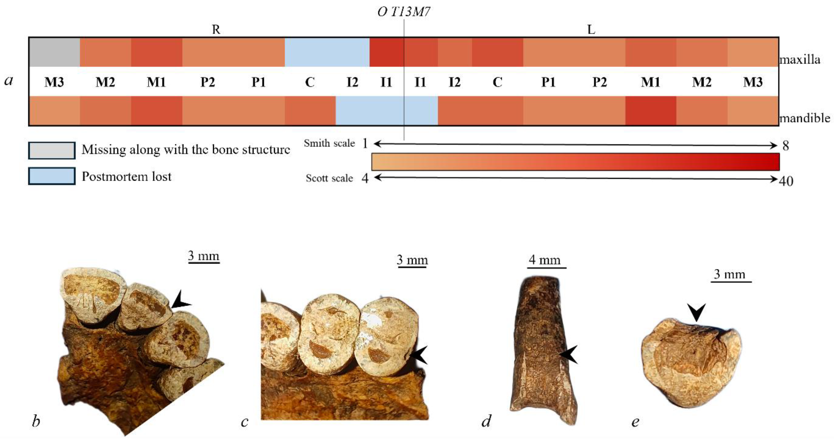
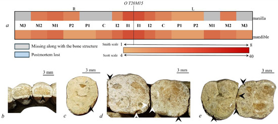
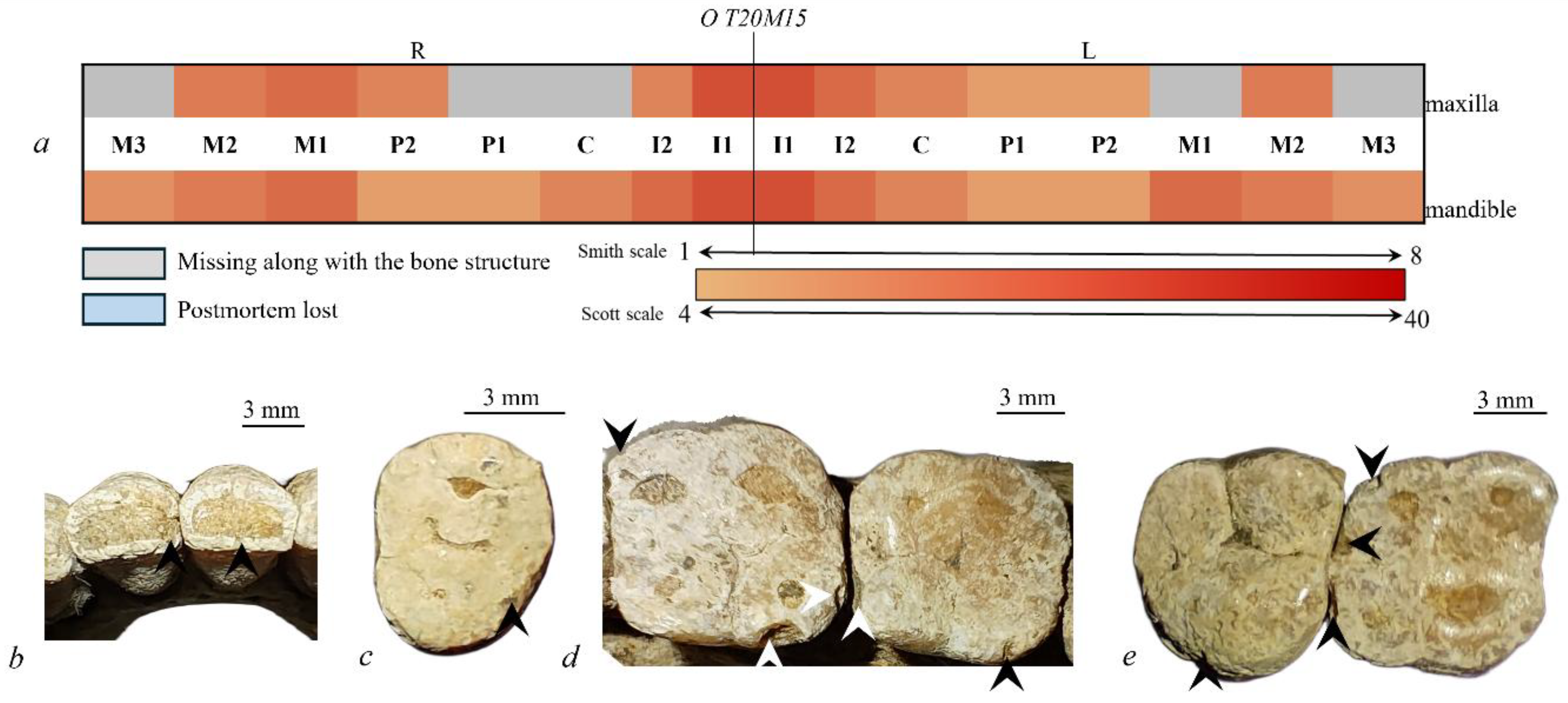
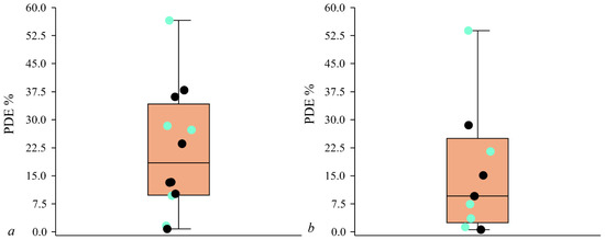
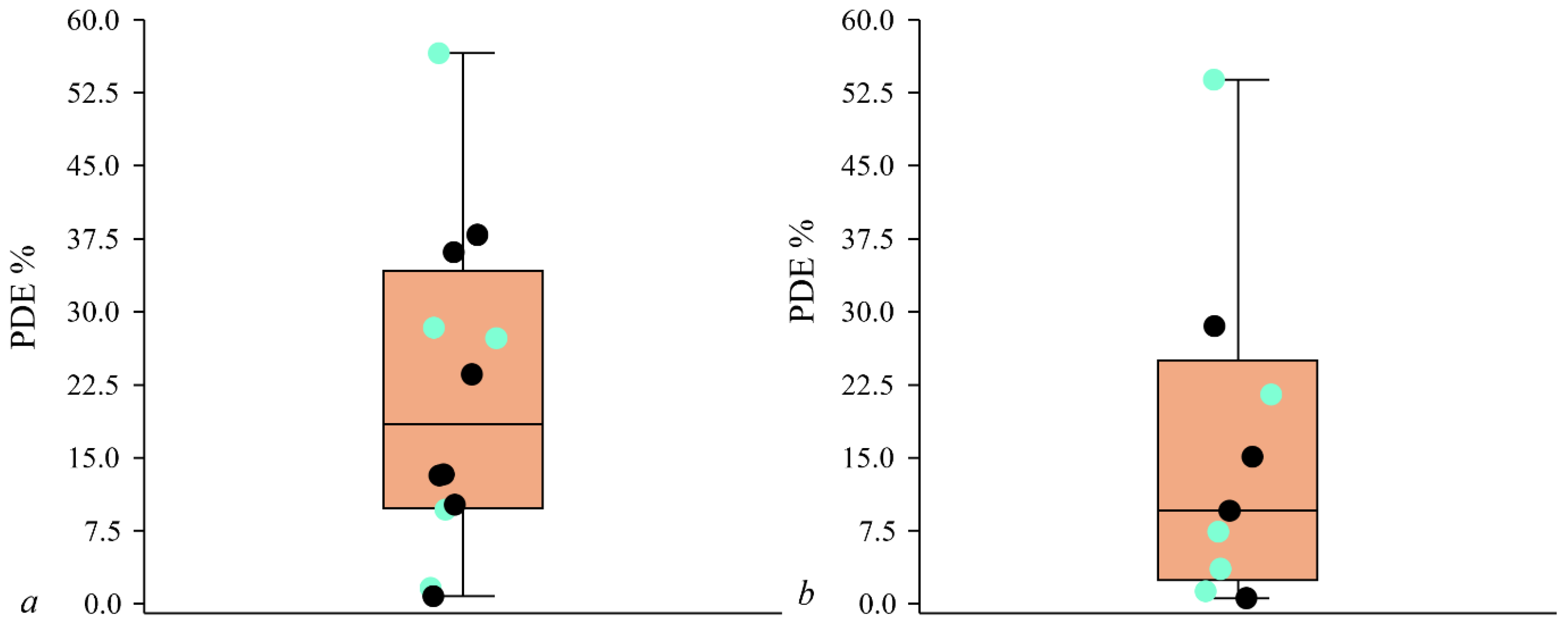
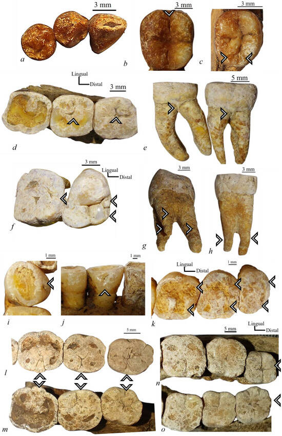
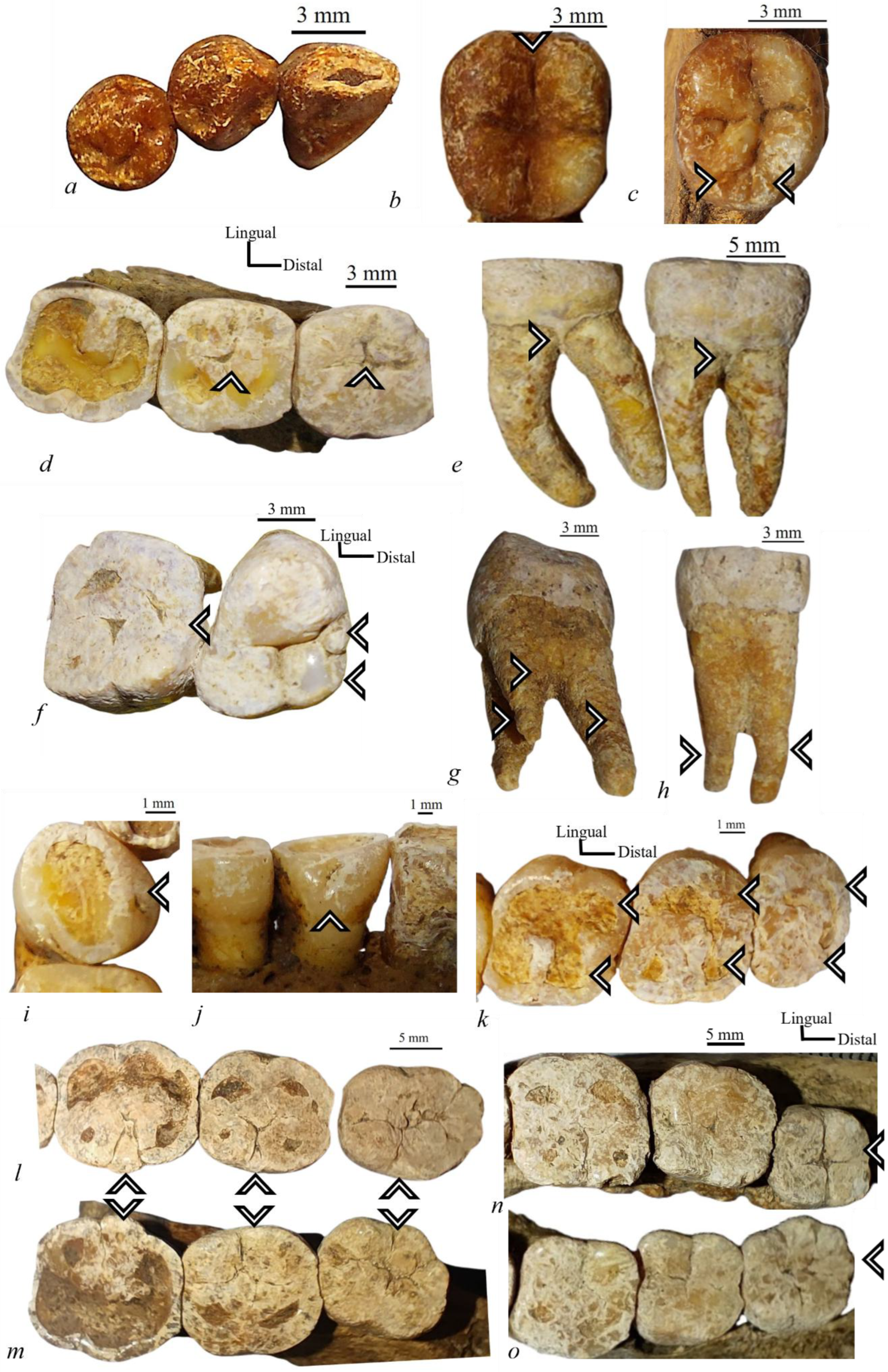
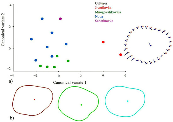
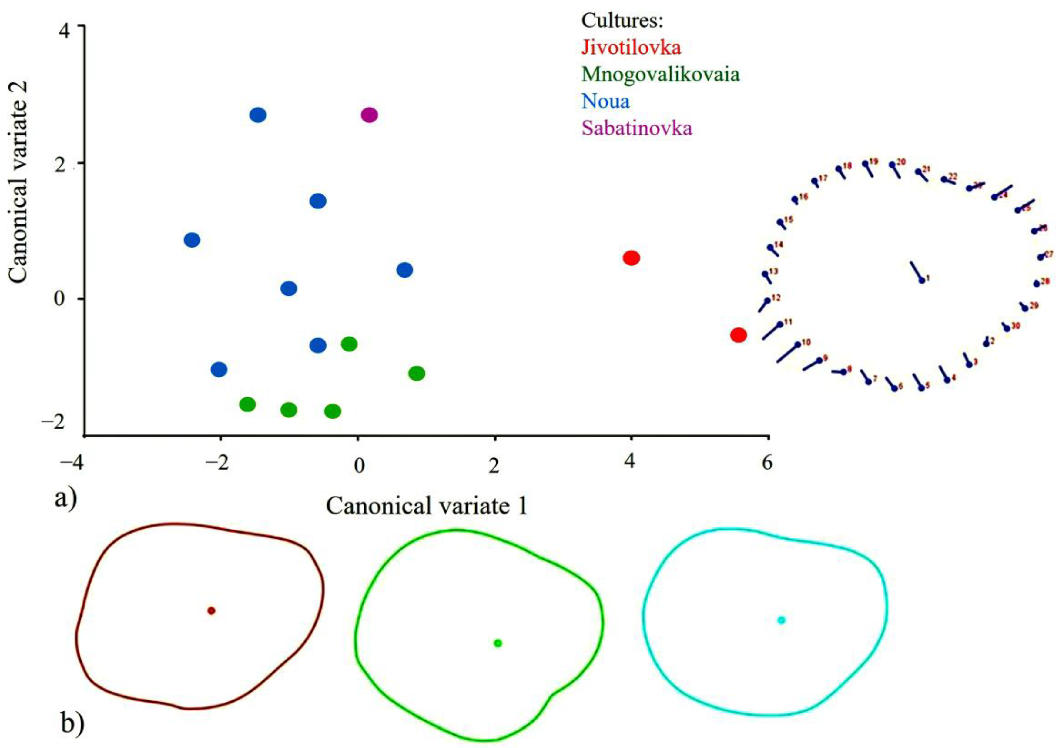
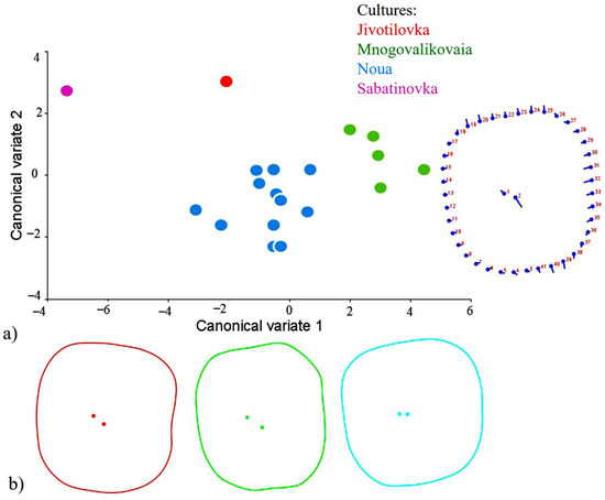
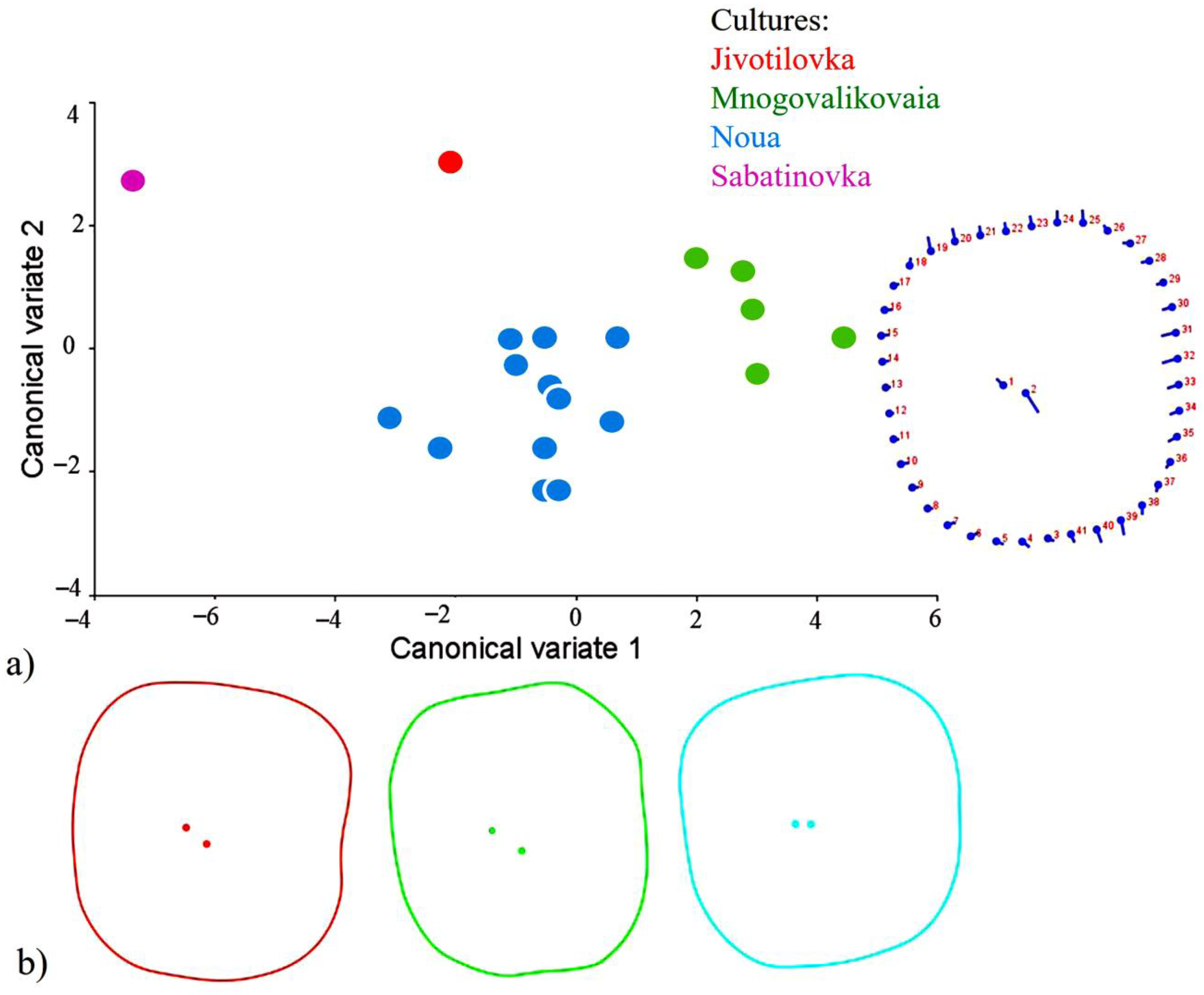

Disclaimer/Publisher’s Note: The statements, opinions and data contained in all publications are solely those of the individual author(s) and contributor(s) and not of MDPI and/or the editor(s). MDPI and/or the editor(s) disclaim responsibility for any injury to people or property resulting from any ideas, methods, instructions or products referred to in the content. |
© 2026 by the authors. Licensee MDPI, Basel, Switzerland. This article is an open access article distributed under the terms and conditions of the Creative Commons Attribution (CC BY) license.