


Abstract
The objective was to investigate the effect of an adhesive-enhancing primer (AEP) on the shear bond strength (SBS) between fresh resin composite and aged resin composite interface, specifically considering the presence or absence of saliva contamination on the aged resin composites. One hundred fifty-three resin composite blocks underwent thermocycling to replicate aging. Nine groups (n = 15) formed: Group 1: no saliva + no surface treatment; Group 2: no saliva + AEP; Group 3: no saliva + bonding agent; Group 4: no saliva + AEP + bonding agent; Group 5: saliva + no surface treatment; Group 6: saliva + AEP; Group 7: saliva + bonding agent; Group 8: saliva + AEP + bonding agent; and Group 9: saliva + phosphoric acid + bonding agent. Fresh resin composite was applied to the surface of each sample and then light-cured. All specimens were stored for 24 h in distilled water at 37 °C. The SBS testing and failure modes were assessed next. One-way ANOVA and the Tukey test (α = 0.05) were used. Group 9 had the highest SBS (15.42 ± 1.37 MPa) but did not significantly differ from Groups 3, 4, 7, and 8 (p > 0.05). Group 5 showed the lowest SBS (3.08 ± 0.41 MPa). Additionally, group 1 demonstrated significantly lower SBS than all other groups except group 5 (p < 0.001). Groups 2 and 6 exhibited comparable SBS (p > 0.05). Groups 3, 4, 7, 8, and 9 displayed both adhesive and mixed failure, whereas the other groups demonstrated a complete adhesive failure. Regardless of saliva contamination, AEP improved the bond strength at the interface between the fresh resin composite and the aged resin composite.
1. Introduction
Dental resin composite is a tooth-colored restorative material commonly used in modern dentistry for fillings, veneers, and various cosmetic and structural applications. It is designed to mimic the natural appearance and function of tooth enamel and dentin. This versatile material offers numerous advantages, including excellent adhesion to tooth structure when used with dental adhesive, as well as resistance to wear and staining. As a result, dental resin composites are becoming increasingly popular for both aesthetic and functional purposes, enhancing patients’ smiles while ensuring durability. These qualities make them a preferred choice among dental professionals, who value their ability to blend seamlessly with existing teeth. With ongoing advancements in technology, the formulation of these materials is expected to improve further, leading to enhanced performance and increased patient satisfaction. Nonetheless, various factors may lead to restoration failure, including persistent cyclic loading, micro shrinkage, and the essential requirement for moisture regulation during application [1,2].
Although resin composites can bond to tooth structure, failures can occur after use. Several factors contribute to the failure of resin composite restorations, including the oral hygiene environment, the manipulation of the composite and bonding system, and continuous cyclic loading, which includes micro-shrinkage during the curing process [3]. As a result, the resin composite restorations were reported to fail due to secondary caries, wear of the restoration, and marginal leakage [3]. To address these issues, ongoing research aims to improve the formulation of resin composites and enhance bonding techniques. Innovations in material science may lead to longer-lasting restorations and improved patient outcomes in restorative dentistry.
Failed composite restorations, characterized by chipping or small fractures, can often be partially repaired because aged resin composite retains the ability to bond with fresh resin composite materials. However, saliva contamination from the oral cavity to the restoration surface might happen unintentionally, and this contamination is challenging to eliminate with water alone because it deposits an organic adhesive coating on the restorative materials within the initial seconds of exposure [4,5]. This coating layer is resistant to washing and acts as a physical or organic barrier (glycoproteins/mucins) that prevents monomer penetration and wetting, reducing bond strength by preventing chemical polymerization with the subsequent resin layer [6].
This highlights the necessity for research focused on interventions aimed at restoring bond strength in these situations. Numerous studies have examined the influence of saliva on the shear bond strength (SBS) of resin composites [7,8,9]. Two studies demonstrated that applying morpholine to an aged resin composite contaminated with saliva, before using the adhesive agent, improved the bond strength of the repaired composite [8,9]. Furthermore, these two studies employed morpholine to address the saliva-contaminated resin composite without prior rinsing before repair. However, there remains insufficient information concerning alternative surface treatments to augment the SBS of repaired resin composites utilizing other chemical agents without rinsing, including the possible efficacy of primers that may also enhance bond strength.
Our study involves an experiment utilizing G-CEM ONE Adhesive Enhancing Primer (AEP), which is applicable in a variety of clinical situations due to its excellent bond strength to both tooth structures and various substrates [10,11]. This information could assist in reducing the sensitivity of the bonding process in resin composite restorations, even in the presence of saliva contamination. Normally, AEP is a primer that is recommended by manufacturers for intraoral use by applying it to the tooth surface before using the resin cement. This primer includes key ingredients, 4-methacryloyloxyethoxycarbonylphthalic acid (4-MET) and 10-Methacryloyloxydecyl dihydrogen phosphate (10-MDP) [10]. The AEP application process enhances adhesion properties, creating a strong bond between the tooth structure and the cement [11,12]. Consequently, clinicians can achieve better long-term outcomes for restorations, thereby reducing the risk of failure associated with compromised bond strength.
According to a study by Pornatitanakul [13], a primer that contains the 10-MDP functional monomer has demonstrated the ability to mitigate the adverse effects of saliva on the SBS of resin–zirconia interfaces. Notably, this primer alters the contaminated surface without requiring rinsing. Based on this result, MDP may function as a surfactant [14]. Consequently, it was intriguing to explore the use of AEP for treating aged resin composite in the context of repairs with fresh resin composite. The aim of this study was to investigate the effect of AEP, which includes 10-MDP, on the SBS of repairs between fresh and aged resin composites, considering both saliva-contaminated and uncontaminated conditions. The primary null hypothesis states that AEP does not influence the SBS between fresh and aged resin composites. The secondary null hypothesis states that AEP does not affect the SBS between fresh and aged resin composites that have been exposed to saliva.
2. Materials and Methods
The experimental part of this study received ethical approval from the Human Research Ethics Committee of the Faculty of Dentistry, Chulalongkorn University (HREC-DCU), under research code 002/2023.The approval number is 069/2023, certified on 7 July 2023.
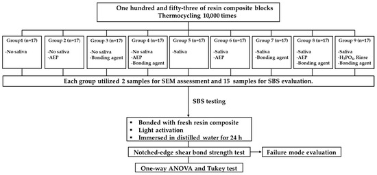
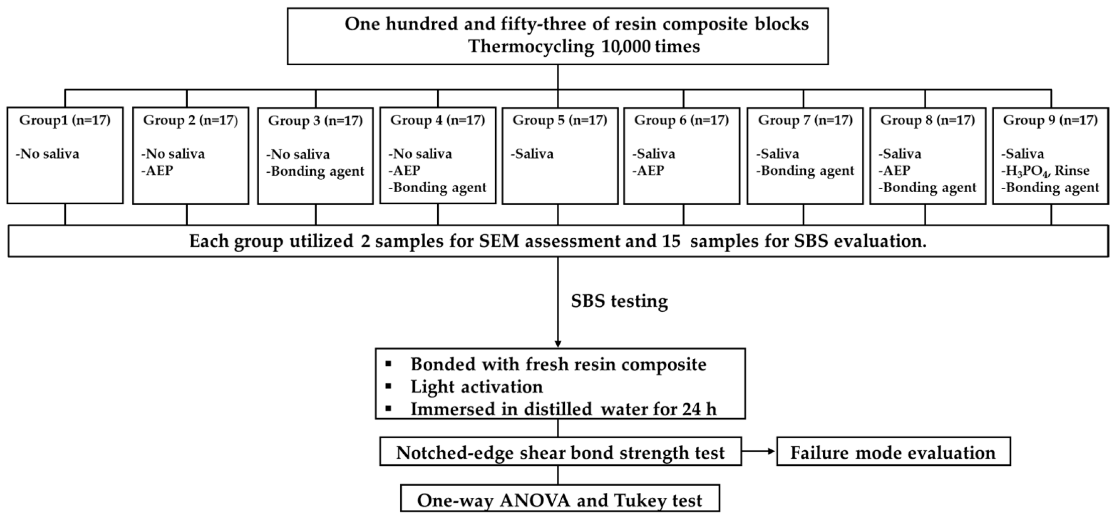
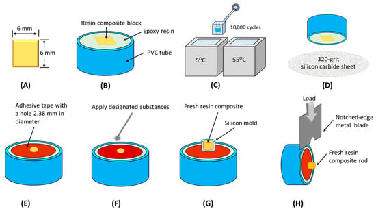
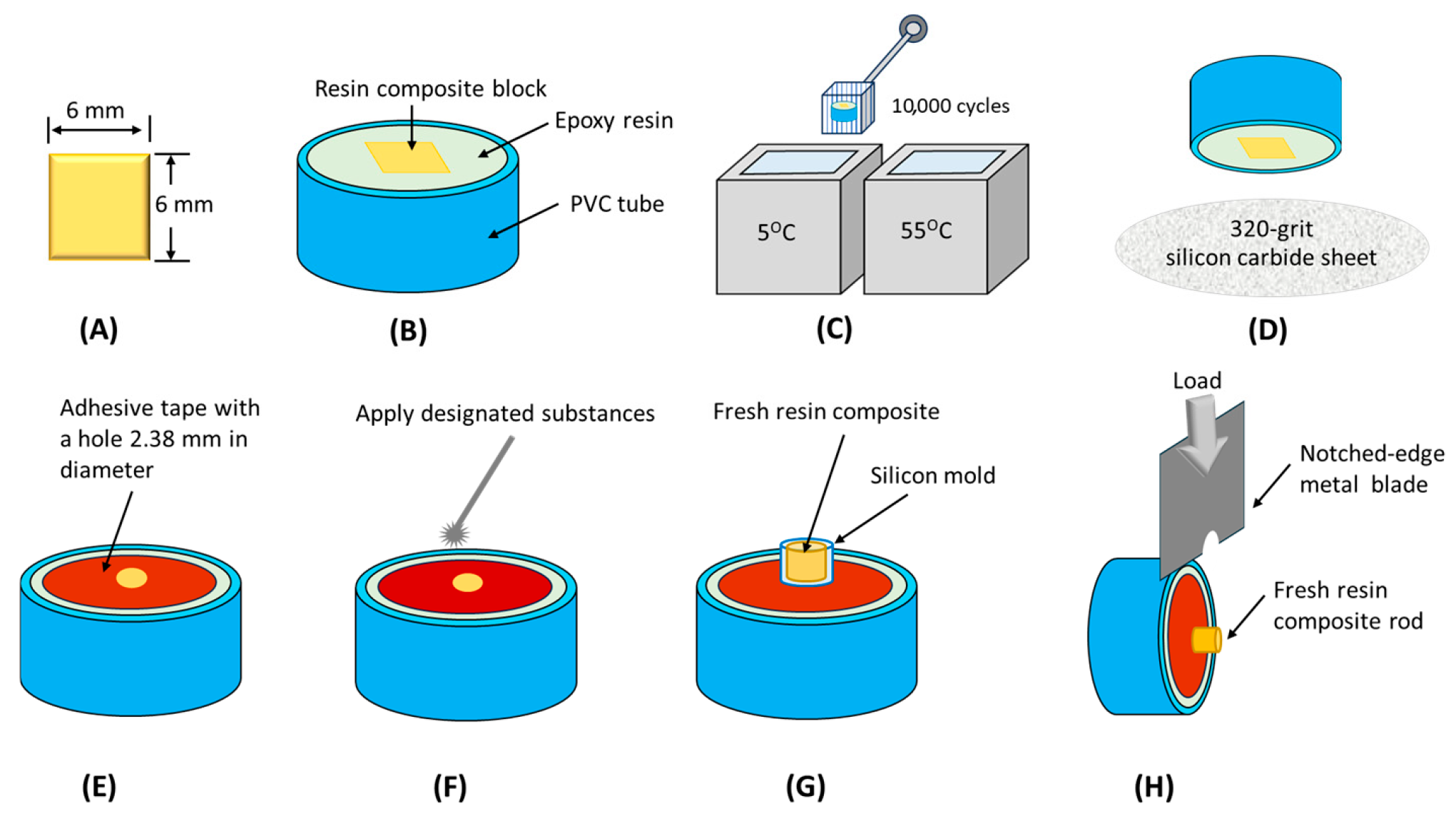
The materials used in this study are shown in Table 1. Additionally, Figure 1 and Figure 2 illustrate the experimental flowchart and the specimen preparation stage, respectively.
Table 1.
Name, manufacturer, lot number, and chemical compositions of the materials.
Figure 1.
A flowchart illustrating the experimental procedure used to determine SEM evaluation and SBS, as well as to identify the mode of failure.
Figure 2.
Depiction of the methodologies employed in this study: (A) Square shape of cured resin composite; (B) Resin composite embedded in epoxy resin within a PVC tube; (C) Aging of the resin composite through thermocycling; (D) Abrasion of the aged resin composite using silicon carbide sheet; (E) Adhesive tape with a central hole applied to the surface of the aged resin composite; (F) Application of designated substances onto the surface of the aged resin composite; (G) Fresh resin composite adhered to the aged resin composite; (H) Notched-edge shear bond strength testing.
2.1. Specimen Preparation
The sample size was analyzed using the G*power program (Version 3.1.9.7, Heinrich-Heine Düsseldorf University, Düsseldorf, Germany). Based on the pilot investigation (n = 5 per group), a total of 117 samples were determined (effect size f = 0.46, α err prob = 0.05, power (1 − β err prob) = 0.95, number of groups = 9). Consequently, a minimum of 13 samples was required for each group. In this study, 15 samples per group were utilized to accommodate a 10% error margin, resulting in 135 samples allocated for SBS testing. Additionally, there were 2 samples designated for SEM evaluation for each surface treatment. Therefore, the total number of samples amounted to 153. The specimens were then randomly divided into 9 groups; each group underwent different resin composite surface treatment methods as follows:
Group 1: No saliva;
Group 2: No saliva, AEP;
Group 3: No saliva, Bonding agent;
Group 4: No Saliva, AEP, Bonding agent;
Group 5: Saliva;
Group 6: Saliva, AEP;
Group 7: Saliva, Bonding agent;
Group 8: Saliva, AEP, Bonding agent;
Group 9: Saliva, Phosphoric acid, Rinse, Bonding agent.
2.2. Aged Resin Composite Block Preparation
A total of 153 resin composite blocks (3M™ ESPE™ Filtek™ Z350 XT, 3M ESPE, St. Paul, Maplewood, MN, USA) were prepared by placing resin composites into a 6 × 6 × 4 mm silicone mold with a flat surface area. The resin composite was condensed with a hand instrument in 2-mm incremental layers and polymerized with a light curing unit (Elipar Freelight2 LED curing light, 3M ESPE, St. Paul, Maplewood, MN, USA) with 400–515 nm wave length range at 1000 mW/cm2 for 20 s. The light cure was positioned perpendicular to the surface of the resin composite, as close to the resin composite as possible. The sample was then taken out of the mold, and a 20-s light cure was conducted on either side of the resin composite in the same manner. The resin composite blocks were placed inside a PVC tube with a diameter of 26 mm, fixed with epoxy resin, and left to set for 24 h. All samples’ surfaces were polished with a polishing machine (Nano 2000 grinder-polisher, Pace Technologies, Tucson, AZ, USA) utilizing 400, 800, and 1200-grit silicon carbide sheet (3M Wetordry abrasive sheet, 3M, Maplewood, MN, USA) at 250 rpm and a force of 50 N, employing continuous water cooling to guarantee uniform surface preparation. To produce aged resin composite, a thermocycler (Thermocycler THE 1100/1200, SD-Mechatronik GMBH, Westerham, Germany) was used to mimic the temperature changes in the oral environment. The resin composite blocks underwent thermocycling for 10,000 cycles, with a dwell time of 30 s at temperatures of 5 °C and 55 °C [15]. Subsequent to thermocycling, the resin composite specimens were subjected to wet abrasion utilizing 320-grit abrasive paper [16]. The procedure was executed at a velocity of 250 rpm and exerted a force of 50 N for a duration of 15 s. The silicon carbide sheet was replaced after processing every 10 samples. The samples’ surfaces were subsequently sprayed with water for 10 s and then dried with oil-free air for 10 s using a triple syringe from a mobile dental unit, which operated at a pressure of 2.5 bars from a distance of 1 cm. This technique replicated dental bur grinding, efficiently eliminating the superficial resin layer and producing uniform repair surfaces.
2.3. Saliva Collection
Unstimulated saliva was collected using the draining method. The subject remained still, kept their mouth open, and tilted their head down to allow saliva to drip passively from the lower lip into a sterile tube. Saliva collection occurred at least two hours after breakfast. The subject refrained from brushing their teeth for 45 min prior to saliva collection. Only one researcher served as the subject for the saliva collection and remained consistent throughout the experiment. The saliva sample was used immediately as fresh saliva.
2.4. Bonding Area Preparation
The single-sided adhesive tape (3M-ESPE, St. Paul, Maplewood, MN, USA), with a circular hole measuring 2.38 mm in diameter (ISO 29022:2013) [17], was securely affixed to the center of the aged resin composite block to define the bonding area.
2.5. Surface Modification Preparation
The procedure for this step was performed at ambient temperature, approximately 24 °C.
The surface of the aged resin composite block of groups 1–4 was free from saliva contamination.
Group 1: The surface did not undergo any treatment with chemical reagents.
Group 2: The surface was treated with G-CEM ONE™ Adhesive Enhancing Primer (AEP). A single drop of AEP was applied using a microbrush to the surface of the aged resin composite, followed by agitation for 5 s. Subsequently, oil-free air was directed onto the surface for 10 s using a triple syringe from a mobile dental unit, with a pressure of 2.5 bars and a distance of 1 cm maintained.
Group 3: The surface was treated with G-Premio BOND™ (Bonding). A single drop of the bonding agent was applied with a microbrush to the surface of the aged resin composite, followed by 5 s of agitation. Subsequently, air was blown onto the surface in the same manner as in Group 2. Finally, the bonding agent was polymerized with light for a duration of 20 s.
Group 4: The surface underwent treatment with AEP followed by Bonding agent. The procedures implemented were identical to those specified in groups 2 and 3.
Groups 5–9 exhibited saliva contamination. A micropipette extracted 50 microliters of saliva, which was subsequently applied to the surface of the aged resin composite with a disposable microbrush, traversing from left to right in a single motion. The region was delicately dried with oil-free air spray for 20 s.
Groups 5, 6, 7, 8 received the same surface treatments as groups 1, 2, 3, 4, respectively.
Group 9: The aged resin composite, which had been contaminated with saliva, was etched using a 37% phosphoric acid gel (Scotchbond™ Etchant, 3M ESPE, Maplewood, MN, USA) for 15 s. It was then rinsed with water and dried for 10 s using a triple syringe air system. The bonding agent was applied in the same manner as described in Group 3.
2.6. Scanning Electron Microscopy (SEM) Evaluation
Two samples were randomly selected from each group for SEM examination at a magnification of 2000× (SEM microscope, FEI Company, Versa 3D, Hillsboro, OR, USA).
2.7. Adhesion Procedures for Fresh Resin Composite to Aged Resin Composite
Subsequent to the application of adhesive tape (2.38 mm diameter hole) to the samples’ surfaces, they were subjected to surface treatment as specified in groups 1–9. A transparent silicone mold featuring a central aperture, measuring 3 mm in diameter and 3 mm in height, was placed over the adhesive tape hole on the surface of each sample. The fresh resin composite was compacted against the aged resin composite using a hand instrument. Subsequently, polymerized using light for a duration of 40 s. All bonded specimens were then stored in distilled water and placed in an incubator (Contherm 1200 lab incubator, Contherm, New Zealand) at 37 °C for 24 h before testing.
All surface treatments and the adhesion of fresh resin composite to aged resin composite were performed by a single trained and calibrated operator to minimize variability related to the operator.
2.8. Shear Bond Strength Test and Mode of Failure Evaluation
The universal testing machine (AGS-X 500N, Shimadzu Corporation, Kyoto, Japan) was used to measure SBS. The notched-edge crosshead blade was aligned parallel to the bonded surface. The crosshead speed was set to 1 mm/min, and data were recorded in megapascals. Each fractured specimen was examined at a magnification of 40× using a stereo microscope (SZ61, Olympus Corporation, Tokyo, Japan) to assess the failure surfaces. The failure modes, adapted from previous studies [18], were classified into three categories: (a) An adhesive failure was defined as occurring when less than 40% of the fresh resin composite was visible on the surface of the aged resin composite. (b) A cohesive failure was identified when at least 60% of the fresh resin composite was exposed on the aged resin composite surface. (c) A mixed failure, which included both cohesive and adhesive failures, was identified when more than 40% but less than 60% of the fresh resin composite was visible on the aged resin composite. The percentage of area for each failure mode was measured using ImageJ software version 1.54p (http://imagej.net/ij/download.html, accessed on 6 October 2025). The area occupied by the fresh resin composite on the aged resin composite surface was measured twice by two independent operators. The mean measurement area was divided by the total bonded surface area (πr2, with r = 2.38/2) and then multiplied by 100.
2.9. Statistical Analysis
IBM SPSS.V29.0.1 (SPSS Inc., Chicago, IL, USA) was used to evaluate the quantitative data from the nine separate groups at a 95% confidence level. Normality was assessed using the Shapiro–Wilk test at a significance level of 0.05. The equality of variation was then examined using Levene’s test. The data had equal variance and a normal distribution, according to the findings. Therefore, the data was analyzed using a one-way ANOVA, and the differences between groups were then ascertained using a post hoc Tukey test (p < 0.05).
3. Result
3.1. SEM Evaluation
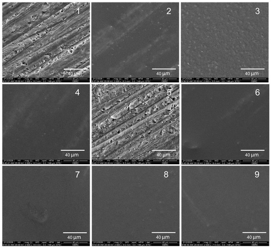
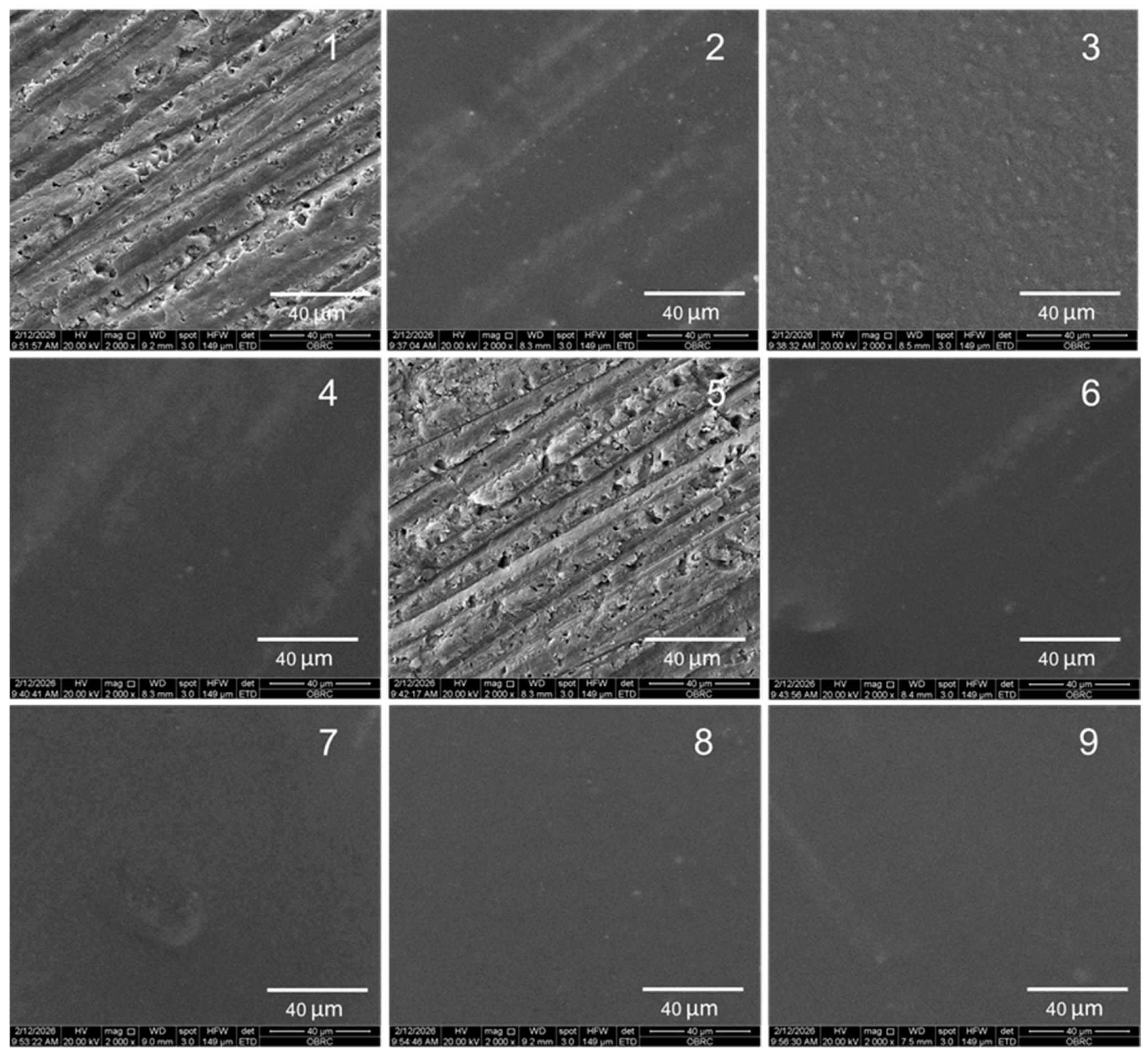
The SEM images reveal distinct surface characteristics of aged resin composites. Image (1) illustrates the surface of aged resin composite from group 1, which received no treatment. In contrast, image (5) depicts the surface of a saliva-contaminated aged resin composite from group 5, also untreated. Both pictures show long grooves and uneven pores on their surfaces. Conversely, images (2), (3), (4), (6), (7), (8), and (9) represent groups 2, 3, 4, 6, 7, 8, and 9, respectively, and they exhibit a noticeable loss of grooves and pores (Figure 3).
Figure 3.
SEM images at 2000× magnification are shown for groups 1 through 9, with each group labeled accordingly. Images (1) and (5) display long grooves and uneven pores on their surfaces. Images (2), (3), (4), (6), (7), (8), and (9) show a noticeable loss of grooves and pores.
3.2. Shear Bond Strength Values
The SBS, with a measurement in MPa, is shown in Table 2 along with its mean and standard deviation. Group 9 exhibited the highest SBS at 15.42 ± 1.37 MPa; however, this value did not significantly differ from the SBS of groups 3, 4, 7, and 8 (p > 0.05). Group 5 showed the lowest SBS at 3.08 ± 0.41 MPa, demonstrating a statistically significant difference from the other groups (p < 0.001). Furthermore, with the exception of group 5, group 1 displayed a significantly lower SBS compared to the other groups (p < 0.001). Statistical analysis revealed no significant difference between groups 2 and 6 in terms of SBS values at 10.16 ± 1.11 and 9.58 ± 1.18 MPa, respectively (p > 0.05).
Table 2.
The shear bond strength means ± standard deviation (in MPa) and percentages (%) of failure mode across groups 1 to 9.
3.3. Failure Mode Analysis
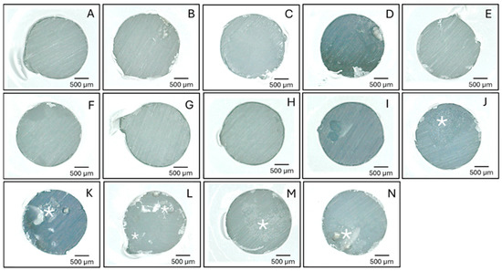
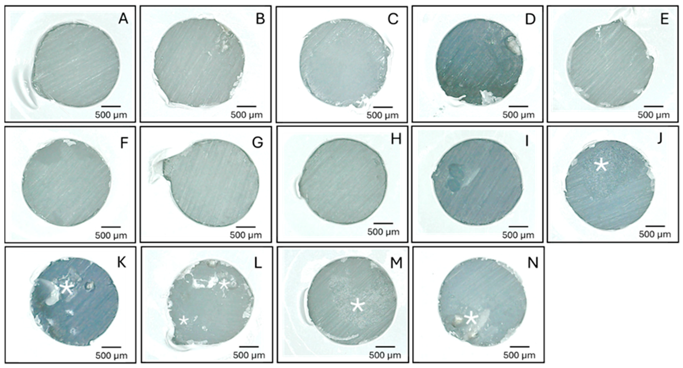
The failure modes were analyzed using a stereomicroscope at 40× magnification, with their respective percentages displayed in Table 2. Representative stereomicroscopic images of the fractured surfaces are presented in Figure 4. Groups 1, 2, 5, and 6 exhibited a complete adhesive failure rate of 100%. Conversely, groups 3, 4, 7, 8, and 9 demonstrated both adhesive and mixed failures, with adhesive failures at 80% or higher.
Figure 4.
The stereo microscope images at 40× magnification: the representations of adhesive failure from groups 1 to 9 are shown in (A–I), respectively, and mixed failure from groups 3, 4, 7, 8, and 9 are displayed in (J–N), respectively. The remaining fresh resin composite is evident on the surface of the aged resin composite, denoted by an asterisk.
4. Discussion
The current study demonstrated that the SBS of group 2 was significantly greater than that of group 1 (p < 0.001), and similarly, the SBS of group 6 was significantly higher than that of group 5 (p < 0.001). These results suggest that the AEP significantly improved the SBS between fresh resin composite and aged resin composite repairs, regardless of saliva contamination. As a result, both the primary and secondary null hypotheses were rejected.
In clinical practice, when resin composite restorations fracture or chip, dentists frequently choose the minimal intervention dentistry (MID) technique to repair the restoration rather than replace it entirely. This methodology offers numerous clinical and patient-focused advantages. The FDI World Dental Federation states that it adheres to the principles of MID, which provides several benefits, including the preservation of tooth structure, pulp protection, enhanced efficiency, and cost-effectiveness [19,20,21]. Furthermore, MID can prolong the durability of dental restorations because it prevents the “restorative death spiral,” a situation where each replacement causes the cavity to grow larger and the tooth to weaken [22]. Therefore, repairs can substantially extend the tooth’s overall longevity.
The present study involved repairing aged resin composites with a new resin composite material. Consequently, simulating the aging condition of the resin composite serves as a control variable for evaluation. Numerous methods are utilized for simulating aging in intraoral environments. In vitro techniques are employed to replicate the oral environmental conditions that lead to material degradation [23]. These methods employed, such as thermal simulation (thermocycling or autoclave), chemical aging (storage and immersion), mechanical simulation (for fatigue and wear assessment), and optical aging (for color stability evaluation), are contingent upon the objectives of the study [24,25,26,27,28].
In the present study, the thermocycling technique (10000 cycles) was chosen to simulate the aging resin composite. That was roughly one year of clinical service [29]. It acts as a crucial, accelerated in vitro method to mimic the oral cavity. The alternating high and low temperatures induced deterioration of the polymeric network and the filler–matrix interface [30], leading to decreased surface hardness, flexural strength, and increased water sorption. After finishing the aging process, the specimens’ surface was prepared by using 320-grit silicon carbide sheets to create a rough surface [16], which was then cleaned with a water spray instead of using ultrasonic cleaning. Although ultrasonic cleaning is superior in removing the smear layer compared to water spray, the latter method is predominantly employed for oral cleaning in patients. This restriction arises from the inability of dentists to utilize an ultrasonic cleaner machine intraorally. This approach ensures that the surfaces of the specimens are treated in a manner that closely mimics real-life conditions. Additionally, it allows for a better evaluation of how the materials will perform in an actual clinical setting.
The SEM images revealed that groups 1 (no surface treatment) and 5 (saliva contamination followed by no surface treatment) displayed elongated grooves and irregular pores on their surfaces, attributable to silicon carbide abrasion. Conversely, the other groups exhibited a loss of these grooves and pores. These features were also observed in the groups that employed AEP, bonding agent, or a combination of AEP followed by bonding agent. This observation indicates that the monomers in both AEP and bonding agent occupied the grooves and pores.
For the SBS testing, the notched-edge shear bond strength test was utilized to measure the bond strength between fresh and aged resin composite interfaces. This technique was modified from ISO 29,022 measuring adhesive bond strength between dental materials and tooth structures [17]. Due to the fact that the crosshead with a notched edge produces the force distribution and yields a more precise test result. It is closely approximating true shear strength rather than the peel strength that a crosshead with a straight edge produces. Thus, it aids in alleviating data misinterpretation caused by non-uniform stress distribution, allowing for better assessment of adhesive performance [31] and a reliable approach to predicting the long-term, in vivo durability of restorations. From the results of failure modes, 100% of adhesive failures were shown in groups 1, 2, 5, and 6. Moreover, 80% or higher of adhesive failures were demonstrated in the remaining groups. Therefore, SBS was closer to the adhesive shear strength. For group 5, 100% adhesive failure was shown with every low SBS (3.08 ± 0.41 MPa); these results may imply that the saliva acts as a barrier [6] for adhesion between the fresh and aged resin composite interface. Furthermore, no cohesive fractures were observed in either the fresh or aged resin composites; these findings suggest that the intrinsic strength of the materials exceeded the bond strength at their interfaces.
The SBS values of the groups with or without saliva contamination suggest that the surface treatments with AEP, bonding agent, or both AEP followed by bonding agent may have played a crucial role in influencing the integrity of the interface bond strength. These findings emphasize the importance of selecting appropriate surface treatments to enhance bond strength, particularly in clinical applications where saliva exposure is a concern. Further research could explore the long-term effects of these treatments on bond durability under various conditions.
The AEP and the bonding agent (G-Premio BOND™) used in this study contain the same functional monomers, including 10-MDP and 4-MET, but they differ in their curing modes. The AEP exhibits touch-cure properties when in contact with G-CEM ONE resin cement [10], which was not utilized in this study. In contrast, the bonding agent contains a photo initiator and employs a light-curing mode. Regarding SBS, the values for groups 2 and 6 were significantly higher than those for groups 1 and 5 (p < 0.001). This difference may be attributed to the monomers in the AEP applied in group 2, which can fill the grooves and pores present in the aged resin composite. Additionally, the AEP enhances surface wettability, while the monomers it contains polymerize through free radicals generated by the fresh resin composite applied on top during light activation. In fact, AEP contains a vanadium derivative that activates touch cure [32,33] when in contact with a specific resin (G-CEM ONE resin cement) [10], but in the present study, this resin cement was not used. Therefore, the functional monomers in AEP can polymerize upon contact with the bonding agent because they receive free radicals generated during the bonding agent’s curing process. In other words, AEP relies on free radicals from the adjacent substances to facilitate polymerization.
This reliance on free radicals from surrounding substance processes resembles the interactions observed between silane coupling agents and resin, as well as those between metal primers and resin [34,35]. The free radicals that come from the resin start the polymerization of the unsaturated monomers in the metal primer or silane coupling agent. This process results in the polymerization of those materials and copolymerization with the resin. Similarly, AEP can undergo polymerization and copolymerization with the fresh resin composite, which contributes to an increase in SBS.
Groups 3 and 4 demonstrated no significant difference in SBS between them; however, both groups exhibited significant differences when compared to groups 1 and 2. The variations can be explained as follows: in group 3, the bonding agent was applied using an agitative motion, ensuring uniform coverage across the entire surface. In group 4, the bonding agent was applied with an agitating motion, which facilitated its integration with the previously applied AEP. After application, the bonding agent was activated by a light-curing unit, enabling the photo initiators in the bonding agent to produce free radicals that initiate polymerization, which creates covalent bonds between the monomers in the bonding agent, or AEP, thereby increasing the degree of conversion. Additionally, the chemical reaction process also formed a chemical bond between the monomers from the bonding agent, or AEP, and the unsaturated monomer in the matrix, as well as the exposed fillers of the aged resin composite surface [36,37]. This enhances the interfacial strength between the aged and fresh resin composites. Furthermore, both the AEP and the bonding agent exhibit lower viscosity, while the fresh resin composite has high viscosity and limited penetration ability. Consequently, lower viscosity agents can enhance micromechanical retention by permeating the micro-irregularities formed through mechanical interlocking. These findings are consistent with prior studies [38,39,40] which demonstrated that low-viscosity bonding agents improve repair bond strength.
Groups 5–9 were differentiated from groups 1–4 due to saliva contamination. Group 5 demonstrated a significantly low SBS of 3.08 ± 0.41 MPa; these results imply that saliva may act as a barrier to adhesion at the interface between fresh and aged resin composites previously referenced [6]. Comparison of the SBS between Group 6, the saliva-contaminated specimen treated with AEP, and Group 5, the saliva-contaminated specimen without treatment, indicated that the SBS of Group 6 is significantly higher than that of Group 5 (p < 0.001). This differentiation can be ascribed to the existence of 10-MDP and 4-MET monomers in AEP. It is hypothesized that 10-MDP and 4-MET, or solely 10-MDP, act as surfactant molecules when in contact with a saliva-coated surface. Both 10-MDP and 4-MET exhibit amphiphilic properties [40]. The 10-MDP exhibits a hydrophilic phosphate group at one terminus and a hydrophobic methacrylate group at the opposing terminus, while 4-MET contains a hydrophilic phosphate group at one end and a hydrophobic methacrylate group at the other end. Amphiphilicity refers to a molecule possessing both a hydrophilic (polar) head and a hydrophobic (nonpolar) tail [41]. This dual property enables the molecule to interact with both polar and nonpolar environments. Upon contact with saliva, these monomers function as surfactants. The hydrophobic regions of their molecules attach to the organic components and proteins in saliva, facilitating the encapsulation of contaminants like saliva into micelles [14,42] and permitting the other monomers to occupy micro-irregularities. During the process, the fresh resin composite was activated, subsequently generating free radicals. The monomers in AEP formed a chemical bond with the unsaturated monomer in the matrix and the exposed fillers of the aged resin composite surface. Leading to an increase in SBS.
The group that used AEP following a bonding agent demonstrated a markedly higher SBS than the group that used the AEP alone. This enhancement can be attributed to AEP reacting to fewer free radicals from the fresh resin composite it contacted, compared to the bonding agent. Because the interfacial adaptation of the AEP–bonding agent was superior to that of the AEP–fresh resin composite. This matter will be examined in subsequent research.
What led the authors to hypothesize that either 10-MDP and 4-MET, or solely 10-MDP, functioned as the surfactant molecule? The studies solely addressed the interaction between the 10-MDP monomer and saliva, illustrating surfactant properties that promote micelle structure formation [14,42]. However, no studies have been performed on the surfactant properties of the 4-MET monomer. The disparity may arise from the molecular configurations of 10-MDP and 4-MET. The molecular structures indicate that the hydrocarbon spacer (-CH2-) in 10-MDP comprises 10 units, surpassing the 2-unit spacer in 4-MET. This distinction may clarify the surfactant properties of these two monomers. The elongated hydrocarbon chain is expected to exhibit enhanced surfactant properties relative to the shorter chain. Extending the spacer length typically results in a diminished Critical Micelle Concentration (CMC) and enhanced efficacy in lowering interfacial tension by distancing the cationic head groups, thereby reducing electrostatic repulsion and facilitating denser packing at the interface; in essence, this indicates a decrease in CMC and an increase in surfactant efficiency [43]. Consequently, this topic will be the focus of future research.
Groups 7 and 8 involved saliva-contaminated specimens treated with a bonding agent and AEP, respectively, following the bonding agent application. These groups exhibited no significant difference in SBS. The lack of difference can be attributed to the presence of the same components, 10-MDP and 4-MET, in both the bonding agent and AEP, which function as surfactant molecules when interacting with saliva.
In group 9, saliva-contaminated specimens were treated with 37% phosphoric acid and then rinsed before the application of a bonding agent, in accordance with the standard protocol for decontaminating resin composite surfaces. Numerous studies have shown that phosphoric acid effectively removes saliva contamination and enhances the bond strength between resin composite interfaces [44,45,46]. The current study indicated that the SBS values for groups 7 to 9 did not significantly differ. However, the SBS of group 9 was greater than that of group 6. This suggests that the bonding agent comprising 10-MDP and 4-MET may enhance the SBS between aged resin composites contaminated with saliva and new resin composites. This improvement may yield SBS comparable to that obtained through protocols employing 37% phosphoric acid for etching and rinsing.
This study provides significant insights; nonetheless, several limitations must be acknowledged. The research was initially conducted in vitro, which may not accurately represent the complexities of the oral environment, including temperature fluctuations, enzymatic activity, and prolonged cyclic loading. Subsequent research should encompass clinical trials to validate the primer’s efficacy in practical applications. Moreover, the effectiveness of the surfactant properties of 10-MDP and 4-MET necessitates additional investigation.
5. Conclusions
The findings indicate that the G-CEM ONE™ Adhesive Enhancing Primer (AEP) has potential for enhancing shear bond strength even in the presence of saliva contamination without the need for rinsing; however, when AEP was used together with a bonding agent, it provided a higher bond strength. Although the effectiveness of AEP as a bonding agent is currently limited, the results of the study may lead to improvements in efficiency in the future. Further research is necessary to explore modifications or combinations with other materials that could enhance its performance. This could ultimately expand its applications in clinical settings, benefiting both practitioners and patients by providing more reliable bonding solutions that improve treatment outcomes.
Author Contributions
Conceptualization, P.L., W.P., A.K. and T.S.; methodology, P.L., W.P., S.Y., A.K., T.S., P.T. and N.T.; formal analysis, P.L., W.P., A.K. and T.S.; investigation, P.L. and N.T.; data curation, P.L., S.Y., P.T., W.P. and T.S.; writing—original draft preparation, P.L., W.P., S.Y., A.K., T.S., P.T. and N.T.; writing—review and editing, W.P., A.K., T.S. and N.T. All authors have read and agreed to the published version of the manuscript.
Funding
This research received no external funding.
Data Availability Statement
The original contributions presented in the study are included in the article; further inquiries can be directed to the corresponding authors.
Acknowledgments
This study was supported by Soranun Chantarangsu of the Faculty of Dentistry, Chulalongkorn University; we thank her for her assistance with statistical consulting.
Conflicts of Interest
The authors declare no conflicts of interest.
Ethics Approval and Consent to Participate
The experimental part of this study received ethical approval from the Human Research Ethics Committee of the Faculty of Dentistry, Chulalongkorn University (HREC-DCU), under research code 002/2023. The approval number is 069/2023, certified on 7 July 2023.
References
- Al-Sulimmani, W.; Al-Rasheed, A.; Al-Daraan, H.; Al-Mutairi, M.; Brahmbhatt, Y.; Al-Hazmi, H.; Al-Qaderi, H. Failure Risk of Composite Resin and Amalgam Restorations: A Systematic Review and Meta-Analysis. Int. Dent. J. 2025, 75, 100871. [Google Scholar] [CrossRef] [Scilit]
- Zhou, X.; Huang, X.; Li, M.; Peng, X.; Wang, S.; Zhou, X.; Cheng, L. Development and status of resin composite as dental restorative materials. J. Appl. Polym. Sci. 2019, 136, 48180. [Google Scholar] [CrossRef] [Scilit]
- Santos, M.J.M.C.; Zare, E.; McDermott, P.; Santos Junior, G.C. Multifactorial Contributors to the Longevity of Dental Restorations: An Integrated Review of Related Factors. Dent. J. 2024, 12, 291. [Google Scholar] [CrossRef] [Scilit] [PubMed]
- Carlen, A.; Nikdel, K.; Wennerberg, A.; Holmberg, K.; Olsson, J. Surface characteristics and in vitro biofilm formation on glass ionomer and composite resin. Biomaterials 2001, 22, 481–487. [Google Scholar] [CrossRef] [Scilit]
- Hannig, M. Transmission electron microscopic study of in vivo pellicle formation on dental restorative materials. Eur. J. Oral Sci. 1997, 105, 422–433. [Google Scholar] [CrossRef] [Scilit]
- Nair, P.; Hickel, R.; Ilie, N. Adverse effects of salivary contamination for adhesives in restorative dentistry: A literature review. Am. J. Dent. 2017, 30, 156–164. [Google Scholar]
- Furuse, A.Y.; da Cunha, L.F.; Benetti, A.R.; Mondelli, J. Bond strength of resin-resin interfaces contaminated with saliva and submitted to different surface treatments. J. Appl. Oral Sci. 2007, 15, 501–505. [Google Scholar] [CrossRef] [Scilit]
- Klaisiri, A.; Suebnukarn, S.; Krajangta, N.; Rakmanee, T.; Sriamporn, T.; Thamrongananskul, N. The Effect of Morpholine on Composite-to-Composite Repair Strength Contaminated with Saliva. Polymers 2022, 14, 4718. [Google Scholar] [CrossRef] [Scilit]
- Klaisiri, A.; Sriamporn, T.; Krajangta, N.; Thamrongananskul, N. Morpholine’s Effects on the Repair Strength of Saliva-Contaminated CAD/CAM Resin-Based Composite Mended with Resin Composite. J. Compos. Sci. 2025, 9, 345. [Google Scholar] [CrossRef] [Scilit]
- Jamel, R.S.; Alkhalidi, E.F.; Dawood, A.E. The effect of touch-cure polymerization on the push-out bond strength of fiber posts. Saudi Dent J. 2024, 36, 627–632. [Google Scholar] [CrossRef] [Scilit] [PubMed]
- Kim, B.N.; Son, S.A.; Park, J.K. Effect of exclusive primer and adhesive on microtensile bond strength of self- adhesive resin cement to dentin. Materials 2020, 13, 2286. [Google Scholar] [CrossRef] [Scilit]
- Ramos, R.Q.; Mercelis, B.; Ahmed, M.H.; Peumans, M.; Lopes, G.C.; Van Meerbeek, B. Bonding of Composite Cements Containing 10-MDP to Zirconia Ceramics Without Dedicated Ceramic Primer. J. Adhes. Dent. 2024, 26, 135–145. [Google Scholar]
- Pornatitanakul, V.; Klaisiri, A.; Sriamporn, T.; Swasdison, S.; Thamrongananskul, N. The Influence of Tooth Primer and Zirconia Cleaners on the Shear Bond Strength of Saliva-Contaminated Zirconia Bonded with Self-Adhesive Resin Cement. Appl. Sci. 2025, 15, 4561. [Google Scholar] [CrossRef] [Scilit]
- Etiennot, L.; Ordies, M.; Yao, C.; Mercelis, B.; Peumans, M.; Van Meerbeek, B. Effective 10-MDP Bonding to Saliva-Contaminated Dentin. J. Adhes. Dent. 2025, 27, 65–74. [Google Scholar]
- Morresi, A.L.; D’Amario, M.; Capogreco, M.; Gatto, R.; Marzo, G.; D’Arcangelo, C.; Monaco, A. Thermal cycling for restorative materials: Does a standardized protocol exist in laboratory testing? A literature review. J. Mech. Behav. Biomed. Mater. 2014, 29, 295–308. [Google Scholar] [CrossRef] [Scilit]
- 3M ESPE. Sof-Lex™ Finishing and Polishing Strip Technical Product Profile. Available online: https://media.dentalcompare.com/m/25/Downloads/Sof-Lex%20Finishing%20and%20Polishing%20Strip%20Technical%20Product%20Profile.pdf (accessed on 20 February 2026).
- ISO 29022:2013; Dentistry-Adhesion-Notched-Edge Shear Bond Strength Test. International Organization for Standardization: Geneva, Switzerland, 2013. Available online: http://www.iso.org/iso/store.htm (accessed on 6 September 2025).
- Matinlinna, J.P.; Lassila, L.V. Enhanced resin-composite bonding to zirconia framework after pretreatment with selected silane monomers. Dent. Mater. 2011, 27, 273–280. [Google Scholar] [CrossRef] [Scilit]
- FDI World Dental Federation. Repair of restorations: Adopted by the General Assembly: September 2019, San Francisco, United States of America. Int. Dent. J. 2020, 70, 7–8. [Google Scholar] [CrossRef] [Scilit]
- Ericson, D. The concept of minimally invasive dentistry. Dent. Update 2007, 34, 9–18. [Google Scholar] [CrossRef] [Scilit]
- Mount, G.J. A new paradigm for operative dentistry. J. Conserv. Dent. 2008, 11, 3–10. [Google Scholar] [CrossRef] [Scilit]
- Qvist, V. Longevity of restorations: ‘The death spiral’. In Dental Caries: The Disease and Its Clinical Management; Fejerskov, O., Nyvad, B., Kidd, E., Eds.; Wiley Blackwell: Hoboken, NJ, USA, 2005; pp. 387–401. [Google Scholar]
- Akçay, H.Ç. Evaluation of In-Vitro Aging Procedures in Dentistry: A Traditional Review. Dent. Med. J.-Rev. 2025, 7, 88–100. [Google Scholar]
- Szczesio-Wlodarczyk, A.; Sokolowski, J.; Kleczewska, J.; Bociong, K. Ageing of dental composites based on methacrylate resins- critical review of the causes and method of assessment. Polymers 2020, 12, 882. [Google Scholar] [CrossRef] [Scilit] [PubMed]
- Lima, V.P.; Machado, J.B.; Zhang, Y.; Loomans, B.A.C.; Moraes, R.R. Laboratory methods to simulate the mechanical degradation of resin composite restorations. Dent. Mater. 2022, 38, 214–229. [Google Scholar] [CrossRef] [Scilit]
- Szczesio-Wlodarczyk, A.; Fronczek, M.; Ranoszek-Soliwoda, K.; Grobelny, J.; Sokolowski, J.; Bociong, K. The first step in standardizing an artificial aging protocol for dental composites-evaluation of basic protocols. Molecules 2022, 27, 3511. [Google Scholar] [CrossRef] [Scilit]
- Oja, J.; Lassila, L.; Vallittu, P.K.; Garoushi, S. Effect of accelerated aging on some mechanical properties and wear of different commercial dental resin composites. Materials 2021, 14, 2769. [Google Scholar] [CrossRef] [Scilit]
- Souza, L.S.; Donato, T.R.; Cerqueira, G.A.; Cavalcanti, A.N.; Mathias, P. Color stability of an artificially aged nanofilled composite resin post-cured with different techniques. J. Dent. Res. Dent. Clin. Dent. Prospects 2021, 15, 53–58. [Google Scholar] [CrossRef] [Scilit]
- Gale, M.S.; Darvell, B.W. Thermal cycling procedures for laboratory testing of dental restorations. J. Dent. 1999, 27, 89–99. [Google Scholar] [CrossRef] [Scilit] [PubMed]
- Ghavami-Lahiji, M.; Firouzmanesh, M.; Bagheri, H.; Jafarzadeh Kashi, T.S.; Razazpour, F.; Behroozibakhsh, M. The effect of thermocycling on the degree of conversion and mechanical properties of a microhybrid dental resin composite. Restor. Dent. Endod. 2018, 43, e26. [Google Scholar] [CrossRef] [Scilit]
- ISO Adopts Ultradent’s Notched Edge Testing Method. Available online: https://www.ultradent.com/company/newsroom/article/iso-adopts-ultradents-notched-edge-testing-method?srsltid=AfmBOootSDXE8hFzN0Em01T296OM-yn7_WG5SgK-IEtKlQXnhh87luEv (accessed on 20 February 2026).
- Yoshihara, K.; Nagaoka, N.; Benino, Y.; Nakamura, A.; Hara, T.; Maruo, Y.; Yoshida, Y.; Van Meerbeek, B. Touchcure polymerization at the composite cement-dentin interface. J. Dent. Res. 2021, 100, 935–942. [Google Scholar] [CrossRef] [Scilit]
- Kondo, T.; Sato, J.; Kakinuma, H.; Ashry, A.; Engkatanachai, K.; Egusa, H. Vanadium-functionalized methyl methacrylate-based primer enhances self-curing and bonding of resin cement to polyetheretherketone restorations. J. Dent. Sci. 2026. [Google Scholar] [CrossRef] [Scilit]
- Matinlinna, J.P.; Lung, C.Y.K.; Tsoi, J.K.H. Silane adhesion mechanism in dental applications and surface treatments: A review. Dent. Mater. 2018, 34, 13–28. [Google Scholar] [CrossRef] [Scilit] [PubMed]
- Ozaki, A.; Shishido, S.; Nakamura, K.; Harada, A.; Katsuda, Y.; Kanno, T.; Egusa, H. Impact of adhesive primer and light-curing on polymerization kinetics of self-adhesive resin cement in association with free radical reaction. Eur. J. Oral Sci. 2021, 129, e12828. [Google Scholar] [CrossRef] [Scilit] [PubMed]
- Tezvergil, A.; Lassila, L.V.; Vallittu, P.K. Composite-composite repair bond strength: Effect of different adhesion primers. J. Dent. 2003, 31, 521–525. [Google Scholar] [CrossRef] [Scilit]
- Brosh, T.; Pilo, R.; Bichacho, N.; Blutstein, R. Effect of combinations of surface treatments and bonding agents on the bond strength of repaired composites. J. Prosthet. Dent. 1997, 77, 122–126. [Google Scholar] [CrossRef] [Scilit]
- Papacchini, F.; Dall’Oca, S.; Chieffi, N.; Goracci, C.; Sadek, F.T.; Suh, B.I.; Tay, F.R.; Ferrari, M. Composite-to-composite microtensile bond strength in the repair of a microfilled hybrid resin: Effect of surface treatment and oxygen inhibition. J. Adhes. Dent. 2007, 9, 25–31. [Google Scholar]
- Rathke, A.; Tymina, Y.; Haller, B. Effect of different surface treatments on the composite-composite repair bond strength. Clin. Oral Investig. 2009, 13, 317–323. [Google Scholar] [CrossRef] [Scilit]
- Lima, A.F.; Ferreira, S.F.; Catelan, A.; Palialol, A.R.; Goncalves, L.S.; Aguiar, F.H.; Marchi, G.M. The effect of surface treatment and bonding procedures on the bond strength of silorane composite repairs. Acta Odontol. Scand. 2014, 72, 71–75. [Google Scholar] [CrossRef] [Scilit]
- Delgado, A.H.S.; Ahmed, M.H.; Nunes Ferreira, M.; Mano Azul, A.; Polido, M.; Yoshihara, K.; Van Meerbeek, B. Physico-Chemical Properties and Performance of Functional Monomers Used in Contemporary Dental Adhesive Technology. J. Adhes. Dent. 2025, 27, 175–193. [Google Scholar]
- Katta, P.K. Strength of Bond Between Adhesives and Low-viscosity Bulk-fill Composites Utilizing 10-methacryloyloxydecyl Dihydrogen Phosphate (10-MDP). Biomed. Pharmacol. J. 2025, 18, 761–772. [Google Scholar] [CrossRef] [Scilit]
- Unal, D.N.; Sadak, S.; Erkmen, C.; Selcuk, Ö.; Uslu, B. Surfactant-Based Sensors in Chemical and Biochemical Detection; Manjunatha, J.G., Ed.; Royal Society of Chemistry: London, UK, 2023; Volume 23, pp. 1–18. [Google Scholar]
- Sismanoglu, S. Efficiency of self-adhering flowable resin composite and different surface treatments in composite repair using a universal adhesive. Niger. J. Clin. Pract. 2019, 22, 1675–1679. [Google Scholar] [CrossRef] [Scilit] [PubMed]
- Yin, H.; Kwon, S.; Chung, S.H.; Kim, R.J.Y. Performance of universal adhesives in composite resin repair. Biomed. Res. Int. 2022, 2022, 7663490. [Google Scholar] [CrossRef] [Scilit] [PubMed]
- de Medeiros, T.C.; de Lima, M.R.; Bessa, S.C.; de Araújo, D.F.; Galvão, M.R. Repair bond strength of bulk fill composites after different adhesion protocols. J. Clin. Exp. Dent. 2019, 11, e1000–e1005. [Google Scholar] [CrossRef] [Scilit] [PubMed]

Disclaimer/Publisher’s Note: The statements, opinions and data contained in all publications are solely those of the individual author(s) and contributor(s) and not of MDPI and/or the editor(s). MDPI and/or the editor(s) disclaim responsibility for any injury to people or property resulting from any ideas, methods, instructions or products referred to in the content. |
© 2026 by the authors. Licensee MDPI, Basel, Switzerland. This article is an open access article distributed under the terms and conditions of the Creative Commons Attribution (CC BY) license.