


Abstract
Background/Objectives: Nasopharyngeal cancer generally has a good survival rate with chemoradiotherapy, but a significant side effect is irreversible hearing loss, for which there is currently no preventive intervention. Given that dexamethasone (Dex) and D-methionine (dMet) have individually shown potential to prevent hearing loss in in vitro and animal studies, we hypothesized that combining Dex and dMet may offer greater protection against cytotoxicity induced by cisplatin and radiation treatments. Methods: Organ-of-Corti-derived OC-k3 cells exposed to cisplatin and/or radiation were used as an in vitro model of treatment-induced cytotoxicity to investigate the protective mechanisms of Dex and dMet. Results: Dex was associated with reduced apoptosis, accompanied by decreased p53 phosphorylation and increased Bcl-XL expression. Dex also reduced COX2 expression and showed trends toward attenuation of ERK1/2 and NFκB signaling. Conversely, dMet effectively blocked cisplatin-induced apoptosis but did not protect against radiation-induced cytotoxicity. Conclusions: The combination of Dex and dMet provided greater protection against cisplatin- and radiation-induced cytotoxicity in the OC-k3 cells than either agent alone. These findings support further investigation of combined Dex and dMet as a potential strategy to mitigate treatment-induced ototoxicity.
1. Introduction
Nasopharyngeal cancer (NPC) is a common cancer in Southeast Asia. Although overall 5-year disease survival is more than 70% for stage 1 to 3 NPC, patients frequently endure side effects from chemoradiation treatments, with 25% of them who suffer from irreversible hearing loss [1,2]. In high risk populations, such as in Singapore, the peak age of incidence is 45 to 49 years [3], leading to significant long-term impacts on the quality of life for these young adults.
NPC treatment is stage-dependent, utilizing three primary modalities: radiation alone, concurrent chemoradiation, and induction chemotherapy with concurrent chemoradiation therapy. Our study examines the effects of dexamethasone (Dex) and D-methionine (dMet) on ear cell cultures exposed to radiation alone, cisplatin chemotherapy alone, and combined chemoradiation. Investigating the effects of these drugs on each treatment modality may elucidate protective mechanisms and identify patient group that could benefit most from these interventions.
Intratympanic Dex has been shown to prevent cisplatin-induced hearing loss in a randomized control trial [4]. In vitro animal studies have shown that Dex reduces apoptosis in ear hair cells exposed to either cisplatin or radiation [5,6]. Our study gives new information by exploring Dex’s effect on cells exposed to both cisplatin and radiation.
Methionine is an essential amino acid with two enantiomers, L-methionine and D-methionine (dMet). dMet has been shown to reduce cisplatin-induced hearing loss in animals when administered through the round window [7] and systemically [8]. An exploratory clinical trial also suggests that oral administration of dMet may be otoprotective against cisplatin [9]. The protective mechanism involves maintaining antioxidant levels, preventing damage to the stria vascularis and reducing outer hair cell loss [10,11]. With regards to the protective effects of dMet on radiation, it has been mainly studied in preventing radiation-induced oral mucositis. dMet has been found to protect oral mucosal from radiation-induced toxicity in a dose-dependent manner in an animal model [12]. A phase 2 trial suggests that dMet may reduce oral mucositis induced by radiation and chemotherapy for head and neck cancers [13].
Studies have found that patients who received chemoradiation treatment experience more severe hearing loss than patients who received radiation alone [14] due to the synergistic ototoxicity from combined cisplatin and radiation treatment [15]. Combining Dex and dMet for hearing protection has not been previously studied. If these drugs protect hearing through separate mechanisms, they could offer greater protection than either agent alone. Our current study explores the protective effects of this drug combination on cell viability and the underlying mechanisms.
2. Materials and Methods
2.1. Cell Lines and Culture Conditions
The immortalized OC-k3 cell line derived from the organ of Corti of the transgenic mice (Immorta-mouse H-2Kb-tsA58, Charles Rivers Laboratories, Wilmington, MA, USA) was used in this study. The OC-k3 cell line was originally established and characterized by Kalinec et al. as a conditionally immortalized organ-of-Corti-derived cell line [16]. The cells were maintained under permissive proliferative conditions in high-glucose Dulbecco’s Modified Eagle’s medium (DMEM, BioWhittaker™, Lonza, Allendale, NJ, USA) supplemented with 10% fetal bovine serum (FBS) (Gibco, Thermo Fisher Scientific, Grand Island, NY, USA), 100 U/mL Penicillin-100 ug/mL Streptomycin (Gibco, Thermo Fisher Scientific, Grand Island, NY, USA) and 50 U/mL gamma-interferon (mouse recombinant, Sigma Aldrich, St Louis, MO, USA). The cultures were maintained at 33 °C with 10% CO2. Experiments were performed using cells between passage 10 and 20. No differentiation protocol was applied prior to experimentation.
2.2. In Vitro OC-k3 Cytotoxicity Model with Drug Protection Treatment
An in vitro model of cytotoxicity was established by seeding OC-k3 cells in 96-well plates at a density of 5000 cells per well. For each assay, cells were seeded into two identical plates. The following day, cells were pre-incubated for 3 h with Dex, dMet, their combination, or solvent control. Subsequently, an equal volume of medium containing either 2× cisplatin or vehicle, together with the corresponding concentration of otoprotective agent (s), was added to each well to achieve the final treatment concentrations. One plate remained non-irradiated (sham-irradiated control) and was used to generate the no treatment (NT) and Cis (0.5 μg/mL) groups, whereas the second plate received 5 Gy irradiation using a Rad Source RS2000 X-Ray irradiator (Rad Source Technologies, Suwanee, GA, USA) immediately after drug addition to generate the radiation (RT) and Cis+RT groups. The RS2000 X-Ray irradiator was operated at 160 kV and 25 mA with a dose rate of 1.885 Gy/min at shelf level 3, as verified by ion chamber dosimetry (Radcal ion chamber model 2086, Radcal Corporation, Monrovia, CA, USA) performed during biannual preventive maintenance. Both the irradiated and sham plates were held at ambient room temperature for the duration of the procedure, after which they were returned to the incubator simultaneously. The treatment concentration with continuous 72 h exposure protocol was adopted from our previously validated OC-k3 ototoxicity model [15]. No medium change or washout step was performed; cisplatin and the otoprotective agent (s) remained in the culture medium throughout the experiment. To determine the optimal concentrations of Dex and dMet for protection against Cis, RT and Cis+RT treatments, cells were pre-treated with Dex (0, 1, 10, 30, 50, 70, 90 and 110/120 μg/mL) or dMet (0, 0.05, 0.1, 0.5, 1, 5 mg/mL) and continuously exposed to the respective agents throughout the 3-day experimental period. Subsequent combination studies were performed using the optimal concentrations of 30 μg/mL Dex and 1 mg/mL dMet. After pre-incubation, the cells were exposed to NT, Cis, RT and Cis+RT treatments.
2.3. Cytotoxicity Measurement as Determined by CellTox Assay
At 72 h post-treatment, cytotoxicity was assessed using CellTox Green cytotoxicity™ assay kit (Promega Corp., Madison, WI, USA). The green dye was added to each well at 2000 × dilution after pre-incubation of Dex and/or dMet and in the presence of Cis, RT and Cis+RT. Fluorescence signals were measured at an excitation wavelength of 485 nm and an emission wavelength of 520 nm using Enspire Multimode Microplate Reader (Perkin Elmer, Shelton, CT, USA). CellTox fluorescence was used as an indicator of cytotoxicity and loss of plasma membrane integrity. Fluorescence signals, with blank subtraction, were presented as mean ± standard deviation (SD) from two independent experiments, each conducted with four replicates.
2.4. Cell Viability Measurement as Determined by MTS Assay
At 72 h post-treatment, cell viability was assessed using MTS assay kit (CellTiter 96 Aqueous One Solution Cell Proliferation Assay, Promega Corp., Madison, WI, USA), which measures the metabolic activity of viable cells. Briefly, MTS solution was added to each well and incubated at 33 °C for 3 h. Absorbance was then measured at 490 nm using a microplate spectrophotometer (Benchmark Plus, Bio-Rad Laboratories, Hercules, CA, USA). The percentage of cell viability in each well was calculated by absorbance after background subtraction, using the formula: 100% × (absorbance of treated cells/average absorbance of solvent control in NT). Data was presented as mean ± standard deviation (SD) from two independent experiments, each conducted with four replicates.
2.5. Immunoblot Analysis
During cell harvesting, proteins were extracted directly by adding 2× Laemmli sample buffer (Bio-Rad Laboratories, Hercules, CA, USA) to the wells. Protein concentration was determined using the RC DC™ Protein Assay (Bio-Rad Laboratories, Hercules, CA, USA) according to manufacturer’s instructions. Equal amounts of protein samples were denatured, separated by 12% SDS-PAGE and transferred onto PVDF membrane using iBlot dry blotting system (Invitrogen, Thermo Fisher Scientific, Waltham, MA, USA). The membranes were blocked with either 5% non-fat milk or 5% BSA, as recommended by the manufacturer, for 1 h. This was followed by overnight incubation with primary antibodies at 4 °C (see Supplementary Table S1). After rinsing with Tris-Buffered Saline containing 0.1% Tween 20 (TBST), the membranes were incubated with corresponding secondary antibodies conjugated with horseradish peroxidase (HRP) in blocking solution for 1 h at room temperature. Following further washes, Immobilon Western chemiluminescent HRP substrate (Merck Millipore, Billerica, MA, USA) was added to the membranes. The chemiluminescence signal was detected using UV Illuminescent Alliance MINI HD4 chemiluminescence imaging system (UVItec, Cambridge, UK). Densitometric analysis of band intensities was performed using UVI Band software (UVItec, Cambridge, UK). Normalized integrated intensities of the proteins of interest were obtained by normalization to β-actin. Three independent batches of cell treatments were processed for immunoblot analysis. Full membrane scans with annotated cropped regions and quantified bands are provided in Supplementary Figure S3.
2.6. Statistical Analysis
Data was analyzed by linear models with two-way interactions. The general model structure was: outcome ~ cancer treatment modality × otoprotective intervention + experiment, where cancer treatment modality (NT, Cis, RT, Cis+RT) was included as a fixed factor and otoprotective intervention was represented either by agent dose (Figures 1 and 2) or treatment group (control, Dex, dMet, Dex+dMet; subsequent figures). Their interaction term was included to evaluate combined effects, while experiment was included as a fixed covariate to adjust for systematic differences between experimental runs. It should be noted that each experiment comprised four technical replicate wells, and with only two independent biological experiments (n = 2) contributing to the CellTox and MTS assays, the technical replicates were not collapsed prior to modeling. This constitutes pseudoreplication, and the fixed covariate for experiment does not fully account for the within-experiment correlation structure. Accordingly, all findings derived from these models should be interpreted as exploratory, and p-values should not be taken as definitive evidence of effect. Model assumptions were evaluated using diagnostic plots, including Q-Q plots to assess residual normality and residual-versus-fitted plots to evaluate homoscedasticity. Estimated marginal means (EMMs) and 95% confidence intervals were derived from the fitted models for each combination of cancer treatment modality and otoprotective intervention. Post hoc pairwise comparisons among EMMs were conducted to determine within group differences, and p-value were adjusted using the Šídák correction. Statistical analyses were performed in R (version 4.2.3) using the {emmeans} package. All graphs were generated using GraphPad Prism 9.2.0.
3. Results
3.1. Dex Demonstrated Protective Effects Against Cytotoxicity Induced by Cisplatin, Radiation and Their Combination
An in vitro cytotoxicity model was established by treating organ-of-Corti-derived OC-k3 cells with 0.5 μg/mL cisplatin (Cis), 5 Gy of radiation (RT) and their combination (Cis+RT). At 3 days post-treatment, when compared with non-treatment controls (NT), we confirmed that Cis, RT and Cis+RT significantly increased cytotoxicity, as evidenced by CellTox assay results (# in Figure 1A, Figure 2A and Figure 3A) and decreased cell viability, as shown by MTS assay results (# in Figure 1B, Figure 2B and Figure 3B). These findings are consistent with treatment-induced cytotoxicity and cellular injury and agree with our previous study demonstrating apoptosis in OC-k3 cells exposed to the same treatment paradigm [15].
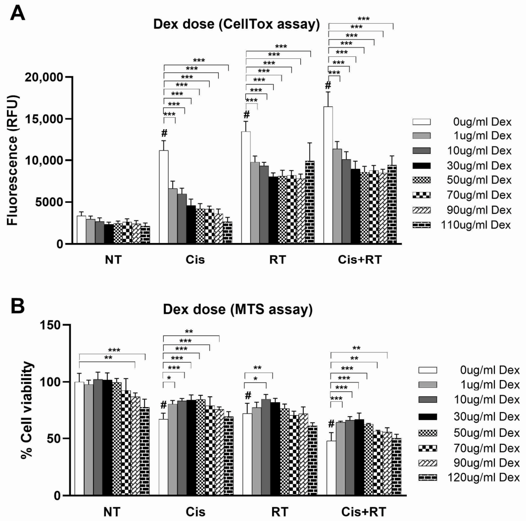
A dose–response assay demonstrated protective effects of Dex against treatment-induced cytotoxicity. This was associated with significantly lower CellTox fluorescence signals (Figure 1A) and a significant restoration of cell viability in cells treated with Cis, RT and Cis+RT (Figure 1B). We noticed that Dex alone at doses above 50 µg/mL tended to reduce cell viability (NT, Figure 1B) without a corresponding increase in CellTox fluorescence signals (NT, Figure 1A), suggesting that high concentration of Dex may influence cellular metabolic activity independently of overt cytotoxicity. Therefore, 30 µg/mL Dex was identified as the optimal concentration for subsequent experiments.
Figure 1.
Dose–response assay demonstrating the protective effect of Dex against cytotoxicity induced by cisplatin, radiation and their combination as determined by (A) CellTox assay and (B) MTS assay. OC-k3 cells were pre-treated with 0, 1, 10, 30, 50, 70, 90 and 110/120 μg/mL Dex for 3 h before receiving either no treatment (NT), 0.5 μg/mL cisplatin (Cis), 5 Gy radiation treatment (RT) or both (Cis+RT). At day 3 post-treatment, cytotoxicity was measured by the increase of green fluorescence signal using the CellTox assay and the % of cell viability was determined using MTS assay (set 0 μg/mL Dex, also known as the solvent control at NT as 100% cell viability). Each treatment condition was measured using four technical replicates within each of two independent biological experiments performed on different days using separate cell passages. Data are presented as bar charts with mean ± SD. CellTox fluorescence (A) and % cell viability (B) were analyzed using linear models with cancer treatment modality (NT, Cis, RT, Cis+RT), Dex dose and their interaction as fixed effects, with experiment included as a covariate. Estimated marginal means were compared using Šídák-adjusted post hoc tests. # indicates significant difference (p < 0.001) found in cells treated with Cis, RT or Cis+RT compared to the NT control (comparison within the white bars). Significant differences between different doses of Dex and the solvent control (0 μg/mL) under Cis, RT or Cis+RT are indicated as asterisks. *** p < 0.001, ** p < 0.01, * p < 0.05.
3.2. dMet Demonstrated Selective Protection Against Cisplatin-Induced Cytotoxicity but Not Radiation-Induced Cytotoxicity
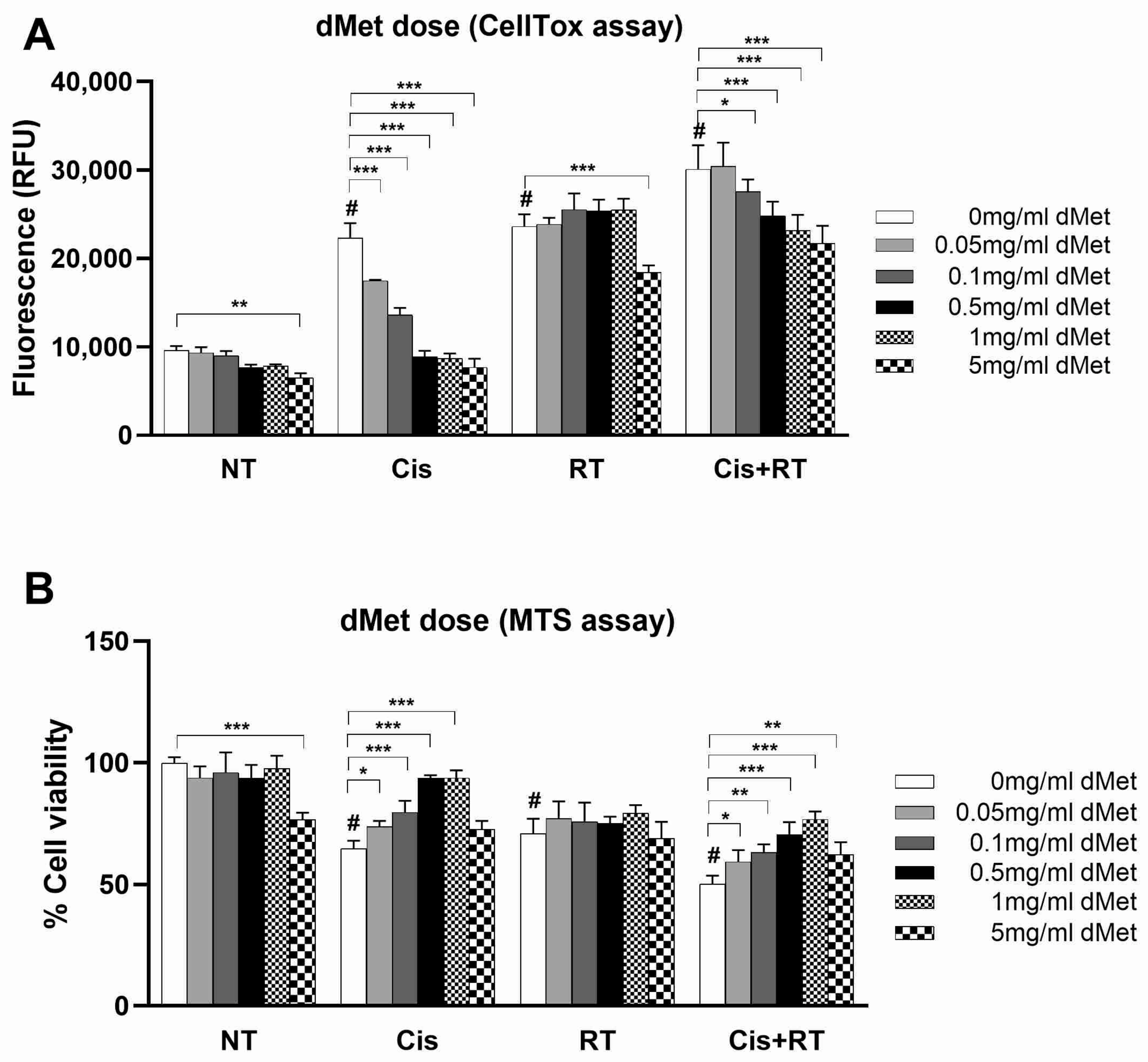
A dose–response assay of dMet demonstrated its specific protection against cisplatin-mediated cytotoxicity. This was evidenced by a significant reduction in CellTox fluorescence signals in cells treated with Cis and Cis+RT, with no change observed in cells treated with RT alone (Figure 2A). Consistently, the MTS assay confirmed that dMet significantly restored cell viability in cells treated with Cis and Cis+RT, but not in those treated with RT alone (Figure 2B). dMet doses ranging from 0.5 to 1 mg/mL provided maximal protection against cisplatin-induced cytotoxicity. However, an adverse effect was noted at 5 mg/mL dMet alone, showing a significant reduction in cell viability (NT in Figure 2B). Note that this reduction in cell viability at high dose corresponded to the overall reduction of CellTox fluorescence signals, which should not be misinterpreted as protective effect of dMet (Figure 2A). Therefore, 1 mg/mL dMet was identified as the optimal concentration for subsequent experiments.
Figure 2.
Dose–response assay demonstrating the protective effect of dMet against cisplatin- and the combined treatment-induced cytotoxicity, but not by radiation-induced cytotoxicity, as determined by (A) CellTox assay and (B) MTS assay. OC-k3 cells were pre-treated with 0, 0.05, 0.1, 0.5, 1, 5 mg/mL dMet for 3 h before receiving either no treatment (NT), 0.5 μg/mL cisplatin (Cis), 5 Gy radiation treatment (RT) or both (Cis+RT). At day 3 post-treatment, cytotoxicity was measured by the increase of green fluorescence signal using the CellTox assay and the % of cell viability was determined using MTS assay (set 0 mg/mL dMet, also known as the solvent control at NT as 100% cell viability). Each treatment condition was measured using four technical replicates within each of two independent biological experiments performed on different days using separate cell passages. Data are presented as bar charts with mean ± SD. CellTox fluorescence (A) and % cell viability (B) were analyzed using linear models with cancer treatment modality (NT, Cis, RT, Cis+RT), dMet dose and their interaction as fixed effects, with experiment included as a covariate. Estimated marginal means were compared using Šídák-adjusted post hoc tests. # indicates significant difference (p < 0.001) in cells treated with Cis, RT or Cis+RT compared to the NT control (comparison within the white bars). Significant differences between different doses of dMet and the solvent control (0 mg/mL) under Cis, RT or Cis+RT are indicated as asterisks. *** p < 0.001, ** p < 0.01, * p < 0.05.
3.3. Combining Dex and dMet Provided Greater Protection Against Cisplatin- and Radiation-Induced Cytotoxicity, Which Was Associated with Reduced Apoptosis and p53 Phosphorylation
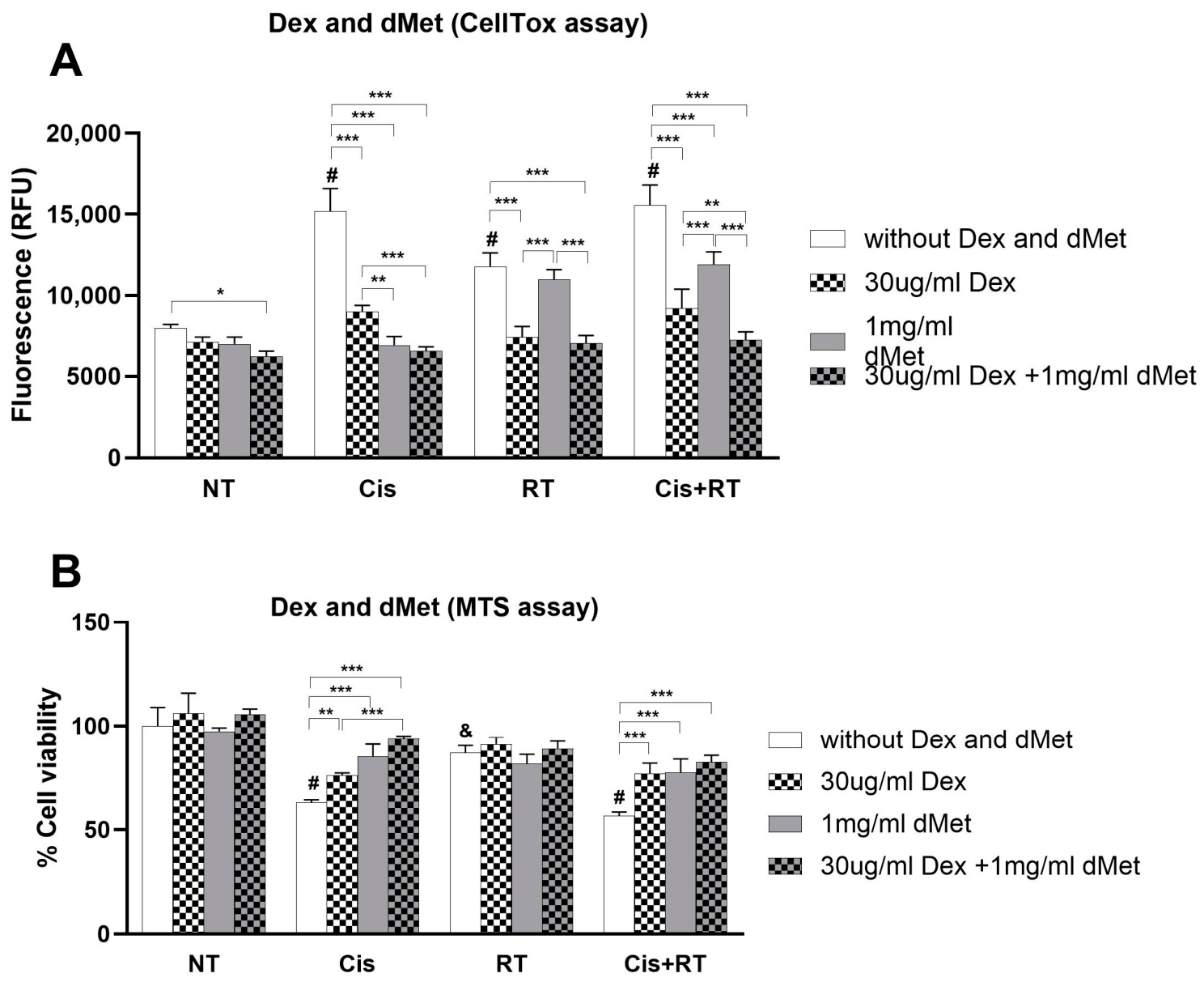
After determining the optimal dosages of Dex (30 µg/mL) and dMet (1 mg/mL), we further monitored their protective effects against cytotoxicity triggered by Cis, RT and Cis+RT. Apparently, cisplatin-induced cytotoxicity was partially inhibited by Dex alone, whereas fully inhibited by dMet alone and the combination of both (Cis in Figure 3A,B). In contrast, radiation-induced cytotoxicity was attenuated by Dex alone and by the combination, but not by dMet alone (RT in Figure 3A,B). Furthermore, combining Dex and dMet significantly attenuated CellTox fluorescence signals compared to the individual treatment of Dex and dMet (Cis+RT, Figure 3A), suggesting greater protection against cytotoxicity induced by the combined treatment of cisplatin and radiation. This finding was supported by Supplementary Figure S1, which showed representative images of cells under Cis+RT treatment, with fewer dead cells observed in the combined treatment compared to the single treatments.
We found that the cytotoxic injury triggered by Cis, RT and Cis+RT was associated with the activation of apoptosis. This was demonstrated by an increase in cleaved caspase 7 levels, as shown by the immunoblot assay (Figure 4A). Further analysis using data from biological triplicates experiments indicated that Dex, dMet and their combination tended to reduce cleaved caspase 7 levels following cisplatin treatment (Cis, Figure 4B). Additionally, Dex, but not dMet, showed a trend of attenuation for radiation-induced cleaved caspase 7 levels (RT, Figure 4B). Notably, under Cis+RT treatment, the combination of Dex and dMet resulted in significant lower cleaved caspase 7 levels than Dex alone (Figure 4A,B). However, given the limited statistical significance observed among treatment groups, these findings should be interpreted as supportive evidence of reduced apoptosis activation.
Figure 3.
Greater protective effects of combined Dex and dMet against cytotoxicity induced by combined cisplatin and radiation treatment as determined by (A) CellTox assay and (B) MTS assay. OC-k3 cells were pre-treated with 30 μg/mL Dex and 1 mg/mL Met for 3 h before receiving either no treatment (NT), 0.5 μg/mL cisplatin (Cis), 5 Gy radiation treatment (RT) or both (Cis+RT). Solvent control is labeled as without Dex and dMet. At day 3 post-treatment, cytotoxicity was measured by the increase of green fluorescence signal using the CellTox assay and the % of cell viability was determined using the MTS assay (set the solvent control at NT as 100% cell viability). Each treatment condition was measured using four technical replicates within each of two independent biological experiments performed on different days using separate cell passages. Data are presented as bar charts with mean ± SD. CellTox fluorescence (A) and % cell viability (B) were analyzed using linear models with cancer treatment modality (NT, Cis, RT, Cis+RT), otoprotective treatment group (control, Dex, dMet, Dex+dMet), and their interaction as fixed effects, with experiment included as a covariate. Estimated marginal means were compared using Šídák-adjusted post-hoc tests. Significant differences between Cis, RT or Cis+RT when compared with NT in solvent controls are indicated by # p < 0.001 and & p = 0.012 (comparison between the white bars). Significant differences between solvent control, Dex, dMet and Dex+dMet under Cis, RT and Cis+RT treatments are indicated as asterisks. *** p < 0.001, ** p < 0.01, * p < 0.05.
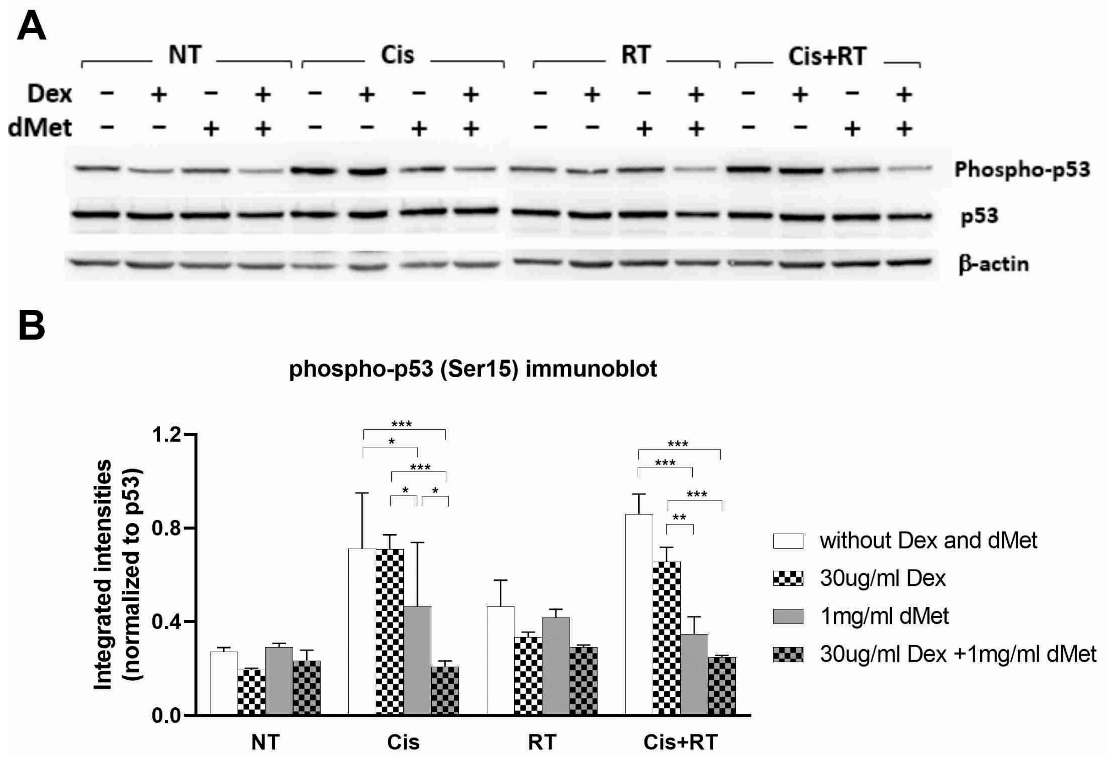
P53 activation in response to DNA damage could be manifested by phosphorylation at Ser15. In line with the treatment-induced cytotoxicity (Figure 3A,B) and apoptosis activation (Figure 4A,B), we detected increased phosphorylation of p53 at Ser15, indicating p53 activation (Figure 5A). The protective effects of Dex and dMet under cisplatin treatment were also evident, as both agents, individually and in combination, attenuated p53 phosphorylation (Cis in Figure 5A,B). Similarly, under radiation treatment, Dex, but not dMet, showed a trend of reducing p53 phosphorylation (RT in Figure 5A,B). Furthermore, the combination of Dex and dMet reduced p53 phosphorylation to a greater extent than either Dex alone or dMet alone under combined Cis+RT treatment (Cis+RT, Figure 5A,B), consistent with enhanced suppression of p53 activation.
Figure 4.
Dex, dMet and their combination were associated with reduced apoptosis during cytotoxicity induced by cisplatin, radiation and their combination. OC-k3 cells were pre-treated with 30 μg/mL Dex and 1 mg/mL Met for 3 h before receiving either no treatment (NT), 0.5 μg/mL cisplatin (Cis), 5 Gy radiation treatment (RT) or both (Cis+RT). Solvent control is labeled as without Dex and dMet. At day 3 post-treatment, cells were harvested and processed for immunoblot analysis to detect apoptosis activation. (A) Representative immunoblots display the immunoreactivities of cleaved caspase 7 and β-actin (as loading control). (B) Quantification of respective immunoreactivities, presented as integrated intensities normalized to β-actin, was carried out in 3 independent experiments. Data are presented as bar charts with mean ± SD. Normalized cleaved caspase-7 immunoreactivity (integrated intensity/β-actin) was analyzed using a linear model with cancer treatment modality (NT, Cis, RT, Cis+RT), otoprotective treatment group (control, Dex, dMet, Dex+dMet), and their interaction as fixed effects, with experiment included as a covariate. Estimated marginal means were compared using Šídák-adjusted post hoc tests. Significant differences between solvent control, Dex, dMet and Dex+dMet under the Cis, RT and Cis+RT treatments are indicated as asterisks. * p < 0.05.
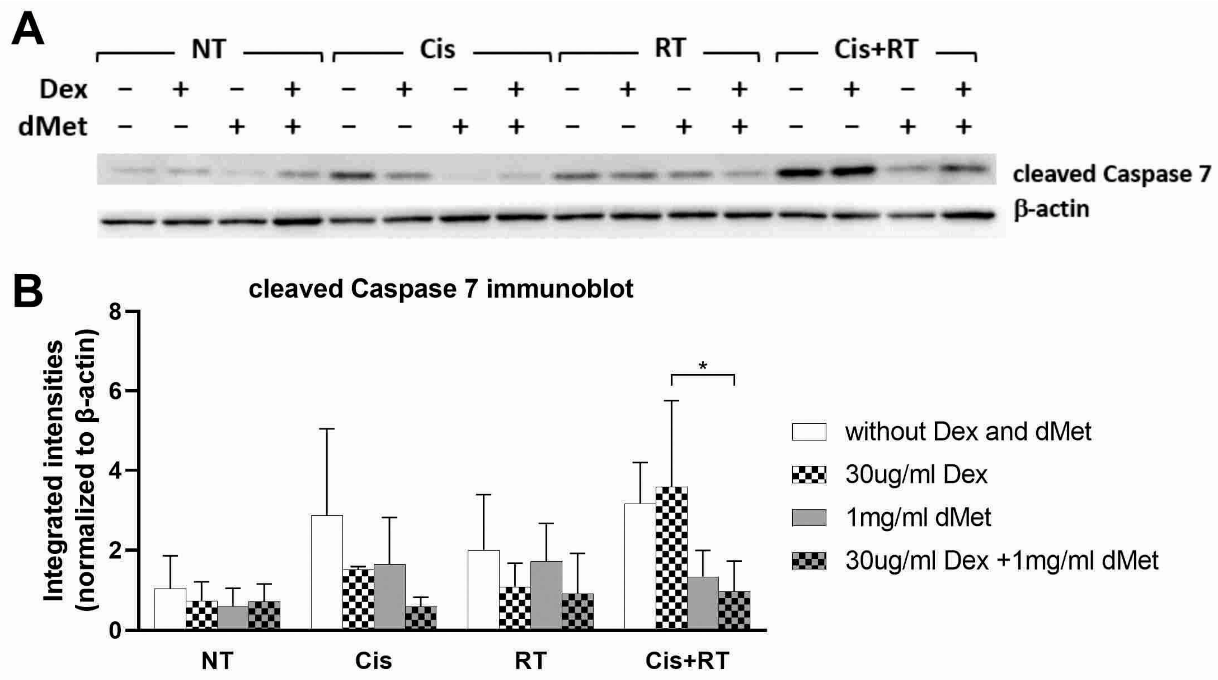
Figure 5.
Reduced p53 phosphorylation following Dex, dMet and their combination during cytotoxicity induced by cisplatin, radiation and their combination. OC-k3 cells were pre-treated with 30 μg/mL Dex and 1 mg/mL Met for 3 h before receiving either no treatment (NT), 0.5 μg/mL cisplatin (Cis), 5 Gy radiation treatment (RT) or both (Cis+RT). Solvent control is labeled as without Dex and dMet. At day 3 post-treatment, cells were harvested and processed for immunoblot analysis. (A) Representative immunoblots display the immunoreactivities of phospho-p53(Ser15), p53 total protein and β-actin (as loading control). (B) Quantification of respective immunoreactivities, presented as integrated intensities normalized to total p53, was carried out in 3 independent experiments. Data are presented as bar charts with mean ± SD. Normalized phospho-p53 immunoreactivity (phospho-p53/p53 total protein) was analyzed using a linear model with cancer treatment modality (NT, Cis, RT, Cis+RT), otoprotective treatment group (control, Dex, dMet, Dex+dMet), and their interaction as fixed effects, with experiment included as a covariate. Estimated marginal means were compared using Šídák-adjusted post hoc tests. Significant differences between solvent control, Dex, dMet and Dex+dMet under Cis, RT and Cis+RT treatment are indicated as asterisks. *** p < 0.001, ** p < 0.01, * p < 0.05.
3.4. The Greater Protective Effect of Dex May Be Associated with Increased Bcl-XL Expression, Reduced COX2 Expression, and Trends Toward Reduced ERK1/2 and NFkB Signaling
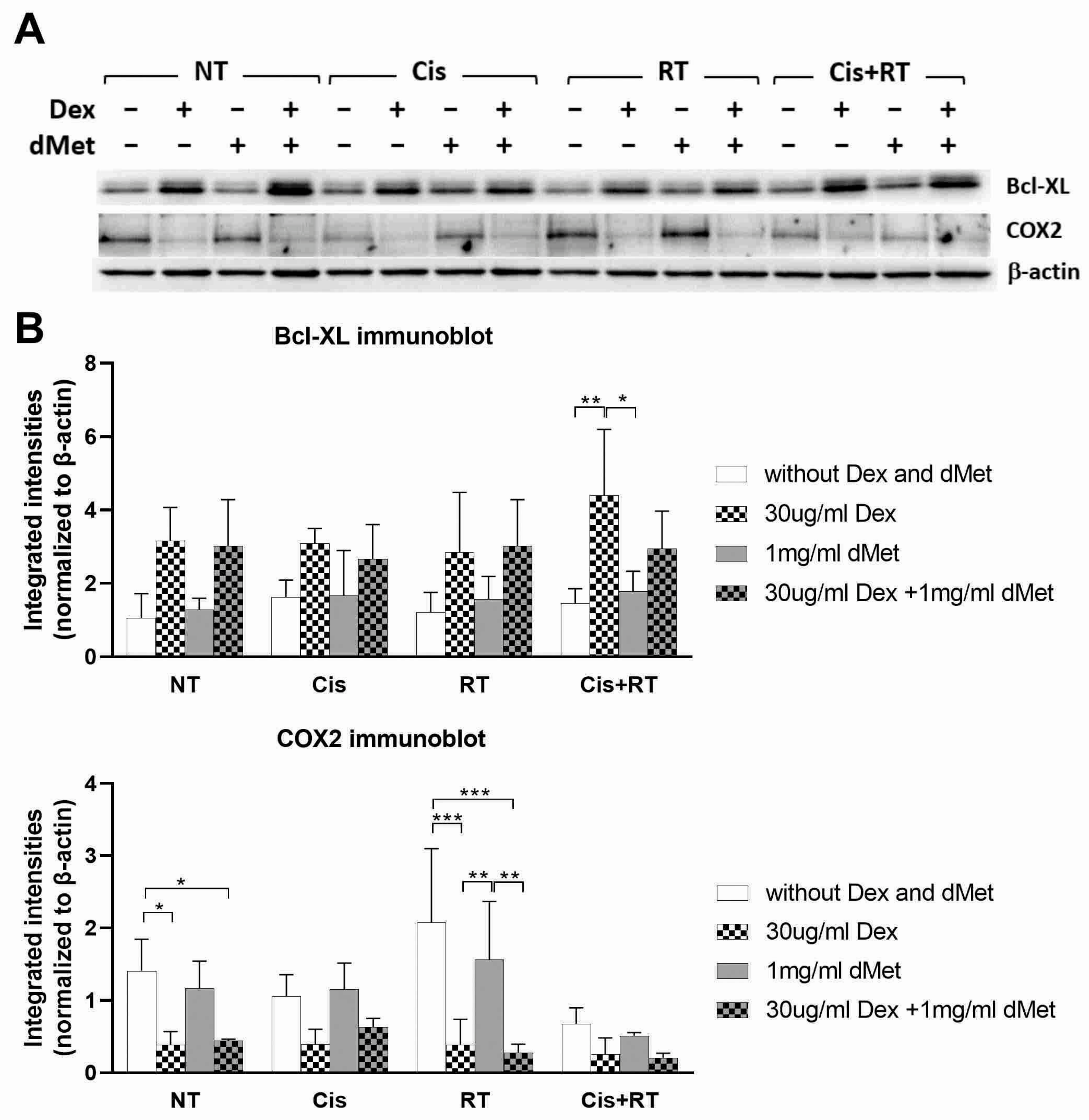
We further investigated whether Dex and dMet might employ alternative protective mechanisms by modulating apoptosis and inflammatory signaling pathways. Notably, Dex consistently upregulated the expression of the anti-apoptotic protein Bcl-XL under all conditions (Figure 6A,B), suggesting that upregulation of Bcl-XL may contribute to the protective effects of Dex through suppression of intrinsic apoptosis. Additionally, Dex consistently downregulated cyclooxygenase 2 (COX2) expression across all conditions (Figure 6A,B), highlighting its anti-inflammatory properties. Dex also exhibited a trend of inhibiting the phosphorylation of key molecules in the ERK1/2 and NFκB signaling pathway activated by Cis, RT and Cis+RT treatment (Supplementary Figure S2A,B). In contrast, dMet did not modulate Bcl-XL expression nor affect inflammation-associated pathways involving COX2, ERK1/2 or NFκB activation under any conditions tested (Figure 6A,B and Figure S2A,B).
Figure 6.
Dex induced consistent upregulation of Bcl-XL expression and downregulation of COX2 expression. OC-k3 cells were pre-treated with 30 μg/mL Dex and 1 mg/mL Met for 3 h before receiving either no treatment (NT), 0.5 μg/mL cisplatin (Cis), 5 Gy radiation treatment (RT) or both (Cis+RT). Solvent control is labeled as without Dex and dMet. At day 3 post-treatment, cells were harvested and processed for immunoblot analysis. (A) Representative immunoblots display the immunoreactivities of Bcl-XL, COX2 and β-actin (as loading control). (B) Quantification of respective immunoreactivities, presented as integrated intensities normalized to β-actin, was carried out in 3 independent experiments. Data are presented as bar charts with mean ± SD. Normalized Bcl-XL and COX2 immunoreactivities (integrated intensity/β-actin) were analyzed using linear models with cancer treatment modality (NT, Cis, RT, Cis+RT), otoprotective treatment group (control, Dex, dMet, Dex+dMet), and their interaction as fixed effects, with experiment included as a covariate. Estimated marginal means were compared using Šídák-adjusted post hoc tests. Significant differences between solvent control, Dex, dMet and Dex+dMet under Cis, RT and Cis+RT treatments are indicated as asterisks. *** p < 0.001, ** p < 0.01, * p < 0.05.
4. Discussion
4.1. Diverse Mechanisms of Otoprotection by Dex and dMet
Cisplatin is a common chemotherapy agent and, when used in larger cumulative doses, predisposes patients to serious side effects such as nephrotoxicity, neurotoxicity and ototoxicity. Cisplatin has been found to affect different parts of the cochlea: the organ of Corti, spiral ganglion cells and lateral wall [17]. Cisplatin inhibits the antioxidant enzyme activity in the cochlear, resulting in build-up of reactive oxygen species (ROS), leading to calcium influx into the cochlear cells and causing apoptosis [18]. Cisplatin also triggers apoptosis by activating caspases which are proapoptotic enzymes.
Radiation is the primary treatment for nasopharyngeal cancer, with cisplatin being given concurrently for more advanced stages. Due to the close proximity of the nasopharynx to the inner ear, radiation predisposes patients to hearing loss. Our previous research highlighted an increase in apoptotic-related gene expression and subsequent apoptotic cell death, potentially explaining the synergistic ototoxicity observed with the combination of cisplatin and radiation [15].
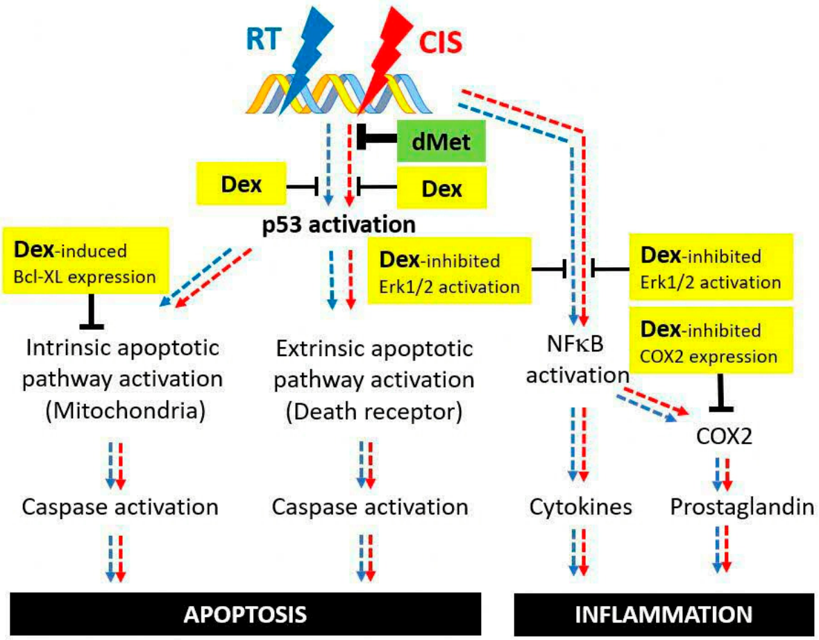
Our present findings suggest that combining Dex and dMet provides greater protection than either agent alone against cisplatin- and radiation-induced cytotoxicity in the OC-k3 model, supporting further investigation of this combination as a potential strategy to mitigate treatment-induced ototoxicity. Notably, Dex and dMet appear to engage distinct mechanisms in protecting against cytotoxicity, particularly in the process of apoptosis and inflammation (see Figure 7). Generally, cisplatin and radiation treatments increased ROS production, inducing DNA damage and activating p53. Subsequently, activation of effector caspase 7 may cause ROS to accumulate, resulting in cells detaching from the extracellular membrane, causing apoptosis [19]. Moreover, both treatments can induce inflammatory responses, exacerbating ototoxicity [20,21,22].
In the present study, we intentionally adopted the same cisplatin exposure paradigm (0.5 µg/mL; approximately 1.7 µM for 72 h without washout) as our previously validated OC-k3 model [15] to enable direct comparison with the molecular mechanisms established in that study. Although many published in vitro cochlear cell models employ substantially higher cisplatin concentrations administered over relatively shorter exposure periods to induce rapid cytotoxicity [23,24], our model was designed to investigate injury following prolonged continuous exposure. Continuous incubation for 72 h without washout results in greater cumulative drug exposure than acute short-pulse protocols and may better approximate the sustained cochlear exposure experienced during clinical cisplatin treatment. This experimental paradigm therefore provides a complementary model for studying ototoxicity under prolonged exposure conditions while maintaining consistency with our previously established dataset.
Our study provides insights into how dMet prevents cisplatin-induced ototoxicity. We observed that dMet could fully protect against cisplatin-induced apoptosis, by inhibiting cleaved caspase 7 and p53 activation to basal level (as shown in Figure 3, Figure 4 and Figure 5). Intriguingly, being a ROS scavenger to protect against cisplatin, dMet did not exhibit the same protection effects against radiation. Given our identification of p53 activation as a shared mechanism for both cisplatin and radiation-induced apoptosis, we speculate that the sources of ROS generated by cisplatin and radiation treatment could be different. Thus, dMet may act directly to impede cisplatin-specific ROS processes, resulting in the complete blockage of the downstream signaling. Future investigations to delineate these ROS differences could elucidate the otoprotective role of dMet. However, despite its effectiveness against cisplatin-induced apoptosis, dMet might not alleviate cisplatin-mediated inflammatory responses.
Our data also confirmed the protective effects of Dex against cisplatin and radiation treatment. Dex upregulated Bcl-XL, an inhibitor of the intrinsic apoptotic pathway. Furthermore, Dex could inhibit ERK1/2 and NFκB activation and COX2 expression to ameliorate inflammatory pathways, which may also indirectly attenuate ototoxicity. Our findings suggested that these alternative protective mechanisms associated with Dex administration may play crucial roles in shielding cells exposed to radiation alone and contribute to the greater protective effects observed with the combined treatment.
Figure 7.
Proposed mechanisms underlying the greater protective effects of combined Dex and dMet against cisplatin- and radiation-induced cytotoxicity. Cisplatin (CIS, pathway activation indicated by red arrows) and radiation (RT, pathway activation indicated by blue arrows) induce DNA damage, leading to p53 activation and subsequent apoptosis. Additionally, CIS and RT may activate inflammation, which have been implicated in the development of ototoxicity. Using the organ-of-Corti-derived OC-k3 cell line as an in vitro model of treatment-induced cytotoxicity, we demonstrated that dMet markedly reduced cisplatin-induced apoptosis via blocking p53 activation. Conversely, Dex was associated with protection against cisplatin- and radiation-induced cytotoxicity, accompanied by attenuating p53 activation and upregulating Bcl-XL expression to inhibit apoptosis. Dex also inhibited ERK1/2 and NFκB activation and downregulated COX2 expression to mitigate inflammation. Together, these findings suggest that Dex and dMet may protect through complementary mechanisms, thereby providing greater protection than either agent alone.
4.2. Study Limitations
A limitation of this study is that CellTox Green and MTS assays measure cytotoxicity and metabolic activity, respectively, rather than directly quantifying cell number or survival. Nevertheless, although performed on separate plates, both assays demonstrated consistent treatment-dependent responses, which, together with the modulation of apoptosis- and stress-related signaling pathways, support the overall protective effects of Dex and dMet in this model.
Formal mycoplasma testing was not performed on the OC-k3 cells used in this study; whilst routine microscopy examination, including DAPI staining, did not reveal evidence of contamination and cell morphology remained consistent throughout, the absence of dedicated mycoplasma testing is acknowledged as a limitation.
Another limitation is that only a single cisplatin concentration (0.5 µg/mL) was evaluated. Although this exposure paradigm was selected based on our previously validated model and utilizes prolonged continuous exposure rather than the higher-dose, short-duration protocols commonly used in vitro, the present study did not directly compare different cisplatin concentrations or exposure regimens. Therefore, whether the protective effects of dexamethasone and D-methionine are maintained under higher cisplatin concentrations remains to be determined. Future studies examining multiple clinically and experimentally relevant exposure conditions would further establish the generalizability of these findings.
In addition, while ototoxicity studies typically benefit from in vivo models, our current study employed an in vitro model utilizing the organ-of-Corti-derived cell lines treated with cisplatin and radiation. This approach enabled us to effectively investigate the dose response of Dex and dMet and elucidate their underlying protective mechanisms. Previous study has indicated that apoptosis manifested in cells three days post-treatment with cisplatin and radiation [15]. Hence, we chose to monitor cells at this time point to assess the protective effect of Dex and dMet. It is worth noting that this time point may not be optimal for studying inflammatory-related pathways, such as ERK1/2 and NFκB signaling, given their rapid and biphasic activation. Consequently, transient changes occurring at earlier time points may have been missed, which could contribute to the greater variation observed among the three independent experiments and the lack of statistically significant changes in ERK1/2 and NFκB signaling. Nonetheless, our findings align with prior reports indicating that Dex can inhibit inflammation by destabilizing COX2 mRNA and inhibiting ERK signaling, thereby repressing NFκB activity [25,26].
5. Conclusions
Clinical trials have explored the administration of systemic or transtympanic steroids to prevent cisplatin-induced ototoxicity [4,27,28]. Despite Dex being a promising drug to protect against ototoxicity, challenges persist regarding its ability to penetrate the inner ear, and the time taken for it to reach and remain in the inner ear with respect to cisplatin dosing. Strategies to address these challenges, such as dexamethasone-loaded nanoparticles and sustained drug delivery systems, have been investigated in animal studies [29,30]. Presently, systemic dexamethasone is routinely administered to patients undergoing chemoradiation for nasopharyngeal cancer to manage nausea. Previous clinical trials have administered oral dMet at a dose of 100 mg/kg prior to cisplatin or 100 mg/kg bid 30 to 60 min before and after radiotherapy, with no observed adverse effects [9,13]. Our findings provide preliminary in vitro evidence that the combination of Dex and dMet exerts additive protective effects against cisplatin- and radiation-induced ototoxic injury in OC-k3 cells. Further studies in animal models and clinical settings are required to determine whether these findings translate into meaningful hearing protection in patients undergoing chemoradiation for nasopharyngeal cancer.
Supplementary Materials
The following supporting information can be downloaded at https://www.mdpi.com/article/10.3390/ohbm7020026/s1, Table S1: Primary and secondary antibodies used in immunoblot analysis; Figure S1: Representative merged fluorescent and bright field images showing the protective effect of Dex and dMet against combined cisplatin and radiation treatment in OC-k3 mouse cochlear cell line at 72 h post-treatment; Figure S2: Dex consistently inhibited phosphorylation of ERK1/2 and NFκB p65; Figure S3: Full Scans of immunoblots presented in this study.
Author Contributions
V.Y.J.T.: Conceptualisation, Writing—Original draft preparation, Project administration, Funding acquisition, G.M.L.: Writing—Original draft preparation, Writing—Review and Editing, C.M.L.: Writing—Review and Editing, J.F.T.: Writing—Review and Editing, W.K.L.: Conceptualisation, Writing—Review and Editing, K.H.D.: Writing—Review and Editing, S.W.T.: Validation, Investigation, Resources, Writing—Original draft preparation, Writing—Review and Editing, E.Z.Z.: Writing—Review and Editing, M.G.K.T.: Formal analysis, Investigation, Methodology, Supervision, Project administration, Writing–Original draft preparation, Writing—Review and Editing, C.Y.B.L.: Validation, Investigation, resources, Writing—Review and Editing, H.B.A.K.: Formal analysis. All authors have read and agreed to the published version of the manuscript.
Funding
This initiative is funded by the SingHealth Duke-NUS Academic Medical Center, facilitated by the Joint Office of Academic Medicine. It is an initiative of Surgery Academic Clinical Program (SURG ACP), sited at the Singapore General Hospital.
Institutional Review Board Statement
Not applicable.
Informed Consent Statement
Not applicable.
Data Availability Statement
The original contributions presented in the study are included in the article/Supplementary Materials; further inquiries can be directed to the corresponding authors.
Acknowledgments
The authors would like to thank Natalie Aw for assisting in culturing of cells.
Conflicts of Interest
The authors declare no conflicts of interest. The funders had no role in the design of the study; in the collection, analyses, or interpretation of data; in the writing of the manuscript; or in the decision to publish the results.
References
- Yip, P.; Mok, K.; Ho, H. Sensorineural Hearing Loss in Nasopharyngeal Carcinoma Survivors in the Modern Treatment Era—The Early and Late Effects of Radiation and Cisplatin. Clin. Oncol. 2021, 34, e160–e167. [Google Scholar]
- Chan, S.; Ng, L.; Goh, X. Time Course and Clinical Characterization of Cisplatin-Induced Ototoxicity after Treatment for Nasopharyngeal Carcinoma in a South East Asian Population. Head Neck 2018, 40, 1425–1433. [Google Scholar] [CrossRef] [Scilit] [PubMed]
- Bray, F.; Haugen, M.; Moger, T.; Tretli, S.; Aalen, O.; Grotmol, T. Age-Incidence Curves of Nasopharyngeal Carcinoma Worldwide: Bimodality in Low-Risk Populations and Aetiologic Implications. Cancer Epidemiol. Biomark. Prev. 2008, 17, 2356–2365. [Google Scholar] [CrossRef] [Scilit]
- Marshak, T.; Steiner, M.; Kaminer, M.; Levy, L.; Shupak, A. Prevention of Cisplatin-Induced Hearing Loss by Intratympanic Dexamethasone: A Randomized Controlled Study. Otolaryngol. Head Neck Surg. 2014, 150, 983–990. [Google Scholar] [CrossRef] [Scilit] [PubMed]
- van As, J.; van den Berg, H.; van Dalen, E. Medical Interventions for the Prevention of Platinum-Induced Hearing Loss in Children with Cancer. Cochrane Database Syst. Rev. 2019, 5, CD009219. [Google Scholar] [CrossRef] [Scilit] [PubMed]
- Dinh, C.; Chen, S.; Bas, E. Dexamethasone Protects Against Apoptotic Cell Death of Cisplatin-Exposed Auditory Hair Cells In Vitro. Otol. Neurotol. 2015, 36, 1566–1571. [Google Scholar] [CrossRef] [Scilit] [PubMed]
- Korver, K.; Rybak, L.; Whitworth, C.; Campbell, K. Round Window Application of D-Methionine Provides Complete Cisplatin Otoprotection. Otolaryngol. Head Neck Surg. 2002, 126, 683–689. [Google Scholar] [CrossRef] [Scilit] [PubMed]
- Campbell, K.; Rybak, L.; Meech, R.; Hughes, L. D-Methionine Provides Excellent Protection from Cisplatin Ototoxicity in the Rat. Hear. Res. 1996, 102, 90–98. [Google Scholar] [CrossRef] [Scilit] [PubMed]
- Campbell, K.; Rehemtulla, A.; Sunkara, P.; Hamstra, D.; Buhnerkempe, M.; Ross, B. Oral D-Methionine Protects against Cisplatin-Induced Hearing Loss in Humans: Phase 2 Randomized Clinical Trial in India. Int. J. Audiol. 2021, 61, 621–631. [Google Scholar] [CrossRef] [Scilit] [PubMed]
- Campbell, K.; Meech, R.; Rybak, L.; Hughes, L. The Effect of D-Methionine on Cochlear Oxidative State with and without Cisplatin Administration: Mechanisms of Otoprotection. J. Am. Acad. Audiol. 2003, 14, 144–156. [Google Scholar] [CrossRef] [Scilit] [PubMed]
- Campbell, K.; Meech, R.; Rybak, L.; Hughes, L. D-Methionine Protects Against Cisplatin Damage to the Stria Vascularis. Hear. Res. 1999, 138, 13–28. [Google Scholar] [CrossRef] [Scilit] [PubMed]
- Vuyyuri, S.; Hamstra, D.; Khanna, D. Evaluation of D-Methionine as a Novel Oral Radiation Protector for Prevention of Mucositis. Clin. Cancer Res. 2008, 14, 2161–2170. [Google Scholar] [CrossRef] [Scilit] [PubMed]
- Hamstra, D.; Lee, K.; Eisbruch, A. Double-Blind Placebo-Controlled Multicenter Phase II Trial to Evaluate D-Methionine in Preventing/Reducing Oral Mucositis Induced by Radiation and Chemotherapy for Head and Neck Cancer. Head Neck 2018, 40, 1375–1388. [Google Scholar] [CrossRef] [Scilit] [PubMed]
- Low, W.; Toh, S.; Wee, J.; Fook-Chong, S.; Wang, D. Sensorineural Hearing Loss after Radiotherapy and Chemoradiotherapy: A Single, Blinded, Randomized Study. J. Clin. Oncol. 2006, 24, 1904–1909. [Google Scholar] [CrossRef] [Scilit] [PubMed]
- Low, W.; Kong, S.; Tan, M. Ototoxicity from Combined Cisplatin and Radiation Treatment: An In Vitro Study. Int. J. Otolaryngol. 2010, 2010, 523976. [Google Scholar] [CrossRef] [Scilit] [PubMed]
- Kalinec, F.; Kalinec, G.; Boukhvalova, M.; Kachar, B. Establishment and Characterization of Conditionally Immortalized Organ of Corti Cell Lines. Cell Biol. Int. 1999, 23, 175–184. [Google Scholar] [CrossRef] [Scilit] [PubMed]
- van Ruijven, M.; de Groot, J.; Hendriksen, F.; Smoorenburg, G. Immunohistochemical Detection of Platinated DNA in the Cochlea of Cisplatin-Treated Guinea Pigs. Hear. Res. 2005, 203, 112–121. [Google Scholar] [CrossRef] [Scilit] [PubMed]
- Clerici, W.; DiMartino, D.; Prasad, M. Direct Effects of Reactive Oxygen Species on Cochlear Outer Hair Cell Shape In Vitro. Hear. Res. 1995, 84, 30–40. [Google Scholar] [CrossRef] [Scilit] [PubMed]
- Brentnall, M.; Rodriguez-Menocal, L.; De Guevara, R.; Cepero, E.; Boise, L. Caspase-9, Caspase-3 and Caspase-7 Have Distinct Roles during Intrinsic Apoptosis. BMC Cell Biol. 2013, 14, 32. [Google Scholar] [CrossRef] [Scilit] [PubMed]
- Astradsson, T.; Sellberg, F.; Berglund, D.; Ehrsson, Y.; Laurell, G. Systemic Inflammatory Reaction in Patients with Head and Neck Cancer-An Explorative Study. Front. Oncol. 2019, 9, 1177. [Google Scholar] [CrossRef] [Scilit] [PubMed]
- Gentilin, E.; Simoni, E.; Candito, M.; Cazzador, D.; Astolfi, L. Cisplatin-Induced Ototoxicity: Updates on Molecular Targets. Trends Mol. Med. 2019, 25, 1123–1132. [Google Scholar] [CrossRef] [Scilit] [PubMed]
- Vyas, D.; Laput, G.; Vyas, A. Chemotherapy-Enhanced Inflammation May Lead to the Failure of Therapy and Metastasis. OncoTargets Ther. 2014, 7, 1015–1023. [Google Scholar] [CrossRef] [Scilit]
- Guidotti, L.; Tomassi, E.; Marracci, S.; Lai, M.; Lapi, D.; Pesi, R.; Pucci, L.; Novellino, E.; Albi, E.; Garcia-Gil, M. Effects of Nutraceuticals on Cisplatin-Induced Cytotoxicity in HEI-OC1 Cells. Int. J. Mol. Sci. 2023, 24, 17416. [Google Scholar] [CrossRef] [Scilit] [PubMed]
- Previati, M.; Lanzoni, I.; Astolfi, L.; Fagioli, F.; Vecchiati, G.; Pagnoni, A.; Martini, A.; Capitani, S. Cisplatin Cytotoxicity in Organ of Corti-Derived Immortalized Cells. J. Cell. Biochem. 2007, 101, 1185–1197. [Google Scholar] [CrossRef] [Scilit] [PubMed]
- Bladh, L.; Johansson-Haque, K.; Rafter, I.; Nilsson, S.; Okret, S. Inhibition of Extracellular Signal-Regulated Kinase (ERK) Signaling Participates in Repression of Nuclear Factor (NF)-kappaB Activity by Glucocorticoids. Biochim. Biophys. Acta 2009, 1793, 439–446. [Google Scholar] [CrossRef] [Scilit] [PubMed]
- Lasa, M.; Brook, M.; Saklatvala, J.; Clark, A. Dexamethasone Destabilizes Cyclooxygenase 2 mRNA by Inhibiting Mitogen-Activated Protein Kinase P38. Mol. Cell. Biol. 2001, 21, 771–780. [Google Scholar] [CrossRef] [Scilit] [PubMed]
- Chen, J.; Zhao, Y.; Zhou, X. Methylprednisolone Use during Radiotherapy Extenuates Hearing Loss in Patients with Nasopharyngeal Carcinoma. Laryngoscope 2016, 126, 100–103. [Google Scholar] [PubMed]
- Moreno, I.; Belinchon, A. Evaluating the Efficacy of Intratympanic Dexamethasone in Protecting Against Irreversible Hearing Loss in Patients on Cisplatin-Based Cancer Treatment: A Randomized Controlled Phase IIIB Clinical Trial. Ear Hear. 2021, 43, 676–684. [Google Scholar] [CrossRef] [Scilit]
- Sun, C.; Wang, X.; Chen, D.; Lin, X.; Yu, D.; Wu, H. Dexamethasone Loaded Nanoparticles Exert Protective Effects against Cisplatin-Induced Hearing Loss by Systemic Administration. Neurosci. Lett. 2016, 619, 142–148. [Google Scholar] [CrossRef] [Scilit] [PubMed]
- Fernandez, R.; Harrop-Jones, A.; Wang, X.; Dellamary, L.; LeBel, C.; Piu, F. The Sustained-Exposure Dexamethasone Formulation OTO-104 Offers Effective Protection against Cisplatin-Induced Hearing Loss. Audiol. Neurotol. 2016, 21, 22–29. [Google Scholar] [CrossRef] [Scilit]
Disclaimer/Publisher’s Note: The statements, opinions and data contained in all publications are solely those of the individual author(s) and contributor(s) and not of MDPI and/or the editor(s). MDPI and/or the editor(s) disclaim responsibility for any injury to people or property resulting from any ideas, methods, instructions or products referred to in the content. |
© 2026 by the authors. Licensee MDPI, Basel, Switzerland. This article is an open access article distributed under the terms and conditions of the Creative Commons Attribution (CC BY) license.