


Abstract
Dysphagia refers either to the difficulty someone may have with the initial
phases of a swallow (usually described as oropharyngeal dysphagia,
“OD”) or to the sensation that foods and or liquids are somehow
being obstructed in their passage from the mouth to the stomach (usually
described as “esophageal dysphagia”). In patients with no
indication of a somatic disease or abnormality, psychiatric conditions must be
considered as a possible cause of OD. Moreover, diagnosis and treatment of
dysphagia are not standardized. There is no universal standard tool for
screening or clinical assessment of OD. Education of health professionals on
early diagnosis and improvement of therapeutic strategies are mainstays to allow
maximal recovery potential in this population. Future studies, clinical trials,
clinical evidence and clear guidelines are needed to manage this condition.
INTRODUCTION
Dysphagia refers either to the difficulty someone may have with the initial phases of
a swallow (usually described as oropharyngeal dysphagia, “OD”) or to
the sensation that foods and or liquids are somehow being obstructed in their
passage from the mouth to the stomach (usually described as “esophageal
dysphagia”) 1. It is
positive in 12.4% after extubation (18.3% of emergency and 4.9% of elective
patients) 2. The prevalence of OD
has been calculated in older people across different setting, with rates between 30
and 40% in geriatric acute care unit 3,4. It may be more
common in an acute hospital setting because patients often have multiple medical
comorbidities. In particular, it observed predominantly in patients with an altered
mental status 5. Well-established
risk factors for dysphagia include advanced age, neurological disease, head-neck
cancer and pulmonary disease 6.
However, Giordano et al, have been reported that in an octogenarian, the only
clinical symptoms of generalized tetanus, dysphagia and tremor 7. It has been reported that
isolated bulbar symptoms, as dysphagia, in elderly, may be included in the
differential diagnosis 8. To this
regard, the European Society for swallowing disorders-European Union Geriatric
Medicine Society describe that dysphagia can cause severe complications such as
malnutrition, dehydration, respiratory infections, aspiration/pneumonia and
increased readmissions, institutionalization and mortality. In fact, OD is a
prevalent and serious problem in older patients. Oro-pharyngeal swallow response is
impaired in older people and can cause aspiration 9. Despite its prevalence and severity, OD is still
underdiagnosed and untreated in many medical centers.
The present minireview highlights the importance of considering crucial causes among
the possible expression and cause of dysphagia.
METHODS
Clinical trials were identified by PubMed until June 30 2020. The search keywords
were “dysphagia, elderly, OD”. The studies were selected, their
references were reviewed for potential inclusion. Studies written in languages other
than English were excluded. Three authors (T.C., G.S., and O.P.) reviewed all study
abstracts. Studies were included if analyzed dysphagia in elderly patients All
selected studies are qualitatively analyzed.
ETIOPATHOGENESIS
Dysphagia can be related to gastrointestinal or not gastrointestinal causes, as
reported in Table I.
GASTROINTESTINAL CAUSES
Eosinophilic esophagitis
Esophageal food bolus impaction is an acute multidisciplinary common
emergency, frequently observed in the Emergency Department 10. The estimated annual
incidence rate is 13 per 100,000 persons after upper and lower
gastrointestinal bleeding 11. The most common presentation of food bolus impaction
in the Western countries are the “steakhouse syndrome”, so
called because the meats represent the most frequently impacted foods
12. Several
abnormalities of esophageal mucosal and neuromuscular layers, as well as
underlying diseases, including edentulism, eosinophilic esophagitis and
esophageal stenosis (as peptic esophageal stricture or Schatzki’s
ring), facilitate food bolus impaction 13. In particular, several studies suggest
that eosinophilic esophagitis (EO) is an emergency cause of food bolus
impaction and dysphagia in adult (Fig.
1) 14,15. It has been reported
that patients who present with food bolus impaction, have approximately 1 in
3 chance of having EO. According to a study, EO was found in 54% of patients
presenting with food bolus impaction 16. EO usually affects young adults with mean age 30
years old 14. The
exclusion of secondary causes of EO, the presence of mucosal eosinophilia
being isolated to the esophagus and persisting after a proton pump inhibitor
trial are also included in the diagnostic criteria for EO 17. According to the
current guidelines, EO is defined histologically with eosinophils count of
15 or more eosinophils/hpf.
Esophageal motility disorders
More infrequently, esophageal motility disorders (as diffuse esophageal
spasm) and other esophageal hypertensive dysmotility’s, may represent
a cause of food bolus impaction 18. This multidisciplinary, as well as the plurality
of underlying esophageal diseases, generate a high heterogeneity in its
management, ranging from observational therapy to surgical treatment
19. Although
more than half of food bolus impaction will resolve spontaneously after a
short observational period without any serious consequences, in 10-20% of
cases a conservative or surgical management is required 20. Several studies have
been conducted on pharmacological and non-pharmacological agents for
treatment of this issue. Pharmacological agents with enzymatic activity such
as papain, trypsin and chymotrypsin are no longer recommended due to high
risk of esophageal perforation and hypernatremia. In the same way, the
administration of hyoscine butyl-bromide (Buscopan), glucagon injection and
diazepam resulted in no significant difference in compare to controls
21. Better
results were obtained by non-pharmacological effervescent agents (fizzy
drinks). However, the level of evidence of these studies is too low to
provide strong conclusions. The current treatment, recommended by American
Society for Gastrointestinal Endoscopy (ASGE) include endoscopic food
extraction and, only in some cases, the advancement of the bolus into the
stomach under endoscopic guide 22. With regards to the management of food bolus
impaction, Ikenberry et al., recommended a time frame of 6 h to remove the
food bolus due to the potential risk of ischemia, necrosis and perforation
23.
Achalasia
It is a relatively rare primary motor esophageal disorder, characterized by
absence of relaxation of the lower esophageal sphincter and of peristalsis
along the esophageal body. As a result, patients typically, present with
dysphagia, regurgitation and occasionally chest pain. High resolution
manometry is recommended for the diagnosis of the esophageal achalasia
(GRADE recommendation: low). It is recommended to perform endoscopy to
exclude neoplastic pseudo-achalasia (Good practice recommendation). There is
no convincing evidence that medical treatment with nitrates or calcium
blocker or phosphodiesterase inhibitors is effective for symptomatic relief
in adult with achalasia. Treatment of achalasia with peroral endoscopic
myotomy (POEM) results in similar outcomes on swallowing functions compared
with alternative treatment 24.
NON-GASTROINTESTINAL CAUSES
Post-stroke dysphagia
Dysphagia is a common complication after stroke with a prevalence to vary
from 51 to 65% of patients. Many of them are asymptomatic or have symptoms
that aren’t related to swallowing problems. Although many patients
recover swallowing spontaneously, 11-50% still have dysphagia at 6 months
25,26. Post-stroke dysphagia
(PSD) is associated with a poor outcome for multiple reasons:
- it is a manifestation of severe stroke and it is associated with increased death, dependency, disability, impairment and institutionalization;
- it causes aspiration of foods, liquids and oral secretions and therefore pneumonia 27,28, which in itself leads to death;
- however, poor recognition and management leads to dehydration and malnutrition 29.
Although multiple advances have been made in the early management of stroke
(e.g. with thrombolysis, aspirin, mechanical thrombectomy and
hemicraniectomy) and secondary prevention (e.g. with antithrombotic, blood
pressure lowering, lipid lowering, carotid endarterectomy), PSD remains a
neglected research area and its optimal management, including treatment, has
yet to be defined. Nevertheless, guidelines recommend assessment of
swallowing within 24h after stroke 1. In respect of drug treatment, a small pilot
randomized trial suggested that nifedipine (a calcium channel blocker that
relaxes esophageal smooth muscle) might improve swallowing, and
metoclopramide (a dopamine D2-receptor antagonist with antiemetic and
gastric prokinetic activity) might reduce the incidence of pneumonia
25. Glyceryl
trinitrate (GTN), given within 6h of stroke, was associated with improved
functional outcome 30,31. In fact, GTN might also
improve swallowing though relaxing esophageal smooth muscle 32-34.
Medically unexplained oropharyngeal dysphagia (MUNOD)
Rarely, OD occurs without demonstrable abnormalities in the anatomy of the
upper aero-digestive tract and/or swallowing physiology, prompting a
diagnosis of medically unexplained oropharyngeal dysphagia
(MUNOD). Functional somatic disorders and comorbid anxiety and
depression are both associated with increased severity of symptoms and
greater illness burden. According to the DSM-V classification, phagophobia
belongs to the category of ‘specific phobias’ (26). Patients
with phagophobia experience an abnormal sensation during swallowing,
sometimes accompanied by behavioral abnormalities during swallowing
examination 35.
MANAGEMENT
Thus, patients with a food bolus impaction in the emergency department should receive
a clinical and instrumental assessment with chest radiograph to rule out evidence of
perforation or a radiopaque object in the esophagus (Fig. 2). Once a foreign object is ruled out, endoscopy
should be considered 36. The
food bolus can be removed during upper endoscopy and is successful in 98% of
patients during the first endoscopy 37.
A recent review explored the level of evidence and classified the types of
interventions into the following categories:
- bolus modification and management;
- swallow postures and maneuvers;
- other interventions (as oral hygiene, feeding);
- facilitation techniques.
BOLUS MODIFICATIONS
The modification of the consistence of solids and or liquids is the main element
of compensatory treatment for patients suffering from OD. The level of evidence
with this treatment is A (randomized controlled trials) and B (non-randomized
trials) 38.
SWALLOW POSTURE
A general directive is to swallow in an upright position (90 seated) and to
maintain this posture after the meal for at least 30 minutes. Generally, level
of evidence for maneuvers or other posture is B 38.
ORAL HYGIENE
Deficient oral hygiene is a risk factor for pneumonia in older patients with OD
39. Minimal oral
hygiene should be performed every 12 hours to avoid dental plaque formation
40. Mouthwashes
should be used at least every 3 days, chlorhexidine being the most effective but
it should not be used for > 15 days 41.
NUTRITIONAL STATUS
Nutritional status is basic to good health and an important part of treatment of
many chronic diseases. The relationship between malnutrition and OD has been
established. It has been recommended that patients following modified texture
diet or being given enteral feeding for OD should have their swallowing and
nutritional status regularly assessed, after the first week and then every 2 or
3 months for the first years and after every 6 months 42.
Oral nutritional supplement should be provided in patients with:
- anorexia;
- dietary restriction due to chronic disease;
- nutritional intake < 75% of their nutritional requirements;
- involuntary weight loss.
Nutritional interventions include different strategies such as adaptation of the
diet and enteral nutrition, administered by nasogastric tube (NGT) or
percutaneous endoscopic gastrostomy (PEG) and should be based on the nutritional
needs.
NGT is recommended for short periods of time (< 2 months) and when there
is no risk of gastro esophageal reflux. It is reported for the care of the
patients with amyotrophic lateral sclerosis 43.
PEG is most commonly used and it can be inserted by endoscopy or surgery. Carrau
et al., reported the use of laryngeal framework surgery for the effective
treatment of aspiration in selected patients (such as with deficits of the
glottic closure secondary to vocal fold paralysis or paresis) 44. PEG may cause an enhanced
risk of gastro esophageal reflux disease (GERD) with increased risk of
aspiration 45. The
recommendations by Wirth et al., underline the importance of an early screening
and assessment of dysphagia and give advice for an evidence based and
comprehensive nutritional management to avoid aspiration, malnutrition and
dehydration 46.
FACILITATION TECHNIQUES
In recent years, new treatment based on stimulation or sensorial and motor
pathways are being assessed. Neuromuscular electrical stimulation therapy (NMES)
stimulates deglution nerves and muscles to improve OD. The main target nerves
are the superior laryngeal nerve, the pharyngeal branch of the glossopharyngeal
nerve and two branches of the vagus nerve, the pharyngeal branch and the
maxillary branch of the trigeminal nerve. Guidelines on the NMES have been
published by the British National Institute for Health and Care Excellence
giving recommendation. The evidence on safety is limited in quality and
quantity. In patients with PSD, treatment with transcutaneous NMES increased
prevalence of safe swallows and reduced the time of the laryngeal vestibule
closure (LVC) 47.
OTHER EVALUATION
In addition, MUNOD is a rare condition that is difficult to diagnose. Patients
deserve a professional approach, particularly because their diagnostic
trajectory has often been long and inconclusive. Affective symptoms are common
in these patients. MUNOD could be a symptom of a psychiatric condition or part
of the alarm falsification defense system, suggesting that physical symptoms and
affective disorders are stress-related and a response to earlier threats.
Consultation of a psychiatrist for patients with MUNOD is recommended as part of
a pathway toward multidisciplinary integrated care 35. In patients with prolonged dysphagia, with
no indication of a somatic disease or abnormality, psychiatric conditions must
be considered as a possible cause of OD. Validated psychological screening
questionnaires could be helpful in the detection of affective conditions but
also of other psychiatric conditions. Involvement of a psychiatrist and/or
psychologist is recommended.
DISCUSSION
The present review describes the gastrointestinal and not gastrointestinal causes of
dysphagia.
Due to its prognostic importance, an early detection of causes related dysphagia and
a suitable nutritional management is therefore of utmost clinical importance
46. A formalized
screening for dysphagia should be carried out in all patients as part of the initial
examination or upon arrival of the patient on the hospital ward. To this regard,
several studies reported associations between a pathological dysphagia screening and
an increased incidence of pneumonia 48 as well as a reduction of infectious complications after
implementation of a systematic screening 49. Unfortunately, despite its prevalence and severity,
dysphagia is still underdiagnosed and undertreated in many medical centers.
Moreover, diagnosis and treatment of dysphagia are not standardized. There is no
universal standard tool for screening or clinical assessment of OD. Education of
health professionals on early diagnosis and improvement of therapeutic strategies
are mainstays to allow maximal recovery potential in this population 50.
Current treatment of OD is usually compensatory, rehabilitative or a combination of
the two. Future studies, clinical trials, clinical evidence and clear guidelines are
needed to manage this condition.
Ethical consideration
None.
Acknowledgement
None.
Funding
This research received no external funding.
Conflict of interest
The Authors declare no conflict of interest.
Figures and tables
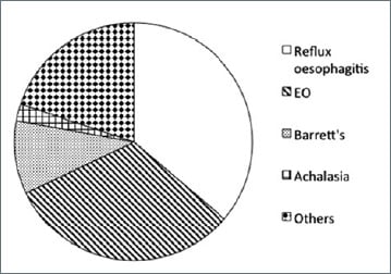
Figure 1.
In this figure we describe the main gastrointestinal causes of dysphagia.
Eosinophilic esophagitis (EO).
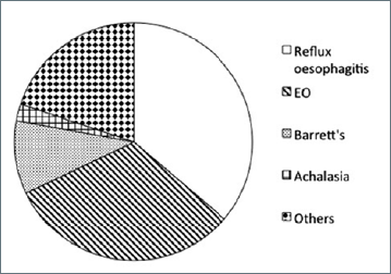
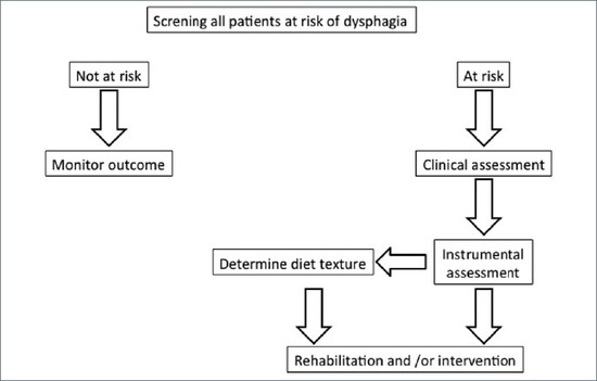
Figure 2.
In this figure we describe the algorithm to use for the management of the
dysphagia.
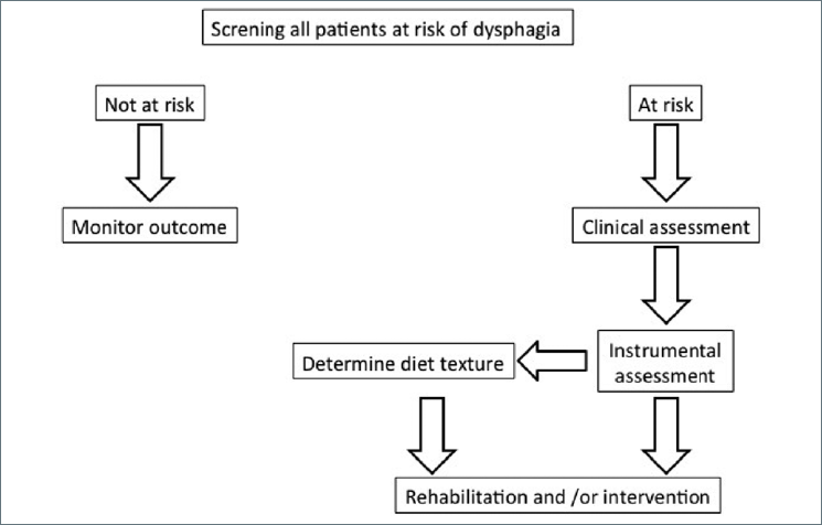

Table I.
Gastrointestinal and not gastrointestinal causes of dysphagia.
References
- Malagelada JR, Bazzoli F, Boeckxstaens G. World gastroenterology organization global guidelines: dysphagia – global guidelines and cascades update September 2014. J Clin Gastroenterol 2015;49:370. https://doi.org/10.1097/MCG.000000000000030710.1097/MCG.0000000000000307
- Schefold J, Berger D, Zurcher P, et al. Dysphagia in mechanically ventilation ICU patients (Dynamics): a prospective observational trial. Crit Care 2017;45:2061-2069. https://doi.org/10.1097/CCM.000000000000276510.1097/CCM.0000000000002765
- Cabre M, Almirall J, Clave P. Aspiration pneumonia: management in Spain. Eur Geriatr Med 2012;3:180-183.
- Clave P, Rofes L, Carrion S. Pathophysiology, relevance and natural history of OD among older people. Nestle Nutr Inst Workshops Ser 2012;72:57-66. https://doi.org/10.1159/00033998610.1159/000339986
- Simning I, Simning A. Severe idiopathic dysphagia in an acute hospital setting: assessment, management, and outcome. J Med Speech Lang Pathol 2013;21:3.
- Garon B, Sierzant T, Ormiston C. Silent Aspiration: results of 2,000 video fluoroscopic evaluations. J Neurosci Nurs 2009;41:178-185.
- Giordano M, Ciarambino T, Di Bella O. Acute dysphagia in an octogenarian: an unusual case of tetanus. Am J Emerg Med 2014;32:691.e1-691.e2. https://doi.org/10.1016/j.ajem.2013.12.02010.1016/j.ajem.2013.12.020
- Mustafa M, Mohamed MB, Hayat A. Tumour lysis syndrome and partial remission occurring after administration of a test dose of obinutuzumab. Eur J Case Rep Intern Med 2017;4:000516. https://doi.org/10.12890/2016_00051610.12890/2016_000516
- Baijens LWJ, Clave P, Cras O. European Society for swallowing disorders-European Union Geriatric Medicine Society white paper: oropharyngeal dysphagia as a geriatric syndrome. Clin Interv Aging. 2016,11:1403-1428.
- Price T, Jones SE, Montgomery PQ. Is current UK management of oesophageal food bolus obstruction evidence based? An e-mail survey and literature review. Eur Arch Otorhinolaryngol 2007;264:329-335. https://doi.org/10.1007/s00405-007-0260-x10.1007/s00405-007-0260-x
- Longstreth GF, Longstreth KJ, Yao JF. Esophageal food impaction: epidemiology and therapy. A retrospective, observational study. Gastrointest Endosc 2001;53:193-198. https://doi.org/10.1067/mge.2001.11270910.1067/mge.2001.112709
- Stadler J, Holscher AH, Feussner H, et al. The “Steakhouse syndrome” – primary and definitive diagnosis and therapy. Surg Endosc 1989;3:195-198. https://doi.org/10.1007/BF0217154510.1007/BF02171545
- Sperry SL, Crockett SD, Miller CB, et al. Esophageal foreign-body impactions: epidemiology, time trends, and the impact of the increasing prevalence of eosinophilic esophagitis. Gastrointest Endosc 2011;74:985-991. https://doi.org/10.1016/j.gie.2011.06.02910.1016/j.gie.2011.06.029
- Heerasing N, Lee SY, Alezander S. Prevalence of eosinophilic oesophagitis in adults presenting with oesophageal food bolus impaction. WJG Pharmacol Therap 2015;6:244-247. https://doi.org/10.4292/wjgpt.v6.i4.24410.4292/wjgpt.v6.i4.244
- Attwod SE, Forutaa GT. Eosinophilic esophagitis: historically perspective on an evolving disease. Gastroenterol Clin North Am 2014;43:185-199. https://doi.org/10.1016/j.gtc.2014.02.01010.1016/j.gtc.2014.02.010
- Desai TK, Stecevic V, Chang CH. Association of eosinophil inflammation with esophageal food impaction in adult. Gastrointest Endosc. 2005,61:795-801. https://doi.org/10.1016/s0016-5107(05)00313-510.1016/s0016-5107(05)00313-5
- Furuta GT, Liacouras CA, Collins MH, et al. Eosinophilic esophagitis in children and adults: a systematic review and consensus recommendations for diagnosis and treatment. Gastroenterology 2007;133:1342-1363. https://doi.org/10.1053/j.gastro.2007.08.01710.1053/j.gastro.2007.08.017
- Chae HS, Lee TK, Kim YW, et al. Two cases of steakhouse syndrome associated with nutcracker esophagus. Dis Esophagus 2002;15:330-333. https://doi.org/10.1046/j.1442-2050.2002.00271.x10.1046/j.1442-2050.2002.00271.x
- Reddy VM, Bennett W, Burrows SA, et al. Recurrence of food bolus impaction of the oesophagus: a retrospective observational study. Int J Surg 2011;9:464–466. https://doi.org/10.1016/j.ijsu.2011.04.01110.1016/j.ijsu.2011.04.011
- Basavaraj S, Penumetcha KR, Cable HR, et al. Buscopan in oesophageal food bolus: is it really effective?Eur Arch Otorhinolaryngol 2005;262:524-527. https://doi.org/10.1007/s00405-004-0852-710.1007/s00405-004-0852-7
- Khayyat YM. Pharmacological management of esophageal food bolus impaction. Emerg Med Int 2013;2013:924015. https://doi.org/10.1155/2013/92401510.1155/2013/924015
- ASGE Standards of Practice Committee(https://www.asge.org/home/resources/key-resources/guidelines)
- Ikenberry SO, Jue TL, Anderson MA. Management of ingested foreign bodies and food impactions. Gastrointes Endosc 2011;73:1085-1091. https://doi.org/10.1016/j.gie.2010.11.01010.1016/j.gie.2010.11.010
- Zaninotto G, Bennett C, Boeckxstaens G. The 2018 ISDE achalasia guidelines. Dis Esophagus 2018;31:1-29. https://doi.org/10.1093/dote/doy07110.1093/dote/doy071
- Martino R, Foley N, Bhogal S, et al. Dysphagia after stroke: incidence, diagnosis, and pulmonary complications. Stroke 2005;36:2756-2763. https://doi.org/10.1161/01.STR.0000190056.76543.eb10.1161/01.STR.0000190056.76543.eb
- Mann G, Hankey GJ, Cameron D. Swallowing disorders following acute stroke: prevalence and diagnostic accuracy. Cerebrovasc Dis 2000;10:380-386.
- Smithard D, Smeeton N, Wolfe C. Long-term outcome after stroke: does dysphagia matter?Age Ageing 2007;36:90-94. https://doi.org/10.1159/00001609410.1159/000016094
- Rofes L, Arreola V, Almirall J, et al. Diagnosis and management of oropharyngeal dysphagia and its nutritional and respiratory complications in the elderly. Gastroenterol Res Pract 2011;2011:13. https://doi.org/10.1155/2011/81897910.1155/2011/818979
- Wilson R. Mortality and cost of pneumonia after stroke for different risk groups. J Stroke Cerebrovasc Dis 2012;21. https://doi.org/10.1016/j.jstrokecerebrovasdis.2010.05.00210.1016/j.jstrokecerebrovasdis.2010.05.002
- Warusevitane A, Karuntilake D, Lally F, et al. Safety and effect of metoclopramide to prevent pneumonia in patients with stroke fed via nasogastric tubes trial. Stroke 2015;46:454-460. https://doi.org/10.1161/STROKEAHA.114.00663910.1161/STROKEAHA.114.006639
- Woodhouse L, Scutt P, Krishnan K, et al. Effect of hyperacute administration (within 6 hours) of transdermal glyceryl trinitrate, a nitric oxide donor, on outcome after stroke: subgroup analysis of the efficacy of nitric oxide in stroke (ENOS) trial. Stroke 2015;46:3194-3201.
- Ankolekar S, Fuller M, Cross I, et al. Feasibility of an ambulance- based stroke trial, and safety of glyceryl trinitrate in ultra-acute stroke: the rapid intervention with glyceryl trinitrate in Hypertensive Stroke Trial (RIGHT, ISRCTN66434824). Stroke 2013;44:3120-3128. https://doi.org/10.1161/STROKEAHA.113.00130110.1161/STROKEAHA.113.001301
- Wei J, Wu T, Yang Q, et al. Nitrates for stable angina: a systematic review and meta-analysis of randomized clinical trials. Int J Cardiol 2011;146:4-12. https://doi.org/10.1016/j.ijcard.2010.05.01910.1016/j.ijcard.2010.05.019
- Perez I, Smithard DG, Davies H, et al. Pharmacological treatment of dysphagia in stroke. Dysphagia 1998;13:12-16. https://doi.org/10.1007/PL0000954310.1007/PL00009543
- Baijens LW, Koetsenruijter K, Pilz W. Diagnosis and treatment of phagophobia: a review. Dysphagia 2013;28:260-270. https://doi.org/10.1007/s00455-013-9454-010.1007/s00455-013-9454-0
- VerdonschoT JCG, Laura WJ. Medically Unexplained oropharyngeal dysphagia at the University Hospital ENT outpatient clinic for dysphagia: a cross-sectional cohort study. Dysphagia 2019;34:43-51.
- Vicari JJ, Johanson JF, Frakes JT. Outcomes of acute esophageal food impaction: success of the push technique. Gastrointest Endosc 2001;53:178-181. https://doi.org/10.1067/mge.2001.11103910.1067/mge.2001.111039
- Speyer R, Baijens L, Heijnen M. Effects of therapy in OD by speech and language therapist: a systematic review. Dysphagia 2010;25:40-65. https://doi.org/10.1007/s00455-009-9239-710.1007/s00455-009-9239-7
- Ortega O, Clave P. Oral hygiene, aspiration, and aspiration pneumonia from pathophysiology to therapeutic strategies. Curr Phys Med Rehabil Rep 2013:292-295.
- Sjogren P, Nilsson E, Forseel M. A systematic review of the preventive effect of oral hygiene on pneumonia and respiratory tract infection in elderly in hospitals and nursing homes. JAGS 2008;56:2114-2130. https://doi.org/10.1111/j.1532-5415.2008.01926.x10.1111/j.1532-5415.2008.01926.x
- Berchier CE, Slot DE, Van der Wenden GA. The efficacy of 0.12% chlorhexidine mouthrise compared with 0.2% on plaque accumulation and periodontal parameters: a systematic review. J Clin Periodont 2010;37:829-839.
- European Society for swallowing disorders: position statement. Dysphagia 2013:28:280-335.
- Miller RG, Rosenberg JA, Gelinas NF. Practice parameter: the care of the patient with amyotrophic lateral sclerosis (an evidence-based review): report of the Quality Standards Subcommittee of the American Academy of Neurology: ALS Practice Parameters Task Force. Neurology 1999;52:1311-1323.
- Carrau RL, Pou A, Eibling DE, et al. Laryngeal framework surgery for the management of aspiration. Head Neck 1999;21:139-145. https://doi.org/10.1002/(sici)1097-0347(199903)21:210.1002/(sici)1097-0347(199903)21:2
- Groher ME. Determination of the risks and benefits of oral feeding. Dysphagia 1994;4:233-235. https://doi.org/10.1007/BF0030191610.1007/BF00301916
- Wirth R, Smoliner C, Jäger M, et alThe DGEM Steering Committee. Guideline clinical nutrition in patients with stroke. Experimental & Translational Stroke Medicine 2013;5:14.
- Gallas S, Marie JP, Leroi AM. Sensory transcutaneous electrical stimulation improve post stroke dysphagia. Dysphagia 2010;24:291-297. https://doi.org/10.1007/s00455-009-9259-310.1007/s00455-009-9259-3
- Sellars C, Bowie L, Bagg J, et al. Risk factors for chest infection in acute stroke. A prospective cohort study. Stroke 2007:38:2284-2291.
- Lakshminarayan K, Tsai AW, Tong X, et al. Utility of dysphagia screening results in predicting poststroke pneumonia. Stroke 2010;41:2849-2854. https://doi.org/10.1161/STROKEAHA.110.59703910.1161/STROKEAHA.110.597039
- Di Pede C, Mantovani ME, Del Felice A, et al. Dysphagia in the elderly: focus on rehabilitation strategies. Aging Clin Exp Res 2016;28:607-617.
Società Italiana di Gerontologia e Geriatria (SIGG) This is an open access article distributed in accordance with the CC-BY-NC-ND (Creative Commons Attribution-NonCommercial-NoDerivatives 4.0 International) license. The article can be used by giving appropriate credit and mentioning the license, but only for non-commercial purposes and only in the original version. For further information: https://creativecommons.org/licenses/by-nc-nd/4.0/deed.en