


Abstract
Chronic diabetic foot ulcers remain a major clinical challenge due to persistent inflammation, impaired tissue repair, and microbial dysbiosis. We report the case of a 62-year-old male with diabetes mellitus presenting with two chronic diabetic foot ulcers managed using different local treatment strategies within the same patient. One wound received conventional wound care, while the second was treated with topical probiotic therapy containing Enterococcus faecium and Saccharomyces boulardii. Wound progression was assessed over eight weeks using the RESVECH 2.0 scale (Results Expected from Chronic Wound Healing), serial wound measurements, and microbiological cultures obtained before and after treatment. Although baseline wound characteristics differed between lesions—with the probiotic-treated ulcer being smaller and less severe at presentation—the probiotic-treated plantar ulcer demonstrated progressive reduction in RESVECH 2.0 scores and complete closure by week 8, whereas the conventionally treated ulcer remained partially open at the end of follow-up. Serial microbiological assessment demonstrated persistent colonization with Providencia stuartii and changes in antimicrobial susceptibility profiles during follow-up. Although limited to a single-patient observation, these findings support further investigation of microbiome-targeted approaches as adjunctive strategies in chronic wound management.
1. Introduction
Wound healing is a dynamic and highly coordinated process that restores tissue integrity after injury. In the normal course, this process occurs through overlapping phases—hemostasis, inflammation, proliferation, and remodeling—with coordinated participation of immune cells, fibroblasts, keratinocytes, endothelial cells, and extracellular matrix components [1]. When this process becomes disrupted, healing may become delayed or incomplete, leading to chronic wounds characterized by persistent inflammation, microbial colonization, impaired angiogenesis, and defective tissue regeneration [2,3,4].
Chronic wounds represent a significant clinical and public health burden, affecting approximately 1–2% of the global population during their lifetime [5]. Diabetic foot ulcers, venous leg ulcers, arterial ulcers, and pressure injuries are among the most common types worldwide [6,7]. These conditions typically arise from the interaction of multiple local and systemic factors—including impaired vascular perfusion, persistent microbial colonization, immune dysfunction, mechanical stress, and underlying metabolic disorders—which together interfere with normal repair processes [2,3,8].
Among these, diabetes mellitus is one of the most important conditions associated with impaired wound healing [9]. Diabetic foot ulcers are characterized by a hostile microenvironment driven by hyperglycemia, microvascular dysfunction, neuropathy, oxidative stress, chronic inflammation, and defective immune responses, all of which contribute to delayed closure and increased risk of complications [9,10,11,12].
In recent years, increasing attention has focused on the role of the wound microbiome in both normal and impaired healing. Healthy skin contains a diverse microbial community that contributes to barrier integrity and immune balance [13]. In chronic wounds, however, this equilibrium is often disrupted, resulting in microbial dysbiosis, pathogen overgrowth, and biofilm formation [14,15]. These changes can prolong inflammation, impair granulation tissue formation, reduce responsiveness to treatment, and contribute to wound persistence [14,15,16].
In this context, topical probiotics have been explored as a potential microbiome-based approach to support wound healing [16]. Experimental and early clinical studies suggest that these agents may help limit pathogen colonization, disrupt biofilm formation, and modulate local inflammatory responses. In addition, probiotics may support tissue repair by promoting angiogenesis, collagen deposition, and epithelialization [16,17,18,19]. Although these findings are encouraging, the current body of evidence remains limited and largely derived from preclinical models and small pilot studies. Topical probiotic approaches should therefore currently be regarded as experimental adjuncts rather than established therapies [14,16,17,19].
Previous clinical reports on topical probiotic therapy have predominantly involved Lactobacillus-based formulations. Peral et al. demonstrated the beneficial effects of Lactobacillus plantarum in burn wound management [17]. Subsequent studies have explored various topical probiotic approaches across different wound types [16,19]. These early reports, while limited in scale, suggest that topical application of probiotic microorganisms may favorably influence the wound microenvironment and support tissue repair.
The present report describes the use of a topical preparation containing E. faecium and S. boulardii. These strains were selected based on their well-characterized antimicrobial and immunomodulatory properties. E. faecium produces bacteriocins and antimicrobial peptides capable of inhibiting pathogenic bacteria and modulating cytokine production, while S. boulardii, a non-pathogenic yeast, interferes with pathogen adhesion, secretes proteases that neutralize bacterial toxins, and reduces pro-inflammatory cytokine expression [20,21]. These biological properties provide a plausible rationale for their potential application in the wound microenvironment, where pathogen overgrowth, biofilm formation, and dysregulated inflammation are central barriers to healing.
Here, we describe a patient with two diabetic foot ulcers treated using different local therapeutic approaches within the same individual, allowing for intra-patient comparison between topical probiotic therapy and conventional wound management. The aim of this report is to present this clinical observation and to discuss the observed outcomes in the context of current knowledge on microbiome-based approaches to wound care.
2. Materials and Methods
2.1. Study Design
This is a descriptive case report with an intra-patient observational comparison. The patient presented with two diabetic foot ulcers managed using different local treatment approaches; one wound received conventional care while the second received topical probiotic therapy. This intra-patient design allowed descriptive comparison while minimizing the influence of systemic patient-related factors. No statistical analysis was performed given the descriptive nature of the study.
2.2. Ethical Considerations
Written informed consent was obtained from the patient for publication of this case report and any accompanying images. The study was conducted in accordance with the Declaration of Helsinki and approved by the Ethics Committee of the University of Medicine, Tirana (approval number: 11, dated 18 March 2024).
2.3. Patient
A 62-year-old male from a rural area presented with two chronic wounds involving the foot and ankle region. He was overweight and had a five-year history of diabetes mellitus with suboptimal glycemic control (HbA1c 8.7%) complicated by diabetes-related sensorimotor neuropathy. No evidence of peripheral arterial disease was identified.
Baseline laboratory findings included: RBC 4.2 × 10⁶/µL, hemoglobin 12.2 g/dL, hematocrit 35%, and leukocyte count 10.40 × 10³/µL with neutrophil predominance (82%). Inflammatory markers were elevated, including C-reactive protein 13.9 mg/L and erythrocyte sedimentation rate 25 mm/h.
One ulcer was located on the plantar surface of the left foot (ulcus pedis sinistra) and the second at the lateral talocrural region. The plantar ulcer received topical probiotic therapy, while the lateral talocrural ulcer was managed with conventional wound care. Baseline microbiological cultures identified Providencia stuartii. Both wounds were evaluated using the RESVECH 2.0 scale with different treatment allocations to allow for intra-patient comparison.
2.4. Treatment Protocol
Probiotic treatment consisted of topical application of a preparation containing Enterococcus faecium and Saccharomyces boulardii (109 CFU per capsule for each probiotic). The preparation was applied to cover the entire wound bed surface following cleansing with sterile saline solution and covered with a hydrogel dressing and secondary protective dressing. Dressings were changed every 48 h.
Conventional wound care consisted of wound cleansing with sterile saline solution followed by application of a silver-impregnated antimicrobial dressing and secondary protective coverage, with dressings changed every 48 h.
No surgical debridement or systemic antibiotic therapy was administered during the treatment period. The patient was followed for eight weeks with periodic clinical evaluation and RESVECH 2.0 scoring.
It should be noted that the patient declined offloading measures for the plantar ulcer throughout the follow-up period. The plantar ulcer nevertheless achieved complete closure despite the absence of offloading, which is generally considered an essential component of treatment for plantar diabetic foot ulcers. Since failure to offload is associated with delayed wound healing, this represents an additional confounding factor that should be considered when interpreting the findings.
2.5. Microbiological Assessment
Microbiological cultures were obtained at baseline and during follow-up using sterile wound swab sampling after wound cleansing with sterile saline solution. Bacterial identification and antimicrobial susceptibility testing were performed using MALDI Biotyper Sirius and VITEK 2 Compact systems.
2.6. Wound Assessment Scale
Wound progression was assessed using the RESVECH 2.0 scale (Results Expected from Chronic Wound Healing), a validated multidimensional instrument for the systematic evaluation of chronic wounds [22]. The scale evaluates six domains: wound size, depth and affected tissues, wound edges, type of tissue in the wound bed, exudate, and infection or inflammation, with scores ranging from 0 (healed wound) to 35 (worst possible wound status). Lower scores reflect improvement in the healing process.
2.7. Wound Area Measurement
Wound area was measured at baseline and at each follow-up assessment (weeks 2, 4, 6, and 8) using iMitoWound digital planimetry software. Sequential measurements were obtained following wound cleansing prior to dressing application.
The local treatment protocols used for probiotic and conventional wound management are summarized in Table 1.
Table 1.
Local treatment protocol used for probiotic and conventional wound management during the study period.
3. Results
At baseline, both wounds demonstrated characteristics typical of chronic diabetic ulcers, including irregular wound edges, devitalized tissue within the wound bed, and varying degrees of exudate. Initial RESVECH 2.0 scores reflected moderate wound severity. Baseline wound characteristics are summarized in Table 2.
Table 2.
Baseline characteristics of the two diabetic foot ulcers at initial evaluation, including anatomical location, treatment allocation, wound area, RESVECH 2.0 score, and microbiological findings.
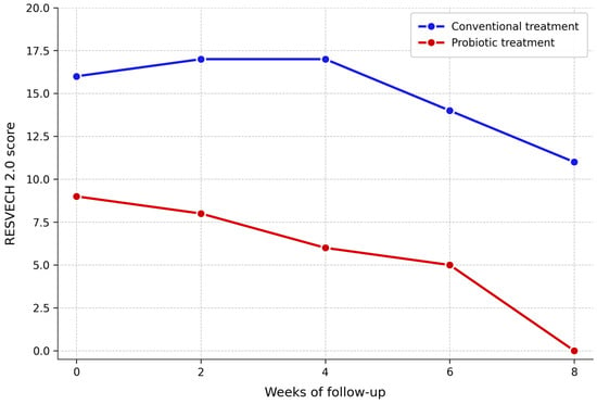
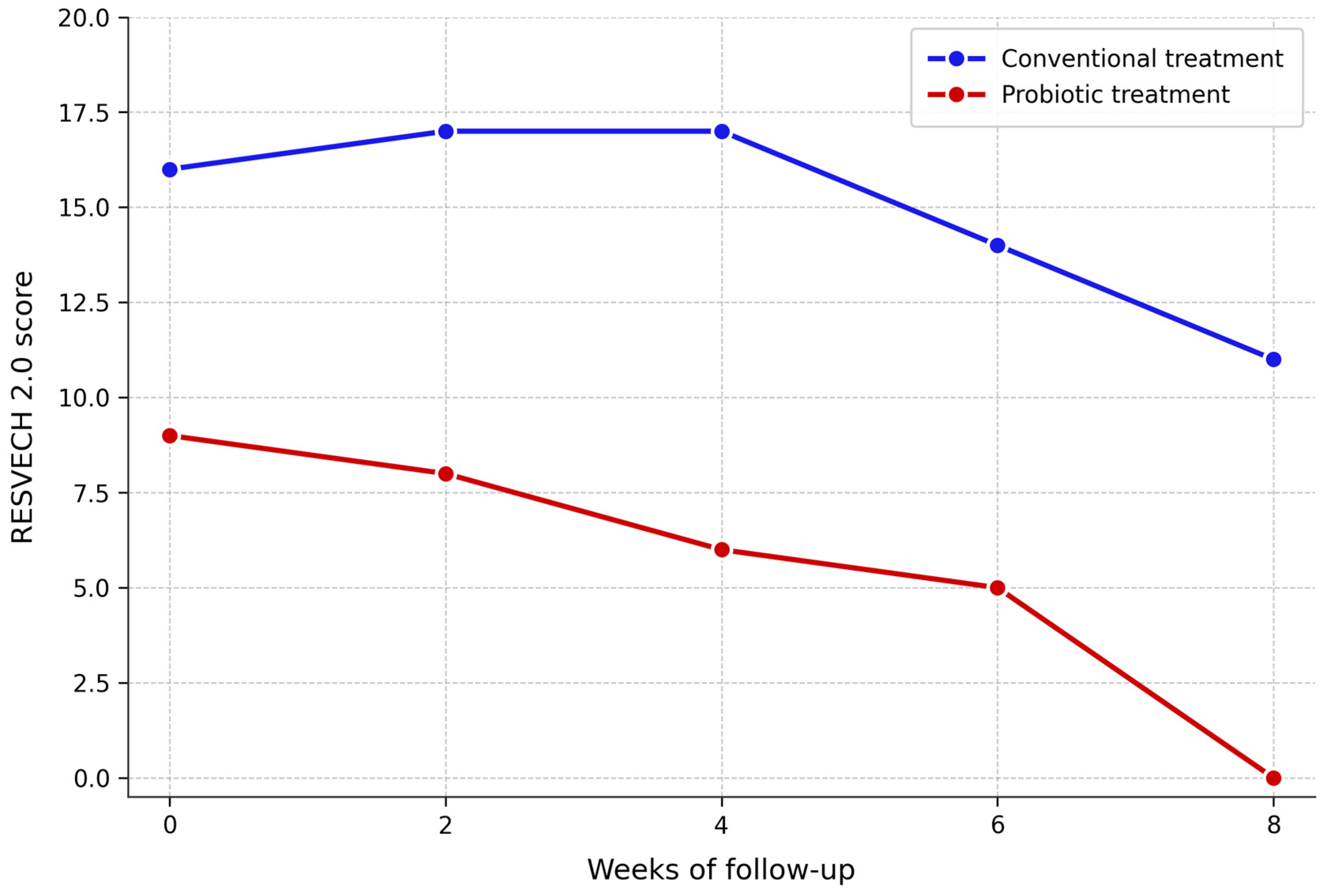
During follow-up, the probiotic-treated plantar ulcer demonstrated progressive improvement across both outcome measures. RESVECH 2.0 scores decreased from 9 at baseline to 8 at week 2, 6 at week 4, and 5 at week 6, followed by complete wound closure at week 8. Wound area measurements confirmed this trajectory, reducing from 10.1 cm2 at baseline to 8.6 cm2 at week 2, 6.8 cm2 at week 4, 2.2 cm2 at week 6, and 0 cm2 at week 8.
In contrast, the conventionally treated ulcer showed no consistent improvement in wound area throughout the follow-up period. Area measurements fluctuated from 25.3 cm2 at baseline to 27.2 cm2 at week 2, 25.3 cm2 at week 4, 33.4 cm2 at week 6, and 25.0 cm2 at week 8, indicating wound persistence without progressive closure. RESVECH 2.0 scores showed only modest improvement, decreasing from 16 at baseline to 11 at week 8, and the wound remained partially open at the end of follow-up.
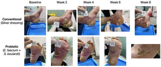
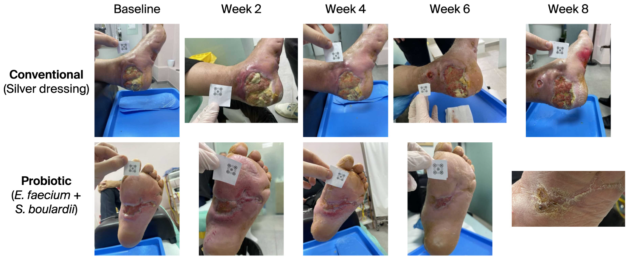
Serial clinical photographs demonstrated progressive improvement in wound appearance during follow-up, with earlier granulation tissue formation and epithelialization observed in the probiotic-treated ulcer (Figure 1).
Figure 1.
Serial clinical photographs showing wound progression during the 8-week follow-up period. The upper row illustrates the conventionally treated diabetic foot ulcer, while the lower row shows the probiotic-treated wound. Images were obtained at baseline and at weeks 2, 4, 6, and 8. The probiotic-treated wound demonstrated faster improvement in wound appearance and earlier closure compared with the conventionally treated lesion. A calibration marker (iMitoWound, imito AG, Zurich, Switzerland) is visible in some images, used for standardized digital wound area measurement.
Baseline and follow-up microbiological cultures identified P. stuartii, with changes observed in antimicrobial susceptibility profiles during follow-up. These microbiological findings should be interpreted cautiously because conventional culture techniques cannot characterize the broader wound microbiome or biofilm architecture.
The evolution of RESVECH 2.0 scores and wound area measurements are presented in Table 3 and illustrated in Figure 1 and Figure 2.
Table 3.
Evolution of RESVECH 2.0 wound assessment scores during the 8-week follow-up period. Lower scores reflect improved wound healing.
Figure 2.
Evolution of RESVECH 2.0 scores during the 8-week follow-up period for probiotic-treated and conventionally treated diabetic foot ulcers. Lower scores indicate improvement in wound status. The probiotic-treated ulcer demonstrated faster reduction in RESVECH 2.0 scores, with complete wound closure observed at week 8.
Given the differences in baseline wound size and anatomical characteristics, these findings should be interpreted descriptively and considered hypothesis-generating rather than evidence of treatment efficacy.
4. Discussion
The present report describes an intra-patient comparison between topical probiotic therapy and conventional wound care in a patient with diabetic foot ulcers. The probiotic-treated wound demonstrated faster reduction in RESVECH 2.0 scores, progressive improvement in wound bed appearance, and complete wound closure by week 8, whereas the conventionally treated ulcer remained partially open at the end of follow-up.
The intra-patient design represents an important strength of this report because it reduces the influence of systemic confounding factors such as age, glycemic control, medication use, and comorbidities. Nevertheless, these observations remain descriptive and should not be interpreted as evidence of treatment efficacy.
Wound progression was assessed using the RESVECH 2.0 scale, a validated multidimensional instrument developed by Restrepo-Medrano and Soriano for the systematic evaluation of chronic wounds [22]. The scale assesses six domains: wound size, depth and affected tissues, wound edges, type of tissue in the wound bed, exudate, and infection or inflammation, with scores ranging from 0 (healed wound) to 35 (worst possible wound status), where lower scores reflect improvement in the healing process [22]. Validation studies have confirmed acceptable internal consistency, high inter-rater agreement, and good construct validity across different wound etiologies and clinical settings [22,23]. The use of RESVECH 2.0 in the present case allowed structured and reproducible tracking of wound progression and facilitated direct comparison between probiotic-treated and conventionally treated wounds within the same patient. Its multidimensional nature—capturing wound bed characteristics, exudate, and signs of infection simultaneously—makes it particularly suitable for monitoring wounds in which microbiome-targeted interventions are being evaluated, as changes in microbial balance may manifest across several wound domains rather than in wound size alone.
These findings align with current understanding of the role of the wound microbiome in chronic wound persistence. Chronic wounds are frequently characterized by microbial dysbiosis and the formation of polymicrobial biofilms, which sustain inflammation and impair normal tissue repair [24,25]. In this context, probiotics have been explored as a microbiome-modulating approach aimed at restoring microbial balance and supporting wound healing [16,26]. Experimental and early clinical studies suggest that probiotics can inhibit the growth of pathogenic bacteria, interfere with biofilm formation, and modulate local inflammatory responses, in addition to supporting tissue repair through angiogenesis, collagen deposition, and epithelialization [16,27]. For example, L. plantarum has demonstrated antimicrobial activity against common wound pathogens and the ability to inhibit biofilm formation in vitro and in experimental models [28,29,30], while probiotic-derived products have been shown to influence cytokine production and promote a more balanced inflammatory response [31,32,33].
The present findings can be placed within a small but growing body of clinical evidence on topical probiotic therapy. Existing reports have predominantly involved Lactobacillus species; to our knowledge, no prior clinical study has specifically evaluated topical E. faecium or S. boulardii in chronic wound management, making this observation potentially novel, though it remains a single-patient report. A published case report by Venosi et al. described an elderly patient with a polymicrobial-infected ischemic chronic ulcer in whom complementary treatment with a multi-strain topical probiotic formulation resulted in progressive wound healing alongside microbiological resolution of the infection [34]. Regarding S. boulardii specifically, Partlow et al. conducted a preliminary experimental study of its topical application in a porcine acute wound model and found no significant difference in healing rate compared to controls, though all wounds healed without complications [35].
The topical probiotic preparation used in the present case contained a combination of E. faecium and S. boulardii. These microorganisms are most commonly used as probiotics in gastrointestinal applications, where both have demonstrated well-characterized antimicrobial and immunomodulatory properties [20,21]. E. faecium strains are known to produce bacteriocins and other antimicrobial peptides capable of inhibiting the growth of pathogenic bacteria and have been shown to modulate host immune responses by influencing cytokine production and promoting anti-inflammatory signaling [30]. S. boulardii, a non-pathogenic yeast, has been reported to interfere with pathogen adhesion to epithelial surfaces, secrete proteases that neutralize bacterial toxins, and modulate inflammatory pathways through reduction of pro-inflammatory cytokine expression [21]. These properties suggest a plausible biological rationale for their use in wound environments, where pathogen overgrowth, biofilm formation, and dysregulated inflammation are central to healing impairment. However, to our knowledge, no published clinical or experimental studies have specifically evaluated the topical application of E. faecium or S. boulardii in chronic wound management. While the mechanisms underlying the observed outcomes cannot be confirmed without microbiological and immunological follow-up data, the biological properties of these strains provide a plausible biological rationale for the clinical observations described in this report. Notably, persistent colonization with P. stuartii during follow-up indicates that clinical improvement cannot necessarily be attributed to microbiological eradication. This underscores the need for dedicated experimental and clinical studies to evaluate these specific strains in wound care settings.
It should also be acknowledged that the conventional dressing used in this report was silver-impregnated, and the lack of progressive improvement in the conventionally treated wound cannot be attributed solely to the absence of probiotic therapy. Silver-based dressings, while effective as antimicrobial agents, have been associated with cytotoxic effects on keratinocytes and fibroblasts, and with delayed re-epithelialization in experimental models [36,37]. No clinical signs of silver-related irritation were specifically documented in this case; however, this possibility cannot be excluded and represents an alternative explanation for the observed differences in healing outcomes.
Several limitations should be considered when interpreting these findings. First, this report describes a single-patient clinical observation and therefore does not provide evidence of treatment efficacy. Second, baseline wound characteristics differed between lesions, particularly with respect to wound size and anatomical location, which may independently influence healing trajectories. Specifically, the plantar surface is subject to repetitive mechanical loading during ambulation, which typically represents an adverse factor for wound closure, while the lateral talocrural region presents distinct vascular supply and tissue composition. These anatomical differences may have independently influenced healing trajectories in ways that cannot be disentangled from any potential treatment effect in a single-patient observation. The allocation of probiotic therapy to the smaller and less severe wound was not based on a predefined randomization protocol but reflected a pragmatic clinical decision, as the novel topical preparation was applied to the wound considered more suitable for an initial observational application. We acknowledge that this represents a significant methodological limitation, as the observed differences in healing outcomes may partly reflect baseline wound severity rather than any treatment effect. This should be considered when interpreting the findings. Regarding systemic factors, no new systemic treatments—including glycemic control adjustments, nutritional supplements, or anti-inflammatory medications—were initiated during the eight-week follow-up period. Furthermore, as both wounds were present in the same patient simultaneously, any fluctuations in systemic parameters such as glycemic control, nutritional status, or inflammatory markers would have affected both wounds equally, representing a key advantage of the intra-patient design. Third, microbiological assessment was based on conventional cultures and antimicrobial susceptibility testing. While these methods are standard in clinical practice, they cannot determine whether the probiotic bacteria actually established themselves in the wound or changed the local microbial environment. More advanced techniques, such as 16S rRNA sequencing, would be needed to answer these questions, but were not available in our setting. We see this as an important area for future research. Finally, RESVECH 2.0 assessments were performed by the treating clinicians without independent blinded review, and observer expectation bias in wound scoring cannot be fully excluded. No attempt was made to have photographs reviewed by a second clinician uninvolved in care, which represents a limitation of this report. Importantly, however, wound area measurements were obtained using iMitoWound digital planimetry software, an objective and operator-independent method, and the progressive reduction in wound area in the probiotic-treated ulcer is consistent with and supported by the serial clinical photographs.
Despite these limitations, this report adds to the growing interest in microbiome-targeted wound care. If future studies confirm benefit in larger cohorts, topical probiotics could become a practical adjunct for chronic wound management, particularly in diabetic foot ulcers, where persistent inflammation and microbial imbalance are common barriers to healing [31].
5. Conclusions
This report describes an intra-patient comparison between topical probiotic therapy and conventional wound care in diabetic foot ulcers. The probiotic-treated ulcer demonstrated faster reduction in RESVECH 2.0 scores and complete wound closure during follow-up compared with the conventionally treated wound.
These observations suggest that microbiome-targeted approaches may warrant further investigation as adjunctive strategies in chronic wound management. However, the findings should be interpreted cautiously given the single-patient observational design, differences in baseline wound characteristics, and the limited microbiological assessment performed using conventional culture techniques.
Well-designed prospective clinical studies incorporating standardized wound measurements and advanced microbiome analysis are required to evaluate the safety, effectiveness, and mechanisms of topical probiotic therapy in chronic wound care.
Author Contributions
Conceptualization, E.Ç., A.D. and L.D.; investigation, E.Ç., A.D., S.H. and L.D.; writing—original draft preparation, E.Ç., A.D. and L.D.; writing—review and editing, E.Ç., A.D. and L.D.; visualization, E.Ç., A.D., S.H. and L.D.; supervision, A.D. and L.D. All authors have read and agreed to the published version of the manuscript.
Funding
This manuscript was funded by the Albanian University Research Fund, grant number 227/2026.
Institutional Review Board Statement
The study was conducted according to the guidelines of the Declaration of Helsinki and approved by the Ethics Committee of the University of Medicine, Tirana (protocol code 11, date of approval: 18 March 2024).
Informed Consent Statement
Written informed consent has been obtained from the patient to publish this paper, including all accompanying clinical images.
Data Availability Statement
Data supporting the findings of this study are available from the corresponding author upon reasonable request.
Acknowledgments
The authors used ChatGPT (OpenAI) for language editing, grammar refinement, and formatting assistance during the preparation of this manuscript. AI tools were not used for data analysis, interpretation of findings, or generation of scientific conclusions. All content was critically reviewed, revised, and approved by the authors, who take full responsibility for the accuracy and integrity of the work.
Conflicts of Interest
The authors declare no conflicts of interest.
Abbreviations
The following abbreviations are used in this manuscript:
| CRP | C-reactive protein |
| HbA1c | Glycated hemoglobin |
| RESVECH | Results Expected from Chronic Wound Healing |
| RBC | Red blood cell count |
References
- Jin, C.; Jin, Y.; Ding, Z.; Nuch, K.S.; Han, M.; Shim, J.; Chien, P.N.; Heo, C.Y. Cellular and Molecular Mechanisms of Wound Repair: From Biology to Therapeutic Innovation. Cells 2025, 14, 1850. [Google Scholar] [CrossRef] [Scilit] [PubMed]
- Falanga, V. Wound healing and its impairment in the diabetic foot. Lancet 2005, 366, 1736–1743. [Google Scholar] [CrossRef] [Scilit] [PubMed]
- Sen, C.K.; Gordillo, G.M.; Roy, S.; Kirsner, R.; Lambert, L.; Hunt, T.K.; Gottrup, F.; Gurtner, G.C.; Longaker, M.T. Human skin wounds: A major and snowballing threat to public health and the economy. Wound Repair. Regen. 2009, 17, 763–771. [Google Scholar] [CrossRef] [Scilit] [PubMed]
- Rodrigues, M.; Kosaric, N.; Bonham, C.A.; Gurtner, G.C. Wound Healing: A Cellular Perspective. Physiol. Rev. 2019, 99, 665–706. [Google Scholar] [CrossRef] [Scilit] [PubMed]
- Falanga, V.; Isseroff, R.R.; Soulika, A.M.; Romanelli, M.; Margolis, D.; Kapp, S.; Granick, M.; Harding, K. Chronic wounds. Nat. Rev. Dis. Primers 2022, 8, 50. [Google Scholar] [CrossRef] [Scilit] [PubMed]
- Armstrong, D.G.; Boulton, A.J.M.; Bus, S.A. Diabetic Foot Ulcers and Their Recurrence. N. Engl. J. Med. 2017, 376, 2367–2375. [Google Scholar] [CrossRef] [Scilit] [PubMed]
- Nunan, R.; Harding, K.G.; Martin, P. Clinical challenges of chronic wounds: Searching for an optimal animal model to recapitulate their complexity. Dis. Model. Mech. 2014, 7, 1205–1213. [Google Scholar] [CrossRef] [Scilit] [PubMed]
- Guo, S.; Dipietro, L.A. Factors affecting wound healing. J. Dent. Res. 2010, 89, 219–229. [Google Scholar] [CrossRef] [Scilit] [PubMed]
- Armstrong, D.G.; Tan, T.W.; Boulton, A.J.M.; Bus, S.A. Diabetic Foot Ulcers: A Review. JAMA 2023, 330, 62–75. [Google Scholar] [CrossRef] [Scilit] [PubMed]
- Schaper, N.C.; van Netten, J.J.; Apelqvist, J.; Bus, S.A.; Fitridge, R.; Game, F.; Monteiro-Soares, M.; Senneville, E. Practical guidelines on the prevention and management of diabetes-related foot disease (IWGDF 2023 update). Diabetes Metab. Res. Rev. 2024, 40, e3657. [Google Scholar] [CrossRef] [Scilit] [PubMed]
- Dawi, J.; Tumanyan, K.; Tomas, K.; Misakyan, Y.; Gargaloyan, A.; Gonzalez, E.; Hammi, M.; Tomas, S.; Venketaraman, V. Diabetic Foot Ulcers: Pathophysiology, Immune Dysregulation, and Emerging Therapeutic Strategies. Biomedicines 2025, 13, 1076. [Google Scholar] [CrossRef] [Scilit] [PubMed]
- Deng, L.; Du, C.; Song, P.; Chen, T.; Rui, S.; Armstrong, D.G.; Deng, W. The Role of Oxidative Stress and Antioxidants in Diabetic Wound Healing. Oxid. Med. Cell Longev. 2021, 2021, 8852759. [Google Scholar] [CrossRef] [Scilit] [PubMed]
- Grice, E.A.; Segre, J.A. The skin microbiome. Nat. Rev. Microbiol. 2011, 9, 244–253. [Google Scholar] [CrossRef] [Scilit] [PubMed]
- Afonso, A.C.; Oliveira, D.; Saavedra, M.J.; Borges, A.; Simões, M. Biofilms in Diabetic Foot Ulcers: Impact, Risk Factors and Control Strategies. Int. J. Mol. Sci. 2021, 22, 8278. [Google Scholar] [CrossRef] [Scilit] [PubMed]
- Pouget, C.; Dunyach-Remy, C.; Pantel, A.; Schuldiner, S.; Sotto, A.; Lavigne, J.P. Biofilms in Diabetic Foot Ulcers: Significance and Clinical Relevance. Microorganisms 2020, 8, 1580. [Google Scholar] [CrossRef] [Scilit] [PubMed]
- Knackstedt, R.; Knackstedt, T.; Gatherwright, J. The role of topical probiotics on wound healing: A review of animal and human studies. Int. Wound J. 2020, 17, 1687–1694. [Google Scholar] [CrossRef] [Scilit] [PubMed]
- Peral, M.C.; Martinez, M.A.; Valdez, J.C. Bacteriotherapy with Lactobacillus plantarum in burns. Int. Wound J. 2009, 6, 73–81. [Google Scholar] [CrossRef] [Scilit] [PubMed]
- Wang, J.; Li, X.; Zhao, X.; Yuan, S.; Dou, H.; Cheng, T.; Huang, T.; Lv, Z.; Tu, Y.; Shi, Y.; et al. Lactobacillus rhamnosus GG-derived extracellular vesicles promote wound healing via miR-21-5p-mediated re-epithelization and angiogenesis. J. Nanobiotechnol. 2024, 22, 644. [Google Scholar] [CrossRef] [Scilit] [PubMed]
- Hussain, A.; Mojgani, N.; Shah, S.M.A.; Kousar, N.; Ali, S.A. The emerging role of probiotics in the management and treatment of diabetic foot ulcer: A comprehensive review. AIMS Microbiol. 2025, 11, 649–678. [Google Scholar] [CrossRef] [Scilit] [PubMed]
- Maldonado Galdeano, C.; Cazorla, S.I.; Lemme Dumit, J.M.; Velez, E.; Perdigon, G. Beneficial Effects of Probiotic Consumption on the Immune System. Ann. Nutr. Metab. 2019, 74, 115–124. [Google Scholar] [CrossRef] [Scilit] [PubMed]
- Pique, N.; Berlanga, M.; Minana-Galbis, D. Health Benefits of Heat-Killed (Tyndallized) Probiotics: An Overview. Int. J. Mol. Sci. 2019, 20, 2534. [Google Scholar] [CrossRef] [Scilit] [PubMed]
- Restrepo-Medrano, J.C.; Soriano, J.V. Desarrollo de un índice de medida de la evolución hacia la cicatrización de las heridas crónicas. Gerokomos 2011, 22, 176–183. [Google Scholar] [CrossRef] [Scilit]
- Menegon, M.R.; Malaquias, S.G.; Silva, J.A.D.; Oliveira, B.; Medrano, J.C.R.; Verdú-Soriano, J.; Bachion, M.M. RESVECH 2.0: Cross-cultural adaptation for Brazil, reliability and validity for the evaluation of venous ulcers. Rev. Bras. Enferm. 2023, 76, e20220185. [Google Scholar] [CrossRef] [Scilit] [PubMed]
- James, G.A.; Swogger, E.; Wolcott, R.; Pulcini, E.; Secor, P.; Sestrich, J.; Costerton, J.W.; Stewart, P.S. Biofilms in chronic wounds. Wound Repair. Regen. 2008, 16, 37–44. [Google Scholar] [CrossRef] [Scilit] [PubMed]
- Bjarnsholt, T.; Kirketerp-Møller, K.; Jensen, P.; Madsen, K.G.; Phipps, R.; Krogfelt, K.; Høiby, N.; Givskov, M. Why chronic wounds will not heal: A novel hypothesis. Wound Repair. Regen. 2008, 16, 2–10. [Google Scholar] [CrossRef] [Scilit] [PubMed]
- Kalan, L.R.; Brennan, M.B. The role of the microbiome in nonhealing diabetic wounds. Ann. N. Y Acad. Sci. 2019, 1435, 79–92. [Google Scholar] [CrossRef] [Scilit] [PubMed]
- Lukic, J.; Chen, V.; Strahinic, I.; Begovic, J.; Lev-Tov, H.; Davis, S.C.; Tomic-Canic, M.; Pastar, I. Probiotics or pro-healers: The role of beneficial bacteria in tissue repair. Wound Repair. Regen. 2017, 25, 912–922. [Google Scholar] [CrossRef] [Scilit] [PubMed]
- Onbas, T.; Osmanagaoglu, O.; Kiran, F. Potential Properties of Lactobacillus plantarum F-10 as a Bio-control Strategy for Wound Infections. Probiotics Antimicrob. Proteins 2019, 11, 1110–1123. [Google Scholar] [CrossRef] [Scilit] [PubMed]
- Meroni, G.P.S.; Zuccotti, G.; Biasibetti, E.; Carli, S.; Pisoni, G. Probiotics as therapeutic tools against pathogenic biofilms: Have we found the perfect weapon? Microbiol. Res. 2021, 242, 126595. [Google Scholar]
- Pompilio, A.; Kaya, E.; Lupetti, V.; Catelli, E.; Bianchi, M.; Maisetta, G.; Esin, S.; Di Bonaventura, G.; Batoni, G. Cell-free supernatants from Lactobacillus strains exert antibacterial, antibiofilm, and antivirulence activity against Pseudomonas aeruginosa from cystic fibrosis patients. Microbes Infect. 2024, 26, 105301. [Google Scholar] [CrossRef] [Scilit] [PubMed]
- Machado, P.; Ribeiro, F.N.; Giublin, F.C.W.; Mieres, N.G.; Tonin, F.S.; Pontarolo, R.; Sari, M.H.M.; Lazo, R.E.L.; Ferreira, L.M. Next-Generation Wound Care: A Scoping Review on Probiotic, Prebiotic, Synbiotic, and Postbiotic Cutaneous Formulations. Pharmaceuticals 2025, 18, 704. [Google Scholar] [CrossRef] [Scilit] [PubMed]
- Dubey, A.K.; Sharma, M.; Raut, S.; Gupta, P.; Khatri, N. Healing wounds, defeating biofilms: Lactiplantibacillus plantarum in tackling MRSA infections. Front. Microbiol. 2023, 14, 1284195. [Google Scholar] [CrossRef] [Scilit] [PubMed]
- Mei, L.; Zhang, D.; Shao, H.; Hao, Y.; Zhang, T.; Zheng, W.; Ji, Y.; Ling, P.; Lu, Y.; Zhou, Q. Injectable and Self-Healing Probiotics-Loaded Hydrogel for Promoting Superbacteria-Infected Wound Healing. ACS Appl. Mater. Interfaces 2022, 14, 20538–20550. [Google Scholar] [CrossRef] [Scilit] [PubMed]
- Venosi, S.; Ceccarelli, G.; de Angelis, M.; Laghi, L.; Bianchi, L.; Martinelli, O.; Maruca, D.; Cavallari, E.N.; Toscanella, F.; Vassalini, P.; et al. Infected chronic ischemic wound topically treated with a multi-strain probiotic formulation: A novel tailored treatment strategy. J. Transl. Med. 2019, 17, 364. [Google Scholar] [CrossRef] [Scilit] [PubMed]
- Partlow, J.; Blikslager, A.; Matthews, C.; Law, M.; Daniels, J.; Baker, R.; Labens, R. Effect of topically applied Saccharomyces boulardii on the healing of acute porcine wounds: A preliminary study. BMC Res. Notes 2016, 9, 210. [Google Scholar] [CrossRef] [Scilit] [PubMed]
- Nešporová, K.; Pavlík, V.; Šafránková, B.; Vágnerová, H.; Odráška, P.; Žídek, O.; Císařová, N.; Skoroplyas, S.; Kubala, L.; Velebný, V. Effects of wound dressings containing silver on skin and immune cells. Sci. Rep. 2020, 10, 15216. [Google Scholar] [CrossRef] [Scilit] [PubMed]
- Burd, A.; Kwok, C.H.; Hung, S.C.; Chan, H.S.; Gu, H.; Lam, W.K.; Huang, L. A comparative study of the cytotoxicity of silver-based dressings in monolayer cell, tissue explant, and animal models. Wound Repair. Regen. 2007, 15, 94–104. [Google Scholar] [CrossRef] [Scilit] [PubMed]

Disclaimer/Publisher’s Note: The statements, opinions and data contained in all publications are solely those of the individual author(s) and contributor(s) and not of MDPI and/or the editor(s). MDPI and/or the editor(s) disclaim responsibility for any injury to people or property resulting from any ideas, methods, instructions or products referred to in the content. |
© 2026 by the authors. Licensee MDPI, Basel, Switzerland. This article is an open access article distributed under the terms and conditions of the Creative Commons Attribution (CC BY) license.