


Abstract
Lavandula angustifolia essential oil (LEO) was obtained by hydrodistillation of air-dried flowers collected in the Mostar region (Bosnia and Herzegovina). Its chemical composition was analyzed by gas chromatography-mass spectrometry, revealing a camphor content of 16.96%, substantially higher than the maximum value specified in the European Pharmacopoeia. Antimicrobial activity was evaluated using quantitative suspension tests according to EN 1276 and EN 1650 under simulated “dirty” conditions with organic load (bovine albumin, 3 g/L) and a 5 min contact time. High-concentration LEO (80% w/v) exhibited strong bactericidal activity against Escherichia coli ATCC 10536 and Staphylococcus aureus ATCC 6538, and yeasticidal activity against Candida albicans ATCC 10231 (>5 log10 CFU/mL reduction for bacteria, >4 log10 CFU/mL reduction for yeast), but was ineffective against Pseudomonas aeruginosa ATCC 15442 and Enterococcus hirae ATCC 10541. Lower concentrations (1.0% and 0.1% w/v) showed no bactericidal and yeasticidal activity, highlighting LEO’s efficacy limits. Antioxidant activity, assessed by DPPH radical scavenging, was dose- and time-dependent. Molecular docking provided insight into the interaction of major constituents with selected microbial and antioxidant-related targets. These findings highlight both the potential and limitations of LEO as a renewable bio-based resource for sustainable disinfectant formulations while emphasizing the importance of chemical composition and regulatory compliance.
1. Introduction
Plant-derived essential oils have attracted increasing attention as renewable and sustainable chemical agents for antimicrobial and disinfectant applications, offering eco-friendly alternatives to synthetic biocides [1,2]. Although essential oils often exhibit strong antimicrobial activity against foodborne pathogens in vitro, effective antimicrobial concentrations in real food matrices are generally much higher than minimum inhibitory concentrations (MIC) determined in laboratory media. In minimally processed foods, studies have shown that realistic antibacterial applications require much higher essential oil concentrations than those indicated by in vitro MIC values, which frequently result in undesirable sensory effects such as strong odour and altered flavour profiles that limit practical use in food products [3,4]. Particular attention has been devoted to the application of essential oils for surface sanitation that come into contact with food, where effective microbial control is essential to prevent contamination and the spread of foodborne pathogens. Studies have shown that essential oils can significantly reduce microbial populations on common food-contact materials, such as stainless steel, glass, and polypropylene surfaces [2], which pose a major challenge for hygiene management in food-processing environments. Unlike many conventional disinfectants, essential oils are generally biodegradable and originate from renewable plant resources [5].
Essential oils represent complex mixtures of volatile, biologically active compounds whose efficacy is strongly influenced by composition, concentration, and application conditions [6,7]. Among them, lavender (Lavandula angustifolia) essential oil has been widely studied due to its chemical richness and reported antioxidant and antimicrobial properties [8,9]. The qualitative and quantitative composition of lavender essential oil is known to vary significantly depending on genetic factors, geographical origin, climatic conditions, plant part, developmental stage, and extraction method. Such variability directly influences the chemical profile determined by gas chromatography–mass spectrometry (GC-MS) [10]. The major constituents of L. angustifolia essential oil typically include linalool and linalyl-acetate, accompanied by varying amounts of 1,8-cineole, camphor, borneol, terpinen-4-ol, lavandulyl-acetate, and other mono- and sesquiterpenes [11,12,13]. The antioxidant potential of LEO has been widely demonstrated using radical-scavenging assays such as DPPH, showing notable free-radical neutralization [10,14]. Similarly, antimicrobial efficacy has been reported against diverse Gram-positive and Gram-negative bacteria as well as yeasts, reflecting the concentration-dependent antimicrobial activity of lavender oil [15,16,17,18,19].
Therefore, this study aimed to define the antioxidant and antimicrobial performance limits of LEO obtained from plants collected in the Mostar region (southern Bosnia and Herzegovina), characterize its chemical composition via GC-MS, and complement experimental findings with molecular docking to elucidate potential mechanisms of action under conditions relevant to sustainable disinfection systems. Antimicrobial activity was assessed against a panel of bacterial strains and one yeast strain using standardized quantitative suspension tests under simulated soiled (dirty) conditions to obtain results as close as possible to real conditions for LEO application in food-related and other industrial systems. In addition to the general evaluation of LEO, particular emphasis was placed on the analysis of essential oil derived from the Mediterranean Balkans, a region characterized by distinct agro-climatic conditions that can markedly influence the chemical composition and biological activity of plant-derived oils. Such region-specific traits may contribute to variations in antimicrobial efficacy, underscoring the need to investigate locally sourced LEO. The antimicrobial potential was assessed using standardized quantitative methods designed to determine bactericidal and yeasticidal activities. These approaches were selected over conventional screening techniques, such as disk diffusion or time-kill assays, as they provide more rigorous, reproducible, and regulatory-compliant evidence of efficacy in line with European Chemicals Agency (ECHA) guidance [20] for biocidal product evaluation. Furthermore, testing under simulated “dirty” conditions allowed a more realistic appraisal of practical performance, providing insights into the potential application of LEO as a natural biocidal agent. Thus, this study not only investigates the intrinsic antimicrobial activity of lavandin essential oil but also evaluates its practical applicability and potential for development as a sustainable and commercializable biocidal product.
2. Materials and Methods
The chemical composition of lavender (Lavandula angustifolia) essential oil obtained by hydrodistillation was analyzed by gas chromatography–mass spectrometry (GC-MS). Its antioxidant potential was evaluated using the DPPH radical-scavenging assay. The antimicrobial activity of the essential oil at an effective concentration of 80% (w/v), as well as its emulsions at 1.0% and 0.1% (w/v), was assessed in situ. Throughout the study, these samples (i.e., the pure essential oil and its emulsions) are collectively referred to as test products. All antimicrobial testing was performed following the adopted EN 1276 [21] and EN 1650 [22] procedures. In addition, molecular docking studies were performed in silico to investigate the potential interactions of key constituents with bacterial targets.
2.1. Plant Material
The plant material used in this study was collected in the area of Mostar (southern Bosnia and Herzegovina), which is characterized by a Mediterranean climate with warm summers and mild winters. The plant material was harvested at the full-flowering stage manually in August 2024 and transported to the laboratory, where it was botanically identified as Lavandula angustifolia Mill. Species identification was performed using standard floristic literature [23]. Air-dried flowers, previously separated from their stems, were used for the isolation of essential oils (Figure S1, Supplementary Materials).
2.2. Hydrodistillation
The essential oil was obtained by hydrodistillation using a Clevenger-type apparatus (Šurlan, Medulin, Croatia). Briefly, 50 g of lavender flowers, obtained as a composite sample of L. angustifolia plants, were placed in a round-bottom flask and mixed with 500 mL of distilled water. The flask was heated on a hot plate, and hydrodistillation was performed for 2 h. The oil yields were in the interval 1.8–2.2%. The collected essential oil was transferred to a graduated glass tube and stored at 4 °C in a refrigerator. The pooled sample obtained from all distillations was used for further analyses.
2.3. Investigation of Chemical Composition
The semiquantitative chemical profiling of the essential oil was performed using gas chromatography (GC; Agilent 6890B GC-FID, Waldbronn, Germany) coupled with mass spectrometry (MS; Agilent 5977 MSD, Waldbronn, Germany). Essential oil samples (20 µL) were diluted in hexane (1:49, w/v) and injected in split mode (1:50) at an inlet temperature of 220 °C. The separation of constituents was achieved on an HP-5MS capillary column (30 m × 0.25 mm i.d., 0.25 μm film thickness; Agilent, Waldbronn, Germany) under the following temperature program: the initial oven temperature of 60 °C was increased at a rate of 3 °C/min to reach 246 °C. Helium was used as the carrier gas at a constant flow rate of 1 mL/min. The MSD transfer line was maintained at 230 °C. Mass spectra were recorded in scan mode (m/z 50–550). Compounds were identified by comparison of the acquired mass spectra with the NIST mass spectral library (v14) and literature data [24].
2.4. Investigation of Antimicrobial Activity
The antimicrobial activity of LEO (at an effective concentration of 80%) and its diluted emulsions (at effective concentrations of 1.0% and 0.1%, w/v) was evaluated based on the EN 1276 standard [21] for bactericidal activity and EN 1650 [22] for yeasticidal activity, with adaptations applied as described in Section 2.4.3 and Section 2.4.5. In accordance with the test protocol, the addition of the microbial suspension and the interfering substance systematically diluted the test product; consequently, a nominal concentration of 100% LEO corresponded to an effective concentration of 80% (w/v) during the assay. Nominal concentrations were used exclusively for sample preparation, whereas all results are reported and discussed based on the effective concentration present in the reaction mixture.
2.4.1. Preparation of Lavender Oil Test Emulsions
Test emulsions of LEO were prepared using sterile distilled water and Tween 80 (HiMedia Laboratories Pvt. Ltd., Maharashtra, India) as the emulsifying agent through vigorous mechanical homogenization. The emulsions were prepared with Tween 80 to prevent the separation of the essential oil upon addition to the aqueous solution of the interfering substance and microbial suspension during a 5 min contact time of the test. For the nominal 1.25% (w/v) emulsion, 1.25 g of LEO was added to 10 mL of sterile water containing 1 g of Tween 80, and the total volume was adjusted to 100 mL, resulting in an effective concentration of 1.0% (w/v). The nominal 0.125% (w/v) emulsion was prepared in the same manner and corresponded to an effective concentration of 0.1% (w/v). All emulsions were freshly prepared and vigorously mixed before testing.
2.4.2. Microbial Suspensions
Bacterial test suspensions were prepared using Pseudomonas aeruginosa ATCC 15442, Escherichia coli ATCC 10536, Staphylococcus aureus ATCC 6538, and Enterococcus hirae ATCC 10541 (all Microbiologics, St. Cloud, MN, USA), with optical densities of 0.8, 0.9, 0.8 and 2.0 McFarland (McF), respectively. For the testing of yeasticidal activity, a suspension of Candida albicans ATCC 10231 (Microbiologics, USA) with an optical density of 4.0 McF was prepared. Optical density was determined using a DEN-1 densitometer (suspension turbidity detector) (SIA Biosan, Riga, Latvia).
For counting bacterial cells in suspension, 1 mL aliquots of 10−6 and 10−7 dilutions were poured onto Tryptone Soya Agar (TSA) (HiMedia Laboratories Pvt. Ltd., Maharashtra, India) in duplicate and incubated for 48 h at 37 ± 1 °C. For yeast cells, 1 mL aliquots of 10−5 and 10−6 dilutions were poured onto Malt Extract Agar (MEA) (Malt agar, HiMedia Laboratories Pvt. Ltd., Maharashtra, India) in duplicate and incubated for 48 h at 30 °C ± 1 °C. The number of microorganisms (N) in the suspension was calculated using the formula provided in Section 11.2.7.2.2 of ISO 7218 [25], which takes into account colony counts from different dilutions and their corresponding dilution factor.
2.4.3. Test Procedure
The standards EN 1276 [21] and EN 1650 [22], which are quantitative suspension tests, were partially modified. Specifically, for the dilution–neutralization method, additional decimal dilutions were performed after neutralization to accurately determine logarithmic reduction below 5 log10 for bactericidal activity and below 4 log10 for yeasticidal activity. All antimicrobial experiments were conducted under aseptic conditions in a double-sided Class II A2 biosafety cabinet (Biobase, Jinan, China). Contact time was 5 min, and the test temperature was 20 °C ± 1 °C.
An interfering substance solution, used to simulate dirty test conditions, was prepared by dissolving 3.0 g/L of bovine albumin fraction V (Roche Diagnostics GmbH, Mannheim, Germany) following European standards [21,22]. The final concentration of bovine albumin in the test procedure is 3.0 g/L. Neutralizing solution was prepared in the laboratory using the following components: L-histidine (CAS No. 9048-46-8, HiMedia Laboratories Pvt. Ltd., Maharashtra, India), saponin (from plant) (CAS No. 8047-15-2, HiMedia Laboratories Pvt. Ltd., Maharashtra, India), Tween 80 (CAS No. 9005-65-6, HiMedia Laboratories Pvt. Ltd., Maharashtra, India), sodium thiosulfate pentahydrate (CAS 10102-17-7, Merck KGaA, Darmstadt, Germany).
The neutralization–dilution method in accordance with the modified EN 1276 [21] was applied for 1.25% and 0.125% (w/v) LEO emulsions (effective concentrations of 1.0% w/v and 0.1% w/v LEO), as Control C (see Section 2.4.4) confirmed effective neutralization. Briefly, 8.0 mL of the test product was added to a mixture of 1 mL of bacterial test suspension and 1.0 mL of an interfering substance and incubated at 37 °C ± 1 °C for a contact time of 5 min. At the end of the contact time, the bactericidal effect was immediately neutralized by transferring 1.0 mL aliquots of the mixture into 9.0 mL of a validated neutralizing solution. Surviving bacteria were enumerated using serial decimal dilution and pour plating on TSA, followed by incubation for 48 h at 37 °C ± 1 °C. Yeasticidal activity was assessed using the neutralization–dilution method in accordance with EN 1650 [22], and C. albicans ATCC 10231 was used as the test organism. After neutralization, surviving yeast cells were enumerated by serial dilution (modification of EN 1650 [22]) and pour plating on MEA, with incubation for 48 h at 30 °C ± 1 °C.
For undiluted LEO (80% effective concentration), neutralization was insufficient for some of the test microorganisms, and the membrane filtration method was used to stop antimicrobial activity. After the defined contact time, 0.1 mL aliquots of the test mixture were filtered through a membrane and plated on the appropriate agar medium and incubated as mentioned above.
After incubation of the plates, the colonies were counted (Tables S1–S5, Supplementary Materials). For the dilution–neutralization method, the acceptable limits for individual CFU values per Petri plate are 14 and 330, whereas for the membrane filtration method, the upper limit is 165.
2.4.4. Verification of Methodology
Control procedures A, B, and C were performed in accordance with EN 1276 [21] and EN 1650 [22] to ensure the reliability of the bactericidal and yeasticidal assays for both the neutralization–dilution method and the membrane filtration method. These controls verified the absence of lethal effects in the test conditions (Control A), verification of the absence of toxicity of neutralizer or validation of the filtration procedure (Control B), and validation of the dilution–neutralization or membrane filtration methods (Control C). The validation microbial suspensions (Nv) were prepared as follows: for bactericidal testing, a 1:3 dilution of the fifth decimal dilution of the bacterial suspension was used; for yeasticidal activity, a 1:3 dilution of the fourth decimal dilution of the Candida albicans suspension was used. Detailed descriptions of the verification of methodologies and control procedures are provided in EN 1276 [21] and EN 1650 [22].
2.4.5. Calculation of Bactericidal and Yeasticidal Activity
The decimal logarithmic reduction in the neutralization–dilution method for each essential oil concentration was calculated using the following formula:
where:
log10 R= log10 N0 − log10 (Xsr × 10 × 10n),
- –
- log R is the reduction (log10 CFU/mL),
- –
- N0 is the number of microorganisms in the test microbial suspension, representing the actual concentration of microorganisms present in the reaction mixture (CFU/mL),
- –
- Xsr is the average of viable microorganisms on the two plates after a contact time of 5 min
- –
- n is decimal dilution (n = 1, 2, 3, 4).
N0 was derived from the prepared microbial suspension (N) by taking into account the dilution resulting from mixing 1 mL of microbial suspension (N) with 1 mL of interfering substance and 8 mL of test product, resulting in a tenfold decrease in microbial concentration. The decimal logarithmic reduction in the membrane filtration method for each essential oil concentration was calculated as described in the EN 1276 [21] and EN 1650 [22].
All experiments were performed in triplicate. Key results of logarithmic reduction are presented in the main text as the mean ± standard deviation, whereas detailed data for all replicates are provided in Tables S1–S5 (Supplementary Materials). No inferential statistical tests were applied, since antimicrobial efficacy was evaluated according to predefined log-reduction thresholds specified by EN 1276 [21] and EN 1650 [22], rather than by hypothesis-testing approaches.
Bactericidal concentration was defined as the concentration of LEO that achieves at least a 5-log10 reduction in test microbial suspension (N0) for each of the four tested bacterial strains, in accordance with EN 1276 [21]. Yeasticidal concentration was defined as the concentration of LEO that achieves at least a 4-log10 reduction in the test microbial suspension (N0) of Candida albicans ATCC 10231, in accordance with EN 1650 [22].
2.5. Molecular Docking
The X-ray crystal structure of Escherichia coli DNA gyrase B in complex with a reference inhibitor (PDB ID: 6KZX) was retrieved from the Protein Data Bank (PDB). This structure, resolved at 2.10 Å, provided a high-resolution template for molecular docking studies. To prepare the protein for docking, AutoDockTools 1.5.7 was used to remove non-conserved water molecules, ensuring they did not interfere with ligand binding, add polar hydrogens to the protein structure and assign Gasteiger charges to optimize electrostatic interactions during docking. The three-dimensional (3D) structures of linalool, camphor, eucalyptol and terpinen-4-ol were obtained from the PubChem database in their respective SDF format. To optimize their geometries, the structures were energy-minimized using Chem3D Ultra (PerkinElmer, Waltham, MA, USA), ensuring adoption of low-energy, stable conformations before docking. Partial atomic charges were assigned, and torsional degrees of freedom were defined to allow flexible docking. The binding site was determined by setting a grid box around the DNA gyrase B active site, using the co-crystallized ligand’s coordinates. Molecular docking simulations were carried out using AutoDock Vina, which generated nine distinct binding poses (conformers) for each ligand, ranked by lowest binding energy (kcal/mol). The docked complexes were visualized and analyzed using Discovery Studio Visualizer (version 21.1.0.20298; Biovia, Dessault Systèmes, San Diego, CA, USA) to identify key molecular interactions.
2.6. Investigation of Antioxidant Potential
The antioxidant activity of LEO was evaluated using the 2,2-diphenyl-1-picrylhydrazyl (DPPH) radical scavenging assay. All reagents were of analytical grade. Ethanol (96%; Gram Mol, Zagreb, Croatia) and DPPH radical (Sigma Chemical Company, St. Louis, MO, USA) were used. Absorbance measurements were performed using a UV-Vis spectrophotometer (UV-1800 spectrophotometer, Shimadzu Corporation, Kyoto, Japan).
DPPH radical was dissolved in ethanol to obtain a final concentration of 3 × 10⁻⁴ mol/L and stored in a dark glass vial. The solution was homogenized by ultrasonic treatment. Subsequently, 1.0 mL of the DPPH solution was mixed with 2.5 mL of an ethanolic LEO solution. The antioxidant activity was assessed by measuring the absorbance at 517 nm, corresponding to the characteristic absorption of the DPPH radical. The assay was conducted at three LEO concentrations (3 mg/mL, 5 mg/mL, and 10 mg/mL). For each concentration, absorbance was recorded immediately after preparation (0 min) and after 30 min of incubation in the dark at room temperature to evaluate the time-dependent radical scavenging activity.
The absorbance at 517 nm was measured for the ethanolic solution of the DPPH radical, which was diluted in the previously mentioned ratio (1 mL of DPPH radical solution with a given concentration, and 2.5 mL of ethanol added). Ethanol was used as the blank. The DPPH radical scavenging activity was calculated according to Equation (2) [26]:
where
DPPH radical scavenging capacity (%) = 100 − [(AS − AB) × (100/AC)]
- –
- As is the absorbance of the sample (ethanolic LEO solution mixed with DPPH)
- –
- AB is the absorbance of the “blank” (ethanolic LEO solution without DPPH),
- –
- AC is the absorbance of the control (ethanolic solution with DPPH).
All measurements were performed in triplicate, and the results are expressed as mean ± standard deviation.
3. Results and Discussion
3.1. Chemical Composition
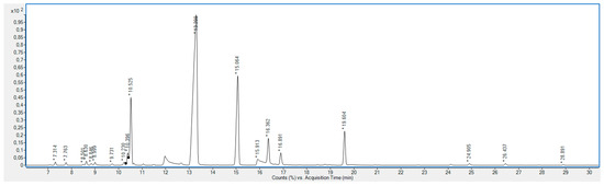
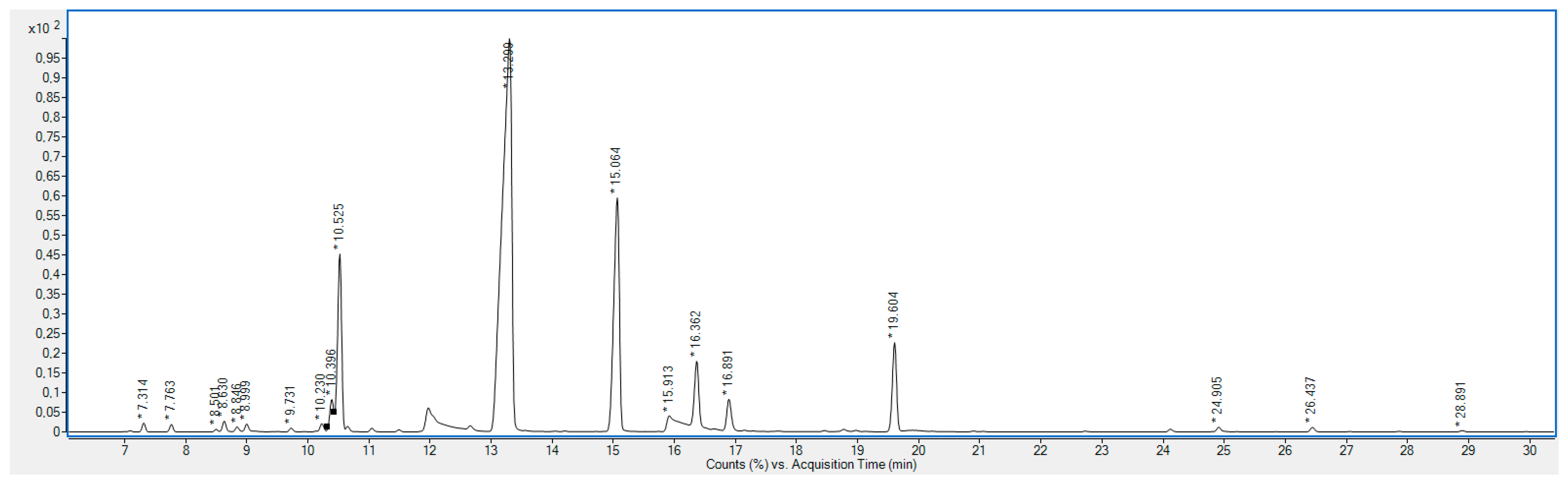
The chemical composition of Lavandula angustifolia essential oil, obtained by hydrodistillation of air-dried flowers collected in the Mostar region (southern Bosnia and Herzegovina) in August 2024, was analyzed by GC-MS. Nineteen constituents were identified, representing 97.79% of the total LEO composition (Table 1, Figure 1).
Table 1.
Chemical composition of lavender essential oil obtained by GC-MS.
Figure 1.
GC-MS analysis of the composition of L. angustifolia essential oil.
The major components were linalool (49.19%), camphor (16.96%), eucalyptol (10.98%), and terpinen-4-ol (5.14%), together accounting for 77.13% of the oil. The essential oil was dominated by oxygenated monoterpenes, including alcohols (59.44%) and ketones/ethers (27.94%).
Several studies report linalool and linalyl acetate as the predominant compounds in Lavandula angustifolia essential oil [12,27,28,29], with linalool content ranging from 24.11% to 40.68% and linalyl acetate from 16.68% to 34.19%. In addition, the content of several key components of lavender essential oil is standardized by the European Pharmacopoeia [30]. In our study, however, the contents of linalyl acetate and camphor did not align with the recommended standardized ranges. The linalyl acetate content in our essential oil was 4.97%, considerably lower than the European Pharmacopoeia [30] range of 25–47% and substantially below the values commonly reported in the literature. Nevertheless, previous reports have also described relatively low linalyl acetate contents [13,31]. For example, Danh et al. [11] conducted a comparative study of lavender oils obtained by hydro-distillation, hexane extraction, and supercritical CO2 extraction, finding linalyl acetate contents of 9.27%, 25.73%, and 23.40%, respectively. Such variation is likely due to partial decomposition of linalyl acetate during hydrodistillation [11,32]. Conversely, the camphor content in our sample was 16.96%, far exceeding the maximum value recommended by the European Pharmacopoeia (≤1.2%). Elevated camphor levels have also been reported in other studies [1,2,33], suggesting that this compound can vary considerably depending on the plant source and method of hydrodistillation. For example, Belhadj-Mostefa et al. [34], in their study of essential oils obtained from fresh flowers of Lavandula angustifolia from Algeria, reported a camphor content of 24.6%. Similarly, Gonzales-Rivera et al. [35], analyzed essential oils extracted from dried leaves and stems and found camphor contents ranging from 23.1% to 25.1%. Another notable feature of our essential oil is its relatively high eucalyptol (1,8-cineole) content, a compound recognized for its multiple health-related benefits [36]. Although comparisons with literature values are informative, the composition of essential oils is governed by plant-related factors, including genotype and ecotype, along with environmental factors such as geographical origin, climatic conditions during the growing season, harvest time, and the extraction method employed [6,13,37,38].
3.2. Antimicrobial Activity
European standards EN 1276 [21] and EN 1650 [22] are widely accepted quantitative suspension tests for evaluating the bactericidal and yeasticidal/fungicidal activity of chemical disinfectants and sanitizers intended for food, industrial, domestic, and institutional applications. Testing according to these standards provides insight into the potential applicability of lavender essential oil (LEO), or its low-concentration emulsions (1.0% w/v and 0.1% w/v) as a sustainable, renewable biocidal agent suitable for the areas defined by the standards. To obtain more detailed information on logarithmic reduction than that provided by the standard protocols, the dilution–neutralization method from EN 1276 [21] and EN 1650 [22] was modified at the inoculation stage following the contact time. The results of the verification of the dilution–neutralization method, presented in the Supplementary Materials (Tables S1–S5), showed no lethal effects under the test conditions (control A), no toxicity of applied neutralized, and the suitability of the dilution–neutralization method (Control C) for testing effective concentration of 1.0% w/v and 0.1% w/v LEO emulsions. As shown in Table 2, the 1.0% w/v and 0.1% w/v LEO emulsions did not achieve a 5-log10 reduction for any of the tested bacterial strains and achieved less than a 4-log10 reduction for Candida albicans ATCC 10231, the yeast strain employed in the assay.
Table 2.
Logarithmic reduction in microorganisms (contact time 5 min) for 1.0% (w/v) and 0.1% (w/v) of lavender essential oil emulsions, assessed using the neutralization–dilution method under simulated dirty conditions.
These results indicate that, under the applied experimental conditions, the low-concentration LEO emulsions did not achieve complete inactivation of the tested bacterial or yeast strains. Table 3 presents the results obtained with undiluted LEO (effective 80% w/v concentration).
Table 3.
Logarithmic reduction in microorganisms (contact time 5 min) for undiluted lavender essential oil (effective 80% w/v concentration), assessed using the membrane filtration method under simulated dirty conditions.
The results of the method verification presented in Supplementary Material (Tables S1–S5) showed that the neutralization–dilution method (Control C) was not suitable for testing undiluted LEO. Accordingly, the membrane filtration method was applied, which met the requirements of Control A, Control B, and Control C (Supplementary material, Tables S1–S5). After a contact time of 5 min under simulated dirty conditions, 80% (w/v) LEO achieved > 5 log10 reduction for S. aureus ATCC 6538 and E. coli ATCC 10536. According to EN 1276 [21], a disinfectant can be classified as bactericidal only when a ≥5 log10 reduction is achieved for all four mandatory bacterial strains. In the present study, this criterion was not fulfilled, even at the highest tested concentration of LEO (80% w/v), due to insufficient activity against P. aeruginosa ATCC 15442 and E. hirae ATCC 10541. Consequently, Lavandula angustifolia essential oil cannot be regarded as bactericidal according to EN 1276 [21] when applied as a single active constituent, which represents limitation for its use as a stand-alone disinfectant.
In contrast, C. albicans ATCC 10231 is the only mandatory strain for assessing yeasticidal activity according to EN 1650 [22]. Based on the results presented in Table 3, 80% (w/v) LEO achieved a ≥4-log10 reduction after 5 min of contact time, thereby fulfilling the standard criterion for yeasticidal activity. This demonstrates that LEO is effective against yeasts at high concentrations. However, from a practical standpoint, such high essential oil concentrations are unlikely to be suitable for routine surface disinfection in industrial environments due to sensory, economic, and formulation-related constraints. The high log10 reduction observed for E. coli ATCC 10536 and S. aureus ATCC 6538 with 80% (w/v) LEO, compared to the low-concentration emulsions, is consistent with previous reports indicating a concentration-dependent bactericidal effect [15]. The limited efficacy against E. hirae ATCC 10541 and P. aeruginosa ATCC 15442 may be attributed to strain-specific susceptibility and differences in cell envelope structure, which influence essential oil penetration and resistance mechanisms [39,40].
Antimicrobial activity of essential oils is typically assessed using disk diffusion or time-kill assays; however, suspension tests under simulated soiled conditions, as applied here, provide more realistic insights for sustainable industrial application. While MIC and MBC values are informative, practical use in minimally processed foods and industrial environments often requires higher concentrations than MIC, balancing efficacy with sensory or material considerations [41,42]. Thus, direct comparison with previously published results is challenging due to differences in the methodologies used to evaluate the effects of lavender essential oil on microorganisms. To provide a clearer understanding of results, logarithmic reduction was expressed as percentages [43]. The experiments were conducted under conditions simulating organic load, representing real-world application scenarios where surfaces were not pre-cleaned or rinsed. The contact time for LEO with the microbial suspension and the organic load was 5 min. Under these conditions, LEO concentrations of 1.0% w/v and 0.1% w/v achieved less than 99% reduction for all tested strains, whereas an active concentration of 80% w/v LEO resulted in reduction exceeding 99.999% for E. coli ATCC 10,536 and S. aureus ATCC 6538 and over 99.99% for C. albicans ATCC 10231. These findings demonstrate a pronounced concentration-dependent antimicrobial effect while presenting the data in a clear and easily interpretable format.
Overall, the present findings establish the realistic limits of antimicrobial performance for LEO under standardized conditions and in line with European Chemicals Agency (ECHA) guidance [20] for biocidal product evaluation. While high concentrations exhibit selective bactericidal and confirmed yesticidal activity, the inability to meet EN 1276 [21] requirements for all mandatory bacterial strains highlights the necessity for future studies focusing on synergistic combinations or formulated systems rather than reliance on LEO as a single-component disinfectant.
3.3. Molecular Docking Study of the Principal Components of Lavender Essential Oil with DNA Gyrase B
An in silico molecular docking study can be an effective approach to investigating the antibacterial mode of action of active compounds in essential oils by predicting their ability to bind to the active sites of enzymes critical for bacterial survival, metabolism, and virulence. One such enzyme is DNA gyrase, which plays a key role in bacterial DNA replication and transcription, making it an attractive target for the development of antibacterial agents, with fluoroquinolones being among the most well-known examples [44,45]. In this context, we conducted a molecular docking study of linalool, camphor, eucalyptol, and terpinen-4-ol, identified as major constituents of L. angustifolia essential oil, to explore whether their demonstrated antibacterial activity against E. coli and S. aureus is associated with inhibition of DNA gyrase. The obtained results indicated that all four compounds were capable of forming stable complexes within the active site of the enzyme, with binding energies ranging from −4.8 to −5.9 kcal/mol (Table 4).
Table 4.
Results of molecular docking of principal compounds of lavender essential oil and DNA gyrase inhibitor ciprofloxacin.
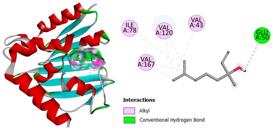
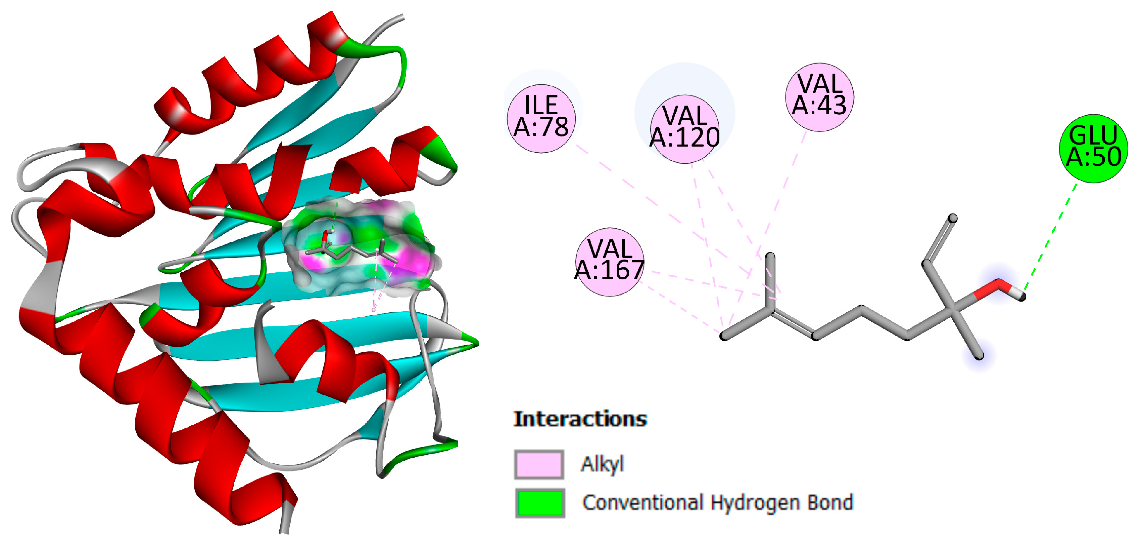
While these binding energies were higher than those of ciprofloxacin (−7.0 kcal/mol), a well-known DNA gyrase inhibitor, they were still comparable and suggest notable binding affinity, particularly for terpinen-4-ol and linalool. It is important to note that these natural compounds have a relatively simple, non-optimized structure that lacks the complex pharmacophores typical of synthetic gyrase inhibitors, which partly explains their lower predicted affinities. Analysis of binding interactions showed that linalool was able to form hydrogen bond interactions with the Glu50 residual amino acid and hydrophobic interactions with Val43, Ile78, Val120 and Val167 amino acid residues within or near the ATP-binding pocket of the gyrase subunit, suggesting that linalool may interfere with ATP-dependent catalytic activity (Figure 2).
Figure 2.
Representation of linalool docked within the binding site of DNA gyrase B (PDB ID: 6KZX) and key molecular interactions with amino acid residues at the active site.
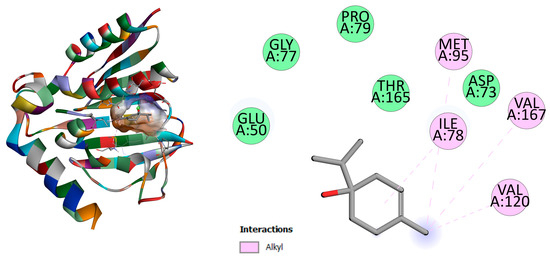
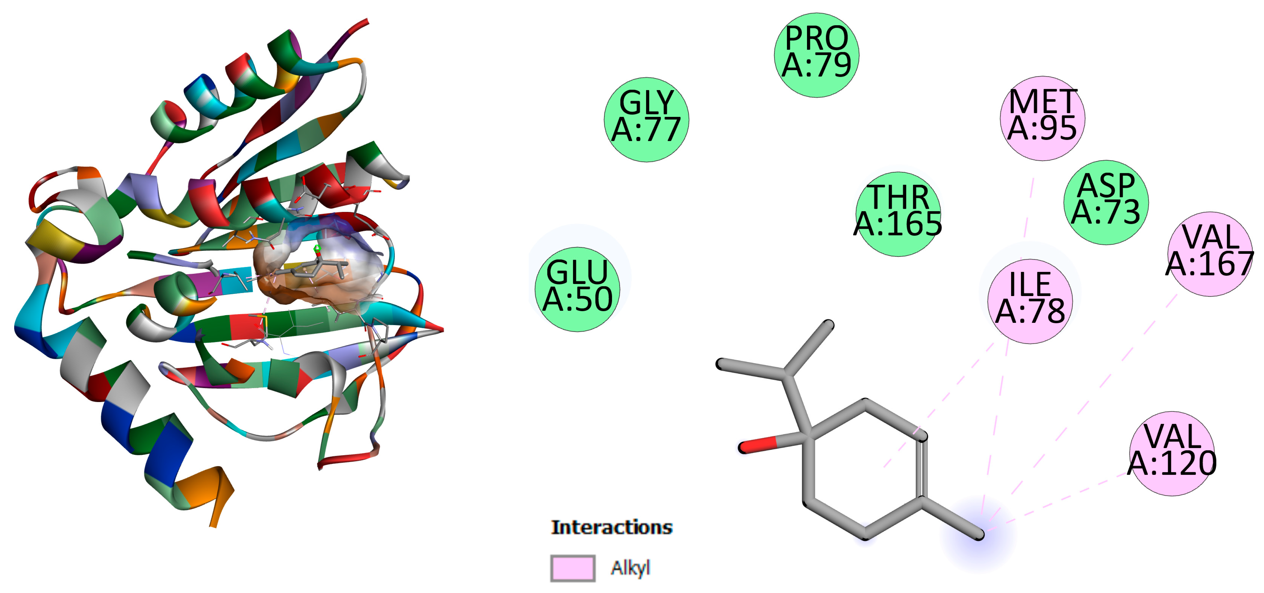
Terpinen-4-ol, which is present in a smaller percentage in lavender essential oil, showed the highest potential for binding to DNA gyrase, with a binding energy of −5.9 kcal/mol. As shown in Figure 3, it can form four alkyl interactions with Ile78, Met95, Val120, and Val167, similar to linalool.
Figure 3.
Representation of terpinen-4-ol docked within the binding site of DNA gyrase B (PDB ID: 6KZX) and key molecular interactions with amino acid residues at the active site.
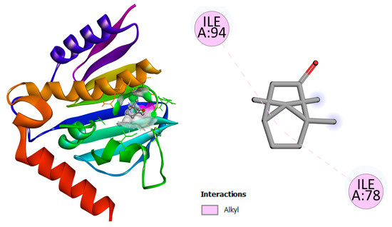
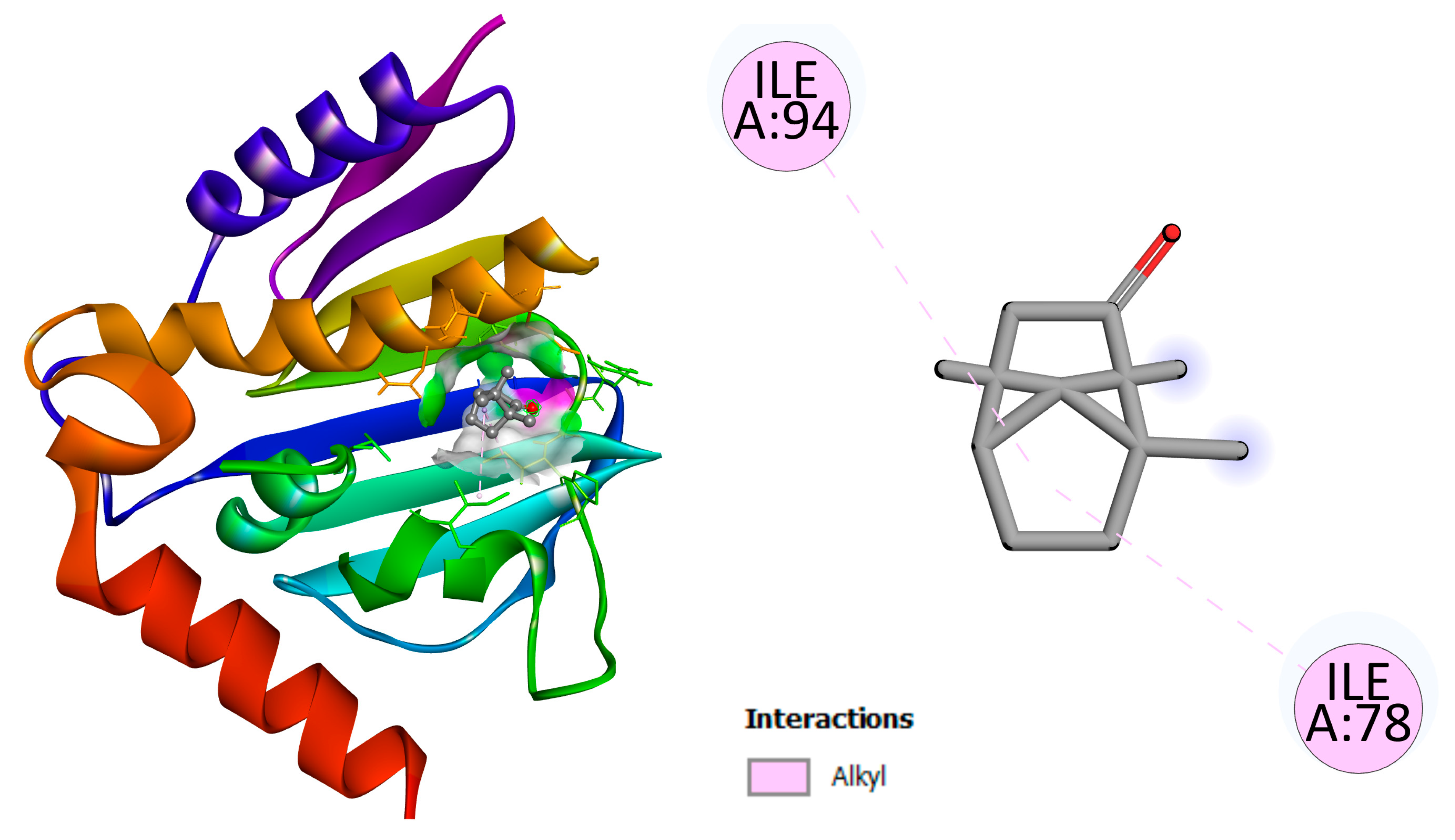
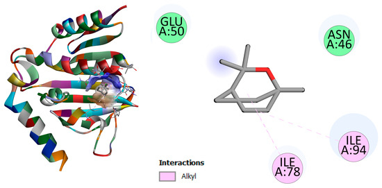
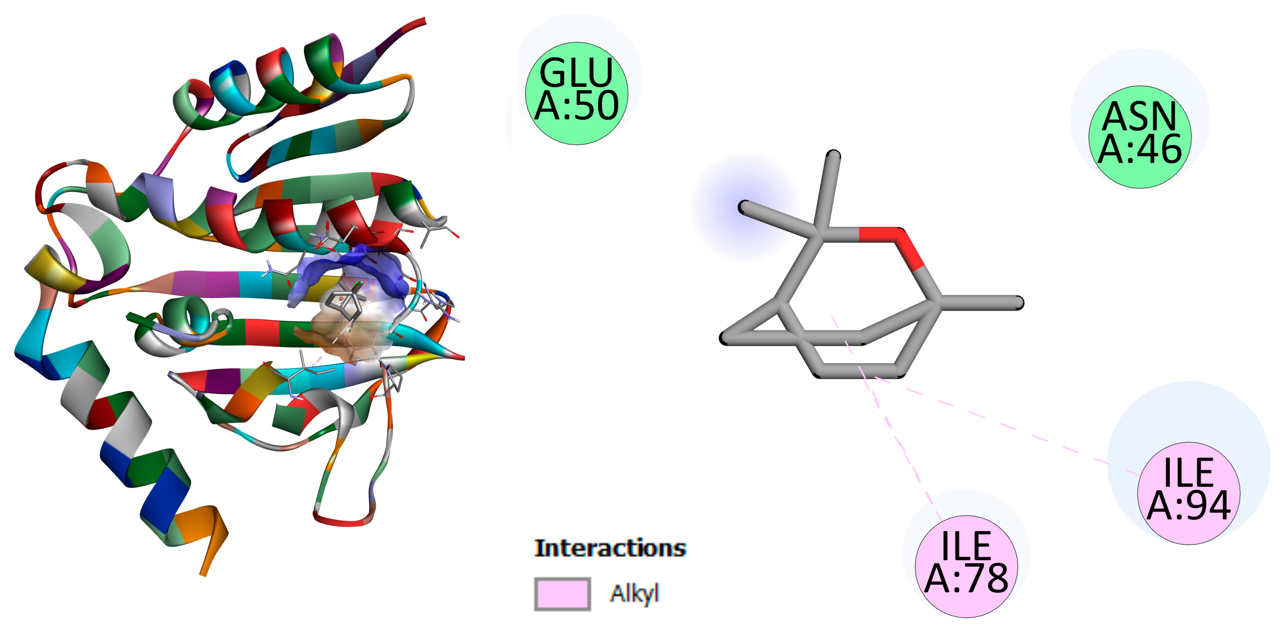
Camphor and eucalyptol, which exhibited the lowest binding affinity and share a similar chemical structure, achieved only two alkyl interactions, with Ile78 and Ile94 (Figure 4 and Figure 5, respectively).
Figure 4.
Representation of camphor docked within the binding site of DNA gyrase B (PDB ID: 6KZX) and key molecular interactions with amino acid residues at the active site.
Figure 5.
Representation of eucalyptol docked within the binding site of DNA gyrase B (PDB ID: 6KZX) and key molecular interactions with amino acid residues at the active site.
These findings build upon previous research on the antimicrobial activity of linalool and camphor, which has primarily been attributed to cell membrane disruption and induction of oxidative stress [46,47,48]. However, increasing evidence suggests that small terpenoids may also interact with intracellular targets, including enzymes involved in nucleic acid processes [49]. Our molecular docking results suggest that DNA gyrase inhibition may contribute to the antibacterial activity of lavender essential oil constituents, providing new insights into their potential as antibacterial agents and into the application of natural antimicrobials in food preservation systems and sanitation of food-contact surfaces. Moreover, these findings may help guide future strategies for developing novel antimicrobial agents. This perspective is supported by recent studies showing that terpene moieties can be successfully incorporated into fluoroquinolone scaffolds, producing terpene–fluoroquinolone conjugates with potent antibacterial activity against both methicillin-sensitive and methicillin-resistant S. aureus and C. albicans [50]. Such approaches highlight the potential of terpenes derived from lavender essential oil as valuable structural motifs in modern antimicrobial drug design.
3.4. Antioxidant Activity
The antioxidant activity of Lavandula angustifolia essential oil was evaluated using the DPPH (2,2-diphenyl-1-picrylhydrazyl) radical-scavenging assay, a commonly employed method for assessment of the free radical scavenging capacity of plant-derived products. The DPPH radical is a stable free radical that exhibits a deep violet color and is reduced in the presence of antioxidants through electron or hydrogen atom donation, resulting in a decrease in absorbance at 517 nm. Radical scavenging activity was calculated based on the decrease in absorbance relative to a control sample and expressed as the percentage of DPPH inhibition. The DPPH radical scavenging activity of LEO increased in a concentration- and time-dependent manner (Table 5).
Table 5.
Antioxidant activity of L. angustifolia essential oil at different concentrations (3, 5, and 10 mg/mL) immediately after solution preparation (0 min) and after 30 min, measured using the DPPH radical-scavenging assay.
At 0 min, inhibition values ranged from 22.0% to 50.6% for concentrations 3 mg/mL, 5 mg/mL, and 10 mg/mL. After 30 min of incubation, a marked increase in radical scavenging activity was observed, reaching a maximum of 89.3% inhibition at 10 mg/mL. These results indicate a clear enhancement of antioxidant activity with both higher concentrations and longer incubation times, consistent with observations reported in previous studies [12,33,51]. The IC50 value of Lavandula angustifolia essential oil after 30 min was 4.2 mg/mL This parameter represents the concentration required to achieve 50% inhibition of the DPPH radical and was calculated by linear interpolation from the plot of DPPH inhibition versus essential oil concentration. Reported IC50 values for Lavandula angustifolia essential oil in DPPH assays vary widely in the literature (Table 6).
Table 6.
Comparation of IC50 values for the antioxidant activity of Lavandula angustifolia essential oil deaminated by the DPPH assay from different geographical origins.
For example, Nikšić et al. [52] found 0.421 mg/mL for lavender oil from Bosnia and Herzegovina, while Massoud et al. [13] reported 5.24 mg/mL for oil from Lebanon. Other studies have reported higher values, such as 31.30 mg/mL for lavender from Spain [12], and 34.92 mg/mL [33] and 27.67 mg/mL [53] in Egyptian and Croatian studies, respectively.
The mechanism underlying the antioxidant activity of essential oils is complex and not fully understood. The activity is primarily associated with the chemical composition of the oil, particularly the presence of constituents that readily donate hydrogen atoms [33,54]. In the present study, the observed scavenging capacity is likely related to the presence of oxygenated monoterpenes, particularly linalool, α-terpineol and terpinen-4-ol, in agreement with previous reports [13,55]. Hydrocarbons such as γ-terpinene may contribute synergistically, enhancing the overall antioxidant response despite their limited hydrogen-donating ability [56]. It should be noted, however, that interactions among constituents can be complex, and further studies would be required to fully elucidate the underlying mechanisms.
Importantly, the antioxidant properties of LEO have practical relevance for sustainable disinfection applications. By mitigating oxidative degradation during storage and use, these properties may enhance the stability and functional efficacy of essential oil-based disinfectants, offering environmentally friendly alternatives to conventional synthetic biocides in food-related and industrial systems [57,58]. This perspective integrates chemical, biological, and sustainability considerations to support the design of robust, eco-friendly disinfection strategies.
4. Conclusions
The chemical characterization of the essential oil obtained from Lavandula angustifolia plants originating from the Mostar region (southern Bosnia and Herzegovina) revealed that linalool, camphor, and eucalyptol represent the predominant constituents. In contrast, the camphor content was markedly higher than values commonly reported for essential oils of this species. The major constituents of Lavandula angustifolia essential oil (linalool, camphor, eucalyptol, and terpinen-4-ol) demonstrated the ability to interact with bacterial DNA gyrase, suggesting that enzyme inhibition may contribute to their antibacterial activity alongside membrane-disruptive effects. These findings highlight the potential of lavender-derived terpenes, not only as natural antimicrobial agents but also for application in food preservation systems and the sanitation of food-contact surfaces. The essential oil of Lavandula angustifolia exhibited strong DPPH radical scavenging activity in a concentration- and time-dependent manner, indicating notable antioxidant potential. Although the results are based on a single in vitro assay, they suggest that oxygenated monoterpenes play a key role in the observed activity. This finding warrants further investigation using antioxidant methods.
The antimicrobial activity results showed that, at an effective concentration 80% (w/v) and a contact time of 5 min, LEO achieved strain-dependent antimicrobial effects, exhibiting >5 log10 reduction against E. coli ATCC 10536 and S. aureus ATCC 6538 It also fulfilled the yeasticidal criterion of EN 1650 [22] against C. albicans ATCC 10231. However, the failure to achieve a ≥5 log10 reduction against P. aeruginosa ATCC 15442 and E. hirae ATCC 10541 indicates that LEO does not meet the bactericidal requirements of EN 1276 [21], even at very high concentrations. This represents a critical limitation for the use of LEO as a stand-alone bactericidal disinfectant under standardized conditions. The absence of bactericidal and yesticidal activity at lower, more application-relevant concentrations (1.0% w/v and 0.1% w/v) further emphasizes the narrow operational window of LEO when evaluated against regulatory performance criteria. While such high concentrations are not practically feasible for routine surface disinfection due to sensory, economic, and formulation constraints, the observed selective efficacy at 80% (w/v) provides valuable boundary conditions for defining realistic performance limits.
This work provides a conceptual basis for the practical application of essential oils in green chemistry, formulation science, and bio-based sustainable technologies. Future research should focus on combination strategies, formulation optimization, and the exploration of potential synergistic effects with other active compounds. In addition, it should consider that various factors may influence the chemical composition of essential oils and, consequently, their antioxidant and antimicrobial activity.
Supplementary Materials
The following supporting information can be downloaded at: https://www.mdpi.com/article/10.3390/sci8050102/s1, Figure S1. Air-dry flowers of Lavandula angustifolia Mill., Table S1: Quantitative suspension test in accordance with modified EN 1276 [21] for the test microorganism Pseudomonas aeruginosa ATCC 15442, Table S2: Quantitative suspension test in accordance with modified EN 1276 [21] for the test microorganism Escherichia coli ATCC 10536, Table S3. Quantitative suspension test in accordance with modified EN 1276 [21] for the test microorganism Staphylococcus aureus ATCC 6538, Table S4. Quantitative suspension test in accordance with modified EN 1276 [21] for test microorganism Enterococcus hirae ATCC 10541, Table S5. Quantitative suspension test in accordance with modified EN 1650 [22] for the test microorganism ATCC 10231.
Author Contributions
Conceptualization, V.A., Ž.M.-B., Ž.G. (Molecular docking) and D.Đ.-M. (Antimicrobial activity); methodology, V.A. (Sample preparation, Antioxidant activity), Ž.M.-B. (Sample preparation), N.K. (GC-MS), D.Đ.-M. (Antimicrobial activity) and Ž.G. (Molecular Docking); software, Ž.G. (AutoDock Vina, Discovery Studio Visualizer v21.1.0.20298); validation, N.K. (GC-MS), V.G.C. (Antioxidant activity) and D.Đ.-M. (Antimicrobial activity); formal analysis, V.A., Ž.M.-B., N.K., V.G.C., V.K. and D.Đ.-M.; investigation, V.A., Ž.M.-B. (Sample preparation, Hydrodistillation, Antioxidant activity), N.K. (GC-MS) and D.Đ.-M. (Antimicrobial activity); resources, V.A. (Plant material); data curation, V.A. and D.Đ.-M.; writing—original draft preparation, V.A., Ž.G. and D.Đ.-M.; writing—review and editing, Ž.M.-B. and V.K.; visualization, V.A., Ž.G. and D.Đ.-M.; supervision, D.Đ.-M. All authors have read and agreed to the published version of the manuscript.
Funding
This study was funded by a project “Hydrodistillation and analysis of essential oils and hydrolates from medicinal and herbal plants from Herzegovina: Research on composition and effect as well as possible application in pharmacy and industry.” No. 125 7064, financed by the Ministry of Scientific and Technological Development and Higher Education of the Republic of Srpska.
Institutional Review Board Statement
Not applicable.
Informed Consent Statement
Not applicable.
Data Availability Statement
The original contributions presented in the study are included in the article, further inquiries can be directed to the corresponding author.
Conflicts of Interest
The authors declare no conflicts of interest.
Abbreviations
The following abbreviations are used in this manuscript:
| ATCC | American Type Culture Collection |
| CFU | colony-forming units |
| DPPH | 2,2-diphenyl-1-picrylhydrazyl |
| EU | European Union |
| IC50 | the concentration required to achieve 50% inhibition of the DPPH radical |
| GC–MS | gas chromatography–mass spectrometry |
| LEO | lavender essential oil |
| MIC | minimum inhibitory concentration |
| MBC | minimum bactericidal concentration |
| MEA | malt extract agar |
| MFC | minimum fungicidal concentration |
| TSA | tryptone soya agar |
References
- Tanasă, F.; Nechifor, M.; Teacă, C.-A. Essential Oils as Alternative Green Broad-Spectrum Biocides. Plants 2024, 13, 3442. [Google Scholar] [CrossRef] [Scilit]
- Falcó, I.; Verdeguer, M.; Aznar, R.; Sánchez, G.; Randazzo, W. Sanitizing food contact surfaces by the use of essential oils. Innov. Food Sci. Emerg. Technol. 2019, 51, 220–228. [Google Scholar] [CrossRef] [Scilit]
- Santos, M.I.S.; Marques, C.; Mota, J.; Pedroso, L.; Lima, A. Applications of Essential Oils as Antibacterial Agents in Minimally Processed Fruits and Vegetables—A Review. Microorganisms 2022, 10, 760. [Google Scholar] [CrossRef] [Scilit]
- Nazzaro, F.; Fratianni, F.; De Martino, L.; Coppola, R.; De Feo, V. Effect of Essential Oils on Pathogenic Bacteria. Pharmaceuticals 2013, 6, 1451–1474. [Google Scholar] [CrossRef] [Scilit]
- Sateriale, D.; Forgione, G.; De Cristofaro, G.A.; Continisio, L.; Pagliuca, C.; Colicchio, R.; Salvatore, P.; Paolucci, M.; Pagliarulo, C. Eco-Friendly Sanitization of Indoor Environments: Effectiveness of thyme essential oil in controlling bioaerosol levels and disinfecting surfaces. BioTech 2024, 13, 12. [Google Scholar] [CrossRef] [Scilit]
- Lopez, V.; Nielsen, B.; Solas, M.; Ramirez, M.J.; Jager, A.K. Exploring Pharmacological Mechanisms of Lavender (Lavandula angustifolia) Essential Oil on Central Nervous System Targets. Front. Pharmacol. 2017, 8, 280. [Google Scholar] [CrossRef] [Scilit]
- Santana de Oliveira, M.; Vostinaru, O.; Rigano, D.; de Aguiar Andrade, E.H. Editorial: Bioactive compounds present in essential oils: Advances and pharmacological applications. Front. Pharmacol. 2023, 14, 1130097. [Google Scholar] [CrossRef] [Scilit]
- Dobros, N.; Zawada, K.; Paradowska, K. Phytochemical Profile and Antioxidant Activity of Lavandula angustifolia and Lavandula × intermedia Cultivars Extracted with Different Methods. Antioxidants 2022, 11, 711. [Google Scholar] [CrossRef] [Scilit]
- Aguerd, O.; Elhrech, H.; El Omari, N.; Benali, T.; Akhazzane, M.; Mostakim, M.; Ouma, S.; Khattabi, L.; Amanullah, M.; Menyiy, N.E.; et al. Chemical composition and biological effects of Lavandula angustifolia Mill., essential oils. AMB Express 2025, 15, 164. [Google Scholar] [CrossRef] [Scilit]
- de Elguea-Culebras, G.O.; Herraiz-Peñalver, D.; Prieto-Blanco, D.; Cerro-Ibáñez, N.; Sánchez-Vioque, R.; Navarro-Rocha, J.; Sanz, M.A.; Asensio-S.-Manzanera, M.C.; Pérez-Magariño, S.; Herrero, B.; et al. Essential oils of lavandin (Lavandula × intermedia Emeric ex Loisel.) of Spain: A case study on clones ‘Grosso’ and ‘Super’. J. Appl. Res. Med. Aromat. Plants 2024, 41, 100550. [Google Scholar] [CrossRef] [Scilit]
- Danh, L.T.; Han, L.N.; Triet, N.D.A.; Zhao, J.; Mammucari, R.; Foster, N. Comparison of chemical composition, antioxidant and antimicrobial activity of lavander (Lavandula angustifolia L.) essential oils extracted by supercritical CO2, hexane and hydrodistillation. Food Bioprocess Technol. 2012, 6, 3481–3489. [Google Scholar] [CrossRef] [Scilit]
- Marin, I.; Sayas-Barbera, E.; Viuda-Martos, M.; Navarro, C.; Sendra, E. Chemical Composition, Antioxidant and Antimicrobial Activity of Essential Oils from Organic Fennel, Parsley, and Lavender from Spain. Foods 2016, 5, 18. [Google Scholar] [CrossRef] [Scilit]
- Massoud, R.I.; Bouaziz, M.; Abdallah, H.; Zeiz, A.; Flamini, G.; El-Dakdouki, M.H. Comparative Study on the Chemical Composition and Biological Activities of the essential oils of Lavandula angustifolia and Lavandula × intermedia cultivated in Lebanon. ACS Omega 2024, 9, 30244–30255. [Google Scholar] [CrossRef] [Scilit]
- Pljevljakusic, D.; Kostadinovic Velickovska, S.; Mihajlov, L.; Cherepnalkoski, A. Chemical composition and biological activity of lavandin and lavender essential oils. Riv. Ital. Sostanze Grasse 2023, 100, 91–103. [Google Scholar]
- Stamova, S.; Ermenlieva, N.; Tsankova, G.; Georgieva, E. Antimicrobial activity of lavender essential oil from Lavandula angustifolia Mill.: In Vitro and in silico evaluation. Antibiotics 2025, 14, 656. [Google Scholar] [CrossRef] [Scilit]
- Betlej, I.; Andres, B.; Cebulak, T.; Kapusta, I.; Balawejder, M.; Jaworski, S.; Lange, A.; Kutwin, M.; Pisulewska, E.; Kidacka, A.; et al. Antimicrobial properties and assessment of the content of bioactive compounds Lavandula angustifolia Mill. cultivated in Southern Poland. Molecules 2023, 28, 6416. [Google Scholar] [CrossRef] [Scilit]
- Perovic, S.; Pantovic, S.; Scepanovic, V.; Perovic, A.; Zivkovic, V.; Damjanovic Vratnica, B. Evaluation of antimicrobial activity and activity on the autonomic nervous system of the lavender essential oils from Montenegro. Prog. Nutr. 2019, 21, 584–590. [Google Scholar]
- Hossain, S.; Heo, H.; De Silva, B.C.J.; Wimalasena, S.H.M.P.; Pathirana, H.N.K.S.; Heo, G.-J. Antibacterial activity of essential oil from lavender (Lavandula angustifolia) against pet turtle-borne pathogenic bacteria. Lab. Anim. Res. 2017, 33, 195–201. [Google Scholar] [CrossRef] [Scilit]
- Imane, M.M.; Houda, F.; Amal, A.H.S.; Kaotar, N.; Mohammed, T.; Imane, R.; Farid, H. Phytochemical Composition and Antibacterial Activity of Moroccan Lavandula angustifolia Mill. J. Essent. Oil-Bear. Plants 2017, 20, 1074–1082. [Google Scholar]
- European Chemical Agency (ECHA). Guidance on the Biocidal Products Regulation. Volume II: Efficacy. Parts B + C: Assessment and Evaluation. Version 6.0. Helsinki, Finland, 2023. Available online: https://echa.europa.eu/documents/10162/2324906/bpr_guidance_assessment_evaluation_part_vol_ii_part_bc_en.pdf/ae2e9a18-82ee-2340-9354-d82913543fb9?t=1691738248873 (accessed on 9 March 2026).
- EN 1276; Chemical Disinfectants and Antiseptics—Quantitative Suspension Test for the Evaluation of Bactericidal Activity of Chemical Disinfectants Used in Food, Industrial, Domestic and Institutional Areas—Test Method and Requirements (Phase 2, Step 1). European Committee for Standardisation: Brussels, Belgium, 2009.
- EN 1650; Chemical Disinfectants and Antiseptics—Quantitative Suspension Test for the Evaluation of Fungicidal or Yeasticidal Activity of Chemical Disinfectants Used in Food, Industrial, Domestic and Institutional Areas—Test Method and Requirements (Phase 2, Step 1). European Committee for Standardisation: Brussels, Belgium, 2009.
- Kovačić, S.; Nikolić, T.; Ruščić, M.; Milović, M.; Stamenković, V.; Mihelj, D.; Jasprica, N.; Bogdanović, S.; Topić, J. Flora Jadranske obale i Otoka: 250 Najčešćih Vrsta; Školska Knjiga: Zagreb, Croatia, 2008. [Google Scholar]
- Adams, R.P. Identification of Essential Oil Components by Gas Chromatography/Mass Spectrometry, 5th ed.; Texensis Publishing: Gruver, TX, USA, 2017; pp. 46–52. [Google Scholar]
- ISO 7218; Microbiology of the Food Chain—General Requirements and Guidance for Microbiological Examinations. International Organization for Standardization: Geneva, Switzerland, 2024.
- Mensor, L.L.; Menezes, F.S.; Leitao, G.G.; Reis, A.S.; dos Santos, T.C.; Coube, C.S.; Leitao, S.G. Screening of Brazilian plant extracts for antioxidant activity by the use of DPPH free radical method. Phytother. Res. 2001, 15, 127–130. [Google Scholar] [CrossRef] [Scilit]
- Ciocarlan, A.; Lupascu, L.; Aricu, A.; Dragalin, I.; Popescu, V.; Geana, E.I.; Ionete, R.E.; Vornicu, N.; Duliu, O.G.; Hristozova, G.; et al. Chemical composition and assessment of antimicrobial activity of Lavandula angustifolia essential oil and some by-products. Plants 2021, 10, 1829. [Google Scholar] [CrossRef] [Scilit]
- Kozuharova, E.; Simeonov, V.; Batovska, D.; Stoycheva, C.; Valchev, H.; Benbassat, N. Chemical composition and comparative analysis of lavender essential oil samples from Bulgaria in relation to the pharmacological effects. Pharmacia 2023, 70, 395–403. [Google Scholar] [CrossRef] [Scilit]
- Smigielski, K.; Raj, A.; Krosowiak, K.; Gruska, R. Chemical Composition of the Essential Oil of Lavandula angustifolia Cultivated in Poland. J. Essent. Oil-Bear. Plants 2013, 12, 338–347. [Google Scholar] [CrossRef] [Scilit]
- European Pharmacopoeia Commission. European Pharmacopoeia, 10th ed.; Council of Europe: Strasbourg, France, 2020. [Google Scholar]
- Lane, W.A.; Mahmoud, S.S. Composition of essential oil from Lavandula angustifolia and L. intermedia varieties grown in British Columbia, Canada. Nat. Prod. Commun. 2008, 3, 1235–1238. [Google Scholar] [CrossRef] [Scilit]
- Reverchon, E.; Porta, G.D.; Senatore, F. Supercritical CO2 extraction and fractionation of lavender essential oil and waxes. J. Agric. Food Chem. 2002, 43, 1654–1658. [Google Scholar] [CrossRef] [Scilit]
- Viuda-Martos, M.; Mohamady, M.A.; Fernández-López, J.; Abd ElRazik, K.A.; Omer, E.A.; Pérez-Alvarez, J.A.; Sendra, E. In vitro antioxidant and antibacterial activities of essential oils obtained from Egyptian aromatic plants. Food Control 2011, 22, 1715–1722. [Google Scholar] [CrossRef] [Scilit]
- Belhadj Mostefa, M.; Kabouche, A.; Abaza, I.; Aburjai, T.; Touzani, R.; Kabouche, Z. Chemotypes investigation of Lavandula essential oils growing at different North African soils. J. Mater. Environ. Sci. 2014, 5, 1896–1901. [Google Scholar]
- Gonzalez-Rivera, J.; Duce, C.; Falconieri, D.; Ferrari, C.; Ghezzi, L.; Piras, A.; Tine, M.R. Coaxial microwave assisted hydrodistillation of essential oils from five different herbs (lavender, rosemary, sage, fennel seeds and clove buds): Chemical composition and thermal analysis. Innov. Food Sci. Emerg. Technol. 2016, 33, 308–318. [Google Scholar] [CrossRef] [Scilit]
- Cai, Z.M.; Peng, J.Q.; Chen, Y.; Tao, L.; Zhang, Y.Y.; Fu, L.Y.; Long, Q.D.; Shen, X.C. 1,8-Cineole: A review of source, biological activities, and application. J. Asian Nat. Prod. Res. 2021, 23, 938–954. [Google Scholar] [CrossRef] [Scilit]
- Gorgini Shabankareh, H.; Khorasaninejad, S.; Soltanloo, H.; Shariati, V. Physiological response and secondary metabolites of three lavender genotypes under water deficit. Sci. Rep. 2021, 11, 19164. [Google Scholar] [CrossRef] [Scilit]
- Betlej, I.; Andres, B.; Cebulak, T.; Kapusta, I.; Balawejder, M.; Żurek, N.; Jaworski, S.; Lange, A.; Kutwin, M.; Pisulewska, E.; et al. Phytochemical composition and antimicrobial properties of new Lavandula angustifolia ecotypes. Molecules 2024, 29, 1740. [Google Scholar] [CrossRef] [Scilit]
- Schelz, Z.; Molnar, J.; Hohmann, J. Antimicrobial and antiplasmid activities of essential oils. Fitoterapia 2006, 77, 279–285. [Google Scholar] [CrossRef] [Scilit]
- Walasek-Janusz, M.; Grzegorczyk, A.; Zalewski, D.; Malm, A.; Gajcy, S.; Gruszecki, R. Variation in the antimicrobial activity of essential oils from cultivars of Lavandula angustifolia and L. × intermedia. Agronomy 2022, 12, 2955. [Google Scholar] [CrossRef] [Scilit]
- Diogo Gonçalves, S.; Paiva-Cardoso, M.d.N.; Caramelo, A. Green preservation strategies: The role of essential oils in sustainable food preservatives. Sustainability 2025, 17, 7326. [Google Scholar] [CrossRef] [Scilit]
- Mith, H.; Duré, R.; Delcenserie, V.; Zhiri, A.; Daube, G.; Clinquart, A. Antimicrobial activities of commercial essential oils and their components against food-borne pathogens and food spoilage bacteria. Food Sci. Nutr. 2014, 2, 403–416. [Google Scholar] [CrossRef] [Scilit] [PubMed]
- U.S. Environmental Protection Agency (EPA). LT1ESWTR Disinfection Profiling and Benchmarking. Technical Guidance Manual; EPA 816-R-03-004; Office of Water: Washington, DC, USA, 2003. Available online: https://books.google.co.jp/books/about/Lt1eswtr_Disinfection_Profiling_and_Benc.html?id=cr-fpwAACAAJ&redir_esc=y (accessed on 10 March 2026).
- Reece, R.J.; Maxwell, A. DNA gyrase: Structure and function. Crit. Rev. Biochem. Mol. Biol. 1991, 26, 335–375. [Google Scholar] [CrossRef] [Scilit]
- Collin, F.; Karkare, S.; Maxwell, A. Exploiting bacterial DNA gyrase as a drug target: Current state and perspectives. Appl. Microbiol. Biotechnol. 2011, 92, 479–497. [Google Scholar] [CrossRef] [Scilit]
- Liu, X.; Cai, J.; Chen, H.; Zhong, Q.; Hou, Y.; Chen, W.; Chen, W. Antibacterial activity and mechanism of linalool against Pseudomonas aeruginosa. Microb. Pathog. 2020, 141, 103980. [Google Scholar] [CrossRef] [Scilit]
- Guo, F.; Chen, Q.; Liang, Q.; Zhang, M.; Chen, W.; Chen, H.; Yun, Y.; Zhong, Q.; Chen, W. Antimicrobial activity and proposed action mechanism of linalool against Pseudomonas fluorescens. Front. Microbiol. 2021, 12, 562094. [Google Scholar] [CrossRef] [Scilit]
- Duda-Madej, A.; Viscardi, S.; Grabarczyk, M.; Topola, E.; Kozłowska, J.; Mączka, W.; Wińska, K. Is camphor the future in supporting therapy for skin infections? Pharmaceuticals 2024, 17, 715. [Google Scholar] [CrossRef] [Scilit]
- Huang, W.; Wang, Y.; Tian, W.; Cui, X.; Tu, P.; Li, J.; Shi, S.; Liu, X. Biosynthesis investigations of terpenoid, alkaloid, and flavonoid antimicrobial agents derived from medicinal plants. Antibiotics 2022, 11, 1380. [Google Scholar] [CrossRef] [Scilit] [PubMed]
- Izmest’ev, E.S.; Pestova, S.V.; Kolesnikova, A.I.; Baidamshina, D.R.; Kayumov, A.R.; Rubtsova, S.A. Terpene/functionalized fluoroquinolones as potential antimicrobials: Synthesis and properties. ChemMedChem 2023, 18, e202300358. [Google Scholar] [CrossRef] [Scilit] [PubMed]
- Gharib, F.A.; Badr, S.E.A.; Al-Ghazali, B.A.S.; Zahran, M.K. Chemical composition, antioxidant and antibacterial activities of lavender and marjoram essential oils. Egypt. J. Chem. 2013, 56, 1–24. [Google Scholar] [CrossRef] [Scilit]
- Nikšić, H.; Kovač-Bešović, E.; Makarević, E.; Durić, K.; Kusturica, J.; Muratović, S. Antiproliferative, antimicrobial, and antioxidant activity of Lavandula angustifolia Mill. essential oil. J. Health Sci. 2026, 7, 35–43. [Google Scholar]
- Blažeković, B.; Yang, W.; Wang, Y.; Li, C.; Kindl, M.; Pepeljnjak, S.; Vladimir-Knežević, S. Chemical composition, antimicrobial and antioxidant activities of essential oils of Lavandula × intermedia ‘Budrovka’ and L. angustifolia cultivated in Croatia. Ind. Crop. Prod. 2018, 123, 173–182. [Google Scholar] [CrossRef] [Scilit]
- Miguel, M.G. Antioxidant activity of medicinal and aromatic plants. A review. Flavour. Frag. J. 2010, 25, 291–312. [Google Scholar] [CrossRef] [Scilit]
- Wojtunik, K.A.; Ciesla, L.M.; Waksmundzka-Hajnos, M. Model studies on the antioxidant activity of common terpenoid constituents of essential oils by means of the 2,2-diphenyl-1-picrylhydrazyl method. J. Agric. Food Chem. 2014, 62, 9088–9094. [Google Scholar] [CrossRef] [Scilit]
- Guo, Y.; Baschieri, A.; Amorati, R.; Valgimigli, L. Synergic antioxidant activity of gamma-terpinene with phenols and polyphenols enabled by hydroperoxyl radicals. Food Chem. 2021, 345, 128468. [Google Scholar] [CrossRef] [Scilit]
- Hafsa, B.A.; Javier, Q.-G.J.; Ahmad, A.; Marina, V.-M. The Natural Disinfectant Role of Essential Oils in Improving Radical Scavenging Activity and Total Phenolic Compounds in Fresh Vegetables. Antioxidants 2025, 14, 1458. [Google Scholar] [CrossRef] [Scilit]
- Sengun, I.Y.; Senturk, S.; Gul, S.; Kilic, G. Potential of essential oil combinations for surface and air disinfection. Lett. App. Microbiol. 2021, 72, 526–534. [Google Scholar] [CrossRef] [Scilit]

Disclaimer/Publisher’s Note: The statements, opinions and data contained in all publications are solely those of the individual author(s) and contributor(s) and not of MDPI and/or the editor(s). MDPI and/or the editor(s) disclaim responsibility for any injury to people or property resulting from any ideas, methods, instructions or products referred to in the content. |
© 2026 by the authors. Licensee MDPI, Basel, Switzerland. This article is an open access article distributed under the terms and conditions of the Creative Commons Attribution (CC BY) license.