Next Issue
Volume 4, June
Previous Issue
Volume 3, December


Quantum Beam Sci., Volume 4, Issue 1 (March 2020) – 17 articles

Cover Story (view full-size image): 
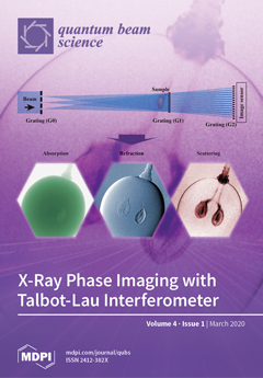
Configuration consisting of gratings for quantum-beam phase imaging (top) and X-ray phase imaging result (absorption, refraction, and scattering images) obtained for a grape (bottom). Internal structures are visible in the phase imaging result, and fibrous connective tissue is especially visible in the scattering image. View this paper.
  • Issues are regarded as officially published after their release is announced to the table of contents alert mailing list.
  • You may sign up for e-mail alerts to receive table of contents of newly released issues.
  • PDF is the official format for papers published in both, html and pdf forms. To view the papers in pdf format, click on the "PDF Full-text" link, and use the free Adobe Reader to open them.
Order results
Result details
Section
Select all
Export citation of selected articles as:
24 pages, 13248 KiB  
Review
Modification of Lattice Structures and Mechanical Properties of Metallic Materials by Energetic Ion Irradiation and Subsequent Thermal Treatments
by Akihiro Iwase and Fuminobu Hori
Quantum Beam Sci. 2020, 4(1), 17; https://doi.org/10.3390/qubs4010017 - 16 Mar 2020
Cited by 3 | Viewed by 2825
Abstract
When materials are irradiated with high-energy ions, their energies are transferred to electrons and atoms in materials, and the lattice structures of the materials are largely changed to metastable or non-thermal equilibrium states, causing the modification of several physical properties. There are two [...] Read more.
When materials are irradiated with high-energy ions, their energies are transferred to electrons and atoms in materials, and the lattice structures of the materials are largely changed to metastable or non-thermal equilibrium states, causing the modification of several physical properties. There are two processes for the material modification by ion irradiation; one is “the irradiation-enhanced process”, and the other is “the irradiation-induced process”. In this review, two kinds of recent results for the microstructural changes and the modifications of mechanical properties will be summarized: one is the hardness modification of dilute aluminum alloys, which is a result of the irradiation-enhanced process, and the other is the hardness modification of Ni-based intermetallic compounds as a result of the irradiation-induced process. The effect of the subsequent thermal treatment on the microstructures and the hardness for ion-irradiated dilute aluminum alloys is quite different from that for Ni-based intermetallic compounds. This result reflects the difference between the irradiation-enhanced process and the irradiation-induced process. Finally, possibilities of the ion irradiation and subsequent thermal treatment to industrial applications will also be discussed. Full article
(This article belongs to the Special Issue Quantum Beams Applying to Innovative Industrial Materials)
Show Figures

Figure 1

14 pages, 7656 KiB  
Article
Microstructural Features and Ductile-Brittle Transition Behavior in Hot-Rolled Lean Duplex Stainless Steels
by O. Takahashi, Y. Shibui, P.G. Xu, S. Harjo, T. Suzuki and Y. Tomota
Quantum Beam Sci. 2020, 4(1), 16; https://doi.org/10.3390/qubs4010016 - 05 Mar 2020
Cited by 6 | Viewed by 4309
Abstract
The characteristics of texture and microstructure of lean duplex stainless steels with low Ni content produced through hot rolling followed by annealing were investigated locally with electron backscatter diffraction and globally with neutron diffraction. Then, the ductile–brittle transition (DBT) behavior was studied by [...] Read more.
The characteristics of texture and microstructure of lean duplex stainless steels with low Ni content produced through hot rolling followed by annealing were investigated locally with electron backscatter diffraction and globally with neutron diffraction. Then, the ductile–brittle transition (DBT) behavior was studied by Charpy impact test. It is found that the DBT temperature (DBTT) is strongly affected by the direction of crack propagation, depending on crystallographic texture and microstructural morphology; the DBTT becomes extremely low in the case of fracture accompanying delamination. A high Ni duplex stainless steel examined for comparison, shows a lower DBTT compared with the lean steel in the same crack propagating direction. The obtained results were also discussed through comparing with those of cast duplex stainless steels reported previously (Takahashi et al., Tetsu-to-Hagané, 100(2014), 1150). Full article
(This article belongs to the Special Issue Analysis of Strain, Stress and Texture with Quantum Beams)
Show Figures

Graphical abstract

9 pages, 2420 KiB  
Article
A Methodology for Reconstructing DSET Pulses from Heavy-Ion Broad-Beam Measurements
by Takahiro Makino, Shinobu Onoda, Takeshi Ohshima, Daisuke Kobayashi, Hirokazu Ikeda and Kazuyuki Hirose
Quantum Beam Sci. 2020, 4(1), 15; https://doi.org/10.3390/qubs4010015 - 04 Mar 2020
Cited by 2 | Viewed by 2330
Abstract
A table-based method for the estimation of heavy-ion-induced Digital Single Event Transient (DSET) voltage pulse-width in a single logic cell has been developed. The estimation method is based on the actual heavy-ion-induced transient current data in a single metal-oxide-semiconductor field effect transistor (MOSFET) [...] Read more.
A table-based method for the estimation of heavy-ion-induced Digital Single Event Transient (DSET) voltage pulse-width in a single logic cell has been developed. The estimation method is based on the actual heavy-ion-induced transient current data in a single metal-oxide-semiconductor field effect transistor (MOSFET) used in the logic cell. The DSET pulse waveform in an inverter is obtained from which the pulse-width was estimated to be 420 ps. This DSET pulse-width value (420 ps) falls within the reasonable range of the DSET pulse-width distribution measured by the self-triggering flip-flop latch chain under heavy-ion irradiation test conditions. Full article
(This article belongs to the Special Issue Quantum Beams Applying to Innovative Industrial Materials)
Show Figures

Figure 1

13 pages, 3972 KiB  
Review
Development of Advanced Biodevices Using Quantum Beam Microfabrication Technology
by Tomoko G. Oyama, Atsushi Kimura, Naotsugu Nagasawa, Kotaro Oyama and Mitsumasa Taguchi
Quantum Beam Sci. 2020, 4(1), 14; https://doi.org/10.3390/qubs4010014 - 02 Mar 2020
Cited by 8 | Viewed by 3668
Abstract
Biodevices with engineered micro- and nanostructures are strongly needed for advancements in medical technology such as regenerative medicine, drug discovery, diagnostic reagents, and drug delivery to secure high quality of life. The authors produced functional biocompatible plastics and hydrogels with physical and chemical [...] Read more.
Biodevices with engineered micro- and nanostructures are strongly needed for advancements in medical technology such as regenerative medicine, drug discovery, diagnostic reagents, and drug delivery to secure high quality of life. The authors produced functional biocompatible plastics and hydrogels with physical and chemical properties and surface microscopic shapes that can be freely controlled in three dimensions during the production process using the superior properties of quantum beams. Nanostructures on a biocompatible poly(L-lactic acid) surface were fabricated using a focused ion beam. Soft hydrogels based on polysaccharides were micro-fabricated using a focused proton beam. Gelatin hydrogels were fabricated using γ-rays and electron beam, and their microstructures and stiffnesses were controlled for biological applications. HeLa cells proliferated three-dimensionally on the radiation-crosslinked gelatin hydrogels and, furthermore, their shapes can be controlled by the micro-fabricated surface of the hydrogel. Long-lasting hydrophilic concave structures were fabricated on the surface of silicone by radiation-induced crosslinking and oxidation. The demonstrated advanced biodevices have potential applications in three-dimensional cell culture, gene expression control, stem cell differentiation induction/suppression, cell aggregation into arbitrary shapes, tissue culture, and individual diagnosis in the medical field. Full article
(This article belongs to the Special Issue Quantum Beams Applying to Innovative Industrial Materials)
Show Figures

Figure 1

9 pages, 2988 KiB  
Article
Novel Approaches for Intensifying Negative C60 Ion Beams Using Conventional Ion Sources Installed on a Tandem Accelerator
by Atsuya Chiba, Aya Usui, Yoshimi Hirano, Keisuke Yamada, Kazumasa Narumi and Yuichi Saitoh
Quantum Beam Sci. 2020, 4(1), 13; https://doi.org/10.3390/qubs4010013 - 02 Mar 2020
Cited by 9 | Viewed by 3086
Abstract
We developed novel methods for producing negative C60 ion beams at the accelerator facility Takasaki Ion Accelerators for Advanced Radiation Application (TIARA) to increase the current intensity of swift C60 ion beams accelerated to the MeV energy region using a tandem [...] Read more.
We developed novel methods for producing negative C60 ion beams at the accelerator facility Takasaki Ion Accelerators for Advanced Radiation Application (TIARA) to increase the current intensity of swift C60 ion beams accelerated to the MeV energy region using a tandem accelerator. We produced negative C60 ion beams with an intensity of 1.3 µA, which is several tens of thousands of times greater than the intensity of beams produced using conventional methods based on the Cs sputtering process. These beams were obtained by temporarily adding an ionization function based on electron attachment to an existing ion source that is widely used in tandem accelerators. The high-intensity swift C60 ion beams can be made available relatively easily to institutes that have tandem accelerators and ion sources of the type used at TIARA because there is no need to change existing ion sources or install new ones. Full article
(This article belongs to the Special Issue Quantum Beams Applying to Innovative Industrial Materials)
Show Figures

Figure 1

16 pages, 5911 KiB  
Article
In Situ Analysis of the Phase Transformation Kinetics in the β-Water-Quenched Ti-5Al-5Mo-5V-3Cr-1Zr Alloy during Ageing after Fast Heating
by Rafael Paiotti Marcondes Guimarães, Bruna Callegari, Fernando Warchomicka, Katherine Aristizabal, Flavio Soldera, Frank Mücklich and Haroldo Cavalcanti Pinto
Quantum Beam Sci. 2020, 4(1), 12; https://doi.org/10.3390/qubs4010012 - 21 Feb 2020
Cited by 6 | Viewed by 3457
Abstract
Thermal treatments are the main route to achieve improvements in mechanical properties of β-metastable titanium alloys developed for structural applications in automotive and aerospace industries. Therefore, it is of vital importance to determine phase transformation kinetics and mechanisms of nucleation and precipitation during [...] Read more.
Thermal treatments are the main route to achieve improvements in mechanical properties of β-metastable titanium alloys developed for structural applications in automotive and aerospace industries. Therefore, it is of vital importance to determine phase transformation kinetics and mechanisms of nucleation and precipitation during heat treatment of these alloys. In this context, the present paper focuses on the assessment of solid-state transformations in a β-water-quenched Ti-5Al-5Mo-5V-3Cr-1Zr alloy during the early stages of ageing treatment at 500 °C. In situ tracking of transformations was performed using high-energy synchrotron X-ray diffraction. The transformation sequence β + ω → α + α”iso + β is proposed to take place during this stage. Results show that isothermal α” phase precipitates from ω and from spinodal decomposition domains of the β phase, whereas α nucleates from ω, β and also from α” with different morphologies. Isothermal α” is considered to be the regulator of transformation kinetics. Hardness measurements confirm the presence of ω, although this phase was not detected by X-ray diffraction during the in situ treatment. Full article
Show Figures

Figure 1

11 pages, 3970 KiB  
Article
Formation of Fe Nanoparticles by Ion Implantation Technique for Catalytic Graphitization of a Phenolic Resin
by Akira Idesaki, Shunya Yamamoto, Masaki Sugimoto, Tetsuya Yamaki and Yasunari Maekawa
Quantum Beam Sci. 2020, 4(1), 11; https://doi.org/10.3390/qubs4010011 - 12 Feb 2020
Cited by 5 | Viewed by 3035
Abstract
Ion implantation technique was employed to introduce iron nanoparticles (Fe NPs) into a carbon precursor polymer with the aim of forming of a graphitic nanostructure through catalytic graphitization by the introduced Fe NPs. A phenolic resin was implanted by 100 keV Fe+ [...] Read more.
Ion implantation technique was employed to introduce iron nanoparticles (Fe NPs) into a carbon precursor polymer with the aim of forming of a graphitic nanostructure through catalytic graphitization by the introduced Fe NPs. A phenolic resin was implanted by 100 keV Fe+ ions with ion fluence of 1 × 10¹⁴–1 × 10¹⁶ ions/cm2 at ambient temperature under vacuum, and subsequently heat-treated at 800 °C in a nitrogen gas atmosphere. It was found that the particle size of Fe NPs could be controlled in the range of 5–30 nm by the Fe+ ion fluence. Additionally, it was found that a nanosized turbostratic graphite structure with mean interlayer distance of 0.3531 nm, which is consisted of shell-like carbon layers and intricately distorted carbon layers, was formed around the Fe NPs. The ion implantation technique is one of the advantageous ways to introduce size-controlled fine metal NPs which are effective for the formation of graphitic nanostructure from a carbon precursor polymer. Full article
(This article belongs to the Special Issue Quantum Beams Applying to Innovative Industrial Materials)
Show Figures

Figure 1

10 pages, 1865 KiB  
Article
Hydration of Nucleobase as Probed by Electron Emission of Uridine-5′-Mono-Phosphate (UMP) in Aqueous Solution Induced by Nitrogen K-Shell Ionization
by Yasuaki Takeda, Hiroyuki Shimada, Ryosuke Miura, Masatoshi Ukai, Kentaro Fujii, Yoshihiro Fukuda and Yuji Saitoh
Quantum Beam Sci. 2020, 4(1), 10; https://doi.org/10.3390/qubs4010010 - 11 Feb 2020
Cited by 1 | Viewed by 2502
Abstract
To identify the precise early radiation processes of DNA lesions, we measure electron kinetic energy spectra emitted from uridine-5′ monophosphate (UMP) in aqueous solution for the photoionization of the N 1s orbital electron and for the following Auger effect using a monochromatic soft [...] Read more.
To identify the precise early radiation processes of DNA lesions, we measure electron kinetic energy spectra emitted from uridine-5′ monophosphate (UMP) in aqueous solution for the photoionization of the N 1s orbital electron and for the following Auger effect using a monochromatic soft X-ray synchrotron radiation at energies above the nitrogen K-shell ionization threshold. The change of photoelectron spectra for UMP in aqueous solutions at different proton concentrations (pH = 7.5 and 11.3) is ascribed to the chemical shift of the N3 nitrogen atom in uracil moiety of canonical and deprotonated forms. The lowest double ionization potentials for aqueous UMP at different pH obtained from the Auger electron spectra following the N 1s photoionization values show the electrostatic aqueous interaction of uracil moiety of canonical (neutral) and deprotonated (negatively charged) forms with hydrated water molecules. Full article
Show Figures

Graphical abstract

21 pages, 9630 KiB  
Review
Recent Progress in X-ray and Neutron Phase Imaging with Gratings
by Atsushi Momose, Hidekazu Takano, Yanlin Wu, Koh Hashimoto, Tetsuo Samoto, Masato Hoshino, Yoshichika Seki and Takenao Shinohara
Quantum Beam Sci. 2020, 4(1), 9; https://doi.org/10.3390/qubs4010009 - 10 Feb 2020
Cited by 19 | Viewed by 4730
Abstract
Under the JST-ERATO project in progress to develop X-ray and neutron phase-imaging methods together, recent achievements have been selected and reviewed after describing the merit and the principle of the phase imaging method. For X-ray phase imaging, recent developments of four-dimensional phase tomography [...] Read more.
Under the JST-ERATO project in progress to develop X-ray and neutron phase-imaging methods together, recent achievements have been selected and reviewed after describing the merit and the principle of the phase imaging method. For X-ray phase imaging, recent developments of four-dimensional phase tomography and phase microscopy at SPring-8, Japan are mainly presented. For neutron phase imaging, an approach in combination with the time-of-flight method developed at J-PARC, Japan is described with the description of new Gd grating fabrication. Full article
(This article belongs to the Special Issue Quantum Beams Applying to Innovative Industrial Materials)
Show Figures

Figure 1

6 pages, 4478 KiB  
Article
Precipitation of Pt Nanoparticles inside Ion-Track-Etched Capillaries
by Shunya Yamamoto, Hiroshi Koshikawa, Tomitsugu Taguchi and Tetsuya Yamaki
Quantum Beam Sci. 2020, 4(1), 8; https://doi.org/10.3390/qubs4010008 - 06 Feb 2020
Cited by 2 | Viewed by 3450
Abstract
Ion-track-etched capillaries containing nanoparticles of precious metals (e.g., Pt, Au, and Ag) can be applied to plasmonic absorber materials. The precipitation of homogeneous and highly dispersed precious metal nanoparticles inside capillaries represents a key process. Ion-track-etched capillaries (diameter: ~500 nm, length: ~25 μm) [...] Read more.
Ion-track-etched capillaries containing nanoparticles of precious metals (e.g., Pt, Au, and Ag) can be applied to plasmonic absorber materials. The precipitation of homogeneous and highly dispersed precious metal nanoparticles inside capillaries represents a key process. Ion-track-etched capillaries (diameter: ~500 nm, length: ~25 μm) were created in polyimide film by 350 MeV Xe irradiation (3 × 10⁷ ions/cm2) and chemical etching (using a sodium hypochlorite solution). The films with capillaries were immersed in an aqueous solution containing 0.1–10 mmol/L H2PtCl6 and 0.5 vol% C2H5OH, and then irradiated with a 2 MeV electron beam up to a fluence of 1.4 × 10¹⁶ e/cm2. The Pt particles inside the capillaries were characterized by scanning electron microscopy (SEM) and transmission electron microscopy (TEM). The precipitation of Pt nanoparticles and isolated aggregates inside the capillaries was confirmed by TEM. The Pt nanoparticles tended to aggregate under increasing concentrations of H2PtCl6 in the aqueous solution; meanwhile, no changes in nanoparticle size were noted under increasing electron beam fluence. The results suggest that the proposed method can be used to form metal nanoparticles in nanosized capillaries with a high aspect ratio. Full article
(This article belongs to the Special Issue Quantum Beams Applying to Innovative Industrial Materials)
Show Figures

Figure 1

28 pages, 13413 KiB  
Perspective
Interactions of Single Particle with Organic Matters: A Facile Bottom-Up Approach to Low Dimensional Nanostructures
by Shugo Sakaguchi, Koshi Kamiya, Tsuneaki Sakurai and Shu Seki
Quantum Beam Sci. 2020, 4(1), 7; https://doi.org/10.3390/qubs4010007 - 05 Feb 2020
Cited by 5 | Viewed by 3145
Abstract
A particle induces a pack of chemical reactions in nanospace: chemical reactions confined into extremely small space provide an ultimate technique for the nanofabrication of organic matter with a variety of functions. Since the discovery of particle accelerators, an extremely high energy density [...] Read more.
A particle induces a pack of chemical reactions in nanospace: chemical reactions confined into extremely small space provide an ultimate technique for the nanofabrication of organic matter with a variety of functions. Since the discovery of particle accelerators, an extremely high energy density can be deposited, even by a single isolated particle with MeV-ordered kinetic energy. However, this was considered to cause severe damages to organic molecules due to its relatively small bond energies, and lack of ability to control the reactions precisely to form the structures while retaining physico-chemical molecular functionalities. Practically, the severely damaged area along a particle trajectory: a core of a particle track has been simply visualized for the detection/dosimetry of an incident particle to the matters, or been removed to lead nanopores and functionalized by refilling/grafting of fresh organic/inorganic materials. The use of intra-track reactions in the so-called “penumbra” or “halo” area of functional organic materials has been realized and provided us with novel and facile protocols to provide low dimensional nano-materials with perfect size controllability in the 21st century. These protocols are now referred to as single particle nanofabrication technique (SPNT) and/or single particle triggered linear polymerization technique (STLiP), paving the way towards a new approach for nanomaterials with desired functionalities from original molecules. Herein, we report on the extremely wide applicability of SPNT/STLiP protocols for the future development of materials for opto-electronic, catalytic, and biological applications among others. Full article
(This article belongs to the Special Issue Quantum Beams Applying to Innovative Industrial Materials)
Show Figures

Figure 1

14 pages, 3081 KiB  
Article
Efficient Adsorption Performance of Lithium Ion onto Cellulose Microspheres with Sulfonic Acid Groups
by Chenxi Xu, Tianlin Yu, Jing Peng, Long Zhao, Jiuqiang Li and Maolin Zhai
Quantum Beam Sci. 2020, 4(1), 6; https://doi.org/10.3390/qubs4010006 - 03 Feb 2020
Cited by 14 | Viewed by 3363
Abstract
The separation of Li+ from an aqueous solution has received much attention in recent years because of its wide application in batteries and nuclear energy. A cellulose microsphere adsorbent with sulfonic acid groups (named as CGS) was successfully prepared by the pre-irradiation-induced [...] Read more.
The separation of Li+ from an aqueous solution has received much attention in recent years because of its wide application in batteries and nuclear energy. A cellulose microsphere adsorbent with sulfonic acid groups (named as CGS) was successfully prepared by the pre-irradiation-induced emulsion graft polymerization of glycidyl methacrylate onto cellulose microspheres through subsequent sulfonation and protonation. The adsorption performance of Li+ onto the CGS adsorbent is investigated in detail. The as-prepared CGS adsorbent exhibited fast adsorption kinetics and a high adsorption capacity of Li+ (16.0 mg/g) in a wide pH range from 4 to 10. The existence of K+ and Na+ was found to have the ability to affect the adsorption capacity of Li+ due to the cation-exchange adsorption mechanism, which was further confirmed by X-ray photoelectron spectroscopy (XPS). The column adsorption experiment indicated that the adsorption capacity of CGS agreed well with the batch adsorption, and a fast desorption could be obtained in 10 min. It is expected that CGS has potential usage in the adsorption separation of Li+ from an aqueous solution. Full article
(This article belongs to the Special Issue Quantum Beams Applying to Innovative Industrial Materials)
Show Figures

Figure 1

1 pages, 162 KiB  
Editorial
Acknowledgement to Reviewers of Quantum Beam Science in 2019
by Quantum Beam Science Editorial Office
Quantum Beam Sci. 2020, 4(1), 5; https://doi.org/10.3390/qubs4010005 - 20 Jan 2020
Viewed by 1545
Abstract
The editorial team greatly appreciates the reviewers who have dedicated their considerable time and expertise to the journal’s rigorous editorial process over the past 12 months, regardless of whether the papers are finally published or not [...] Full article
10 pages, 5729 KiB  
Article
A Compact Ultrafast Electron Diffractometer with Relativistic Femtosecond Electron Pulses
by Jinfeng Yang, Kazuki Gen, Nobuyasu Naruse, Shouichi Sakakihara and Yoichi Yoshida
Quantum Beam Sci. 2020, 4(1), 4; https://doi.org/10.3390/qubs4010004 - 20 Jan 2020
Cited by 9 | Viewed by 3590
Abstract
We have developed a compact relativistic femtosecond electron diffractometer with a radio-frequency photocathode electron gun and an electron lens system. The electron gun generated 2.5-MeV-energy electron pulses with a duration of 55 ± 5 fs containing 6.3 × 10⁴ electrons per pulse. [...] Read more.
We have developed a compact relativistic femtosecond electron diffractometer with a radio-frequency photocathode electron gun and an electron lens system. The electron gun generated 2.5-MeV-energy electron pulses with a duration of 55 ± 5 fs containing 6.3 × 10⁴ electrons per pulse. Using these pulses, we successfully detected high-contrast electron diffraction images of single crystalline, polycrystalline, and amorphous materials. An excellent spatial resolution of diffraction images was obtained as 0.027 ± 0.001 Å−1. In the time-resolved electron diffraction measurement, a laser-excited ultrafast electronically driven phase transition in single-crystalline silicon was observed with a temporal resolution of 100 fs. The results demonstrate the advantages of the compact relativistic femtosecond electron diffractometer, including access to high-order Bragg reflections, single shot imaging with the relativistic femtosecond electron pulse, and the feasibility of time-resolved electron diffraction to study ultrafast structural dynamics. Full article
(This article belongs to the Special Issue Quantum Beams Applying to Innovative Industrial Materials)
Show Figures

Figure 1

11 pages, 3886 KiB  
Article
Spatial Internal Material Load and Residual Stress Distribution Evolution in Synchrotron In Situ Investigations of Deep Rolling
by Heiner Meyer and Jérémy Epp
Quantum Beam Sci. 2020, 4(1), 3; https://doi.org/10.3390/qubs4010003 - 13 Jan 2020
Cited by 2 | Viewed by 2570
Abstract
Mechanical loading scenarios, comparable to a deep rolling process, were reproduced in static indentation experiments on AISI 4140H steel samples with a cylindrical deep rolling tool and investigated in situ with synchrotron radiation at the European Synchrotron Radiation Facility (ESRF) on beamline ID11. [...] Read more.
Mechanical loading scenarios, comparable to a deep rolling process, were reproduced in static indentation experiments on AISI 4140H steel samples with a cylindrical deep rolling tool and investigated in situ with synchrotron radiation at the European Synchrotron Radiation Facility (ESRF) on beamline ID11. Through the use of spatially resolved diffraction data, two-dimensional (2D) equivalent von Mises stress maps were recorded during loading and after unloading. The material modifications were analyzed in the material below the contact zone for different loading conditions. It was demonstrated that the characteristics of internal material load and residual stress distributions can be evaluated through data fitting and the effect of the applied force could be linked to the stress fields by an empirical model. The experimental values were then compared to a contact mechanics approach in order to analyze the correlation between the theoretical maximum loading stresses and the stored elastic residual stresses remaining by considering the dissipation of a certain amount of energy through plastic deformation. Full article
Show Figures

Figure 1

10 pages, 4387 KiB  
Review
Targeting Specific Sites in Biological Systems with Synchrotron X-Ray Microbeams for Radiobiological Studies at the Photon Factory
by Akinari Yokoya and Noriko Usami
Quantum Beam Sci. 2020, 4(1), 2; https://doi.org/10.3390/qubs4010002 - 09 Jan 2020
Cited by 6 | Viewed by 2970
Abstract
X-ray microbeams have been used to explore radiobiological effects induced by targeting a specific site in living systems. Synchrotron radiation from the Photon Factory, Japan, with high brilliance and highly parallel directionality is a source suitable for delivering a particular beam size or [...] Read more.
X-ray microbeams have been used to explore radiobiological effects induced by targeting a specific site in living systems. Synchrotron radiation from the Photon Factory, Japan, with high brilliance and highly parallel directionality is a source suitable for delivering a particular beam size or shape, which can be changed according to target morphology by using a simple metal slit system (beam size from 5 μm to several millimeters). Studies have examined the non-targeted effects, called bystander cellular responses, which are thought to be fundamental mechanisms of low-dose or low-dose-rate effects in practical radiation risk research. Narrow microbeams several tens of micrometers or less in their size targeted both the cell nucleus and the cytoplasm. Our method combined with live-cell imaging techniques has challenged the traditional radiobiological dogma that DNA damage is the only major cause of radiation-induced genetic alterations and is gradually revealing the role of organelles, such as mitochondria, in these biological effects. Furthermore, three-dimensionally cultured cell systems have been used as microbeam targets to mimic organs. Combining the spatial fractionation of X-ray microbeams and a unique ex vivo testes organ culture technique revealed that the tissue-sparing effect was induced in response to the non-uniform radiation fields. Spatially fractionated X-ray beams may be a promising tool in clinical radiation therapy. Full article
Show Figures

Figure 1

17 pages, 2308 KiB  
Article
Use of Space-Resolved in-Situ High Energy X-ray Diffraction for the Characterization of the Compositional Dependence of the Austenite-to-Ferrite Transformation Kinetics in Steels
by Imed-Eddine Benrabah, Hugo Paul Van Landeghem, Frédéric Bonnet, Florence Robaut and Alexis Deschamps
Quantum Beam Sci. 2020, 4(1), 1; https://doi.org/10.3390/qubs4010001 - 18 Dec 2019
Cited by 2 | Viewed by 2404
Abstract
In-situ high energy X-Ray diffraction (HEXRD) was used on compositionally graded steels to study the effect of substitutional elements on ferrite growth kinetics in Fe–C–X and Fe–C–X–Y systems. Two systems were selected to illustrate the applicability of the combinatorial approach in studying such [...] Read more.
In-situ high energy X-Ray diffraction (HEXRD) was used on compositionally graded steels to study the effect of substitutional elements on ferrite growth kinetics in Fe–C–X and Fe–C–X–Y systems. Two systems were selected to illustrate the applicability of the combinatorial approach in studying such transformations, Fe–C–Mn and Fe–C–Mn–Mo. Comparison between the measured ferrite growth kinetics using HEXRD and the predicted ones using Para-Equilibrium (PE) and Local Equilibrium with Negligible Partitioning (LENP) models indicates that the fractions reached at the stasis of transformation are lower than the predicted ones. Experiments indicated a deviation of measured kinetics from both PE and LENP models when increasing Mn and decreasing Mo (in Fe–C–Mn–Mo system). The large amount of data that can be obtained using this approach can be used for validating existing models describing ferrite growth kinetics. Full article
(This article belongs to the Special Issue Microstructural and Phase Transformations in Materials)
Show Figures

Graphical abstract

Previous Issue
Next Issue
Back to TopTop