


Abstract
Background: Several risk factors have been identified for knee anterior cruciate ligament (ACL) injuries. Yet, their contributions still raise questions when attempting to identify at-risk populations with whom they are associated. Objectives: The purpose of this study was to identify combinations of characteristics in athlete populations at risk of non-contact ACL injuries using principal component analysis. Methods: From a cohort of 161 sports practitioners aged 28.6 ± 8 years who had presented a non-contact ACL injury, five risk factors were assessed on the healthy contralateral knee: passive knee alignment (valgus and recurvatum), knee laxity (KT-1000 test), and isokinetic hamstring strength at 60°/s and at 180°/s. Female sex, age and BMI were considered as individual risk factors. The profile of the population at risk of non-contact ACL injury was determined using a principal component analysis. Results: A total of 31% of the patients were females, 34% were under 24 years old, and 63% had a BMI > 22.5 Kg/m2. The knee axis was valgus in 37% and recurvatum in 80%. An anterior–posterior laxity ≥ 4 mm was present in 38% of the cases. Weakness of the hamstring was present at 60°/s (<1.3 Nm/kg) and 180°/s (<1.0 Nm/kg) in 49% and 54% of the cases, respectively. The principal component analysis explained only 61% of the variance in non-contact ACL injury. Three profiles of the population could be identified: “female with dynamical factors” (knee valgus and weakness of the hamstring strength), “male with physiological factors” (old age and high BMI), and “knee morphology” (valgus, recurvatum and laxity). Conclusions: In the absence of a comparison with a non-injured population, combinations of the potential factors associated with non-contact ACL injuries have been identified, and three independent populations could be at risk.
1. Introduction
Every year, several hundred thousand anterior cruciate ligament (ACL) injuries occur during sports practice. The estimated incidence is 0.06 injuries per 1000 h of exposure in high school athletes [1]. The mechanism of ACL injury is mostly non-contact in 70 to 75% of the cases [2,3]. Several risk factors have been identified to explain non-contact ACL injuries [4,5,6,7,8]. These risk factors are classified into two distinct categories: modifiable (e.g., bodyweight or muscle strength) and non-modifiable (e.g., sex, anatomical knee structure, joint laxity) [6]. Female sex and young age (<24 years old) are particularly identified as risk factors for non-contact ACL rupture [9,10,11,12,13]. Indeed, female sex must be particularly taken into consideration, because women suffer from ACL injuries at a 2- to 6-fold greater rate than men [11,14,15]. In women, general joint laxity, passive knee extension (recurvatum) and anterior–posterior knee laxity could be risk factors for the occurrence of non-contact ACL injuries [10,16,17,18,19,20]. Concerning modifiable intrinsic risk factors, high body mass index (BMI) would represent a questionable risk factor for non-contact ACL injuries [5,8,12,16]. In women, dynamic knee valgus would be poorly controlled during a non-contact ACL injury, based on an increase in hip varus, knee valgus and hamstring strength deficit (or an imbalance of hamstring/quadriceps ratio) [8,10,21,22,23,24]. Yet the links with the static morphological valgus (a non-modifiable factor) are poorly known [25,26,27].
Therefore, the objective of this study was to identify combinations of characteristics (female sex, age, BMI, passive knee alignment, anterior–posterior laxity and poor hamstring knee strength) among athletes with non-contact ACL injuries. To confirm the associations, we used a principal component analysis [28].
2. Materials and Methods
2.1. Population
Methodologically, it was hypothesized that the two knees were almost identical in the same athlete, and that patients who had a non-contact ACL injury had more frequently associated predisposing risk factors for ACL injury. Indeed, bilateral neuromuscular defects have been reported after ACL injury [21]. This particular method was used because of the difficulty of tracking athletes for many years, pending the occurrence of a possible primary ACL injury [29,30]. All the athletes aged between 18 and 45 years old who performed an isokinetic knee evaluation as part of the usual 6-month follow-up from an ACL surgical reconstruction using hamstring auto-graft were included during a 2-year follow-up (January 2022 to December 2023). Patients were not included if they had undergone bilateral ACL reconstruction, a second ACL reconstruction on the same knee, a posterior cruciate ligament reconstruction and/or multiple peripheral ligaments reconstructions, or a modification of the knee alignment by bone surgical correction. Patients were also not included if they refused to participate in this study. The written consent of the patients participating in this study was sought. This study followed the World Medical Association Declaration of Helsinki [31] and was approved by the local Ethics Committee of the Sports Medicine department (CNEMS)—2022-006.
2.2. Anthropometric Parameters
Due to the already known risk factors associated with non-contact ACL injury, the female sex was considered at risk. The cut-off of age <24 years old was taken into consideration in accordance with our study published in 2022 [13]. The body mass index (BMI) was calculated from the bodyweight and height, and the cut-off >22.5 kg/m2 was considered at risk [5,13].
2.3. Knee Anatomical Parameters
Knee anatomical parameters were measured in the reconstructed knee against to the contralateral healthy knee by the same observer. Knee alignment was measured in the frontal plane in a standing position according to knee morphologic types [13]. Passive knee valgus was quantified by measuring, to the nearest millimeter, the inter-malleolar distance using a ruler and was considered present when the inter-malleolar distance was measured with the knees straight and in contact, and not present when the inter-malleolar distance was not measurable. Passive knee extension was evaluated in degrees in the sagittal plane in dorsal decubitus. Knee recurvatum was considered present when the heel was not in contact with the examination table with the knee in hyperextension.
Knee laxity was measured in millimeters using a KT-1000® arthrometer (MEDmetric Corp., San Diego, CA, USA) at 134 Newtons [8,10,17]. Because a cut-off ≥4 mm corresponds to the threshold of pathological laxity, this cut-off was taken into consideration to characterize the risk factor associated with ACL injury [13,17,32,33]. The intra-examiner reliability is good for 134 Newtons [34]. The knee laxity was considered present if the cut-off ≥4 mm was reached.
2.4. Isokinetic Knee Parameters
Muscle strength was assessed using an isokinetic dynamometer Humac® dynamometer (Medimex, Sainte-Foy-lès-Lyon, France) according to an identical protocol for each subject. For the healthy knee, quadriceps (extensors) and hamstring (flexors) strength was assessed in concentric mode at 60 and 180°/s of angular velocities. The knee range of motion was limited to 100 degrees (from the full extension to 100 degrees of flexion). Gravity correction was used for all the tests. The subject was installed and given the instructions for the tests. Three repetitions at 60°/s and then 5 repetitions at 180°/s were performed. Only the hamstring strength was considered, because this parameter was identified as a risk factor for non-contact ACL injury [13]. The reliability of the hamstring strength measurement (ICC between 0.93 and 0.97) is excellent at 60 and 180°/s of angular speed [35]. The hamstring strength was reported along with the bodyweight, and values <1.3 Nm/kg and <1 Nm/kg were considered at risk for the 60 and 180°/s isokinetic angular speeds, respectively, according to the study by Dauty et al. [13].
2.5. Definition of Non-Contact ACL Rupture
Non-contact ACL injury during sports practice was defined by a knee twisting mechanism that occurred without collision and without high kinetic reception (high-impact rotation landing) [36].
2.6. Statistical Analysis
The statistical analyses were performed using SPSS 29.0 software (IBM Corp. Armonk, NY, USA). Quantitative parameters were presented as mean and standard deviation, and qualitative parameters as frequency. To confirm or negate the potential factors associated with a non-contact ACL injury, a principal component analysis (PCA) was performed [28]. This factorial analysis was justified to explore combinations of variables based on the total variance of the studied variables (potentially associated factors) and allowed for the extraction of a minimum of factors that explained the majority of the specific variance of the phenomenon (non-contact ACL injury). Thus, from 8 studied variables (sex, age, BMI, hamstring strength (60 and 180°/s), knee valgus, extension, and laxity), it was decided to condense the information according to a number of factors as defined by an eigenvalue ≥ 1 and a screen plot (Cattell’s elbow). First of all, the correlations between the studied variables were sought in order to obtain the correlation matrix. The determinant of this correlation matrix verified the absence of multiple collinearity between the variables (determinant ≠ 0 and ≠1). Sampling adequacy was verified according to the Kaiser–Meyer–Olkin index (KMO > 0.6). The Bartlett’s test of sphericity verified that all variables were independent of each other (p < 0.05). Then, an orthogonal “Varimax” rotation was used to select the most interesting factorization to define the principal components or independent profile populations associated with non-contact ACL injuries. The small coefficients were suppressed for an absolute value <0.3. Thus, different profiles were constructed from the variance of the potentially associated factors considering eigenvalues >1. A regression analysis was then carried out in order to display the matrix of factor coefficients. This matrix represented the relationship between each variable and each profile of an at-risk population by taking into consideration the original relationship between the pairs of variables.
3. Results
One hundred and sixty-one patients were included, 111 men and 50 women (age: 28.6 ± 8.0 years, weight: 73.8 ± 13.1 kg, height: 175 ± 8.5 cm, BMI: 24 ± 3.6 kg/m2). No patient refused to participate in this study. The sports participation before the ACL injury was soccer (42.6%), basketball (18%), ski (15.4%), handball (7.4%) and other sports (16.6%). The delay between ACL rupture and ACL reconstruction was 154 ± 250 days.
From the potential factors point of view, 50 patients were female (31.1%), and 55 were aged between 18 and 24 years old (34%). One hundred two patients had a BMI > 22.5 kg/m2 (63.4%). The number of patients with hamstring knee strength at 60°/s < 1.3 Nm/kg (mean of population 1.32 ± 0.30 Nm/Kg) and at 180°/s < 1.0 Nm/kg (mean of population 0.99 ± 0.23 Nm/kg) was 79 (49.1%) and 87 (54%), respectively. From a knee morphology point of view, 60 (37.3%), 129 (80.1%) and 61 (37.9%) patients presented a passive knee valgus (mean of population 38.2 ± 28.9 mm), a passive knee extension (mean of population 6.1 ± 4.5°) and a knee laxity ≥ 4 mm (mean of population 3.7 ± 1.6 mm), respectively.
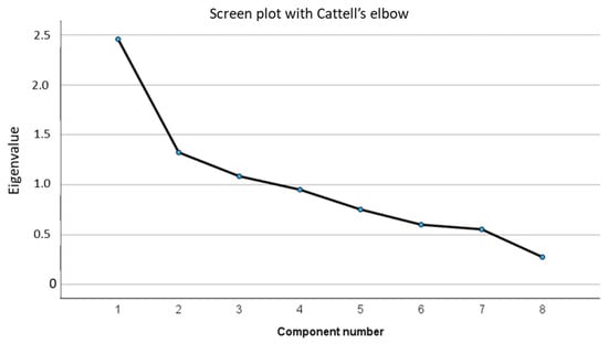
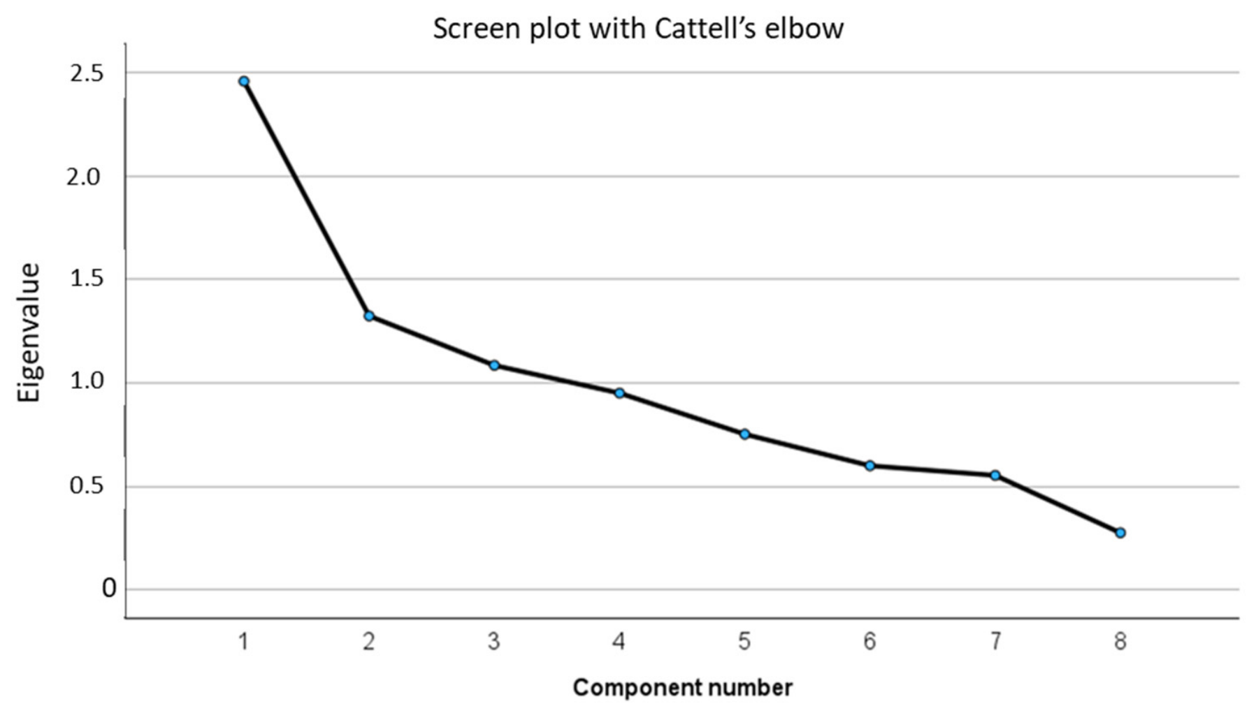
The principal component analysis was possible because the determinant of the correlation matrix was 0.230 (Table 1), the KMO index to measure the sampling quality was >0.6 (of 0.67) and the Bartlett’s test of sphericity was significant (χ2 = 229; p < 0.001). For coding sex, female was 1. The anti-image matrix and the communalities are shown in Table 2 and Table 3. Three principal components have been selected because the eigenvalue was >1. However, these three components only explained 60.8% of the variance from the total variance of the eight potentially associated factors studied (Table 4 and Figure 1). The third component has an eigenvalue of 1.08, only marginally above the KMO threshold, with possible over-extraction of the components.
Table 1.
Correlation matrix of all potentially associated factors.
Table 2.
Anti-image matrix with Kaiser–Meyer–Olkin index (in bold).
Table 3.
Communalities.
Table 4.
Principal components and total variance explanation.
Figure 1.
Components. The Cattell’s elbow is after the third component, which corresponds to an eigenvalue > 1.
After the “Varimax” rotation, the first component was composed of low hamstring strength, knee valgus and female sex, and named “female with dynamical factors”. The second component was composed of male sex, older age and higher BMI, and named “male with physiological factors”. The third component was composed of knee valgus, recurvatum and knee laxity, and named “knee morphology” (Table 5). The component transformation matrix (Table 6) was used to realize the rotation of the factorial axis. The factorial matrix is shown in Table 7 and corresponds to the component-score coefficient matrix. For instance, the component “’female with dynamical factors” is obtained by adding the eight variables multiplied by the corresponding coefficient. Thus, all eight variables were confirmed as factors potentially associated with non-contact ACL injury.
Table 5.
Component matrix after “Varimax” rotation.
Table 6.
Component transformation matrix for axis rotation.
Table 7.
Factorial matrix that defines the 3 profiles of populations with potential association factors for risk of non-contact ACL injuries.
4. Discussion
Multivariate analyses are necessary to explore combinations of factors associated with non-contact ACL injury because the occurrence of this injury is multifactorial. We have chosen to carry out a factorial analysis using principal components to define populations at potential risk of ACL injury. The choice of using a “Varimax” rotation allowed the identification of separate profiles of potentially at-risk populations. Thus, three distinct populations were identified. However, given the methodological approach, no predictions or causal inferences regarding ACL injury could be made, as the analysis only examined combinations of variables to identify a limited number of population profiles potentially at risk. The first population was called “female with dynamical factors” and corresponded to a population at risk of non-contact ACL injury in reference to the literature. Indeed, recently, a sex-specific approach showed that non-contact ACL injury in females was associated with biomechanical differences compared to males [37]. Greater knee abduction angles (knee valgus) and higher adduction moments during landing were linked to an increased risk for ACL injury. Moreover, female sex and dynamic knee valgus are known to be risk factors for ACL injury when these factors are considered alone [22,38]. Knee valgus was also associated with non-contact ACL injury [13,23]. From our analysis, this population could be more at risk when a weak hamstring was also associated. This profile of an at-risk population is very interesting to consider because in clinical practice, dynamic valgus and, particularly, hamstring strength could be modifiable by motor control exercises and muscle strengthening. According to the literature, a training program to correct movement including dynamic valgus by focusing on muscle strength, balance and proprioception could reduce ACL injuries by up to 51% [39]. However, Raffeuddin et al. have reported that the evidence-based injury prevention programs remain uncertain after systematic review [40]. To summarize, the sex-based characteristic differences could support the necessity to develop training and rehabilitation protocols specific to the sexes [41].
The second population was called “male with physiological factors”. Male sex is considered the same risk as female sex during collision, limited contact and non-contact sports practice [36]. In contrast, relative old age > 24 years old was not considered at risk, while younger patients are very vulnerable to ACL injury [16,42]. Bodyweight was identified as an associated factor, but only in women [8,12]. However, Evans et al. have shown a relationship in a male military population [5]. From our analysis, male sex was a potential factor when it was associated with age > 24 years and BMI > 22.5 kg/m2. In clinical practice, losing weight might be interesting for non-contact ACL injury prevention, even though our study cannot prove this because of the method used.
The third population identified was called “knee morphology”. Despite having kept this profile, which is debatable due to the possibility of over-extraction, this population is interesting to consider, although these factors are not modifiable (knee valgus, recurvatum and laxity) except by surgery when ACL reconstruction is performed [43]. Antero-posterior knee laxity is known to be associated with non-contact ACL rupture, particularly in women and in young athletes [8,10,17,44]. This relationship can be extended to a large population whatever the sex or the age, when this parameter is analyzed apart [13]. From a clinical point of view, this last profile of an at-risk population should be followed for several years to know if the ACL injury risk is confirmed in order to help select future high-level athletes, particularly in contact sports [36].
Our study has several limitations. First, the categorical nature of the variables included in the principal component analysis (PCA) may have resulted in information loss or distorted the underlying correlation structure. Second, the exploratory, non-prospective design precludes any conclusions regarding the risk of ACL injury. The inclusion of an uninjured control group might have provided additional insights, since assuming that the contralateral, non-operated limb had the same risk of ACL rupture as the operated limb represents a limitation. Finally, the time elapsed between ACL injury and reconstruction, together with the interruption of sports participation during this period, may have influenced the strength of the contralateral knee. Only adults were studied, so the results cannot be applied to adolescents or children. Knee morphology was not measured using x-rays or MRI, though many osseous morphological risk factors have been identified, including intercondylar notch stenosis, increase tibial slope, and reduced ACL size [27,42,43,45]. Moreover, the dynamic valgus of the knee has not been studied using a kinematic analysis of movement, but only in static position [26]. Despite all these limitations, we have reported morphological parameters that are easily measurable, with cut-offs that are easy to define, that could be useful in clinical practice, and that would allow researchers to prospectively follow different populations potentially at risk of ACL rupture.
5. Conclusions
Three independent athletic adult populations were identified, with combinations of characteristics, that may be at risk of non-contact ACL injuries. Because some of the factors are modifiable, preventive care could be proposed. In women, hamstring strengthening and knee valgus controlling could be interesting to prevent ACL injuries. In men, weight loss could be proposed after 24 years old if the BMI is above 22.5 kg/m2. In the case of knee valgus, recurvatum and laxity association, an athlete selection or a different choice of sport could be advised to avoid the risk of ACL injury. Prospective studies should be conducted to confirm whether these factor combinations really represent a risk of ACL injury.
Author Contributions
Conceptualization, M.D. and A.F.-C.; methodology, M.D.; validation, P.M., N.B. and M.D.; formal analysis, M.D.; investigation, M.D. and A.F.-C.; data curation, M.D.; writing—original draft preparation, M.D. and A.F.-C.; writing—review and editing, P.M. and N.B. All authors have read and agreed to the published version of the manuscript.
Funding
This research received no external funding.
Institutional Review Board Statement
This study was conducted in accordance with the Declaration of Helsinki and approved by the local Ethics Committee of the Sports Medicine department (CNEMS) (Approval No. 2022-006; approved on 19 July 2021).
Informed Consent Statement
Informed consent was obtained from all subjects involved in the study.
Data Availability Statement
The data presented in this study are available on request from the corresponding author due to ethical reasons.
Acknowledgments
The authors thank Annie Chailloux for proofreading.
Conflicts of Interest
The authors declare no conflicts of interest.
References
- Gornitzky, A.L.; Lott, A.; Yellin, J.L.; Fabricant, P.D.; Lawrence, J.T.; Ganley, T.J. Sport-Specific Yearly Risk and Incidence of Anterior Cruciate Ligament Tears in High School Athletes: A Systematic Review and Meta-Analysis. Am. J. Sports Med. 2016, 44, 2716–2723. [Google Scholar] [CrossRef] [Scilit] [PubMed]
- Griffin, L.Y.; Agel, J.; Albohm, M.J.; Arendt, E.A.; Dick, R.W.; Garrett, W.E.; Garrick, J.G.; Hewett, T.E.; Huston, L.; Ireland, M.L.; et al. Noncontact Anterior Cruciate Ligament Injuries: Risk Factors and Prevention Strategies. J. Am. Acad. Orthop. Surg. 2000, 8, 141–150. [Google Scholar] [CrossRef] [Scilit] [PubMed]
- Wetters, N.; Weber, A.E.; Wuerz, T.H.; Schub, D.L.; Mandelbaum, B.R. Mechanism of Injury and Risk Factors for Anterior Cruciate Ligament Injury. Oper. Tech. Sports Med. 2016, 24, 2–6. [Google Scholar] [CrossRef] [Scilit]
- Pfeifer, C.E.; Beattie, P.F.; Sacko, R.S.; Hand, A. Risk Factors Associated with Non-Contact Anterior Cruciate Ligament Injury: A Systematic Review. Int. J. Sports Phys. Ther. 2018, 13, 575–587. [Google Scholar] [CrossRef] [Scilit]
- Evans, K.N.; Kilcoyne, K.G.; Dickens, J.F.; Rue, J.-P.; Giuliani, J.; Gwinn, D.; Wilckens, J.H. Predisposing Risk Factors for Non-Contact ACL Injuries in Military Subjects. Knee Surg. Sports Traumatol. Arthrosc. Off. J. ESSKA 2012, 20, 1554–1559. [Google Scholar] [CrossRef] [Scilit] [PubMed]
- Smith, H.C.; Vacek, P.; Johnson, R.J.; Slauterbeck, J.R.; Hashemi, J.; Shultz, S.; Beynnon, B.D. Risk Factors for Anterior Cruciate Ligament Injury: A Review of the Literature—Part 1: Neuromuscular and Anatomic Risk. Sports Health 2012, 4, 69–78. [Google Scholar] [CrossRef] [Scilit] [PubMed]
- Amraee, D.; Alizadeh, M.H.; Minoonejhad, H.; Razi, M.; Amraee, G.H. Predictor Factors for Lower Extremity Malalignment and Non-Contact Anterior Cruciate Ligament Injuries in Male Athletes. Knee Surg. Sports Traumatol. Arthrosc. Off. J. ESSKA 2017, 25, 1625–1631. [Google Scholar] [CrossRef] [Scilit] [PubMed]
- Uhorchak, J.M.; Scoville, C.R.; Williams, G.N.; Arciero, R.A.; St Pierre, P.; Taylor, D.C. Risk Factors Associated with Noncontact Injury of the Anterior Cruciate Ligament: A Prospective Four-Year Evaluation of 859 West Point Cadets. Am. J. Sports Med. 2003, 31, 831–842, Erratum in Am. J. Sports Med. 2005, 33, 614. [Google Scholar] [CrossRef] [Scilit] [PubMed]
- Hughes, G.; Watkins, J. A Risk-Factor Model for Anterior Cruciate Ligament Injury. Sports Med. 2006, 36, 411–428. [Google Scholar] [CrossRef] [Scilit] [PubMed]
- Myer, G.D.; Ford, K.R.; Paterno, M.V.; Nick, T.G.; Hewett, T.E. The Effects of Generalized Joint Laxity on Risk of Anterior Cruciate Ligament Injury in Young Female Athletes. Am. J. Sports Med. 2008, 36, 1073–1080. [Google Scholar] [CrossRef] [Scilit] [PubMed]
- Beynnon, B.D.; Vacek, P.M.; Newell, M.K.; Tourville, T.W.; Smith, H.C.; Shultz, S.J.; Slauterbeck, J.R.; Johnson, R.J. The Effects of Level of Competition, Sport, and Sex on the Incidence of First-Time Noncontact Anterior Cruciate Ligament Injury. Am. J. Sports Med. 2014, 42, 1806–1812. [Google Scholar] [CrossRef] [Scilit] [PubMed]
- Hägglund, M.; Waldén, M. Risk Factors for Acute Knee Injury in Female Youth Football. Knee Surg. Sports Traumatol. Arthrosc. Off. J. ESSKA 2016, 24, 737–746. [Google Scholar] [CrossRef] [Scilit] [PubMed]
- Dauty, M.; Crenn, V.; Louguet, B.; Grondin, J.; Menu, P.; Fouasson-Chailloux, A. Anatomical and Neuromuscular Factors Associated to Non-Contact Anterior Cruciate Ligament Injury. J. Clin. Med. 2022, 11, 1402. [Google Scholar] [CrossRef] [Scilit] [PubMed]
- Arendt, E.; Dick, R. Knee Injury Patterns among Men and Women in Collegiate Basketball and Soccer. NCAA Data and Review of Literature. Am. J. Sports Med. 1995, 23, 694–701. [Google Scholar] [CrossRef] [Scilit] [PubMed]
- Hewett, T.E.; Myer, G.D.; Ford, K.R.; Paterno, M.V.; Quatman, C.E. Mechanisms, Prediction, and Prevention of ACL Injuries: Cut Risk with Three Sharpened and Validated Tools. J. Orthop. Res. Off. Publ. Orthop. Res. Soc. 2016, 34, 1843–1855. [Google Scholar] [CrossRef] [Scilit] [PubMed]
- Vauhnik, R.; Morrissey, M.C.; Rutherford, O.M.; Turk, Z.; Pilih, I.A.; Pohar, M. Knee Anterior Laxity: A Risk Factor for Traumatic Knee Injury among Sportswomen? Knee Surg. Sports Traumatol. Arthrosc. Off. J. ESSKA 2008, 16, 823–833. [Google Scholar] [CrossRef] [Scilit] [PubMed]
- Woodford-Rogers, B.; Cyphert, L.; Denegar, C.R. Risk Factors for Anterior Cruciate Ligament Injury in High School and College Athletes. J. Athl. Train. 1994, 29, 343–346. [Google Scholar] [PubMed]
- Ramesh, R.; Von Arx, O.; Azzopardi, T.; Schranz, P.J. The Risk of Anterior Cruciate Ligament Rupture with Generalised Joint Laxity. J. Bone Jt. Surg. Br. 2005, 87, 800–803. [Google Scholar] [CrossRef] [Scilit] [PubMed]
- Loudon, J.K.; Jenkins, W.; Loudon, K.L. The Relationship between Static Posture and ACL Injury in Female Athletes. J. Orthop. Sports Phys. Ther. 1996, 24, 91–97. [Google Scholar] [CrossRef] [Scilit] [PubMed]
- Kramer, L.C.; Denegar, C.R.; Buckley, W.E.; Hertel, J. Factors Associated with Anterior Cruciate Ligament Injury: History in Female Athletes. J. Sports Med. Phys. Fit. 2007, 47, 446–454. [Google Scholar]
- Konishi, Y.; Aihara, Y.; Sakai, M.; Ogawa, G.; Fukubayashi, T. Gamma Loop Dysfunction in the Quadriceps Femoris of Patients Who Underwent Anterior Cruciate Ligament Reconstruction Remains Bilaterally. Scand. J. Med. Sci. Sports 2007, 17, 393–399. [Google Scholar] [CrossRef] [Scilit] [PubMed]
- Söderman, K.; Alfredson, H.; Pietilä, T.; Werner, S. Risk Factors for Leg Injuries in Female Soccer Players: A Prospective Investigation during One out-Door Season. Knee Surg. Sports Traumatol. Arthrosc. Off. J. ESSKA 2001, 9, 313–321. [Google Scholar] [CrossRef] [Scilit] [PubMed]
- Hewett, T.E.; Myer, G.D.; Ford, K.R.; Heidt, R.S.; Colosimo, A.J.; McLean, S.G.; van den Bogert, A.J.; Paterno, M.V.; Succop, P. Biomechanical Measures of Neuromuscular Control and Valgus Loading of the Knee Predict Anterior Cruciate Ligament Injury Risk in Female Athletes: A Prospective Study. Am. J. Sports Med. 2005, 33, 492–501. [Google Scholar] [CrossRef] [Scilit] [PubMed]
- Boden, B.P.; Sheehan, F.T.; Torg, J.S.; Hewett, T.E. Noncontact Anterior Cruciate Ligament Injuries: Mechanisms and Risk Factors. J. Am. Acad. Orthop. Surg. 2010, 18, 520–527. [Google Scholar] [CrossRef] [Scilit] [PubMed]
- Alentorn-Geli, E.; Myer, G.D.; Silvers, H.J.; Samitier, G.; Romero, D.; Lázaro-Haro, C.; Cugat, R. Prevention of Non-Contact Anterior Cruciate Ligament Injuries in Soccer Players. Part 1: Mechanisms of Injury and Underlying Risk Factors. Knee Surg. Sports Traumatol. Arthrosc. Off. J. ESSKA 2009, 17, 705–729. [Google Scholar] [CrossRef] [Scilit] [PubMed]
- Pantano, K.J.; White, S.C.; Gilchrist, L.A.; Leddy, J. Differences in Peak Knee Valgus Angles between Individuals with High and Low Q-Angles during a Single Limb Squat. Clin. Biomech. 2005, 20, 966–972. [Google Scholar] [CrossRef] [Scilit] [PubMed]
- Pangaud, C.; Laumonerie, P.; Dagneaux, L.; LiArno, S.; Wellings, P.; Faizan, A.; Sharma, A.; Ollivier, M. Measurement of the Posterior Tibial Slope Depends on Ethnicity, Sex, and Lower Limb Alignment: A Computed Tomography Analysis of 378 Healthy Participants. Orthop. J. Sports Med. 2020, 8, 2325967119895258. [Google Scholar] [CrossRef] [Scilit] [PubMed]
- Jolliffe, I.T.; Cadima, J. Principal Component Analysis: A Review and Recent Developments. Philos. Trans. Ser. A Math. Phys. Eng. Sci. 2016, 374, 20150202. [Google Scholar] [CrossRef] [Scilit] [PubMed]
- Swärd, P.; Kostogiannis, I.; Roos, H. Risk Factors for a Contralateral Anterior Cruciate Ligament Injury. Knee Surg. Sports Traumatol. Arthrosc. Off. J. ESSKA 2010, 18, 277–291. [Google Scholar] [CrossRef] [Scilit] [PubMed]
- Paterno, M.V.; Rauh, M.J.; Schmitt, L.C.; Ford, K.R.; Hewett, T.E. Incidence of Second ACL Injuries 2 Years After Primary ACL Reconstruction and Return to Sport. Am. J. Sports Med. 2014, 42, 1567–1573. [Google Scholar] [CrossRef] [Scilit] [PubMed]
- World Medical Association. World Medical Association Declaration of Helsinki: Ethical Principles for Medical Research Involving Human Subjects. JAMA 2013, 310, 2191–2194. [Google Scholar] [CrossRef] [Scilit] [PubMed]
- Jardin, C.; Chantelot, C.; Migaud, H.; Gougeon, F.; Debroucker, M.J.; Duquennoy, A. Reliability of the KT-1000 arthrometer in measuring anterior laxity of the knee: Comparative analysis with Telos of 48 reconstructions of the anterior cruciate ligament and intra- and interobserver reproducibility. Rev. Chir. Orthop. Reparatrice Appar. Mot. 1999, 85, 698–707. [Google Scholar] [PubMed]
- Robert, H.; Nouveau, S.; Gageot, S.; Gagnière, B. A New Knee Arthrometer, the GNRB: Experience in ACL Complete and Partial Tears. Orthop. Traumatol. Surg. Res. OTSR 2009, 95, 171–176. [Google Scholar] [CrossRef] [Scilit] [PubMed]
- Boyer, P.; Djian, P.; Christel, P.; Paoletti, X.; Degeorges, R. Reliability of the KT-1000 arthrometer (Medmetric) for measuring anterior knee laxity: Comparison with Telos in 147 knees. Rev. Chir. Orthop. Reparatrice Appar. Mot. 2004, 90, 757–764. [Google Scholar] [CrossRef] [Scilit] [PubMed]
- Impellizzeri, F.M.; Bizzini, M.; Rampinini, E.; Cereda, F.; Maffiuletti, N.A. Reliability of Isokinetic Strength Imbalance Ratios Measured Using the Cybex NORM Dynamometer. Clin. Physiol. Funct. Imaging 2008, 28, 113–119. [Google Scholar] [CrossRef] [Scilit] [PubMed]
- Montalvo, A.M.; Schneider, D.K.; Webster, K.E.; Yut, L.; Galloway, M.T.; Heidt, R.S.; Kaeding, C.C.; Kremcheck, T.E.; Magnussen, R.A.; Parikh, S.N.; et al. Anterior Cruciate Ligament Injury Risk in Sport: A Systematic Review and Meta-Analysis of Injury Incidence by Sex and Sport Classification. J. Athl. Train. 2019, 54, 472–482. [Google Scholar] [CrossRef] [Scilit] [PubMed]
- Rodríguez-Molina, P.A.; Barrera, R.; Gonzalez, L.S.; Ortiz, J.G.; Tuta-Quintero, E. Risk Factors Associated with Anterior Cruciate Ligament Injuries in Athletes During Physical Activity According to Sex: A Scoping Review. Sports 2026, 14, 243. [Google Scholar] [CrossRef] [Scilit] [PubMed]
- Meyer, C.A.G.; Gette, P.; Mouton, C.; Seil, R.; Theisen, D. Side-to-Side Asymmetries in Landing Mechanics from a Drop Vertical Jump Test Are Not Related to Asymmetries in Knee Joint Laxity Following Anterior Cruciate Ligament Reconstruction. Knee Surg. Sports Traumatol. Arthrosc. Off. J. ESSKA 2018, 26, 381–390. [Google Scholar] [CrossRef] [Scilit] [PubMed]
- Mehl, J.; Diermeier, T.; Herbst, E.; Imhoff, A.B.; Stoffels, T.; Zantop, T.; Petersen, W.; Achtnich, A. Evidence-Based Concepts for Prevention of Knee and ACL Injuries. 2017 Guidelines of the Ligament Committee of the German Knee Society (DKG). Arch. Orthop. Trauma Surg. 2018, 138, 51–61. [Google Scholar] [CrossRef] [Scilit] [PubMed]
- Rafeeuddin, R.; Sharir, R.; Staes, F.; Dingenen, B.; George, K.; Robinson, M.A.; Vanrenterghem, J. Mapping Current Research Trends on Neuromuscular Risk Factors of Non-Contact ACL Injury. Phys. Ther. Sport 2016, 22, 101–113. [Google Scholar] [CrossRef] [Scilit] [PubMed]
- Deodato, M.; Saponaro, S.; Šimunič, B.; Martini, M.; Murena, L.; Buoite Stella, A. Trunk Muscles’ Characteristics in Adolescent Gymnasts with Low Back Pain: A Pilot Study on the Effects of a Physiotherapy Intervention Including a Postural Reeducation Program. J. Man. Manip. Ther. 2024, 32, 310–324. [Google Scholar] [CrossRef] [Scilit] [PubMed]
- Fernández-Jaén, T.; López-Alcorocho, J.M.; Rodriguez-Iñigo, E.; Castellán, F.; Hernández, J.C.; Guillén-García, P. The Importance of the Intercondylar Notch in Anterior Cruciate Ligament Tears. Orthop. J. Sports Med. 2015, 3, 2325967115597882. [Google Scholar] [CrossRef] [Scilit] [PubMed]
- Bayer, S.; Meredith, S.J.; Wilson, K.W.; de Sa, D.; Pauyo, T.; Byrne, K.; McDonough, C.M.; Musahl, V. Knee Morphological Risk Factors for Anterior Cruciate Ligament Injury: A Systematic Review. J. Bone Jt. Surg. Am. 2020, 102, 703–718, Erratum in J Bone Jt. Surg Am. 2020, 102, e85. [Google Scholar] [CrossRef] [Scilit] [PubMed]
- Vauhnik, R.; Morrissey, M.C.; Rutherford, O.M.; Turk, Z.; Pilih, I.A.; Perme, M.P. Rate and Risk of Anterior Cruciate Ligament Injury among Sportswomen in Slovenia. J. Athl. Train. 2011, 46, 92–98. [Google Scholar] [CrossRef] [Scilit] [PubMed]
- Kızılgöz, V.; Sivrioğlu, A.K.; Ulusoy, G.R.; Aydın, H.; Karayol, S.S.; Menderes, U. Analysis of the Risk Factors for Anterior Cruciate Ligament Injury: An Investigation of Structural Tendencies. Clin. Imaging 2018, 50, 20–30. [Google Scholar] [CrossRef] [Scilit] [PubMed]

Disclaimer/Publisher’s Note: The statements, opinions and data contained in all publications are solely those of the individual author(s) and contributor(s) and not of MDPI and/or the editor(s). MDPI and/or the editor(s) disclaim responsibility for any injury to people or property resulting from any ideas, methods, instructions or products referred to in the content. |
© 2026 by the authors. Licensee MDPI, Basel, Switzerland. This article is an open access article distributed under the terms and conditions of the Creative Commons Attribution (CC BY) license.