


Abstract
Infectious agents shape fish populations by inducing lethal and sublethal changes that alter nutrient metabolism and metal bioaccumulation. These shifts can manifest as changes in the ionome—the specific combination of essential and non-essential chemical elements defining the whole-body composition of an individual. Understanding how pathogens shape the fish ionome is critical for developing advanced monitoring tools and clarifying the ecological roles of hosts and their pathogens. This study reports the first documented outbreak of Citrobacter freundii, Pseudomonas aeruginosa, Pseudomonas mosselii, and Shewanella xiamenensis bacterial infections in wild-caught eastern mosquitofish (Gambusia holbrooki) from three populations in Extremadura, southwestern Spain. Under laboratory-controlled conditions, we established associations between these bacterial outbreaks and the whole-fish body ionome of G. holbrooki. We compared 19 chemical elements and seven elemental ratios among diseased fish, healthy fish at the outbreak, and individuals fully recovered 100 days post-infection following antibiotic treatment. The fish ionome clearly discriminated between diseased and healthy states, and the response was consistent across all three populations. Our findings support the utility of whole-fish body elemental composition in G. holbrooki as a biomarker for environmental monitoring. Furthermore, as the bacterial infections were associated with the capture and transport-induced stress of wild individuals, this study provides critical data on the opportunistic pathogens that may be co-introduced into recipient ecosystems through the release of this widely distributed invasive fish species.
Keywords:
mosquitofish; invasive species; ionomics; trace elements; metals; opportunistic pathogens; ecological monitoring Key Contribution:
We found that G. holbrooki individuals naturally infected with C. freundii, P. aeruginosa, P. mosselii, or S. xiamenensis have a characteristic elemental signature, supporting the use of animal ionomes as biomarkers in ecological and ecotoxicological research.
1. Introduction
Infectious agents shape fish populations, inducing lethal and sublethal changes that alter nutrient metabolism and metal bioaccumulation [1,2]. These alterations can be quantified through biomarkers, which can be defined as any chemical and biological response capable of detecting sublethal effects of environmental stressors on individuals [1,3]. Determining how pathogens alter biomarker responses is critical for advancing disease ecology and refining the use of wild fish as bioindicators in environmental monitoring [2,3]. However, developing diagnostic tools requires establishing clear cause–effect relationships, which is often hindered in wild populations by the co-occurrence of environmental stressors and other confounding factors [3,4]. Consequently, studying wild fish populations under controlled laboratory conditions is essential for the identification of reliable biomarkers [4,5].
Many biomarkers, such as enzymatic and non-enzymatic antioxidants and blood cell profiles, are frequently used in fish studies [4,6,7]. However, a major caveat is their often-limited ecological relevance due to a lack of reference values for many wild species and the poor link between biomarker values and species’ ecological performance in the ecosystem [4,8]. To address these limitations, the analysis of whole-body elemental composition—the ionome—is a promising tool [9,10]. The ionome reflects the systemic homeostasis of essential elements such as zinc (Zn), copper (Cu), iron (Fe), and the accumulation of non-essential toxic metal(oids)s such as mercury (Hg) and arsenic (As) [9,11]. Because all these elements are trophically transferred, infection-induced ionomic shifts can also provide critical insights into prey quality, nutrient cycling, and energy flow within aquatic ecosystems [9,10].
The usefulness of the ionome as a biomarker in disease ecology stems from the redistribution of minerals and trace elements between the host and pathogens [12,13], a phenomenon known as “nutritional immunity”. For example, hosts can mobilize Zn for the synthesis of immune metalloenzymes, and bacterial pathogens often manipulate host mineral and trace element pools, such as sequestering Fe, to facilitate replication [13,14]. Infections also destroy tissues and alter fish osmoregulation [15], which may result in characteristic chemical signatures in hosts through altered ion uptake and excretion. However, our understanding of these shifts in chemical elements remains limited because studies typically measure a few elements in specific tissues [9,16]. Overlooking whole-body changes is a significant gap in fish ecology, as predators consume the entire organism [17], thereby ingesting the “total ionome” of the prey.
In this study, we address this question using the eastern mosquitofish (Gambusia holbrooki Girard, 1859) as a sentinel model, a widely distributed fish species [18]. This species is an ideal candidate due to its small size, extensive use in ecotoxicological and ecological research, and its role as a primary food source for piscivores [4,17,18]. Our study is also novel in that we report the first case of lethal bacterial infections in wild invasive populations of G. holbrooki, a notable occurrence given that invasive populations of this species are typically characterized by wide environmental tolerance and reduced (nearly absent) parasite loads [19,20,21,22]. The aim of this study was to determine whether there are significant associations between the whole-body elemental composition and bacterial infections in G. holbrooki after fish transport under controlled laboratory conditions. Our hypothesis was that infection may significantly alter absolute chemical elemental concentrations and key physiological ratios (e.g., Fe:Zn) [12,14]. By comparing diseased and healthy fish at the outbreak with fish individuals that were fully recovered at 100 days post-infection following antibiotic treatment, we provide whole-body elemental biomarker data in G. holbrooki under variable health status conditions.
2. Materials and Methods
2.1. Host and Infection Origins
In September 2023, a total of 180 female eastern mosquitofish G. holbrooki (average length: 35.7 ± 3.1 mm) were caught using hand nets at three different locations in Extremadura, Spain (60 individuals per location): two agricultural ponds (Talayuela, 276,538.30 E–4,430,798.64 N; Valdeíñigos 256,252.68 E–4,427,890.19 N) and a polluted stream (Malpartida de Plasencia, 754,278.444 E–4,426,764.331 N). These sites are separated by a linear distance of 15–35 km and lack natural connectivity, which likely indicates isolated populations; we will therefore refer to them hereafter as Population 1 (Valdeíñigos), Population 2 (Malpartida de Plasencia), and Population 3 (Talayuela). The fish were transported by car to the aquatic facility of the University of Barcelona (UB) in plastic bags containing oversaturated oxygenated water and housed in isothermal EPS insulated boxes (Lab box®, Barcelona, Spain), following UB ethical standards for transport and fish keeping (License No. #11946). The transport took approximately 12 h for ~1000 km, with a mean velocity of 90–100 km/h. The vehicle was equipped with climate control (air conditioning) to maintain a stable internal temperature (25 °C) throughout the journey.
The three distinct fish populations were kept separate in three individually, fully equipped 500 L tanks, with 60 fish per tank. Fish were maintained in a mixture of 50% dechlorinated tap water and 50% distilled water so that the high conductivity of the tap water from Barcelona was reduced and the fish were kept in water with similar levels to the field (Tables S1 and S2). The aquatic facility was also at 25 ± 1 °C and 12 h light:12 h dark to mirror late summer temperature and daylight duration in their natural habitats (www.chtajo.es, accessed on 20 February 2026). Water quality was maintained through three biological filters and air-pumps (Eheim®, Deizisau, Germany), ensuring proper water oxygenation and safe water quality (Tables S1 and S2). Fish were fed daily with frozen chironomids Petra® (Prague, Czech Republic) and SERA VIPAN® (Heinsberg, Germany) fish flakes. To prevent cross-contamination, each tank utilized separate handling equipment.
During the second week of a planned 4-week acclimatization period for the original use of these fish (EU project INVASOMICS—101024805), an unplanned bacterial outbreak occurred, likely due to handling stress. Individuals from all three populations showed clinical signs of disease compatible with bacterial infection, including hemorrhagic ulcers, eroded fins, and petechiae [23]. Symptoms emerged simultaneously across all tanks by day 10 and the clinical signs of disease were visible in 5% of fish in Populations 1 and 2 and in 20% of the fish from Population 3. There was also an initial mortality of 2 fish in Population 3 on day 10, prompting the current study. Although the onset was concurrent, cumulative mortality at the end of the treatment varied by population, reaching 26.4% in Population 3, 6.4% in Population 2, and 8% in Population 1.
2.2. Experimental Design and Screening Procedure for Pathogens
All three fish populations were sampled at two distinct stages: at the peak of infection, including individuals with (D0) and without (H0) clinical signs of disease, and at 100 days post-antibiotic treatment (H100). Tetracycline was added to the tanks (including D0 and H0 fishes) three days after the detection of infection at 100 mg/L for 10 days [23]. The fish were treated again on the third day after a 50% water change and the sequence was repeated until all fish were fully recovered. The tanks had diffuse light to reduce tetracycline photoinactivation, as recommended by Noga [23]. After the treatment was successful and no clinical signs of disease were detected, antibiotic residuals were removed by a 50% water exchange and the addition of activated charcoal to the filtration system [23]. To maintain a balanced experimental design and meet the sample size of the original project requirements, four size-matched females (average length: 32 ± 1 mm and wet weight: 0.4 ± 0.1 g) were sampled per tank for each group (D0, H0, and H100), resulting in 12 diseased fish (D0; 4 per stock tank), 12 healthy fish sampled during the outbreak (H0), and 12 healthy fish sampled 100 days post-treatment (H100). Healthy fish were identified based on their normal behavior and the lack of overt external signs of disease. The health status was confirmed during their dissection, as detailed below. We focused on females because G. holbrooki populations are strongly female-biased in summer [24].
Fish were euthanized with an overdose (250 mg/L) of buffered tricaine methane sulfonate (MS-222®, Sigma-Aldrich, Ferndale, WA, USA) [23,25]. All fish were processed for the analysis of bacteria, other parasites, and whole-fish body elemental composition. Systematic necropsy was conducted in sterile Petri dishes for bacteria and other parasites, as described below [20,26,27]. Firstly, external surfaces were inspected for parasites using a dissecting microscope with fiber-optic illumination (NexiusZoom 1903P, Euromex®, Duiven, The Netherlands). Plastic pipette tips were employed to manipulate the fish and thoroughly inspect all surfaces. Secondly, the body cavity was opened with a sterile scalpel, and a sterile swab was used to sample renal tissue, the liver, and the surface of all internal organs, except the gastrointestinal tract to prevent fecal contamination. To identify the bacterial pathogens, swab samples from all analyzed fish were sent in sterile 10 mL tubes with tryptic soya broth (TSB, Scharlab S.L., Barcelona, Spain) to the coauthor YS at the University of Santiago de Compostela, Galicia, Spain. Samples of broth cultures were seeded on plates of Brain Heart Infusion (BHI) agar and Trypticase Soy Agar (TSA) (Scharlab S.L., Barcelona, Spain) with 0.5% (w/v) NaCl (TSA-1 and BHI-1) and incubated for 24 to 72 h at 25 ± 1 °C. Bacterial cultures recovered from fish tissues were transferred onto TSA-1 and incubated at 25 ± 1 °C for 24 h for further characterization. Pure cultures of the isolates were stored in commercial Microbank storage medium (Pro-Lab Diagnostics, Richmond Hill, ON, Canada) at −30 °C until use. Initial characterization of the bacterial colony forming units was carried out using light microscopy for analyzing cellular morphology, motility, and Gram staining alongside the oxidase and Oxidative/Fermentative (O/F) test, following the guidelines of Austin and Austin [28]. Oxidase test sticks (Oxidase reagent, BioMerieux, Lyon, France) were used to determine oxidase activity.
Bacterial identification was confirmed using matrix-assisted laser desorption/ionization time-of-flight mass spectrometry (MALDI-TOF MS) [29]. The spectra were imported into BioTyper software (V. 3.1, Bruker Daltonics, Bremen, Germany) and analyzed by matching against standard patterns for bacterial identification, and the results were expressed using the criteria proposed by the manufacturer, e.g., [29,30,31]. The analysis had sterilized water as the negative control and Aeromonas jandaei CECT 4228 as the positive control. Finally, all internal organs (gut, liver, swim bladder, gonads, other organs, and tissues) were all individually screened for parasites. Muscle and skin were also crushed between two glass slides and examined by transparency.
2.3. Chemical Element Analysis
Whole-fish elemental composition was determined for all individuals subjected to necropsy (n = 36): 12 diseased fish (D0), 12 clinically healthy fish sampled during the outbreak (H0), and 12 healthy fish sampled 100 days post-treatment (H100). Individual fish were acid-digested HNO3:H2O2 at 90 °C for 12 h using Instra-quality reagents (J. T. Baker®, Phillipsburg, NJ, USA). Elemental concentrations were determined using a Perkin-Elmer (Shelton, CT, USA) OPTIMA-3200RL Inductively Coupled Plasma Optical Spectrometer (ICP-OES) and a Perkin-Elmer ELAN-6000 Inductively Coupled Plasma Mass Spectrometer (ICP-MS), both at the University of Barcelona (UB) Technical Scientific Services, where analytical accuracy was ensured through calibration curves, blanks, and certified tissue standards (http://www.ccit.ub.edu/EN/tq01.html, accessed on 20 February 2026), e.g., [32,33].
We analyzed 13 essential elements involved in physiological processes and tissue structure: calcium (Ca), cobalt (Co), chromium (Cr), copper (Cu), iron (Fe), potassium (K), magnesium (Mg), molybdenum (Mo), sodium (Na), phosphorus (P), sulfur (S), selenium (Se), and zinc (Zn). Additionally, we measured non-essential elements and potential pollutants, including arsenic (As), mercury (Hg), lithium (Li), and lead (Pb), as well as boron (B) [34,35]. Certain essential elements (e.g., Cu, Zn) were also monitored as potential environmental pollutants [34,35].
To further explore physiological shifts associated with health status, we also calculated the following chemical elemental ratios: Ca:P, Na:K, Zn:Cu, Se:Hg, Na:Mg, Ca:K, and Fe:Cu. While the fish-specific literature for some of these ratios is emerging, their clinical relevance in metabolic regulation, stress response, and toxicology is well-documented in other vertebrates [36,37,38,39,40,41]. For example, Fe:Cu and Cu:Zn (or Zn:Cu) ratios have been associated with infection susceptibility and immune alterations [37,38]. The Na:K ratio is regarded as a potential indicator of osmotic and immune-related responses [40]. The Se:Hg ratio is widely used to assess the antagonistic protective effect of selenium against mercury toxicity [42].
2.4. Statistical Analysis
All analyses were performed in the R software v. 4.5.1 [43] using the libraries vegan [44], nlme [45], and car [46]. To visualize primary patterns of variation in the 19 chemical elements and seven elemental ratios, a partial principal component analysis (pPCA) was conducted using the rda function. Tank identity was included as a “partial” effect to account for the nested structure of repeated samplings. The varimax rotation was applied to facilitate the interpretation of PC axes. Chemical elements or their ratios with loadings > |0.70| were identified as the primary drivers of the PCA ordination. To compare their values among fish groups (D0, H0, and H100), linear mixed models (LMMs) were implemented using the lme function. A separate model was constructed for the scores of each PC and the selected individual chemical elements or their ratios, with tank identity included as a random effect.
Statistical significance of fish group effects was determined using the R function Anova. Significant results were followed by pairwise post hoc comparisons at p ≤ 0.05 using the R function emmeans [47]. Model assumptions were validated through visual inspection of residual diagnostic plots following the protocols of Zuur et al. [48].
3. Results
The preliminary characterization of the bacterial colony forming units isolated from fish revealed that all were Gram-negative rod-shaped bacteria. Isolates from Population 1 were oxidase-negative and fermentative, while bacterial isolates from Populations 2 and 3 included oxidative-positive bacteria with oxidative (Population 2) or fermentative and oxidative (Population 3) metabolism.
The analysis of the proteomic profiles obtained by MALDI-TOF MS identified bacterial pathogens characteristic of each fish population isolated in a separate tank. The outbreak in Population 1 was due to Citrobacter freundii (score value = 2.39 ± 0.026), in Population 2 it was due to Pseudomonas aeruginosa (score value = 2.26 ± 0.026) and P. mosselii (score value = 1.91 ± 0.023), and in Population 3 it was due to Shewanella xiamenensis (score value = 2.24 ± 0.131) and P. aeruginosa (score value = 2.10 ± 0.071). The strain used as a positive control for MALDI assays was correctly identified as A. jandaei CECT 4228 (score value = 1.90 ± 0.042). Since fish from each tank were processed in pools, it could not be determined if individual fish were co-infected. The bacteria-induced shifts in whole-fish elemental composition were not confounded by the presence of other parasites; only two colonies of the protozoan Epistylis sp. were observed on the caudal fins of a single G. holbrooki individual from Population 1.
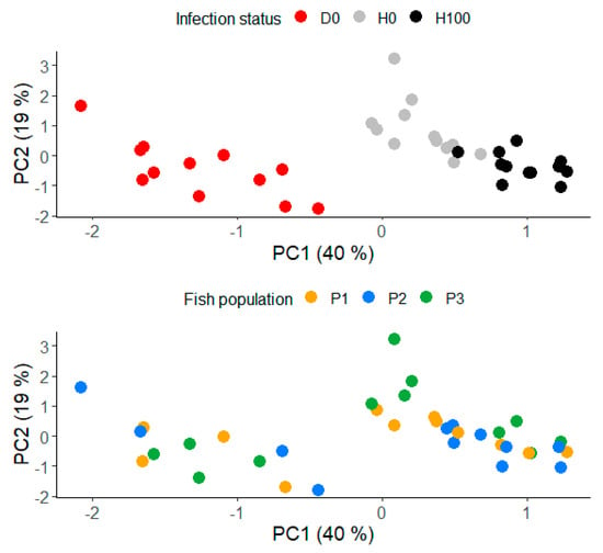
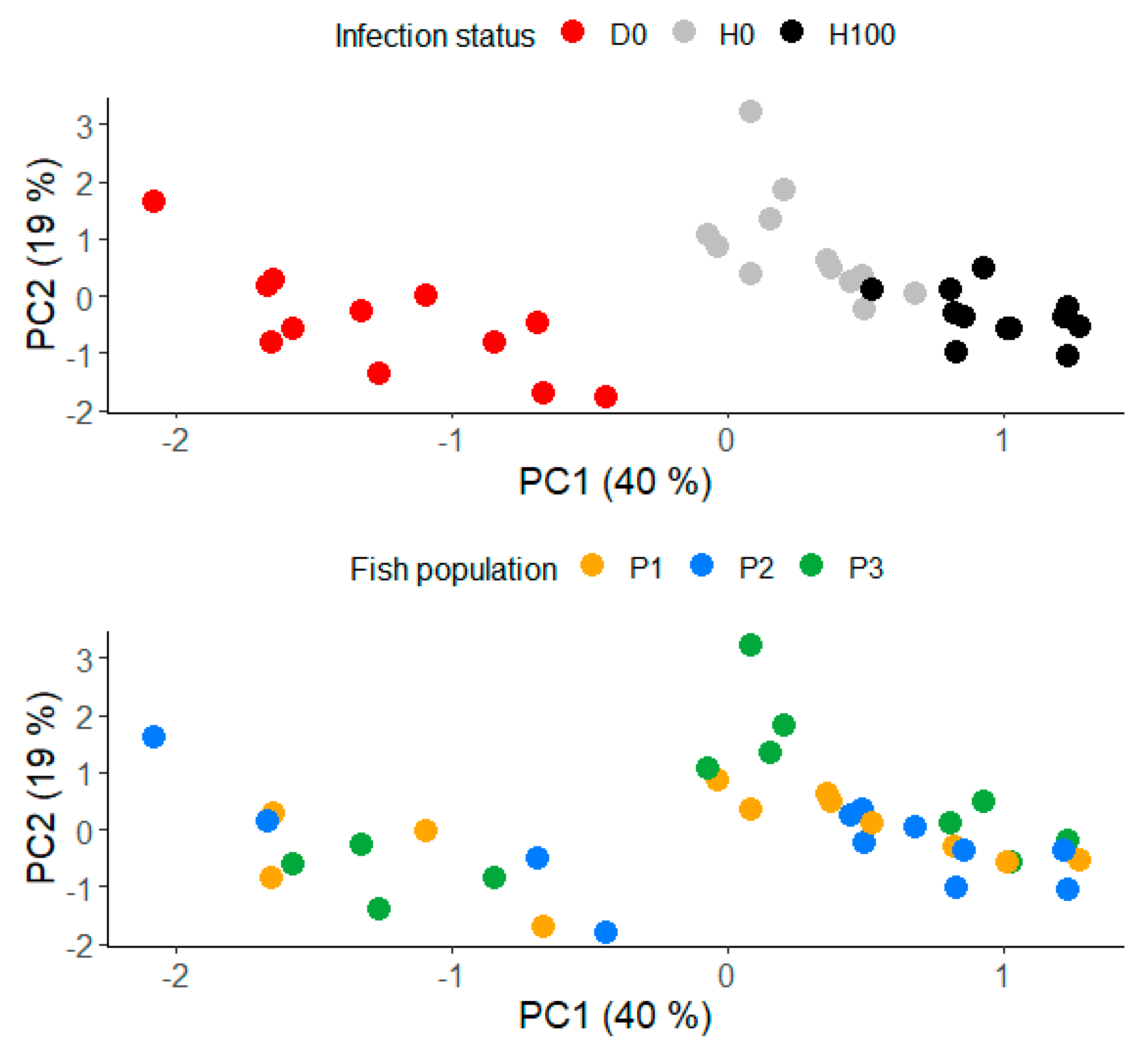
Given that absolute concentrations of chemical elements or their ratios were all intercorrelated to some extent, PCA effectively summarized their variation into two principal components (Figure 1). PC1 explained 40% of the variance and represented the best indicator of infection status (F2,33 = 128; p < 0.001) (Figure 1). Along this axis, diseased fish D0 were clearly distinct from both groups of healthy fish H0 and H100 (p < 0.01). PC1 loaded positively on Cu, Se, As, K, S, and the Na:Mg and Se:Hg ratios and negatively on Ca, Ca:P, Zn:Cu, and Ca:K (Table 1). PC2 explained 19% of the variance and was primarily defined by Mg, Fe, and P. While there were overall significant differences regarding fish infection status (F2,33 = 9.85; p < 0.001), PC2 did not differentiate diseased fish D0 from the healthy fish group H100 (p = 0.92) (Figure 1).
Figure 1.
Principal component analysis ordination based on the whole-body chemical element concentrations in Gambusia holbrooki. The top plot shows differences in PC1 and PC2 scores among diseased (D0) and healthy (H0) fish during the bacterial outbreak, and healthy fish at 100 days post-outbreak (H100) to obtain baseline data. The bottom plot illustrates that the differences between healthy and diseased fish in PC1 and PC2 scores are not obscured by the effect of different fish populations (P1, P2, and P3). To assist in interpreting the PCA biplots, the strength and direction of associations of chemical elements with each PC axis are detailed in Table 1.
Table 1.
Loadings of a principal component analysis (PCA) built based on the whole-body chemical element concentrations in Gambusia holbrooki. The elements or ratios that made the largest contribution (≥0.74) to the variation explained by PC1 and PC2 are highlighted in bold.
Separate linear mixed models confirmed the individual association of chemical elements with infection status, supporting all variables highlighted by the PCA except P (Table 2). At the time of the outbreak, healthy fish (H0) exhibited 1.2- to 2.3-fold higher concentrations of the elements Cu, Fe, Se, As, Mg, K, and S compared to diseased individuals (D0). These differences widened to 1.9–3.4-fold for all elements except Mg and Fe when compared to the healthy fish group at 100 days (H100) (Table 3). Conversely, diseased fish D0 were characterized by 1.3-fold higher levels of Ca than the healthy group at the time of infection outbreak (H0). Regarding elements with less influence on the PCA ordination, diseased fish D0 were significantly depleted in Na (χ2 = 352, df = 2, p < 0.001), Cr (χ2 = 12.4, df = 2, p = 0.002), and Zn (χ2 = 10.5, df = 2, p = 0.005), whereas they were enriched in B (χ2 = 30.8, df = 2, p < 0.001) (Table 3).
Table 2.
Results of linear mixed models comparing whole-body elemental concentrations and elemental ratios among fish groups D0, H0, and H100. The standard deviations for the random term population identity are also shown.
Table 3.
Whole-body chemical element concentrations and ratios (mean, standard deviation, minimum and maximum) in Gambusia holbrooki during a bacterial infection outbreak (diseased: D0, healthy: H0) and at 100 days post-antibiotic therapy and fish full recovery (H100). The table shows in the upper side the elements with |loadings| > 0.75 in the PCA and the rest of the elements in the bottom side.
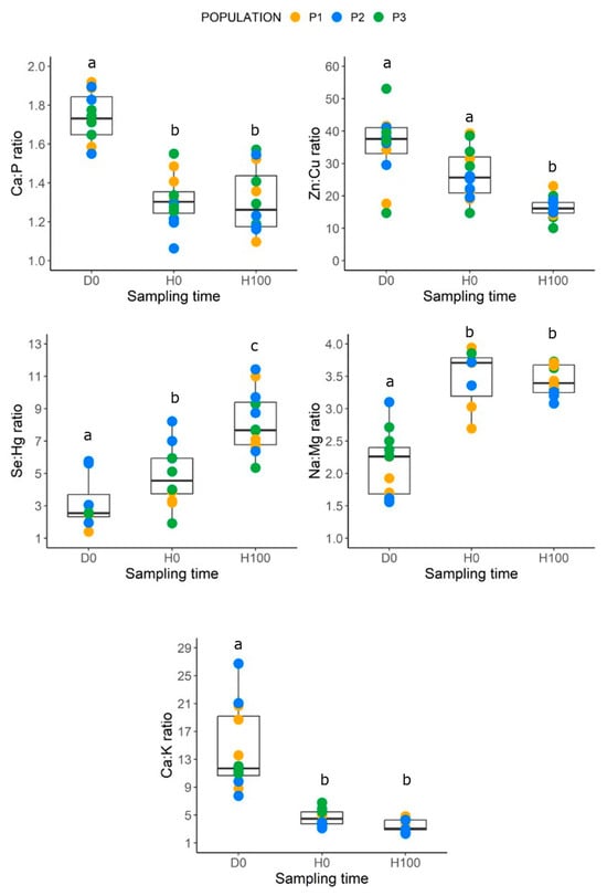
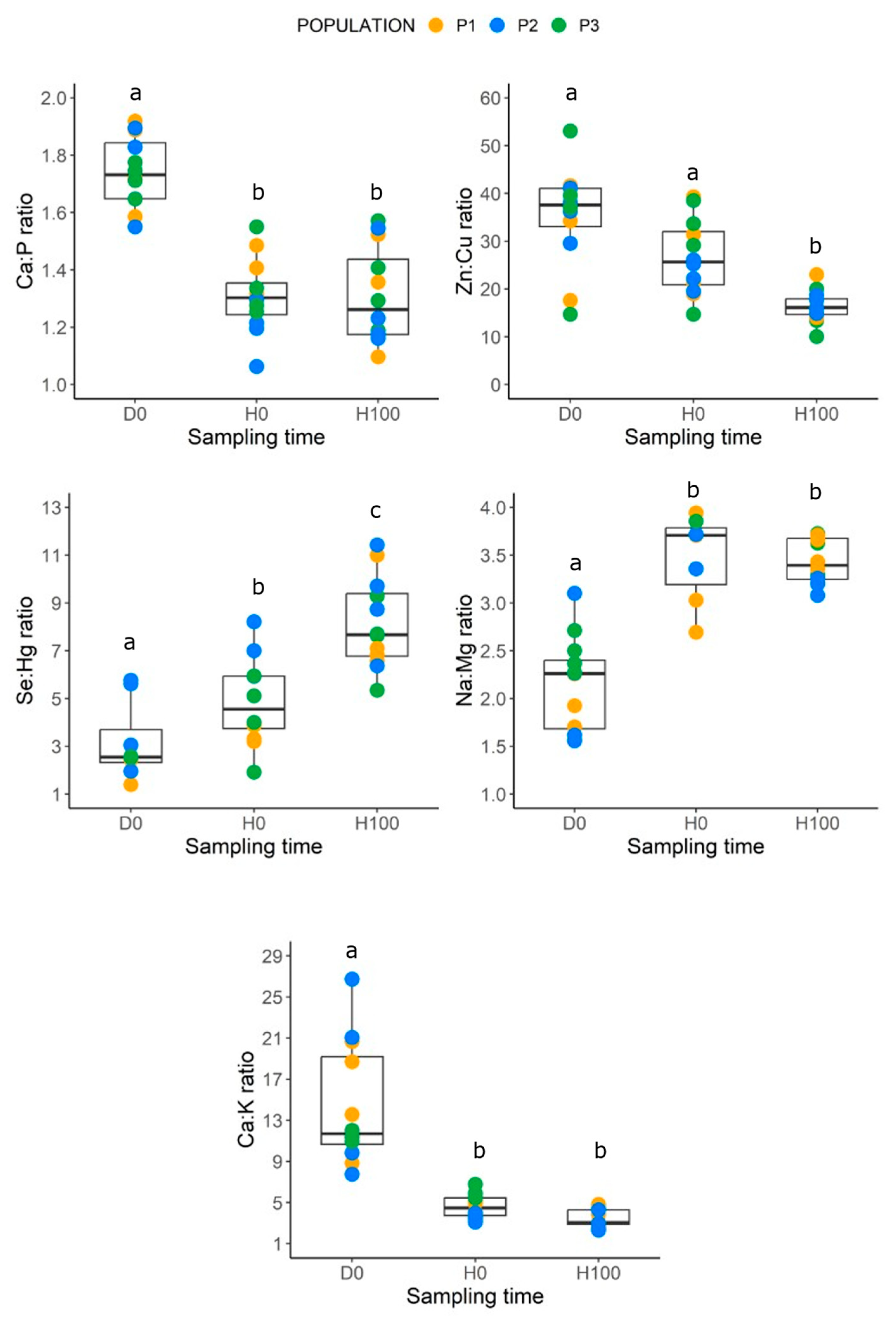
As with absolute elemental levels, the elemental ratios highlighted by the PCA also effectively discriminated between healthy and diseased fish (Table 2). Specifically, the ratios of Ca:P and Ca:K were significantly higher in diseased fish than in healthy fish at the time of infection, with values remaining stable in healthy fish until 100 days (H100) (Figure 2). In contrast, the Na:Mg ratio was markedly lower in the diseased group (Figure 2). Temporal shifts in healthy fish were most evident in the Se:Hg ratio, which increased steadily between H0 and H100 relative to the diseased group D0 (Figure 2). While the Zn:Cu ratio tended to be higher in diseased fish, it reached statistical significance only when compared against the healthy group at 100 days (H100) (Figure 2). Regarding the other two ratios less influential to PCA ordination, diseased and healthy fish significantly differed in both the Na:K ratio (χ2 = 69.2, df = 2, p < 0.001) and the Fe:Cu ratio (χ2 = 72.6, df = 2, p < 0.001). However, these differences were not seen at the time of the outbreak (D0 vs. H0) but emerged at 100 days (D0 vs. H100) (all p < 0.01; Table S3).
Figure 2.
Boxplots of chemical elemental ratios (from Table 1) in diseased and healthy fish during the bacterial outbreak (D0 and H0, respectively) and in healthy fish at 100 days (H100) after full recovery to obtain baseline data in controlled laboratory conditions. The line inside the box indicates the median, the box itself shows the interquartile range IQR, and the whiskers the minimum and maximum data points. Dots represent individual observations, categorized by fish population (P1, P2, and P3). Different letters show significant differences among groups at p ≤ 0.05.
4. Discussion
The present study reports for the first time an outbreak of C. freundii, P. aeruginosa, P. mosselii, and S. xiamenensis infections in wild-caught G. holbrooki. The clinical manifestations observed—including erythema, skin ulcers, and lethargy—align with the symptomatic profiles of these pathogens in other fish species [23,28,49]. Notably, the highest mortality was recorded in fish Population 3 (26.4%), where fish hosted potentially both S. xiamenensis and P. aeruginosa, suggesting that this specific pathogen combination or the presence of S. xiamenensis alone may pose a greater threat to G. holbrooki, as reported for other fish species [50]. Despite this, the success of the tetracycline treatment is consistent with the known susceptibility of these bacteria to this broad-spectrum antibiotic, which inhibits protein synthesis [23,28]. In natural settings, moribund fish are typically removed rapidly by water currents or through predation; however, our laboratory observations provided a unique opportunity to quantify associations between bacterial infections and the whole-fish body elemental composition or “ionome” sensu Filipiak & Filipiak [9]. Capture and transport-induced stress probably catalyzed the bacterial infection outbreak in G. holbrooki, as overcrowding and the accumulation of metabolic waste in transport bags are well-established triggers for opportunistic pathogen replication in fish [51,52,53].
The variation in the elemental composition of the whole fish body was strongly associated with the presence of clinical signs at the time of infection (groups D0 and H0) and the subsequent 100-day recovery following antibiotic treatment. It is essential to highlight that the H100 fish group represents a physiological state influenced not only by the absence of infection but also by fish in captivity and the potential legacy effects of antibiotic treatment. Specifically, tetracycline, while effective in halting the outbreak, can affect the fish gut microbiota [54], with potential ramifications for nutrient assimilation and the overall fish physiology [55,56]. Interestingly, a low overall random effect variance for the term fish tank in the models (mean ± SD = 0.017 ± 0.036 in elements and 0.046 ± 0.082 in ratios) suggests a consistent pattern of elemental shifts across the three G. holbrooki populations. It is important to note that we confirmed that elemental shifts were not masked by other parasites; this is an aspect that is often overlooked in ecotoxicological studies, even though it is known to skew physiological data [1,2]. The negligible presence of Epistylis spp. on one fish fin was likely harmless, as these commensal ciliates only compromise fish health at much higher densities [23]. The elemental concentrations recorded here were consistent with previous data on metal bioaccumulation in G. holbrooki, even when studies focused on specific organs such as the liver [34,35]. In this regard, our whole-fish body measurements offer greater ecological relevance, as typical predators of G. holbrooki, including the Eurasian kingfisher Alcedo atthis, the squacco heron Ardeola ralloides, and the Eurasian bitten Botaurus stellaris, among many others, ingest the entire fish [17,57]. All elements regulated by EU legislation for fish consumption remained below safe limits (e.g., Pb = 0.3 mg/kg; Hg = 0.5 mg/kg; As = 0.1 mg/kg) [58,59].
The significant depletion of Cu, Se, Mg, and K in diseased fish likely reflects a disruption of physiological homeostasis, as the individuals sampled during the infection outbreak were moribund. The markedly higher levels of Cu and Se in healthy fish support their roles as essential cofactors for antioxidant enzymes, such as superoxide dismutase (Cu/Zn-SOD) and glutathione peroxidase (Se/GPx) [60]. Reactive oxygen species are typically produced during inflammatory and infection processes [60]; when the host’s capacity to mitigate this oxidative stress is overwhelmed, widespread tissue damage and the loss of cellular homeostasis can occur [4]. Damage to internal tissues has also been associated with a significant reduction in Mg and K levels in diseased individuals due to passive efflux or alterations in Mg2+-dependent and Na/K-dependent ATPases [39,40,41]. Furthermore, this tissue damage may be linked to the lower levels of arsenic (As), a non-essential metalloid, observed in diseased fish; this might suggest a functional failure of the tissues involved in its bioaccumulation, such as the gills and kidneys [35].
We acknowledge that, while it is tempting to view all these elemental shifts as a direct result of infection, the present study design can only support strong associations but not direct cause–effect relationships. While infection can impair mineral uptake through reduced feeding and compromised gastrointestinal function in many animals, including fish [4,61], it is also plausible that the fish had lower baseline levels of essential elements prior to the outbreak due to differences in microhabitat quality, which in turn may have increased their susceptibility to opportunistic pathogens [11,12,13,14]. As this outbreak was unexpected, no baseline profiles were established upon arrival at the laboratory; however, given that the outbreak occurred immediately after arrival, it is possible that such profiles would also have reflected a poorer physical condition of the fish in the field or differences between individuals within the population. There may be differences in tolerance to changes in environmental conditions among individuals of the same population due to behavioral hierarchies, given that fish were in the breeding season [62], all of which can influence food access and body condition [61] and hence potentially the fish ionome. In this context, the depletion of Cu, Se, K, and S in diseased fish might indicate a pre-existing deficit in sulfur-containing amino acids and impaired protein metabolism [16,36]. Physiological impairments are expected in the field, as the sampled individuals were collected from sites surrounded by an extensive agricultural area, where the authors (OCR and AMV) saw the repeated use of pesticides and other chemicals. Nevertheless, the consistent changes found in elemental composition associated with infection across all populations suggest that differences in diet or in environmental conditions while the fish were in the field may have played a secondary role in the patterns we observed in captivity.
Beyond individual elements, our study supports the utility of elemental ratios as biomarkers of health status in G. holbrooki. The significantly higher Ca:P and Ca:K ratios in diseased fish were mainly associated with a peak in Ca concentration, as P levels remained stable across groups. Given the high mineral content of the water in the aquatic facility (Ca = 122 ± 1 mg/L), this increase was probably associated with an alteration in gill ion exchange caused by infection, as the gills are the main site of calcium entry and exit [41]. In part, the influx and efflux of Ca and other elements, such as B, were probably related to the water stock tanks having greater values of Ca and B than very soft waters at the fish collection sites in Extremadura (~10-fold). The observed Zn:Cu trends are concordant with the copper depletion often seen during active infections in mammals [12,38], suggesting a potential conserved physiological pattern. Conversely, the PCA outputs suggest that the Na:Mg and Ca:K ratios provided limited additional insight compared to individual elemental levels in this specific context, although these ratios have been associated with hormonal disorders in mammals [63]. Finally, the steady increase in the Se:Hg ratio over time in healthy fish (H0 and H100) may reflect the cumulative benefits of a balanced laboratory diet. While Se levels are known to provide protective antagonism against Hg toxicity [42], the very low Hg concentrations detected suggest that this shift was primarily associated with improved Se assimilation rather than a response to toxic stress in the studied G. holbrooki individuals.
5. Conclusions
Our study shows that G. holbrooki individuals naturally infected with C. freundii, P. aeruginosa, P. mosselii, or S. xiamenensis exhibit a characteristic elemental signature, supporting the use of animal ionomes as biomarkers in ecological and ecotoxicological research [9,10,11]. While the elemental composition observed after 100 days in the aquatic facility provides a tentative baseline for the 19 chemical elements and seven elemental ratios analyzed, we caution that these values may be influenced by the antibiotic treatment and the environmental conditions the fish experienced during acclimatization from the field to the laboratory, including changes in water chemistry and diet. Beyond physiological relevance, our findings underscore potentially biosecurity risks associated with the transport and release of G. holbrooki, a highly successful invasive species globally [18]. Future research should determine whether these ionomic signatures are pathogen-specific or represent a generalized stress response of G. holbrooki to infection and shifts in environmental conditions, which would further refine the use of elemental profiling as a biomarker in environmental monitoring.
Supplementary Materials
The following supporting information can be downloaded at https://www.mdpi.com/article/10.3390/fishes11050257/s1, Table S1. Physicochemical characteristics of the water at the three fish collection points. Data represent the range of minimum–maximum values for all variables. The “Historic NO3− + NO2− (mg N/L)” column (green) is derived from the RECAREX and Confederación Hidrográfica del Tajo (www.chtajo.es) monitoring programs. For the Talayuela site, where historical records were unavailable, the maximum NO3− + NO2− values from the current study’s four samplings were utilized as a reference. Table S2. Essential and non-essential elements measured in water from the three fish collection sites and from the three stocking tanks. Data represent the range of minimum–maximum values for all variables. Elements are classified based on the periodic table: non-metals in green, metalloids in yellow, alkali and alkaline earth metals in light gray, and metals in dark gray. Table S3. Whole-body chemical element ratios (mean, standard deviation, minimum, and maximum) in Gambusia holbrooki during a bacterial infection outbreak (diseased: D0, healthy: H0) and at 100 days after antibiotic treatment and fish full recovery (H100). The table reports the exploratory statistics of the five significant element ratios in Figure 2 and the remaining two ratios for completeness, despite the fact that these were not highlighted in the PCA.
Author Contributions
Conceptualization, O.C.-R., Y.S. and A.M.-V.; methodology, O.C.-R., Y.S., R.M.A., G.W. and A.M.-V.; software, O.C.-R. and Y.S.; validation, O.C.-R., Y.S., R.M.A., H.S., G.W., M.P., M.T.-D., A.R. and A.M.-V.; formal analysis, O.C.-R., Y.S. and A.M.-V.; investigation, O.C.-R., Y.S., H.S., A.R. and A.M.-V.; resources, O.C.-R., Y.S., R.M.A., H.S. and A.M.-V.; data curation, O.C.-R., Y.S. and A.M.-V.; writing—original draft, O.C.-R. and A.M.-V.; writing—review and editing, O.C.-R., Y.S., R.M.A., H.S., G.W., M.P., M.T.-D., A.R. and A.M.-V.; visualization, A.M.-V.; supervision, A.M.-V.; project administration, O.C.-R. and A.M.-V.; funding acquisition, O.C.-R. and A.M.-V. All authors have read and agreed to the published version of the manuscript.
Funding
Fieldwork was funded by European Union’s Horizon 2020 programme under the Marie Skłodowska-Curie (grant No. 101024805), and the analyses were funded by remnants of the contract 2023-CE137 awarded to YS and “Contracte Fauna” awarded to AMV by Departament de Territori, Habitatge i Transició Ecològica-Generalitat de Catalunya.
Institutional Review Board Statement
Fish keeping and experimentation were conducted by qualified personnel, and the experimental protocol was approved by the Ethical Committee for Animal Experimentation at the University of Barcelona (code number #11946, 14 December 2022). Fish capture and transport was authorized by regional governments under the Acts RCP006/2023 (13 February 2023) and CN0055/21/ACA (14 March 2023).
Data Availability Statement
The data presented in this study are available on request from the corresponding author.
Acknowledgments
We are grateful to four anonymous referees and the editor for their constructive review and the time spent handling this manuscript. We also thank Martina Griful, Marc García-Bargalló, Júlia Fulleda, and the team at the CCiTUB (Maite Romero and Bárbara Baena) for laboratory assistance in metal quantification and Manel Soler and Sergio Pacho for assistance in field work.
Conflicts of Interest
The authors declare no conflicts of interest.
References
- Grabner, D.M.; Rothe, L.E.; Sures, B. Parasites and pollutants: Effects of multiple stressors on aquatic organisms. Environ. Toxicol. Chem. 2023, 42, 1946–1959. [Google Scholar] [CrossRef] [Scilit]
- Timi, J.T.; Poulin, R. Why ignoring parasites in fish ecology is a mistake. Int. J. Parasitol. 2020, 50, 755–761. [Google Scholar] [CrossRef] [Scilit]
- Van der Oost, R.; Beyer, J.; Vermeulen, N.P. Fish bioaccumulation and biomarkers in environmental risk assessment: A review. Environ. Toxicol. Pharmacol. 2003, 13, 57–149. [Google Scholar] [CrossRef] [Scilit] [PubMed]
- Colin, N.; Porte, C.; Fernandes, D.; Barata, C.; Padrós, F.; Carrassón, M.; Monroy, M.; Cano-Rocabayera, O.; de Sostoa, A.; Piña, B.; et al. Ecological relevance of biomarkers in monitoring studies of macro-invertebrates and fish in Mediterranean rivers. Sci. Total Environ. 2016, 540, 307–323. [Google Scholar] [CrossRef] [Scilit] [PubMed]
- La Point, T.W.; Fairchild, J.F.; Little, E.E.; Finger, S.E. Laboratory and field techniques in ecotoxicological research: Strengths and limitations. In Aquatic Ecotoxicology; CRC Press: Boca Raton, FL, USA, 2018; pp. 239–256. [Google Scholar]
- Brosset, P.; Cooke, S.J.; Schull, Q.; Trenkel, V.M.; Soudant, P.; Lebigre, C. Physiological biomarkers and fisheries management. Rev. Fish. Biol. Fish. 2021, 31, 797–819. [Google Scholar] [CrossRef] [Scilit]
- Rossi, A.S.; Bacchetta, C.; Mora, C.; Michlig, M.P.; Repetti, M.R.; Cazenave, J. Can digestive biomarkers detect early effects of bifenthrin exposure in native fish? Environ. Toxicol. Pharmacol. 2025, 17, 104751. [Google Scholar] [CrossRef] [Scilit]
- Forbes, V.E.; Palmqvist, A.; Bach, L. The use and misuse of biomarkers in ecotoxicology. Environ. Toxicol. Chem. 2006, 25, 272–280. [Google Scholar] [CrossRef] [Scilit] [PubMed]
- Filipiak, M.; Filipiak, Z.M. Application of ionomics and ecological stoichiometry in conservation biology: Nutrient demand and supply in a changing environment. Biol. Conserv. 2022, 272, 109622. [Google Scholar] [CrossRef] [Scilit]
- Kaspari, M. The invisible hand of the periodic table: How micronutrients shape ecology. Annu. Rev. Ecol. Evol. Syst. 2021, 52, 199–219. [Google Scholar] [CrossRef] [Scilit]
- Braga, C.P.; Adamec, J.; de Magalhães Padilha, P. Metallomics in Fish. In Metallomics: The Science of Biometals; Springer: Cham, Switzerland, 2018; pp. 101–110. [Google Scholar]
- Hood, M.I.; Skaar, E.P. Nutritional immunity: Transition metals at the pathogen–host interface. Nat. Rev. Microbiol. 2012, 10, 525–537. [Google Scholar] [CrossRef] [Scilit]
- Bagheri, S.; Gholamhosseini, A.; Hoseinifar, S.H.; Banaee, M. Investigation of the effects of heavy metals (copper, cobalt, manganese, selenium, and zinc) on fish immune systems—An overview. Ann. Anim. Sci. 2024, 24, 1025–1035. [Google Scholar] [CrossRef] [Scilit]
- Palmer, L.D.; Skaar, E.P. Transition metals and virulence in bacteria. Annu. Rev. Genet. 2016, 50, 67–91. [Google Scholar] [CrossRef] [Scilit] [PubMed]
- Senthamarai, M.D.; Rajan, M.R.; Bharathi, P.V. Current risks of microbial infections in fish and their prevention methods: A review. Microb. Pathog. 2023, 185, 106400. [Google Scholar] [CrossRef] [Scilit]
- Lall, S.P.; Kaushik, S.J. Nutrition and metabolism of minerals in fish. Animals 2021, 11, 2711. [Google Scholar] [CrossRef] [Scilit]
- García, J.A.; Hernández, M.Á.; Nunes, L.; Sánchez-Sastre, L.F.; Casanueva, P.; Campos, F. Predation by the Common Kingfisher Alcedo atthis of non-native fish species during the winter. Bird. Study 2022, 69, 97–102. [Google Scholar] [CrossRef] [Scilit]
- Jourdan, J.; Riesch, R.; Cunze, S. Off to new shores: Climate niche expansion in invasive mosquitofish (Gambusia spp.). Ecol. Evol. 2021, 11, 18369–18400. [Google Scholar] [CrossRef] [Scilit]
- Benejam, L.; Alcaraz, C.; Sasal, P.; Simon-Levert, G.; García-Berthou, E. Life history and parasites of the invasive mosquitofish (Gambusia holbrooki) along a latitudinal gradient. Biol. Invasions 2009, 11, 2265–2277. [Google Scholar] [CrossRef] [Scilit]
- Maceda-Veiga, A.; Cano-Rocabayera, O.; de Sostoa, A.; Cable, J. Low parasite prevalence in the invasive fish Gambusia holbroki in riverine and stagnant waters in north-eastern Spain. Bull. Eur. Assoc. Fish. Pathol. 2019, 39, 70–76. [Google Scholar]
- Lebedeva, D.I.; Petrovskiy, A.B.; Reshetnikov, A.N. Potential role of the eastern mosquitofish (Gambusia holbrooki) in the spread of the harmful fish parasite, Asian tapeworm Schyzocotyle (Bothriocephalus) acheilognathi. Parasitologia 2024, 4, 358–368. [Google Scholar] [CrossRef] [Scilit]
- Dove, A.D.M. Richness patterns in the parasite communities of exotic poeciliid fishes. Parasitology 2000, 120, 609–623. [Google Scholar] [CrossRef] [Scilit]
- Noga, E.J. Fish Disease: Diagnosis and Treatment, 2nd ed.; Wiley-Blackwell: Ames, IA, USA, 2011; p. 519. [Google Scholar]
- Vargas, M.J.; De Sostoa, A. Life history of Gambusia holbrooki (Pisces, Poeciliidae) in the Ebro delta (NE Iberian peninsula). Hydrobiologia 1996, 341, 215–224. [Google Scholar] [CrossRef] [Scilit]
- Topic Popovic, N.; Strunjak-Perovic, I.; Coz-Rakovac, R.; Barisic, J.; Jadan, M.; Persin Berakovic, A.; Sauerborn Klobucar, R. Tricaine methane-sulfonate (MS-222) application in fish anaesthesia. J. Appl. Ichthyol. 2012, 28, 553–564. [Google Scholar] [CrossRef] [Scilit]
- Das, B.K. An Overview of Sample Collection and Standard Necropsy Procedures for Fish. In Laboratory Techniques for Fish Disease Diagnosis; Springer: Singapore, 2025; pp. 1–16. [Google Scholar]
- Maceda-Veiga, A.; Green, A.J.; Poulin, R.; Lagrue, C. Body condition peaks at intermediate parasite loads in the common bully Gobiomorphus cotidianus. PLoS ONE 2016, 11, e0168992. [Google Scholar] [CrossRef] [Scilit]
- Austin, B.; Austin, D.A. Bacterial Fish Pathogens: Diseases of Farmed and Wild Fish; Springer: Dordrecht, The Netherlands, 2007; p. 552. [Google Scholar]
- Tsuchida, S.; Umemura, H.; Nakayama, T. Current status of matrix-assisted laser desorption/ionization–time-of-flight mass spectrometry (MALDI-TOF MS) in clinical diagnostic microbiology. Molecules 2020, 25, 4775. [Google Scholar] [CrossRef] [Scilit]
- Saltos Rosero, N.; Torres Corral, Y.; Fernández Álvarez, C.; Aguilera Guirao, A.; Seoane Prado, R.; Santos, Y. Usefulness of matrix-assisted laser desorption ionization/time of flight mass spectrometry for the identification of Streptococcus mutans. Appl. Microbiol. Biotechnol. 2020, 104, 10601–10612. [Google Scholar] [CrossRef] [Scilit]
- Fernández-Álvarez, C.; Torres-Corral, Y.; Santos, Y. Use of ribosomal proteins as biomarkers for identification of Flavobacterium psychrophilum by MALDI-TOF mass spectrometry. J. Proteomics 2018, 170, 59–69. [Google Scholar] [CrossRef] [Scilit]
- Monroy, M.; Maceda-Veiga, A.; de Sostoa, A. Metal concentration in water, sediment and four fish species from Lake Titicaca reveals a large-scale environmental concern. Sci. Total Environ. 2014, 487, 233–244. [Google Scholar] [CrossRef] [Scilit]
- Maceda-Veiga, A.; Monroy, M.; Navarro, E.; Viscor, G.; de Sostoa, A. Metal concentrations and pathological responses of wild native fish exposed to sewage discharge in a Mediterranean river. Sci. Total Environ. 2013, 449, 9–19. [Google Scholar] [CrossRef] [Scilit]
- Wood, C.M.; Farrell, A.P.; Brauner, C.J. Homeostasis and Toxicology of Essential Metals; Fish Physiology; Academic Press Inc.: London, UK, 2012; Volume 31A, p. 475. [Google Scholar]
- Wood, C.M.; Farrell, A.P.; Brauner, C.J. Homeostasis and Toxicology of Non-Essential Metals; Fish Physiology; Academic Press Inc.: London, UK, 2012; Volume 31B, p. 484. [Google Scholar]
- Botella, H.; Stadthagen, G.; Lugo-Villarino, G.; de Chastellier, C.; Neyrolles, O. Metallobiology of host–pathogen interactions: An intoxicating new insight. Trends Microbiol. 2012, 20, 106–112. [Google Scholar] [CrossRef] [Scilit] [PubMed]
- El-Sayed, A.F.M.; Figueiredo-Silva, C.; Zeid, S.M.; Makled, S.O. Metal–amino acid complexes (Zn, Se, Cu, Fe, and Mn) enhance immune response, antioxidant capacity, liver function enzymes, and expression of cytokine genes in Nile Tilapia reared under field conditions. J. Aquat. Anim. Health 2023, 35, 248–262. [Google Scholar] [CrossRef] [Scilit] [PubMed]
- Djoko, K.Y.; Cheryl-lynn, Y.O.; Walker, M.J.; McEwan, A.G. The role of copper and zinc toxicity in innate immune defense against bacterial pathogens. J. Biol. Chem. 2015, 290, 18954–18961. [Google Scholar] [CrossRef] [Scilit]
- Zhang, K.; Gao, S.; Xing, G.; Hao, Y.; Nie, Z.; Wei, J.; Ai, T.; Zhang, S.; Zhang, J.; Huang, Z. Physiological Thresholds and Adaptation Mechanisms of the Ili Perch (Perca schrenkii) to Chloride-Type Saline Water. Animals 2025, 16, 63. [Google Scholar] [CrossRef] [Scilit]
- Suvetha, L.; Ramesh, M.; Saravanan, M. Influence of cypermethrin toxicity on ionic regulation and gill Na+/K+-ATPase activity of a freshwater teleost fish Cyprinus carpio. Environ. Toxicol. Pharmacol. 2010, 29, 44–49. [Google Scholar] [CrossRef] [Scilit]
- Griffith, M.B. Toxicological perspective on the osmoregulation and ionoregulation physiology of major ions by freshwater animals: Teleost fish, crustacea, aquatic insects, and Mollusca. Environ. Toxicol. Chem. 2017, 36, 576–600. [Google Scholar] [CrossRef] [Scilit]
- Ralston, N.V.; Raymond, L.J. Dietary selenium’s protective effects against methylmercury toxicity. Toxicology 2010, 278, 112–123. [Google Scholar] [CrossRef] [Scilit]
- R Core Team. R: A Language and Environment for Statistical Computing; R Foundation for Statistical Computing: Vienna, Austria, 2025; Available online: https://www.R-project.org/ (accessed on 18 April 2026).
- Oksanen, J.; Simpson, G.; Blanchet, F.; Kindt, R.; Legendre, P.; Minchin, P.; O’Hara, R.B.; Solymos, P.; Stevens, M.H.H.; Szoecs, E.; et al. Vegan: Community Ecology Package, R package version 2.7-2; R Foundation for Statistical Computing: Viena, Austria, 2025. [CrossRef] [Scilit]
- Pinheiro, J.C.; Bates, D.M. Mixed-Effects Models in S and S-PLUS; Springer: New York, NY, USA, 2000; p. 528. [Google Scholar]
- Fox, J.; Weisberg, S. An R Companion to Applied Regression, 3rd ed.; Sage: Thousand Oaks, CA, USA, 2019; p. 608. [Google Scholar]
- Lenth, R.; Piaskowski, J. Emmeans: Estimated Marginal Means, aka Least-Squares Means, R package version 2.0.0; R Foundation for Statistical Computing: Viena, Austria, 2026. [CrossRef] [Scilit]
- Zuur, A.F.; Ieno, E.N.; Elphick, C.S. A protocol for data exploration to avoid common statistical problems. Methods Ecol. Evol. 2010, 1, 3–14. [Google Scholar] [CrossRef] [Scilit]
- Walczak, N.; Puk, K.; Guz, L. Bacterial flora associated with diseased freshwater ornamental fish. J. Vet. Res. 2017, 61, 445. [Google Scholar] [CrossRef] [Scilit] [PubMed]
- Rich, A.F.; Naguib, M. Shewanella xiamenensis-associated ulcerative dermatitis in koi carp (Cyprinus rubrofuscus). J. Fish. Dis. 2024, 47, e13942. [Google Scholar] [CrossRef] [Scilit] [PubMed]
- Ramsay, J.M.; Watral, V.; Schreck, C.B.; Kent, M.L. Husbandry stress exacerbates mycobacterial infections in adult zebrafish, Danio rerio (Hamilton). J. Fish Dis. 2009, 32, 931–941. [Google Scholar] [CrossRef] [Scilit]
- Pramod, P.K.; Sajeevan, T.P.; Ramachandran, A.; Thampy, S.; Pai, S.S. Effects of two anesthetics on water quality during simulated transport of a tropical ornamental fish, the Indian tiger barb Puntius filamentosus. N. Am. J. Aquac. 2010, 72, 290–297. [Google Scholar] [CrossRef] [Scilit]
- Duman, M.; Satıcıoğlu, I.B.; Janda, J.M. A review of the industrial importance, common bacterial diseases, and zoonotic risks of freshwater aquarium fish. Vector-Borne Zoonotic Dis. 2024, 24, 69–85. [Google Scholar] [CrossRef] [Scilit] [PubMed]
- Jia, P.; Deng, S.; Lin, X.; Song, L.; Wang, Y.; Pei, D.S. Chronic exposure to environmentally relevant concentrations of tetracycline perturbs gut homeostasis in zebrafish. Environ. Health 2023, 1, 258–269. [Google Scholar] [CrossRef] [Scilit]
- Romero, J.; Ringø, E.; Merrifield, D.L. The gut microbiota of fish. In Aquaculture Nutrition: Gut Health, Probiotics and Prebiotics; John Wiley & Sons Ltd.: Chichester, UK, 2014; pp. 75–100. [Google Scholar]
- Li, Z.; Lu, T.; Li, M.; Mortimer, M.; Guo, L.H. Direct and gut microbiota-mediated toxicities of environmental antibiotics to fish and aquatic invertebrates. Chemosphere 2023, 329, 138692. [Google Scholar] [CrossRef] [Scilit] [PubMed]
- Goutner, S.K.V. The diet of nestlings of three Ardeidae species (Aves, Ciconiiformes) in the Axios Delta, Greece. Belg. J. Zool. 2005, 135, 165–170. [Google Scholar]
- EU Commission. Regulation 2023/915 of 25 April 2023 on Maximum Levels for Certain Contaminants in Food and Repealing Regulation (EC) No 1881/2006; EU Commission: Brussels, Belgium, 2023. [Google Scholar]
- Storelli, M.M.; Marcotrigiano, G.O. Organic and inorganic arsenic and lead in fish from the South Adriatic Sea, Italy. Food Addit. Contam. 2000, 17, 763–768. [Google Scholar] [CrossRef] [Scilit]
- Chowdhury, S.; Saikia, S.K. Oxidative stress in fish: A review. J. Sci. Res. 2020, 12, 145–160. [Google Scholar] [CrossRef] [Scilit]
- Amiard-Triquet, C.; Amiard, J.C. Behavioral ecotoxicology. In Ecological Biomarkers: Indicators of Ecotoxicological Effects; CRC Press: Boca Raton, FL, USA, 2012; pp. 253–278. [Google Scholar]
- Gorlick, D.L. Dominance hierarchies and factors influencing dominance in the guppy Poecilia reticulata (Peeters). Anim. Behav. 1976, 24, 336–346. [Google Scholar] [CrossRef] [Scilit]
- Demir Cendek, B.; Bayraktar, B.; Sapmaz, M.A.; Yıldırım, A.E.; Can Ibanoglu, M.; Engin Ustun, Y. The Role of Inflammatory and Nutritional Indices in Postmenopausal Osteoporosis: A Retrospective Study. J. Clin. Med. 2024, 13, 7741. [Google Scholar] [CrossRef] [Scilit]

Disclaimer/Publisher’s Note: The statements, opinions and data contained in all publications are solely those of the individual author(s) and contributor(s) and not of MDPI and/or the editor(s). MDPI and/or the editor(s) disclaim responsibility for any injury to people or property resulting from any ideas, methods, instructions or products referred to in the content. |
© 2026 by the authors. Licensee MDPI, Basel, Switzerland. This article is an open access article distributed under the terms and conditions of the Creative Commons Attribution (CC BY) license.