


Abstract
The intensification of aquaculture often leads to dissolved oxygen depletion, posing a significant hypoxic stress to aquatic organisms such as the mudskipper (Boleophthalmus pectinirostris). Identifying compounds that can mitigate hypoxic damage is therefore of great importance. This study investigated the protective effects of puerarin and dexamethasone against acute hypoxic stress in mudskippers. Four experiment groups were formulated: fish in the control group (HC) and the hypoxia group (HH) were injected with NaCl, fish in the puerarin group (HP) were injected with puerarin, and fish in the dexamethasone group (HD) were injected with dexamethasone, then the HH, HP, and HD groups were treated with a hypoxia condition sustained for 48 h. Compared with the HC and HP groups, the HH group showed significantly reduced activities of protein kinase B (Akt), glycogen synthase kinase (GSK-3β), lactate dehydrogenase (LDH), and pyruvate kinase (PK) in the liver at 24 and 48 h. The gene transcription levels of GSK-3β and Akt in the liver and gill of mudskipper were the smallest, but p53, VEGF, HIF-1, and BNIP3 in the liver of fish in the HH group were significantly highest. The gene transcription levels of GSK-3β and Akt in the liver of mudskippers in the HP group were observably lower than those of mudskippers in the HD group at 24 h, but HIF-1 was significantly higher. On the contrary, at 48 h, the mRNA expression levels of GSK-3β and Akt in the liver of fish in the HP group were significantly higher than those of fish in the HD group, but HIF-1 was significantly lower. The results indicated that mudskippers suffering from hypoxia led to oxygen homeostasis disturbances and apoptosis, but exogenous puerarin or dexamethasone could mitigate the adverse effects. The mRNA of HIF-1 regulation has an important role in enhancing hypoxia tolerance.
Key Contribution:
This study revealed that puerarin and dexamethasone can effectively alleviate physiological disorders and cell apoptosis caused by hypoxia stress in mudskippers by regulating the expression of key genes such as HIF-1, providing a potential strategy for enhancing the hypoxia tolerance of aquaculture animals.
1. Introduction
Hypoxia, characterized by critically low levels of dissolved oxygen, represents a major environmental stressor threatening global aquaculture [1]. Its prevalence is exacerbated by factors such as eutrophication, climate change, and high-density farming practices, often leading to severe physiological disturbances, reduced growth, and elevated mortality in cultured fish [2,3]. To survive hypoxic conditions, fish initiate a suite of compensatory adjustments, including behavioral modifications and a metabolic shift toward anaerobic glycolysis [4]. This shift is mediated by key enzymes like lactate dehydrogenase (LDH) and pyruvate kinase (PK), which are crucial for maintaining energy homeostasis when aerobic respiration is compromised [5,6]. However, prolonged or severe hypoxia inevitably inflicts damage, highlighting the urgent need to understand the underlying molecular pathways and identify effective mitigants.
Hypoxic stress activates a molecular response mediated by complex and interconnected signaling networks. Notably, the PI3K/Akt pathway (phosphatidylinositol 3-kinase/protein kinase B) is one of the major pathways involved in apoptosis, which is also a significant pattern of transducing cell membrane receptor signals into intracellular signals [7,8]. PKB (Protein kinase B), also known as Akt, is part of an endogenous pathway and an essential protein upstream of GSK-3β (glycogen synthase kinase-3β); phosphorylated Akt inhibits GSK-3β activity [7]. GSK-3β is involved in several pathways, including regulating the cell cycle, invasion, cell polarization, apoptosis, migration, and cell polarization, which also acts as an adjustment switch that determines the export of numerous signaling pathways, including viability, transcriptional regulation, cell multiplication, and metabolism [8]. Under hypoxia, the PI3K-Akt-GSK-3β signaling pathway has been shown to regulate hypoxia-inducible factor (HIF-1) activity [9]. Hypoxia-inducible factor 1 (HIF-1) is a central physiological hypoxia regulator and mediates adaptation to hypoxic stress by controlling cell growth and death [10]. In medaka Oryzias latipes, the HIF-1 signaling pathway contributes to hypoxia adaptation by regulating hypoxia-inducible genes, including vascular endothelial growth factor (VEGF), Bcl-2/adenovirus E1B 19kD interacting protein 3 (BNIP3), and tumor suppressor protein p53 (p53) [11]. VEGF is not only for angiogenesis and vasculogenesis but also for the protection of existing blood vessels, which is the crucial vehicle of acclimatization under hypoxic stress [12]. Li et al. [13] showed that HIF-1 and VEGF gene expression in pufferfish Takifugu fasciatus during hypoxia is upregulated, which could be associated with maintaining energy supply and the formation of blood vessels and cells. Vivek et al. [14] reported that Western blotting analysis in zebrafish Danio rerio embryos revealed a significant increase in VEGF expression under hypoxic exposure. However, the hypoxia tolerance of different fishes depends on tolerance ability, and the molecular mechanisms involved in hypoxia stress in fish are still poorly understood.
Puerarin can be used to treat cardiocerebrovascular diseases; it is widely found in many plants and herbs, and its chemical essence is an isoflavone [15,16]. Recent studies have suggested that puerarin may have therapeutic effects on inflammatory disease induced by hypoxia and hypobaric [17]; it can obviously prevent lung and brain damage caused by hypoxia and hypobaric conditions in rats [18]. There is a novel protective mechanism of puerarin in anoxic injury. Puerarin can increase BAG3 (bcl-2 associated athanogene 3) translation and transcription in cardiomyocytes after anoxic injury directly; elevated BAG3 expression presents protective effects on anoxic injury through enhancing autophagy and reducing apoptosis [19]. In aquatic animals, puerarin has been confirmed to effectively alleviate cardiotoxicity induced by chemical pollutants such as Tetrabromobisphenol A (TBBPA) [20], whereas the action of puerarin on anoxia damage in fish is rarely reported. In addition, dexamethasone also has the same effect as puerarin in alleviating anoxic injury. The anti-inflammatory properties of dexamethasone, a synthetic glucocorticoid, can be used to treat encephaledema caused by hypoxia at high altitude localities, and it has been successfully applied in the clinic [21]. Inada et al. [22] reported that dexamethasone can inhibit the activation of retinal microglia in mice induced by hypoxia, as well as the gene expression of proinflammatory cytokines. Feng et al. [23] reported that dexamethasone provides robust neuroprotection against persistent hypoxia in rats through glucocorticoid receptors. However, the effects of puerarin and dexamethasone on hypoxic stress in fish are still poorly understood.
The mudskipper (Boleophthalmus pectinirostris) inhabits the brackish waters of estuaries and intertidal mudflats. The water content is insufficient in caves constructed by mudskippers, and oxygen deprivation and high carbonic acid content often occur in this aquatic environment. The mudskipper is susceptible to hypoxic stress because of its unique living behavior and environment, which is an excellent model for the study of hypoxic stress reactions [24]. To understand the physiological regulatory mechanism of this fish’s response to hypoxia, this research investigated the action of acute hypoxia stress on activities of Akt, GSK, LDH, and PK, the expression levels of Akt, GSK-3β, HIF-1, BNIP3, VEGF, and p53, and the mitigation of exogenous puerarin and dexamethasone. The result is a supplement to the fish’s hypoxic physiology.
2. Materials and Methods
2.1. Rationale for Model Selection and Experimental Approach
The mudskipper was selected as the experimental model due to its innate high tolerance to hypoxia, a trait evolved from its natural intertidal habitat. This makes it a sensitive and informative model for elucidating the core molecular mechanisms underlying hypoxia adaptation in fish. To investigate the specific roles of puerarin and dexamethasone in modulating these mechanisms, an intraperitoneal injection route was employed. This method was chosen to ensure precise and consistent dosage delivery, thereby enabling a clear interpretation of the compounds’ effects on the targeted signaling pathways. This study is designed as a foundational investigation into the mechanistic action of these compounds, with the understanding that any potential future applications in aquaculture would require the development of more practical delivery methods, such as dietary supplementation.
2.2. Animal and Experimental Design
Mudskippers (21.76 ± 1.22) g were obtained from the Lulin aquatic product market in Ningbo (Zhejiang, China). Fish were acclimated in 15‰ salinity seawater for 14 d, then randomly distributed into twelve 300-L rectangular plastic tanks at 40 fish each in triplicate. Four experiment groups were formulated: fish in the control group (HC) and the hypoxia group (HH) were intraperitoneally injected with NaCl (0.9%, Sangon Biotech Co., Ltd., Shanghai, China), fish in the puerarin group (HP) were intraperitoneally injected with puerarin (100 mg kg−1, Sigma-Aldrich, Shanghai, China), and fish in the dexamethasone group (HD) were intraperitoneally injected with dexamethasone (0.5 mg kg−1, Sigma-Aldrich, Shanghai, China). All fish of the four groups were intraperitoneally injected once a day for three times. Then, the HC group was continuously aerated using air stones (dissolved oxygen 6.96 ± 0.08 mg L−1). On the other hand, the HH, HP, and HD groups were subjected to hypoxia treatment, in which water dissolved oxygen was reduced from normoxia to hypoxia (dissolved oxygen 1.10 ± 0.20 mg L−1) by bubbling 100% nitrogen into the water. Water dissolved oxygen was measured every 30 min using a YSI ProPlus Multi-Parameter Water Quality Meter (YSI Inc., Yellow Springs, OH, USA). During the trial, the experimental water with a daily exchange rate of 1/3 tank, the water temperature ranged from 22 °C to 25 °C, pH 6.5–6.7, and 16 h light and 8 h dark. Although the plastic tanks were not completely sealed, we observed that the mudskippers did not surface to gulp air under this hypoxic condition. Under these conditions, all groups of mudskippers achieved 100% survival.
2.3. Sample Collection
Experimental sampling was performed in accordance with the standard operating procedures of Ningbo University’s guide for the use of experimental animals, following approval from the university’s Institutional Animal Care and Use Committee. During the hypoxia treatment, experimental fish were sampled at 0, 24 and 48 h. For each exposure time, three fish from each tank were randomly collected and anesthetized with MS-222 (0.02‰, Sigma-Aldrich, Shanghai, China). Liver and gill were quickly removed and freeze-clamped with liquid nitrogen-precooled aluminum tongs and stored at −80 °C for RNA extraction. The remaining livers were mixed and stored at −20 °C for protein detection using the ELISA method.
2.4. Protein Content Analysis
Hepatic protein concentrations of Akt, GSK, LDH, and PK were quantified using commercial ELISA kits (Jiangsu Meibiao Biotechnology Co., Ltd., Nanjing, China). Briefly, frozen liver samples were weighed, homogenized in nine volumes of ice-cold 0.9% sodium chloride buffer, and centrifuged at 6000× g for 20 min at 4 °C. The resulting supernatants were collected, and absorbance was measured at 450 nm using a microplate reader; sample concentrations were then calculated accordingly.
2.5. Total RNA Extraction, cDNA Synthesis, and Real-Time qPCR
Total RNA was isolated from liver and gill tissues using RNAiso Plus (Takara Bio Inc., Dalian, China). RNA quality was assessed by the A260/A280 ratio using a Nano-100 spectrophotometer (SuperMicro, San Jose, CA, USA). cDNA was synthesized from 1 μg DNase I-treated RNA using the TransScript All-in-one First-Strand cDNA Synthesis SuperMix (TransGen Biotech, Beijing, China). Quantitative real-time PCR was performed on a Roche LightCycler 480 II system using SYBR Premix Ex Taq (Takara Bio Inc., Dalian, China) with gene-specific primers (Table 1). Each 20 μL reaction contained 10 μL Master Mix, 2 μL cDNA, and 0.8 μL each primer. Cycling conditions were 95 °C for 5 min, followed by 40 cycles of 95 °C for 20 s, 57 °C for 25 s, and 72 °C for 25 s. All reactions were performed in duplicate. Relative expression was normalized to β-actin and calculated using the 2−ΔΔCt method [25].
Table 1.
Primers used for real-time PCR analysis from mudskippers.
2.6. Statistical Analysis
Prior to statistical analysis, all data were tested for normality of distribution and homogeneity of variances. The results were subjected to two-way ANOVA and Tukey’s multiple range test. In cases where the assumption of homoscedasticity was violated, we employed appropriate non-parametric alternatives (Kruskal–Wallis test). The data are shown as mean ± standard deviation, and the significance level was set at p < 0.05. All analyses were performed using SPSS 19.0.0 (Chicago, IL, USA) for Windows.
3. Results
Prior to detailed comparisons, all data were analyzed by two-way ANOVA to assess the main effects of treatment (four groups: HC, HH, HP, HD) and time (0, 24, 48 h), as well as their interaction. The complete two-way ANOVA results (p-values) for all measured variables in the liver and the gill are summarized in Table 2. In the liver: for all hepatic parameters measured—including protein concentrations of Akt, GSK-3β, LDH, and PK, as well as mRNA expression levels of AKT, GSK-3β, HIF-1, VEGF, p53, and BNIP3—two-way ANOVA revealed highly significant main effects of treatment and time (p < 0.001 for all, except liver Akt protein time effect: p = 0.001). Importantly, significant treatment × time interactions were also detected for all liver variables (p < 0.001 for all), indicating that the effects of hypoxia and drug treatments on these parameters varied significantly over time. In the gill: in gill tissue, gene expression analysis yielded similar but not identical patterns. For GSK-3β, HIF-1, VEGF, p53, and BNIP3, two-way ANOVA showed highly significant main effects of treatment and time (p < 0.001 for all, except VEGF time effect: p = 0.001), as well as significant treatment × time interactions (p < 0.001 for all). In contrast, for gill AKT mRNA expression, two-way ANOVA revealed a significant main effect of treatment (p = 0.01), but no significant effect of time (p = 0.856) and no significant treatment × time interaction (p = 0.191). This indicates that while treatment conditions affected gill Akt expression, the expression levels remained stable over the 48-h experimental period, and the treatment effects did not vary with time.
Table 2.
Two-way ANOVA results (p-values) for the effects of treatment, time, and their interaction on enzyme concentrations and gene expression levels in the liver and the gill of Boleophthalmus pectinirostris under acute hypoxic stress. n.s.: not significant.
There were no significant differences in the protein contents of Akt, GSK, LDH, and PK among all groups at hour 0 (p > 0.05; Table 2). At 24 h, the liver Akt protein content of fish in the HH group reflected the lowest, followed by fish in the HD group; the highest values appeared in the HC and HP groups (p < 0.05). The liver GSK protein content of fish in the HC group was significantly higher than that of fish in the HH and HP groups; the significantly lowest value appeared in the HD group (p < 0.05). The liver LDH and PK protein contents of fish in the HH and HD groups were significantly lower than those of fish in the HC and HP groups (p < 0.05). At 48 h, the liver Akt content of fish in the HH group remained the lowest, and the highest values appeared in the HP group (p < 0.05). In the HD group, Akt protein content recovered to normal levels (p > 0.05). In the HH group, liver GSK protein content was the lowest, followed by fish in the HD group; the GSK protein content in the HP group was highest except for the HC group (p < 0.05). LDH and PK protein contents of the liver in the HH and HD groups were also significantly lower than the other two groups (p < 0.05) (Table 3).
Table 3.
Effects of hypoxia on enzyme protein concentrations of Akt, GSK, LDH, and PK in the liver of mudskipper and the mitigation of exogenous puerarin and dexamethasone.
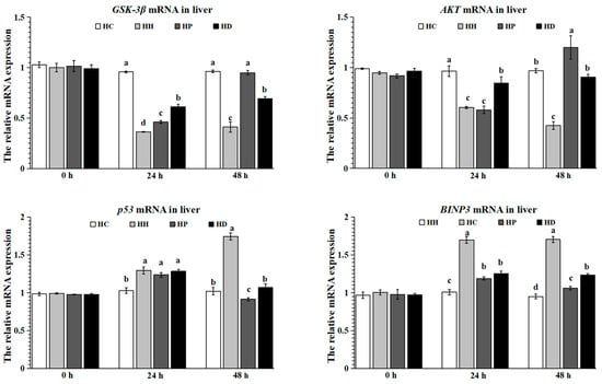
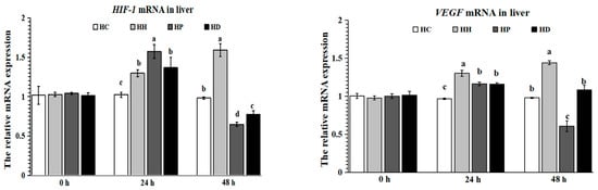
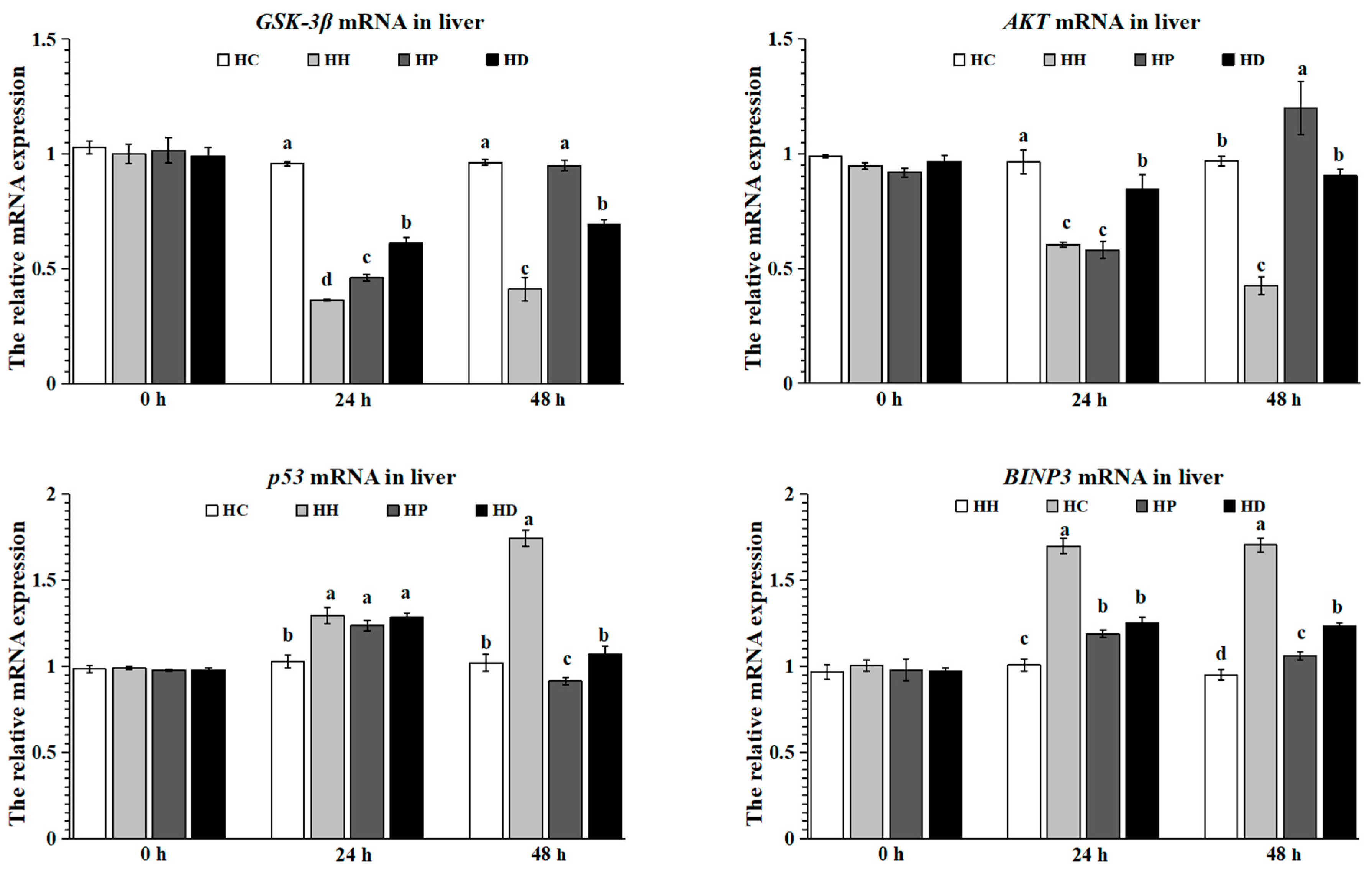
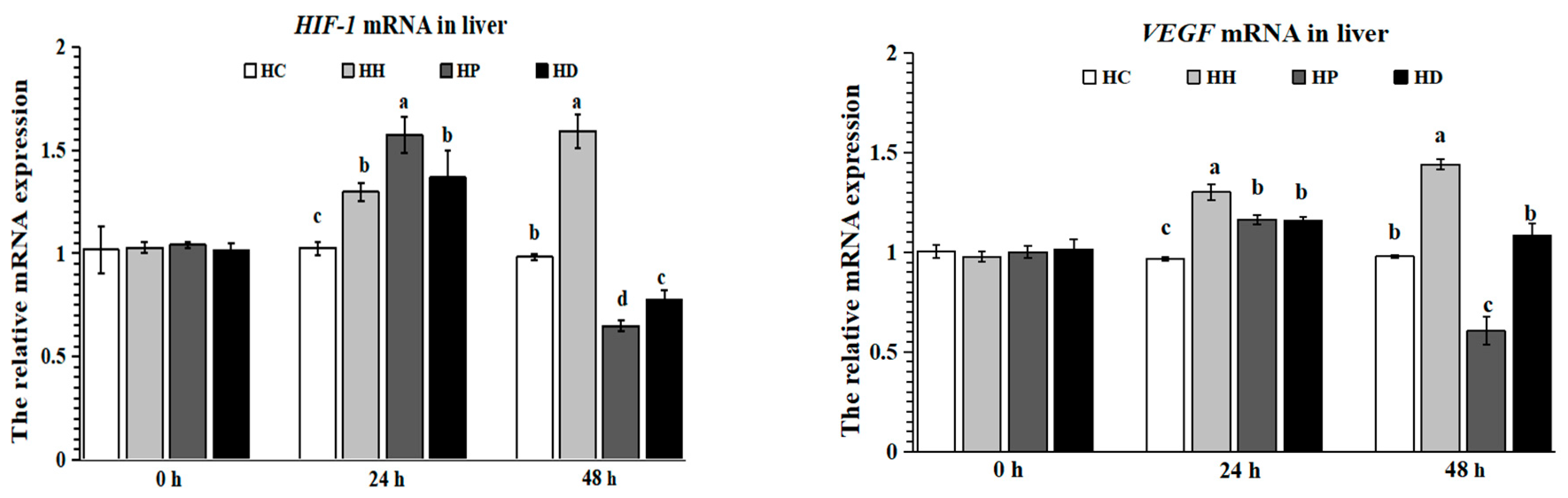
The mRNA expression levels of liver GSK-3β, Akt, HIF-1, VEGF, p53, and BNIP3 in all groups at hour 0 had no significant difference (p > 0.05, Figure 1). At 24 h, the mRNA expression level of GSK-3β was the lowest in the HH group, and the highest value appeared in the HC group (p < 0.05); The mRNA expression level of Akt was the lowest in the HH and HP groups, followed by fish in the HD group; the highest value showed in the HC group (p < 0.05). The mRNA expression level of p53 in the HC group was the lowest (p < 0.05), and there were no significant differences among the HH, HP and HD groups (p > 0.05). The mRNA expression level of BNIP3 in the HH group was the highest, followed by the HP and HD groups; the lowest showed in the HC group (p < 0.05). The mRNA expression level of HIF-1 in the HP group was the highest, followed by the HH and HD groups; the lowest HIF-1 level showed in the HC group (p < 0.05). The mRNA expression level of VEGF in the HH group was the highest, and the lowest value was found in the HC group (p < 0.05). At 48 h, the mRNA expression level of GSK-3β was the lowest in the HH group, and the mRNA expression level in the HD group was lower than that in the HC and HP groups (p < 0.05), whereas no significant difference was observed between the HP and HC groups (p > 0.05). The mRNA expression level of Akt was the lowest in the HH group, followed by fish in the HC and HD groups, and the highest value was found in the HP group (p < 0.05). The highest values of p53 and BNIP3 appeared in the HH group (p < 0.05). The mRNA expression level of HIF-1 in the HH group was the highest, followed by the HC group, and the lowest value was found in the HP group (p < 0.05); The mRNA expression level of VEGF in the HH group was the highest, and the next was the HD group; the lowest values were the HC and HP groups (p < 0.05).
Figure 1.
Relative expression levels of apoptosis-related genes GSK-3β, Akt, HIF-1, VEGF, p53, and BNIP3 in the liver. Relative mRNA expression of target genes was determined by qPCR, normalized to β-actin, and calculated using the standard curve method. Different letters indicate significant differences between groups (p < 0.05).
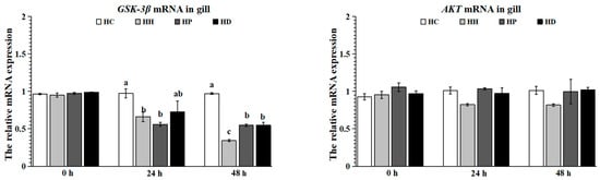
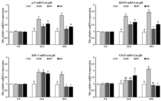
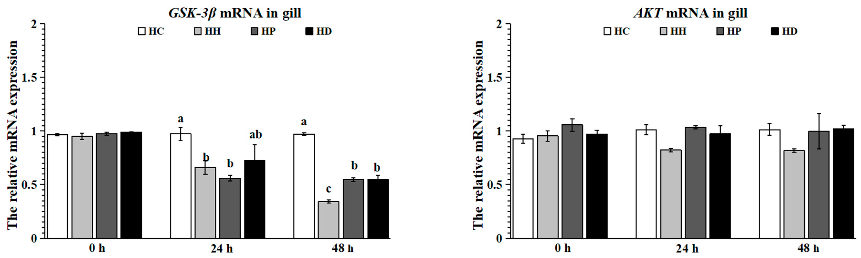
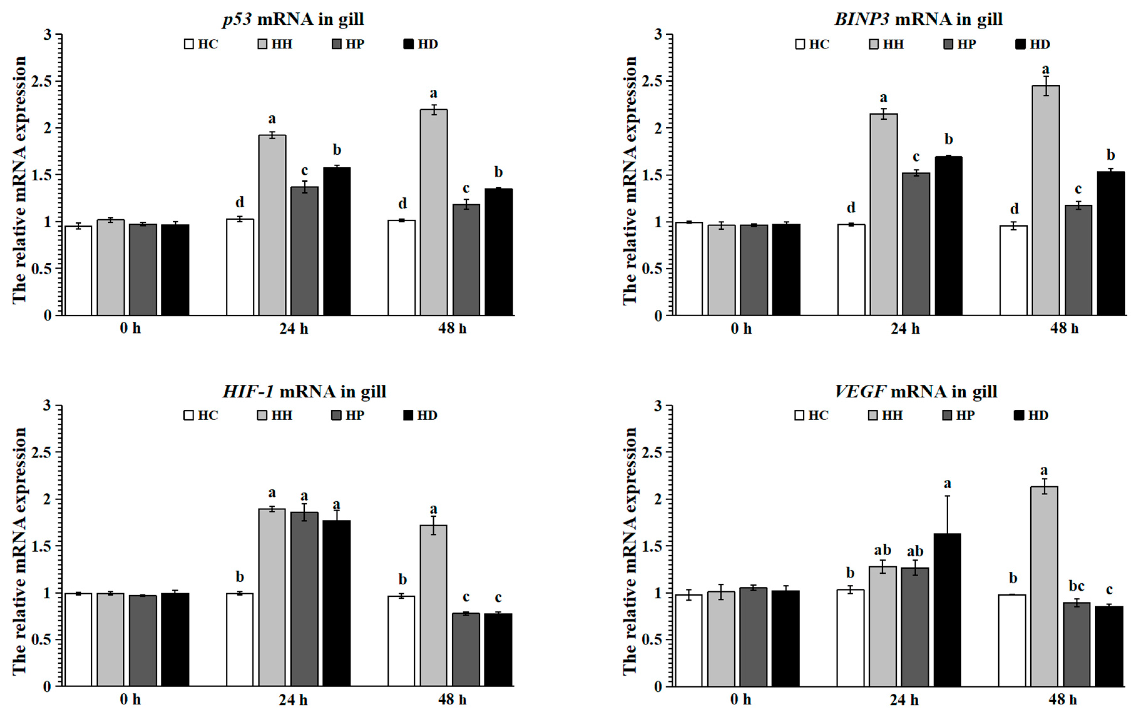
No significant differences in the mRNA expression levels of gill GSK-3β, Akt, HIF-1, VEGF, p53, and BNIP3 in all groups at hour 0 were observed (p < 0.05; Figure 2). The mRNA expression level of GSK-3β in the HC group was higher than in other groups (p < 0.05), while no significant difference was found in Akt levels in all groups at 24 h (p > 0.05). The mRNA expression level of p53 in the HH group was the highest, followed by fish in the HD and HP groups; the lowest value appeared in the HH group (p < 0.05). The mRNA expression level of BNIP3 was the highest in the HH group, and the lowest value appeared in the HH group (p < 0.05). The mRNA expression level of HIF-1 in the HC group was the lowest (p < 0.05). The mRNA expression level of VEGF in the HD group was the highest (p < 0.05), and no significant difference was found in the HH, HP and HD groups (p > 0.05). At 48 h, the mRNA expression level of GSK-3β in the HH group was the lowest, followed by fish in the HP and HD groups (p < 0.05). No significant differences were found in Akt levels in all groups (p > 0.05). The mRNA expression levels of p53 and BNIP3 in the HH group were the highest, and in the HH group were the lowest (p < 0.05). The mRNA expression levels of HIF-1 and VEGF in the HH group were the highest (p < 0.05).
Figure 2.
Relative expression levels of apoptosis-related genes GSK-3β, Akt, HIF-1, VEGF, p53, and BNIP3 in the gill. Relative mRNA expression of target genes was determined by qPCR, normalized to β-actin, and calculated using the standard curve method. Different letters indicate significant differences between groups (p < 0.05).
4. Discussion
The Akt/GSK-3β signaling pathway plays a crucial role in the cellular response to hypoxia. The Akt regulates diverse biological processes by regulating GSK-3, p70S6K1, mTOR, and HIF-1 expression under low-oxygen conditions [26]. In the present study, acute hypoxic stress decreased the contents of liver GSK and Akt at both 24 and 48 h. This suppression aligns with findings in human hepatoma cells under severe hypoxia, where Akt/GSK-3β signaling is similarly inhibited [27]. This study also found that intraperitoneal injection of puerarin and dexamethasone significantly increased Akt and GSK contents at 48 h, but pretreated with puerarin increased GSK content only 24 h, which suggested that both puerarin and dexamethasone could alleviate the negative effects of hypoxia stress on the physiology of mudskipper, but dexamethasone alleviated the GSK content level later than puerarin, which indicated that the mitigation of dexamethasone was slower than puerarin and cannot be used as an alleviator to rescue acute hypoxia stress. This result is consistent with previous findings. Qiao et al. [28] reported that the protein expression levels of Akt and GSK-3β were downregulated in the rat H9c2 cardiomyocyte cell line under hypoxic eupatilin pretreatment, increasing the activation of the Akt/GSK-3β signaling pathway in H9c2 cells. Wang et al. [29] found that hypoxia significantly decreased the levels of Akt and GSK phosphorylation. One limitation of this study is that we measured enzyme protein concentrations using ELISA, rather than direct enzyme activities. Although changes in enzyme concentration can reflect altered expression or turnover under hypoxic stress, they do not necessarily correlate with catalytic activity due to post-translational modifications, allosteric regulation, and other cellular factors. Therefore, our findings regarding metabolic enzyme changes should be interpreted as indicators of altered protein abundance, and future studies employing direct activity assays (e.g., spectrophotometric methods) are warranted to fully elucidate the functional consequences of these changes.
Under hypoxic stress, fish rely on anaerobic glycolysis for energy production: carbohydrates are broken down into glucose, which is taken up by tissues, and pyruvate derived from glucose is converted to lactate via LDH, accompanied by ATP generation [30,31]. Glycolysis fuels energy needs but inevitably leads to marked lactate accumulation. In the present study, fish in the HH group exhibited lower liver LDH contents at 24 and 48 h, confirming the inhibition of LDH under hypoxic exposure. Yang et al. [32] reported that hypoxia stress disrupted the balance between pyruvate and lactate metabolism, leading to a significant accumulation of lactic acid in tissues. This accumulation is a hallmark of enhanced anaerobic glycolysis under hypoxic conditions, wherein pyruvate is preferentially converted to lactate via LDH. The authors suggested that this metabolic shift may be accompanied by complex regulatory changes in LDH activity over time. LDH content decreased may be a protective mechanism under hypoxic conditions that may ameliorate the cytotoxicity induced by the conversion of pyruvate to lactic acid. Previous studies found that pretreatment using puerarin significantly decreased LDH production in rat PC12 cells, and dexamethasone increased liver LDH mRNA levels of rats [33,34]. In the present study, pretreatment with puerarin can recover liver LDH content to normal levels at 24 and 48 h, but dexamethasone only increased LDH content at 48 h, which suggests that pretreatment with puerarin or dexamethasone can rescue LDH content disorder induced by hypoxia, and the mitigation of puerarin was obviously better than dexamethasone. Pyruvate kinase (PK) mediates the conversion of glucose to pyruvate under anaerobic conditions [35]. The present study showed that liver PK content was decreased at 24 and 48 h, suggesting that acute hypoxia stress inhibits the tricarboxylic acid cycle and glycolysis by reducing the production of pyruvate in the liver. This study also found that pretreatment with puerarin increased liver PK content at 24 h, but pretreatment with dexamethasone increased PK content at 48 h, which indicated that intraperitoneal injection of puerarin or dexamethasone can ameliorate the reduction of PK content induced by hypoxia; the mitigation of puerarin was more effective than dexamethasone. Ruan et al. [36] reported that puerarin treatment upregulated the PK levels in the liver of mice with D-galactose—induced aging. Pantaleao et al. [34] found that sixty-hour fasted dexamethasone-treated rats showed increased mRNA levels of PK in rats.
GSK-3β, a key downstream target of Akt, is critically involved in hypoxic signaling [29]. In the present study, hypoxic stress significantly decreased the mRNA levels of liver Akt and GSK-3β at 24 and 48 h, which suggested, in cases of severe hypoxic stress, Akt/GSK-3β signaling is inhibited in the liver. In addition, hypoxic stress significantly decreased the mRNA level of gill GSK-3β at 24 and 48 h, but the mRNA level of Akt had no significant change among all groups, which indicated prolonged hypoxia mainly inhibited the gene expression level of GSK-3β to protect against organ hypoxic injury and oxidative stress [37]. Previous studies reported that puerarin pretreatment can attenuate myocardial hypoxic injury by inhibiting autophagy via the Akt signaling pathway [38], and dexamethasone provides robust neuroprotection against subsequent hypoxic injury in rats in part via activation of the Akt/GSK-3β pathway [39]. The present study demonstrated that puerarin or dexamethasone pretreatment increased the mRNA levels of liver Akt and GSK-3β at 48 h compared with the HH group, which indicated that the protective effect of puerarin or dexamethasone against hypoxic injury in the liver and the gill of mudskippers is associated with the Akt/GSK-3β signaling pathways.
Akt regulates multiple downstream factors, including transcription factors, kinases, and apoptotic regulators such as p53 [40]. Akt regulates multiple downstream factors, including transcription factors, kinases, and apoptotic regulators such as p53 [7]. In the present study, acute hypoxic stress can up-regulate the mRNA levels of liver p53 and BNIP3 at 24 h and 48 h, which suggests hypoxia may induce apoptosis in the liver through the p53/BNIP3 signaling pathway. And in gills, the mRNA levels of p53 and BNIP3 were significantly increased, more than tripling during 48 h, which may be because gills are the primary respiratory organs of fish that come into contact with the water environment directly, and therefore gills are more vulnerable to hypoxia. Felix-Portillo et al. [41] further reported that p53 expression is significantly upregulated under hypoxic stress in whiteleg shrimp (Litopenaeus vannamei). As a general sensor of environmental stress, p53 overexpression may exacerbate oxidative stress and promote apoptosis in exposed cells [11]. This study found that pretreatment with puerarin or dexamethasone can decrease the mRNA level of p53 and BNIP3 in the liver and the gill at 24 and 48 h, which suggests that puerarin or dexamethasone may mitigate apoptosis induced by hypoxia through down-regulating the gene expression levels of p53 and BNIP3, which is mediated by the Akt/GSK-3β pathway. However, after pretreatment with puerarin and dexamethasone, the gene expression levels of p53 and BNIP3 in gills were still higher than the HC group at 24 and 48 h, which suggests puerarin and dexamethasone have limited effectiveness on apoptosis induced by hypoxia. Interestingly, the absence of a significant time effect (p = 0.856) and treatment × time interaction (p = 0.191) for gill Akt mRNA suggests that transcriptional regulation of Akt in gill tissue is not a primary response mechanism to acute hypoxia. This is consistent with the gill’s role as a direct environmental interface, where rapid physiological adjustments—such as changes in blood flow and oxygen extraction efficiency—are likely achieved through post-translational modifications (e.g., phosphorylation) of existing proteins rather than de novo gene expression. In contrast, the highly significant interactions for downstream targets such as GSK-3β, HIF-1, and VEGF indicate that the pathway is still functionally engaged, but at a post-transcriptional level.
The HIF-1 signaling pathway also upregulates genes involved in oxygen transport and utilization, such as vascular endothelial growth factor (VEGF), which supports oxygen delivery for energy metabolism [13]. Under adverse conditions, elevated VEGF can promote endothelial proliferation [42], angiogenesis [43], and vascular permeability [44], potentially exacerbating hypoxic damage. In our experiment, hepatic HIF-1α and VEGF mRNA levels were markedly upregulated at 24 and 48 h post-hypoxia, likely facilitating the vascular adaptations required for acute oxygen shortage. In gills, the response differed: VEGF mRNA increased only at 48 h, indicating a delayed angiogenic response compared with the liver. This may be because gills are already densely vascularized, conferring greater inherent oxygen-transport capacity and hypoxia tolerance. Comparable inductions of HIF-1α and VEGF have been reported in hypoxic European sea bass [45] and orange-spotted grouper [46]. Both puerarin and dexamethasone pretreatment downregulated HIF-1α and VEGF expression relative to the hypoxic group, implying that they can normalize the hypoxia-driven dysregulation of these genes in the liver and the gill, possibly via Akt/GSK-3β signaling. Previous studies corroborate that puerarin modulates VEGF and HIF-1α expression under stress [47], and dexamethasone potently suppresses their induction in human systems [48]. As the primary interface with the aquatic environment, the gills are expected to mount an early response to hypoxia. Indeed, we observed significant upregulation of HIF-1α and VEGF mRNA in gill tissue at 48 h post-hypoxia, consistent with the gill’s role in oxygen sensing and vascular adaptation. Interestingly, the temporal pattern of gene expression in the gill differed from that in the liver, with the liver showing earlier and more pronounced changes in metabolic genes (e.g., LDH, PK), reflecting its central role in systemic energy metabolism. These tissue-specific responses underscore the complex, multi-organ coordination required for hypoxia adaptation. Together, these results suggest that puerarin and dexamethasone can ameliorate hypoxic injury partly by regulating the HIF-1α/VEGF axis.
One limitation of this study is the absence of plasma metabolic parameters such as glucose, lactate, and cortisol levels. These indicators provide valuable systemic context for interpreting the tissue-level changes in metabolic enzymes and signaling pathways observed in the liver and the gill. Although our tissue-focused approach has elucidated organ-specific molecular responses, integrating plasma biochemical profiling in future studies will be essential to achieve a more comprehensive understanding of the systemic hypoxia response in fish.
5. Conclusions
This study leads to the following conclusions: On the one hand, hypoxic stress triggers apoptotic signaling through the p53/BNIP3 pathway. On the other hand, it concurrently promotes compensatory adaptation by upregulating HIF-1 and VEGF expression, mediated via the Akt/GSK-3β pathway. Furthermore, pretreatment with puerarin or dexamethasone was shown to alleviate the adverse physiological effects induced by hypoxia in mudskippers.
Author Contributions
D.F.: investigation, methodology, writing—original draft, funding acquisition; Q.H.: investigation, methodology; X.S.: writing—review and editing, project administration; G.W.: methodology, data analysis; Z.L.: supervision, writing—review and editing. All authors have read and agreed to the published version of the manuscript.
Funding
This research was supported by the Open Project Program of the Key Laboratory of Fishery Drug Development, the Ministry of Agriculture and Rural Affairs; the Guangdong Provincial Key Laboratory of Aquatic Animal Immunology and Sustainable Aquaculture (202308); and the Special Fund for Henan Agriculture Research System (HARS-22-16-G3).
Institutional Review Board Statement
The animal study protocol was approved by the Ethics Committee of Xinyang Agriculture and Forestry University (permit no. 2018-2 and approval date: 11 March 2018).
Data Availability Statement
The original contributions presented in this study are included in the article. Further inquiries can be directed to the corresponding authors.
Conflicts of Interest
The authors declare no conflict of interest.
References
- Shen, Y.; You, W.; Luo, X.; Lu, Y.; Huang, M.Q.; Ke, C.H. An overview of the mechanisms underlying hypoxia tolerance differences in aquatic animals and their inspirations for aquaculture. Rev. Fish Biol. Fish. 2023, 33, 1223–1236. [Google Scholar] [CrossRef] [Scilit]
- Sun, J.L.; Zhao, L.L.; Wu, H.; Liu, Q.; Liao, L.; Luo, J.; Lian, W.Q.; Cui, C.; Jin, L.; Ma, J.D.; et al. Acute hypoxia changes the mode of glucose and lipid utilization in the liver of the largemouth bass (Micropterus salmoides). Sci. Total Environ. 2020, 713, 135157. [Google Scholar] [CrossRef] [Scilit]
- Zhang, W.X.; Pan, S.; Yu, L.Q.; Zhang, H.Y.; Chen, F.J.; Song, G.S.; Hu, J.T.; Wei, Q.S.; Zhao, H.D.; Chen, J.F.; et al. Dissolved oxygen depletion in Chinese coastal waters. Water Res. 2025, 272, 123004. [Google Scholar] [CrossRef] [Scilit] [PubMed]
- Timmerman, C.M.; Chapman, L.J. Behavioral and physiological compensation for chronic hypoxia in the sailfin molly (Poecilia latipinna). Physiol. Biochem. Zool. 2004, 77, 601–610. [Google Scholar] [CrossRef] [Scilit] [PubMed]
- Chippari-Gomes, A.R.; Gomes, L.C.; Lopes, N.P. Metabolic adjustments in two amazonian cichlids exposed to hypoxia and anoxia. Comp. Biochem. Phys. B 2005, 141, 347–355. [Google Scholar] [CrossRef] [Scilit]
- Bickler, P.E.; Buck, L.T. Hypoxia tolerance in reptiles, amphibians, and fishes: Life with variable oxygen availability. Annu. Rev. Physiol. 1993, 69, 145–170. [Google Scholar] [CrossRef] [Scilit]
- Li, X.; Zhang, J.; Chai, S.; Wang, X. Progesterone alleviates hypoxic-ischemic brain injury via the Akt/GSK-3beta signaling pathway. Exp. Ther. Med. 2014, 8, 1241–1246. [Google Scholar] [CrossRef] [Scilit]
- Zhang, M.; Zhang, J.; Chen, X.; Cho, S.J. Glycogen synthase kinase 3 promotes p53 mrna translation via phosphorylation of rnpc1. Genes Dev. J. Devoted Mol. Anal. Gene Expr. Eukaryotes Prokaryotes Viruses 2013, 20, 27. [Google Scholar] [CrossRef] [Scilit]
- Denis, M. Regulation of hypoxia-inducible factor-1alpha protein level during hypoxic conditions by the phosphatidylinositol 3-kinase/Akt/glycogen synthase kinase 3beta pathway in HepG2 cells. J. Biol. Chem. 2003, 33, 31277–31285. [Google Scholar]
- Carmeliet, P.; Dor, Y.; Herbert, J.M.; Fukumura, D.; Brusselmans, K.; Dewerchin, M.; Neeman, M.; Bono, F.; Abramovitch, R.; Maxwell, P. Role of HIF-1 in hypoxia-mediated apoptosis, cell proliferation and tumour angiogenesis. Nature 1998, 394, 485–490. [Google Scholar] [CrossRef] [Scilit]
- Qiang, J.; Zhu, X.W.; He, J.; Tao, Y.F.; Bao, J.W.; Zhu, J.H.; Xu, P. miR-34a regulates the activity of HIF-1α and p53 signaling pathways by promoting GLUT1 in genetically improved farmed Tilapia (GIFT, Oreochromis niloticus) under hypoxia stress. Front. Physiol. 2020, 11, 670. [Google Scholar] [CrossRef] [Scilit] [PubMed]
- Horowitz, J.R.; Rivard, A.; vanderZee, R.; Hariawala, M.; Sheriff, D.D.; Esakof, D.D.; Chaudhry, G.M.; Symes, J.F.; Isner, J.M. Vascular endothelial growth factor vascular permeability factor produces nitric oxide-dependent hypotension-evidence for a maintenance role in quiescent adult endothelium. Arterioscler. Thromb. Vasc. Biol. 1997, 17, 2793–2800. [Google Scholar] [CrossRef] [Scilit]
- Li, X.; Wang, T.; Yin, S.; Zhang, G.; Cao, Q.; Wen, X.; Zhang, H.; Wang, D.; Zhu, W. The improved energy metabolism and blood oxygen-carrying capacity for pufferfish, takifugu fasciatus, against acute hypoxia under the regulation of oxygen sensors. Fish Physiol. Biochem. 2019, 45, 323–340. [Google Scholar] [CrossRef] [Scilit]
- Vivek, S.R.; Paneerselvan, N.; Jagadeeshan, S.; Malathi, R. Hypoxia induced angiogenesis and upregulation of VEGF: An in vivo study using zebrafish model. Int. J. Entific Eng. Res. 2015, 6, 831. [Google Scholar]
- Luo, C.F.; Hou, N.; Tian, J.; Yuan, M.; Liu, S.M.; Xiong, L.G.; Luo, J.D.; Chen, M.S. Metabolic profile of puerarin in rats after intragastric administration of puerarin solid lipid nanoparticles. Int. J. Nanomed. 2013, 8, 933–940. [Google Scholar] [CrossRef] [Scilit]
- Zhou, Y.X.; Zhang, H.; Peng, C. Puerarin: A review of pharmacological effects. Phytother. Res. 2014, 28, 961–975. [Google Scholar] [CrossRef] [Scilit]
- Pang, W.; Lan, X.-M.; Wang, C.-B. Effect of puerarin on the release of interleukin-8 in co-culture of human bronchial epithelial cells and neutrophils. Chin. J. Integr. Med. 2012, 4, 283–287. [Google Scholar] [CrossRef] [Scilit]
- Wang, C.; Yan, M.; Jiang, H.; Wang, Q.; Guan, X.; Chen, J.; Wang, C. Protective effects of puerarin on acute lung and cerebrum injury induced by hypobaric hypoxia via the regulation of aquaporin (AQP) via NF-κB signaling pathway. Int. Immunopharmacol. 2016, 40, 300–309. [Google Scholar] [CrossRef] [Scilit]
- Ma, Y.; Gai, Y.; Yan, J.; Li, J.; Zhang, Y. Puerarin attenuates anoxia/reoxygenation injury through enhancing Bcl-2 associated athanogene 3 expression, a modulator of apoptosis and autophagy. Med. Sci. Monit. Int. Med. J. Exp. Clin. Res. 2016, 22, 977–983. [Google Scholar] [CrossRef] [Scilit] [PubMed]
- Yang, S.; Wang, S.; Sun, F.; Zhang, M.; Wu, F.; Xu, F.; Ding, Z. Protective effects of puerarin against tetrabromobisphenol a-induced apoptosis and cardiac developmental toxicity in zebrafish embryo-larvae. Environ. Toxicol. 2015, 30, 1014–1023. [Google Scholar] [CrossRef] [Scilit] [PubMed]
- Luks, A.M.; Swenson, E.R. Medication and dosage considerations in the prophylaxis and treatment of high-altitude illness. Chest 2008, 133, 744–755. [Google Scholar] [CrossRef] [Scilit]
- Inada, M.; Taguchi, M.; Harimoto, K.; Karasawa, Y.; Takeuchi, M.; Ito, M. Protective effects of dexamethasone on hypoxia-induced retinal edema in a mouse model. Exp. Eye Res. 2019, 178, 82–90. [Google Scholar] [CrossRef] [Scilit]
- Feng, Y.; Rhodes, P.G.; Bhatt, A.J. Dexamethasone pre-treatment protects brain against hypoxic-ischemic injury partially through up-regulation of vascular endothelial growth factor a in neonatal rats. Neuroscience 2011, 179, 223–232. [Google Scholar] [CrossRef] [Scilit]
- Jing, D.D.; Li, M.; Zhang, Y.; Yuan, L.X.; Wang, R.X.; Gong, Y.F. Differential induction of enzymes and genes involved in oxidative stress in gill and liver tissues of mudskipper Boleophthalmus pectinirostris Exposed to Lead. Turk. J. Fish. Aquat. Sci. 2017, 17, 437–443. [Google Scholar] [CrossRef]
- Livak, K.J.; Schmittgen, T.D. Analysis of relative gene expression data using real-time quantitative PCR and the 2(T)(-Delta Delta C) method. Methods 2001, 25, 402–408. [Google Scholar] [CrossRef] [Scilit]
- Datta, S.R.; Brunet, A.; Greenberg, M.E. Cellular survival: A play in three Akts. Gene Dev. 1999, 13, 2905–2927. [Google Scholar] [CrossRef] [Scilit]
- Loberg, R.D.; Vesely, E.; Brosius, F.C. Enhanced glycogen synthase kinase-3 beta activity mediates hypoxia-induced apoptosis of vascular smooth muscle cells and is prevented by glucose transport and metabolism. J. Biol. Chem. 2002, 277, 41667–41673. [Google Scholar] [CrossRef] [Scilit]
- Qiao, Z.Y.; Xu, Y.W.; Yang, J.Y. Eupatilin inhibits the apoptosis in H9c2 cardiomyocytes via the Akt/GSK-3 beta pathway following hypoxia/reoxygenation injury. Biomed. Pharmacother. 2016, 82, 373–378. [Google Scholar] [CrossRef] [Scilit] [PubMed]
- Wang, M.; Liu, Y.; Pan, R.L.; Wang, R.Y.; Ding, S.L.; Dong, W.R.; Sun, G.B.; Ye, J.X.; Sun, X.B. Protective effects of Myrica rubra flavonoids against hypoxia/reoxygenation-induced cardiomyocyte injury via the regulation of the PI3K/Akt/GSK3beta pathway. Int. J. Mol. Med. 2019, 43, 2133–2143. [Google Scholar] [PubMed]
- Mizock, B.A. Alterations in carbohydrate-metabolism during stress—A review of the literature. Am. J. Med. 1995, 98, 75–84. [Google Scholar] [CrossRef] [Scilit] [PubMed]
- Wang, Y.; Richards, J.G. Hypoxic|Anaerobic metabolism in fish. In Encyclopedia of Fish Physiology; Academic Press: Cambridge, MA, USA, 2011; pp. 1757–1763. [Google Scholar]
- Yang, S.; Wu, H.; He, K.; Yan, T.; Zhou, J.; Zhao, L.L.; Sun, J.L.; Lian, W.Q.; Zhang, D.M.; Du, Z.J.; et al. Response of AMP-activated protein kinase and lactate metabolism of largemouth bass (Micropterus salmoides) under acute hypoxic stress. Sci. Total Environ. 2019, 666, 1071–1079. [Google Scholar] [CrossRef] [Scilit]
- Cheng, Y.; Leng, W.; Zhang, J.S. Protective effect of puerarin against oxidative stress injury of neural cells and related mechanisms. Med. Sci. Monit. 2016, 22, 1244–1249. [Google Scholar] [CrossRef] [Scilit]
- Pantaleao, L.C.; Murata, G.; Teixeira, C.J.; Payolla, T.B.; Santos-Silva, J.C.; Duque-Guimaraes, D.E.; Sodre, F.S.; Lellis-Santos, C.; Vieira, J.C.; de Souza, D.N.; et al. Prolonged fasting elicits increased hepatic triglyceride accumulation in rats born to dexamethasone-treated mothers. Sci. Rep. 2017, 7, 10367. [Google Scholar] [CrossRef] [Scilit]
- Richards, J.G. Physiological, behavioral and biochemical adaptations of intertidal fishes to hypoxia. J. Exp. Biol. 2011, 214, 191–199. [Google Scholar] [CrossRef] [Scilit]
- Ruan, H.; Li, W.; Chang, H.; Wen, M.; Luo, S.; Song, F.; Ye, L.; Mei, J.; Zhu, X.; Liu, X.; et al. Antioxidant, Hypoglycemic, and Hypolipidemic Effects of Puerarin In Vivo. Food Sci. Nutr. 2025, 13, e70257. [Google Scholar] [CrossRef] [Scilit]
- Martin, M.; Rehani, K.; Jope, R.S.; Michalek, S.M. Toll-like receptor-mediated cytokine production is differentially regulated by glycogen synthase kinase 3. Nat. Immunol. 2005, 6, 777–784. [Google Scholar] [CrossRef] [Scilit]
- Tang, H.; Song, X.; Ling, Y.; Wang, X.; Yang, P.; Luo, T.; Chen, A. Puerarin attenuates myocardial hypoxia/reoxygenation injury by inhibiting autophagy via the Akt signaling pathway. Mol. Med. Rep. 2017, 15, 3747–3754. [Google Scholar] [CrossRef] [Scilit] [PubMed]
- Peng, Y.Z.; Lu, S.Q.; Wang, J.M.; Kumar, P.; Zhang, L.; Bhatt, A.J. Dexamethasone-induced neuroprotection in hypoxic-ischemic brain injury in newborn rats is partly mediated via Akt activation. Brain Res. 2014, 1589, 68–77. [Google Scholar]
- Chong, Z.Z.; Li, F.; Maiese, K. Activating Akt and the brain’s resources to drive cellular survival and prevent inflammatory injury. Histol. Histopathol. 2005, 20, 299–315. [Google Scholar] [PubMed]
- Felix-Portillo, M.; Martínez-Quintana, J.A.; Arenas-Padilla, M.; Mata-Haro, V.; Gómez-Jiménez, S.; Yepiz-Plascencia, G. Hypoxia drives apoptosis independently of p53 and metallothionein transcript levels in hemocytes of the whiteleg shrimp Litopenaeus vannamei. Chemosphere 2016, 161, 454–462. [Google Scholar] [CrossRef] [Scilit]
- Faes, S.; Uldry, E.; Planche, A.; Santoro, T.; Pythoud, C.; Demartines, N.; Dormond, O. Acidic pH reduces VEGF-mediated endothelial cell responses by downregulation of VEGFR-2; relevance for anti-angiogenic therapies. Oncotarget 2016, 7, 86026–86038. [Google Scholar] [CrossRef] [Scilit]
- Shams, N.; Ianchulev, T. Role of Vascular endothelial growth factor in ocular angiogenesis. Ophthalmol. Clin. N. Am. 2006, 19, 335. [Google Scholar]
- Lee, I.S.; Kim, Y.S.; Jung, S.H.; Yu, S.Y.; Kim, J.H.; Sun, H.; Kim, J.S. Lignans from the stems and leaves of Brandisia hancei and their effects on VEGF-induced vascular permeability and migration of HRECs and DLAV formation in zebrafish. Biosci. Biotechnol. Biochem. 2015, 79, 581–586. [Google Scholar] [CrossRef] [Scilit] [PubMed]
- Terova, G.; Rimoldi, S.; Cora, S.; Bernardini, G.; Gornati, R.; Saroglia, M. Acute and chronic hypoxia affects HIF-1 alpha mRNA levels in sea bass (Dicentrarchus labrax). Aquaculture 2008, 279, 150–159. [Google Scholar] [CrossRef] [Scilit]
- Yu, R.M.K.; Ng, P.K.S.; Tan, T.F.; Chu, D.L.H.; Wu, R.S.S.; Kong, R.Y.C. Enhancement of hypoxia-induced gene expression in fish liver by the aryl hydrocarbon receptor (AhR) ligand, benzo[a]pyrene (BaP). Aquat. Toxicol. 2008, 90, 235–242. [Google Scholar] [CrossRef] [Scilit]
- Zhang, S.; Chen, S.; Shen, Y.; Yang, D.; Liu, X.; Sun-Chi, A.C.; Xu, H. Puerarin induces angiogenesis in myocardium of rat with myocardial infarction. Biol. Pharm. Bull. 2006, 29, 945–950. [Google Scholar] [CrossRef] [Scilit] [PubMed]
- Wu, Y.; Lucia, K.; Lange, M.; Kuhlen, D.; Stalla, G.K.; Renner, U. Hypoxia inducible factor-1 is involved in growth factor, glucocorticoid and hypoxia mediated regulation of vascular endothelial growth factor-A in human meningiomas. J. Neuro-Oncol. 2014, 119, 263–273. [Google Scholar] [CrossRef] [Scilit] [PubMed]

Disclaimer/Publisher’s Note: The statements, opinions and data contained in all publications are solely those of the individual author(s) and contributor(s) and not of MDPI and/or the editor(s). MDPI and/or the editor(s) disclaim responsibility for any injury to people or property resulting from any ideas, methods, instructions or products referred to in the content. |
© 2026 by the authors. Licensee MDPI, Basel, Switzerland. This article is an open access article distributed under the terms and conditions of the Creative Commons Attribution (CC BY) license.