


Abstract
The theoretical underpinnings and empirical data supporting oxytocin as a context-dependent biomarker of prosocial affiliation, bonding, attachment, emotional regulation, cognitive development, and the modulation of stress responses are summarized. We propose an interdisciplinary consensus against a backdrop of inconsistencies concerning behavioral and experimental findings for the oxytocinergic system. Current knowledge gaps demonstrating the inability to establish a hierarchy of findings are related to the following: (a) the theory and practice of oxytocinergic therapeutic interventions; (b) identification, characterization, and quantification of functional, mechanistic, and molecular biomarkers and matrices; (c) context- and time-dependent resilience factors associated with mind–brain–body development, (d) measures of intergenerational transmission, and (e) summative indicators of psychopathological co-morbidities. We analyze prospective objective measures of oxytocin against subjective caregiver reports of positive childhood experiences (PCEs, resulting from secure nurturing, established daily routines, pleasurable social interactions, opportunistic and spontaneous play, secure environments), which favor healthy developmental outcomes in preschool children. Few studies prospectively use the painless, noninvasive sampling of risk and resilience biomarker levels to index the health status, emotional regulation, and cognitive development in preschool children. We present the methodology for novel measures of risk (cortisol) and resilience (oxytocin) in human scalp hair. Future studies validating the therapeutic efficacy of oxytocin or the stress-mediating effects of oxytocin on socioemotional regulation and health status should systematically address these gaps.
1. Introduction
Historically, many theories have been put forth to explain the effects of oxytocin in adaptive ecological and psychological resilience in the setting of a dysregulated stress pathway. This review is centered on the role of oxytocin in preverbal/preschool child development, attachment theory, resilience theory, theory of the mind, and self-identity theory as forerunners for the socially affiliative model of resilience. These are discussed in the context of the three childhood developmental domains, i.e., emotions, cognition, and behaviors, each of which cumulatively adds to child wellbeing and health outcomes.
Allostatic load is the common term assigned to the cumulative temporal experiences of chronic stress exposures that impinge upon the mind–brain–body connectome, leading to dysregulation of the central and peripheral nervous systems, poor health, impaired self-regulation, and identity disruption. This model proffers that parental/primary caregiver nurturing, followed by additional supportive relationships, activates oxytocin release for stress axis regulation, thereby curbing the long-term detrimental effects of allostatic load. Because many children have primary caregivers other than a parent, it is important to understand that the effects of oxytocinergic affiliative pathways apply equally to all primary caregivers. These effects occur within the limbic system of the brain at the level of the hypothalamic–pituitary–adrenal (HPA) axis (physiologic homeostasis), the amygdala (emotionality: fear, anxiety, etc.), and the prefrontal cortex (cognition: coping, planning, self-regulation). To fully comprehend the power of oxytocin to inhibit a deleterious cycle of allostatic loading with potential transmission across generations, we focus on advances in stress and resilience theory to address knowledge gaps centered upon early childhood–family adaptation and interventions designed to improve child well-being.
1.1. Current State of Oxytocin Research
1.1.1. Early Experiences and Neurodevelopment
Foundational neuronal pathways are shaped by both negative and positive experiences during sensitive periods of brain development [1]. Early childhood is a sensitive period when exposures to adverse childhood experiences (ACEs) or early life stress (ELS) in the presence or absence of positive childhood experiences (PCEs) may shape lifetime outcomes. ACEs can include events that affect children directly, such as physical abuse or neglect, and indirectly in their social environment, such as parent conflict, substance abuse, or mental illness [2]. Early life adversity (ELA) includes maternal cumulative lifetime exposures to stress, including childhood experiences with past or present insecure environments or relationships, community violence, household instability, and limited access to prenatal care. The four common domains of PCEs include nurturing, consistent child–family routines, access to extended networks providing pleasurable, enriching activities (e.g., play, green space), and provisions for safety [3]. The neural pathways most involved during early-stage affiliative child development are those of the limbic system, including the HPA axis [4].
1.1.2. HPA Axis Function and Toxic Stress
The HPA axis is the central node of the stress network that maintains body homeostasis through cortisol release and its negative feedback loop. This neuromodulatory system comprises pathways responsible for the release of neuroendocrine hormones, such as cortisol and oxytocin, among others, that modulate adaptive regulation of the sympathetic and parasympathetic nervous systems. Toxic stress, i.e., prolonged or repeated ELA experienced without the buffering presence of PCEs, affects neuroplasticity to perturb a child’s emotional and cognitive development and health [5]. A myriad of mental and physical health disorders, such as anxiety, depression, cardiovascular disease, asthma, and others, are consequential to toxic stress exposures, HPA axis dysregulation [6,7,8], and the pediatric onset of adult diseases [7].
Childhood trauma has been declared a worldwide public health crisis [9]. Children are particularly susceptible to creating maladaptive stress responses when faced with higher ACEs and few PCEs during critical periods of neuroplasticity in early life. Recent estimates show that 45.7 percent of U.S. children aged 2 to 17 have experienced two or more ACEs [10]. In a systematic review conducted by Hughes and colleagues, individuals experiencing four or more ACEs had increased negative health outcomes, inclusive of problematic drug use and interpersonal or self-directed harm [2]. Furthermore, individuals exposed to ACEs are at higher risk for adverse behavioral, neurobiological, and physical consequences [11].
1.1.3. Social and Behavioral Impacts
Beyond physiological effects, toxic stress has a profound impact on a child’s social functioning [12,13,14]. A qualitative study conducted with kindergarten to 3rd-grade teachers focused on child behavioral observations in classrooms. Teachers reported that the children exposed to toxic stress lacked effective emotional regulation strategies and the ability to form and maintain social relationships with them and their peers [13]. A different study conducted a community-based intervention to mitigate the impact of toxic stress amongst youth at risk [14]. Interviews of these children revealed their inability to create and maintain meaningful relationships due to their home environments [14]. In this context, a potential mediator of the feelings of distrust and inability to build secure connections can be related to oxytocin’s role in social bonding and modulating the stress response system [15,16,17].
1.1.4. Oxytocin Pathway Knowledge Gaps
There is a wealth of knowledge centered on the role of glucocorticoid response element (GRE) genes in fetal organ development, growth, tissue regeneration, and health maintenance through the lifespan [18]. In contrast, the roles of the greater than 150 genes associated with the oxytocin signaling pathway remains underexplored [19], particularly in preverbal children. No studies to date have analyzed oxytocin-related genes or factors regulating their gene expression in preschool children. Oxytocin mitigates cortisol synthesis primarily by dampening the HPA axis and stimulating parasympathetic nervous system activity. Thus, oxytocin downregulates physiological stress responses, promotes autonomic balance, and suppresses peak cortisol reactivity, returning the body to a restful, restorative state under conditions of caregiver warmth and responsiveness; consistent family routines; access to resources that give rise to enjoyment, competence, and social stimulation; and provisions for safety. Sartorius et al. (2024) conducted functional analyses to reveal the evolutionary timeline for the development of human oxytocin pathway genes [19]. They report that the function of “modern genes in the oxytocin pathway … primarily expressed in the brain and muscle tissue” is to guide parturition, lactation, metabolism, cardiovascular tone, social cognition, decision-making, and learning. They point to “a persistent knowledge gap regarding the function and purpose of the human oxytocin system and its treatment potential” in psychiatric disorders and conditions associated with dysfunctions in social behavior and cognition.
1.2. Theoretical Basis, Models/Frameworks of Social Cognition in Stress Resilience
1.2.1. Resilience Frameworks: Attachment, Biopsychosocial, and Synchrony Models
Attachment theory, resilience theory, the biopsychosocial framework, and affiliative neuroscience models explain how PCEs become physiologically embedded. Attachment theory describes how early caregiver relationships shape developmental and emotional outcomes in young children. The ecological resilience framework highlights environmental factors distinct from psychological resilience that contribute to progressive adaptive functioning. The biopsychosocial framework emphasizes interactions among biological, psychological, and social domains in determining the health status. The affiliative neuroscience model outlines the role of oxytocin in forming brain networks that promote prosocial attachment and stress regulation. Quintana and Guastella propose that oxytocin functions as an allostatic hormone due to its widespread developmental, social, and regulatory effects [20]. Instead of simply triggering fixed prosocial behaviors like empathy, oxytocin heightens sensory and cognitive awareness for both social and non-social cues. This allows an organism to respond flexibly—by prompting cooperation within a safe group or promoting defensive, protective behaviors (like maternal aggression) in a life-threatening environment [20]. Research demonstrates that attachment styles are linked to outcomes in child psychopathology, cognitive and emotional brain networks related to empathy and compassion, school readiness, and immune function [5].
Garmezy’s early resilience work [21,22], later expanded by Masten and colleagues [23,24,25], demonstrated a shift in the focus on pathology towards competence-based developmental outcomes. Fleming and Ledogar described resilience as temporal and contextual, extending across individual, family, community, cultural, and ecosystem levels. Ruth Feldman integrated biological and behavioral plasticity, affiliative brain centers, and parent–child biobehavioral synchrony into a neuroscience-based resilience theory, connecting these processes to oxytocin pathways [26,27,28,29,30]. These concepts support Gottlieb’s model of experience-guided brain development via behavioral canalization [31,32] with predictive epigenetic trajectories, which provides one explanation for behavioral patterns observed in the intergenerational transmission of toxic stress (ITTS).
Maternal calibration and entrainment of a child’s neuroendocrine stress and prosocial pathways can be correlated with maternal and child ACE and PCE domain scores, although simple dose–response relationships may have limited value [5]. Assessing ELA including poverty, domestic or neighborhood violence, immigration-related stress, and acculturation is necessary to identify risks across the lifespan.
Together, these perspectives show that resilience is relational and dynamic. It results from the union of biological and social contours, exemplified in the synchronicity found between a baby and primary caregiver, as well as in the flexibility of families to adapt, which enables people to regulate their stress through human connection or bonding [5]. This perspective sees resilience as the capacity to employ a variety of social and biological processes to transmute contextual stressors into opportunities for growth, as opposed to simple stress reduction.
1.2.2. An Emerging Mechanistic Model: The Neurobiology of Oxytocin
The literature highlights an interdependence of oxytocin release, caregiver synchrony, and caregiver behaviors in shaping resilience capacities, as outlined in the affiliative neuroscience model of resilience [29]. Parent–child alignment in physiology and behavior offers recurring opportunities for co-regulation and contributes to the development of empathy, trust, and adaptive coping. Research on the theory of mind further shows how neural substrates supporting social cognition overlap with those involved in attachment, emotional understanding, and protection against psychopathology under conditions of toxic stress [33].
1.3. Oxytocin as a Biomarker of Stress Mediation, Development, and Health
Few studies exist for measures of oxytocin levels in young children. Oxytocin may be considered a biomarker when measuring actual physiological responses in individuals. Oxytocin is also a neuroendocrine hormone, responsible for parasympathetic regulation in response to stress and prosocial affiliation or bonding. Because of its role as a neuromodulator, it is frequently identified as a therapy for symptom control in children with developmental disorders, such as autism spectrum disorders (ASDs), or attention deficit hyperactivity disorder (ADHD). Despite its study across many human subjects and healthcare settings, significant disagreements exist in both methodology and conceptualization regarding the role of oxytocin in therapeutic interventions. Questions remain about whether measured oxytocin levels reflect the synthesis and secretion of endogenous oxytocin or are influenced by the exogenous dosing of synthetic oxytocin. Variability across studies can stem from differences in the methodology used with sample-specific measurements (saliva, serum, CSF, urine, tissue, or hair), matrix-dependent extraction procedures, and assay platforms such as liquid chromatography/mass spectrometry, aptamers, or the enzyme-linked-immunosorbent assays (ELISAs) used for oxytocin quantification. The high level of temporal and context-sensitivity of central versus peripheral circulating oxytocin levels across various populations adds additional confusion to study results. The impact of oxytocin on behavior varies based upon an individual’s developmental background, gender, mental health status, and social–emotional conditions; thus, generalizing across studies and varying populations is difficult [34,35,36]. See Table 1.
Table 1.
A critical appraisal: inconsistencies, gaps, and future directions. This table identifies the primary issues with inconsistencies, gaps in empirical knowledge, and unanswered questions in the role of oxytocin in the development of preverbal/preschool children as discussed throughout this review. It provides a framework for understanding previous, divergent findings and developing research goals. Code for abbreviations: ACTH (adrenocorticotropic hormone), ASD (autism spectrum disorder), AVP (arginine vasopressin), AVP V1b (arginine vasopressin V1b receptor), CNS (central nervous system, CSF (cerebrospinal fluid), EEG (electroencephalogram), GWAS (genome-wide association studies), IV (intravenous), LC-MS/MS (liquid chromatography-tandem, mass spectrometry), OT (oxytocin), OXTR (oxytocin receptors), PK (pharmacokinetics), RCTs (research clinical trials), SNP (single-nucleotide polymorphism), ToM (theory of the mind), TPJ (temporoparietal junction).
2. Publication Search Strategy
We found no publications centered on early childhood development of the affiliative oxytocinergic system or the oxytocin–arginine vasopressin pathway. Interdisciplinary perspectives on the role of oxytocin typically focus on adolescents or adults, with frequent inconsistencies concerning the behavioral and observational findings on the oxytocinergic system. Systematic reviews, especially those offering meta-analysis of clinical trials, demonstrate a lack of studies conducted with young children below the age of 5 years. Thus, additional search terms included a combination of the following major themes: (a) theory and practice guiding oxytocinergic therapeutic interventions; (b) identification, characterization, and quantification of functional, mechanistic, and molecular biomarkers of the social affiliative pathway in its capacity to buffer chronic stress; (c) interactions of parental factors, child age- and sex-associated brain and behavioral development with or without intervention, (d) measures of intergenerational transmission of biopsychosocial factors; and (e) measures of psychopathological comorbidities.
Recent evidence linking risk and resilience factors to poor health outcomes with(out) oxytocin treatment comprise the largest number of references (52%), mostly published from 2020 to 2025, discussed in our narrative review. Concepts of the affiliative neuroendocrine framework publications gained the greatest traction during 2017–2019 and comprise 16% of the references included in our review. The bulk of these studies did not test for or report the role of oxytocin in early childhood development. Seminal publications from animal and human research demonstrating the ability of oxytocin to modulate the cortisol feedback loop were most concentrated during 2000–2016 and constituted 25% of references under discussion. Early childhood is important because neuroplasticity and network formation, especially in the HPA axis, peak prior to a child’s fourth birthday. Theoretical frameworks for interactions among stress, development, and poor health outcomes have progressed throughout the last century, providing 6% of our references. Research on the childhood onset of adult noncommunicable diseases, psychopathologies, and suicide is sharply increasing, yet these studies have not linked the socially affiliative role of oxytocin to health trajectories across the lifespan.
3. Biology and Regulation of the Oxytocinergic System
3.1. Oxytocin–Arginine Vasopressin Pathways and Brain Development
The oxytocin–arginine vasopressin (OT-AVP) system serves as a critical neurobiological network connecting early adversity, neural development, social–emotional functions, and stress-related outcomes. Genetic, epigenetic, and environmental factors converge on this system, supporting its potential as a biomarker for identifying children at risk for neurodevelopmental and psychiatric vulnerabilities [69,70]. Childhood stress exposure gives rise to dysregulation of the stress-response system, altered neuroimmune signaling, chronic inflammation (central and peripheral), and poor health [71]. Chronic childhood stress impairs the functioning of brain regions important for developing self-regulation and coping skills [72]. Clinical psychiatry emphasizes that stress exposures may manifest as risk-taking or resilience-building behaviors. Behavioral investigators describe stress adaptation as a balance between protective and vulnerable elements that influence recovery or worsening vulnerability [21,22]. Clinical evidence links oxytocin dysregulation to psychiatric disorders, including depression, anxiety, and post-traumatic stress disorder (PTSD) [70]. Recent syntheses that incorporate oxytocin into broader neurotransmitter systems linked to mental illness emphasize its multifaceted effects on social function and regulation [73]. Given that oxytocin and AVP coregulate social behavior and stress reactivity, the cooperative function of these neuropeptides is important for studies on neurodevelopmental psychopathology [74].
Neuroimaging studies reveal specific neural circuits that are especially susceptible to ELS, particularly in the salience network, responsible for detecting and filtering environmental stimuli, and the sensorimotor processing network, which integrates sensory input and coordinates motor responses [75]. Oxytocin and AVP are important for social and emotional development according to the brain underpinnings of affiliative behaviors [76]. A further understanding of how these networks function at various stages of child development is provided by evaluating parent–child neural synchrony in real-world contexts. Neural synchrony supports brain plasticity and adaptive socioemotional growth [77].
The OT-AVP system plays a key role in modulating stress responses and socioemotional behaviors. Oxytocin influences experiences of loneliness [78], grief [79], compassion [80], fear [16,78,81], anxiety and fear memory [82], reward and motivation [83], trust and cooperation [84], and symptoms of PTSD [85], primarily via its effects on limbic regions such as the amygdala [82]. Arginine vasopressin, in contrast, helps to modulate arousal, vigilance, and social communication, complementing oxytocin’s anxiolytic and affiliative actions noted in neuropsychiatric disorders [74]. Genetic variations in OT-AVP signaling were also linked to differences in parental behaviors, such as emotional warmth or child intolerance [80].
Arginine vasopressin emerges as a modulator of stress-linked psychopathology from a risk-resilience perspective [21]. Its signaling through the V1b receptor (AVP V1B) enhances ACTH release and modifies HPA axis reactivity, proposed as a therapeutic target in stress-related disorders [64]. While daily oxytocin rhythms were associated with psychopathy traits and childhood trauma in high-risk youth [86], AVP linkages to arousal, vigilance, and heightened stress responsivity suggest that AVP-sensitive phenotypes may map onto externalizing risk profiles. Although AVP is less directly linked to psychiatric disorders than oxytocin, its role in HPA axis regulation suggests it may influence vulnerability to stress-related conditions, and targeting the AVP V1b receptor system has been proposed as a potential therapeutic approach [64]. Together, these data support a model in which AVP contributes to psychiatric vulnerability via HPA potentiation and social-affective tuning, with dysfunction shaped by the developmental history and context [64,74].
Maternal behaviors can induce epigenetic programming in their children [87], and any discussion of child developmental trajectories must include the potency of epigenetic mechanisms. Epigenetic mechanisms triggered by early-life experiences can have profound, lasting effects on early childhood development as described in Section 3.4.1.
The OT-AVP system and its brain circuits are critically sensitive to parental–child interactions during childhood. Comparative research in mammals demonstrates that receptor distributions for oxytocin and AVP are primarily located in limbic structures such as the amygdala, hypothalamus, and septum, as well as in the cortical regions involved with social cognition. These receptor distributions underlie social organization and affiliative behaviors, indicating conserved biological mechanisms across species [88]. Consistent maternal behaviors of positive (praise) and negative (criticism) feedback in early and middle childhood shape not only the brain but also sculpt the long-term socioemotional outcomes of children [89,90]. Low parental engagement with infants and early-life traumatic experiences have been linked to increased methylation of the oxytocin receptor [69,91].
3.2. Oxytocin Modulation of the Cortisol Feedback Loop
Oxytocin amelioration of the cortisol biofeedback loop has been studied for several decades [92,93]. The hypothalamic paraventricular nucleus (PVN) produces corticotropin-releasing hormone (CRH) in response to contextual stressors, which signals ACTH production from the anterior pituitary, to stimulate cortisol secretion from the adrenal cortex. Typically, plasma cortisol inhibits CRH and ACTH release for sustaining the diurnal pattern of physiologically healthy cortisol production (homeostasis), which is highest at 30 min after wakening, followed by an afternoon plateau, and ending with the lowest bedtime cortisol, which aids in sleep induction. As cortisol dips at night, oxytocin and AVP peak to promote restful sleep [94]. These opposing rhythms imply temporally distinct roles for cortisol and oxytocin. The second feedback mechanism for cortisol regulation is the OT-AVP pathway, prominently known for parental–infant bonding, sensory experience-induced neuroplasticity, and neuroendocrine entrainment in the infant [95] (see Figure 1).
Figure 1.
ACTH (adrenocorticotrophic stimulating hormone), CRH (corticotropin-releasing hormone), and PVN (paraventricular nucleus). The red font, arrow, and shadowing indicate the sympathetic action of the cortisol feedback loop in response to stressful stimuli. The blue font distinguishes the parasympathetic action of oxytocin in the regulation of the cortisol feedback loop in response to affiliative stimuli.
Mechanistically, oxytocin acts at multiple points in the cortisol feedback loop: it modulates CRH neurons in the PVN, influences autonomic (vagal) tone and heart-rate variability linked to affect regulation and cognitive coping under stress, and thereby promotes allostatic recovery [36,96,97]. Parents may not be reliable historians and even if so, they cannot know with certainty the biological and physiological impact of chronic stress on their preverbal child. Consequently, any parental report concerning a child’s development is purely subjective. The use of a child’s hair biomarkers such as cortisol (measuring stress) and oxytocin (measuring social affiliation/bonding) is an objective, non-invasive measure of the child’s mind–brain–body connectome in response to contextual experiences of ACEs/PCEs. Oxytocin effects may in part emerge after enzymatic degradation into bioactive fragments that have more specific action profiles, a hypothesis that demands attention when linking peripheral measures to central functions [92]. The human hair shaft develops from the neuroectoderm [98]; therefore, hair may be optimal for measuring summative (up to 6 months) levels of circulating hormones with daily incorporation into hair. Hair oxytocin levels have been reported in adults reporting work-related burnout [99], across early childhood, and in their mothers [100,101,102]. Circulating oxytocin levels vary by age and by sex, with the highest levels present in females during their reproductive years [103].
Oxytocin and AVP are structurally similar cyclic nonapeptides that differ by only two peptides at positions three and eight (OT: isoleucine/leucine; AVP: phenylalanine/arginine), changes that produce distinct receptor affinities and divergent physiological roles (e.g., the affiliative, anti-stress, anxiolytic oxytocin versus the antidiuretic, vasoconstrictive, and social activator AVP). This molecular proximity raises analytic and conceptual questions about fragment identity and specificity, notably whether measured peptides are functionally “oxytocin-like” or cross-reactive with AVP-related fragments.
The clinical and developmental context matters: early adversity and maltreatment are associated with altered oxytocin/cortisol dynamics and downstream social–emotional risk [86,104,105,106]. Rare oxytocin receptor (OXTR) variants link trauma exposure to cognitive and emotional processing differences (e.g., reduced perceptual organization, slower processing speeds, and changes in affect regulation and negative symptom severity) [107]. Because cortisol and oxytocin operate on different time scales and are subject to rapid clearance, protein binding, and fragment formation, single time-point sampling is insufficient. Integrative, longitudinal protocols that sample both hormones across circadian cycles, combine autonomic (vagal/heart-rate variability) and behavioral measures, and use fragment-sensitive assays are required to test how oxytocin mediates cortisol feedback, promotes resilience, and strikes the balance between trauma and recovery. These considerations set the stage for examining how oxytocin–cortisol dynamics interface with the HPA–immune axis signaling in perinatal and caregiving contexts that impact maternal-child neuroplasticity [108,109,110,111,112,113,114,115,116,117,118,119,120,121,122,123] (see Figure 2).
Figure 2.
This figure integrates four bodies of evidence relevant to early development. First, work on neuroplasticity and sensitive periods establishes the timing of synaptic and neuronal pruning from the fetal period through early childhood. Second, studies of epigenetics, heredity, and transgenerational inheritance describe the mechanisms through which experience and ancestry shape neurodevelopment. Third, evidence on environmental, nutritional, and toxic stress exposures identifies common contributors to CNS dysregulation. Fourth, research on psychosocial factors and oxytocin signaling links the caregiving environment to stress-regulatory, social-emotional, and cognitive outcomes. Together, this evidence supports a reciprocal, developmentally timed model in which maternal–child interactions can be integrated and measured through hair oxytocin as a non-invasive biomarker [108,109,110,111,112,113,114,115,116,117,118,119,120,121,122,123].
3.3. HPA–Immune Axis
3.3.1. Maternal Psychiatric History and HPA Axis (Dys)Regulation
Maternal immunity and physical [124] and mental health [95] during the perinatal period play intricate, complex roles influencing her infant’s HPA axis regulation and stress responsivity, setting a child’s enduring health status and general sense of well-being. HPA axis (dys)regulation is cumulative during early life and becomes detectable closer to the child’s age for HPA axis maturation (namely, 3-4 years of age). Maternal psychiatric disorders such as major depressive disorder (MDD), postpartum depression (PD), or postpartum psychosis (PP) are linked to severe morbidity for both the mother and infant [125]. Cattarinussi et al. found that mothers with psychiatric histories presented with worse executive functions and processing speeds across all cognitive domains, i.e., verbal learning, verbal memory, visual memory, executive function, verbal comprehension, and processing speed [126]. Children born to mothers with a psychiatric history display problematic internalizing and externalizing behaviors [95]. These outcomes unfold against a background of neuroendocrine–immune recalibration during late pregnancy and the postpartum period, with the HPA axis tightly coupled to OT-AVP-related processes that support lactation, bonding, and stress regulation [127,128].
3.3.2. Perinatal Adaptations and PD Risk
Adaptive changes in immunological setpoints, feedback sensitivity, and HPA axis tone are necessary during pregnancy and lactation [127,129]. PD risk increases when these adaptations are dysregulated. Reviews indicate perinatal HPA axis dysregulation as a likely cause of postpartum mood disorders [130]. Oxytocin is linked to HPA axis reactivity in postpartum women, with greater levels of oxytocin corresponding to decreased cortisol responses during mother–infant interactions [128,131]. Women with a childhood history of maltreatment are at greater risk for PD and long-term HPA axis dysregulation [132] detected by significant increases in hair cortisol concentration and stress reactivity that is amplified and sustained through middle life when experiencing new stressors [133]. Stress reactivity between mothers and children has been attributed to elevated maternal prenatal cortisol linked to mothers’ childhood maltreatment, resulting in their children presenting with lower executive functioning [134].
High-risk mothers, based on their own childhood histories, experience a disconnect between the perception of their child’s health, behavioral, and developmental status that is not consistent with their child’s actual developmental outcomes [100,101,102].
3.3.3. Developmental Calibration of HPA Function
Physiological cortisol levels and stress-reactivity are calibrated by the quality of caregiving from early childhood through adolescence. In a large longitudinal cohort, higher maternal insensitivity and greater time spent in center-based care during the first three years of life predicted cumulative lower awakening cortisol at age 15 years [135]. Early psychological distress is linked to allostasis to explain vulnerability to chronic disease, with proposed behavioral and biological mechanisms connecting childhood stress exposures to immune and metabolic dysregulation in adulthood [136].
There is no clear consensus in the calculation of allostatic load. Investigators call for clearer, more standardized methods incorporating age, sex, race, and temporal exposures to ACEs and PCEs for determining loading scores [137]. HPA axis function and neurodevelopmental progression during early life can be influenced by vaginal vs. cesarean birth due to transfer of the maternal microbiome [138]. The vaginal microbiome modifies the mucosal immunity of an infant and communicates with central stress circuits via immunological and vagal pathways. Additionally, maternal breast milk contains oxytocin and secretory (s)-IgA that provide a direct interface between the immunological and microbiological status of the mother, thereby regulating the newborn’s physiology. The maternal influence of the child’s gut–immune axis in child metabolism has not been fully explored [139]. Nonetheless, an allostatic algorithm revealed moderate inflammation and the earlier onset of childhood obesity following exposures to childhood adversity [139,140].
Postpartum women exposed to adversity show clustering between immune-cell mitochondrial oxygen consumption and alterations in cortisol and oxytocin, linking endocrine regulation to cellular energy metabolism [141]. These factors can influence the quality and quantity of milk production. Human breast milk oxytocin and s-IgA vary with maternal anxiety, suggesting that the maternal mood shapes immune signals available to the infant [139]. Breastfeeding mothers asymptomatic for anxiety or depression, but not symptomatic mothers, have higher circulating levels of oxytocin and lower cortisol in the postpartum period [128].
3.3.4. Social Buffering and Dyadic Biology
The social buffering properties of oxytocin discussed through this narrative review have addressed the ability of oxytocin to affect glucocorticoid gene activity driven by cortisol. GREs, short DNA sequences in immune-related genes, bind to the glucocorticoid receptor after cortisol-mediated activation. In adults, real-time partner dialogue increases interbrain synchrony between partners, which is associated with lower cortisol, higher oxytocin, and reductions in s-IgA, which suggest a coordinated adjustment of neuroendocrine and immune systems during affiliative exchange [142]. In romantic and co-parenting pairs, hyperscanning shows that social interactions and touch strengthen brain-to-brain coupling, and stronger coupling relates to better empathic accuracy and more coordinated behaviors [142,143,144]. Longitudinal work connects co-parental bonding with corticostriatal pathways that support coordinated caregiving and family formation, consistent with a partner-synchrony mechanism [145]. In youth, supportive friends show similar stress-buffering effects on physiology that engage the oxytocin system, which indicates that social context shapes HPA axis output across development [131]. Computational methods now incorporate parent–child interaction patterns with pediatric obsessive–compulsive disorder (OCD) cohorts to interpret how child oxytocin levels relate to their developmental stage and psychopathology [146,147].
3.3.5. Mechanistic Bridges Between Oxytocin and the HPA–Immune Axis
Immune dysregulation with altered pain perception and psychological distress are hallmark features of toxic stress (https://osg.ca.gov/wp-content/uploads/sites/266/2020/12/Part-I-3.-The-Biology-of-Toxic-Stress.pdf) (accessed on 23 June 2026) [148]. Several lines of evidence suggest that oxytocin directly interacts with immune pathways and microglial function [149,150]. Experimental data show that OXTR agonists can reduce perinatal brain injury by targeting microglia (macrophages of the brain and spinal cord), due to anti-inflammatory neuroprotective effects in early life [150]. Oxytocin can have varying results due to distinct astrocytic G-protein signaling in neuronal targets. Conceptually, oxytocin may support neuronal differentiation and curb inflammatory signaling by inhibiting glycogen synthase kinase-3β, a protein that regulates cell survival and inflammation. It may also function by modulating the Wnt/β-catenin pathway, which guides cell fate and tissue repair [151].
Complement signaling, a part of the innate immune system that serves as a bridge to adaptive immunity, enhancing the ability of antibodies to suppress inflammation and fight pathogens, can also influence oxytocin biology. In mice, C4a, a cleaved product in the classical complement pathway, binds to oxytocin, changes its circulating levels, and shifts social behavior [152]. Yamamoto et al. (2025) conclude that innate immunity can fine-tune peptide availability and may influence coupling between oxytocin and the HPA–immune axis [152].
Emerging evidence also implicates mitochondria as a downstream effector of oxytocin signaling. In human postpartum groups, measures of mitochondrial oxidative metabolism vary with oxytocin under stress exposure. Preclinical studies suggest that oxytocin dosing can help restore mitochondrial function and limit oxidative/inflammatory damage, linking peptide signaling to cellular energy and redox balance [141,153].
In clinical settings, intranasal oxytocin has been associated with changes in cytokine profiles alongside social symptom outcomes in autism trials, again tying oxytocin to immune results [154]. At the systems level, hypothalamic circuits that integrate oxytocin and AVP support socioemotional behaviors from approach/affiliation to defensive responses, providing anatomical substrates for coordinated endocrine–immune adjustments during social interactions [155].
Testing clinical outcomes of oxytocin therapy to moderate stress–immune reactivity remains challenging due to the diurnal expression patterns of these hormones. The benefit of measuring neuroendocrine hormones and immune factors in hair is that hair stably binds circulating levels of these biomarkers to signify the cumulative average of physiological responses over weeks and months.
3.3.6. Cautions and Boundary Conditions
It is vital to operationalize the social context, the temporal or diurnal timing, the endogenous release, and synthetic oxytocin dosage in therapeutic interventions. Partner availability appears to be an important moderator of perinatal maternal physiology: when a co-parent is absent or unavailable during labor and the early postpartum period, the dyadic social buffering seen in partner hyperscanning studies is reduced, removing a natural brake on cortisol and coordinated affiliative responses, and may heighten maternal vulnerability to postpartum depression [142]. Exogenous oxytocin appears unlikely to substitute for partner-based synchrony and may have different effects in its absence [142,143].
An animal study revealed that giving high-dose intranasal oxytocin (50 μg) daily across three days in early life dysregulated the HPA axis and the formation of social behaviors as opposed to supporting neuroprotection [156]. Rault et al. (2013) acknowledged that their findings could be attributed to the dosage and age at dosing [156]. A recent study demonstrated that low concentrations (10 pmol/L) excite oxytocin neurons and higher concentrations (1 nmol/L) inhibit oxytocin neurons [149]. These animal studies illuminate how critical it is to establish a dose–response curve for appropriate therapeutic administration. Apart from the inconsistencies noted for oxytocin dosing regimens, it must be noted that problematic symptoms related to immune–neuroendocrine models of psychiatric disorders (i.e., PTSD) can be effectively modified via psychotherapy, as opposed to dosing with oxytocin [85].
Overall, findings summarized in this section support the idea that endogenous oxytocin and perhaps exogenous oxytocin optimizes prenatal stress and bonding by interacting with the HPA–immune axis. Future research could outline the ways in which social context, prior exposure, and the timing of intervention can affect these processes.
3.4. Genetics and Epigenetics of the Affiliative Brain Pathway
3.4.1. Epigenetic Mechanisms and Biological Embedding
Epigenetics is the study of biological embedding of positive and negative environmental factors capable of silencing protein expression. The phrase “biologically embedded” can be conceptualized as the adjustments made to bodily systems (e.g., endocrine, hormonal, immune, and neuronal) under varying environmental and behavioral exposures [136]. Oxytocin, cortisol, their receptors, and their interactions with response element genes are susceptible to epigenetic modifications. ACEs and PCEs contribute to divergent neural networking during sensitive neuroplasticity periods in the fetal brain up to a child’s fifth year of age. ACEs unbuffered by PCEs contribute to HPA axis dysregulation with the manifestation of cardiovascular and metabolic-associated diseases during later childhood. The association between HPA axis dysregulation and epigenetic factors must be explored to determine therapeutic means to reduce epigenetic transmission from parent to child. Expert opinions grounded in empirical studies suggest that epigenetic mechanisms are reversible and even preventable [157]. Processes involved in the reversal of these mechanisms that shape ITTS remain underexplored. A basic exploration requires the consideration of risk factors, measures, and interventions (see Figure 3) [132,133,158,159,160,161,162,163,164,165,166,167,168,169,170,171,172,173,174,175,176,177,178].
Figure 3.
A cascade of neurodevelopmental risks, biomarkers, and interventions. This figure proposes a longitudinal, multifaceted framework of understanding and assessing neurodevelopmental conditions across six developmental time periods, ranging from the prenatal to ages 5–7. Content is organized in three rows representing distinct categories: risk factors (orange), the biological predispositions and environmental exposures that may promote atypical neurodevelopment; measures (blue), the biological and behavioral markers used to assess neurodevelopmental status at each stage; and interventions (green), the evidence-based strategies that may reduce or prevent symptom onset. This figure is grounded in the concept of cumulative allostatic loading: each developmental stage inherits the unresolved biological atypicalities of the previous one, such that a child’s clinical presentation of symptoms at ages 5–7 represents years of biological compounding that cannot be as easily treated [132,133,158,159,160,161,162,163,164,165,166,167,168,169,170,171,172,173,174,175,176,177,178].
Parental histories of adversity shape their children’s development and long-term wellness [157,179] via heritable epigenetic gene-silencing mechanisms. One study conducted by Leon et al. (2022) examined the extent to which ELS was inherited across mother–child dyads by identifying differentially methylated regions (DMRs) between children and their mothers [179]. Mothers and neglected children showed overlapping genome-wide DNA methylation patterns linked directly to the emotional intensity of ACEs the mother had faced. These findings help explain how ELS and dysfunctional parenting are biologically transmitted across generations [180,181]. A question remains as to whether maternal treatments prior to or after childbirth may moderate sensitive DMR regions to prevent developmental disorders such as autism or ADHD in their children.
PCEs remain underexplored in preverbal and preschool children. A gradual shift in epigenetic healthcare practices over the last decade is gaining traction by focusing on health maintenance opposed to curing disease. For example, a recent review discusses the transgenerational benefits of parental physical activity and exercise training in mediating fetal metabolic and cardiac programming [182]. Nonetheless, the healthcare industry remains predominantly fixated on treating, as opposed to preventing, disease [183,184], often by administering synthetic (pharmaceutical and biotechnological) methods (Section 3.7).
The preclinical implications for oxytocin therapy surged when some began viewing oxytocin as a pharmacological “oasis” for psychiatric disorders [185]. Pharmaceuticals and biotechnological approaches to reverse the epigenetic mechanisms of DNA methylation, transcriptomic (i.e., microRNA), and/or histone modification are available. DNA methylation is the process of covalently binding a methyl group to effectively silence a heritable gene [186]. Histone modifications refer to the chemical alterations on lysine, arginine, and other residues, including methylation, acetylation, phosphorylation, and ubiquitination. ncRNAs, also known as microRNAs (miRNAs), bind to messenger RNA (mRNA) to cause its degradation, thereby blocking protein translation [187]. These changes occur at the level of the “chromosome rather than in the DNA sequence” [187]. Thus, epigenetic therapies are being investigated for the early onset of noncommunicable diseases: cancer [188,189], accelerated aging in MDD [190] and COVID-19 survivors [191], Parkinson’s Disease [192,193], pulmonary tuberculosis [194], and osteoarthritis [195]. Current standard operating procedures guiding pharmaceutical dosing with oxytocin to treat the childhood onset of developmental disorders frequently fail to incorporate family or individual biobehavioral therapies that may sustain transgenerational remission. Positive emotion-based interventions are proven to “alleviate mood disorders, improve cognitive function, and enhance social and physiological health” [196], yet they remain underexplored.
3.4.2. OXTR Methylation and Social–Cognitive Function
Epigenetic mechanisms within the oxytocin pathway that regulate social cognition and behavior are largely unknown [197,198,199]. Although the oxytocinergic system is associated with social and behavioral development, there are very few direct links between epigenetic modifications and social cognition [197,198]. Puglia et al. (2015) investigated OXTR methylation as a link to amygdala activity when healthy, adult participants were presented with stimuli intended to produce fear and anger [199]. Results of the study showed that lower methylation led to increased endogenous oxytocin, which attenuated the perception of negative stimuli [199]. Adults with decreased OXTR methylation were less likely to have disordered social perceptions. Oxytocin has numerous functions in physiology, reproduction, parent–child bonding, and additional socioemotional and behavioral roles [200]. Distinctions should be drawn between epigenetic regulation of oxytocin synthesis in comparison to receptor turnover or the binding expression of oxytocin response element genes. Future research should continue to explore the links between the oxytocinergic system and social–cognitive functioning, particularly in pediatric populations.
Genetic variations in the OT-AVP pathway genes or the polymorphisms in the oxytocin receptor impact social behavior, stress, and psychiatric conditions such as autism, anxiety, and depression. Genetic factors affecting the oxytocin pathway involve polymorphisms in OXTR genes, the structural oxytocin gene, and the CD38 gene. Pathway genes involved in psychiatric disorders are linked to an OXTR single-nucleotide polymorphism (SNP) related to autism, depression, and schizophrenia. Two specific SNPs are rs53576 and rs2254298 [27]. Figure 3 positions these genetic and epigenetic factors within the broader developmental timeline, demonstrating how OXTR SNPs operating prenatally interact with early life stress, caregiver synchrony, and the accumulating allostatic load across early childhood, informing neurodevelopmental trajectories. As indicated in Figure 3, molecule-level variation in the OXTR gene, be it genetic or environmental, compounds over time, affecting immune, sleep, and HPA axis dysregulation by mid-childhood. The oxytocin receptor is a G-protein coupled receptor that mediates oxytocin effects in the periphery and brain through downstream intracellular signal transduction [130,201]. Intriguingly, the most studied OXTR polymorphism, rs53576, is associated with empathy, social behavior, and stress, where G alleles show increased emotional support seeking and sociability, whereas A alleles exhibit higher stress sensitivity with lower social tendencies [47,202]. OXTR polymorphisms vary by race and ethnic backgrounds [37]. Future research should combine epigenetic data with diverse populations to view how receptor expression and brain pathways mediate genotype-linked neural circuit behaviors. While OXTR polymorphisms represent fixed genetic variation, epigenetic modifications of this system (Figure 4) offer a potentially reversible pathway through which parental stress becomes biologically embedded and transmitted to offspring [132,133,158,159,160,161,162,163,164,165,166,167,168,178].
Figure 4.
Pathways for intergenerational transmission. This figure proposes a pathway showing how parents’ own childhood adversity (ACEs) may become biologically embedded and passed on to their children through epigenetic mechanisms, including oxytocin system dysregulation and methylation changes. These factors can influence conception, pregnancy, and birth outcomes, which in turn shape children’s developmental outcomes. The curved arrow indicates that targeted therapeutic interventions may help mitigate this intergenerational transmission by reducing or reversing epigenetic signatures [132,133,158,159,160,161,162,163,164,165,166,167,168,178].
3.5. The Power of Parenting and Interbrain Synchrony
3.5.1. Early Synchrony and Brain Plasticity
Many empirical studies clearly define the bi-directional role of parent–child behaviors that shape synchronous brain plasticity. The majority of these studies are based on neuroimaging and EEG technology measuring maternal–child interactions in real time.
Parenting functions as a behavioral system, as well as a neurobiological system, for regulating behavior. Neurobiologically, maternal endogenous oxytocin is highest during the perinatal period [103]. Behaviorally, oxytocin signals bidirectionally through the parent–child interactions. Through the interaction of the caregiver’s and the infant’s eye contact, voices, and touch, they can develop and create rhythmic patterns of multi-sensory synchrony with one another, thus affecting the infant’s brain development and its ability to maintain emotional equilibrium. Inter-brain plasticity is defined as the process by which the infant’s brain develops from repeated episodes of synchronization with the caregiver, and this process helps to refine the neural circuitry associated with empathy and the regulation of emotions [203]. Swain and colleagues expanded upon the previous research by identifying that parental attachment activates areas of the brain that are associated with emotional regulation, reward, and a motivation to care for others [204,205]. They also identified oxytocin as the primary neuromodulator responsible for maintaining these interactive processes [204,205]. While the research provides valuable information about how neural coupling in early life trains the brain’s stress systems to recover, reorganize, and ultimately build resilience, it raises several questions. Parenting is therefore both experiential and neuroendocrine, operating as a dynamic system that organizes physiology, emotions, and meaning.
A central purpose for this comprehensive narrative review on oxytocin is to demonstrate the use of oxytocin as a measure of childhood outcomes to explain the potential for child flourishing. Flourishing is well established in the literature as the ability of an individual to thrive in the presence of positive experiences that are capable of buffering a multitude of stressful or adverse experiences [5]. Recent research on PCEs provides a lens to quantify relational processes. We proposed a PCE-Preschool scale to identify nurturing relationships, supportive environments, emotional safety, and opportunities for growth, each of which represents a different aspect of building resiliency in early life [5].
3.5.2. Biobehavioral Synchrony and Stress Regulation
The role of the parent is a biologically synchronized process that transmits regulatory control from one nervous system (the parent) to another (the child). The relationship of oxytocin-mediated attachment, daily routine consistency, and positive parental interactions in childhood all contribute to internalizing the models of regulation (balance and recovery) that contribute to a child’s emotional resilience throughout their lifetime. These early relationships serve not only to protect against adverse events, but they also provide a foundation for the child to develop the skills necessary to be empathetic, self-regulate, and maintain good physical and mental health.
Feldman developed the biobehavioral synchrony framework that defined parent–infant synchrony as the coordination of physiological and behavioral rhythms, including gaze, affect, and vocalizations, which serve as the foundation of self-regulation [26,27]. According to Feldman, synchrony is not only the social, but also physiological training of circadian biorhythms. Establishing consistent bedtime habits/routines (e.g., rocking, cuddling, singing, or reading for pre-verbal and preschool children) supports a rise in oxytocin for sleep induction and restful duration [58,94]. The concept of the sleep–social–oxytocin nexus illustrates how sleep quality, social interaction, and oxytocin signaling support the recovery of biological stress systems [68]. Together, these systems indicate that resilience is created through the biological dance between caregivers and young children.
The maternal stress and depression experienced during pregnancy program fetal regulatory systems, thus influencing how the infant will respond to future emotional challenges [206]. Early exposure to environmental toxins disrupts oxytocin and vasopressin pathways in the developing brain and indicates that biological stress regulation is active even before birth [207]. This potentially leads to dysregulation of the infant’s stress physiology, which may persist throughout development due to neuronal network connectivity established during sensitive periods of brain neuroplasticity.
Together, these findings suggest a coherent pathway where parental stress and caregiver warmth become biologically embedded and transmitted to the offspring. Table 2 illustrates this pathway across five stages, from early-life parental stress through epigenetic and prenatal mechanisms, to childhood outcomes and potential points of therapeutic reversal [132,133,134,158,159,160,161,162,163,164,165,166,167,168,178].
Table 2.
Intergenerational transmission of toxic stress. Proposes a five-step pathway linking parental early-life stress to offspring developmental outcomes and critical developmental windows as potential points for therapeutic intervention [132,133,158,159,160,161,162,163,164,165,166,167,168,178].
Following birth, the caregiver’s attunement serves as a behavioral extension of the prenatal influences. Cardenas identified paternal sensitivity as a predictive factor of a child’s attunement [208], while Moilanen and Rambo-Hernandez identified maternal relationship quality as predictive of self-regulatory growth in adolescents [209]. Boyer and Nelson further established the link between parental sensitivity and physiological outcomes, demonstrating that parental sensitivity mediated the relationship between social competence and cardiovascular health, evidence that the quality of a relationship can influence the child’s allostatic load during development [210]. Together, these studies demonstrate that emotional safety and synchrony within the context of caregiving relationships provide the necessary mechanisms to regulate the child’s biological stress systems throughout childhood and adolescence.
Oxytocin plays a dual role at the mechanistic level. It facilitates approach behaviors, increases sensitivity to social cues, and decreases HPA axis reactivity during stressful events [211]. Due to its presence in both the central and peripheral systems, oxytocin serves as a unique biomarker, allowing researchers to connect measurable physiological responses to subjective social experiences and vice versa. Thus, oxytocin is not only a correlate of human bonding, but rather a bridge connecting social experience to recovery. Further complexity is added by the influence of a family of genetic studies examining variants of OXTR rs53576 on how caregivers perceive and respond to their children’s distress [212,213]. While gene–environment interactions were found to modulate the development of resilience using cellular aging markers (telomere length) as an indicator [214], it is also important to examine culturally- and contextually-based variables when examining the impact of genetic factors. In this way, Chander and group demonstrated that the relationship between OXTR-based empathy and the caregiver’s culture was an important consideration [37].
3.5.3. Metaplasticity of Oxytocin Neuroendocrine Function
Oxytocin also plays a critical role in intra- and inter-individual neuroplasticity. Neuroplasticity research shows how experience-dependent changes within individual brain mechanisms has long-term potentiation with synaptic transmission and dendritic remodeling during the formation of new or the maturation of connected neural circuits [215]. Intra-individually, neuroplastic processes provide learning and adaptation through Hebbian (synaptic) plasticity. Homeostatic plasticity, on the other hand, serves to stabilize global network activity [216]. During pregnancy, oxytocin functions at the cellular and molecular level to induce neuroplasticity in mothers that is driven by synaptic remodeling, neurogenesis, and glial activity to prepare for caregiving and infant bonding [217]. Inter-brain plasticity is a curious phenomenon illuminating how socialization influences the neural synchronization observed during coupling interactions such as romantic relationships, co-parenting partners, or parent–child bonding [203,218].
During parental–infant socialization, neuroplasticity in the infant is driven at the network level for connectivity that is heightened by infants seeking and responding to familiar faces and facial expressions [219]. Early predispositions reveal innate neural detectors for social stimuli and substantial developmental variations in face and motion perception arising through early life [220]. Longitudinal studies exhibit that mother–child behavioral synchrony from infancy predicts the magnitude of neural activation to attachment cues, neuroendocrine systems involved in stress regulation, social bonding, and immunity [28]. Neural coordination, established by parent–child interactions developing through adolescence, becomes predominantly attuned to neural systems in social contexts, amplified sensitivity to social influence, and peer feedback [221].
3.6. Developmental Disorders
3.6.1. Internalizing and Externalizing Problems
Multiple factors, including the environment, parental background (e.g., histories of ACEs, PCEs, and mental health), and child temperament, can serve as risk factors for behavioral problems such as internalizing and externalizing behaviors. Familial dysfunction and the home environment have significant effects on child behavior. Family discord, violence, parental alcoholism, partner conflict, mental health concerns, and neuroticism [222] are common measures included in inventories. Parental mental health interacts with child temperament, and high parental psychopathology predicts greater child behavioral instability [222]. Even when parental psychopathology is low, children with temperament difficulties still show higher risk for externalizing problems such as aggression, defiance, and impulsivity [222].
Child psychopathology plays a major role in internalizing problems. High neuroticism is one of the strongest predictors of internalizing behaviors, whereas low extraversion and low agreeableness contribute to increased social anxiety and reduced emotional control [223]. Children who report deviations from typical Big Five profiles (openness, conscientiousness, extraversion, agreeableness, and neuroticism) show high levels of both externalizing and internalizing behaviors [224]. When a child’s personality shifts away from developmental norms over time, they present a higher risk for both categories of problems [224]. Oxytocin-related pathways appear to underlie functions involved in the internalizing and externalizing symptoms in ways that intersect with environmental and temperamental factors.
Maternal depression (MDD and/or PD) is a major health concern associated with oxytocin pathway dysregulation. Low maternal oxytocin predicts children’s internalizing problems in early childhood through age 6 [225]. Child maltreatment is also a significant risk factor consistently associated with oxytocin alterations. Children with a history of maltreatment show lower salivary oxytocin levels and reduced social cue recognition [226]. This study had limitations, including a broad age range (ages 2 to 15) and interindividual sample collection across an eight-hour period, both of which affect diurnal concentrations.
Child maltreatment has long been associated with HPA axis dysregulation. Maltreated adolescents, specifically girls with a history of abuse, have higher levels of urinary oxytocin and lower levels of salivary cortisol [227]. Given the biological mechanisms, dysregulation of the HPA axis, as indicated through these findings, can lead to externalizing problems (poor emotion regulation, impulsivity, oppositional behavior) and internalizing problems (anxiety and depression).
3.6.2. Neurodevelopmental and Psychiatric Disorders
New evidence indicates optimal intranasal oxytocin dosing capable of “rescuing” connectivity within salience and sensorimotor networks of adult males with autism [75]. Andari and colleagues concluded that the baseline/resting network strongly predicted individual responsiveness. Additionally, individuals with pre-existing connectivity patterns demonstrated greater oxytocin-induced changes, suggesting high heterogeneity in ASD [75]. This is also present at the genetic level in common OXTR polymorphisms, with rs53576 and rs2254298 associated with greater social impairment with ASD but also with fewer social deficits in ADHD [228]. Both findings suggest diagnosis-specific modifier alleles rather than uniform risk factors.
In comparison, peripheral biomarkers in plasma oxytocin concentrations have reported no reduction in ASD but rather correlate positively with symptom severity [229]. Broader social implications have been shown in mouse models of ASD as well, with probiotic treatments enhancing social behavior while also increasing oxytocin levels and improving receptor signaling [230,231]. Empirical evidence from these models demonstrates the action of probiotics within the gut where there is a high concentration of oxytocin receptors. Greater than half of the cells present in the human body are microbial cells, most populous in the gut [232]. Therefore, it stands to reason that the rise in synthesized, secreted, circulating oxytocin occurs via microbial symbionts boosting oxytocin through vagus-nerve-dependent gut–brain–immune axis mechanisms [232]. These findings, as a whole show, how the oxytocin system in neurodevelopmental disorders can change and adapt based on genetic, neural, social, and microbial environmental factors.
ASD, ADHD, and schizophrenia manifest with impaired oxytocin and dopamine functions linked to faltering adaptive development of socialization skills [233]. It is important to note that investigators of ASD and ADHD focus primarily on the level of protein synthesis, whereas other psychiatric disorders tend to focus on receptor expression or methylation. Mice studies show an alignment with protein synthesis mechanisms with one study investigating mutations in the SHANK3 postsynaptic scaffold protein [234]. Naturally occurring forms of SHANK3 are responsible for synaptic development, brain function, and eye tracking. Knockout mouse ASD models for SHANK3 revealed increased global cerebral protein synthesis [234]. Knockout dog ASD models for SHANK3 demonstrated that exogenous oxytocin increases the eye gaze duration [235]. With schizophrenia, aberrant elevations in vasopressin (AVPR1a) and oxytocin (OXTR) receptor expression occur in the superior temporal sulcus and hypothalamus in humans with advancing age, perhaps as a compensatory mechanism for reduced vasopressin and oxytocin release [236]. Reductions in oxytocin are also confirmed in borderline personality disorder [237]. Schizophrenia studies have described schizophrenia as a condition of dopaminergic dysfunction with “dopamine system overdrive” [238]. Adolescent stress impairs socialization skills and is strongly associated with the emergence of schizophrenia [238] and borderline personality disorder during adolescence and early adulthood.
3.6.3. Sources of Therapeutic Oxytocin Preparations
The role of oxytocin as a central neuropeptide involved in the regulation of stress reactivity across systems (i.e., stress, affiliative, dopaminergic, acetylcholinergic), along with age- and sex-specific findings in humans, has been discussed. Much of this information has been gathered through in vivo and in vitro evidence, particularly in the arena preparing exogenous, synthetic oxytocin administration for preclinical and clinical trials. Other than pitocin or syntocinon for the induction of labor, it is unclear how and why certain preclinical and clinical trials have selected the synthetic formulations of oxytocin. See Table 3 for a list of suppliers.
Table 3.
FDA-approved synthetic oxytocin manufacturers. Provides FDA Establishment Identifier (FEI) numbers and facility registration details obtained from the U.S. FDA Drug Establishment Registration & Listing Database and DailyMed FDA drug labels. Addresses were verified via company websites.
3.6.4. Brief History of Synthetic Oxytocin Therapy
Synthetic oxytocin (pitocin or syntocinon) was first developed in 1954 by Vincent de Vigneaud, an American biochemist [239]. De Vigneaud and colleagues determined the structure of the neurohypophysial hormone oxytocin, which was the first peptide hormone to be synthesized [130,201]. Usage was focused on labor and childbirth, primarily on uterine contractions in labor [130,201]. Limited clinical trials began in the UK, with 31 labor patients being administered either synthetic oxytocin or purified natural oxytocin [240]. In comparing these two treatments, clinicians found synthetic oxytocin to be equal in efficacy to purified natural oxytocin produced in the body [240]. The ability to synthesize oxytocin served as a major milestone in peptide chemistry and pharmacology.
Apart from its longstanding clinical use for the induction of labor, animal studies show that oxytocin treatment reduces physiological responses to acute stress. In rodents, treatment via nasal cannula reduced pulmonary edema, neutrophil influx, and signs of oxidative stress following a heat challenge [241]. These findings suggest that oxytocin regulates systemic responses to environmental stress, such that recovery occurs through the coordinated inhibition of inflammation and reduction of oxidative stress via oxytocin and glucocorticoid response element genes [19]. Human preclinical and clinical trial studies provide parallel data within psychosocial contexts.
At the molecular level, oxytocin works together with vasopressin and the related receptors involved in empathy, pain perception, and social modulation [242,243,244]. Receptor dynamics may account for the variance in how children with neurodevelopmental disorders such as ASD, ADHD, and others process pain. Oxytocin receptor polymorphisms were associated with ELS experience as a pathway to psychiatric illness later in life, including schizophrenia [245]. Overall, the studies discussed indicate that oxytocin is both an immediate mediator of acute stress responses, as well as a long-term marker of psychiatric illness risk.
Adolescents who have experienced parental psychopathology (e.g., schizophrenia, bipolar disorder) have lower levels of serum oxytocin and heat shock protein HSP-70, a cellular stress-regulating protein [246]. Lower levels of these biomarkers may indicate increased stress reactivity and decreased resilience in youth at higher genetic and environmental risk. Overall, the evidence supports the idea that oxytocin and heat shock proteins may be biomarkers of ELS and mediate the subsequent long-term effects of ELS on emotional and physiological regulation.
3.6.5. Modern Clinical Usage
Childhood perinatal development is largely dependent on a mother’s experience during parturition. All precautions should favor a mother’s smooth birthing experience to benefit the child’s birthing outcomes. The clinical usage of synthetic oxytocin spans a wide variety of procedures, including labor induction, uterine synchrony, control of post-partum hemorrhage, and adjunctive therapy for incomplete or inevitable abortion [62]. It is worth noting that oxytocin has a half-life of 3–5 min with continuous dosing required [62]. Since the 1950s, it is commonly used with labor and childbirth to favor positive maternal and neonatal outcomes, primarily with lactation ejection and stimulation of contractions [201]. Synthetic oxytocin is associated with a lower maternal PD risk when adjusted for history of depression and other risk factors [247]. A systematic review concluded that synthetic oxytocin raises maternal plasma concentrations during labor while fetal exposures show little to no increase [248]. Still, others point out that oxytocin induction, compared to spontaneous labor, especially with higher or prolonged dosing, increases perineal trauma and postpartum hemorrhage but appears to have no impact on lactation or breastfeeding [249].
3.6.6. Clinical Trials
Numerous clinical trials have investigated the use of intranasal synthetic oxytocin. Intravenous (IV) or intramuscular (IM) oxytocin is primarily aimed at labor and postpartum care. A meta-analysis of randomized controlled clinical trials to investigate high vs. low dosages in labor found that low-dose regimens tend to be as effective as high-dose regimens for labor augmentation and more effective in lowering rates of uterine tachysystole [250].
Meta-analyses warn that precautions are warranted during prophylactic labor induction because dosages and dosage timing play significant roles for favorable maternal and neonatal outcomes. One systematic review with a meta-analysis concluded that prophylactic administration post-vaginal delivery can reduce average postpartum blood loss [251]. Rapid IV administration can cause maternal hypotension and tachycardia, and dilution with an isotonic solution mitigates these adverse side effects [252]. In a separate meta-analysis, oxytocin administration, before or after placenta delivery, resulted in no significant change in postpartum hemorrhaging [253]. Additional randomized trials investigated the role of oxytocin in wound healing. Physical intimacy in relationships, along with administered oxytocin, resulted in a decrease in the cortisol response during wound healing [254]. Interestingly, intranasal oxytocin alone was not associated with wound healing, but the combination of physical intimacy resulted in faster recovery. This further illuminates the role of a supportive co-parent during maternal postpartum recovery.
3.6.7. Oxytocin and the Transdiagnostic Psychopathology Factor
Psychiatric research has shifted towards identifying transdiagnostic mechanisms underlying comorbidity across mental health disorders. The psychopathology factor (p-factor) model is a single unified dimension of mental illness that explains the shared variance across internalizing disorders, externalizing disorders, and thought disorders [255]. In pursuit of the p-factor, oxytocin has been identified as a possible molecular mechanism alongside other endocrine and inflammatory markers like cortisol, testosterone, and C-reactive protein [256,257]. Multiple modeling strategies demonstrated that neuroticism, impulsivity, and functional impairment were most highly correlated with elevated p-factor scores [258]. Family and twin studies support the notion of common genetic factors contributing to these patterns of a common biological risk process [259]. Given the known roles of oxytocin in regulating stress and in social functioning, testing oxytocin dysregulation may be a putative route by which transdiagnostic vulnerability may be evaluated.
Developmental studies have also attempted to refine the gold standard for measuring the p-factor. An approach using multiple quantitative models to determine the p-factor found that a total problem score (weighted equally) was the best clinical diagnostic tool [260]. Similarly, a neuroimaging analysis demonstrated that individuals with high p-factor scores had abnormal activation of the default mode and visual networks [261]. Brain regions where abnormal activation occurs overlap with oxytocin-sensitive circuitry and, therefore, provide evidence of a common biological substrate linking stress physiology to general psychiatric risk. Oxytocin is a strong candidate as the regulatory molecule that serves as a bridge between the domains of social behavior, stress adaptation, and transdiagnostic psychopathology. As a neuromodulator of resilience and perhaps a biomarker of vulnerability, a new theory harmonizing the mechanisms behind ITTS is warranted.
3.6.8. Current Gaps and Limitations
At a basic level, endogenous oxytocin is predominantly synthesized in the hypothalamus and to a lesser degree in other limbic structures. Oxytocin is also expressed through the ovaries, testes, mammary glands, kidneys, heart, pancreas, thymus [262] and the gut [232]. The synthesis of oxytocin in tissues other than the brain leads to complexity in interpreting the effects of exogenous oxytocin observed in clinical trials. For example, oxytocin treatment of children with ASD have yielded mixed results.
Research conducted on ASD is a prime example of how a lack of data harmonization across disciplines impedes progress in effective oxytocin therapeutic designs to intervene in stress physiology and neurodevelopmental disorders. A randomized double-blind 4-week clinical trial showed an increase in social responsiveness in children with ASD who were treated with intranasal oxytocin [263]. Though these results differ from other RCTs showing no therapeutic effect, these inconsistencies may be partially explained by differences in the synthetic formulation, route of administration, or dose regimen. Another limitation causing discrepant findings may be due to under-powered trials including low sample sizes or broad age ranges. One well-designed 24-week-long RCT using daily intranasal oxytocin therapy aimed to address both underpowered and age-associated outcomes by enrolling 290 children aged 3–17 years equally split between placebo and treatment groups [264]. There were no significant differences found in social functioning between the groups pre- or post- dosing [264]. Smaller-scale RCTs have shown promise with combinations of applied behavioral analysis (ABA) and/or social skills training with intranasal oxytocin, albeit with a smaller sample size [265].
Several gaps and limitations remain within oxytocin research, specifically with endogenous oxytocin production in humans compared to current exogenous oxytocin formulations and their potential for fragmentation. Clinical trials have also focused on the effects of intranasal oxytocin in potentially mitigating stress responses. Schneider et al. (2025) recruited 80 heterosexual couples to receive daily intranasal oxytocin to test concurrent stress buffering under varying social, stress, and reproductive events [254]. Not all synthetic forms of oxytocin appear to replicate the full repertoire of natural signaling patterns of endogenous oxytocin most likely due to the route of administration. The chemical properties are thought to be identical; however, synthetic oxytocin administered peripherally via IV or IM during labor induction or postpartum hemorrhage control does not readily activate oxytocinergic brain centers. On the other hand, intranasal dosing is unique in that it has both peripheral and central action. Central action occurs via nose-to-brain pathways (i.e., olfactory/trigeminal nerves) and brain–gut pathways (i.e., vagal nerves).
3.7. Biologically Active Oxytocin Fragments as Biomarkers
Oxytocin released into the bloodstream and other body fluids does not remain as a complete nonapeptide; it may be broken down by peptidases into smaller pieces with different bioactivities and reactivities [39,266]. This biochemical heterogeneity is important, as many frequently used immunoassays cannot differentiate between intact oxytocin and its fragments. Hence, reported concentrations may be a mixture of forms rather than one analyte. Oxytocin fragments can have unique behavioral and physiological effects in animal models, different from oxytocin [266]. If humans experience similar fragment-specific actions, then these fragments may be helpful as informative biomarkers or confound attempts to interpret levels as a readout of central oxytocin release or intact-peptide signaling [267].
Measurement problems are numerous: immunoassays (e.g., ELISA, radioimmunoassay (RIA)) usually target peptide epitopes, and consequently, they cross-react with its metabolites. Laboratories also vary in their extraction procedures, the choice of antibody, and the timing, resulting in very low comparability across studies [39,267]. Currently, the best method for distinguishing between intact oxytocin and fragments is mass spectrometry (MS) or those immunoassays validated against MS. Only a handful of human studies have employed these techniques [267]. Pharmacokinetics make it even harder to interpret results: oxytocin attaches to plasma proteins and has a rapid clearance rate. Hence, plasma levels reflect the dynamics of both binding and clearance, as well as release [268]. The rapid turnover implies that samples taken at a single time point are a poor representation of ongoing central activity.
The choice of biofluid is also relevant for the content and stability of fragments. Saliva and urine are very attractive for pediatric studies because they are semi-non-invasive methods, but the enzymatic activity in these matrices can degrade the peptides differently, so that the measured levels might be fragment-biased when compared with plasma [267,269]. Breast milk contains oxytocin along with many immune factors, and milk oxytocin varies depending on the maternal state and caregiving context, which is a potentially rich but complicated source of information [139]. Multi-fluid studies, e.g., Feldman. (2012), that link peripheral oxytocin to parent–infant synchrony are indeed valuable but should be interpreted with the understanding that assays may reflect fragments and that the assay choice influences outcomes [50].
Succinctly, to make the most of intact (and fragmented) oxytocin as a biomarker, future studies should give priority to LC–MS/MS or aptamer assays validated against MS and provide a detailed report of sample preparation. Only methodologically rigorous, fragment-aware measurements will make it possible to determine whether intact oxytocin or their specific fragments, or a combination of both, is the best indicator of caregiving, stress regulation, and child health.
3.8. Operational Challenges and Inconsistent Findings in Oxytocin Research
3.8.1. Genetic, Epigenetic, and Contextual Variability in Oxytocin Findings
Genetic changes and epigenetic modifications in the oxytocin system (mainly OXTR, OXT, and CD38) are among numerous reasons why humans vary significantly in their attachment styles, social cognitive skills, and responsiveness to care. The effect sizes are small and inconsistent across different studies and populations [37,73,270]. The literature on rs53576 is a classic example of the candidate-gene period: minor studies have reported links between empathy and prosocial traits, but meta-analyses and preregistered replications indicate only modest, domain-specific effects and great diversity concerning the ancestry and sample features [37,38]. These conflicting results demonstrate the risks of underpowered studies, publication bias, and culturally or ancestrally limited sampling. Thus, multiethnic, well-powered designs are indispensable for eliminating biased inference [37]. See Table 1.
Oxytocin’s role in enhancing empathy and theory of mind has existed in limbo across decades of conflicting study results. Initially, several researchers were able to correlate various common OXTR rs53576 receptor gene mutations to increased empathic capacity and better social cognition performance [195,271]. However, subsequent attempts to confirm such results failed to yield statistically significant correlations [38]. Moreover, the failure to replicate results further contributed to a more general concern in behavioral genetics regarding the tendency for underpowered, small-scale studies to produce flawed but statistically significant results.
These contradictions are currently addressed with recent advances in the field. A large-scale meta-analytic study conducted by Chander et al. (2022) revealed that the role of oxytocin in empathy is not universal, but rather, will depend on who is being studied and the racial/cultural background of the participants [37]. Furthermore, the nature of the relationship between oxytocin and empathic behaviors (i.e., cognitive vs. emotional) was also shown to affect the size and direction of the correlation.
In addition to inconclusive results, the findings also indicate that early environmental exposures may influence an individual’s responsiveness to oxytocin at a later point in time through epigenetic means [270].
3.8.2. Behavioral and Experimental Inconsistencies: Context, Tasks, and Reproducibility
The relationship between oxytocin and social behavior is equally intricate and context dependent. Different styles of interaction, such as touch, breastfeeding, and parent–child synchrony, increase peripheral oxytocin levels and are associated with bonding and regulation [30,272], but experimental manipulations (especially via intranasal oxytocin) show inconsistent results. Smaller studies usually reported social advantages, but larger and neuroimaging studies often encounter null or conflicting outcomes influenced by the subject’s gender, baseline attributes, drug dosage, and experimental task performance [43,44]. Neuroimaging points to the amygdala and temporoparietal junction (TPJ) as regions involved in oxytocin-sensitive processes (e.g., emotion and mentalizing), which could be one reason why effects on “mind-reading” tasks are inconsistent [53,270].
Similarly, the intranasal administration of oxytocin has produced mixed results. While some studies have demonstrated improvement in the subjects’ abilities to recognize emotions and to understand what another person is thinking [43], others conducting a large-scale, preregistered neuroimaging study have found no significant differences in either brain activity or behavior [44]. The one area of potential interest involves the temporoparietal junction (TPJ), which has been associated with the ability to take the perspective of another individual [53]. However, at present, there is insufficient evidence to conclude that oxytocin reliably modulates neural processes related to social reasoning.
Theoretical disagreements exist concerning how oxytocin can be conceptualized. Feldman (2021) views oxytocin as a transdiagnostic marker of resilience, shown to promote both social connection and stress recovery across all psychiatric disorders [30]. In contrast, Florea et al. (2022) argue that oxytocin is heavily modulated by multiple neurotransmitter systems, including dopamine and serotonin [73]. Therefore, the extent to which one can view the behavioral effects of oxytocin in isolation is limited. The interactions among these systems may help explain the variability of results seen across studies.
Methodological inconsistencies also pose a major obstacle to progress. Despite considerable variation in dosing, timing, and participant populations across studies, very few studies have sufficient sample sizes to detect subtle effects or culturally based variations. With the increasing focus on more stringent pre-registered methodology, the field is making progress towards standardization, although there is currently still no standardization in terms of study design or measurements. The same limitations are evident in attempts to define the p-factor, where overlapping symptoms and biological mechanisms make the differentiation between distinct diagnoses increasingly difficult.
Current data indicate that oxytocin plays a complex and context-dependent role in social behavior and psychopathology. While it appears to be an interesting candidate for use in transdiagnostic research, the mechanisms of action underlying the effects of oxytocin are still unclear. Further research will require the use of larger samples, more consistent task designs, and clearly defined operationalizations that connect molecular findings to real-world social outcomes.
3.8.3. Obstetric, Perinatal, and Postpartum Inconsistencies: Clinical and Developmental Contexts
Oxytocin is associated with uterine contractility and hemorrhage prevention, analgesia, lactation physiology, mother–infant synchronization, and stress management during the perinatal and postpartum window. Oxytocin administered during labor has analgesic and anti-hypersensitivity effects under a proper dosage, which may contribute to the low incidence of chronic post-cesarean pain [55]. Despite a dual role in neuropsychiatric and obstetric health, its clinical uses are still conceptually and methodologically fragmented. While oxytocin is frequently used in obstetrics to induce birth and prevent postpartum hemorrhage [55,273], there is disagreement on its therapeutic scope because the FDA has not approved its intranasal administration for mental health or PD. Intranasal oxytocin has been tested in an RCT to discern if there were increases in oxytocin secreted in breast milk comparing mothers with and without PD [274]. This RCT found that mothers with PD given intranasal oxytocin remained at a baseline level of oxytocin in the breast milk, while mothers without PD had an increase in oxytocin in their breast milk.
As maternal oxytocin can prevent hypertension acquired in utero and improve baby immunological and social development, there is growing evidence linking breastfeeding-associated oxytocin to cardiovascular and developmental advantages [139,275]. Nevertheless, conflicting findings from meta-analyses evaluating neurodevelopmental outcomes after perinatal oxytocin exposure highlight the need for longitudinal biomarker validation, while also pointing to an inconsistent rise in the risk of neurodevelopmental disorders [56]. Oxytocin’s role in maternal empathy and “mentalizing” networks, domains that may underpin resilience and postnatal adjustment [30], is highlighted by the neuroepigenetic dimension of oxytocin function [270] and temporoparietal modulation of self-other processing [53].
4. Discussion
This review depicts a strong emerging pattern for the role of oxytocin in the developing neurobiology and resilience of a child as it is influenced by the quality and stability of parent–child relationships partnered with temporal exposures to varying stressors. The foundations of stress and coping were established through work by Selye (1936), Lazarus (1974), and Lazarus and Folkman (1984) to understand how children experience and deal with chronic adversity [276,277,278]. Building on these foundational works, research on resilience has shown that stable, supportive caregiving can buffer children from these stressors, which helps to develop the ability of children to regulate their emotions and improve their overall long-term health [21,279,280]. Affiliative neuroscience research shows oxytocin to be one of the pathways connecting relational experiences with neurobiological adaptations [29] in response to stress. The central oxytocinergic system originates from the same embryonic neuroectoderm lineage as does the stress system. Therefore, the overlap of social and stress-regulatory processes makes oxytocin a compelling and methodologically challenging transdiagnostic tool in developmental research.
In addition to the previously mentioned sections regarding genetic, epigenetic, and environmental moderators, these sections also illustrate how variability in caregiving, synchrony, traumatic exposure, and family stress impacts oxytocin functions across childhood years [71,143,281]. Research focused on the allostatic load, parent/child attunement, and interbrain synchrony demonstrates that oxytocin should be considered not as a single, momentary measure, but as a system that develops over repeated interactions and across developmental periods. For this reason, cumulative biological measures are appropriate for assessing pre-verbal children who communicate via physiological and relational cues rather than verbally.
Collectively, these themes illustrate an increasing need for methods to assess oxytocin’s long-term patterns of functioning versus its short-term fluctuation. Trimmed hair sampling represents a practical and theoretically consistent way to measure oxytocin, as hair is a matrix that integrates oxytocin and cortisol from the past several weeks or months, providing a chronology of biological activity that parallels the experiences that shape early development. In addition to being a minimally invasive procedure to collect data, hair is also less difficult to collect than blood samples and is suitable for infants and toddlers who cannot provide reliable biofluid samples. Given that human scalp hair is of neuroectodermal and mesodermal origin, it measures both central and peripheral oxytocin. Hair oxytocin concentrations provide a reliable and ecologically valid window into the broader physiological context that influences caregiving, stress, and therapeutic dosing efficacy.
5. Future Directions
Future studies examining preverbal children will benefit from longitudinal designs that integrate hair-based oxytocin measurements with behavioral observations of the child, caregiver–child synchrony, environmental variables, and additional physiological indicators. Integrating hair oxytocin measurements within multi-system frameworks, including stress physiology, immune responses, PCEs, and resilience processes, will facilitate a clearer understanding of how early environments become biologically embedded. As the field moves toward more integrated and developmentally aligned methods, hair biomarkers may provide a means to address longstanding inconsistencies about measuring oxytocin and how early relational experiences influence long-term health outcomes. The Stanford Child Wellness Lab (CWL) is vetting a pediatric transdiagnostic clinical tool using biomarkers detected in the scalp hair of preverbal children to prospectively monitor the health status with advancing age, using validated and patented multi-omic methodology. Greater than 3000 nonstructural proteins were detected in the hair of mothers and their children, inclusive of cortisol, oxytocin, arginine vasopressin, HSP, histone-associated, and glucocorticoid response element proteins [282].
A dominant theme emerges through the integration of biological, developmental, and methodological concepts reviewed here. Oxytocin needs to be measured in a manner that captures the complex temporal nature of human development in early childhood. Painlessly trimmed hair shafts present a matrix for biomarker evaluation that is practical and developmentally relevant to support stable inferences and increase participation in early childhood research. Though challenges in standardizing and interpreting exist, the convergence of theoretical, developmental, and methodological perspectives suggests that hair oxytocin represents a viable means of improving the clarity of oxytocin research. As future studies continue to combine behavioral observations, the environmental context, and multi-system biological measures, developing research will be able to better clarify the role that early experiences play in becoming biologically embedded during critical windows in early childhood development.
Author Contributions
Conceptualization, C.R.R.; methodology and resources, C.R.R. and H.-N.W.; investigation, C.R.R., H.K.N., H.-N.W., M.H., A.C., L.M.I., A.S., and J.T.; original draft preparation, C.R.R. and H.K.N.; writing, review and editing, H.K.N., C.R.R., M.H., A.C., L.M.I., J.T., H.-N.W., A.S., and K.J.S.A.; visualization, C.R.R., M.H., A.C., A.S., and H.K.N.; supervision, C.R.R.; grant funding, K.J.S.A. All authors have read and agreed to the published version of the manuscript.
Funding
This research received no external funding.
Institutional Review Board Statement
Not applicable.
Informed Consent Statement
Not applicable.
Data Availability Statement
No new data were created or analyzed in this study.
Conflicts of Interest
The authors declare no conflicts of interest.
Abbreviations
The following abbreviations are used in this manuscript:
| ACEs | adverse childhood experiences |
| ACTH | adrenocorticotropic hormone |
| ADHD | attention deficit hyperactivity disorder |
| ANS | autonomic nervous system |
| ASD | autism spectrum disorder |
| CNS | central nervous system |
| CRH | corticotropin-releasing hormone |
| CSF | cerebrospinal fluid |
| CWL | Child Wellness Laboratory |
| DNA | deoxyribonucleic acid |
| ELA | early life adversity |
| ELISA | enzyme-linked-immunosorbent assays |
| ELS | early life stress |
| FDA | Food and Drug Administration of the U.S.A. |
| FEI | FDA Establishment Identifier |
| GRE | glucocorticoid response element |
| G-protein | guanine nucleotide-binding protein |
| GWAS | genome wide association studies |
| HPA axis | hypothalamic-pituitary-adrenal axis |
| IV | intravenous |
| IM | intramuscular |
| LC-MS/MS | liquid chromatography-tandem, mass spectrometry |
| MDD | major depressive disorder |
| miRNA | micro ribonucleic amino acid |
| mRNA | messenger ribonucleic acid |
| MS | mass spectrometry |
| ncRNA | non-coding ribonucleic acid |
| OXTR | oxytocin receptor |
| OT-AVP | oxytocin-arginine vasopressin |
| PCEs | positive childhood experiences |
| PD | postpartum depression |
| PK | pharmacokinetics |
| PVN | paraventricular nucleus |
| RCT | randomized clinical trial |
| SNP | single nucleotide polymorphism |
| ToM | theory of the mind |
| ToNA | Theory of Neurogenesis Abruption |
| TPJ | temporoparietal junction |
References
- Knudsen, E.I. Sensitive Periods in the Development of the Brain and Behavior. J. Cogn. Neurosci. 2004, 16, 1412–1425. [Google Scholar] [CrossRef] [Scilit] [PubMed]
- Hughes, K.; Bellis, M.A.; Hardcastle, K.A.; Sethi, D.; Butchart, A.; Mikton, C.; Jones, L.; Dunne, M.P. The effect of multiple adverse childhood experiences on health: A systematic review and meta-analysis. Lancet Public Health 2017, 2, e356–e366. [Google Scholar] [CrossRef] [Scilit] [PubMed]
- Sege, R.D.; Browne, C.H. Responding to ACEs With HOPE: Health Outcomes From Positive Experiences. Acad. Pediatr. 2017, 17, S79–S85. [Google Scholar] [CrossRef] [Scilit] [PubMed]
- Ross, H.E.; Young, L.J. Oxytocin and the neural mechanisms regulating social cognition and affiliative behavior. Front. Neuroendocrinol. 2009, 30, 534–547. [Google Scholar] [CrossRef] [Scilit] [PubMed]
- Rovnaghi, C.R.; Castilla-Liu, D.; Lee, A.M.; Shrivastava, A.; Anand, K.J.S. Promoting Child Wellness: A Narrative Review of Positive Childhood Experiences. Behav. Sci. 2025, 15, 1432. [Google Scholar] [CrossRef] [Scilit] [PubMed]
- McEwen, B.S.; Stellar, E. Stress and the individual. Mechanisms leading to disease. Arch. Intern. Med. 1993, 153, 2093–2101. [Google Scholar] [CrossRef] [Scilit]
- Shonkoff, J.P.; Boyce, W.T.; Levitt, P.; Martinez, F.D.; McEwen, B. Leveraging the Biology of Adversity and Resilience to Transform Pediatric Practice. Pediatrics 2021, 147, e20193845. [Google Scholar] [CrossRef] [Scilit] [PubMed]
- Shonkoff, J.P.; Garner, A.S. The lifelong effects of early childhood adversity and toxic stress. Pediatrics 2012, 129, e232–e246. [Google Scholar] [CrossRef] [Scilit] [PubMed]
- Dube, S.R. Continuing conversations about adverse childhood experiences (ACEs) screening: A public health perspective. Child Abus. Negl. 2018, 85, 180–184. [Google Scholar]
- Offerman, E.C.; Asselman, M.W.; Bolling, F.; Stams, G.-J.J.; Bosman, R.J.; Lindauer, R.J.; Hoeve, M. Prevalence of adverse childhood experiences among students with emotional and behavioral difficulties or disorders—A multi-level meta-analysis. J. Appl. Dev. Psychol. 2025, 101, 101871. [Google Scholar] [CrossRef] [Scilit]
- Nelson, C.A.; Scott, R.D.; Bhutta, Z.A.; Harris, N.B.; Danese, A.; Samara, M. Adversity in childhood is linked to mental and physical health throughout life. BMJ 2020, 371, m3048. [Google Scholar] [CrossRef] [Scilit] [PubMed]
- Garner, A.S.; Shonkoff, J.P. Early childhood adversity, toxic stress, and the role of the pediatrician: Tanslating developmental science into lifelong health. Pediatrics 2012, 129, e224–e231. [Google Scholar] [CrossRef] [Scilit] [PubMed]
- Abell, S. Early Childhood Teachers’ Perspectives of Teaching Children Who Are Experiencing Toxic Stress. Ph.D. Thesis, College of Education, Walden University, Minneapolis, MN, USA, 2021. [Google Scholar]
- Burke, N.; Brezack, N.; Woodward, A. Children’s social networks in developmental psychology: A network approach to capture and describe early social environments. Front. Psychol. 2022, 13, 1009422. [Google Scholar] [CrossRef] [Scilit] [PubMed]
- Carter, C.S. Oxytocin Pathways and the Evolution of Human Behavior. Annu. Rev. Psychol. 2014, 65, 17–39. [Google Scholar] [CrossRef] [Scilit] [PubMed]
- Kirsch, P.; Esslinger, C.; Chen, Q.; Mier, D.; Lis, S.; Siddhanti, S.; Gruppe, H.; Mattay, V.S.; Gallhofer, B.; Meyer-Lindenberg, A. Oxytocin Modulates Neural Circuitry for Social Cognition and Fear in Humans. J. Neurosci. 2005, 25, 11489–11493. [Google Scholar] [CrossRef] [Scilit] [PubMed]
- Uvnäs-Moberg, K.; Gross, M.M.; Calleja-Agius, J.; Turner, J.D. The Yin and Yang of the oxytocin and stress systems: Opposites, yet interdependent and intertwined determinants of lifelong health trajectories. Front. Endocrinol. 2024, 15, 1272270. [Google Scholar] [CrossRef] [Scilit] [PubMed]
- Erhuma, A.M. Glucocorticoids: Biochemical Group That Play Key Role in Fetal Programming of Adult Disease; Qian, X., Ed.; IntechOpen: London, UK, 2012; pp. 449–478. [Google Scholar]
- Sartorius, A.M.; Rokicki, J.; Birkeland, S.; Bettella, F.; Barth, C.; de Lange, A.-M.G.; Haram, M.; Shadrin, A.; Winterton, A.; Steen, N.E.; et al. An evolutionary timeline of the oxytocin signaling pathway. Commun. Biol. 2024, 7, 471. [Google Scholar] [CrossRef] [Scilit] [PubMed]
- Quintana, D.S.; Guastella, A.J. An Allostatic Theory of Oxytocin. Trends Cogn. Sci. 2020, 24, 515–528. [Google Scholar] [CrossRef] [Scilit] [PubMed]
- Garmezy, N. Resilience and Vulnerability: Protective Factors Influencing Adaptation to Stress; University of Minnesota Press: Minneapolis, MN, USA, 1974; pp. 81–104. [Google Scholar]
- Garmezy, N.; Streitman, S. Children at risk: The search for the antecedents of schizophrenia. Part I. Conceptual models and research methods. Schizophria Bull. 1974, 1, 14–90. [Google Scholar] [CrossRef] [Scilit] [PubMed][Green Version]
- Masten, A.S.; Coatsworth, J.D. The development of competence in favorable and unfavorable environments. Lessons from research on successful children. Am. Psychol. 1998, 53, 205–220. [Google Scholar] [CrossRef] [Scilit] [PubMed]
- Masten, A.S.; Coatsworth, J.D.; Neemann, J.; Gest, S.D.; Tellegen, A.; Garmezy, N. The Structure and Coherence of Competence from Childhood Through Adolescence. Child Dev. 1995, 66, 1635. [Google Scholar] [CrossRef] [Scilit]
- Masten, A.S.; Hubbard, J.J.; Gest, S.D.; Tellegen, A.; Garmezy, N.; Ramirez, M. Competence in the context of adversity: Pathways to resilience and maladaptation from childhood to late adolescence. Dev. Psychopathol. 1999, 11, 143–169. [Google Scholar] [CrossRef] [Scilit] [PubMed]
- Feldman, R. Parent-infant synchrony and the construction of shared timing; physiological precursors, developmental outcomes, and risk conditions. J. Child Psychol. Psychiatry 2007, 48, 329–354. [Google Scholar] [CrossRef] [Scilit] [PubMed]
- Feldman, R. The neurobiology of mammalian parenting and the biosocial context of human caregiving. Horm. Behav. 2016, 77, 3–17. [Google Scholar] [CrossRef] [Scilit] [PubMed]
- Feldman, R. The neurobiology of human attachments. Trends Cogn. Sci. 2017, 21, 80–99. [Google Scholar] [CrossRef] [Scilit] [PubMed]
- Feldman, R. What is resilience: An affiliative neuroscience approach. World Psychiatry 2020, 19, 132–150. [Google Scholar] [CrossRef] [Scilit] [PubMed]
- Feldman, R. Social behavior as a transdiagnostic marker of resilience. Annu. Rev. Clin. Psychol. 2021, 17, 153–180. [Google Scholar] [CrossRef] [Scilit] [PubMed]
- Gottlieb, G. Experiential Canalization of Behavioral Development: Theory. Dev. Psychol. 1991, 27, 4–13. [Google Scholar] [CrossRef] [Scilit]
- Gottlieb, G. Experiential Canalization of Behavioral Development: Results. Dev. Psychol. 1991, 27, 35–39. [Google Scholar] [CrossRef] [Scilit]
- de la Higuera-González, P.; Galvez-Merlin, A.; Rodríguez-Toscano, E.; Andreo-Jover, J.; de la Torre-Luque, A. Theory of Mind and Psychopathology: A Comprehensive Assessment and an Overview of Impairments in Neuropsychiatric Disorders; Springer: Cham, Switzerland, 2023. [Google Scholar]
- Heinrichs, M.; Baumgartner, T.; Kirschbaum, C.; Ehlert, U. Social support and oxytocin interact to suppress cortisol and subjective responses to psychosocial stress. Biol. Psychiatry 2003, 54, 1389–1398. [Google Scholar] [CrossRef] [Scilit] [PubMed]
- MacDonald, K.; Feifel, D. Oxytocin’s role in anxiety: A critical appraisal. Brain Res. 2014, 1580, 22–56. [Google Scholar] [CrossRef] [Scilit] [PubMed]
- Takayanagi, Y.; Onaka, T. Roles of Oxytocin in Stress Responses, Allostasis and Resilience. Int. J. Mol. Sci. 2021, 23, 150. [Google Scholar] [CrossRef] [Scilit] [PubMed]
- Chander, R.J.; Mather, K.A.; Cleary, R.; Grainger, S.A.; Thalamuthu, A.; Numbers, K.; Kochan, N.A.; Armstrong, N.J.; Brodaty, H.; Henry, J.D.; et al. The influence of rs53576 polymorphism in the oxytocin receptor (OXTR) gene on empathy in healthy adults by subtype and ethnicity: A systematic review and meta-analysis. Prog. Neurobiol. 2021, 33, 43–57. [Google Scholar] [CrossRef] [Scilit] [PubMed]
- Opitz, T.; Schuwerk, T.; Paulus, M.; Kloo, D.; Osterhaus, C.; Lesch, K.; Sodian, B. No links between genetic variation and developing theory of mind: A preregistered replication attempt of candidate gene studies. Dev. Sci. 2021, 24, e13100. [Google Scholar] [CrossRef] [Scilit] [PubMed]
- Burenkova, O.V.; Dolgorukova, T.A.; An, I.; Kustova, T.A.; Podturkin, A.A.; Shurdova, E.M.; Talantseva, O.I.; Zhukova, M.A.; Grigorenko, E.L. Endogenous oxytocin and human social interactions: A systematic review and meta-analysis. Psychol. Bull. 2023, 149, 549–579. [Google Scholar] [CrossRef] [Scilit] [PubMed]
- Valstad, M.; Alvares, G.A.; Egknud, M.; Matziorinis, A.M.; Andreassen, O.A.; Westlye, L.T.; Quintana, D.S. The correlation between central and peripheral oxytocin concentrations: A systematic review and meta-analysis. Neurosci. Biobehav. Rev. 2017, 78, 117–124. [Google Scholar] [CrossRef] [Scilit] [PubMed]
- Quintana, D.S.; Lischke, A.; Grace, S.; Scheele, D.; Ma, Y.; Becker, B. Advances in the field of intranasal oxytocin research: Lessons learned and future directions for clinical research. Mol. Psychiatry 2020, 26, 80–91. [Google Scholar] [CrossRef] [Scilit] [PubMed]
- Striepens, N.; Kendrick, K.M.; Hanking, V.; Landgraf, R.; Wüllner, U.; Maier, W.; Hurlemann, R. Elevated cerebrospinal fluid and blood concentrations of oxytocin following its intranasal administration in humans. Sci. Rep. 2013, 3, 3440. [Google Scholar] [CrossRef] [Scilit] [PubMed]
- Macchia, A.; Zebhauser, P.T.; Salcedo, S.; Burum, B.; Gold, E.; Alonso-Alonso, M.; Pascual-Leone, A.; Gilbert, D.; Brem, A.-K. Divergent effects of oxytocin on “mind-reading” in healthy males. Cogn. Affect. Behav. Neurosci. 2021, 22, 112–122. [Google Scholar] [CrossRef] [Scilit] [PubMed]
- Straccia, M.A.; Teed, A.R.; Katzman, P.L.; Tan, K.M.; Parrish, M.H.; Irwin, M.R.; Eisenberger, N.I.; Lieberman, M.D.; Tabak, B.A. Null results of oxytocin and vasopressin administration on mentalizing in a large fMRI sample: Evidence from a randomized controlled trial. Psychol. Med. 2021, 53, 2285–2295. [Google Scholar] [CrossRef] [Scilit] [PubMed]
- Audunsdottir, K.; Sartorius, A.M.; Kang, H.; Glaser, B.D.; Boen, R.; Nærland, T.; Alaerts, K.; Kildal, E.S.; Westlye, L.T.; Andreassen, O.A.; et al. The effects of oxytocin administration on social and routinized behaviors in autism: A preregistered systematic review and meta-analysis. Psychoneuroendocrinology 2024, 167, 107067. [Google Scholar] [CrossRef] [Scilit] [PubMed]
- Loparo, D.; Waldman, I.D. The oxytocin receptor gene (OXTR) is associated with autism spectrum disorder: A meta-analysis. Mol. Psychiatry 2015, 20, 640–646. [Google Scholar] [CrossRef] [Scilit] [PubMed]
- Rodrigues, S.M.; Saslow, L.R.; Garcia, N.; John, O.P.; Keltner, D. Oxytocin receptor genetic variation relates to empathy and stress reactivity in humans. Proc. Natl. Acad. Sci. USA 2009, 106, 21437–21441. [Google Scholar] [CrossRef] [Scilit] [PubMed]
- Triki, Z.; Daughters, K.; De Dreu, C.K.W. Oxytocin has ‘tend-and-defend’ functionality in group conflict across social vertebrates. Philos. Trans. R. Soc. B Biol. Sci. 2022, 377, 20210137. [Google Scholar] [CrossRef] [Scilit] [PubMed]
- Schultebraucks, K.; Maslahati, T.; Wingenfeld, K.; Hellmann-Regen, J.; Kraft, J.; Kownatzki, M.; Behnia, B.; Ripke, S.; Otte, C.; Roepke, S. Intranasal oxytocin administration impacts the acquisition and consolidation of trauma-associated memories: A double-blind randomized placebo-controlled experimental study in healthy women. Neuropsychopharmacology 2021, 47, 1046–1054. [Google Scholar] [CrossRef] [Scilit] [PubMed]
- Feldman, R. Oxytocin and social affiliation in humans. Horm. Behav. 2012, 61, 380–391. [Google Scholar] [CrossRef] [Scilit] [PubMed]
- Rigney, N.; de Vries, G.J.; Petrulis, A.; Young, L.J. Oxytocin, vsopressin, and social behavior: From neural circuits to cinical opportunities. Endocrinology 2022, 163, bqac111. [Google Scholar] [CrossRef] [Scilit] [PubMed]
- Wang, D.; Yan, X.; Li, M.; Ma, Y. Neural substrates underlying the effects of oxytocin: A quantitative meta-analysis of pharmaco-imaging studies. Soc. Cogn. Affect. Neurosci. 2017, 12, 1565–1573. [Google Scholar] [CrossRef] [Scilit] [PubMed]
- Sun, F.; Yang, T.; Liu, N.; Wan, X. The Causal Role of Temporoparietal Junction in Mediating Self-Other Mergence during Mentalizing. J. Neurosci. 2023, 43, 8442–8455. [Google Scholar] [CrossRef] [Scilit] [PubMed]
- Grace, S.A.; Rossell, S.L.; Heinrichs, M.; Kordsachia, C.; Labuschagne, I. Oxytocin and brain activity in humans: A systematic review and coordinate-based meta-analysis of functional MRI studies. Psychoneuroendocrinology 2018, 96, 6–24. [Google Scholar] [CrossRef] [Scilit] [PubMed]
- Monks, D.T.; Palanisamy, A. Oxytocin: At birth and beyond. A systematic review of the long-term effects of peripartum oxytocin. Anaesthesia 2021, 76, 1526–1537. [Google Scholar] [CrossRef] [Scilit] [PubMed]
- Lønfeldt, N.N.; Verhulst, F.C.; Strandberg-Larsen, K.; Plessen, K.J.; Lebowitz, E.R. Assessing risk of neurodevelopmental disorders after birth with oxytocin: A systematic review and meta-analysis. Psychol. Med. 2018, 49, 881–890. [Google Scholar] [CrossRef] [Scilit] [PubMed]
- Seckl, J.R.; Lightman, S.L. Intracerebroventricular arginine vasopressin causes intracranial pressure to rise in conscious goats. Brain Res. 1987, 423, 279–285. [Google Scholar] [CrossRef] [Scilit] [PubMed]
- Forsling, M.L. Diurnal rhythms in neurohypophysial function. Exp. Physiol. 2000, 85, 179S–186S. [Google Scholar] [CrossRef] [Scilit] [PubMed]
- Kagerbauer, S.M.; Debus, J.M.; Martin, J.; Gempt, J.; Jungwirth, B.; Hapfelmeier, A.; Podtschaske, A.H. Absence of a diurnal rhythm of oxytocin and arginine-vasopressin in human cerebrospinal fluid, blood and saliva. Neuropeptides 2019, 78, 101977. [Google Scholar] [CrossRef] [Scilit] [PubMed]
- García-Álvarez, R.; Arboleda-Salazar, R. Vasopressin in Sepsis and Other Shock States: State of the Art. J. Pers. Med. 2023, 13, 1548. [Google Scholar] [CrossRef] [Scilit] [PubMed]
- US Food and Drug Administration. Pitocin (Oxytocin Injection, USP) Synthetic; US FDA: Silver Spring, MD, USA, 2014.
- Osilla, E.V.; Patel, P.; Sharma, S. Oxytocin; StatPearls: Treasure Island, FL, USA, 2025. [Google Scholar]
- Park, K.S.; Yoo, K.Y. Role of vasopressin in current anesthetic practice. Korean J. Anesthesiol. 2017, 70, 245–257. [Google Scholar] [CrossRef] [Scilit] [PubMed]
- Kanes, S.J.S.; Dennie, L.; Perera, P. Targeting the Arginine Vasopressin V1b Receptor System and Stress Response in Depression and Other Neuropsychiatric Disorders. Neuropsychiatr. Dis. Treat. 2023, 19, 811–828. [Google Scholar] [CrossRef] [Scilit] [PubMed]
- Hu, H.; Zarate, C.A., Jr.; Verbalis, J. Arginine vasopressin in mood disorders: A potential biomarker of disease pathology and a target for pharmacologic intervention. Psychiatry Clin. Neurosci. 2024, 78, 495–506. [Google Scholar] [CrossRef] [Scilit] [PubMed]
- Raymond, J.S.; A Everett, N.; Gururajan, A.; Bowen, M.T. Quiet wakefulness: The influence of intraperitoneal and intranasal oxytocin on sleep–wake behavior and neurophysiology in rats. SLEEP 2023, 46, zsad112. [Google Scholar] [CrossRef] [Scilit] [PubMed]
- Jain, V.; Marbach, J.; Kimbro, S.; Andrade, D.C.; Jain, A.; Capozzi, E.; Mele, K.; Del Rio, R.; Kay, M.W.; Mendelowitz, D. Benefits of oxytocin administration in obstructive sleep apnea. Am. J. Physiol. Cell. Mol. Physiol. 2017, 313, L825–L833. [Google Scholar] [CrossRef] [Scilit] [PubMed]
- Raymond, J.S.; Troxel, W.M.; Bowen, M.T. A bench-to-bedside narrative review of the sleep-social-oxytocin nexus. Sleep Med. Rev. 2025, 81, 102077. [Google Scholar] [CrossRef] [Scilit] [PubMed]
- Bales, K.L. Oxytocin: A developmental journey. Compr. Psychoneuroendocrinol. 2023, 16, 100203. [Google Scholar] [CrossRef] [Scilit] [PubMed]
- Matsushita, H.; Latt, H.M.; Koga, Y.; Nishiki, T.; Matsui, H. Oxytocin and Stress: Neural Mechanisms, Stress-Related Disorders, and Therapeutic Approaches. Neuroscience 2019, 417, 1–10. [Google Scholar] [CrossRef] [Scilit] [PubMed]
- Berens, A.E.; Jensen, S.K.G.; Nelson, C.A., 3rd. Biological embedding of childhood adversity: From physiological mechanisms to clinical implications. BioMed Cent. Med. 2017, 15, 135. [Google Scholar] [CrossRef] [Scilit] [PubMed]
- Compas, B.E. Psychobiological processes of stress and coping: Implications for resilience in children and adolescents—Comments on the papers of Romeo & McEwen and Fisher et al. Ann. N. Y. Acad. Sci. 2006, 1094, 226–234. [Google Scholar] [PubMed]
- Florea, T.; Palimariciuc, M.; Cristofor, A.C.; Dobrin, I.; Chiriță, R.; Bîrsan, M.; Dobrin, R.P.; Pădurariu, M. Oxytocin: Narrative Expert Review of Current Perspectives on the Relationship with Other Neurotransmitters and the Impact on the Main Psychiatric Disorders. Medicina 2022, 58, 923. [Google Scholar] [CrossRef] [Scilit] [PubMed]
- Cid-Jofré, V.; Moreno, M.; Reyes-Parada, M.; Renard, G.M. Role of Oxytocin and Vasopressin in Neuropsychiatric Disorders: Therapeutic Potential of Agonists and Antagonists. Int. J. Mol. Sci. 2021, 22, 12077. [Google Scholar] [CrossRef] [Scilit] [PubMed]
- Andari, E.; Gopinath, K.; O’Leary, E.; Caceres, G.A.; Nishitani, S.; Smith, A.K.; Ousley, O.; Rilling, J.K.; Cubells, J.F.; Young, L.J. Random forest and Shapley Additive exPlanations predict oxytocin targeted effects on brain functional networks involved in salience and sensorimotor processing, in a randomized clinical trial in autism. Neuropsychopharmacology 2025, 50, 1385–1394. [Google Scholar] [CrossRef] [Scilit] [PubMed]
- Bora, E.; Yucel, M.; Allen, N.B. Neurobiology of human affiliative behaviour: Implications for psychiatric disorders. Curr. Opin. Psychiatry 2009, 22, 320–325. [Google Scholar] [CrossRef] [Scilit] [PubMed]
- Levy, J.; Lankinen, K.; Hakonen, M.; Feldman, R. The integration of social and neural synchrony: A case for ecologically valid research using MEG neuroimaging. Soc. Cogn. Affect. Neurosci. 2020, 16, 143–152. [Google Scholar] [CrossRef] [Scilit] [PubMed]
- Abu Elheja, R.; Palgi, Y.; Feldman, R.; Zagoory-Sharon, O.; Keisari, S.; Shamay-Tsoory, S. The role of oxytocin in regulating loneliness in old age. Psychoneuroendocrinology 2021, 133, 105413. [Google Scholar] [CrossRef] [Scilit] [PubMed]
- Demarchi, L.; Pawluski, J.L.; Bosch, O.J. The brain oxytocin and corticotropin-releasing factor systems in grieving mothers: What we know and what we need to learn. Peptides 2021, 143, 170593. [Google Scholar] [CrossRef] [Scilit] [PubMed]
- Dobewall, H.; Keltikangas-Järvinen, L.; Saarinen, A.; Lyytikäinen, L.; Zwir, I.; Cloninger, R.; Raitakari, O.T.; Lehtimäki, T.; Hintsanen, M. Genetic differential susceptibility to the parent–child relationship quality and the life span development of compassion. Dev. Psychobiol. 2021, 63, e22184. [Google Scholar] [CrossRef] [Scilit] [PubMed]
- Acheson, D.; Feifel, D.; de Wilde, S.; Mckinney, R.; Lohr, J.; Risbrough, V. The effect of intranasal oxytocin treatment on conditioned fear extinction and recall in a healthy human sample. Psychopharmacology 2013, 229, 199–208. [Google Scholar] [CrossRef] [Scilit] [PubMed]
- Baldi, E.; Costa, A.; Rani, B.; Passani, M.B.; Blandina, P.; Romano, A.; Provensi, G. Oxytocin and Fear Memory Extinction: Possible Implications for the Therapy of Fear Disorders? Int. J. Mol. Sci. 2021, 22, 10000. [Google Scholar] [CrossRef] [Scilit] [PubMed]
- Coria-Avila, G.A.; Manzo, J.; Garcia, L.I.; Carrillo, P.; Miquel, M.; Pfaus, J.G. Neurobiology of social attachments. Neurosci. Biobehav. Rev. 2014, 43, 173–182. [Google Scholar] [CrossRef] [Scilit] [PubMed]
- De Dreu, C.K. Oxytocin modulates the link between adult attachment and cooperation through reduced betrayal aversion. Psychoneuroendocrinology 2012, 37, 871–880. [Google Scholar] [CrossRef] [Scilit] [PubMed]
- Avgana, H.; Toledano, R.S.; Akirav, I. Examining the Role of Oxytocinergic Signaling and Neuroinflammatory Markers in the Therapeutic Effects of MDMA in a Rat Model for PTSD. Pharmaceuticals 2024, 17, 846. [Google Scholar] [CrossRef] [Scilit] [PubMed]
- Fragkaki, I.; Verhagen, M.; van Herwaarden, A.E.; Cima, M. Daily oxytocin patterns in relation to psychopathy and childhood trauma in residential youth. Psychoneuroendocrinology 2019, 102, 105–113. [Google Scholar] [CrossRef] [Scilit] [PubMed]
- Lester, B.M.; Conradt, E.; Lagasse, L.L.; Tronick, E.Z.; Padbury, J.F.; Marsit, C.J. Epigenetic Programming by Maternal Behavior in the Human Infant. Pediatrics 2018, 142, e20171890. [Google Scholar] [CrossRef] [Scilit] [PubMed]
- Kalamatianos, T.; Faulkes, C.G.; Oosthuizen, M.K.; Poorun, R.; Bennett, N.C.; Coen, C.W. Telencephalic binding sites for oxytocin and social organization: A comparative study of eusocial naked mole-rats and solitary cape mole-rats. J. Comp. Neurol. 2009, 518, 1792–1813. [Google Scholar] [CrossRef] [Scilit] [PubMed]
- Aupperle, R.L.; Morris, A.S.; Silk, J.S.; Criss, M.M.; Judah, M.R.; Eagleton, S.G.; Kirlic, N.; Byrd-Craven, J.; Phillips, R.; Alvarez, R.P. Neural responses to maternal praise and criticism: Relationship to depression and anxiety symptoms in high-risk adolescent girls. NeuroImage Clin. 2016, 11, 548–554. [Google Scholar] [CrossRef] [Scilit] [PubMed]
- Else-Quest, N.M.; Clark, R.; Owen, M.T. Stability in Mother–Child Interactions from Infancy Through Adolescence. Parenting 2011, 11, 280–287. [Google Scholar] [CrossRef] [Scilit] [PubMed]
- Danoff, J.S.; Connelly, J.J.; Morris, J.P.; Perkeybile, A.M. An epigenetic rheostat of experience: DNA methylation of OXTR as a mechanism of early life allostasis. Compr. Psychoneuroendocrinol. 2021, 8, 100098. [Google Scholar] [CrossRef] [Scilit] [PubMed]
- Moberg, K.U.; Handlin, L.; Kendall-Tackett, K.; Petersson, M. Oxytocin is a principal hormone that exerts part of its effects by active fragments. Med. Hypotheses 2019, 133, 109394. [Google Scholar] [CrossRef] [Scilit] [PubMed]
- Godoy, L.D.; Rossignoli, M.T.; Delfino-Pereira, P.; Garcia-Cairasco, N.; de Lima Umeoka, E.H. A Comprehensive Overview on Stress Neurobiology: Basic Concepts and Clinical Implications. Front. Behav. Neurosci. 2018, 12, 127. [Google Scholar] [CrossRef] [Scilit] [PubMed]
- Azmi, N.A.S.M.; Juliana, N.; Azmani, S.; Effendy, N.M.; Abu, I.F.; Teng, N.I.M.F.; Das, S. Cortisol on Circadian Rhythm and Its Effect on Cardiovascular System. Int. J. Environ. Res. Public Health 2021, 18, 676. [Google Scholar] [CrossRef] [Scilit] [PubMed]
- Laurent, H. Early calibration of the HPA axis by maternal psychopathology. Psychoneuroendocrinology 2017, 78, 177–184. [Google Scholar] [CrossRef] [Scilit] [PubMed]
- Moss, D. Brain-Heart Interactions and Optimizing Psychotherapy. Appl. Psychophysiol. Biofeedback 2025, 50, 189–196. [Google Scholar] [CrossRef] [Scilit] [PubMed]
- Kuchenbecker, S.Y.; Pressman, S.D.; Celniker, J.; Grewen, K.M.; Sumida, K.D.; Jonathan, N.; Everett, B.; Slavich, G.M. Oxytocin, cortisol, and cognitive control during acute and naturalistic stress. Stress 2021, 24, 370–383. [Google Scholar] [CrossRef] [Scilit] [PubMed]
- Ruiz, M.O.; Rovnaghi, C.R.; Tembulkar, S.; Qin, F.; Truong, L.; Shen, S.; Anand, K.J.S. Linear hair growth rates in preschool children. Pediatr. Res. 2023, 95, 359–366. [Google Scholar] [CrossRef] [Scilit] [PubMed]
- Tawfik, D.S.; Rovnaghi, C.; Profit, J.; Cornell, T.T.; Anand, K.J. Prevalence of burnout and its relation to the neuroendocrine system among pediatric residents during the early COVID-19 pandemic: A pilot feasibility study. Compr. Psychoneuroendocrinol. 2023, 14, 100174. [Google Scholar] [CrossRef] [Scilit] [PubMed]
- Gupta, A.; Rovnaghi, C.R.; Anand, K.J.S. Associations of Hair Biomarkers with Height, Weight, and Body Mass Index in Preschool Children. Children 2025, 12, 1264. [Google Scholar] [CrossRef] [Scilit] [PubMed]
- Gupta, A.; Hartshorn, M.; Ivery, L.; Carter, Z.; Tapia, J.; Nijjer, H.; Martinez, I.; Tolomeo, C.; Rovnaghi, C.R.; Anand, K.J. Influence of Early Life Adversity on Academic, Behavioral, and Health Outcomes at Elementary School. Acad. Pediatr. 2026, 26, 103260. [Google Scholar] [CrossRef] [Scilit] [PubMed]
- Rovnaghi, C.R.; Gupta, A.; Ramsundarsingh, S.; Newman, R.I.; Shen, S.; Vedelli, J.K.; Reichert, E.L.; Anand, K.J. Using hair biomarkers to examine social-emotional resilience in adolescence: A feasibility study. Compr. Psychoneuroendocrinol. 2025, 22, 100287. [Google Scholar] [CrossRef] [Scilit] [PubMed]
- Colby, A.E.; Jud, D.C.; Baettig, V.; Martin, J.S.; Scaff, C.; Gurven, M.D.; Trumble, B.C.; Beheim, B.A.; Hooper, P.L.; Cummings, D.K.; et al. Oxytocin varies across the life course in a sex-specific way in a human subsistence population. Proc. Natl. Acad. Sci. USA 2025, 122, e2509977122. [Google Scholar] [CrossRef] [Scilit] [PubMed]
- Al-Ali, Z.; Yasseen, A.A.; Al-Dujailli, A.; Al-Karaqully, A.J.; McAllister, K.A.; Jumaah, A.S. The oxytocin receptor gene polymorphism rs2268491 and serum oxytocin alterations are indicative of autism spectrum disorder: A case-control paediatric study in Iraq with personalized medicine implications. PLoS ONE 2022, 17, e0265217. [Google Scholar] [CrossRef] [Scilit] [PubMed]
- Almeida, D.; Fiori, L.M.; Chen, G.G.; Aouabed, Z.; Lutz, P.-E.; Zhang, T.-Y.; Mechawar, N.; Meaney, M.J.; Turecki, G. Oxytocin receptor expression and epigenetic regulation in the anterior cingulate cortex of individuals with a history of severe childhood abuse. Psychoneuroendocrinology 2022, 136, 105600. [Google Scholar] [CrossRef] [Scilit] [PubMed]
- Felitti, V.J.; Anda, R.F.; Nordenberg, D.; Williamson, D.F.; Spitz, A.M.; Edwards, V.; Koss, M.P.; Marks, J.S. REPRINT OF: Relationship of Childhood Abuse and Household Dysfunction to Many of the Leading Causes of Death in Adults: The Adverse Childhood Experiences (ACE) Study. Am. J. Prev. Med. 2019, 56, 774–786. [Google Scholar] [CrossRef] [Scilit] [PubMed]
- Veras, A.B.; Getz, M.; Froemke, R.C.; Nardi, A.E.; Alves, G.S.; Walsh-Messinger, J.; Chao, M.V.; Kranz, T.M.; Malaspina, D. Rare missense coding variants in oxytocin receptor (OXTR) in schizophrenia cases are associated with early trauma exposure, cognition and emotional processing. J. Psychiatr. Res. 2018, 97, 58–64. [Google Scholar] [CrossRef] [Scilit] [PubMed]
- Abdul, F.; Sreenivas, N.; Kommu, J.V.S.; Banerjee, M.; Berk, M.; Maes, M.; Leboyer, M.; Debnath, M. Disruption of circadian rhythm and risk of autism spectrum disorder: Role of immune-inflammatory, oxidative stress, metabolic and neurotransmitter pathways. Prog. Neurobiol. 2021, 33, 93–109. [Google Scholar] [CrossRef] [Scilit] [PubMed]
- Alberini, C.M.; Travaglia, A. Infantile Amnesia: A Critical Period of Learning to Learn and Remember. J. Neurosci. 2017, 37, 5783–5795. [Google Scholar] [CrossRef] [Scilit] [PubMed]
- Barks, A.K.; Liu, S.X.; Georgieff, M.K.; Hallstrom, T.C.; Tran, P.V. Early-Life Iron Deficiency Anemia Programs the Hippocampal Epigenomic Landscape. Nutrients 2021, 13, 3857. [Google Scholar] [CrossRef] [Scilit] [PubMed]
- Bjørklund, G.; Harutyunyan, H.; Mkrtchyan, Z.; Baghdasaryan, E.; Avetisyan, T.; Meguid, N.A.; Hussein, F.; Yenkoyan, K. Iron dysregulation in the central nervous system: Implications for autism spectrum disorder. BioMetals 2026, 39, 1339–1359. [Google Scholar] [CrossRef] [Scilit] [PubMed]
- Carmel, M.; Michaelovsky, E.; Weinberger, R.; Frisch, A.; Mekori-Domachevsky, E.; Gothelf, D.; Weizman, A. Differential methylation of imprinting genes and MHC locus in 22q11.2 deletion syndrome-related schizophrenia spectrum disorders. World J. Biol. Psychiatry 2020, 22, 46–57. [Google Scholar] [CrossRef] [Scilit] [PubMed]
- Cerdo, T.; Dieguez, E.; Campoy, C. Infant growth, neurodevelopment and gut microbiota during infancy: Which nutrients are crucial? Curr. Opin. Clin. Nutr. Metab. Care 2019, 22, 434–441. [Google Scholar] [PubMed]
- Georgieff, M.K. The Effects of Maternal Iron Deficiency and Iron Deficiency Anemia on the Developing Fetal and Neonatal Brain. J. Obstet. Gynaecol. Can. 2026, 48, 103269. [Google Scholar] [CrossRef] [Scilit] [PubMed]
- Goli, P.; Yazdi, M.; Poursafa, P.; Kelishadi, R. Intergenerational influence of paternal physical activity on the offspring’s brain: A systematic review and meta-analysis. Int. J. Dev. Neurosci. 2020, 81, 10–25. [Google Scholar] [CrossRef] [Scilit] [PubMed]
- Harshitha, P.; Parithathvi, A.; Mumbrekar, K.D.; Dsouza, H.S. Neurodevelopmental Consequences Induced by Early-Life Lead Exposure Through Disruption of Neurotransmitter Pathways and Molecular Mechanisms: A Systematic Review. Dev. Neurobiol. 2026, 86, e70043. [Google Scholar] [CrossRef] [Scilit] [PubMed]
- Herrero-Roldán, S.; Martín-Rodríguez, A. Neglect and Neurodevelopment: A Narrative Review Understanding the Link Between Child Neglect and Executive Function Deficits. Biomedicines 2025, 13, 1565. [Google Scholar] [CrossRef] [Scilit] [PubMed]
- Jubair, H. Epigenetics and Environmental Exposures in Early Neurodevelopment: Implications for Pediatric Neurological Disorders. Sage Open Pediatr. 2025, 12, 30502225251380331. [Google Scholar] [CrossRef] [Scilit] [PubMed]
- Meyer, D.N.; Crofts, E.J.; Akemann, C.; Gurdziel, K.; Farr, R.; Baker, B.B.; Weber, D.; Baker, T.R. Developmental exposure to Pb2+ induces transgenerational changes to zebrafish brain transcriptome. Chemosphere 2020, 244, 125527. [Google Scholar] [CrossRef] [Scilit] [PubMed]
- Parvizi, D.; Kandikattu, S.; Ayzenberg, G.; Aavula, M.; Khaleefa, M.; Agrawal, D.K. Anesthetic-Induced Developmental Neurotoxicity: Cognitive Sequelae of Early Exposure. Anesth. Crit. Care 2026, 8, 45–53. [Google Scholar] [CrossRef] [Scilit]
- Satyapal, H.; Wankhede, N.; Yadav, A.; Rahangdale, S.; Aglawe, M.; Taksande, B.; Koppula, S.; Umekar, M.; Kale, M. Transgenerational effects of sleep deprivation: Behavioral and epigenetic implications for maternal and offspring health. Physiol. Behav. 2025, 305, 115199. [Google Scholar] [CrossRef] [Scilit] [PubMed]
- Ciernia, A.V.; Laufer, B.I.; Dunaway, K.W.; Mordaunt, C.E.; Coulson, R.L.; Totah, T.S.; Stolzenberg, D.S.; Frahm, J.C.; Singh-Taylor, A.; Baram, T.Z.; et al. Experience-dependent neuroplasticity of the developing hypothalamus: Integrative epigenomic approaches. Epigenetics 2018, 13, 318–330. [Google Scholar] [CrossRef] [Scilit] [PubMed]
- Wang, R.-X.; Afzal, A.; Jing, X.-Y.; Zhou, Y.; Feng, J.-X.; Chen, Z.-X.; Cao, D.-Z.; Liu, X.-A. Intergenerational effects of the microbiota on neurodevelopment: Mechanisms and therapeutic perspectives. Acta Pharmacol. Sin. 2026, 47, 1695–1717. [Google Scholar] [CrossRef] [Scilit] [PubMed]
- Balasundaram, P.; Farhana, A. Immunology at the Maternal-Fetal Interface; StatPearls: Treasure Island, FL, USA, 2025. [Google Scholar]
- Carlson, K.; Mughal, S.; Azhar, Y.; Siddiqui, W. Perinatal Depression (Nursing); StatPearls: Treasure Island, FL, USA, 2026. [Google Scholar]
- Cattarinussi, G.; Segre, G.; Biaggi, A.; Hazelgrove, K.; Sambataro, F.; Russo, M.; Lawrence, A.; Fusté, M.; Mehta, M.A.; Seneviratne, G.; et al. Neuropsychological performance in women at risk of postpartum depression and postpartum psychosis. Arch. Women’s Ment. Health 2024, 28, 55–65. [Google Scholar] [CrossRef] [Scilit] [PubMed]
- Brunton, P.J.; Russell, J.A.; Douglas, A.J. Adaptive Responses of the Maternal Hypothalamic-Pituitary-Adrenal Axis during Pregnancy and Lactation. J. Neuroendocr. 2008, 20, 764–776. [Google Scholar] [CrossRef] [Scilit] [PubMed]
- Cox, E.; Stuebe, A.; Pearson, B.; Grewen, K.; Rubinow, D.; Meltzer-Brody, S. Oxytocin and HPA stress axis reactivity in postpartum women. Psychoneuroendocrinology 2015, 55, 164–172. [Google Scholar] [CrossRef] [Scilit] [PubMed]
- Brunton, P.J. Programming the Brain and Behaviour by Early-Life Stress: A Focus on Neuroactive Steroids. J. Neuroendocr. 2015, 27, 468–480. [Google Scholar] [CrossRef] [Scilit] [PubMed]
- Glynn, L.M.; Davis, E.P.; Sandman, C.A. New insights into the role of perinatal HPA-axis dysregulation in postpartum depression. Neuropeptides 2013, 47, 363–370. [Google Scholar] [CrossRef] [Scilit] [PubMed]
- Doom, J.R.; Doyle, C.M.; Gunnar, M.R. Social stress buffering by friends in childhood and adolescence: Effects on HPA and oxytocin activity. Soc. Neurosci. 2016, 12, 8–21. [Google Scholar] [CrossRef] [Scilit] [PubMed]
- Blecker, M.K.; Klusmann, H.; Engel, S.; Haering, S.; Meyer, C.; Skoluda, N.; Nater, U.M.; Knaevelsrud, C.; Schumacher, S. The predictive role of childhood maltreatment for long-term HPA axis regulation, chronic stress and postpartum depression. J. Affect. Disord. 2025, 391, 119914. [Google Scholar] [CrossRef] [Scilit] [PubMed]
- Champagne, F.A.; Courtney, K.; Bellatin, A.; Miller, M.; Widom, C.S. Association between childhood maltreatment, stressful life events and hair cortisol concentration in late midlife: A prospective investigation. Psychoneuroendocrinology 2025, 180, 107561. [Google Scholar] [CrossRef] [Scilit] [PubMed]
- De Rubeis, V.; Wang, E.; Joshi, D.; Catherine, N.; MacMillan, H.L.; Waddell, C.; Boyle, M.; Tonmyr, L.; Atkinson, L.; Jack, S.M.; et al. History of childhood maltreatment, prenatal cortisol levels, and executive functioning: A cross-sectional study using data from the Healthy Foundations Study. Child Abus. Negl. 2025, 167, 107602. [Google Scholar] [CrossRef] [Scilit] [PubMed]
- Roisman, G.I.; Susman, E.; Barnett-Walker, K.; Booth-LaForce, C.; Owen, M.T.; Belsky, J.; Bradley, R.H.; Houts, R.; Steinberg, L. The NICHD Early Child Care Research Network. Early Family and Child-Care Antecedents of Awakening Cortisol Levels in Adolescence. Child Dev. 2009, 80, 907–920. [Google Scholar] [CrossRef] [Scilit] [PubMed]
- Miller, G.E.; Chen, E.; Parker, K.J. Psychological stress in childhood and susceptibility to the chronic diseases of aging: Moving toward a model of behavioral and biological mechanisms. Psychol. Bull. 2011, 137, 959–997. [Google Scholar] [CrossRef] [Scilit] [PubMed]
- Carbone, J.T.; Clift, J.; Alexander, N. Measuring allostatic load: Approaches and limitations to algorithm creation. J. Psychosom. Res. 2022, 163, 111050. [Google Scholar] [CrossRef] [Scilit] [PubMed]
- Zhou, L.; Qiu, W.; Wang, J.; Zhao, A.; Zhou, C.; Sun, T.; Xiong, Z.; Cao, P.; Shen, W.; Chen, J.; et al. Effects of vaginal microbiota transfer on the neurodevelopment and microbiome of cesarean-born infants: A blinded randomized controlled trial. Cell Host Microbe 2023, 31, 1232–1247.E5. [Google Scholar] [CrossRef] [Scilit] [PubMed]
- Zagoory-Sharon, O.; Yirmiya, K.; Peleg, I.; Shimon-Raz, O.; Sanderlin, R.; Feldman, R. Breast milk oxytocin and s-IgA modulate infant biomarkers and social engagement; The role of maternal anxiety. Compr. Psychoneuroendocrinol. 2023, 17, 100219. [Google Scholar] [CrossRef] [Scilit] [PubMed]
- Calcaterra, V.; Vinci, F.; Casari, G.; Pelizzo, G.; de Silvestri, A.; De Amici, M.; Albertini, R.; Regalbuto, C.; Montalbano, C.; Larizza, D.; et al. Evaluation of Allostatic Load as a Marker of Chronic Stress in Children and the Importance of Excess Weight. Front. Pediatr. 2019, 7, 335. [Google Scholar] [CrossRef] [Scilit] [PubMed]
- Boeck, C.; Gumpp, A.M.; Calzia, E.; Radermacher, P.; Waller, C.; Karabatsiakis, A.; Kolassa, I.-T. The association between cortisol, oxytocin, and immune cell mitochondrial oxygen consumption in postpartum women with childhood maltreatment. Psychoneuroendocrinology 2018, 96, 69–77. [Google Scholar] [CrossRef] [Scilit] [PubMed]
- Djalovski, A.; Kinreich, S.; Zagoory-Sharon, O.; Feldman, R. Social dialogue triggers biobehavioral synchrony of partners’ endocrine response via sex-specific, hormone-specific, attachment-specific mechanisms. Sci. Rep. 2021, 11, 12421. [Google Scholar] [CrossRef] [Scilit] [PubMed]
- Atzil, S.; Hendler, T.; Zagoory-Sharon, O.; Winetraub, Y.; Feldman, R. Synchrony and Specificity in the Maternal and the Paternal Brain: Relations to Oxytocin and Vasopressin. J. Am. Acad. Child Adolesc. Psychiatry 2012, 51, 798–811. [Google Scholar] [CrossRef] [Scilit] [PubMed]
- Djalovski, A.; Dumas, G.; Kinreich, S.; Feldman, R. Human attachments shape interbrain synchrony toward efficient performance of social goals. NeuroImage 2021, 226, 117600. [Google Scholar] [CrossRef] [Scilit] [PubMed]
- Abraham, E.; Gilam, G.; Kanat-Maymon, Y.; Jacob, Y.; Zagoory-Sharon, O.; Hendler, T.; Feldman, R. The Human Coparental Bond Implicates Distinct Corticostriatal Pathways: Longitudinal Impact on Family Formation and Child Well-Being. Neuropsychopharmacology 2017, 42, 2301–2313. [Google Scholar] [CrossRef] [Scilit] [PubMed]
- Mora-Jensen, A.-R.C.; Clemmensen, L.K.H.; Grønberg, M.G.; Lebowitz, E.R.; Quintana, D.S.; Jørgensen, N.R.; Larsen, C.S.; Bak, L.K.; Christensen, G.L.; Pretzmann, L.; et al. The association between salivary oxytocin, age, and puberty in children with and without OCD. Sci. Rep. 2024, 14, 28693. [Google Scholar] [CrossRef] [Scilit] [PubMed]
- Mora-Jensen, A.-R.C.; Thoustrup, C.L.; Lebowitz, E.R.; Hagstrøm, J.; Pretzmann, L.; Korsbjerg, N.L.J.; Thorsen, E.D.; Uhre, V.F.; Christensen, S.H.; Uhre, C.; et al. Computationally derived parent-child interaction patterns and oxytocin in children with and without OCD. J. Anxiety Disord. 2025, 111, 102996. [Google Scholar] [CrossRef] [Scilit] [PubMed]
- What Is Toxic Stress? 2026. Available online: https://osg.ca.gov/wp-content/uploads/sites/266/2020/12/Part-I-3.-The-Biology-of-Toxic-Stress.pdf (accessed on 23 June 2026).
- Liu, X.-Y.; Ling, S.; Liu, Y.; Jiang, Y.; Wang, X.; Jia, S.; Hou, C.; Li, Y.; Qiu, D.; Wang, Y.-F. G Proteins-Associated Dose-Dependent Effects of Oxytocin on Oxytocin Neuronal Activity and Astrocytic Plasticity of the Supraoptic Nucleus. Mol. Neurobiol. 2025, 62, 15654–15668. [Google Scholar] [CrossRef] [Scilit] [PubMed]
- Mairesse, J.; Zinni, M.; Pansiot, J.; Hassan-Abdi, R.; Demene, C.; Colella, M.; Charriaut-Marlangue, C.; Novais, A.R.B.; Tanter, M.; Maccari, S.; et al. Oxytocin receptor agonist reduces perinatal brain damage by targeting microglia. Glia 2018, 67, 345–359. [Google Scholar] [CrossRef] [Scilit] [PubMed]
- Paudel, P.; Kumarchinchole, R.P.; Gharai, P.K. Can Oxytocin Neuropeptide Promote Human Mesenchymal Stem Cells to Neuron Conversion?: A Novel Approach in Future Neurotherapeutic Research. ACS Chem. Neurosci. 2025, 16, 2753–2755. [Google Scholar] [CrossRef] [Scilit] [PubMed]
- Yamamoto, Y.; Zhang, A.; Yuhi, T.; Harashima, A.; Shabalova, A.A.; Nishizawa, E.; Moriya, M.; Munesue, S.; Oshima, Y.; Gerasimenko, M.; et al. Complement component C4a binds to oxytocin and modulates plasma oxytocin concentrations and social behavior in male mice. Biochem. Biophys. Res. Commun. 2025, 771, 152004. [Google Scholar] [CrossRef] [Scilit] [PubMed]
- Kamrani-Sharif, R.; Hayes, A.W.; Gholami, M.; Salehirad, M.; Allahverdikhani, M.; Motaghinejad, M.; Emanuele, E. Oxytocin as neuro-hormone and neuro-regulator exert neuroprotective properties: A mechanistic graphical review. Neuropeptides 2023, 101, 102352. [Google Scholar] [CrossRef] [Scilit] [PubMed]
- Wakuda, T.; Benner, S.; Uemura, Y.; Nishimura, T.; Kojima, M.; Kuroda, M.; Matsumoto, K.; Kanai, C.; Inada, N.; Harada, T.; et al. Oxytocin-induced increases in cytokines and clinical effect on the core social features of autism: Analyses of RCT datasets. Brain Behav. Immun. 2024, 118, 398–407. [Google Scholar] [CrossRef] [Scilit] [PubMed]
- Caria, A. A Hypothalamic Perspective of Human Socioemotional Behavior. Neuroscientist 2023, 30, 399–420. [Google Scholar] [CrossRef] [Scilit] [PubMed]
- Rault, J.-L.; Carter, C.S.; Garner, J.P.; Marchant, J.N.; Richert, B.T.; Lay, D.C. Repeated intranasal oxytocin administration in early life dysregulates the HPA axis and alters social behavior. Physiol. Behav. 2013, 112–113, 40–48. [Google Scholar] [CrossRef] [Scilit] [PubMed]
- Lappé, M.; Hein, R.J. You Are What Your Mother Endured: Intergenerational Epigenetics, Early Caregiving, and the Temporal Embedding of Adversity. Med. Anthr. Q. 2021, 35, 458–475. [Google Scholar] [CrossRef] [Scilit] [PubMed]
- Boulton, K.A.; Guastella, A.J. Oxytocin and autism—A precision medicine framework to unpack mechanisms and evidence. Neurosci. Biobehav. Rev. 2026, 187, 106746. [Google Scholar] [CrossRef] [Scilit] [PubMed]
- Burenkova, O.V.; Shurdova, E.M.; Manasevich, V.; Norkina, M.; Pashkina, P.E.; Podturkin, A.A.; Vodneva, A.; Zhukova, M.A.; Grigorenko, E.L. Oxytocin dynamics and synchrony in mother-child dyads: Long-term neuroendocrine effects of maternal institutionalization. Psychoneuroendocrinology 2026, 188, 107853. [Google Scholar] [CrossRef] [Scilit] [PubMed]
- Cafaro, M.; Ambrosecchia, L.; Cioffi, V.; Tortora, E.; Sperandeo, R.; Cantone, D. The Early Emotional Bond: An Evolutionary-Developmental Perspective Integrating Psychoanalysis, Neuroscience, and Cross-Cultural Evidence. Brain Sci. 2026, 16, 355. [Google Scholar] [CrossRef] [Scilit] [PubMed]
- Consales, A.; Arioli, M.; Addabbo, M.; Bulf, H.; Silvestri, V.; Turati, C.; Battezzati, A.; Ravasenghi, S.; Colombo, L.; Petrelli, A.; et al. Oxytocin and early life stress in late preterm newborns: An exploratory study. Psychoneuroendocrinology 2026, 188, 107851. [Google Scholar] [CrossRef] [Scilit] [PubMed]
- Rege, S.; Graham, J. The Impact of Maternal Stress on the Fetal Brain—A Summary of Key Mechanisms. 2017. Available online: https://psychscenehub.com/psychinsights/maternal-stress-and-the-fetal-brain/ (accessed on 31 August 2026).
- Feneberg, A.C.; Fischer, S.; Skoluda, N. Seasonal variation in hair cortisol concentration: A systematic review. Front. Neuroendocrinol. 2025, 78, 101199. [Google Scholar] [CrossRef] [Scilit] [PubMed]
- Fulun, L.; Yanan, L.; Bing, G.; Qian, L.; Aishu, L.; Xia, L.; Qian, W.; Qianlong, Z. Hypothalamic-pituitary-adrenal axis dysfunction in children with ADHD: A systematic review and meta-analysis. Psychoneuroendocrinology 2025, 181, 107605. [Google Scholar] [CrossRef] [Scilit] [PubMed]
- Getachew, E.D.; Wronski, M.; Van De Water, A.L.; Breithaupt, L.; Becker, K.; Burton-Murray, H.; Kambanis, P.E.; Misra, M.; Eddy, K.; Plessow, F.; et al. Clinical Phenotypes and Neurobiology of Youth With Avoidant/Restrictive Food Intake Disorder (ARFID) Across Sex. Int. J. Eat. Disord. 2026, 59, 1983–1991. [Google Scholar] [CrossRef] [Scilit] [PubMed]
- Li, C.; Xu, Y.; Lin, B.; Huang, X.; Huang, Z.; Zheng, F.; Zhu, L.; Zhu, Y.; Yang, C. Oxytocin ameliorates early life stress-induced anxiety-like behavior by normalizing corticotropin-releasing hormone neuron activity in the PVN. iScience 2026, 29, 115349. [Google Scholar] [CrossRef] [Scilit] [PubMed]
- Mascheroni, E.; Butti, N.; Mambretti, F.; Cordolcini, L.; Castagna, A.; Maucci, V.; Citterio, A.; Bettinelli, A.; Montirosso, R. Exploring the relationship between OXTR DNA methylation and temperament in children with neurodevelopmental disabilities. Front. Psychol. 2026, 17, 1763938. [Google Scholar] [CrossRef] [Scilit] [PubMed]
- Zhang, Y.; Zhang, Z.-Q.; Zhai, Z.-J.; Ren, X.; Zuo, S.; Ma, Z.; Liu, M.; Li, D.; Wang, Y.; Xu, P. Medial prefrontal cortex neurons integrate amygdala and hypothalamic oxytocin signals to mediate stress-induced social alterations. Commun. Biol. 2026, 9, 870. [Google Scholar] [CrossRef] [Scilit] [PubMed]
- Ding, M.; Shi, S.; Qie, S.; Li, J.; Xi, X. Association between heavy metals exposure (cadmium, lead, arsenic, mercury) and child autistic disorder: A systematic review and meta-analysis. Front. Pediatr. 2023, 11, 1169733. [Google Scholar] [CrossRef] [Scilit] [PubMed]
- Axsom, J.E.; Libonati, J.R. Impact of parental exercise on epigenetic modifications inherited by offspring: A systematic review. Physiol. Rep. 2019, 9, 14287. [Google Scholar] [CrossRef] [Scilit] [PubMed]
- Kaşak, M.; Okumuş, H.G.; Çelik, Y.S.; Kırşan, F.Z.; Coşkun, M.; Öztürk, Y.; Efe, A.; Öğütlü, H. Selective Eating and Sensory Sensitivity in Children with ADHD: A Comparative Study of ARFID Symptom Profiles. Int. J. Eat. Disord. 2025, 58, 1991–2002. [Google Scholar] [CrossRef] [Scilit] [PubMed]
- Yin, H.; Yang, D.; Yang, L.; Wu, G. Relationship between sleep disorders and attention-deficit-hyperactivity disorder in children. Front. Pediatr. 2022, 10, 919572. [Google Scholar] [CrossRef] [Scilit] [PubMed]
- Chen, C.; Shih, P.; Su, C.; Cheng, C.; Lee, M.; Lane, H. Association between infant feeding and ADHD development in childhood: A birth cohort study in Taiwan. J. Child Psychol. Psychiatry 2024, 66, 881–891. [Google Scholar] [CrossRef] [Scilit] [PubMed]
- El Mlili, N.; Ahabrach, H.; Cauli, O. Hair Cortisol Concentration as a Biomarker of Sleep Quality and Related Disorders. Life 2021, 11, 81. [Google Scholar] [CrossRef] [Scilit] [PubMed]
- Barranco-Ruiz, Y.; Etxabe, B.E.; Ramírez-Vélez, R.; Villa-González, E. Interventions Based on Mind–Body Therapies for the Improvement of Attention-Deficit/Hyperactivity Disorder Symptoms in Youth: A Systematic Review. Medicina 2019, 55, 325. [Google Scholar] [CrossRef] [Scilit] [PubMed]
- Hatch, B.; Kadlaskar, G.; Miller, M. Diagnosis and treatment of children and adolescents with autism and ADHD. Psychol. Sch. 2022, 60, 295–311. [Google Scholar] [CrossRef] [Scilit] [PubMed]
- Omar, A.; Barakat, M.; Alzaghari, L.F.; Abdulrazzaq, S.B.; Hasen, E.; Chellappan, D.K.; Al-Najjar, M.A.A. The effect of Jordanian essential oil from coriander seeds on antioxidant, anti-inflammatory, and immunostimulatory activities using RAW 246.7 murine macrophages. PLoS ONE 2024, 19, e0297250. [Google Scholar] [CrossRef] [Scilit] [PubMed]
- Grossmann, T.; Miller, H.; Allison, O. Predicting Positive Affect in Infancy. Dev. Sci. 2026, 29, e70192. [Google Scholar] [CrossRef] [Scilit] [PubMed]
- León, I.; Roldán, S.H.; Rodrigo, M.J.; Rodríguez, M.L.; Fisher, J.; Mitchell, C.; Lage-Castellanos, A. The shared mother-child epigenetic signature of neglect is related to maternal adverse events. Front. Physiol. 2022, 13, 966740. [Google Scholar] [CrossRef] [Scilit] [PubMed]
- León, I.; Góngora, D.; Rodrigo, M.J.; Herrero-Roldán, S.; Rodríguez, M.L.; Mitchell, C.; Fisher, J.; Iturria-Medina, Y. Maternal epigenetic index links early neglect to later neglectful care and other psychopathological, cognitive, and bonding effects. Clin. Epigenet. 2025, 17, 46. [Google Scholar] [CrossRef] [Scilit] [PubMed]
- Labaut, L.; Lage-Casellanos, A.; Rodrigo, M.J.; Herrero-Roldán, S.; Mitchell, C.; Fisher, J.; León, I. Mother adversity and co-residence time impact mother-child similarity in genome-wide and gene-specific methylation profiles. Clin. Epigenet. 2024, 16, 44. [Google Scholar] [CrossRef] [Scilit] [PubMed]
- Félix-Soriano, E.; Stanford, K.I. Parental exercise mediates fetal metabolic and cardiac programming. Nat. Rev. Endocrinol. 2025, 22, 262–273. [Google Scholar] [CrossRef] [Scilit] [PubMed]
- Levine, S.; Malone, E.; Lekiachvili, A.; Briss, P. Health Care Industry Insights: Why the Use of Preventive Services Is Still Low. Prev. Chronic Dis. 2019, 16, E30. [Google Scholar] [CrossRef] [Scilit] [PubMed]
- Young, P.L.; Saunders, R.S.; Olsen, L. The Healthcare Imperative: Lowering Costs and Improving Outcomes: Workshop Series Summary; Institute of Medicine of the National Academies: Washington, DC, USA, 2010; p. 668. [Google Scholar]
- MacDonald, K.; Feifel, D. Helping oxytocin deliver: Considerations in the development of oxytocin-based therapeutics for brain disorders. Front. Neurosci. 2013, 7, 35. [Google Scholar] [CrossRef] [Scilit] [PubMed]
- Kiefer, J.C. Epigenetics in development. Dev. Dyn. 2007, 236, 1144–1156. [Google Scholar] [CrossRef] [Scilit] [PubMed]
- Al Aboud, N.M.; Tupper, C.; Jialal, I. Genetics, Epigenetic Mechanism; StatPearls: Treasure Island, FL, USA, 2025. [Google Scholar]
- Alemany-Cosme, E.; Cruz-Chamorro, D.; Hervas, D.; Garcia, D.; Martinez-Tomas, R.; Briones-Gomez, A.; Galbis-Caravajal, J.; Calvo, A.; Juan, O.; Lahoz, A.; et al. Genome-wide methylation analysis of circulating cell-free DNA identifies an episignature in lung cancer: Validation of EMP2 as a potential biomarker. Clin. Epigenet. 2025, 18, 8. [Google Scholar] [CrossRef] [Scilit] [PubMed]
- Ameen, A.; Alganmi, N.; Bajnaid, N. Stacked deep-learning ensemble for multi-omics cancer-type classification: Development and validation study. JMIR Bioinform. Biotechnol. 2024, 6, e70709. [Google Scholar] [CrossRef] [Scilit] [PubMed]
- Chen, X.; Wang, Z.; Song, Y.; Su, Y.; Zhao, Y.; Li, J.; Wang, W.; Zhang, J.; Daniels, C.; Wu, X.; et al. Targeting TET3 suppresses group 3 medulloblastoma stemness and progression via impairing hypomethylation of Otx2 super-enhancer. Cell Rep. Med. 2025, 6, 102474. [Google Scholar] [CrossRef] [Scilit] [PubMed]
- Gupta, M.K.; Vadde, R. DNA methylation-based epigenetic clocks highlight immune-driven aging acceleration in COVID-19 across diverse populations. Biogerontology 2025, 27, 12. [Google Scholar] [CrossRef] [Scilit] [PubMed]
- Gemici, Y.I.; Dundar, M.; Gozukara, H.G.; Koc, A. Adaptive and terminal endoplasmic reticulum stress genes methylation levels in Parkinson patients’ peripheral blood. Clin. Neurosci. 2025, 78, 417–423. [Google Scholar] [CrossRef] [Scilit] [PubMed]
- Celik, F.; Gemici, Y.I.; Aktas, F.; Ulman, C.; Batum, M. Serum pentraxin-3 and systematic inflammatory biomarkers in Parkinson’s disease with and without freezing of gait. Neurol. Sci. 2026, 47, 728. [Google Scholar] [CrossRef] [Scilit] [PubMed]
- Wang, H.; Wang, C.-C.; Huang, Q.; Xiao, Y.; Han, Y.; Xie, L.; Liu, Y.; Xiong, Y.-J. Diagnostic potential of WNT signaling gene methylation in pulmonary tuberculosis. Front. Immunol. 2025, 16, 1639997. [Google Scholar] [CrossRef] [Scilit] [PubMed]
- Wu, L.; Wang, L.; Zhuang, Y.; Luo, Y.; Zhu, Q.; Maimaitizunong, M.; Wang, Y.; Cheng, Z.; Li, Y.; Sheng, X.; et al. Integrative analysis reveals synergistic regulation of Sp7 by BRD9 and Wnt/β-catenin signaling during osteogenic differentiation. Commun. Biol. 2025, 9, 23. [Google Scholar] [CrossRef] [Scilit] [PubMed]
- El-Mansoury, B.; Laadraoui, J.; Razouqi, Y.; Haj-Khlifa, A.; El Fatimi, H.; El Hiba, O.; Jayakumar, A.R. Therapeutic effects of positive emotions on neural pathways and neurotransmitter systems. Prog. Brain Res. 2025, 293, 149–178. [Google Scholar] [CrossRef] [Scilit] [PubMed]
- Park, H.Y.; Lee, S.; Koo, S.J.; Li, Z.; Seo, E.; Lee, E.; An, S.K. Epigenetic modulation of social cognition: Exploring the impact of methylation in brain-derived neurotrophic factor and oxytocin receptor genes across sex. Sci. Rep. 2025, 15, 3412. [Google Scholar] [CrossRef] [Scilit] [PubMed]
- Kumsta, R.; Hummel, E.; Chen, F.S.; Heinrichs, M. Epigenetic regulation of the oxytocin receptor gene: Implications for behavioral neuroscience. Front. Neurosci. 2013, 7, 83. [Google Scholar] [CrossRef] [Scilit] [PubMed]
- Puglia, M.H.; Lillard, T.S.; Morris, J.P.; Connelly, J.J. Epigenetic modification of the oxytocin receptor gene influences the perception of anger and fear in the human brain. Proc. Natl. Acad. Sci. USA 2015, 112, 3308–3313. [Google Scholar] [CrossRef] [Scilit] [PubMed]
- McCall, C.; Singer, T. The animal and human neuroendocrinology of social cognition, motivation and behavior. Nat. Neurosci. 2012, 15, 681–688. [Google Scholar] [CrossRef] [Scilit] [PubMed]
- Gimpl, G.; Fahrenholz, F. The Oxytocin Receptor System: Structure, Function, and Regulation. Physiol. Rev. 2001, 81, 629–683. [Google Scholar] [CrossRef] [Scilit] [PubMed]
- Bakermans-Kranenburg, M.J.; van Ijzendoorn, M.H. Oxytocin receptor (OXTR) and serotonin transporter (5-HTT) genes associated with observed parenting. Soc. Cogn. Affect. Neurosci. 2008, 3, 128–134. [Google Scholar] [CrossRef] [Scilit] [PubMed]
- Schwartz, L.; Levy, J.; Salomonski, C.; Peleg, I.; Hayut, O.; Zagoory, O.; Feldman, R. Social interactions between attachment partners increase inter-brain plasticity. Brain Res. 2025, 1865, 149856. [Google Scholar] [CrossRef] [Scilit] [PubMed]
- Swain, J.; Kim, P.; Spicer, J.; Ho, S.; Dayton, C.; Elmadih, A.; Abel, K. Approaching the biology of human parental attachment: Brain imaging, oxytocin and coordinated assessments of mothers and fathers. Brain Res. 2014, 1580, 78–101. [Google Scholar] [CrossRef] [Scilit] [PubMed]
- Swain, J.E.; Lorberbaum, J.P.; Kose, S.; Strathearn, L. Brain basis of early parent–infant interactions: Psychology, physiology, and in vivo functional neuroimaging studies. J. Child Psychol. Psychiatry 2007, 48, 262–287. [Google Scholar] [CrossRef] [Scilit] [PubMed]
- Jones, N.A.; Sloan, A. Neurohormones and temperament interact during infant development. Philos. Trans. R. Soc. B Biol. Sci. 2018, 373, 20170159. [Google Scholar] [CrossRef] [Scilit] [PubMed]
- Martin, E.M.; Xue, J.; Smith, C.J. Effects of environmental toxicant exposures on oxytocin and vasopressin systems in the developing brain: Factors imparting risk and resilience. Behav. Brain Res. 2025, 494, 115723. [Google Scholar] [CrossRef] [Scilit] [PubMed]
- Cardenas, S.I.; Stoycos, S.A.; Sellery, P.; Marshall, N.; Khoddam, H.; Kaplan, J.; Goldenberg, D.; Saxbe, D.E. Theory of mind processing in expectant fathers: Associations with prenatal oxytocin and parental attunement. Dev. Psychobiol. 2021, 63, 1549–1567. [Google Scholar] [CrossRef] [Scilit] [PubMed]
- Moilanen, K.L.; Rambo-Hernandez, K.E. Effects of Maternal Parenting and Mother-Child Relationship Quality on Short-Term Longitudinal Change in Self-Regulation in Early Adolescence. J. Early Adolesc. 2016, 37, 618–641. [Google Scholar] [CrossRef] [Scilit]
- Boyer, B.P.; Nelson, J.A. Longitudinal Associations of Childhood Parenting and Adolescent Health: The Mediating Influence of Social Competence. Child Dev. 2015, 86, 828–843. [Google Scholar] [CrossRef] [Scilit] [PubMed]
- Weisman, O.; Zagoory-Sharon, O.; Feldman, R. Oxytocin administration alters HPA reactivity in the context of parent–infant interaction. Eur. Neuropsychopharmacol. 2013, 23, 1724–1731. [Google Scholar] [CrossRef] [Scilit] [PubMed]
- Nishitani, S.; Ikematsu, K.; Takamura, T.; Honda, S.; Yoshiura, K.-I.; Shinohara, K. Genetic variants in oxytocin receptor and arginine-vasopressin receptor 1A are associated with the neural correlates of maternal and paternal affection towards their child. Horm. Behav. 2017, 87, 47–56. [Google Scholar] [CrossRef] [Scilit] [PubMed]
- Truzzi, A.; Poquérusse, J.; Setoh, P.; Shinohara, K.; Bornstein, M.H.; Esposito, G. Oxytocin receptor gene polymorphisms (rs53576) and early paternal care sensitize males to distressing female vocalizations. Dev. Psychobiol. 2018, 60, 333–339. [Google Scholar] [CrossRef] [Scilit] [PubMed]
- Smearman, E.L.; Yu, T.; Brody, G.H. Variation in the oxytocin receptor gene moderates the protective effects of a family-based prevention program on telomere length. Brain Behav. 2016, 6, e00423. [Google Scholar] [CrossRef] [Scilit] [PubMed]
- Citri, A.; Malenka, R.C. Synaptic Plasticity: Multiple Forms, Functions, and Mechanisms. Neuropsychopharmacology 2008, 33, 18–41. [Google Scholar] [CrossRef] [Scilit] [PubMed]
- Yee, A.X.; Hsu, Y.-T.; Chen, L. A metaplasticity view of the interaction between homeostatic and Hebbian plasticity. Philos. Trans. R. Soc. B Biol. Sci. 2017, 372, 20160155. [Google Scholar] [CrossRef] [Scilit] [PubMed]
- Chiang, Y.-T.; Chen, J.-H.; Chen, K.-H. The Molecular and Cellular Basis of Physiological Changes in Pregnancy and Its Implications in Neurologic and Ophthalmic Pathologies. Int. J. Mol. Sci. 2025, 26, 5220. [Google Scholar] [CrossRef] [Scilit] [PubMed]
- Hasson, U.; Ghazanfar, A.A.; Galantucci, B.; Garrod, S.; Keysers, C. Brain-to-brain coupling: A mechanism for creating and sharing a social world. Trends Cogn. Sci. 2012, 16, 114–121. [Google Scholar] [CrossRef] [Scilit] [PubMed]
- Simion, F.; Giorgio, E.D. Face perception and processing in early infancy: Inborn predispositions and developmental changes. Front. Psychol. 2015, 6, 969. [Google Scholar] [CrossRef] [Scilit] [PubMed]
- Farroni, T.; Csibra, G.; Simion, F.; Johnson, M.H. Eye contact detection in humans from birth. Proc. Natl. Acad. Sci. USA 2002, 99, 9602–9605. [Google Scholar] [CrossRef] [Scilit] [PubMed]
- Sherman, L.E.; Payton, A.A.; Hernandez, L.M.; Greenfield, P.M.; Dapretto, M. The power of the like in adolescence: Effects of peer Influence on neural and behavioral responses to social media. Psychol. Sci. 2016, 27, 1027–1035. [Google Scholar] [PubMed]
- Mun, E.Y.; Fitzgerald, H.E.; Von Eye, A.; Puttler, L.I.; Zucker, R.A. Temperamental characteristics as predictors of externalizing and internalizing child behavior problems in the contexts of high and low parental psychopathology. Infant Ment. Health J. 2001, 22, 393–415. [Google Scholar] [CrossRef] [Scilit]
- Wang, F.; Christ, S.L.; Mills-Koonce, W.R.; Garrett-Peters, P.; Cox, M.J. Association between maternal sensitivity and externalizing behavior from preschool to preadolescence. J. Appl. Dev. Psychol. 2013, 34, 89–100. [Google Scholar] [CrossRef] [Scilit] [PubMed]
- Akker, A.L.V.D.; Prinzie, P.; Deković, M.; De Haan, A.D.; Asscher, J.J.; Widiger, T. The development of personality extremity from childhood to adolescence: Relations to internalizing and externalizing problems. J. Pers. Soc. Psychol. 2013, 105, 1038–1048. [Google Scholar] [CrossRef] [Scilit] [PubMed]
- Apter-Levi, Y.; Zagoory-Sharon, O.; Feldman, R. Oxytocin and vasopressin support distinct configurations of social synchrony. Brain Res. 2014, 1580, 124–132. [Google Scholar] [CrossRef] [Scilit] [PubMed]
- Suzuki, S.; Fujisawa, T.X.; Sakakibara, N.; Fujioka, T.; Takiguchi, S.; Tomoda, A. Development of Social Attention and Oxytocin Levels in Maltreated Children. Sci. Rep. 2020, 10, 7407. [Google Scholar] [CrossRef] [Scilit] [PubMed]
- Seltzer, L.J.; Ziegler, T.; Connolly, M.J.; Prososki, A.R.; Pollak, S.D. Stress-Induced Elevation of Oxytocin in Maltreated Children: Evolution, Neurodevelopment, and Social Behavior. Child Dev. 2013, 85, 501–512. [Google Scholar] [CrossRef] [Scilit] [PubMed]
- Baribeau, D.A.; Dupuis, A.; Paton, T.A.; Scherer, S.W.; Schachar, R.J.; Arnold, P.D.; Szatmari, P.; Nicolson, R.; Georgiades, S.; Crosbie, J.; et al. Oxytocin Receptor Polymorphisms are Differentially Associated with Social Abilities across Neurodevelopmental Disorders. Sci. Rep. 2017, 7, 11618. [Google Scholar] [CrossRef] [Scilit] [PubMed]
- Taurines, R.; Schwenck, C.; Lyttwin, B.; Schecklmann, M.; Jans, T.; Reefschläger, L.; Geissler, J.; Gerlach, M.; Romanos, M. Oxytocin plasma concentrations in children and adolescents with autism spectrum disorder: Correlation with autistic symptomatology. ADHD Atten. Deficit Hyperact. Disord. 2014, 6, 231–239. [Google Scholar] [CrossRef] [Scilit] [PubMed]
- Sabatini, G.; Boccadoro, I.; Prete, R.; Battista, N.; Corsetti, A. Autism Spectrum Disorder: From Experimental Models to Probiotic Application with a Special Focus on Lactiplantibacillus plantarum. Nutrients 2025, 17, 2470. [Google Scholar] [CrossRef] [Scilit] [PubMed]
- Chen, C.-M.; Wu, C.-C.; Kim, Y.; Hsu, W.-Y.; Tsai, Y.-C.; Chiu, S.-L. Enhancing social behavior in an autism spectrum disorder mouse model: Investigating the underlying mechanisms of Lactiplantibacillus plantarum intervention. Gut Microbes 2024, 16, 2359501. [Google Scholar] [CrossRef] [Scilit] [PubMed]
- Varian, B.J.; Weber, K.T.; Erdman, S.E. Oxytocin and the microbiome. Compr. Psychoneuroendocrinol. 2023, 16, 100205. [Google Scholar] [CrossRef] [Scilit] [PubMed]
- Petersson, M.; Uvnäs-Moberg, K. Interactions of Oxytocin and Dopamine—Effects on Behavior in Health and Disease. Biomedicines 2024, 12, 2440. [Google Scholar] [CrossRef] [Scilit] [PubMed]
- Torossian, A.; Saré, R.M.; Loutaev, I.; Smith, C.B. Increased rates of cerebral protein synthesis in Shank3 knockout mice: Implications for a link between synaptic protein deficit and dysregulated protein synthesis in autism spectrum disorder/intellectual disability. Neurobiol. Dis. 2021, 148, 105213. [Google Scholar] [CrossRef] [Scilit] [PubMed]
- Li, Y.; Wang, Q.; Yuan, S.; Lam, T.K.; Guo, K.; Zhang, Y.Q.; Yi, L. Reduced attention to human eyes in autism-associated Shank3 mutant laboratory beagle dogs. Mol. Psychiatry 2025, 30, 3765–3773. [Google Scholar] [CrossRef] [Scilit] [PubMed]
- Snowden, A.W.; Schwartz, S.E.; Smith, A.L.; Goodman, M.M.; Freeman, S.M. Oxytocin and vasopressin 1a receptor alterations in the superior temporal sulcus and hypothalamus in schizophrenia. Soc. Neurosci. 2025, 20, 95–108. [Google Scholar] [CrossRef] [Scilit] [PubMed]
- Galvez-Merlin, A.; López-Villatoro, J.M.; de la Higuera-González, P.; de la Torre-Luque, A.; McDowell, K.; Díaz-Marsá, M.; Leza, J.C.; Carrasco, J.L. Decreased oxytocin levels related to social cognition impairment in borderline personality disorder. Acta Psychiatr. Scand. 2024, 149, 458–466. [Google Scholar] [CrossRef] [Scilit] [PubMed]
- Freitas, Í.S.; Guimarães, F.S.; Gomes, F.V. Protective Effects of N-Acetylcysteine Against Schizophrenia-Related Behavioral and Parvalbumin Interneuron Deficits Induced by Adolescent Stress. Schizophr. Bull. Open 2025, 6, sgaf029. [Google Scholar] [CrossRef] [Scilit] [PubMed]
- Hertog, C.D.; de Groot, A.; van Dongen, P. History and use of oxytocics. Eur. J. Obstet. Gynecol. Reprod. Biol. 2001, 94, 8–12. [Google Scholar] [CrossRef] [Scilit] [PubMed]
- Francis, H.H.; Francis, W.J.A. Clinical Trial of Synthetic Oxytocin. BMJ 1956, 1, 1136–1137. [Google Scholar] [CrossRef] [Scilit] [PubMed][Green Version]
- Lin, C.-H.; Tsai, C.-C.; Chen, T.-H.; Chang, C.-P.; Yang, H.-H. Oxytocin maintains lung histological and functional integrity to confer protection in heat stroke. Sci. Rep. 2019, 9, 18390. [Google Scholar] [CrossRef] [Scilit] [PubMed]
- Baptista-De-Souza, D.; Oliveira, M.K.C.; Braga-Dias, A.C.; Otero, S.J.C.; Carneiro, N.U.; Avalo-Zuluaga, J.H.; Canto-De-Souza, L.; Crestani, C.C.; Nunes-De-Souza, R.L. The interplay of social isolation, sex, and hyperalgesic priming on behavior and hormone levels in a mouse model. Physiol. Behav. 2025, 305, 115191. [Google Scholar] [CrossRef] [Scilit] [PubMed]
- Ma, M.; Zhang, Y.; Tao, K.; Lu, Z. Neurochemical crossroads: Exploring the neurotransmitter network in chronic pain and depression comorbidity. Front. Mol. Neurosci. 2025, 18, 1675814. [Google Scholar] [CrossRef] [Scilit] [PubMed]
- Zhang, X.; Liu, Q.; Liu, C.; Wang, Z.; Fu, K.; Lan, C.; Xu, T.; Zhou, X.; Kendrick, K.M.; Yao, D.; et al. Oxytocin and vasopressin enhance social pain empathy via common and distinct of neural expressions, genetic pathways, and networks. Proc. Natl. Acad. Sci. USA 2025, 122, e2520651122. [Google Scholar] [CrossRef] [Scilit] [PubMed]
- Goh, K.K.; Kanahara, N.; Chiu, Y.-H.; Lu, M.-L. The impact of childhood trauma exposure on social functioning in schizophrenia: The moderated mediation role of oxytocin and oxytocin receptor gene polymorphisms. Psychol. Med. 2023, 54, 980–992. [Google Scholar] [CrossRef] [Scilit] [PubMed]
- Öz, B.O.; Alpay, M.; Kaban, Ş.Ö.; Sungur, M.A.; Ataoğlu, A. The Relationship between attachment and serum oxytocin and heat shock protein-70 levels in adolescents of parents with schizophrenia and bipolar disorder. Psychiatr. Danub. 2023, 35, 199–209. [Google Scholar] [CrossRef] [Scilit] [PubMed]
- Takács, L.; Seidlerová, J.M.; Štěrbová, Z.; Čepický, P.; Havlíček, J. The effects of intrapartum synthetic oxytocin on maternal postpartum mood: Findings from a prospective observational study. Arch. Women’s Ment. Health 2018, 22, 485–491. [Google Scholar] [CrossRef] [Scilit] [PubMed]
- Buckley, S.; Uvnäs-Moberg, K.; Pajalic, Z.; Luegmair, K.; Ekström-Bergström, A.; Dencker, A.; Massarotti, C.; Kotlowska, A.; Callaway, L.; Morano, S.; et al. Maternal and newborn plasma oxytocin levels in response to maternal synthetic oxytocin administration during labour, birth and postpartum—A systematic review with implications for the function of the oxytocinergic system. BMC Pregnancy Childbirth 2023, 23, 137. [Google Scholar] [CrossRef] [Scilit] [PubMed]
- Kartal, Y.A.; Kaya, L.; Yazıcı, S. Effects of oxytocin induction on early postpartum hemorrhage, perineal integrity, and breastfeeding: A case-control study. Front. Public Health 2024, 70, e20231002. [Google Scholar] [CrossRef] [Scilit] [PubMed]
- de Moraes, F.C.A.; Kelly, F.A.; Leite, M.G.H.S.J.; Moro, L.D.; Morbach, V.; Burbano, R.M.R. High-Dose versus Low-Dose Oxytocin for Labor Augmentation: A Meta-Analysis of Randomized Controlled Trials. J. Pers. Med. 2024, 14, 724. [Google Scholar] [CrossRef] [Scilit] [PubMed]
- Hébert, V.; D’Souza, R.; Santesso, N.; Zeraatkar, D.; Oltean, I.; Furnivall, M.; Sadeghirad, B.; Darling, E.K. Prophylactic oxytocin dose following vaginal birth to prevent postpartum hemorrhage: A systematic review and dose-response meta-analysis. Am. J. Obstet. Gynecol. 2025, 234, 350–389. [Google Scholar] [CrossRef] [Scilit] [PubMed]
- Shea, A.E.; Woolfolk, C.L.; Cahill, A.G.; Tuuli, M.G.; Tita, A.T.; Srinivas, S.K.; Caughey, A.B.; Macones, G.A.; Subramaniam, A. Association of postpartum oxytocin dose and postpartum bleeding outcomes in nulliparous patients at term. Am. J. Obstet. Gynecol. 2025, 233, 118.e1–118.e11. [Google Scholar] [CrossRef] [Scilit] [PubMed]
- Angarita, A.M.; Hersh, A.R.; Ullah, N.; Tolosa, J.E.; Berghella, V. Oxytocin before versus after placenta delivery for postpartum hemorrhage prevention: Meta-analysis of randomized controlled trials. Am. J. Obstet. Gynecol. 2023, 228, S430. [Google Scholar] [CrossRef] [Scilit]
- Schneider, E.; Hernandez, C.; Brock, R.; Eckstein, M.; Bodenmann, G.; Heinrichs, M.; Ehlert, U.; Lauchli, S.; Ditzen, B. Intranasal oxytocin and physical intimacy for dermatological wound healing and neuroendocrine stress: A randomized cinical trial. J. Am. Med. Assoc. Psychiatry 2025, 83, 118–127. [Google Scholar]
- Caspi, A.; Houts, R.M.; Belsky, D.W.; Goldman-Mellor, S.J.; Harrington, H.; Israel, S.; Meier, M.H.; Ramrakha, S.; Shalev, I.; Poulton, R.; et al. The p factor: One general psychopathology factor in the structure of psychiatric dsorders? Clin. Psychol. Sci. 2014, 2, 119–137. [Google Scholar] [CrossRef] [Scilit]
- Alachkar, A.; Lee, J.; Asthana, K.; Monfared, R.V.; Chen, J.; Alhassen, S.; Samad, M.; Wood, M.; Mayer, E.A.; Baldi, P. The hidden link between circadian entropy and mental health disorders. Transl. Psychiatry 2022, 12, 281. [Google Scholar] [CrossRef] [Scilit] [PubMed]
- Goulter, N.; Kaouar, S.; Kimonis, E.R. Signaling molecules and callous-unemotional traits in young people: A systematic review and methodological and future considerations. Neurosci. Biobehav. Rev. 2025, 176, 106259. [Google Scholar] [CrossRef] [Scilit] [PubMed]
- Southward, M.W.; Cheavens, J.S.; Coccaro, E.F. Defining the p-factor: An empirical test of five leading theories. Psychol. Med. 2022, 53, 2732–2743. [Google Scholar] [CrossRef] [Scilit] [PubMed]
- Liu, Y.; Lichtenstein, P.; Kotov, R.; Larsson, H.; D’Onofrio, B.M.; Pettersson, E. Exploring the genetic etiology across the continuum of the general psychopathology factor: A Swedish population-based family and twin study. Mol. Psychiatry 2024, 29, 2921–2928. [Google Scholar] [CrossRef] [Scilit] [PubMed]
- Achenbach, T.M.; Ivanova, M.Y.; Turner, L.V.; Ritz, H.; Almqvist, F.; Bilenberg, N.; Bird, H.; Chahed, M.; Döpfner, M.; Erol, N.; et al. P-Factor(s) for Youth Psychopathology Across Informants and Models in 24 Societies. J. Clin. Child Adolesc. Psychol. 2024, 54, 318–327. [Google Scholar] [CrossRef] [Scilit] [PubMed]
- Hwang, J.; Kang, J.-E.; Jeon, S.; Lee, K.H.; Kim, J.-W.; Lee, J.-H. Transfer Learning of Deep Neural Networks Pretrained Using the ABCD Dataset for General Psychopathology Prediction in Korean Adolescents. Biol. Psychiatry Cogn. Neurosci. Neuroimaging 2025, 10, 926–935. [Google Scholar] [CrossRef] [Scilit] [PubMed]
- Hermesch, A.C.; Kernberg, A.S.; Layoun, V.R.; Caughey, A.B. Oxytocin: Physiology, pharmacology, and clinical application for labor management. Am. J. Obstet. Gynecol. 2023, 230, S729–S739. [Google Scholar] [CrossRef] [Scilit] [PubMed]
- Parker, K.J.; Oztan, O.; Libove, R.A.; Sumiyoshi, R.D.; Jackson, L.P.; Karhson, D.S.; Summers, J.E.; Hinman, K.E.; Motonaga, K.S.; Phillips, J.M.; et al. Intranasal oxytocin treatment for social deficits and biomarkers of response in children with autism. Proc. Natl. Acad. Sci. USA 2017, 114, 8119–8124. [Google Scholar] [CrossRef] [Scilit] [PubMed]
- Sikich, L.; Kolevzon, A.; King, B.H.; McDougle, C.J.; Sanders, K.B.; Kim, S.-J.; Spanos, M.; Chandrasekhar, T.; Trelles, P.; Rockhill, C.M.; et al. Intranasal Oxytocin in Children and Adolescents with Autism Spectrum Disorder. N. Engl. J. Med. 2021, 385, 1462–1473. [Google Scholar] [CrossRef] [Scilit] [PubMed]
- Karbasi, A.; Isfahani, S.S.; Maracy, M.R.; Sabzghabaee, A.M. Effect of intranasal oxytocin combination therapy with applied behavior analysis on social impairments in pediatric’s children with autism spectrum disorder. Middle East Curr. Psychiatry 2023, 30, 35. [Google Scholar] [CrossRef] [Scilit]
- Burbach, J.H.; Bohus, B.; Kovács, G.L.; Van Nispen, J.W.; Greven, H.M.; de Wied, D. Oxytocin is a precursor of potent behaviourally active neuropeptides. Eur. J. Pharmacol. 1983, 94, 125–131. [Google Scholar] [CrossRef] [Scilit] [PubMed]
- Gnanadesikan, G.E.; Hammock, E.A.; Tecot, S.R.; Lewis, R.J.; Hart, R.; Carter, C.S.; MacLean, E.L. What are oxytocin assays measuring? Epitope mapping, metabolites, and comparisons of wildtype & knockout mouse urine. Psychoneuroendocrinology 2022, 143, 105827. [Google Scholar] [CrossRef] [Scilit] [PubMed]
- Fabian, M.; Forsling, M.L.; Jones, J.J.; Lee, J. The release, clearance and plasma protein binding of oxytocin in the anaesthetized rat. J. Endocrinol. 1969, 43, 175–189. [Google Scholar] [CrossRef] [Scilit] [PubMed]
- López-Arjona, M.; Botía, M.; Martínez-Subiela, S.; Cerón, J.J. Oxytocin measurements in saliva: An analytical perspective. BMC Vet. Res. 2023, 19, 96. [Google Scholar] [CrossRef] [Scilit] [PubMed]
- Skyberg, A.M.; Beeler-Duden, S.; Goldstein, A.M.; Gancayco, C.A.; Lillard, A.S.; Connelly, J.J.; Morris, J.P. Neuroepigenetic impact on mentalizing in childhood. Dev. Cogn. Neurosci. 2022, 54, 101080. [Google Scholar] [CrossRef] [Scilit] [PubMed]
- Lucht, M.J.; Barnow, S.; Sonnenfeld, C.; Ulrich, I.; Grabe, H.J.; Schroeder, W.; Völzke, H.; Freyberger, H.J.; John, U.; Herrmann, F.H.; et al. Associations between the oxytocin receptor gene (OXTR) and “mind-reading” in humans—An exploratory study. Nord. J. Psychiatry 2012, 67, 15–21. [Google Scholar] [CrossRef] [Scilit] [PubMed]
- Torres, N.; Martins, D.; Monteiro, L.; Santos, A.J.; Vaughn, B.E.; Veríssimo, M. Salivary oxytocin after play with parents predicts behavioural problems in preschool children. Psychoneuroendocrinology 2022, 136, 105609. [Google Scholar] [CrossRef] [Scilit] [PubMed]
- Amaral, S.; Provinciatto, H.; Gewehr, D.M.; Lombardi, R.A.; D’Souza, R.S.; Terres, M.T.; Pereira, E.M.M.; da Silveira, C.A.B.; Assis, M.L.M.; Pereira, L.C.; et al. Prophylactic strategies for prevention of postpartum haemorrhage in caesarean delivery: A systematic review and Bayesian network meta-analysis of randomised controlled trials. Lancet Glob. Health 2025, 13, e1415–e1424. [Google Scholar] [CrossRef] [Scilit] [PubMed]
- Baron-Cohen, K.L.; Fearon, P.; Feldman, R.; Hardiman, P.; Zagoory-Sharon, O.; Meins, E.; Fonagy, P. Intranasal oxytocin increases breast milk oxytocin, but has a reduced effect in depressed mothers: A randomized controlled trial. Psychoneuroendocrinology 2025, 174, 107374. [Google Scholar] [CrossRef] [Scilit] [PubMed]
- Vargas-Martínez, F.; Schanler, R.J.; Abrams, S.A.; Hawthorne, K.M.; Landers, S.; Guzman-Bárcenas, J.; Muñoz, O.; Henriksen, T.; Petersson, M.; Uvnäs-Moberg, K.; et al. Oxytocin, a main breastfeeding hormone, prevents hypertension acquired in utero: A therapeutics preview. Biochim. Biophys. Acta (BBA)-Gen. Subj. 2017, 1861, 3071–3084. [Google Scholar] [CrossRef] [Scilit] [PubMed]
- Lazarus, R.S. Psychological Stress and Coping in Adaptation and Illness. Int. J. Psychiatry Med. 1974, 5, 321–333. [Google Scholar] [CrossRef] [Scilit] [PubMed]
- Lazarus, R.S.; Folkman, S. Stress, Appraisal, and Coping; Springer: Berlin/Heidelberg, Germany, 1984. [Google Scholar]
- Selye, H. A Syndrome produced by Diverse Nocuous Agents. Nature 1936, 138, 32. [Google Scholar] [CrossRef] [Scilit]
- Walsh, F. Applying a Family Resilience Framework in Training, Practice, and Research: Mastering the Art of the Possible. Fam. Process 2016, 55, 616–632. [Google Scholar] [CrossRef] [Scilit] [PubMed]
- Holling, C.S. Resilience and stability of ecological systems. Annu. Rev. Ecol. Syst. 1973, 4, 1–23. [Google Scholar] [CrossRef] [Scilit]
- Ellis, B.J.; Horn, A.J.; Carter, C.S.; van Ijzendoorn, M.H.; Bakermans-Kranenburg, M.J. Developmental programming of oxytocin through variation in early-life stress: Four meta-analyses and a theoretical reinterpretation. Clin. Psychol. Rev. 2021, 86, 101985. [Google Scholar] [CrossRef] [Scilit] [PubMed]
- Rovnaghi, C.R.; Singhal, K.; Leib, R.D.; Xenochristou, M.; Aghaeepour, N.; Chien, A.S.; Dinakarpandian, D.; Anand, K.J.S. Proteins in Scalp Hair of Preschool Children. Psych 2024, 6, 143–162. [Google Scholar] [CrossRef] [Scilit] [PubMed]
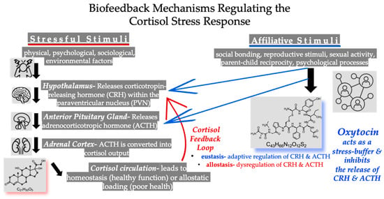
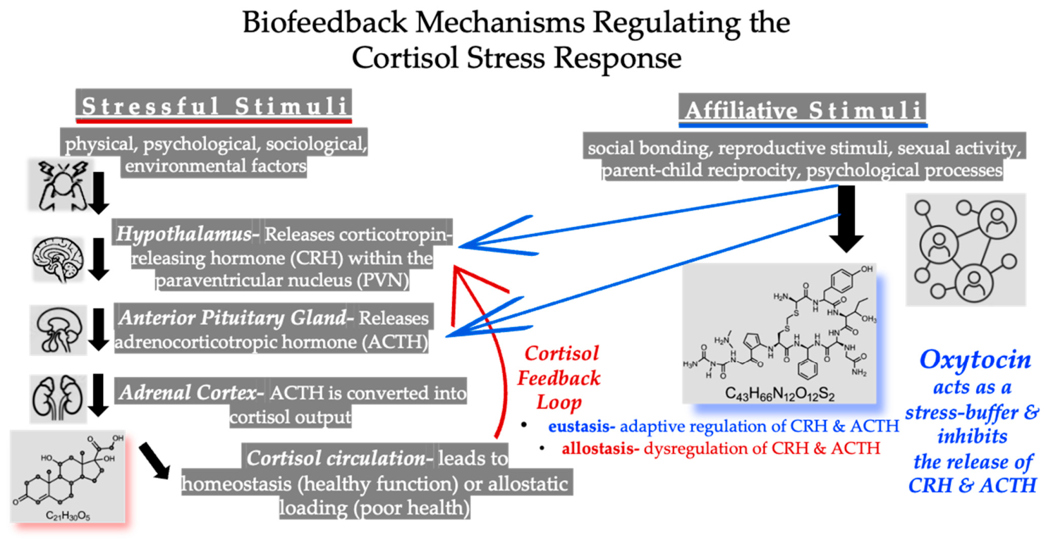
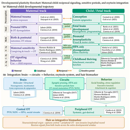
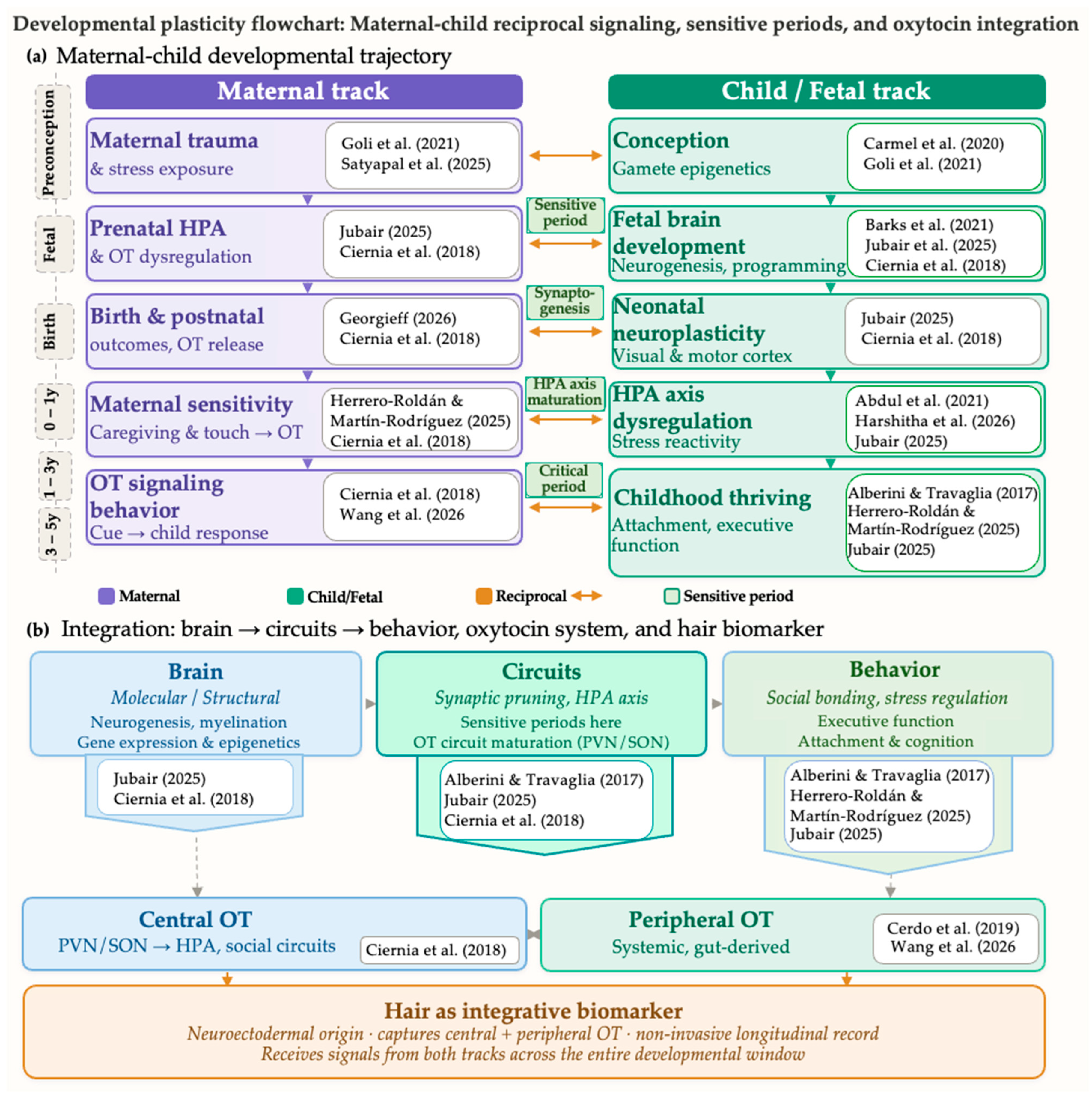
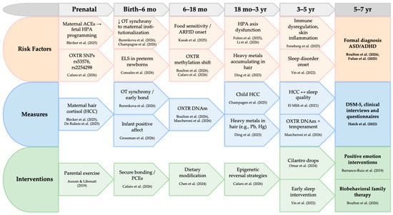
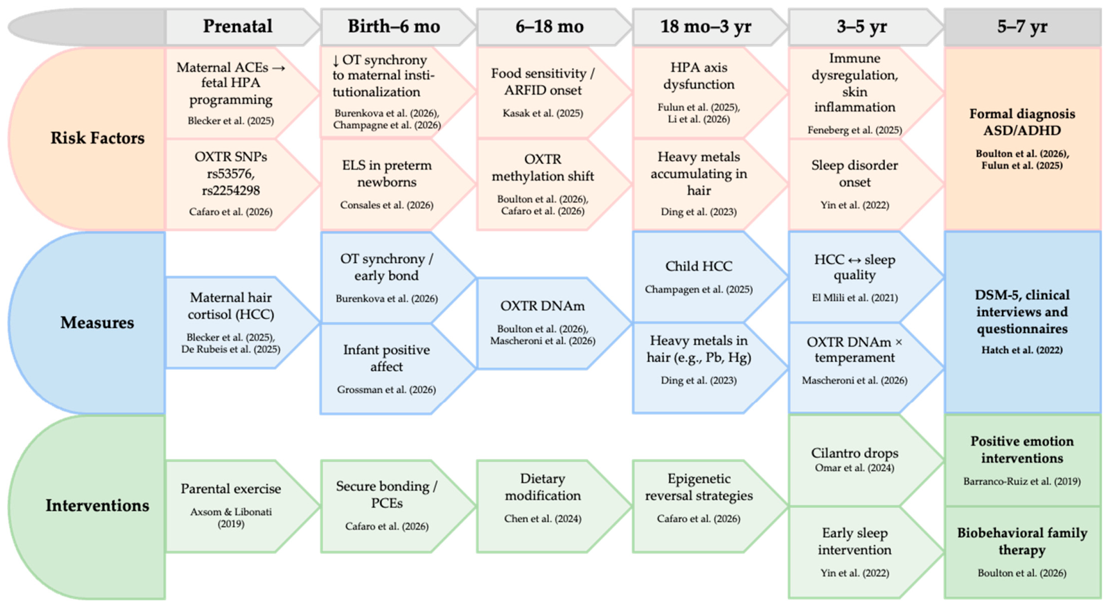
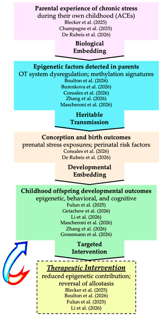
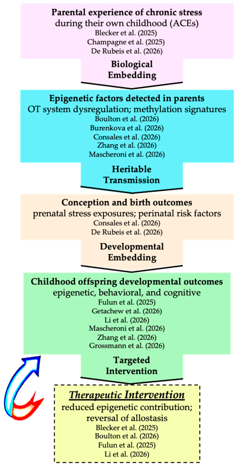

Disclaimer/Publisher’s Note: The statements, opinions and data contained in all publications are solely those of the individual author(s) and contributor(s) and not of MDPI and/or the editor(s). MDPI and/or the editor(s) disclaim responsibility for any injury to people or property resulting from any ideas, methods, instructions or products referred to in the content. |
© 2026 by the authors. Published by MDPI on behalf of the Journal of Mind and Medical Sciences. Licensee MDPI, Basel, Switzerland. This article is an open access article distributed under the terms and conditions of the Creative Commons Attribution (CC BY) license.