


Simple Summary
Orthodontists use head X-rays to measure the teeth and jaws when planning treatment. We reviewed 20 studies comparing measurements produced by artificial intelligence with those made by clinicians. Results varied across software programs and measurements. Small differences on average sometimes hid larger errors in individual patients. Checking and correcting automatically marked points reduced some errors but did not eliminate all differences. Variation in study methods and incomplete reporting limited firm conclusions. Artificial intelligence may assist orthodontic assessment, but its measurements still need professional checking. Future studies should clearly define acceptable errors and use consistent comparison methods.
Abstract
Background: Artificial intelligence has revolutionized the field of dentistry. This systematic review evaluated the clinical performance of artificial intelligence (AI)-assisted cephalometric tracing compared with manual or expert digital tracing. Methods: We searched the PubMed/MEDLINE, Scopus, and Web of Science databases from inception to 20 May 2026. Studies directly comparing AI-assisted tracing with manual or expert digital tracing of the same lateral cephalograms and reporting at least one clinically relevant cephalometric outcome were included in the review. Two authors independently screened records. Risk of bias was assessed using an adapted QUADAS-2 framework. A quantitative meta-analysis was planned but not performed. Findings were synthesised narratively by measurement, platform, and workflow, with reporting informed by the Synthesis Without Meta-analysis (SWiM) guideline. Results: Twenty studies were included. Direct AI-versus-reference ICCs were reported in more than three studies, but their formulations and reference procedures varied. Small average differences sometimes coexisted with wide individual limits of agreement. The numerical evidence did not establish a consistent ranking of SNA, SNB, and ANB. Conclusions: AI-assisted tracing may be useful as a clinician-verified adjunct, but its performance depends on the measurement, platform, and workflow. The evidence does not establish uniform clinical interchangeability with manual or expert digital tracing.
1. Introduction
Since its introduction in 1931, the lateral cephalometric (LC) radiograph has been a fundamental diagnostic imaging tool for the diagnosis and treatment planning of orthodontic cases. It provides a standardised two-dimensional (2D) map of the craniofacial complex, which guides orthodontists accurately [1]. Thus, the LC radiograph is primarily used to assess a patient’s progressive response to treatment and provide a comprehensive view of the relationship between the patient’s skeletal, dental, and soft tissue morphology [1]. In cephalometric analyses, the orthodontist needs to manually detect anatomical landmarks, and this process is time-consuming. In addition, some variability may appear in the results because of image quality and observer skills. In recent years, artificial intelligence (AI) has become helpful in improving diagnostic accuracy, automating analyses, and reducing errors and measurement bias, which improves treatment outcomes. Furthermore, AI can detect cephalometric landmarks in less than one minute. It can also determine which landmarks require more time to identify and which are identified incorrectly. However, the efficiency of AI systems has not yet reached the level of an expert because of the complexity of datasets influenced by factors such as age, gender, size and shape of anatomical structures, class of malocclusion, ethnicity, and bone density. Greater dataset complexity makes AI learning and prediction more challenging, which may increase analytical errors and consequently lead to inaccuracies in diagnosis and treatment planning [2,3,4]. While AI has demonstrated promising capabilities in cephalometric landmark identification, the clinically relevant question is not whether AI can function independently but whether its performance is comparable to that of experienced clinicians. Recent comparative studies have shown that the accuracy of AI-based cephalometric analysis varies considerably across different software platforms and measurement parameters. Research conducted by Bor et al. noted statistically significant differences in several of the main angular and linear measurements produced by AI-assisted tracing software, as compared with those created through an orthodontist performing a manual digital tracing process. The research also indicated that these differences may be due to the methods used to generate these measurements, which can ultimately affect diagnostic conclusions [5]. Additionally, Baig et al. reported similar findings when they evaluated commercially developed AI-based tracing software, which produced measurements that differed from expert-created measurements and was unreliable in measuring some of the most important cephalometric parameters [2]. Besides comparing AI-based tracing with expert-created measurements, Mercier et al. examined the efficiency of completely automated and semi-automated AI systems. They found that, although complete automation significantly reduced the time required to perform the analysis, semi-automation achieved higher levels of accuracy and was more closely aligned with traditional digital tracing results because clinicians could verify and correct the data [6]. These findings suggest that clinician involvement remains essential and that corrected or semi-automated AI workflows may currently offer greater clinical reliability than fully automated analyses alone. Previous reviews have examined both landmark localisation and derived clinical measurements [7,8,9]. Polizzi et al. reviewed fully automatic linear and angular measurements [7], and Narkhede et al. compared digital with manual tracing using meta-analysis [8]. The present review therefore provides an incremental update, rather than the first synthesis of clinical cephalometric measurements. It focuses on direct AI–human comparisons and distinguishes inter-method agreement from examiner reliability, software repeatability, and clinician-corrected workflows. Its additional contribution is the measurement- and platform-specific numerical synthesis of the 20 included studies, not heterogeneity itself.
2. Materials and Methods
2.1. Study Protocol and Registration
This systematic review was conducted in accordance with the Preferred Reporting Items for Systematic Reviews and Meta-Analyses (PRISMA) guidelines 2020. The protocol was registered with INPLASY (INPLASY202660101) on 21 June 2026, at the data-extraction stage [10]. Registration followed the searches conducted on 20 May 2026 and was not prospective to study selection. The registered strategy allowed narrative synthesis and meta-analysis of compatible estimates. No meta-analysis was performed. During revision, study-level numerical reporting was expanded and the grouping and interpretation of the narrative synthesis were clarified. The original eligibility criteria and 20-study synthesis set were retained.
2.2. Search Question
The research question was based on PICO elements and formulated as follows: ‘What is the performance of AI-assisted cephalometric analysis compared with conventional methods?’
Accordingly, PICO elements were as follows:
Population Patients indicated for cephalometry.
Intervention AI-assisted cephalometry.
Comparator Conventional approaches, including human experts’ performance in cephalometry.
Outcome Performance of AI-assisted cephalometry.
2.3. Eligibility Criteria
The inclusion criteria comprised studies that reported subjects who had standard LC X-rays or digital lateral cephalograms, along with a direct comparison of AI-assisted cephalometric analysis versus manual/traditional digital tracing by an expert, as well as studies that reported clinically relevant cephalometric output(s), including but not limited to angular/linear measurements, diagnostic category, agreement metrics, reproducibility metrics, and time outcomes. On the other hand, studies were excluded from the primary clinical review if their main purpose was to assess the performance of reduced-field prediction of missing cephalometric values, education/student training, single-image reproducibility without patient-level diagnostic comparison, or landmark detection without clinically relevant cephalometric output.
2.4. Search Strategy
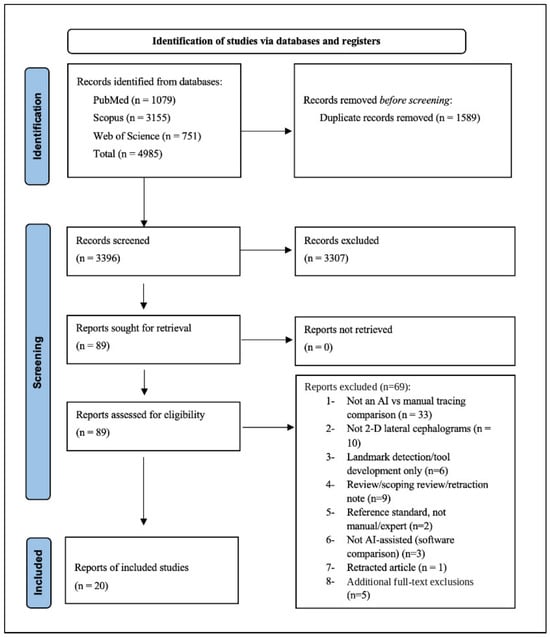
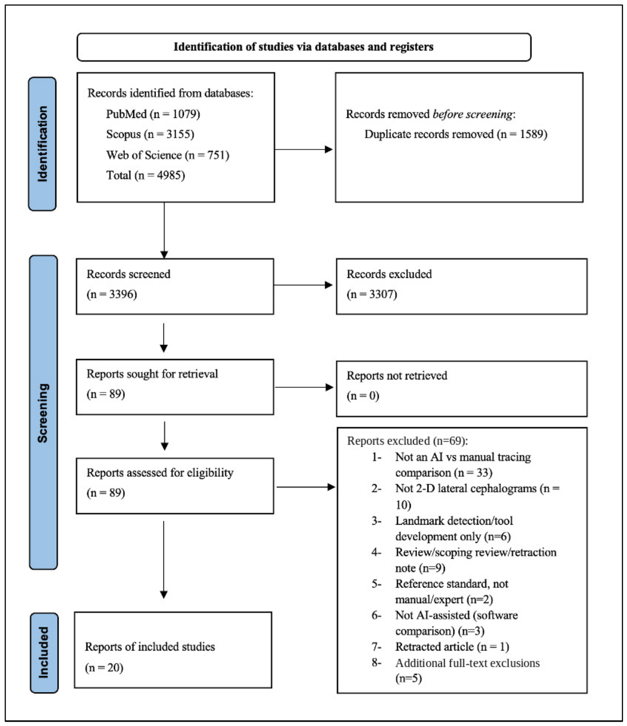
The searches of PubMed/MEDLINE, Scopus, and Web of Science were conducted on 20 May 2026, covering records from database inception. The search strategy for each database is detailed in Table 1. All retrieved records were uploaded to Rayyan®, where duplicates were identified and removed. Two authors independently assessed the titles and abstracts against the eligibility criteria. After that, the same two authors screened the full texts of the relevant studies, and any uncertainty was resolved through group discussion with the senior author. The reviewers then determined the studies suitable for data extraction and quality assessment. This process followed the PRISMA flowchart (Figure 1).
Table 1.
Search strategies for the databases searched on 20 May 2026.
Figure 1.
Flowchart of PRISMA.
2.5. Data Extraction
Data were extracted for study design, population, sample size, AI platform, human reference, cephalometric measurements, and reported statistical comparisons. Direct AI–reference agreement was distinguished from examiner reliability and software repeatability. Fully automatic and clinician-corrected outputs were retained separately. Signed mean differences, mean absolute errors, confidence intervals, Bland–Altman limits of agreement, ICC formulations, and numerical clinical acceptability criteria were recorded where available. Table 2 and Table 4 identify the statistical meaning and source of the results. Missing numerical information was not imputed or interpreted as evidence of acceptable agreement.
2.6. Quality Assessment
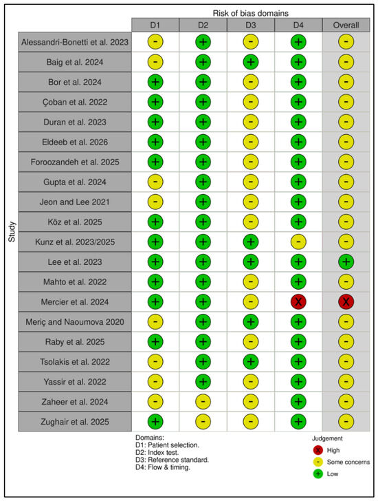
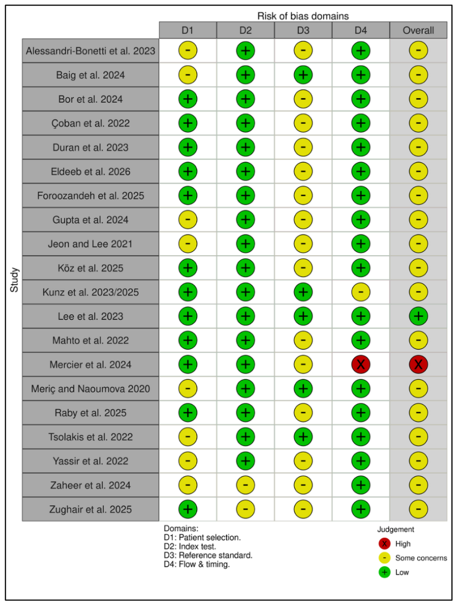
Risk of bias was assessed using an adapted QUADAS-2 framework covering patient selection, the AI index test, the human reference standard, and flow and timing [11]. The framework was used because these studies compared paired image-based measurements with a human reference, rather than primarily developing or validating multivariable clinical prediction models. PROBAST addresses prediction-model risk of bias and was not used as a substitute for appraisal of measurement agreement [12]. The QUADAS-2 adaptation is not a validated instrument for continuous method-comparison studies. Patient-selection judgments considered recruitment, sampling, and exclusions, rather than sample size alone. Index-test judgments considered whether automatic outputs or clinician corrections were obtained independently of the reference. Reference-standard judgments considered examiner expertise, independent tracing, repeatability, and consensus procedures; a single examiner was not automatically judged to create high risk. Flow-and-timing judgments considered whether all selected images received both measurements and whether failures or exclusions depended on AI performance. Each domain was rated low, unclear, or high risk; overall risk was high if any domain was high, low only if all domains were low, and unclear otherwise. Applicability was considered separately. Two reviewers assessed the studies independently and resolved disagreements through discussion, as specified in the original Methods and registered protocol [10]. The screening process also involved two authors. Table 3 presents the corrected judgments, with their reasons explained below. Figure 2 displays the same risk-of-bias judgments.
Figure 2.
Traffic-light display of the adapted QUADAS-2 risk-of-bias assessment for the 20 included studies. Domain and overall judgments correspond to Table 3. Each row represents one of the 20 included studies. Green denotes low risk, yellow denotes unclear risk (labelled “some concerns” in the graphic), and red denotes high risk. D1, patient selection; D2, index test; D3, reference standard; D4, flow and timing. The domain and overall risk-of-bias ratings correspond to Table 3. These ratings are not certainty-of-evidence grades [2,5,6,13,14,15,16,17,18,19,20,21,22,23,24,25,26,27,28,29].
Table 2.
Data extraction of the retrieved studies.
Table 3.
Methodological quality and risk of bias of the included studies according to the modified QUADAS-2 tool.
2.7. Evidence Certainty
No formal certainty-of-evidence framework was applied. Interpretation considered recruitment and exclusion procedures, reference-standard limitations, imprecision, inconsistency across measurements and software, and incomplete numerical reporting. Domain-level risk-of-bias judgments were not converted into a certainty rating for the body of evidence.
2.8. Data Synthesis
No quantitative meta-analysis was performed. The narrative synthesis was organised by clinical measurement, AI platform, human reference, and fully automatic versus clinician-corrected workflow. Multiple software or correction arms using the same images were retained as separate comparisons but not counted as independent samples. Direct AI–reference ICCs were separated from examiner reliability, software repeatability, and coefficients combining several methods. The ICC model, absolute-agreement or consistency definition, and single- or average-measure unit were recorded when reported [30]. Signed mean differences, mean absolute errors, and Bland–Altman limits of agreement were considered separately. Numerical estimates, uncertainty, and the original studies’ clinical margins took priority over significance tests or qualitative labels. Conflicting findings were interpreted within their population, platform, reference, workflow, and risk-of-bias context; no significance-based vote counting or statistical weighting was used. A clinical margin not defined by the source was recorded as absent. During revision, mean-difference pooling was considered separately from ICC pooling. Paired standard deviations were available in Çoban, Duran, Gupta, and Jeon and Lee, and mean-bias confidence intervals were available in other reports [6,14,15,16,18,19,21,22,27]. A restricted synthesis of average bias was therefore feasible in principle. It was not undertaken because an average-bias estimate alone would not answer the principal question of individual clinical interchangeability, and compatible individual-agreement estimates and clinical margins were not consistently available. This was a decision not to implement the protocol’s conditional mean-difference analysis, not evidence that all quantitative synthesis was impossible. Reporting followed SWiM where applicable [31]. No pooled agreement estimate, equivalence estimate, or statistical ranking of SNA, SNB, and ANB was calculated. Descriptive analyses were performed in Stata 19/BE (StataCorp, 2025. Stata Statistical Software: Release 19. College Station, TX, USA: StataCorp LLC).
3. Results
The searches identified 4985 records: PubMed/MEDLINE, 1079; Scopus, 3155; and Web of Science, 751. Records were uploaded to Rayyan for deduplication. After removal of 1589 duplicates, two authors screened 3396 records and excluded 3307. The same two authors assessed 89 full-text reports. The recorded exclusions comprised 64 reports at the initial full-text stage and five additional reports at detailed full-text assessment, giving 69 excluded reports and 20 included studies. Table S1 provides the original reasons for these five decisions [32,33,34,35,36]. Lee Part 2 was excluded to avoid duplicate use of the 84-cephalogram cohort already represented by Part 1 [22,35]. Panesar was excluded because its ICCs assessed intra-rater precision and its gold-standard comparison used mean absolute error rather than a poolable AI–reference ICC [36]. These were not exclusions based on QUADAS-2 ratings. Figure 1 presents the reconciled counts.
3.1. Risk of Bias Assessment
Patient selection was rated low risk in 12 studies and unclear in eight. The index-test domain was low risk in 18 studies and unclear in two; the reference-standard domain was low risk in five and unclear in 15. Flow and timing were low risk in 18 studies, unclear in one, and high risk in one. Overall, one study was low risk, 18 were unclear, and one was high risk. Applicability concerns were predominantly unclear: 16 studies were unclear, three low, and one high. Table 3 reports these judgments, and Figure 2 displays the same domain and overall risk-of-bias ratings.
The judgments concern recruitment and exclusions, not sample size alone. In Alessandri-Bonetti et al., the sample comprised records with existing AI analyses; selection independent of successful AI processing was unclear, but a sample of 13 was not itself evidence of high selection bias [13]. In Gupta et al., the reporting of recruitment and the reference procedure supported uncertainty rather than a high-risk rating based only on sample size or unstated examiner experience [18]. Kunz et al. randomly selected 50 images from 3000 and excluded three for radiographic artefacts, not documented AI-recognition failures; flow was therefore rated unclear rather than high [21]. Mahto et al. randomly selected eligible images, supporting low selection risk despite the small sample [23]. By contrast, Mercier et al. excluded 25 of 433 images because AI could not recognise or adequately landmark them, supporting high flow-and-timing risk [6]. Duran and Raby used randomly selected clinical images but imposed image-quality or anatomical restrictions; the independence of their single-examiner reference from the AI results was not clearly established [15,25]. Eldeeb used random selection, randomised software order, and a blinded statistician, but the tracing operator was not blinded [16]. Unreported independence and restricted patient or image spectra were retained as uncertainty or applicability concerns rather than assumed absent.
3.2. Synthesis of Findings
Table 4 reports numerical findings for all 20 included studies, including the additional software arms and clinician-corrected comparisons. Direct AI–reference ICCs were reported by Çoban, Duran, Köz, Lee, Mahto, Raby, Tsolakis, and Yassir [14,15,20,22,23,25,26,27]. Eldeeb also reported between-method ICC results, separately from examiner reliability [16]. Foroozandeh and Gupta reported coefficients combining several methods; these do not estimate a single AI–manual pair [17,18]. For SNA, SNB, and ANB, respectively, Köz reported ICCs of 0.692 (95% CI 0.390–0.826), 0.883 (0.829–0.920), and 0.845 (0.434–0.934); Mahto reported 0.879 (0.745–0.943), 0.899 (0.770–0.954), and 0.908 (0.747–0.961) [20,23]. Neither study defined a numerical angular acceptability margin. Duran reported corresponding ICCs of 0.750/0.846/0.876 for OrthoDx and 0.736/0.862/0.791 for WebCeph against Dolphin, with signed differences and paired standard deviations reported in Table 4 [15]. Raby reported automatic CephX ICCs of 0.93/0.96/0.95 and automatic WebCeph ICCs of 0.81/0.91/0.83, yet its assessment of individual differences supported SNA and SNB only after landmark correction [25]. Its single confidence-interval column could not be assigned to the four software/workflow estimates and was not treated as four separate intervals. The evidence did not support a uniform ranking of SNB, SNA, and ANB. Baig reported a human-minus-WebCeph SNB difference of −2.3°, exceeding that study’s 2° clinical convention [2]. In Lee, automatic WebCeph SNB bias was −0.24° (95% CI −0.71 to 0.23), but the 95% limits of agreement were −4.48° to 4.00°; none of the four automatic platforms met the study’s interchangeability criteria for all three sagittal angles [22]. A missing clinical threshold or a non-significant test was not interpreted as evidence of equivalence. Eldeeb’s main text reported WebCeph–manual ICCs above 0.8 except for 1u–NA (0.529), and Carestream–manual ICCs of 0.386–0.833 except for anterior facial height (0.970); exact parameter-level between-method values were in a separate supplement that could not be retrieved [16].
Table 4.
Numerical synthesis of the original 20 included studies.
4. Discussion
This review examined how closely AI-assisted cephalometric tracing reproduces manual or expert digital measurements. The results support a measurement- and platform-specific interpretation, rather than uniformly good agreement. A small mean difference can coexist with wide limits of agreement for individual patients, as illustrated by the sagittal results in Lee et al. [22]. Non-significant ANOVA or paired-test results do not establish equivalence. A consistency ICC may remain high despite systematic bias; an absolute-agreement ICC incorporates disagreement attributable to such bias but also depends on sample variability, model and measurement unit [30]. Neither form alone establishes clinical interchangeability. Signed bias, mean absolute error, confidence intervals, limits of agreement, and a justified clinical margin answer different questions and were not treated as interchangeable outcomes.
Previous reviews have addressed both landmark localisation and clinically derived measurements. Schwendicke et al. and de Queiroz et al. synthesised landmark-detection performance [37,38], while Hendrickx et al. pooled landmark-error data from 27 two-dimensional studies [3]. These landmark-level findings cannot by themselves establish acceptable error for a particular clinical angle. The present review therefore interprets clinical measurements separately, without claiming that earlier syntheses were confined to landmark error.
Polizzi et al. included 10 studies of fully automatic clinical linear and angular measurements and searched through 22 March 2025 [7]. Seven also appear in the present review: Duran, Mahto, Jeon and Lee, Meriç and Naoumova, Yassir, Mercier, and Zaheer. Their remaining three studies were Prince, El-Dawlatly, and Sadek; the present 20-study set contains 13 other studies. Narkhede et al. included 20 studies of digital-versus-manual tracing published from January 2013 to July 2023, with 14 contributing to meta-analysis, including SNA, SNB, and ANB [8]. None of its 20 Table 1 entries is the same report as the 20 studies in the present review; its Mahto report is from 2016, not the 2022 study included here. Narkhede’s review was not restricted to AI. In contrast, our synthesis is confined to direct AI–human clinical comparisons and separates inter-method agreement, repeatability, and corrected workflows. These differences define an incremental contribution; they do not establish that clinical measurement synthesis is new or that this review supersedes the earlier reviews.
Clinician correction reduced some discrepancies but did not establish universal interchangeability. Mercier et al. reported smaller sagittal mean differences after correction, while Yassir et al. reported persistent error for the lower-incisor-to-mandibular-plane angle even after correction [6,27]. Fully automatic and corrected results were therefore reported separately; neither workflow was pooled. The platform- and parameter-specific variability in Baig and Bor also argues against interpreting a single overall accuracy label as evidence that all clinical measurements are acceptable [2,5].
Strengths and Limitations
Strengths include independent screening by two authors and a numerical synthesis that distinguishes inter-method agreement from repeatability. The search was restricted to PubMed/MEDLINE, Scopus, and Web of Science; missed studies and publication bias cannot be excluded. Registration occurred at the data-extraction stage, after study selection. The adapted QUADAS-2 framework has not been validated specifically for continuous cephalometric agreement studies, and no formal certainty framework was applied. Some agreement statistics were incompletely specified: Duran did not report the ICC model, form, or unit, and Raby provided an unassigned confidence-interval column alongside four software-specific ICCs [15,25]. Eldeeb’s separate between-method ICC supplement was not available for parameter-level extraction [16]. Reference methods, patient spectra, platforms, correction procedures, and clinical margins varied. Although average-bias pooling was feasible in principle for some comparisons, it was not performed; no pooled agreement estimate or formal between-parameter ranking is available. Findings for sagittal angles should not be generalised to all vertical, dental, or soft-tissue measurements.
5. Conclusions
AI-assisted cephalometric tracing may be useful as a clinician-verified adjunct, but performance depends on the measurement, platform, and workflow. The available evidence does not establish uniformly acceptable SNA, SNB, or ANB measurements, a consistent ranking of these parameters, or interchangeability for individual patients. Interpretation is limited by heterogeneous methods, reference-standard concerns, incomplete numerical reporting, and the absence of a pooled estimate or formal certainty assessment. AI-assisted analysis should complement rather than replace expert verification. Future studies should use multi-observer or consensus reference standards and report agreement across skeletal, dental, and soft-tissue parameters.
Supplementary Materials
The following supporting information can be downloaded at: https://www.mdpi.com/article/10.3390/tomography12100146/s1.
Author Contributions
H.A.A.: Conceptualization, Formal analysis, Writing—review & editing, Supervision. H.A. (Huda Alnami): Investigation, Writing—original draft. A.A.A.: Investigation, Writing—original draft, Writing—review & editing. H.A. (Hana Almujthel): Formal analysis, Writing—original draft, Writing—review & editing. Z.A.: Methodology and data curation. M.A.: Writing—original draft and resources. All authors have read and agreed to the published version of the manuscript.
Funding
The authors extend their appreciation to the Deanship of Research and Graduate Studies at King Khalid University, KSA, for funding this work through the General Research Project (grant number RGP2/494/47). The sponsors had no role in the design, execution, interpretation, or writing of the study.
Institutional Review Board Statement
Not applicable.
Informed Consent Statement
Not applicable.
Data Availability Statement
All data analysed are included in this study. Further enquiries are available upon request from the corresponding author.
Acknowledgments
During the preparation of the manuscript, a generative AI assistant was used to support drafting and language editing. In addition, professional human language-editing services were used. All study design, data extraction, statistical analysis, interpretation, content review, and manuscript revisions were performed by the authors. The authors reviewed and verified each other’s work and accept full responsibility for the published content.
Conflicts of Interest
The authors declare no conflict of interest.
References
- Dinesh, A.; Mutalik, S.; Feldman, J.; Tadinada, A. Value-Addition of Lateral Cephalometric Radiographs in Orthodontic Diagnosis and Treatment Planning. Angle Orthod. 2020, 90, 665–671. [Google Scholar] [CrossRef] [Scilit] [PubMed]
- Baig, N.; Gyasudeen, K.S.; Bhattacharjee, T.; Chaudhry, J.; Prasad, S. Comparative Evaluation of Commercially Available AI-Based Cephalometric Tracing Programs. BMC Oral Health 2024, 24, 1241. [Google Scholar] [CrossRef] [Scilit] [PubMed]
- Hendrickx, J.; Gracea, R.S.; Vanheers, M.; Winderickx, N.; Preda, F.; Shujaat, S.; Jacobs, R. Can Artificial Intelligence-Driven Cephalometric Analysis Replace Manual Tracing? A Systematic Review and Meta-Analysis. Eur. J. Orthod. 2024, 46, cjae029. [Google Scholar] [CrossRef] [Scilit] [PubMed]
- Ribas-Sabartés, J.; Sánchez-Molins, M.; d’Oliveira, N.G. The Accuracy of Algorithms Used by Artificial Intelligence in Cephalometric Points Detection: A Systematic Review. Bioengineering 2024, 11, 1286. [Google Scholar] [CrossRef] [Scilit] [PubMed]
- Bor, S.; Ciğerim, S.Ç.; Kotan, S. Comparison of AI-Assisted Cephalometric Analysis and Orthodontist-Performed Digital Tracing Analysis. Prog. Orthod. 2024, 25, 41. [Google Scholar] [CrossRef] [Scilit] [PubMed]
- Mercier, J.-P.; Rossi, C.; Sanchez, I.N.; Renovales, I.D.; Sahagún, P.M.-P.; Templier, L. Reliability and Accuracy of Artificial Intelligence-Based Software for Cephalometric Diagnosis. A Diagnostic Study. BMC Oral Health 2024, 24, 1309. [Google Scholar] [CrossRef] [Scilit] [PubMed]
- Polizzi, A.; Nucci, L.; Serra, S.; Isola, G.; Leonardi, R. Accuracy of Artificial Intelligence Fully-Automatic Cephalometric Analysis in Linear and Angular Measurement: A Critical Scoping Review. Semin. Orthod. 2026, 32, 397–403. [Google Scholar] [CrossRef] [Scilit]
- Narkhede, S.; Rao, P.; Sawant, V.; Sachdev, S.S.; Arora, S.; Pawar, A.M.; Reda, R.; Testarelli, L. Digital versus Manual Tracing in Cephalometric Analysis: A Systematic Review and Meta-Analysis. J. Pers. Med. 2024, 14, 566. [Google Scholar] [CrossRef] [Scilit] [PubMed]
- Polizzi, A.; Leonardi, R. Automatic Cephalometric Landmark Identification with Artificial Intelligence: An Umbrella Review of Systematic Reviews. J. Dent. 2024, 146, 105056. [Google Scholar] [CrossRef] [Scilit] [PubMed]
- INPLASY. Protocol Registration; INPLASY202660101; INPLASY: Middletown, DE, USA, 2026. [Google Scholar] [CrossRef] [Scilit]
- Whiting, P.F.; Rutjes, A.W.S.; Westwood, M.E.; Mallett, S.; Deeks, J.J.; Reitsma, J.B.; Leeflang, M.M.G.; Sterne, J.A.C.; Bossuyt, P.M.M.; QUADAS-2 Group. QUADAS-2: A Revised Tool for the Quality Assessment of Diagnostic Accuracy Studies. Ann. Intern. Med. 2011, 155, 529–536. [Google Scholar] [CrossRef] [Scilit] [PubMed]
- Wolff, R.F.; Moons, K.G.M.; Riley, R.D.; Whiting, P.F.; Westwood, M.; Collins, G.S.; Reitsma, J.B.; Kleijnen, J.; Mallett, S.; PROBAST Group. PROBAST: A Tool to Assess the Risk of Bias and Applicability of Prediction Model Studies. Ann. Intern. Med. 2019, 170, 51–58. [Google Scholar] [CrossRef] [Scilit] [PubMed]
- Alessandri-Bonetti, A.; Sangalli, L.; Salerno, M.; Gallenzi, P. Reliability of Artificial Intelligence-Assisted Cephalometric Analysis. A Pilot Study. BioMedInformatics 2023, 3, 44–53. [Google Scholar] [CrossRef] [Scilit]
- Çoban, G.; Öztürk, T.; Hashimli, N.; Yağci, A. Comparison between Cephalometric Measurements Using Digital Manual and Web-Based Artificial Intelligence Cephalometric Tracing Software. Dent. Press J. Orthod. 2022, 27, e222112. [Google Scholar] [CrossRef] [Scilit] [PubMed]
- Duran, G.S.; Gökmen, Ş.; Topsakal, K.G.; Görgülü, S. Evaluation of the Accuracy of Fully Automatic Cephalometric Analysis Software with Artificial Intelligence Algorithm. Orthod. Craniofac. Res. 2023, 26, 481–490. [Google Scholar] [CrossRef] [Scilit] [PubMed]
- Eldeeb, O.A.K.; Yacout, Y.M.; Abouelkheir, H.M.; Gaweesh, Y.S.E.; Elkersh, N.M. Evaluating a Cost-Effective, Web-Based AI Platform for Lateral Cephalometric Analysis: A Comparative in-Silico Study. BMC Oral Health 2026, 26, 7. [Google Scholar] [CrossRef] [Scilit] [PubMed]
- Foroozandeh, M.; Salemi, F.; Shokri, A.; Farhadian, N.; Aeini, N.; Hassanzadeh, R. Comparative Accuracy of Artificial Intelligence-Based AudaxCeph Software, Dolphin Software, and the Manual Technique for Orthodontic Landmark Identification and Tracing of Lateral Cephalograms. Imaging Sci. Dent. 2025, 55, 11–21. [Google Scholar] [CrossRef] [Scilit] [PubMed]
- Gupta, S.; Shetty, S.; Natarajan, S.; Nambiar, S.; MV, A.; Agarwal, S. A Comparative Evaluation of Concordance and Speed between Smartphone App-Based and Artificial Intelligence Web-Based Cephalometric Tracing Software with the Manual Tracing Method: A Cross-Sectional Study. J. Clin. Exp. Dent. 2024, 16, e11–e17. [Google Scholar] [CrossRef] [Scilit] [PubMed]
- Jeon, S.; Lee, K.C. Comparison of Cephalometric Measurements between Conventional and Automatic Cephalometric Analysis Using Convolutional Neural Network. Prog. Orthod. 2021, 22, 14. [Google Scholar] [CrossRef] [Scilit] [PubMed]
- Koz, S.; Uslu-Akcam, O. Artificial Intelligence-Supported and App-Aided Cephalometric Analysis: Which One Can We Trust? Diagnostics 2025, 15, 559. [Google Scholar] [CrossRef] [Scilit] [PubMed]
- Kunz, F.; Stellzig-Eisenhauer, A.; Widmaier, L.M.; Zeman, F.; Boldt, J. Assessment of the Quality of Different Commercial Providers Using Artificial Intelligence for Automated Cephalometric Analysis Compared to Human Orthodontic Experts. J. Orofac. Orthop. 2025, 86, 145–160. [Google Scholar] [CrossRef] [Scilit] [PubMed]
- Lee, J.; Bae, S.-R.; Noh, H.-K. Commercial Artificial Intelligence Lateral Cephalometric Analysis: Part 1-the Possibility of Replacing Manual Landmarking with Artificial Intelligence Service. J. Clin. Pediatr. Dent. 2023, 47, 106–118. [Google Scholar] [CrossRef] [Scilit] [PubMed]
- Mahto, R.K.; Kafle, D.; Giri, A.; Luintel, S.; Karki, A. Evaluation of Fully Automated Cephalometric Measurements Obtained from Web-Based Artificial Intelligence Driven Platform. BMC Oral Health 2022, 22, 132. [Google Scholar] [CrossRef] [Scilit] [PubMed]
- Meric, P.; Naoumova, J. Web-Based Fully Automated Cephalometric Analysis: Comparisons between App-Aided, Computerized, and Manual Tracings. Turk. J. Orthod. 2020, 33, 142–149. [Google Scholar] [CrossRef] [Scilit] [PubMed]
- Raby, I.; Rojas, V.; Celis, A.; García-Duhalde, C.; Martinac, M. Accuracy and Reliability of 2 Artificial Intelligence Platforms for Cephalometric Analysis Compared with a Semiautomatic Computer Program. Am. J. Orthod. Dentofac. Orthop. 2025, 168, 505–514. [Google Scholar] [CrossRef] [Scilit] [PubMed]
- Tsolakis, I.A.; Tsolakis, A.I.; Elshebiny, T.; Matthaios, S.; Palomo, J.M. Comparing a Fully Automated Cephalometric Tracing Method to a Manual Tracing Method for Orthodontic Diagnosis. J. Clin. Med. 2022, 11, 6854. [Google Scholar] [CrossRef] [Scilit] [PubMed]
- Yassir, Y.A.; Salman, A.R.; Nabbat, S.A. The Accuracy and Reliability of WebCeph for Cephalometric Analysis. J. Taibah Univ. Med. Sci. 2022, 17, 57–66. [Google Scholar] [CrossRef] [Scilit] [PubMed]
- Zaheer, R.; Shafique, H.Z.; Khalid, Z.; Shahid, R.; Jan, A.; Zahoor, T.; Nawaz, R.; ul Hassan, M. Comparison of Semi and Fully Automated Artificial Intelligence Driven Softwares and Manual System for Cephalometric Analysis. BMC Med. Inform. Decis. Mak. 2024, 24, 271. [Google Scholar] [CrossRef] [Scilit] [PubMed]
- Zughair, S.A.K.; Ishaq, R.A.R.; Al-dossary, O.A.I.; Aldhorae, K.; Saber, N.H.; Elayah, S.A. Evaluation of Artificial Intelligence-Based Cephalometric Tracing versus Semi-Automatic and Manual Tracing. BMC Oral Health 2025, 25, 1429. [Google Scholar] [CrossRef] [Scilit] [PubMed]
- Koo, T.K.; Li, M.Y. A Guideline of Selecting and Reporting Intraclass Correlation Coefficients for Reliability Research. J. Chiropr. Med. 2016, 15, 155–163. [Google Scholar] [CrossRef] [Scilit] [PubMed]
- Campbell, M.; McKenzie, J.E.; Sowden, A.; Katikireddi, S.V.; Brennan, S.E.; Ellis, S.; Hartmann-Boyce, J.; Ryan, R.; Shepperd, S.; Thomas, J.; et al. Synthesis without Meta-analysis (SWiM) in Systematic Reviews: Reporting Guideline. BMJ 2020, 368, l6890. [Google Scholar] [CrossRef] [Scilit] [PubMed]
- Zecca, P.A.; Caccia, M.; Levrini, L.; Carganico, A.; Serafin, M.; Basso, P.R.; Reguzzoni, M. An Open-Source AI Tool for Predicting Cephalometric Measurements from Clinical Data and Photographic Images. BMC Oral Health 2025, 25, 1524. [Google Scholar] [CrossRef] [Scilit] [PubMed]
- Fracchia, D.E.; Bignotti, D.; Lai, S.; Cubeddu, S.; Curreli, F.; Lombardo, M.; Verdecchia, A.; Spinas, E. Reproducibility of AI in Cephalometric Landmark Detection: A Preliminary Study. Diagnostics 2025, 15, 2521. [Google Scholar] [CrossRef] [Scilit] [PubMed]
- Lin, J.; Liao, Z.; Dai, J.; Wang, M.; Yu, R.; Yang, H.; Liu, C. Digital and Artificial Intelligence-Assisted Cephalometric Training Effectively Enhanced Students’ Landmarking Accuracy in Preclinical Orthodontic Education. BMC Oral Health 2025, 25, 623. [Google Scholar] [CrossRef] [Scilit] [PubMed]
- Lee, J.; Bae, S.-R.; Noh, H.-K. Commercial Artificial Intelligence Lateral Cephalometric Analysis: Part 2-Effects of Human Examiners on Artificial Intelligence Performance, a Pilot Study. J. Clin. Pediatr. Dent. 2023, 47, 130–141. [Google Scholar] [CrossRef] [Scilit] [PubMed]
- Panesar, S.; Zhao, A.; Hollensbe, E.; Wong, A.; Bhamidipalli, S.S.; Eckert, G.; Dutra, V.; Turkkahraman, H. Precision and Accuracy Assessment of Cephalometric Analyses Performed by Deep Learning Artificial Intelligence with and without Human Augmentation. Appl. Sci. 2023, 13, 6921. [Google Scholar] [CrossRef] [Scilit]
- Schwendicke, F.; Chaurasia, A.; Arsiwala, L.; Lee, J.-H.; Elhennawy, K.; Jost-Brinkmann, P.-G.; Demarco, F.; Krois, J. Deep Learning for Cephalometric Landmark Detection: Systematic Review and Meta-Analysis. Clin. Oral Investig. 2021, 25, 4299–4309. [Google Scholar] [CrossRef] [Scilit] [PubMed]
- de Queiroz Tavares Borges Mesquita, G.; Vieira, W.A.; Vidigal, M.T.C.; Travençolo, B.A.N.; Beaini, T.L.; Spin-Neto, R.; Paranhos, L.R.; de Brito Júnior, R.B. Artificial Intelligence for Detecting Cephalometric Landmarks: A Systematic Review and Meta-Analysis. J. Digit. Imaging 2023, 36, 1158–1179. [Google Scholar] [CrossRef] [Scilit] [PubMed]

Disclaimer/Publisher’s Note: The statements, opinions and data contained in all publications are solely those of the individual author(s) and contributor(s) and not of MDPI and/or the editor(s). MDPI and/or the editor(s) disclaim responsibility for any injury to people or property resulting from any ideas, methods, instructions or products referred to in the content. |
© 2026 by the authors. Licensee MDPI, Basel, Switzerland. This article is an open access article distributed under the terms and conditions of the Creative Commons Attribution (CC BY) license.