Next Issue
Volume 10, February
Previous Issue
Volume 9, December


Tomography, Volume 10, Issue 1 (January 2024) – 14 articles

Cover Story (view full-size image): 
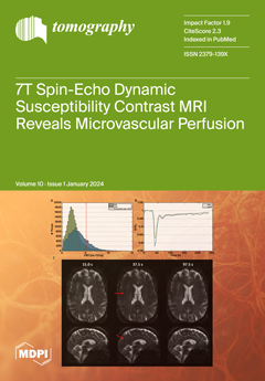
This study assesses the merits of using spin-echo (SE) dynamic susceptibility contrast (DSC) magnetic resonance imaging (MRI) at 7 Tesla to study cerebral perfusion in elderly adults. By tracking susceptibility changes during the passage of a contrast agent (CA) bolus caused by the in- and out-flowing blood, regional quantitative perfusion measures can be obtained. In contrast with the gold standard, Gradient Echo (GE) DSC MRI, SE DSC MRI has a higher sensitivity to the microvasculature (up to ~10 µm diameter). Furthermore, ultra-high-field MRI (≥7 T) provides unique possibilities to provide better images, due to increased signal sensitivity. This makes it possible to obtain the specific capillary blood volume and flow, enabling one to further dive into the physiology at a more microvascular level. View this paper
  • Issues are regarded as officially published after their release is announced to the table of contents alert mailing list.
  • You may sign up for e-mail alerts to receive table of contents of newly released issues.
  • PDF is the official format for papers published in both, html and pdf forms. To view the papers in pdf format, click on the "PDF Full-text" link, and use the free Adobe Reader to open them.
Order results
Result details
Section
Select all
Export citation of selected articles as:
12 pages, 2204 KiB  
Article
Cerebral Microvascular Perfusion Assessed in Elderly Adults by Spin-Echo Dynamic Susceptibility Contrast MRI at 7 Tesla
by Elles P. Elschot, Walter H. Backes, Marieke van den Kerkhof, Alida A. Postma, Abraham A. Kroon and Jacobus F. A. Jansen
Tomography 2024, 10(1), 181-192; https://doi.org/10.3390/tomography10010014 - 18 Jan 2024
Viewed by 1886
Abstract
Perfusion measures of the total vasculature are commonly derived with gradient-echo (GE) dynamic susceptibility contrast (DSC) MR images, which are acquired during the early passes of a contrast agent. Alternatively, spin-echo (SE) DSC can be used to achieve specific sensitivity to the capillary [...] Read more.
Perfusion measures of the total vasculature are commonly derived with gradient-echo (GE) dynamic susceptibility contrast (DSC) MR images, which are acquired during the early passes of a contrast agent. Alternatively, spin-echo (SE) DSC can be used to achieve specific sensitivity to the capillary signal. For an improved contrast-to-noise ratio, ultra-high-field MRI makes this technique more appealing to study cerebral microvascular physiology. Therefore, this study assessed the applicability of SE-DSC MRI at 7 T. Forty-one elderly adults underwent 7 T MRI using a multi-slice SE-EPI DSC sequence. The cerebral blood volume (CBV) and cerebral blood flow (CBF) were determined in the cortical grey matter (CGM) and white matter (WM) and compared to values from the literature. The relation of CBV and CBF with age and sex was investigated. Higher CBV and CBF values were found in CGM compared to WM, whereby the CGM-to-WM ratios depended on the amount of largest vessels excluded from the analysis. CBF was negatively associated with age in the CGM, while no significant association was found with CBV. Both CBV and CBF were higher in women compared to men in both CGM and WM. The current study verifies the possibility of quantifying cerebral microvascular perfusion with SE-DSC MRI at 7 T. Full article
Show Figures

Figure 1

12 pages, 1272 KiB  
Article
Accelerated Hypofractionated Magnetic Resonance Guided Adaptive Radiation Therapy for Ultracentral Lung Tumors
by Alonso La Rosa, Kathryn E. Mittauer, Nema Bassiri, Amy E. Rzepczynski, Michael D. Chuong, Sreenija Yarlagadda, Tugce Kutuk, Nicole C. McAllister, Matthew D. Hall, Alonso N. Gutierrez, Ranjini Tolakanahalli, Minesh P. Mehta and Rupesh Kotecha
Tomography 2024, 10(1), 169-180; https://doi.org/10.3390/tomography10010013 - 17 Jan 2024
Viewed by 1035
Abstract
Radiotherapy for ultracentral lung tumors represents a treatment challenge, considering the high rates of high-grade treatment-related toxicities with stereotactic body radiation therapy (SBRT) or hypofractionated schedules. Accelerated hypofractionated magnetic resonance-guided adaptive radiation therapy (MRgART) emerged as a potential game-changer for tumors in these [...] Read more.
Radiotherapy for ultracentral lung tumors represents a treatment challenge, considering the high rates of high-grade treatment-related toxicities with stereotactic body radiation therapy (SBRT) or hypofractionated schedules. Accelerated hypofractionated magnetic resonance-guided adaptive radiation therapy (MRgART) emerged as a potential game-changer for tumors in these challenging locations, in close proximity to central organs at risk, such as the trachea, proximal bronchial tree, and esophagus. In this series, 13 consecutive patients, predominantly male (n = 9), with a median age of 71 (range (R): 46–85), underwent 195 MRgART fractions (all 60 Gy in 15 fractions) to metastatic (n = 12) or primary ultra-central lung tumors (n = 1). The median gross tumor volumes (GTVs) and planning target volumes (PTVs) were 20.72 cc (R: 0.54–121.65 cc) and 61.53 cc (R: 3.87–211.81 cc), respectively. The median beam-on time per fraction was 14 min. Adapted treatment plans were generated for all fractions, and indications included GTV/PTV undercoverage, OARs exceeding tolerance doses, or both indications in 46%, 18%, and 36% of fractions, respectively. Eight patients received concurrent systemic therapies, including immunotherapy (four), chemotherapy (two), and targeted therapy (two). The crude in-field loco-regional control rate was 92.3%. No CTCAE grade 3+ toxicities were observed. Our results offer promising insights, suggesting that MRgART has the potential to mitigate toxicities, enhance treatment precision, and improve overall patient care in the context of ultracentral lung tumors. Full article
Show Figures

Figure 1

10 pages, 541 KiB  
Article
Lumbar Disc Degeneration Is Linked to Dorsal Subcutaneous Fat Thickness at the L1–L2 Intervertebral Disc Level Measured by MRI
by Ibrahem Hussain Kanbayti, Abdulrahman S. Al-Buqami, Mohammad H. Alsheikh, Saad M. Al-Malki, Ibrahim Hadadi, Adnan Alahmadi, Bander S. Almutairi and Hamzah H. Ahmed
Tomography 2024, 10(1), 159-168; https://doi.org/10.3390/tomography10010012 - 17 Jan 2024
Viewed by 1396
Abstract
Background: Obese individuals have a higher risk of degenerative disc disease (DDD). Currently, body mass index is not sensitive enough to differentiate between muscle and fat distribution, and obesity-related health issues are linked to the way body fat is distributed. Therefore, this study [...] Read more.
Background: Obese individuals have a higher risk of degenerative disc disease (DDD). Currently, body mass index is not sensitive enough to differentiate between muscle and fat distribution, and obesity-related health issues are linked to the way body fat is distributed. Therefore, this study aims to investigate the association between the dorsal subcutaneous fat thickness (DSFT) of the lumbar spine, an alternative measurement tool of body fat distribution, and DDD. Methods: A total of 301 patients with DDD and 123 participants without the disease were recruited. Using length functions of magnetic resonance imaging (MRI) console, the DSFT of L1 to S1 intervertebral disc levels was measured in mid-sagittal spin-echo T2 weighted image. The Mann–Whitney U test and Chi-squared test (X2) were utilized to examine any variations between the case and control groups. Logistic regression models were built to explore the association of the DSFT with DDD. Results: The logistical regression model showed a positive association between DDD and DSFT [OR: 1.30, 95% CI: 1.02–1.64, p = 0.03]. In the stratified logistic regression analysis, a positive association was found between DDD and DSFT among younger participants and females [OR young: 1.48; 95% CI (1.02–2.20); p = 0.04—OR female: 1.37; 95% CI (1–1.88); p = 0.05]. Conclusions: Younger females with thicker DSFT at the L1–L2 level are more likely to develop DDD. This suggests that increased DSFT may be a contributing factor to DDD. Full article
Show Figures

Figure 1

26 pages, 32082 KiB  
Article
Dual-Domain Reconstruction Network Incorporating Multi-Level Wavelet Transform and Recurrent Convolution for Sparse View Computed Tomography Imaging
by Juncheng Lin, Jialin Li, Jiazhen Dou, Liyun Zhong, Jianglei Di and Yuwen Qin
Tomography 2024, 10(1), 133-158; https://doi.org/10.3390/tomography10010011 - 16 Jan 2024
Viewed by 782
Abstract
Sparse view computed tomography (SVCT) aims to reduce the number of X-ray projection views required for reconstructing the cross-sectional image of an object. While SVCT significantly reduces X-ray radiation dose and speeds up scanning, insufficient projection data give rise to issues such as [...] Read more.
Sparse view computed tomography (SVCT) aims to reduce the number of X-ray projection views required for reconstructing the cross-sectional image of an object. While SVCT significantly reduces X-ray radiation dose and speeds up scanning, insufficient projection data give rise to issues such as severe streak artifacts and blurring in reconstructed images, thereby impacting the diagnostic accuracy of CT detection. To address this challenge, a dual-domain reconstruction network incorporating multi-level wavelet transform and recurrent convolution is proposed in this paper. The dual-domain network is composed of a sinogram domain network (SDN) and an image domain network (IDN). Multi-level wavelet transform is employed in both IDN and SDN to decompose sinograms and CT images into distinct frequency components, which are then processed through separate network branches to recover detailed information within their respective frequency bands. To capture global textures, artifacts, and shallow features in sinograms and CT images, a recurrent convolution unit (RCU) based on convolutional long and short-term memory (Conv-LSTM) is designed, which can model their long-range dependencies through recurrent calculation. Additionally, a self-attention-based multi-level frequency feature normalization fusion (MFNF) block is proposed to assist in recovering high-frequency components by aggregating low-frequency components. Finally, an edge loss function based on the Laplacian of Gaussian (LoG) is designed as the regularization term for enhancing the recovery of high-frequency edge structures. The experimental results demonstrate the effectiveness of our approach in reducing artifacts and enhancing the reconstruction of intricate structural details across various sparse views and noise levels. Our method excels in both performance and robustness, as evidenced by its superior outcomes in numerous qualitative and quantitative assessments, surpassing contemporary state-of-the-art CNNs or Transformer-based reconstruction methods. Full article
Show Figures

Figure 1

0 pages, 3631 KiB  
Article
RETRACTED: Modern Subtype Classification and Outlier Detection Using the Attention Embedder to Transform Ovarian Cancer Diagnosis
by S. M. Nuruzzaman Nobel, S M Masfequier Rahman Swapno, Md. Ashraful Hossain, Mejdl Safran, Sultan Alfarhood, Md. Mohsin Kabir and M. F. Mridha
Tomography 2024, 10(1), 105-132; https://doi.org/10.3390/tomography10010010 - 15 Jan 2024
Cited by 1 | Viewed by 1631 | Retraction
Abstract
Ovarian cancer, a deadly female reproductive system disease, is a significant challenge in medical research due to its notorious lethality. Addressing ovarian cancer in the current medical landscape has become more complex than ever. This research explores the complex field of Ovarian Cancer [...] Read more.
Ovarian cancer, a deadly female reproductive system disease, is a significant challenge in medical research due to its notorious lethality. Addressing ovarian cancer in the current medical landscape has become more complex than ever. This research explores the complex field of Ovarian Cancer Subtype Classification and the crucial task of Outlier Detection, driven by a progressive automated system, as the need to fight this unforgiving illness becomes critical. This study primarily uses a unique dataset painstakingly selected from 20 esteemed medical institutes. The dataset includes a wide range of images, such as tissue microarray (TMA) images at 40× magnification and whole-slide images (WSI) at 20× magnification. The research is fully committed to identifying abnormalities within this complex environment, going beyond the classification of subtypes of ovarian cancer. We proposed a new Attention Embedder, a state-of-the-art model with effective results in ovarian cancer subtype classification and outlier detection. Using images magnified WSI, the model demonstrated an astonishing 96.42% training accuracy and 95.10% validation accuracy. Similarly, with images magnified via a TMA, the model performed well, obtaining a validation accuracy of 94.90% and a training accuracy of 93.45%. Our fine-tuned hyperparameter testing resulted in exceptional performance on independent images. At 20× magnification, we achieved an accuracy of 93.56%. Even at 40× magnification, our testing accuracy remained high, at 91.37%. This study highlights how machine learning can revolutionize the medical field’s ability to classify ovarian cancer subtypes and identify outliers, giving doctors a valuable tool to lessen the severe effects of the disease. Adopting this novel method is likely to improve the practice of medicine and give people living with ovarian cancer worldwide hope. Full article
Show Figures

Figure 1

4 pages, 188 KiB  
Editorial
Introduction to Special Issue Imaging in Cancer Diagnosis
by Chiara Zanon, Emilio Quaia and Filippo Crimì
Tomography 2024, 10(1), 101-104; https://doi.org/10.3390/tomography10010009 - 15 Jan 2024
Viewed by 867
Abstract
In the field of oncology, the precision of cancer imaging is the cornerstone of oncological patient care [...] Full article
(This article belongs to the Special Issue Imaging in Cancer Diagnosis)
11 pages, 858 KiB  
Article
Assessment of Hounsfield Units and Factors Associated with Fragmentation of Renal Stones by Extracorporeal Shock Wave Lithotripsy: A Computerized Tomography Study
by Abdallah Saud Alharbi, Moawia Gameraddin, Awadia Gareeballah, Zahra Jibril Shrwani, Moa’ath Abdullah Sindi, Hassan Ibrahim Alsaedi, Abdulaziz A. Qurashi, Khalid M. Aloufi, Abdullah Fahad A. Alshamrani and Amel F. Alzain
Tomography 2024, 10(1), 90-100; https://doi.org/10.3390/tomography10010008 - 11 Jan 2024
Viewed by 1271
Abstract
The success rate of extracorporeal shock wave lithotripsy (ESWL) is influenced by various factors, including stone density, and is determined through computed tomography scans in terms of Hounsfield units (HU). Materials and Methods: This retrospective single-center study was conducted in the King Fahad [...] Read more.
The success rate of extracorporeal shock wave lithotripsy (ESWL) is influenced by various factors, including stone density, and is determined through computed tomography scans in terms of Hounsfield units (HU). Materials and Methods: This retrospective single-center study was conducted in the King Fahad Hospital. Sixty-seven adult patients with renal and ureteric stones were selected randomly and enrolled in the study. Their ages ranged from 20 to 69 years. The patients were examined with non-contrast enhancement (NCCT) to assess the HU of their stones and were consequently treated with ESWL. Results: Of the 67 patients, 37.3% had stones that were completely fragmented, while 62.7% had stones that were partially fragmented. The HU, location of the stone, multiplicity of the stone, and patient age were found to be significant factors contributing to stone fragility (p-values < 0.05). The HU data were found to have a positive significant linear correlation with serum calcium (r = 0.28, p-value = 0.036), while serum acid had a negative correlation (r = −0.55, p-value < 0.001). Thus, the probability of calcium-containing stone formation increases with increased HU. In contrast, uric acid stone formation likely develops with decreasing HU with serum uric acid. Renal stones in patients with diabetes mellitus and hypertension were not completely fragmented compared to those without clinical history. Conclusions: Mean HU, location of the stone, laterality, stone status, and the number of ESWL sessions are the most significant factors affecting stone fragility. CT attenuation values can predict the composition of stones from serum calcium and uric acid examinations. Hypertension and diabetes mellitus are risk factors for renal stone fragmentation. Full article
(This article belongs to the Section Abdominal Imaging)
Show Figures

Figure 1

11 pages, 935 KiB  
Article
The Use of Pre-Chemoradiotherapy Total Masseter Muscle Volume as a Novel Predictor of Radiation-Induced Trismus in Locally Advanced Nasopharyngeal Carcinoma Patients
by Efsun Somay, Erkan Topkan, Umur Anil Pehlivan, Busra Yilmaz, Ali Ayberk Besen, Huseyin Mertsoylu, Berrin Pehlivan and Ugur Selek
Tomography 2024, 10(1), 79-89; https://doi.org/10.3390/tomography10010007 - 10 Jan 2024
Viewed by 802
Abstract
Background: We sought to determine whether pretreatment total masseter muscle volume (TMMV) measures can predict radiation-induced trismus (RIT) in patients with locally advanced nasopharyngeal carcinoma (LA-NPC) receiving concurrent chemoradiotherapy (C-CRT). Methods: We retrospectively reviewed the medical records of LA-NPC patients who received C-CRT [...] Read more.
Background: We sought to determine whether pretreatment total masseter muscle volume (TMMV) measures can predict radiation-induced trismus (RIT) in patients with locally advanced nasopharyngeal carcinoma (LA-NPC) receiving concurrent chemoradiotherapy (C-CRT). Methods: We retrospectively reviewed the medical records of LA-NPC patients who received C-CRT and had pretreatment maximum mouth openings (MMO) greater than 35 mm. MMO of 35 mm or less after C-CRT were considered RIT. We employed receiver operating characteristic (ROC) curve analysis to explore the correlation between pre-treatment TMMV readings and RIT status. Results: Out of the 112 eligible patients, 22.0% of them received a diagnosis of RIT after C-CRT. The optimal TMMV cutoff that was significantly linked to post-C-CRT RIT rates was determined to be 35.0 cc [area under the curve: 79.5%; sensitivity: 75.0%; and specificity: 78.6%; Youden index: 0.536] in the ROC curve analysis. The incidence of RIT was significantly higher in patients with TMMV ≤ 5.0 cc than in those with TMMV > 35.0 cc [51.2% vs. 8.7%; Odds ratio: 6.79; p < 0.001]. A multivariate logistic regression analysis revealed that pre-C-CRT MMO ≤ 41.6 mm (p = 0.001), mean masticatory apparatus dose V56.5 ≥ 34% group (p = 0.002), and TMMV ≤ 35 cc were the independent predictors of significantly elevated rates of RIT. Conclusion: The presence of a smaller pretreatment TMMV is a reliable and independent novel biological marker that can confidently predict higher RIT rates in LA-NPC patients who receive C-CRT. Full article
(This article belongs to the Section Cancer Imaging)
Show Figures

Figure 1

13 pages, 3844 KiB  
Article
The Role of Major Salivary Gland Ultrasound in the Diagnostic Workup of Sicca Syndrome: A Large Single-Centre Study
by Giulia Vallifuoco, Paolo Falsetti, Marco Bardelli, Edoardo Conticini, Stefano Gentileschi, Caterina Baldi, Suhel Gabriele Al Khayyat, Luca Cantarini and Bruno Frediani
Tomography 2024, 10(1), 66-78; https://doi.org/10.3390/tomography10010006 - 8 Jan 2024
Viewed by 722
Abstract
(1) Objective: To determine the diagnostic accuracy of major salivary gland ultrasonography (SGUS) in primary Sjogren’s syndrome (SS), we used the Outcome Measures in Rheumatology Clinical Trials (OMERACT) scoring system on a large single-centre cohort of patients with sicca syndrome. (2) Method: We [...] Read more.
(1) Objective: To determine the diagnostic accuracy of major salivary gland ultrasonography (SGUS) in primary Sjogren’s syndrome (SS), we used the Outcome Measures in Rheumatology Clinical Trials (OMERACT) scoring system on a large single-centre cohort of patients with sicca syndrome. (2) Method: We retrospectively collected the clinical, imaging and serological data of all the patients referred with a suspicion of SS who underwent SGUS and minor salivary glands biopsy. (3) Results: A total of 132 patients were included. The SGUS scores were correlated between the two sides (p < 0.001). The diagnostic cut-off for SS (AUROC: 0.7408) was 6 for the SGUS-global sum (sensitivity: 32.43%; specificity: 96.84%). The cut-off with the highest specificity for SS diagnosis was 7. In the patients with a final diagnosis of SS, the mean SGUS score was significantly higher (p < 0.001) than that of the non-SS patients (3.73 vs. 1.32 for the SGUS-global sum). A significant correlation was demonstrated between the SGUS scores and final SS diagnosis (p < 0.001), biopsy positivity (p < 0.001), ANA positivity (p = 0.016), Ro-SSA positivity (p = 0.01), and gland fibrosis (p = 0.02). (4) Conclusions: SGUS, using the OMERACT scoring system, has moderate sensitivity and high specificity for the diagnosis of SS. The scoring showed a strong and direct correlation with all the clinical hallmarks of SS diagnosis, such as the positivity of a labial salivary gland biopsy, ANA and Ro-SSA statuses, and salivary gland fibrosis. Because of its high specificity, a SGUS-global score > 6 could be therefore employed for the diagnosis of SS in the case of ANA negativity or the unavailability of a biopsy. Full article
Show Figures

Figure 1

19 pages, 10870 KiB  
Review
Primary Sclerosing Cholangitis: Diagnostic Criteria
by Nora Cazzagon, Samantha Sarcognato, Elisa Catanzaro, Emanuela Bonaiuto, Matteo Peviani, Francesco Pezzato and Raffaella Motta
Tomography 2024, 10(1), 47-65; https://doi.org/10.3390/tomography10010005 - 7 Jan 2024
Viewed by 1382
Abstract
Primary sclerosing cholangitis is a chronic cholestatic liver disease characterized by inflammation and fibrosis of intra- and/or extrahepatic bile ducts leading to the formation of multifocal strictures alternated to bile duct dilatations. The diagnosis of the most common subtype of the disease, the [...] Read more.
Primary sclerosing cholangitis is a chronic cholestatic liver disease characterized by inflammation and fibrosis of intra- and/or extrahepatic bile ducts leading to the formation of multifocal strictures alternated to bile duct dilatations. The diagnosis of the most common subtype of the disease, the large duct PSC, is based on the presence of elevation of cholestatic indices, the association of typical cholangiographic findings assessed by magnetic resonance cholangiography and the exclusion of causes of secondary sclerosing cholangitis. Liver biopsy is not routinely applied for the diagnosis of large duct PSC but is mandatory in the case of suspicion of small duct PSC or overlap with autoimmune hepatitis. Full article
Show Figures

Figure 1

10 pages, 753 KiB  
Article
Cone-Beam Computerized Tomography Evaluation of the Relationship between Orthodontic Vertical Direction Parameters and the Distance from the Apex of the Upper Central Tooth to the Nasal Floor and Anterior Nasal Spine
by Saadet Çınarsoy Ciğerim and Türkan Sezen Erhamza
Tomography 2024, 10(1), 37-46; https://doi.org/10.3390/tomography10010004 - 5 Jan 2024
Cited by 1 | Viewed by 891
Abstract
The aim of this study was to examine the relationship between the vertical cephalometric values and the distance from the apex tip of the upper central tooth (U1A) to the anterior nasal spine (ANS) and nasal floor (NF) using cone-beam computerized tomography (CBCT). [...] Read more.
The aim of this study was to examine the relationship between the vertical cephalometric values and the distance from the apex tip of the upper central tooth (U1A) to the anterior nasal spine (ANS) and nasal floor (NF) using cone-beam computerized tomography (CBCT). One hundred and twenty-two patients who applied to the Department of Orthodontics between January 2011 and June 2019 were included. The distances between the U1A and the NF and ANS were measured using CBCT. Statistical significance was considered as p < 0.05. Of the 122 individuals, 73.8% (n = 90) were female and 26.2% (n = 32) were male, with a mean age of 22.8 ± 3.3 years. A statistically significant moderate positive correlation was found between the mean NF-U1A values and the N-Me, ANS-Me, ANS-Gn, S-Go, and N-ANS measurements (p < 0.01). A statistically significant positive correlation was found between the mean ANS-U1A values and the Ar-Go-Me, total posterior angles, N-Me, SN/GoGn and Y-axis angle, ANS-Me, and ANS-Gn measurements (p < 0.01). The distance from the U1A to the ANS and NF was related to the orthodontic vertical direction parameters. The ANS-U1A and NF-U1A distances can serve as reference points for identifying the orthodontic vertical growth pattern from CBCT scans. Full article
Show Figures

Figure 1

12 pages, 1596 KiB  
Review
Residual Lung Abnormalities in Survivors of Severe or Critical COVID-19 at One-Year Follow-Up Computed Tomography: A Narrative Review Comparing the European and East Asian Experiences
by Andrea Borghesi, Pietro Ciolli, Elisabetta Antonelli, Alessandro Monti, Alessandra Scrimieri, Marco Ravanelli, Roberto Maroldi and Davide Farina
Tomography 2024, 10(1), 25-36; https://doi.org/10.3390/tomography10010003 - 30 Dec 2023
Viewed by 926
Abstract
The literature reports that there was a significant difference in the medical impact of the coronavirus disease (COVID-19) pandemic between European and East Asian countries; specifically, the mortality rate of COVID-19 in Europe was significantly higher than that in East Asia. Considering such [...] Read more.
The literature reports that there was a significant difference in the medical impact of the coronavirus disease (COVID-19) pandemic between European and East Asian countries; specifically, the mortality rate of COVID-19 in Europe was significantly higher than that in East Asia. Considering such a difference, our narrative review aimed to compare the prevalence and characteristics of residual lung abnormalities at one-year follow-up computed tomography (CT) after severe or critical COVID-19 in survivors of European and East Asian countries. A literature search was performed to identify articles focusing on the prevalence and characteristics of CT lung abnormalities in survivors of severe or critical COVID-19. Database analysis identified 16 research articles, 9 from Europe and 7 from East Asia (all from China). Our analysis found a higher prevalence of CT lung abnormalities in European than in Chinese studies (82% vs. 52%). While the most prevalent lung abnormalities in Chinese studies were ground-glass opacities (35%), the most prevalent lung abnormalities in European studies were linear (59%) and reticular opacities (55%), followed by bronchiectasis (46%). Although our findings required confirmation, the higher prevalence and severity of lung abnormalities in European than in Chinese survivors of COVID-19 may reflect a greater architectural distortion due to a more severe lung damage. Full article
(This article belongs to the Special Issue The Challenge of Advanced Medical Imaging Data Analysis in COVID-19)
Show Figures

Figure 1

11 pages, 5378 KiB  
Article
Age-Dependent Changes in Effective Dose in Pediatric Brain CT: Comparisons of Estimation Methods
by Yusuke Inoue, Masahiro Mori, Hiroyasu Itoh, Kohei Mitsui, Hiroki Miyatake, Takuro Yamane and Hirofumi Hata
Tomography 2024, 10(1), 14-24; https://doi.org/10.3390/tomography10010002 - 24 Dec 2023
Cited by 1 | Viewed by 1064
Abstract
The effective dose (ED) in computed tomography (CT) may be calculated by multiplying the dose–length product (DLP) by a conversion factor. As children grow, automatic exposure control increases the DLP, while the conversion factor decreases; these two changes affect the ED in opposite [...] Read more.
The effective dose (ED) in computed tomography (CT) may be calculated by multiplying the dose–length product (DLP) by a conversion factor. As children grow, automatic exposure control increases the DLP, while the conversion factor decreases; these two changes affect the ED in opposite ways. The aim of this study was to investigate the methods of ED estimation according to age in pediatric brain CT. We retrospectively analyzed 980 brain CT scans performed for various clinical indications in children. The conversion factor at each age, in integer years, was determined based on the values at 0, 1, 5, and 10 years provided by the International Commission on Radiological Protection (ICRP), using a curve (curve method) or lines (linear method). In the simple method, the ED was estimated using the ICRP conversion factor for the closest age. We also analyzed the ED estimated by a radiation dose management system. Although the median DLP at each age increased with age, the median ED estimated by the curve method was highest at 0 years, decreased with age, and then plateaued at 9 years. The linear method yielded mildly different results, especially at 2 and 3 years. The ED estimated by the simple method or the radiation dose management system showed inconsistent, up-and-down changes with age. In conclusion, the ED in pediatric brain CT decreases with age despite increased DLP. Determination of the conversion factor at each age using a curve is expected to contribute to estimating the ED in pediatric CT according to age. Full article
Show Figures

Figure 1

13 pages, 8828 KiB  
Review
Advantages of Photon-Counting Detector CT in Aortic Imaging
by Chiara Zanon, Filippo Cademartiri, Alessandro Toniolo, Costanza Bini, Alberto Clemente, Elda Chiara Colacchio, Giulio Cabrelle, Florinda Mastro, Michele Antonello, Emilio Quaia and Alessia Pepe
Tomography 2024, 10(1), 1-13; https://doi.org/10.3390/tomography10010001 - 19 Dec 2023
Cited by 4 | Viewed by 1331
Abstract
Photon-counting Computed Tomography (PCCT) is a promising imaging technique. Using detectors that count the number and energy of photons in multiple bins, PCCT offers several advantages over conventional CT, including a higher image quality, reduced contrast agent volume, radiation doses, and artifacts. Although [...] Read more.
Photon-counting Computed Tomography (PCCT) is a promising imaging technique. Using detectors that count the number and energy of photons in multiple bins, PCCT offers several advantages over conventional CT, including a higher image quality, reduced contrast agent volume, radiation doses, and artifacts. Although PCCT is well established for cardiac imaging in assessing coronary artery disease, its application in aortic imaging remains limited. This review summarizes the available literature and provides an overview of the current use of PCCT for the diagnosis of aortic imaging, focusing mainly on endoleaks detection and characterization after endovascular aneurysm repair (EVAR), contrast dose volume, and radiation exposure reduction, particularly in patients with chronic kidney disease and in those requiring follow-up CT. Full article
(This article belongs to the Special Issue Feature Reviews for Tomography 2023)
Show Figures

Figure 1

Previous Issue
Next Issue
Back to TopTop