Recent Advances in Microelectrode Array Interfaces for Organoids


Abstract
1. Introduction
2. MEA Architectures
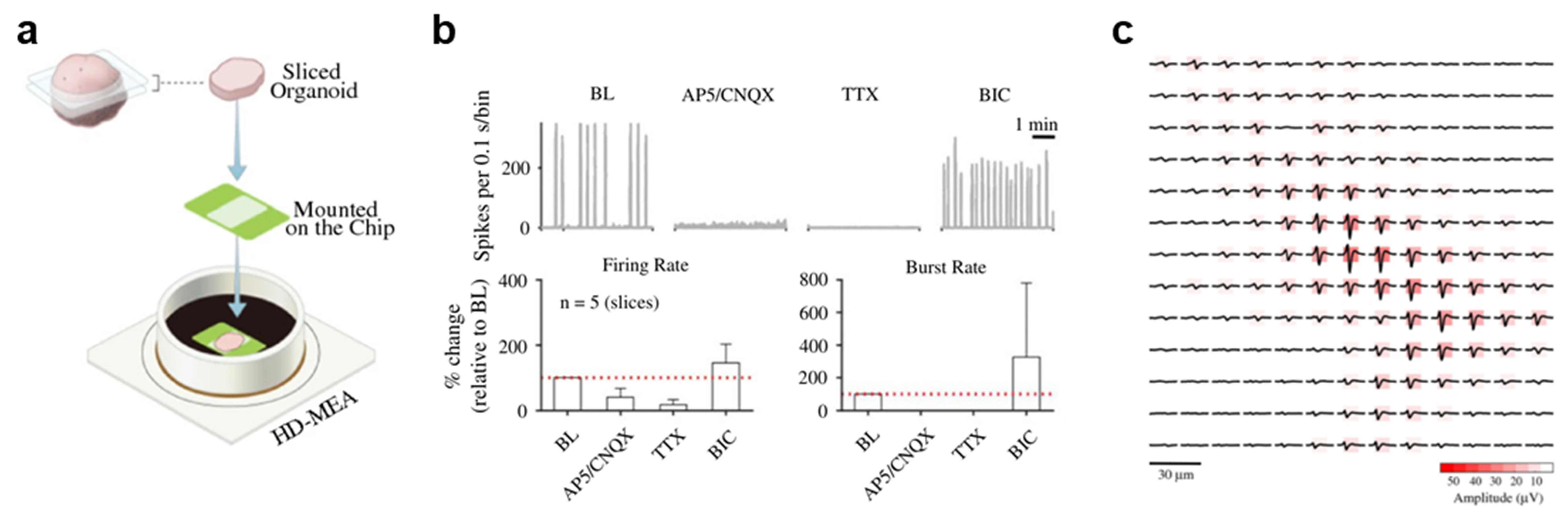
2.1. High-Density CMOS MEAs for Single-Unit Analysis in Brain Organoids
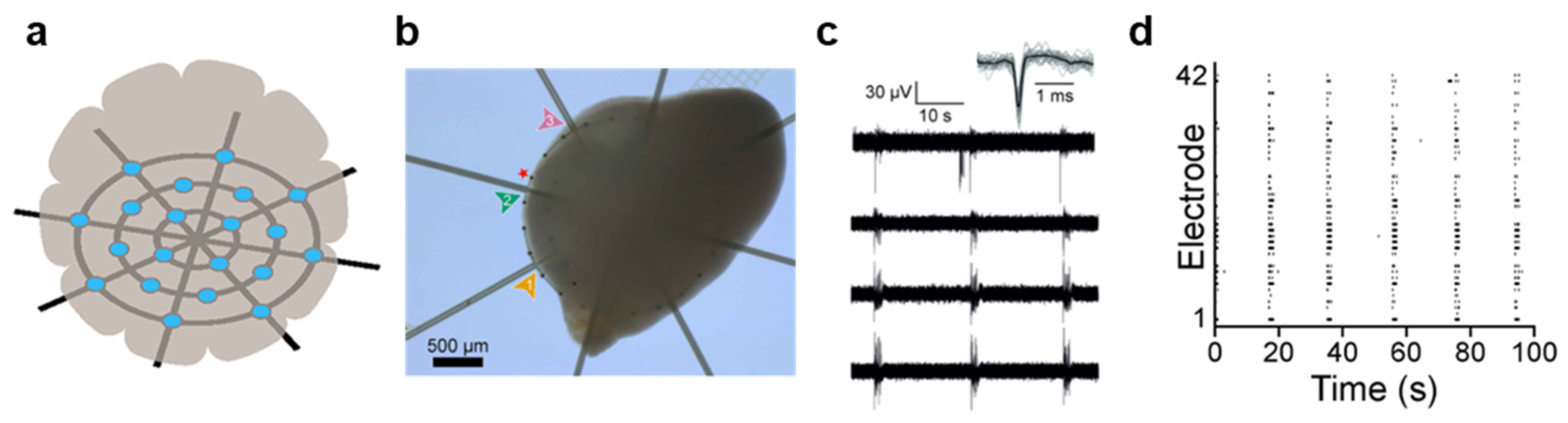
2.2. Mesh MEAs for Long-Term Recording of Organoids
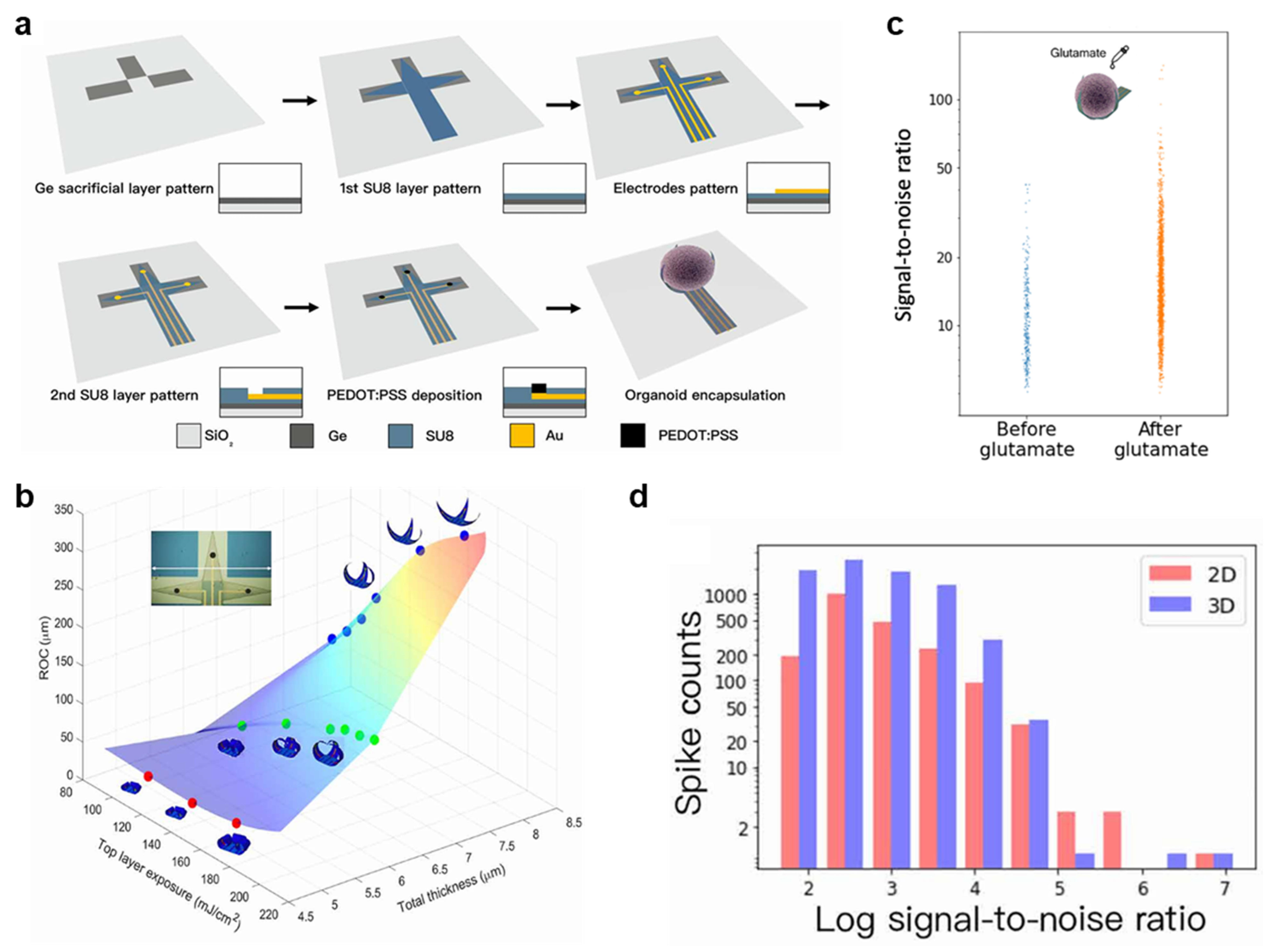
2.3. Self-Folding Shell MEAs for Encapsulating Organoids
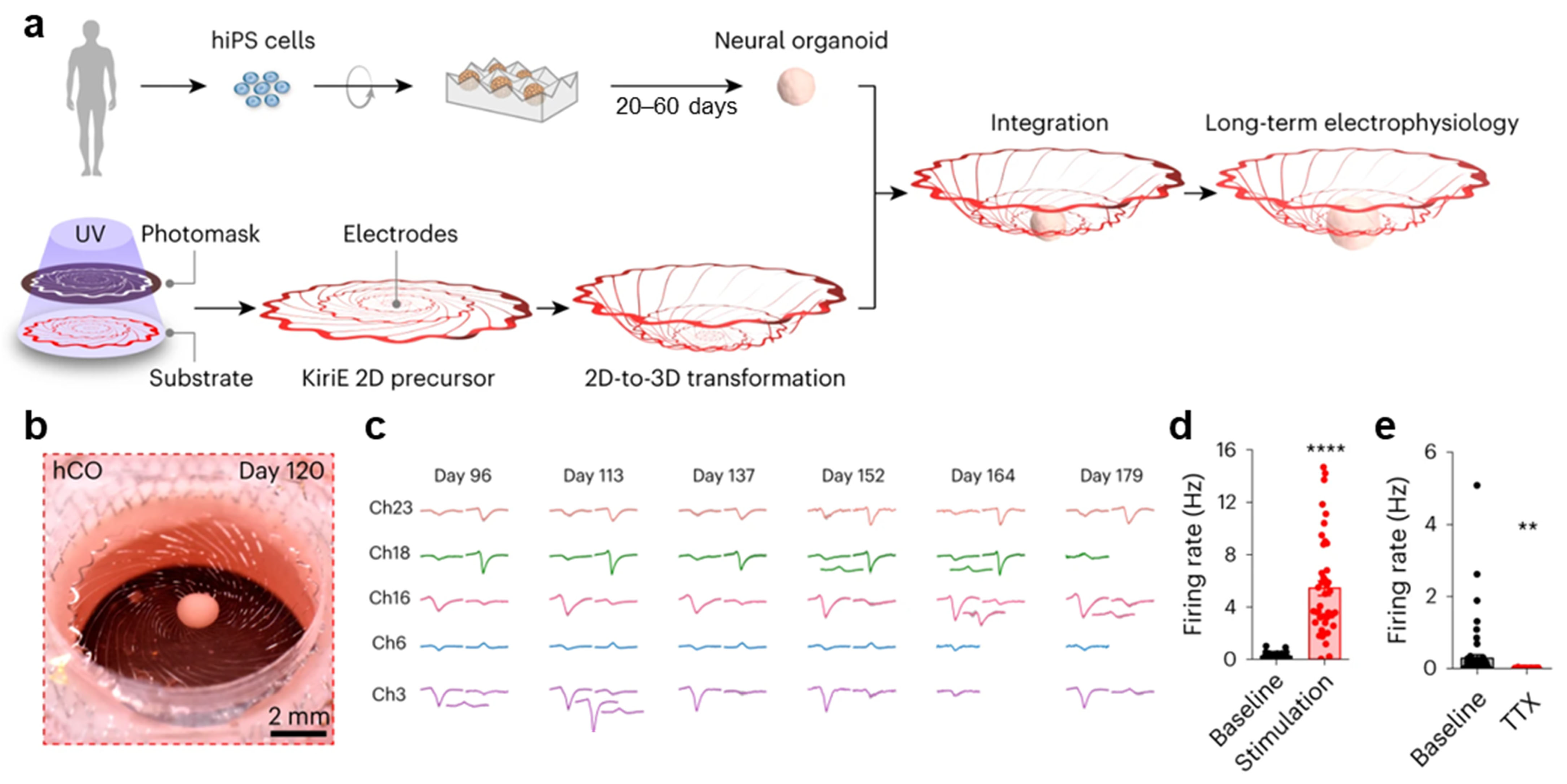
2.4. Kirigami MEAs for Self-Transforming 3D Interfacing with Suspended Organoids
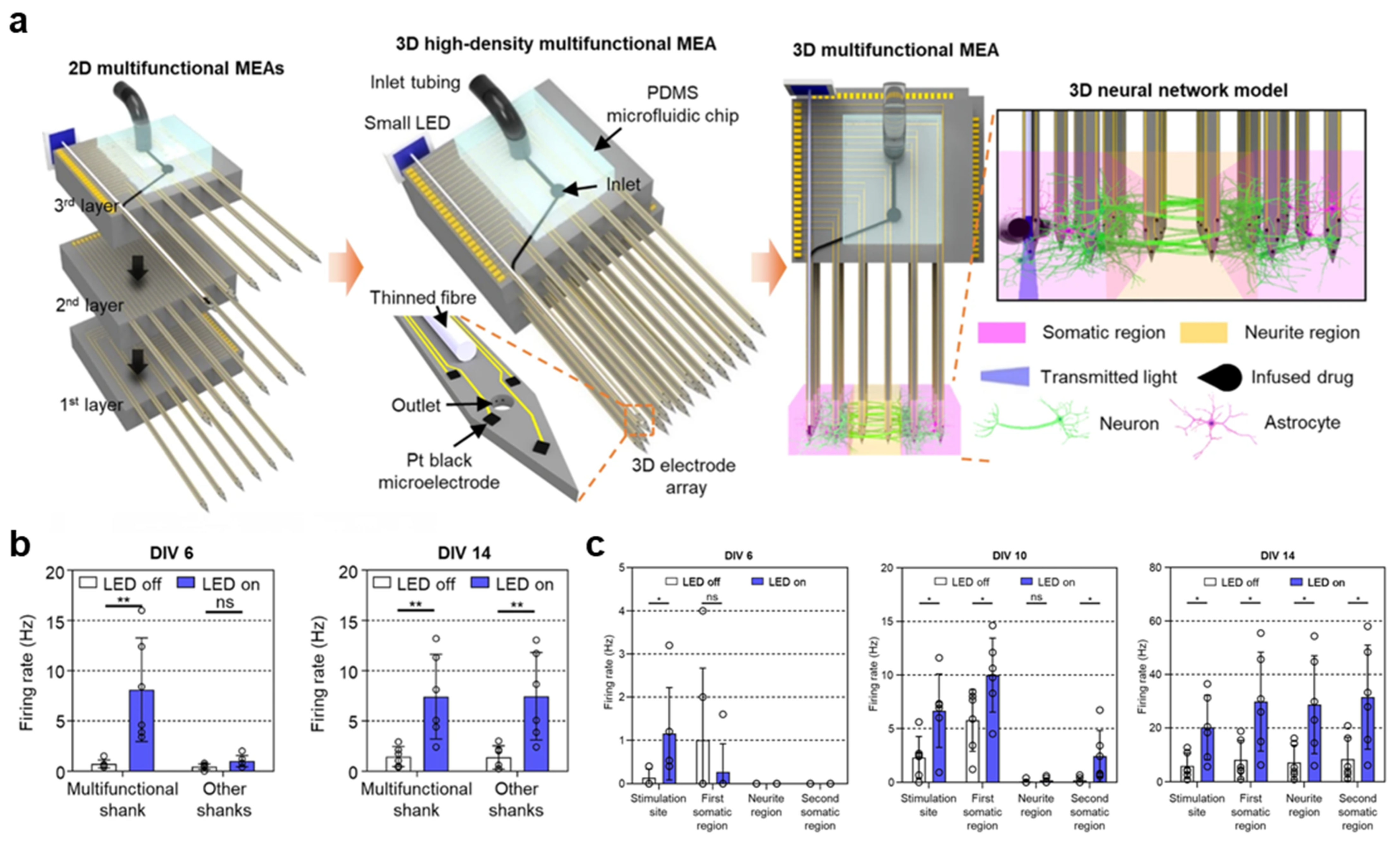
2.5. Multifunctional 3D MEAs for Neural Circuit Analysis
3. Conclusions
Author Contributions
Funding
Institutional Review Board Statement
Informed Consent Statement
Data Availability Statement
Conflicts of Interest
References
- Di Lullo, E.; Kriegstein, A.R. The use of brain organoids to investigate neural development and disease. Nat. Rev. Neurosci. 2017, 18, 573–584. [Google Scholar] [CrossRef] [Scilit] [PubMed]
- Lancaster, M.A.; Renner, M.; Martin, C.-A.; Wenzel, D.; Bicknell, L.S.; Hurles, M.E.; Homfray, T.; Penninger, J.M.; Jackson, A.P.; Knoblich, J.A. Cerebral organoids model human brain development and microcephaly. Nature 2013, 501, 373–379. [Google Scholar] [CrossRef] [Scilit] [PubMed]
- Chiaradia, I.; Lancaster, M.A. Brain organoids for the study of human neurobiology at the interface of in vitro and in vivo. Nat. Neurosci. 2020, 23, 1496–1508. [Google Scholar] [CrossRef] [Scilit] [PubMed]
- Ogawa, J.; Pao, G.M.; Shokhirev, M.N.; Verma, I.M. Glioblastoma model using human cerebral organoids. Cell Rep. 2018, 23, 1220–1229. [Google Scholar] [CrossRef] [Scilit]
- Smits, L.M.; Reinhardt, L.; Reinhardt, P.; Glatza, M.; Monzel, A.S.; Stanslowsky, N.; Rosato-Siri, M.D.; Zanon, A.; Antony, P.M.; Bellmann, J.; et al. Modeling Parkinson’s disease in midbrain-like organoids. NPJ Park. Dis. 2019, 5, 5. [Google Scholar] [CrossRef] [Scilit]
- Poewe, W.; Seppi, K.; Tanner, C.M.; Halliday, G.M.; Brundin, P.; Volkmann, J.; Schrag, A.-E.; Lang, A.E. Parkinson disease. Nat. Rev. Dis. Primers 2017, 3, 17013. [Google Scholar] [CrossRef] [Scilit]
- Smith, M.A. Alzheimer disease. Int. Rev. Neurobiol. 1998, 42, 1–54. [Google Scholar]
- Hamill, O.P.; Marty, A.; Neher, E.; Sakmann, B.; Sigworth, F.J. Improved patch-clamp techniques for high-resolution current recording from cells and cell-free membrane patches. Pflügers Arch. 1981, 391, 85–100. [Google Scholar] [CrossRef] [Scilit]
- Birey, F.; Andersen, J.; Makinson, C.D.; Islam, S.; Wei, W.; Huber, N.; Fan, H.C.; Metzler, K.R.C.; Panagiotakos, G.; Thom, N.; et al. Assembly of functionally integrated human forebrain spheroids. Nature 2017, 545, 54–59. [Google Scholar] [CrossRef] [Scilit]
- Grienberger, C.; Konnerth, A. Imaging calcium in neurons. Neuron 2012, 73, 862–885. [Google Scholar] [CrossRef] [Scilit]
- Samarasinghe, R.A.; Miranda, O.A.; Buth, J.E.; Mitchell, S.; Ferando, I.; Watanabe, M.; Allison, T.F.; Kurdian, A.; Fotion, N.N.; Gandal, M.J.; et al. Identification of neural oscillations and epileptiform changes in human brain organoids. Nat. Neurosci. 2021, 24, 1488–1500. [Google Scholar] [CrossRef] [Scilit] [PubMed]
- Stett, A.; Egert, U.; Guenther, E.; Hofmann, F.; Meyer, T.; Nisch, W.; Haemmerle, H. Biological application of microelectrode arrays in drug discovery and basic research. Anal. Bioanal. Chem. 2003, 377, 486–495. [Google Scholar] [CrossRef] [Scilit] [PubMed]
- Buzsáki, G.; Anastassiou, C.A.; Koch, C. The origin of extracellular fields and currents—EEG, ECoG, LFP and spikes. Nat. Rev. Neurosci. 2012, 13, 407–420. [Google Scholar] [CrossRef] [Scilit] [PubMed]
- Cotterill, E.; Hall, D.; Wallace, K.; Mundy, W.R.; Eglen, S.J.; Shafer, T.J. Characterization of early cortical neural network development in multiwell microelectrode array plates. J. Biomol. Screen. 2016, 21, 510–519. [Google Scholar] [CrossRef] [Scilit]
- Phouphetlinthong, O.; Partiot, E.; Bernou, C.; Sebban, A.; Gaudin, R.; Charlot, B. Protruding cantilever microelectrode array to monitor the inner electrical activity of cerebral organoids. Lab A Chip 2023, 23, 3603–3614. [Google Scholar] [CrossRef] [Scilit]
- Najar-Asl, M.; Halvaei, M.; Abolhasani, R.; Mirsadeghi, S.; Simorgh, S.; Rahmani, S.; Pooyan, P.; Yektadoost, E.; Kiani, S.; Abolghasemi-Dehaqani, M.-R.; et al. Enhanced development of human pluripotent stem cell-derived cerebral organoids via an electrical stimulation bioreactor. Chem. Eng. J. 2024, 487, 150368. [Google Scholar] [CrossRef] [Scilit]
- Li, X.-H.; Hu, N.; Chang, Z.-H.; Shi, J.-X.; Fan, X.; Chen, M.-M.; Bao, S.-Q.; Chen, C.; Zuo, J.-C.; Zhang, X.-W.; et al. Brain organoid maturation and implantation integration based on electrical signals input. J. Adv. Res. 2025, 73, 375–395. [Google Scholar] [CrossRef] [Scilit]
- Trujillo, C.A.; Gao, R.; Negraes, P.D.; Gu, J.; Buchanan, J.; Preissl, S.; Wang, A.; Wu, W.; Haddad, G.G.; Chaim, I.A.; et al. Complex oscillatory waves emerging from cortical organoids model early human brain network development. Cell Stem Cell 2019, 25, 558–569.e557. [Google Scholar] [CrossRef] [Scilit]
- Schröter, M.; Wang, C.; Terrigno, M.; Hornauer, P.; Huang, Z.; Jagasia, R.; Hierlemann, A. Functional imaging of brain organoids using high-density microelectrode arrays. MRS Bull. 2022, 47, 530–544. [Google Scholar] [CrossRef] [Scilit]
- Ronchi, S.; Fiscella, M.; Marchetti, C.; Viswam, V.; Müller, J.; Frey, U.; Hierlemann, A. Single-cell electrical stimulation using CMOS-based high-density microelectrode arrays. Front. Neurosci. 2019, 13, 208. [Google Scholar] [CrossRef] [Scilit]
- Ballini, M.; Muller, J.; Livi, P.; Chen, Y.; Frey, U.; Stettler, A.; Shadmani, A.; Viswam, V.; Lloyd Jones, I.; Jackel, D.; et al. A 1024-channel CMOS microelectrode array with 26,400 electrodes for recording and stimulation of electrogenic cells in vitro. IEEE J. Solid-State Circuits 2014, 49, 2705–2719. [Google Scholar] [CrossRef] [Scilit]
- Bakkum, D.J.; Frey, U.; Radivojevic, M.; Russell, T.L.; Müller, J.; Fiscella, M.; Takahashi, H.; Hierlemann, A. Tracking axonal action potential propagation on a high-density microelectrode array across hundreds of sites. Nat. Commun. 2013, 4, 2181. [Google Scholar] [CrossRef] [Scilit] [PubMed]
- Ronchi, S.; Buccino, A.P.; Prack, G.; Kumar, S.S.; Schröter, M.; Fiscella, M.; Hierlemann, A. Electrophysiological phenotype characterization of human iPSC-derived neuronal cell lines by means of high-density microelectrode arrays. Adv. Biol. 2021, 5, 2000223. [Google Scholar] [CrossRef] [Scilit] [PubMed]
- Li, Q.; Nan, K.; Le Floch, P.; Lin, Z.; Sheng, H.; Blum, T.S.; Liu, J. Cyborg organoids: Implantation of nanoelectronics via organogenesis for tissue-wide electrophysiology. Nano Lett. 2019, 19, 5781–5789. [Google Scholar] [CrossRef] [Scilit] [PubMed]
- McDonald, M.; Sebinger, D.; Brauns, L.; Gonzalez-Cano, L.; Menuchin-Lasowski, Y.; Mierzejewski, M.; Psathaki, O.-E.; Stumpf, A.; Wickham, J.; Rauen, T.; et al. A mesh microelectrode array for non-invasive electrophysiology within neural organoids. Biosens. Bioelectron. 2023, 228, 115223. [Google Scholar] [CrossRef] [Scilit]
- Spira, M.E.; Hai, A. Multi-electrode array technologies for neuroscience and cardiology. Nano-Enabled Med. Appl. 2013, 8, 83–94. [Google Scholar] [CrossRef] [Scilit]
- Huang, Q.; Tang, B.; Romero, J.C.; Yang, Y.; Elsayed, S.K.; Pahapale, G.; Lee, T.-J.; Pantoja, I.E.M.; Han, F.; Berlinicke, C.; et al. Shell microelectrode arrays (MEAs) for brain organoids. Sci. Adv. 2022, 8, eabq5031. [Google Scholar] [CrossRef] [Scilit]
- Yang, X.; Forró, C.; Li, T.L.; Miura, Y.; Zaluska, T.J.; Tsai, C.-T.; Kanton, S.; McQueen, J.P.; Chen, X.; Mollo, V.; et al. Kirigami electronics for long-term electrophysiological recording of human neural organoids and assembloids. Nat. Biotechnol. 2024, 42, 1836–1843. [Google Scholar] [CrossRef] [Scilit]
- Frega, M.; Tedesco, M.; Massobrio, P.; Pesce, M.; Martinoia, S. Network dynamics of 3D engineered neuronal cultures: A new experimental model for in-vitro electrophysiology. Sci. Rep. 2014, 4, 5489. [Google Scholar] [CrossRef] [Scilit]
- Soscia, D.A.; Lam, D.; Tooker, A.C.; Enright, H.A.; Triplett, M.; Karande, P.; Peters, S.K.G.; Sales, A.P.; Wheeler, E.K.; Fischer, N.O. A flexible 3-dimensional microelectrode array for in vitro brain models. Lab A Chip 2020, 20, 901–911. [Google Scholar] [CrossRef] [Scilit]
- Tian, B.; Liu, J.; Dvir, T.; Jin, L.; Tsui, J.H.; Qing, Q.; Suo, Z.; Langer, R.; Kohane, D.S.; Lieber, C.M. Macroporous nanowire nanoelectronic scaffolds for synthetic tissues. Nat. Mater. 2012, 11, 986–994. [Google Scholar] [CrossRef] [Scilit]
- Shin, H.; Jeong, S.; Lee, J.-H.; Sun, W.; Choi, N.; Cho, I.-J. 3D high-density microelectrode array with optical stimulation and drug delivery for investigating neural circuit dynamics. Nat. Commun. 2021, 12, 492. [Google Scholar] [CrossRef] [Scilit]
- Martinelli, E.; Akouissi, O.; Liebi, L.; Furfaro, I.; Maulà, D.; Savoia, N.; Remy, A.; Nikles, L.; Roux, A.; Stoppini, L.; et al. The e-Flower: A hydrogel-actuated 3D MEA for brain spheroid electrophysiology. Sci. Adv. 2024, 10, eadp8054. [Google Scholar] [CrossRef] [Scilit]
- Fekete, Z.; Zátonyi, A.; Kaszás, A.; Madarász, M.; Slézia, A. Transparent neural interfaces: Challenges and solutions of microengineered multimodal implants designed to measure intact neuronal populations using high-resolution electrophysiology and microscopy simultaneously. Microsyst. Nanoeng. 2023, 9, 66. [Google Scholar] [CrossRef] [Scilit]
- Wilson, M.N.; Thunemann, M.; Liu, X.; Lu, Y.; Puppo, F.; Adams, J.W.; Kim, J.-H.; Ramezani, M.; Pizzo, D.P.; Djurovic, S.; et al. Multimodal monitoring of human cortical organoids implanted in mice reveal functional connection with visual cortex. Nat. Commun. 2022, 13, 7945. [Google Scholar] [CrossRef] [Scilit]
- Saif-Ur-Rehman, M.; Lienkämper, R.; Parpaley, Y.; Wellmer, J.; Liu, C.; Lee, B.; Kellis, S.; Andersen, R.; Iossifidis, I.; Glasmachers, T.; et al. SpikeDeeptector: A deep-learning based method for detection of neural spiking activity. J. Neural Eng. 2019, 16, 056003. [Google Scholar] [CrossRef] [Scilit]
- Hilgen, G.; Sorbaro, M.; Pirmoradian, S.; Muthmann, J.-O.; Kepiro, I.E.; Ullo, S.; Ramirez, C.J.; Encinas, A.P.; Maccione, A.; Berdondini, L.; et al. Unsupervised spike sorting for large-scale, high-density multielectrode arrays. Cell Rep. 2017, 18, 2521–2532. [Google Scholar] [CrossRef] [Scilit]



| Electrode Type | Cell Type | Electrode Area (μm2) | Number of Electrodes | Electrode Impedance (at 1 kHz) | Signal Voltage (µV) | Ref. |
|---|---|---|---|---|---|---|
| Active CMOS | iPSCs and ESCs | 49.29 | 26,400 | 1–10 kΩ | 20–60 | [19] |
| Passive 2D planar | iPSCs | 702 | 61 | 100 kΩ | 50 | [25] |
| Passive 3D | iPSCs and ESCs | - | 3 | 10 kΩ | 200 | [27] |
| iPSCs | 490 | 32 | 300 kΩ | 30–140 | [28] | |
| Rat cortical tissue | 400 | 63 | 15 kΩ | 100 | [32] |
| Data Processing | Strengths | Limitations | Ref. | |
|---|---|---|---|---|
| HD-MEAs | - Spike sorting to single units - LFP and burst analysis - Functional connectivity analysis using cross-correlograms and transfer entropy. | - Enables long-term recordings from brain organoid slices with single-unit spike sorting - Axonal conduction velocity measurements - Quantitative analysis of functional connectivity. | - Access is restricted to networks near the slice surface rather than the full 3D volume of the organoid. | [19] |
| Mesh MEAs | - High-rate sampling - High-pass filtering - Noise-based threshold detection - Analysis of spontaneous burst patterns. | - Uses flexible mesh electrodes to minimize tissue damage - Allows long-term recording inside organoids. | - Provides relatively low electrode counts and spatial resolution compared with CMOS-based MEAs - Recordings are often limited to electrodes located near the basal surface. | [25] |
| Shell MEAs | - Threshold-based spike detection - Spike count - SNR evaluation. | - Wraps around spherical organoids - Increasing contact area and spike detection efficiency. | - Requires manual folding and organoid insertion - Limits throughput and can introduce user-to-user variability. | [27] |
| Kirigami MEAs | - Single-unit spike sorting - Long-term firing-rate tracking - Quantification of optogenetic - Pharmacological responses. | - Conforms to the shape of the organoid - Enables stable long-term attachment and recording. | - Involves complex pattern design and fabrication, resulting in a high operational barrier - Currently offers a limited number of channels. | [28] |
| Multifunctional 3D MEAs | - Spike sorting - Amplitude-threshold (≈3× noise) spike detection - Burst and synchrony/network analysis. | - Integrates multiple functions (e.g., electrical recording, stimulation, optical or chemical modulation) to enable detailed analysis of neural circuit dynamics. | - Requires customized packaging and complex fabrication, - Insertion-type structure may impose mechanical burden for long-term applications. | [32] |
Disclaimer/Publisher’s Note: The statements, opinions and data contained in all publications are solely those of the individual author(s) and contributor(s) and not of MDPI and/or the editor(s). MDPI and/or the editor(s) disclaim responsibility for any injury to people or property resulting from any ideas, methods, instructions or products referred to in the content. |
© 2026 by the authors. Licensee MDPI, Basel, Switzerland. This article is an open access article distributed under the terms and conditions of the Creative Commons Attribution (CC BY) license.
Share and Cite
Kim, D.; Ryu, H. Recent Advances in Microelectrode Array Interfaces for Organoids. Biomimetics 2026, 11, 142. https://doi.org/10.3390/biomimetics11020142
Kim D, Ryu H. Recent Advances in Microelectrode Array Interfaces for Organoids. Biomimetics. 2026; 11(2):142. https://doi.org/10.3390/biomimetics11020142
Chicago/Turabian StyleKim, Dongha, and Hanjun Ryu. 2026. "Recent Advances in Microelectrode Array Interfaces for Organoids" Biomimetics 11, no. 2: 142. https://doi.org/10.3390/biomimetics11020142
APA StyleKim, D., & Ryu, H. (2026). Recent Advances in Microelectrode Array Interfaces for Organoids. Biomimetics, 11(2), 142. https://doi.org/10.3390/biomimetics11020142