


Abstract
Women with suspected ischemic heart disease remain underrecognized, underdiagnosed, and undertreated, in part because conventional diagnostic pathways emphasize obstructive epicardial coronary disease and insufficiently account for life-stage-dependent vascular and myocardial biology. Reproductive and hormonal transitions do not create a single “female phenotype”; rather, they may modify arterial-wall integrity, vascular tone, endothelial and microvascular function, inflammation and myocardial remodeling. Cardiovascular imaging does not measure hormonal exposure directly, but it can identify these downstream phenotypes through assessment of coronary anatomy and plaque, myocardial blood flow, vasomotor function, tissue injury, fibrosis, and hemodynamics. This review proposes a practical, question-driven multimodality framework for evaluating chest pain and related ischemic presentations across four life stages: young and premenopausal women, pregnancy and postpartum, midlife and the menopause transition, and older age. The dominant differential and imaging priority evolve from spontaneous coronary artery dissection, myocardial injury with non-obstructive coronary arteries, and congenital coronary anomalies; to pregnancy-associated vascular and myocardial emergencies; to non-obstructive plaque, coronary microvascular and vasomotor dysfunction; and finally to calcific coronary disease, heart failure phenotypes, and multimorbidity. Matching echocardiography, coronary computed tomography angiography, cardiac magnetic resonance, nuclear perfusion imaging, and invasive physiology to the unresolved clinical question may reduce premature diagnostic closure. Future priorities include integrating reproductive history into imaging algorithms, establishing life-stage-specific reference standards, and validating quantitative plaque, perfusion, and artificial intelligence biomarkers in women.
1. Introduction
Women with symptoms suggestive of myocardial ischemia are often evaluated within a stenosis-centered framework focused on obstructive epicardial coronary artery disease (CAD). Yet women frequently present with ischemic phenotypes and syndromes beyond fixed epicardial obstruction, including non-obstructive atherosclerosis, coronary microvascular dysfunction (CMD), vasomotor dysfunction, spontaneous coronary artery dissection (SCAD), and myocardial injury with non-obstructive coronary arteries (MINOCA), reflecting the updated definition in the Fifth Universal Definition of Myocardial Infarction [1,2,3]. Although chest pain commonly initiates diagnostic evaluation, the clinical objective extends beyond excluding obstructive CAD [4], to characterizing, through imaging, the anatomic, physiologic, or myocardial substrate that best explains symptoms, ischemia, or myocardial injury, ultimately guiding management [2,5]. Accordingly, a normal or non-obstructive coronary study may be anatomically reassuring, but diagnostically incomplete [6,7].
A lifespan framework provides a clinically useful approach to this diagnostic complexity. Life stage, reproductive history, pregnancy-related exposures, and hormonal milieu influence vascular biology and myocardial response to injury through changes in arterial-wall integrity, thrombosis, endothelial function, microvascular reserve, inflammation, and remodeling [8,9,10,11]. These biologic processes are not measured directly by routine imaging. Rather, imaging captures their downstream expression as plaque composition and pericoronary inflammation on computed tomography (CT), impaired myocardial blood flow (MBF) or myocardial flow reserve (MFR) on positron emission tomography (PET) or cardiac magnetic resonance (CMR), vasomotor abnormalities on invasive coronary function testing (ICFT), and changes in chamber geometry, strain, diastolic reserve, edema, or fibrosis on transthoracic echocardiography (TTE) and CMR [8,9,10,11]. Thus, biological context should primarily inform the pretest probability and interpretation of imaging findings, though it may also help guide the choice of imaging modality.
The dominant clinical questions evolve across the lifespan. In young and premenopausal women, the differential extends beyond premature atherosclerosis to include SCAD, MINOCA, coronary embolism or spasm, congenital coronary anomalies, and nonischemic myocardial injury [12,13,14,15]. During pregnancy and the postpartum period, major hemodynamic, coagulation, and arterial-wall changes broaden the differential to include pregnancy-associated SCAD, pulmonary embolism, acute aortic syndromes, peripartum cardiomyopathy (PPCM), and hypertensive heart disease [14,16]. Across midlife and the menopause transition, the diagnostic emphasis often shifts from detecting obstructive disease to characterizing the mechanisms underlying angina with non-obstructive coronary arteries (ANOCA) or Ischemia with non-obstructive coronary arteries (INOCA), including non-obstructive plaque, endothelial dysfunction, coronary microvascular dysfunction, and epicardial or microvascular vasospasm [2,7]. In older women, coronary calcification, myocardial fibrosis, overlapping heart failure phenotypes, and multimorbidity increasingly complicate both modality selection and image interpretation [17,18].
Accordingly, we use life stage as the primary organizing framework, linking the likely mechanism and unresolved clinical question to the most informative initial modality, its major blind spots, and indications for complementary testing. A subsequent multimodality synthesis compares what each test adds and what it cannot exclude. The Central Illustration summarizes this life-stage-informed framework.
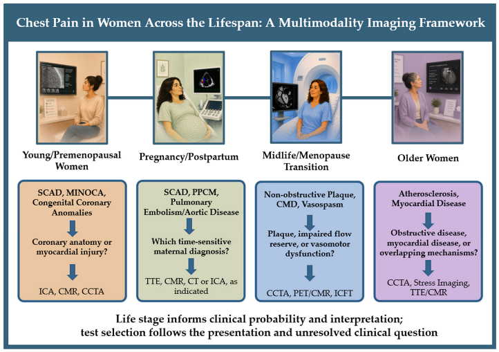
- Central Illustration. Life-stage-informed imaging of chest pain in women. The dominant differential and the unresolved clinical question that guides initial imaging change across young/premenopausal women, pregnancy and postpartum, midlife and menopause, and older age. Abbreviations: CCTA, coronary computed tomography angiography; CMD, coronary microvascular dysfunction; CMR, cardiac magnetic resonance; CT, computed tomography; ICA, invasive coronary angiography; ICFT, invasive coronary function testing; MINOCA, myocardial injury with non-obstructive coronary arteries; PET, positron emission tomography; PPCM, peripartum cardiomyopathy; SCAD, spontaneous coronary artery dissection; TTE, transthoracic echocardiography.
This article presents a narrative review of the available evidence, with priority given to clinical guidelines, scientific statements, systematic reviews and meta-analyses, and large clinical trials and registries. Additional articles were identified through reference-list screening. No predefined systematic search strategy, formal quality appraisal, or quantitative synthesis was performed.
The recommendations discussed in this review are supported by the relevant guidelines according to their respective scopes: the Fifth Universal Definition of Myocardial Infarction (2026) [3] for definitions and classification, the 2025 ACC/AHA Acute Coronary Syndromes Guideline [19] for acute coronary syndrome (ACS) management, the 2021 AHA/ACC Chest Pain Guideline [4] for chest pain evaluation and diagnosis, and the 2025 ESC Guidelines for the Management of Cardiovascular Disease and Pregnancy [20] for pregnancy-related cardiovascular considerations. Together, these documents provide complementary guidance for the clinical contexts addressed in the manuscript.
Throughout the article, the terms “women” and “men” refer to individuals of female and male sex, respectively, based on biological characteristics, including chromosomal, hormonal, and physiological features. We use sex-based terms because the studies cited here report participants by sex, and most do not separately report gender identity. The evidence base for transgender and gender-diverse individuals is limited and is not addressed here, though we recognize this as an area for further research and acknowledge that systemic biases impact people with these backgrounds as well.
2. Sex-Based Bias in Chest Pain Evaluation and Imaging
The sex- and -gender-bias within medicine was a strong motivator for this review, as systemic biases result in less timely and appropriate care for women presenting with cardiac symptoms [3,4,21]. Despite known sex-differences in cardiac physiology and disease risk, research conducted primarily in male cohorts has been extrapolated to females. Consequently, evidence for guideline-directed CAD therapy is derived predominantly from men, and current diagnostic pathways and guidelines do not routinely incorporate sex-specific imaging strategies or fully account for reproductive life stages [19,22,23]. This bias has improved over the years through initiatives promoting inclusion of women in research and consideration of sex-differences [23] though it still persists, and women remain underrepresented despite growing recognition of sex-related differences in cardiovascular outcomes.
Accordingly, the sex-based bias in the evaluation of chest pain extends directly into cardiovascular imaging, and it operates in two reinforcing ways: women are imaged less often and less invasively than men, and when they are imaged, their results are more likely to be misclassified [19,24,25,26,27,28]. Women often have a distinct coronary phenotype, with a greater likelihood of non-obstructive CAD. Therefore, diagnostic frameworks centered on obstructive epicardial stenosis may fail to identify the mechanism responsible for ischemic symptoms. Non-obstructive anatomy should not end the evaluation when symptoms or findings suggest microvascular or vasomotor disease. This review aims to address this gap and provide an imaging approach specifically designed for women at distinct phases of the female lifespan.
3. Young and Premenopausal Women
Young and premenopausal women constitute a distinct diagnostic population in whom obstructive calcific epicardial CAD is less prevalent than in older women and nonatherosclerotic coronary and myocardial disorders merit earlier consideration. The differential should be guided by presentation. Acute chest pain with ischemic electrocardiographic changes or troponin elevation raises concern for SCAD and MINOCA, while myocarditis and Takotsubo syndrome are important alternative causes of myocardial injury. Stable or exertional symptoms may reflect premature or non-obstructive atherosclerosis, congenital coronary anomalies, or functional ischemic mechanisms. Imaging should therefore be selected according to whether the unresolved question concerns coronary anatomy, myocardial injury, inducible ischemia, or coronary vascular function [2,10,12,29]. Table 1 summarizes the key causes of chest pain and myocardial injury in young women, their typical presentation, and the imaging modalities most useful for their evaluation.
Table 1.
Chest pain in young women—not always atherosclerosis.
3.1. Acute Chest Pain and Myocardial Injury in Young Women
Among young women presenting with ACS, SCAD is a key nonatherosclerotic entity that should be considered early. SCAD is characterized by spontaneous separation within the coronary arterial wall, producing an intramural hematoma—with or without intimal disruption—that compresses the true lumen. Its pathobiology remains incompletely understood. Proposed contributors include arterial-wall vulnerability, mechanical stress, and associated arteriopathies, particularly fibromuscular dysplasia. Female predominance and the recognized association with pregnancy and the early postpartum period suggest that reproductive factors may modify susceptibility; SCAD has also been associated with fluctuations in endogenous sex hormone levels during the menstrual cycle and with exogenous hormonal exposures, including combined oral contraceptives and infertility treatments; however, the significance of these relationships remains uncertain and should not be interpreted as evidence of causality [14,15,30,31]. These associations carry important implications for risk stratification and reproductive counseling in survivors [15,30].
Invasive coronary angiography (ICA) remains the principal diagnostic test when SCAD presents as ACS. Optical coherence tomography (OCT) or intravascular ultrasound (IVUS) may clarify equivocal findings but should be used selectively because coronary instrumentation may propagate the dissection. Coronary computed tomography angiography (CCTA) can provide complementary information in clinically stable patients and during follow-up, although its performance is limited for distal and small-caliber vessels. CMR does not directly establish the coronary diagnosis but can define the pattern and extent of associated myocardial injury. These modalities therefore capture vascular and myocardial consequences of SCAD with test selection tailored to disease acuity and patient stability [15,30,32].
MINOCA is a working diagnosis in patients with clinical features of possible myocardial infarction (MI) and no obstructive CAD (no stenosis ≥ 50%) on coronary angiography [3]. Accurate recognition of myocardial injury is an important first step in this diagnostic pathway. Sex-specific 99th-percentile troponin thresholds are recommended because the reference upper limits for high-sensitivity troponin assays are substantially lower in women than in men. Compared with uniform thresholds, the use of sex-specific thresholds reclassifies a considerably higher proportion of women as having myocardial injury, helping address a historical source of underdiagnosis in this population [3]. Once myocardial injury is established, further evaluation is required to establish the underlying cause and final diagnosis, which may include a coronary mechanism such as plaque disruption without flow-limiting stenosis, epicardial or microvascular spasm, coronary thromboembolism, and SCAD. Alternatively, additional testing may identify a non-coronary diagnosis, such as myocarditis or Takotsubo syndrome, which can produce similar symptoms, electrocardiographic changes, troponin elevation, and wall-motion abnormalities [3,19,29].
Early multiparametric CMR is central to this evaluation because it can distinguish ischemic from nonischemic injury, identify myocardial edema and fibrosis, and define infarct extent. In a 2023 systematic review and meta-analysis of 3624 patients with an initial MINOCA working diagnosis, CMR led to diagnostic reclassification in 68%, identifying myocarditis in 31%, Takotsubo syndrome in 10%, and MI in 22%. For clinicians, this high reclassification rate means that the initial diagnosis may change in roughly two-thirds of patients, highlighting the importance of performing CMR early, before myocardial edema and transient wall-motion abnormalities resolve, rather than deferring imaging [33].
3.2. Stable Chest Pain in Young Women
In young and premenopausal women with stable or exertional symptoms, congenital coronary anomalies and premature or non-obstructive atherosclerosis are important anatomic considerations. Congenital coronary anomalies may be discovered incidentally or during evaluation of exertional symptoms; selected high-risk patterns may also present with acute ischemia or sudden cardiac events [34]. CCTA is the preferred noninvasive modality when the principal question concerns coronary origin, proximal course, or the relationship of an anomalous vessel to adjacent structures. It also assesses obstructive and non-obstructive plaque [35,36]. Given the greater lifetime attributable risk of radiation-induced cancer in younger patients, particularly women, radiation-free testing should be considered when it can adequately address the diagnostic question [37].
Exercise electrocardiography retains a limited role in selected low-risk women with an interpretable resting electrocardiogram (ECG) and adequate functional capacity, particularly when exercise tolerance and symptom reproduction are the primary objectives. In a meta-analysis of 19 exercise ECG studies, sensitivity and specificity were 61% and 70%, respectively, in women, compared with 72% and 77% among men [37,38]. Because its specificity is lower in women, an isolated positive result should be interpreted with symptoms, workload, hemodynamic response, and pretest likelihood rather than prompting reflexive invasive angiography [2,39,40].
Stress echocardiography is appropriate when functional assessment is preferred or the resting ECG is uninterpretable, whereas CCTA is favored when coronary anatomy is the principal unresolved question [4]. Normal CCTA excludes clinically important obstructive epicardial disease with high confidence in appropriately selected patients. However, neither TTE nor CCTA exclude CMD, endothelial dysfunction, or vasospasm. When symptoms persist despite non-obstructive anatomy or nondiagnostic conventional stress testing, evaluation should shift from stenosis detection toward quantitative perfusion imaging or, in selected patients, ICFT [4,41]. Detailed test-performance characteristics are outlined in the multimodality section.
The key distinction in this life stage is acute versus stable presentation, followed by the unresolved question, coronary anatomy, myocardial injury, or coronary physiology. A mechanism-directed sequence reduces both underdiagnosis and redundant testing while avoiding premature reassurance after a normal or non-obstructive initial study.
4. Pregnancy and Postpartum: Imaging Time-Sensitive Causes of Chest Pain
Physiologic adaptations of pregnancy alter cardiovascular hemodynamics, coagulation, and vascular structure, broadening the differential diagnosis and modifying image interpretation. Maternal blood volume, heart rate, stroke volume, and cardiac output increase, while systemic vascular resistance decreases; pregnancy also promotes hypercoagulability, arterial remodeling, and increased myocardial wall stress [16,42,43,44]. These changes may alter chamber size, transvalvular flow, pulmonary pressures, and vascular dimensions and can be followed with TTE or CMR when clinically indicated. Findings must be interpreted in relation to gestational age and postpartum recovery [16,44]. During pregnancy, evaluation must balance timely maternal diagnosis with fetal safety, whereas after delivery fetal exposure is no longer relevant although lactation and maternal recovery may continue to influence decisions [4]. Figure 1 addresses pregnancy specific diagnostic and safety considerations.
Figure 1.
Overview of cardiovascular imaging modalities used during pregnancy and postpartum, highlighting their clinical applications and safety considerations. Abbreviations: ACS, acute coronary syndrome; CTPA, computed tomography pulmonary angiogram; CCTA, coronary computed tomography angiography; CMR, cardiac magnetic resonance; CT, computed tomography; GBCA, gadolinium-based contrast agent; ICA, invasive coronary angiography; PE, pulmonary embolism; PET, positron emission tomography; SPECT, single-photon emission computed tomography; STEMI, ST-segment elevation myocardial infarction; TEE, transesophageal echocardiography; TTE, transthoracic echocardiography.
4.1. Pregnancy and Postpartum Specific Diagnoses
Pregnancy-associated SCAD occurs predominantly in the postpartum period, particularly the early postpartum, with a smaller proportion of cases in late pregnancy [45,46]. Clinical presentations may include ST-segment elevation MI, non-ST-segment elevation MI, and hemodynamic instability, among others. Hormonal and hemodynamic changes may contribute to arterial-wall vulnerability, but the relevant imaging priority is prompt recognition of the coronary lesion and its myocardial consequences rather than attribution to a single hormonal mechanism. The general SCAD imaging approach is discussed above; here, the principal considerations are diagnostic urgency and minimization of unnecessary coronary instrumentation [10,14,16].
Other time-sensitive diagnoses include pulmonary embolism, whose risk increases during pregnancy and remains elevated in the early postpartum period; acute aortic syndromes, particularly in women with underlying aortopathy; PPCM; myocarditis; Takotsubo syndrome; and hypertensive disorders of pregnancy. Noncardiac causes remain common, but they should not lead to premature exclusion of cardiovascular disease when symptoms, biomarkers, or hemodynamics are concerning [43,47,48].
4.2. Imaging Evaluation: Balancing Diagnostic Urgency and Safety
The suspected diagnosis and maternal stability should take priority in imaging decisions during pregnancy, with radiation and contrast considerations guiding the choice and optimization of the study rather than preventing necessary testing [4,43,47].
ECG remains an essential initial test in pregnant and postpartum women with chest pain or suspected ACS [20,43,47]. Pregnancy-related sinus tachycardia and nonspecific ST segment or T wave abnormalities may complicate interpretation, while dynamic regional ST-segment abnormalities should increase concern for ischemia [20,43,47,49]. ECG findings alone do not exclude ACS or pulmonary embolism [50].
TTE is typically the first-line cardiac imaging modality because it is portable, radiation-free, and rapidly evaluates ventricular function, regional wall motion, valvular disease, pulmonary pressures, and pericardial effusion. It is particularly useful in suspected PPCM, hypertensive heart disease, myocarditis, Takotsubo syndrome, and complications of ACS [20,43,47]. Transesophageal echocardiography (TEE) may be used when TTE is inadequate and aortic, valvular, or prosthetic pathology remains suspected, with attention to positioning, aspiration risk, sedation, and fetal monitoring when appropriate [4,16,42,47,51,52].
CMR provides complementary assessment when TTE is inconclusive or when more detailed evaluation of ventricular function, myocardial injury, or aortic dimensions is needed [16]. Non-contrast CMR to measure aortic dimensions and assess myocardial function is considered safe during pregnancy, while gadolinium-based contrast agents are generally avoided because of uncertain fetal risk and placental transfer. In lactating patients, gadolinium exposure through breast milk is minimal, and recommendations regarding breastfeeding interruption vary; counseling should reflect these differences and consider patient preferences [20].
When definitive thoracic or coronary anatomic imaging is required, CT should not be withheld solely because of pregnancy. Computed tomography pulmonary angiogram (CTPA) or ventilation-perfusion imaging may be selected for suspected pulmonary embolism according to clinical presentation and local expertise, while CCTA may be considered selectively when coronary anatomy remains the unresolved question [4,47,53]. Fetal radiation exposure from contemporary CT protocols is generally below thresholds for deterministic harm, and iodinated contrast should be used when the anticipated maternal diagnostic benefit justifies exposure [4,10,43,47]. Table 2 summarizes contrast-agent considerations during pregnancy and lactation.
Table 2.
Contrast Agents in Pregnant and Lactating Women.
ICA is indicated for ST-segment elevation MI, hemodynamic instability, or another high-risk acute coronary syndrome presentation and should not be delayed solely because of pregnancy [19]. When SCAD is identified, coronary instrumentation should be limited because intervention may propagate the dissection; conservative management is often preferred when the patient is clinically stable and coronary flow is preserved [14,16].
Exercise stress testing, if needed during pregnancy, should preferably use individualized, submaximal protocols [20,54,55,56]. Pharmacologic stress data remains limited; vasodilator agents are generally avoided [16,20,54,57]. Imaging during pregnancy should therefore be diagnosis-first and safety-conscious, not diagnosis-limiting. Adverse pregnancy outcomes should also be recorded because they inform later-life cardiovascular risk, even when they do not determine the acute imaging test [58].
5. Midlife and Menopause Transition
Menopause is a critical life stage that signifies the permanent cessation of ovarian function and transition to the non-reproductive phase of life with remarkable changes in hormonal patterns and physiological and psychosocial symptoms. The median age of natural menopause is 50 years. Menopause is considered premature when it occurs before age 40 and early if it occurs between ages 40–45 [11]. The menopause transition begins with changes in menstrual-cycle regularity and extends into the early postmenopausal period [59].
CAD generally develops later in women than in men, but its incidence increases substantially during and after the menopause transition [60,61,62]. Declining ovarian function is accompanied by changes in vascular function, lipid profiles, blood pressure, body composition, and metabolic risk that are not fully captured by conventional cardiovascular risk models [11]. Premature or early menopause, surgical menopause, and a high burden of vasomotor symptoms may further identify women with additional risk [63,64]. These factors should inform interpretation of imaging findings rather than serve as automatic indications for imaging in asymptomatic women.
5.1. Lower Plaque Burden, Greater Risk, and Different Mechanisms
The menopause transition is an important period of plaque progression [65]. Changes in endogenous sex hormones, including lower sex hormone–binding globulin and greater free androgen exposure, have been associated with endothelial dysfunction and greater carotid plaque burden, but these relationships are intertwined with chronological aging and cardiometabolic risk [11,66]. These biological influences provide important context for the age- and sex-specific patterns of coronary plaque observed on imaging.
In a large cross-sectional CCTA cohort, plaque burden was higher in men across age groups, whereas the age-associated increase in plaque burden was more pronounced in older women [67]. Plaque composition also shifts with age. In a large international cohort, noncalcified plaque predominated in younger patients, present in approximately 31% of those younger than 45 years compared with 5.9% at 66–75 years and 2.9% above 75 years. With increasing age, the noncalcified fraction of total plaque volume declined from 84.9% to 75.4%, while the calcified fraction increased from 15.1% to 24.6% [68]. This maturation also differs by sex: in PARADIGM, total plaque progression was similar after adjustment, but women had greater calcified-plaque progression (β = 2.83; p = 0.004), slower noncalcified-plaque progression (β = −3.39; p = 0.008), and less high-risk plaque progression (β = −0.18; p = 0.049) [65].
These differences in plaque burden and composition also translate into differences in prognostic significance. The CONFIRM2 artificial intelligence (AI)–based quantitative computed tomography (QCT), showed that men had greater total, noncalcified, and calcified plaque burden and more high-risk plaque (9.2% vs. 2.5%; p < 0.0001), but after adjustment for age and cardiovascular risk factors, each 50-mm3 increase in plaque volume was associated with significantly greater major adverse cardiovascular event risk in women than men: 17.7% vs. 5.3% for total plaque, 27.1% vs. 11.6% for noncalcified plaque, and 22.9% vs. 5.4% for calcified plaque [69]. Model-derived associations from The PROMISE trial further suggest that risk in women may emerge at a lower coronary plaque burden than in men (hazard ratio crossed 1.0 at 20% vessel volume in women vs. 28% in men), so relatively limited plaque should not automatically be considered low risk in a symptomatic woman [26]. Taken together, these findings suggest that the relationship between plaque burden and composition and clinical risk evolves across the female life course, particularly around and after the menopause transition. CCTA and coronary artery calcium (CAC) capture the resulting atherosclerotic phenotype, but not the hormonal transition itself, underscoring the importance of interpreting plaque findings in the context of age, reproductive stage, and other cardiovascular risk factors.
Traditional diagnostic pathways have centered on flow-limiting stenosis. However, women have more frequently non-obstructive plaque and CMD, reflecting a broader spectrum of coronary disease that may not be adequately characterized by stenosis alone [2]. This spectrum includes ANOCA and INOCA. ANOCA describes anginal symptoms in the absence of obstructive epicardial CAD, whereas INOCA additionally reflects objective evidence of myocardial ischemia. Both are associated with persistent symptoms and adverse cardiovascular outcomes and should not be considered benign [7,70].
The underlying pathophysiology is heterogeneous and often overlapping. CMD may reflect impaired vasodilatory capacity, microvascular remodeling or rarefaction, increased resistance, endothelial dysfunction, or microvascular spasm; epicardial vasospasm represents dynamic constriction of larger coronary arteries. Menopause-related changes in endothelial signaling, arterial stiffness, adiposity, and inflammation may contribute to this substrate, but imaging identifies the resulting physiology—reduced flow reserve, perfusion abnormalities, or vasomotor responses—rather than the hormonal exposure itself [70,71].
Women with ANOCA or INOCA may present with chest pressure or discomfort, but dyspnea, fatigue, nausea, and exertional symptoms are also common. These presentations should be recognized as clinically relevant manifestations of possible ischemia rather than dismissed as “atypical”, particularly when symptoms are recurrent, exertional, or associated with functional limitation [3,70,71].
5.2. Imaging the Ischemic Spectrum: From Plaque Burden to Flow Reserve
A structured multimodality imaging approach is increasingly important in midlife women because no single test characterizes the full spectrum of obstructive, non-obstructive, and functional coronary disease. CAC scoring can contribute to atherosclerotic risk stratification, particularly in women at intermediate atherosclerotic cardiovascular disease risk, although it does not capture noncalcified plaque. When coronary anatomy is the principal clinical question, CCTA provides anatomical assessment of obstructive and non-obstructive plaque, total and noncalcified plaque burden, positive remodeling, and other high-risk plaque features. These characteristics may have particular prognostic significance in women, in whom risk may become evident at a lower overall plaque burden [26,65,66].
Risk assessment requires additional caution in women with cardiometabolic or inflammatory risk-enhancing conditions, including diabetes, obesity, human immunodeficiency virus (HIV), and systemic autoimmune diseases. Systemic autoimmune disorders such as systemic lupus erythematosus and systemic sclerosis disproportionately affect women, while all four conditions may amplify atherosclerotic, inflammatory, or microvascular risk beyond that captured by conventional risk estimates. CCTA studies have identified a substantial burden of noncalcified plaque in women with HIV and systemic lupus erythematosus, while diabetes, obesity, and systemic inflammatory disease have been associated with impaired MFR [72,73,74,75,76]. These findings do not support routine advanced imaging in asymptomatic women; however, they underscore that a low CAC score may not fully characterize risk when noncalcified plaque or CMD is suspected. In symptomatic women whose clinical risk appears discordant with the CAC score, CCTA or quantitative perfusion imaging may provide more complete phenotyping when otherwise clinically indicated.
When symptoms persist despite non-obstructive coronary anatomy, or when the clinical question concerns coronary vascular function rather than plaque alone, quantitative perfusion imaging becomes important. PET with absolute quantification of MBF and MFR is a well-established noninvasive method for detecting CMD. An MFR below 2.0 is commonly used as a clinically relevant threshold, but interpretation should incorporate resting and hyperemic flow, radiotracer and technical factors, age, sex, and the broader clinical context [77]. Specifically, depth-independent attenuation correction and superior spatial resolution provide key advantages that directly overcome the sex-specific imaging limitations of breast tissue attenuation and small left ventricular size [71].
Quantitative stress CMR offers a complementary radiation-free approach to myocardial perfusion assessment. Automated pixel-wise myocardial perfusion mapping has been validated against invasive fractional flow reserve (FFR) for obstructive CAD and index of microcirculatory resistance (IMR) for microvascular dysfunction, providing a rapid, radiation-free strategy for differentiating CMD from multivessel epicardial disease [78,79]. Additionally, the superior spatial resolution of stress CMR captures non-transmural ischemia often missed by other modalities, while its combined perfusion imaging and tissue characterization capabilities position it as a key diagnostic modality for midlife women with suspected INOCA and CMD [2]. In the current AHA/ACC chest pain guidelines, stress imaging including CMR or PET carries a Class I recommendation for patients with intermediate-high risk stable chest pain and no known CAD for diagnosis of ischemia. PET and CMR quantitative assessment of myocardial blood flow reserve is considered reasonable for diagnosing CMD and improving risk stratification in patients with INOCA and persistent chest pain (Class IIa, Level of Evidence B-NR) [4]. See details in Section 7.
PET and CMR can identify impaired perfusion and flow reserve but cannot reliably provoke or exclude all vasospastic endotypes. ICFT adds measurement of coronary flow reserve and microvascular resistance during adenosine-mediated hyperemia and assessment of endothelial function and epicardial or microvascular spasm with acetylcholine [56,70,80,81,82]. It is most useful when symptoms persist, noninvasive findings are discordant or inconclusive, or endotype clarification is expected to alter treatment. The Central Illustration depicts this progression from plaque to perfusion to coronary vascular endotype.
6. Older Women: Risk, Function and Diagnostic Complexity
Older women (≥65 years) have a higher likelihood of obstructive CAD, but stenosis is often only one component of a more complex presentation. Chronological aging, cumulative postmenopausal exposure, and accumulated cardiometabolic and inflammatory burden converge to increase arterial stiffness, coronary calcification, microvascular dysfunction, myocardial fibrosis, and ventricular–arterial uncoupling [4,11,83]. Imaging captures these downstream phenotypes through CAC and CCTA, quantitative PET or CMR perfusion, TTE assessment of diastolic and exercise reserve, strain, and CMR tissue characterization. Frailty, reduced exercise capacity, baseline ECG abnormalities, and multimorbidity further complicate test selection [2,83].
6.1. Anatomic Imaging in the Setting of Coronary Calcification
Coronary calcification becomes increasingly common with age and can limit CCTA through blooming artifact, which may obscure the lumen and overestimate stenosis severity [83,84]. CCTA remains useful when coronary anatomy is the primary unresolved question and image quality is expected to be adequate, but functional testing may be preferable when extensive calcification or known CAD is likely to reduce specificity. The CAC Consortium showed that over 50% of women have detectable coronary calcium by age 65 and that number continues to increase linearly reaching about 90% by age 80 [85]. Notably, women have lower absolute CAC scores but greater calcified-plaque density than men within comparable CAC strata. The combination of smaller coronary vessel caliber in women, a difference that has been reported to persist even after adjustment for body habitus and left ventricular mass, and presence of denser calcium may reduce luminal assessment accuracy and potentially render studies nondiagnostic despite modest CAC scores [85,86,87,88]. Thus, CAC scoring may support risk assessment, but a high calcium burden can also signal that subsequent CCTA interpretation will be challenging. When CCTA demonstrates an intermediate lesion, CT-derived fractional flow reserve may add lesion-specific physiologic information; however, it does not evaluate CMD or vasomotor disease [2,4].
6.2. Functional and Tissue Imaging: Beyond Stenosis
Functional imaging provides important complementary information regarding the hemodynamic significance of coronary disease. When the resting ECG is interpretable and the patient can achieve an adequate workload, exercise testing with imaging can provide both diagnostic and prognostic information [40,83]. However, baseline ST-segment abnormalities, left bundle branch block, and left ventricular hypertrophy are more common in older women and may reduce the specificity of exercise electrocardiography [4,40].
Stress echocardiography identifies inducible regional wall-motion abnormalities, whereas single-photon emission computed tomography (SPECT), PET, and stress CMR assess relative or absolute myocardial perfusion [2]. In the PROMISE trial, positive functional testing in adults aged 65–74 years and those aged ≥ 75 years was associated with higher risks of cardiovascular death or MI, with hazard ratios of 3.18 and 6.55, respectively [83]. In the CIAO-ISCHEMIA trial, participants had moderate or severe ischemia at enrollment; at one year, approximately half had normal stress echocardiograms and 23% had moderate or severe ischemia [27]. These findings reinforce the diagnostic and prognostic value of functional assessment in older adults.
CMR is particularly useful when the question extends beyond inducible ischemia to infarct extent, ischemic versus nonischemic cardiomyopathy, infiltrative disease, or fibrosis. PET or quantitative stress CMR may identify CMD when symptoms persist despite non-obstructive epicardial disease. TTE and exercise echocardiography can assess diastolic reserve and exertional pulmonary pressures, while CMR late gadolinium enhancement (LGE) and extracellular-volume (ECV) techniques characterize replacement and diffuse fibrosis. These complementary measures help separate—and recognize overlap among—ischemia, heart failure with preserved ejection fraction (HFpEF), hypertensive remodeling, and infiltrative disease [2,18,83,89].
6.3. Exercise Capacity as a Diagnostic and Prognostic Variable
Exercise capacity is itself an important prognostic marker in older women and should be interpreted alongside the imaging result. Failure to achieve an adequate workload may indicate increased cardiovascular risk and can also make a negative study less reassuring [83]. Modified exercise protocols, including Balke or Naughton protocols with smaller workload increments, may improve test completion and interpretation in women with limited functional capacity [40,83].
A negative stress test should therefore be interpreted in the context of achieved workload, symptom reproduction, hemodynamic response, and image quality [83]. Limited acoustic windows on stress TTE and soft-tissue attenuation on SPECT may reduce diagnostic confidence [2]. When exercise is not feasible, pharmacologic stress imaging is appropriate. In selected patients who can exercise, stress TTE may additionally reveal exertional diastolic dysfunction or pulmonary hypertension, helping distinguish ischemic symptoms from overlapping heart failure physiology [83,90].
6.4. Escalating from Noninvasive to Invasive Evaluation
The decision to proceed to ICA should integrate clinical likelihood, noninvasive findings, symptom burden, and the expected impact of the result on management [2,4]. Invasive evaluation is appropriate when noninvasive imaging demonstrates high-risk anatomy or substantial ischemia, when symptoms remain limiting despite treatment, or when testing is inconclusive despite persistent clinical concern. Frailty, renal function, bleeding risk, and overall comorbidity burden should be considered because they may alter the balance between diagnostic benefit and procedural risk [4,83].
Mixed phenotypes are common in older women and may include obstructive CAD, non-obstructive plaque, CMD, hypertensive or infiltrative myocardial disease, and HFpEF. Imaging should identify which component most plausibly explains the presentation and whether additional testing will alter care. The objective is therefore to integrate anatomy, ischemic burden, myocardial phenotype, hemodynamics, and functional capacity rather than to establish stenosis alone [2].
7. Multimodality Imaging Across the Lifespan: Matching the Modality to the Clinical Question
The practical role of each imaging modality is determined by the unresolved clinical question and by recognition of its principal diagnostic blind spots. Figure 2 translates this principle into a decision pathway for acute and stable chest pain, showing how presentation and suspected mechanism determine the initial test, when complementary imaging is added, and when invasive coronary function testing is considered.
Figure 2.
Imaging decision pathway for acute (A) and stable (B) chest pain in women across the lifespan. All diagnoses and etiologies can occur across all life stages. The circles indicate the stages in which each is most common, not the only stages in which it occurs. Abbreviations: ACS, acute coronary syndrome; CAD, coronary artery disease; CCTA, coronary computed tomography angiography; CMD, coronary microvascular dysfunction; CMR, cardiac magnetic resonance; CT-FFR, CT-derived fractional flow reserve; ECG, electrocardiogram; ICA, invasive coronary angiography; ICFT, invasive coronary function testing; IVUS, intravascular ultrasound; MINOCA, myocardial injury with non-obstructive coronary arteries; NSTEMI, non-ST-segment elevation myocardial infarction; OCT, optical coherence tomography; PET, positron emission tomography; SCAD, spontaneous coronary artery dissection; SPECT, single-photon emission computed tomography; STEMI, ST-segment elevation myocardial infarction; TEE, transesophageal echocardiography; TTE, transthoracic echocardiography.
7.1. Echocardiography: The First-Line Foundation
TTE is frequently the initial cardiac imaging modality when the presentation raises concern for ventricular dysfunction, regional wall-motion abnormalities, valvular or pericardial disease, pulmonary hypertension, or hemodynamic instability [4]. It is widely available, portable, and does not involve ionizing radiation. TTE can rapidly evaluate biventricular size and function, valvular pathology, filling pressures, pericardial effusion, and the hemodynamic consequences of acute coronary syndrome, pulmonary embolism, or aortic disease; although it does not definitively exclude coronary, pulmonary vascular, or aortic pathology [91,92].
Assessment of diastolic function is particularly relevant in women, who have a greater burden of concentric remodeling, ventricular-arterial stiffness, and HFpEF. Exercise TTE may provide additional information regarding symptom reproduction, inducible regional wall-motion abnormalities, diastolic reserve, and exertional pulmonary pressures [40,90].
The principal limitations of TTE are dependence on acoustic windows and operator expertise [51,91]. Stress-induced regional wall-motion analysis is also relatively insensitive to diffuse or balanced reductions in myocardial perfusion [90], including CMD [41]. Doppler-derived coronary flow velocity reserve may provide physiologic information in experienced laboratories, but it is technically demanding and generally limited to the left anterior descending (LAD) coronary territory [93]. The diagnostic limits of TTE are outlined in the multimodality section.
7.2. CMR: Myocardial Injury, Tissue Characterization, and Perfusion
CMR provides accurate assessment of ventricular volumes and function together with myocardial tissue characterization. In women with acute chest pain, troponin elevation, and non-obstructive coronary arteries, multiparametric CMR can distinguish ischemic infarction from myocarditis, Takotsubo syndrome, and other causes of myocardial injury by integrating cine imaging, edema-sensitive techniques, parametric mapping, and late gadolinium enhancement [2,19,33].
Stress perfusion CMR extends the examination from tissue characterization to assessment of inducible ischemia. Quantitative perfusion mapping can identify diffuse or subendocardial perfusion abnormalities and evaluate suspected CMD, while the same examination can characterize scar, inflammation, ventricular remodeling, and mixed ischemic and nonischemic cardiomyopathies. These combined capabilities are particularly valuable when the clinical presentation cannot be explained by coronary anatomy alone [94].
These diagnostic strengths must be balanced against practical and temporal limitations. Access, cost, examination duration, patient tolerance, implanted-device considerations, and the need for specialized acquisition and interpretation may limit widespread use. Further discussion is provided in Section 7.7. In addition, because myocardial edema and transient wall-motion abnormalities may resolve over time, diagnostic yield can decline when CMR is delayed. Life-stage-specific considerations also apply: during pregnancy, non-contrast CMR may be performed when clinically indicated, whereas gadolinium-enhanced and pharmacologic stress examinations require individualized risk–benefit assessment, as discussed in the pregnancy section [4,16].
7.3. CT/CCTA: Defining Anatomy, but Not the Full Phenotype
In women with chest pain, CCTA is most useful when the diagnostic uncertainty centers on coronary anatomy, but its interpretation must explicitly account for the fact that women often have ischemia without obstructive epicardial disease. Compared with men, women more frequently exhibit non-obstructive plaque, diffuse atherosclerosis, and positive remodeling, all of which may be missed or underappreciated in epicardial stenosis-focused paradigms. Molnar et al. found that women had the highest event rate in Coronary Artery Disease–Reporting and Data System (CAD-RADS) 3 (1.1% MI and 1.6% ACS), while event rates were highest in CAD-RADS 4 for men (1.9% MI and 2.5% ACS) suggesting that current CCTA risk stratification tools may underestimate risk in women [95]. This was further supported by the ISCHEMIA trial which found that anginal symptom burden was independently associated with the female sex even when the extent of CAD by CCTA was similar across the sexes (odds ratio, 1.41; 95% CI, 1.13–1.76) [28]. In this context, CCTA’s ability to visualize early and non-obstructive plaque is particularly valuable, as even low plaque burden carries prognostic significance in women. Quantitative plaque assessment is particularly relevant in women because risk may emerge at lower plaque burden [26,65,66,69,96,97]. CT-derived fractional flow reserve is limited to the lesion-specific significance of epicardial stenosis. Additional limitations include ionizing radiation, iodinated contrast, distal-vessel resolution, and blooming artifact from extensive calcification—considerations that vary across pregnancy, younger age, and older women [41,96,98]. See Section 7.5.
Pericoronary adipose tissue (PCAT) attenuation and related fat-attenuation metrics are emerging CT biomarkers of the downstream inflammatory response surrounding coronary arteries. They should be described as measures of coronary inflammatory phenotype—not direct measures of hormonal status—and require further validation in women and across reproductive stages [99,100,101].
7.4. Nuclear Imaging
Nuclear imaging plays a critical role in evaluating ischemia in women, but its strengths and limitations are tightly linked to female-specific disease patterns. SPECT remains widely used and provides strong prognostic value; however, its diagnostic performance is affected by attenuation artifacts from breast tissue and body habitus, which reduce specificity and contribute to false-positive results in women [102,103,104]. Furthermore, sensitivity is also affected and typically, does not increase with attenuation-corrected SPECT perfusion imaging [104,105]. This limitation appears particularly relevant in overweight and obese women, who continue to demonstrate lower sensitivity than men of similar weight despite the use of combined supine and prone imaging (82% vs. 93% and 83% vs. 95%, respectively) [104,106].
These limitations are further underscored by head-to-head comparisons demonstrating superior diagnostic performance with PET. In the flurpiridaz F-18 trial, PET demonstrated greater sensitivity than SPECT for CAD detection (80.3% vs. 68.7%), with noninferior specificity (63.8% vs. 61.7%) and a higher Area Under the Curve (0.80 vs. 0.68). This sensitivity advantage was maintained in women (82.9% vs. 65.9%), obese patients (76.9% vs. 69.2%), and patients with diabetes (75.8% vs. 71.4%) [107].
Beyond diagnostic accuracy, PET provides an important physiological advantage by quantifying absolute MBF and MFR. In contrast, SPECT primarily assesses relative perfusion, which may limit its ability to detect diffuse ischemia or balanced reductions in flow, patterns that are particularly relevant in women with CMD [41]. As a result, SPECT may underestimate ischemic burden in women with persistent symptoms despite non-obstructive coronary arteries, whereas PET can provide quantitative evidence of impaired coronary vasodilator function, a major mechanism of ischemia and adverse outcomes in women [41,108]. PET also offers improved spatial resolution, robust attenuation correction, and lower radiation exposure compared with SPECT, enhancing diagnostic accuracy in female populations [102]. See Section 7.5. In women with persistent chest pain and non-obstructive coronary findings, PET shifts the diagnostic paradigm from identifying obstructive lesions to characterizing impaired coronary physiology, aligning more closely with sex-specific disease mechanisms [41,108]. Limitations of PET include availability, cost, radiotracer-specific requirements, and exposure to ionizing radiation, which are discussed further in Section 7.7. Interpretation of MFR should also account for resting flow, hyperemic flow, technical quality, and clinical context, rather than relying on a single threshold in isolation [2,77,97,108].
7.5. Integrating Anatomy, Myocardial Substrate, and Physiology
The modalities are best understood by the biologic question they answer. Echocardiography evaluates structure, hemodynamics, strain, and reserve; CCTA defines coronary anatomy, plaque, and emerging inflammatory phenotypes; CMR characterizes injury, remodeling, fibrosis, and perfusion; SPECT assesses relative perfusion; PET quantifies MBF and flow reserve; invasive angiography defines high-risk epicardial anatomy; and ICFT identifies microvascular resistance, endothelial dysfunction, and epicardial or microvascular spasm. These measurements can reveal the downstream consequences of changing vascular biology without implying that imaging can directly measure hormonal exposure.
The clinical value of each test lies in recognizing both what it identifies and what it cannot exclude. A normal structural examination does not exclude ischemia; non-obstructive anatomy does not exclude coronary vascular dysfunction; and normal relative perfusion does not always exclude diffuse microvascular disease. Table 3 provides a consolidated overview of cardiovascular imaging modalities, highlighting the clinical questions they address, diagnostic trade-offs, and considerations specific to sex and life stage.
Table 3.
Selecting cardiovascular imaging in women with chest pain: clinical questions, diagnostic trade-offs, and life-stage considerations.
7.6. Emerging Technologies: Toward More Precise Phenotyping in Women
Emerging technologies may strengthen the link between life-stage biology and measurable phenotype. Speckle-Tracking Echocardiography is an established technique that can quantify global longitudinal strain (GLS) and subsequently detect subclinical dysfunction prior to a reduction in left ventricular ejection fraction, which can offer prognostic information and further intervention, if needed [109,110,111]. In patients with normal left ventricular function and suspected coronary disease, reduced GLS, early systolic lengthening, and postsystolic shortening may help identify myocardial ischemia [109,112].
CMR has expanded the assessment of myocardial deformation through several emerging strain techniques. Strain-encoded imaging (SENC), including fast-SENC, directly measures regional strain and can be incorporated into stress imaging for detection of ischemia and assessment of viability [112]. Displacement encoding with stimulated echoes (DENSE) provides highly reproducible regional and global strain and enables early detection of subclinical myocardial dysfunction, though it requires a dedicated sequence and remains largely a research tool [113]. Feature-tracking (FT) derives strain from routinely acquired cine images without additional imaging, offering potential prognostic value; however, variation across vendors and software platforms and the lack of standardized sex-specific reference ranges currently limit its broader clinical adoption and longitudinal use [114].
Emerging cadmium-zinc-telluride (CZT)-SPECT technology may enable quantitative assessment of myocardial blood flow with substantially lower radiation and shorter acquisition times. Although global flow estimates show good agreement with PET, limited regional accuracy, moderate correlation with invasive microvascular indices, and the absence of harmonized thresholds remain barriers to routine clinical use [81,115].
Photon-counting detector CT (PCD-CT) enhances spatial resolution and reduces calcium blooming, potentially improving plaque assessment in older women with heavily calcified vessels [116,117,118,119]. CT-derived PCAT metrics may quantify the local inflammatory response around coronary arteries, and AI-enabled QCT can integrate plaque burden and composition at scale. Recent AI-QCT data suggest that similar increments in plaque may confer greater relative risk in women than in men, reinforcing the need for sex-aware rather than sex-neutral interpretation. AI and radiomics may eventually combine plaque, flow, strain, tissue, reproductive history, and clinical context to support individualized pathways [69].
These technologies offer opportunities for more precise characterization of inflammation, remodeling, and vascular dysfunction. However, most AI-enabled quantitative CCTA and radiomic approaches have been evaluated in retrospective or registry-based cohorts and require prospective, externally validated confirmation before routine clinical use [69,120,121]. See Section 8 for details. The clinical applications and current translational status of these techniques are summarized in Table 4.
Table 4.
Emerging imaging techniques and their current role in clinical practice.
7.7. Imaging Access Across Care Settings
The multimodality strategy proposed here assumes access to advanced functional and physiologic imaging, which remains unevenly distributed across healthcare settings. When access to advanced diagnostic tools are not available, alternatives can help triage higher-risk patients to the appropriate centers.
The evaluation of obstructive epicardial disease is feasible across many healthcare settings. CCTA is a widely available first-line anatomic test, CT-FFR, where offered, adds functional gatekeeping for intermediate stenoses, and SPECT and stress echocardiography are broadly accessible alternatives, including community-based practices and freestanding imaging centers. ICA remains broadly accessible at hospitals with catheterization laboratories [4,81,122]. Access to more advanced imaging techniques including PET, quantitative CMR, and ICFT are less equitably distributed, and primarily concentrated in well-resourced centers and less available to hospitals serving racially and ethnically minoritized and lower-income communities, with access further attenuated by rural residence, insurance gaps, and travel and wait-time burdens [123,124]. When necessary expertise or testing is unavailable locally, an available test may answer part of the clinical question, though referral to a more advanced imaging center should be prioritized.
A life stage informed strategy therefore requires attention to equitable access, because directing evaluation toward mechanism specific tools may otherwise widen existing disparities. Poverty, sexism, structural racism, geographic isolation, educational opportunities, and access to health insurance and health care can all influence whether patients receive timely and appropriate diagnostic evaluation [124,125,126].
8. Clinical Implications, Common Pitfalls, and Future Directions
In summary, a life-stage-informed approach to imaging chest pain in women should integrate reproductive history and biological context without substituting age or hormonal status for clinical assessment. The first decision is the unresolved question: structure and hemodynamics, coronary anatomy and plaque, myocardial injury, perfusion and flow reserve, or vasomotor function. This framing reduces one-size-fits-all testing and clarifies when complementary imaging is needed [4,127].
Common pitfalls include assuming low risk because a patient is young; treating pregnancy as a reason to defer necessary imaging; labeling non-obstructive coronary anatomy, a normal TTE, or normal relative perfusion as exclusion of ischemic disease; and attributing symptoms in older women solely to calcific CAD. CMR remains underused after MINOCA, quantitative perfusion remains underused in suspected CMD, and vasospastic disease cannot be excluded by anatomy alone [2,4,10,29,77].
Important knowledge and access gaps remain. Reproductive history, adverse pregnancy outcomes, menopause timing, and hormonal therapies are rarely incorporated into imaging algorithms despite associations with later cardiovascular risk [58,127]. Access to quantitative PET and CMR perfusion, ICFT, and advanced plaque analysis remains uneven, and reference standards are not consistently stratified by sex, age, body size, life stage or ancestry [1,71,77].
It should also be acknowledged that much of the foundational evidence on multimodality imaging, ANOCA, INOCA, MINOCA, CMD, and sex-specific plaque phenotypes originates from high-income settings and predominantly Caucasian cohorts, with limited representation of older women and of Black, Hispanic, South Asian, East Asian, and lower-socioeconomic-status populations. Because eco-geographical and socioeconomic factors can influence cardiovascular phenotype, disease prevalence, and access to and timing of diagnosis, the thresholds and modality preferences discussed here may not transfer uniformly across populations and warrant validation in more diverse cohorts [4,124,125,128,129,130,131]. Multimodality validation must also include adequate representation across pregnancy, menopause stage, age, race and ethnicity, body size, and relevant inflammatory and cardiometabolic conditions.
Future studies should prospectively link reproductive and hormonal transitions to serial imaging phenotypes rather than relying on cross-sectional associations. Priorities include defining life-stage-specific reference ranges for chamber geometry, strain, MBF, MFR, plaque composition, pericoronary inflammation, and fibrosis; studying trajectories across pregnancy and postpartum and through the menopause transition; determining whether these biomarkers track response to therapy; and validating AI models in diverse populations of women. Imaging can then serve as a bridge between changing vascular biology and clinically actionable phenotype, while avoiding the inaccurate implication that a single modality or hormonal state explains disease [11,58,69,77,109,120].
Chest pain in women is etiologically diverse and evolves across the lifespan. Selecting and interpreting imaging according to the clinical question, life-stage context, and likely vascular or myocardial mechanism may improve diagnostic evaluation beyond a stenosis-centered approach.
Author Contributions
Writing—original draft preparation: C.C.S., M.E.S., L.O.M., S.W., C.R.-S., M.C.E., T.A., A.T., P.P., M.P.W., B.P., H.K., J.A.P., P.L.S., H.P., N.H. and P.F.R.-L.; C.C.S. and P.F.R.-L.; figures, M.E.S., H.K., C.C.S. and P.F.R.-L.; writing—review and editing: C.C.S. and P.F.R.-L.; figures, M.E.S., H.K., C.C.S. and P.F.R.-L.; All authors have read and agreed to the published version of the manuscript.
Funding
This research received no external funding. P.F.R.-L. is supported by the Leducq AtheroGEN consortium (22CVD04) and the National Heart, Lung, and Blood Institute (K01HL174889).
Institutional Review Board Statement
Not applicable.
Informed Consent Statement
Not applicable.
Data Availability Statement
No new data were created or analyzed in this study.
Acknowledgments
During the preparation of this manuscript, the authors used BioRender and AI tools for the creation of figures and an AI-assisted writing tool for language editing and refinement (BioRender, web-based software platform, continuously updated online; BioRender.com, accessed October 2026; Claude Sonnet 5.5, Anthropic; ChatGPT, powered by GPT-6, OpenAI).The authors have reviewed and edited the output and take full responsibility for the content of this publication.
Conflicts of Interest
The authors declare no conflicts of interest.
Abbreviations
The following abbreviations are used in this manuscript:
| ACS | Acute Coronary Syndrome |
| AI | Artificial Intelligence |
| ANOCA | Angina with Non-Obstructive Coronary Arteries |
| CAC | Coronary Artery Calcium |
| CAD | Coronary Artery Disease |
| CAD-RADS | Coronary Artery Disease–Reporting and Data System |
| CCTA | Coronary Computed Tomography Angiography |
| CMD | Coronary Microvascular Dysfunction |
| CMR | Cardiac Magnetic Resonance |
| CT | Computed Tomography |
| CTPA | Computed Tomography Pulmonary Angiogram |
| CZT | Cadmium-zinc-telluride |
| DENSE | Displacement Encoding with Stimulated Echoes |
| ECG | Electrocardiogram |
| ECV | Extracellular-volume |
| FFR | Fractional Flow Reserve |
| FT | Feature Tracking |
| GLS | Global Longitudinal Strain |
| HFpEF | Heart Failure with Preserved Ejection Fraction |
| HIV | Human Immunodeficiency Virus |
| ICA | Invasive Coronary Angiography |
| ICFT | Invasive Coronary Function Testing |
| IMR | Index of Microcirculatory Resistance |
| INOCA | Ischemia with Non-Obstructive Coronary Arteries |
| IVUS | Intravascular Ultrasound |
| LAD | Left Anterior Descending |
| LGE | Late Gadolinium Enhancement |
| MBF | Myocardial Blood Flow |
| MFR | Myocardial Flow Reserve |
| MI | Myocardial Infarction |
| MINOCA | Myocardial Injury with Non-Obstructive Coronary Arteries |
| OCT | Optical Coherence Tomography |
| PCAT | Pericoronary Adipose Tissue |
| PCD-CT | Photon-Counting Detector Computed Tomography |
| PET | Positron Emission Tomography |
| PPCM | Peripartum Cardiomyopathy |
| QCT | Quantitative Computed Tomography |
| SCAD | Spontaneous Coronary Artery Dissection |
| SENC | Strain-Encoded Imaging |
| SPECT | Single-Photon Emission Computed Tomography |
| TEE | Transesophageal Echocardiography |
| TTE | Transthoracic Echocardiography |
References
- Solola Nussbaum, S.; Henry, S.; Yong, C.M.; Daugherty, S.L.; Mehran, R.; Poppas, A. Sex-Specific Considerations in the Presentation, Diagnosis, and Management of Ischemic Heart Disease: JACC Focus Seminar 2/7. J. Am. Coll. Cardiol. 2022, 79, 1398–1406. [Google Scholar] [CrossRef] [Scilit] [PubMed]
- Rodriguez Lozano, P.F.; Rrapo Kaso, E.; Bourque, J.M.; Morsy, M.; Taylor, A.M.; Villines, T.C.; Kramer, C.M.; Salerno, M. Cardiovascular Imaging for Ischemic Heart Disease in Women: Time for a Paradigm Shift. JACC Cardiovasc. Imaging 2022, 15, 1488–1501. [Google Scholar] [CrossRef] [Scilit] [PubMed]
- Mills, N.L.; Kristin Newby, L.; Zaman, S.; Almahmeed, W.A.; Bucciarelli-Ducci, C.; Colvin, M.M.; Dangas, G.D.; DeVon, H.A.; Dharmakumar, R.; Gaziano, T.A.; et al. Fifth Universal Definition of Myocardial Infarction (2026): On Behalf of the Joint European Society of Cardiology (ESC)/American College of Cardiology (ACC)/American Heart Association (AHA)/World Heart Federation (WHF) Task Force for the Universal Definition of Myocardial Infarction. Circulation 2026. [Google Scholar] [CrossRef] [Scilit] [PubMed]
- Gulati, M.; Levy, P.D.; Mukherjee, D.; Amsterdam, E.; Bhatt, D.L.; Birtcher, K.K.; Blankstein, R.; Boyd, J.; Bullock-Palmer, R.P.; Conejo, T.; et al. 2021 AHA/ACC/ASE/CHEST/SAEM/SCCT/SCMR Guideline for the Evaluation and Diagnosis of Chest Pain: A Report of the American College of Cardiology/American Heart Association Joint Committee on Clinical Practice Guidelines. Circulation 2021, 144, e368–e454, Erratum in Circulation 2021, 144, e455. https://doi.org/10.1161/CIR.0000000000001047; Erratum in Circulation 2023, 148, e281. https://doi.org/10.1161/CIR.0000000000001198. [Google Scholar] [CrossRef] [Scilit] [PubMed]
- Mehta, L.S.; Beckie, T.M.; DeVon, H.A.; Grines, C.L.; Krumholz, H.M.; Johnson, M.N.; Lindley, K.J.; Vaccarino, V.; Wang, T.Y.; Watson, K.E.; et al. Acute Myocardial Infarction in Women: A Scientific Statement from the American Heart Association. Circulation 2016, 133, 916–947. [Google Scholar] [CrossRef] [Scilit] [PubMed]
- Hosadurg, N.; Watts, K.; Wang, S.; Wingerter, K.E.; Taylor, A.M.; Villines, T.C.; Patel, A.R.; Bourque, J.M.; Lindner, J.R.; Kramer, C.M.; et al. Emerging Pathway to a Precision Medicine Approach for Angina with Nonobstructive Coronary Arteries in Women. JACC Adv. 2024, 3, 101074. [Google Scholar] [CrossRef] [Scilit] [PubMed]
- Kunadian, V.; Chieffo, A.; Camici, P.G.; Berry, C.; Escaned, J.; Maas, A.; Prescott, E.; Karam, N.; Appelman, Y.; Fraccaro, C.; et al. An EAPCI Expert Consensus Document on Ischaemia with Non-Obstructive Coronary Arteries in Collaboration with European Society of Cardiology Working Group on Coronary Pathophysiology & Microcirculation Endorsed by Coronary Vasomotor Disorders International Study Group. EuroIntervention 2021, 16, 1049–1069. [Google Scholar] [CrossRef] [Scilit] [PubMed]
- Davis, M.B.; Arendt, K.; Bello, N.A.; Brown, H.; Briller, J.; Epps, K.; Hollier, L.; Langen, E.; Park, K.; Walsh, M.N.; et al. Team-Based Care of Women with Cardiovascular Disease from Pre-Conception Through Pregnancy and Postpartum: JACC Focus Seminar 1/5. J. Am. Coll. Cardiol. 2021, 77, 1763–1777. [Google Scholar] [CrossRef] [Scilit] [PubMed]
- O’Kelly, A.C.; Michos, E.D.; Shufelt, C.L.; Vermunt, J.V.; Minissian, M.B.; Quesada, O.; Smith, G.N.; Rich-Edwards, J.W.; Garovic, V.D.; El Khoudary, S.R.; et al. Pregnancy and Reproductive Risk Factors for Cardiovascular Disease in Women. Circ. Res. 2022, 130, 652–672. [Google Scholar] [CrossRef] [Scilit] [PubMed]
- Kovacic, J.C.; Reynolds, H.R.; Alasnag, M.; Blakeman, J.R.; Ijioma, N.N.; Kim, E.S.H.; Sandner, S.; Sanghavi, M.; Saw, J.; Tamis-Holland, J.E.; et al. Acute Coronary Syndromes in Premenopausal Women: A Scientific Statement from the American Heart Association. Circulation 2026, 153, e89–e108. [Google Scholar] [CrossRef] [Scilit] [PubMed]
- El Khoudary, S.R.; Aggarwal, B.; Beckie, T.M.; Hodis, H.N.; Johnson, A.E.; Langer, R.D.; Limacher, M.C.; Manson, J.E.; Stefanick, M.L.; Allison, M.A.; et al. Menopause Transition and Cardiovascular Disease Risk: Implications for Timing of Early Prevention: A Scientific Statement from the American Heart Association. Circulation 2020, 142, e506–e532. [Google Scholar] [CrossRef] [Scilit] [PubMed]
- Minissian, M.B.; Mehta, P.K.; Hayes, S.N.; Park, K.; Wei, J.; Bairey Merz, C.N.; Cho, L.; Volgman, A.S.; Elgendy, I.Y.; Mamas, M.; et al. Ischemic Heart Disease in Young Women: JACC Review Topic of the Week. J. Am. Coll. Cardiol. 2022, 80, 1014–1022. [Google Scholar] [CrossRef] [Scilit] [PubMed]
- Parwani, P.; Kang, N.; Safaeipour, M.; Mamas, M.A.; Wei, J.; Gulati, M.; Naidu, S.S.; Merz, N.B. Contemporary Diagnosis and Management of Patients with MINOCA. Curr. Cardiol. Rep. 2023, 25, 561–570. [Google Scholar] [CrossRef] [Scilit] [PubMed]
- Singulane, C.C.; Wang, S.; Watts, K.; Stahl, M.E.; Denlinger, L.; Lloyd, R.; Pallinti, P.; Preston, L.; Morsy, M.; Quesada, O.; et al. Spontaneous Coronary Artery Dissection (SCAD): Unveiling the Enigma of the Unexpected Coronary Event. Curr. Atheroscler. Rep. 2025, 27, 81. [Google Scholar] [CrossRef] [Scilit] [PubMed]
- Crousillat, D.; Sarma, A.; Wood, M.; Naderi, S.; Leon, K.; Gibson, C.M.; Aday, A.; Grodzinsky, A.; Izard, K.; Kovacic, J.C.; et al. Spontaneous Coronary Artery Dissection: Current Knowledge, Research Gaps, and Innovative Research Initiatives: JACC Advances Expert Panel. JACC Adv. 2024, 3, 101385. [Google Scholar] [CrossRef] [Scilit] [PubMed]
- Bello, N.A.; Bairey Merz, C.N.; Brown, H.; Davis, M.B.; Dickert, N.W.; El Hajj, S.C.; Giullian, C.; Quesada, O.; Park, K.; Sanghani, R.M.; et al. Diagnostic Cardiovascular Imaging and Therapeutic Strategies in Pregnancy: JACC Focus Seminar 4/5. J. Am. Coll. Cardiol. 2021, 77, 1813–1822. [Google Scholar] [CrossRef] [Scilit] [PubMed]
- Damluji, A.A.; Forman, D.E.; Wang, T.Y.; Chikwe, J.; Kunadian, V.; Rich, M.W.; Young, B.A.; Page, R.L., 2nd; DeVon, H.A.; Alexander, K.P.; et al. Management of Acute Coronary Syndrome in the Older Adult Population: A Scientific Statement from the American Heart Association. Circulation 2023, 147, e32–e62. [Google Scholar] [CrossRef] [Scilit] [PubMed]
- Tzoumas, A.; Tapp, D.; Crousillat, D.; Honigberg, M.C.; Rodriguez-Lozano, P.F.; Aggarwal, N.R.; Ebong, I.A.; Briller, J.; De Oliveira, G.M.; Harrington, C.M.; et al. The Role of Coronary Microvascular Dysfunction in Heart Failure with Preserved Ejection Fraction. JACC Adv. 2025, 4, 102345. [Google Scholar] [CrossRef] [Scilit] [PubMed]
- Rao, S.V.; O’Donoghue, M.L.; Ruel, M.; Rab, T.; Tamis-Holland, J.E.; Alexander, J.H.; Baber, U.; Baker, H.; Cohen, M.G.; Cruz-Ruiz, M.; et al. 2025 ACC/AHA/ACEP/NAEMSP/SCAI Guideline for the Management of Patients with Acute Coronary Syndromes: A Report of the American College of Cardiology/American Heart Association Joint Committee on Clinical Practice Guidelines. Circulation 2025, 151, e771–e862, Erratum in Circulation 2025, 151, e865. https://doi.org/10.1161/CIR.0000000000001328; Erratum in Circulation 2025, 151, e1098. https://doi.org/10.1161/CIR.0000000000001346; Erratum in Circulation 2025, 152, e402. https://doi.org/10.1161/CIR.0000000000001397. [Google Scholar] [CrossRef] [Scilit] [PubMed]
- De Backer, J.; Haugaa, K.H.; Hasselberg, N.E.; de Hosson, M.; Brida, M.; Castelletti, S.; Cauldwell, M.; Cerbai, E.; Crotti, L.; de Groot, N.M.S.; et al. 2025 ESC Guidelines for the management of cardiovascular disease and pregnancy. Eur. Heart J. 2025, 46, 4462–4568. [Google Scholar] [CrossRef] [Scilit] [PubMed]
- Meisel, Z.F.; Armstrong, K.; Mechem, C.C.; Shofer, F.S.; Peacock, N.; Facenda, K.; Pollack, C.V. Influence of sex on the out-of-hospital management of chest pain. Acad. Emerg. Med. 2010, 17, 80–87. [Google Scholar] [CrossRef] [Scilit] [PubMed]
- Gaudino, M.; Di Franco, A.; Cao, D.; Giustino, G.; Bairey Merz, C.N.; Fremes, S.E.; Kirtane, A.J.; Kunadian, V.; Lawton, J.S.; Masterson Creber, R.M.; et al. Sex-Related Outcomes of Medical, Percutaneous, and Surgical Interventions for Coronary Artery Disease: JACC Focus Seminar 3/7. J. Am. Coll. Cardiol. 2022, 79, 1407–1425. [Google Scholar] [CrossRef] [Scilit] [PubMed]
- Iribarren, A.; Diniz, M.A.; Merz, C.N.B.; Shufelt, C.; Wei, J. Are we any WISER yet? Progress and contemporary need for smart trials to include women in coronary artery disease trials. Contemp. Clin. Trials 2022, 117, 106762. [Google Scholar] [CrossRef] [Scilit] [PubMed]
- Shaw, L.J.; Miller, D.D.; Romeis, J.C.; Kargl, D.; Younis, L.T.; Chaitman, B.R. Gender differences in the noninvasive evaluation and management of patients with suspected coronary artery disease. Ann. Intern. Med. 1994, 120, 559–566. [Google Scholar] [CrossRef] [Scilit] [PubMed]
- Heydari, B.; Ge, Y.; Antiochos, P.; Islam, S.; Steel, K.; Bingham, S.; Abdullah, S.; Mikolich, J.R.; Arai, A.E.; Bandettini, W.P.; et al. Sex-Specific Stress Perfusion Cardiac Magnetic Resonance Imaging in Suspected Ischemic Heart Disease: Insights from SPINS Retrospective Registry. JACC Cardiovasc. Imaging 2023, 16, 749–764. [Google Scholar] [CrossRef] [Scilit] [PubMed]
- Brendel, J.M.; Mayrhofer, T.; Karady, J.; Kolossvary, M.; Kerkovits, N.M.; Langenbach, I.L.; Jung, M.; Kelsey, M.D.; Langenbach, M.C.; Pagidipati, N.; et al. Risk in Women Emerges at Lower Coronary Plaque Burden Than in Men: PROMISE Trial. Circ. Cardiovasc. Imaging 2026, 19, e019011. [Google Scholar] [CrossRef] [Scilit] [PubMed]
- Reynolds, H.R.; Picard, M.H.; Spertus, J.A.; Peteiro, J.; Lopez Sendon, J.L.; Senior, R.; El-Hajjar, M.C.; Celutkiene, J.; Shapiro, M.D.; Pellikka, P.A.; et al. Natural History of Patients with Ischemia and No Obstructive Coronary Artery Disease: The CIAO-ISCHEMIA Study. Circulation 2021, 144, 1008–1023. [Google Scholar] [CrossRef] [Scilit] [PubMed]
- Reynolds, H.R.; Shaw, L.J.; Min, J.K.; Spertus, J.A.; Chaitman, B.R.; Berman, D.S.; Picard, M.H.; Kwong, R.Y.; Bairey-Merz, C.N.; Cyr, D.D.; et al. Association of Sex with Severity of Coronary Artery Disease, Ischemia, and Symptom Burden in Patients with Moderate or Severe Ischemia: Secondary Analysis of the ISCHEMIA Randomized Clinical Trial. JAMA Cardiol. 2020, 5, 773–786, Erratum in JAMA Cardiol. 2021, 6, 980. https://doi.org/10.1001/jamacardio.2021.1509. [Google Scholar] [CrossRef] [Scilit] [PubMed]
- Tamis-Holland, J.E.; Jneid, H.; Reynolds, H.R.; Agewall, S.; Brilakis, E.S.; Brown, T.M.; Lerman, A.; Cushman, M.; Kumbhani, D.J.; Arslanian-Engoren, C.; et al. Contemporary Diagnosis and Management of Patients with Myocardial Infarction in the Absence of Obstructive Coronary Artery Disease: A Scientific Statement from the American Heart Association. Circulation 2019, 139, e891–e908. [Google Scholar] [CrossRef] [Scilit] [PubMed]
- Hayes, S.N.; Tweet, M.S.; Adlam, D.; Kim, E.S.H.; Gulati, R.; Price, J.E.; Rose, C.H. Spontaneous Coronary Artery Dissection: JACC State-of-the-Art Review. J. Am. Coll. Cardiol. 2020, 76, 961–984. [Google Scholar] [CrossRef] [Scilit] [PubMed]
- Gowda, S.N.; Garapati, S.S.; Kurrelmeyer, K. Spectrum of Ischemic Heart Disease Throughout a Woman’s Life Cycle. Methodist Debakey Cardiovasc. J. 2024, 20, 81–93. [Google Scholar] [CrossRef] [Scilit] [PubMed]
- Tweet, M.S.; Gulati, R.; Williamson, E.E.; Vrtiska, T.J.; Hayes, S.N. Multimodality Imaging for Spontaneous Coronary Artery Dissection in Women. JACC Cardiovasc. Imaging 2016, 9, 436–450. [Google Scholar] [CrossRef] [Scilit] [PubMed]
- Mileva, N.; Paolisso, P.; Gallinoro, E.; Fabbricatore, D.; Munhoz, D.; Bergamaschi, L.; Belmonte, M.; Panayotov, P.; Pizzi, C.; Barbato, E.; et al. Diagnostic and Prognostic Role of Cardiac Magnetic Resonance in MINOCA: Systematic Review and Meta-Analysis. JACC Cardiovasc. Imaging 2023, 16, 376–389. [Google Scholar] [CrossRef] [Scilit] [PubMed]
- Brothers, J.A.; Frommelt, M.A.; Jaquiss, R.D.B.; Myerburg, R.J.; Fraser, C.D., Jr.; Tweddell, J.S. Expert consensus guidelines: Anomalous aortic origin of a coronary artery. J. Thorac. Cardiovasc. Surg. 2017, 153, 1440–1457. [Google Scholar] [CrossRef] [Scilit] [PubMed]
- Dell’Aversana, F.; Tedeschi, C.; Comune, R.; Gallo, L.; Ferrandino, G.; Basco, E.; Tamburrini, S.; Sica, G.; Masala, S.; Scaglione, M.; et al. Advanced Cardiac Imaging and Women’s Chest Pain: A Question of Gender. Diagnostics 2023, 13, 2611. [Google Scholar] [CrossRef] [Scilit] [PubMed]
- Vallurupalli, A.; Thachathodiyl, R. Decoding Hidden Pathways: A Comprehensive Exploration of Anomalous Coronary Arteries. Cureus 2025, 17, e86852. [Google Scholar] [CrossRef] [Scilit] [PubMed]
- Mieres, J.H.; Gulati, M.; Bairey Merz, N.; Berman, D.S.; Gerber, T.C.; Hayes, S.N.; Kramer, C.M.; Min, J.K.; Newby, L.K.; Nixon, J.V.; et al. Role of noninvasive testing in the clinical evaluation of women with suspected ischemic heart disease: A consensus statement from the American Heart Association. Circulation 2014, 130, 350–379, Erratum in Circulation 2014, 130, e86. https://doi.org/10.1161/CIR.0000000000000102. [Google Scholar] [CrossRef] [Scilit] [PubMed]
- Kwok, Y.; Kim, C.; Grady, D.; Segal, M.; Redberg, R. Meta-analysis of exercise testing to detect coronary artery disease in women. Am. J. Cardiol. 1999, 83, 660–666. [Google Scholar] [CrossRef] [Scilit] [PubMed]
- Shaw, L.J.; Bairey Merz, C.N.; Pepine, C.J.; Reis, S.E.; Bittner, V.; Kelsey, S.F.; Olson, M.; Johnson, B.D.; Mankad, S.; Sharaf, B.L.; et al. Insights from the NHLBI-Sponsored Women’s Ischemia Syndrome Evaluation (WISE) Study: Part I: Gender differences in traditional and novel risk factors, symptom evaluation, and gender-optimized diagnostic strategies. J. Am. Coll. Cardiol. 2006, 47, S4–S20. [Google Scholar] [CrossRef] [Scilit] [PubMed]
- Fletcher, G.F.; Ades, P.A.; Kligfield, P.; Arena, R.; Balady, G.J.; Bittner, V.A.; Coke, L.A.; Fleg, J.L.; Forman, D.E.; Gerber, T.C.; et al. Exercise standards for testing and training: A scientific statement from the American Heart Association. Circulation 2013, 128, 873–934. [Google Scholar] [CrossRef] [Scilit] [PubMed]
- Taqueti, V.R.; Di Carli, M.F. Coronary Microvascular Disease Pathogenic Mechanisms and Therapeutic Options: JACC State-of-the-Art Review. J. Am. Coll. Cardiol. 2018, 72, 2625–2641. [Google Scholar] [CrossRef] [Scilit] [PubMed]
- Chang, J.; Streitman, D. Physiologic adaptations to pregnancy. Neurol. Clin. 2012, 30, 781–789. [Google Scholar] [CrossRef] [Scilit] [PubMed]
- 43. American College of Obstetricians and Gynecologists’ Presidential Task Force on Pregnancy and Heart Disease and Committee on Practice Bulletins—Obstetrics. ACOG Practice Bulletin No. 212: Pregnancy and Heart Disease. Obstet. Gynecol. 2019, 133, e320–e356. [Google Scholar] [CrossRef] [Scilit] [PubMed]
- Sanghavi, M.; Rutherford, J.D. Cardiovascular physiology of pregnancy. Circulation 2014, 130, 1003–1008. [Google Scholar] [CrossRef] [Scilit] [PubMed]
- Tweet, M.S.; Hayes, S.N.; Codsi, E.; Gulati, R.; Rose, C.H.; Best, P.J.M. Spontaneous Coronary Artery Dissection Associated with Pregnancy. J. Am. Coll. Cardiol. 2017, 70, 426–435. [Google Scholar] [CrossRef] [Scilit] [PubMed]
- Hayes, S.N.; Kim, E.S.H.; Saw, J.; Adlam, D.; Arslanian-Engoren, C.; Economy, K.E.; Ganesh, S.K.; Gulati, R.; Lindsay, M.E.; Mieres, J.H.; et al. Spontaneous Coronary Artery Dissection: Current State of the Science: A Scientific Statement from the American Heart Association. Circulation 2018, 137, e523–e557. [Google Scholar] [CrossRef] [Scilit] [PubMed]
- Moussa, H.N.; Rajapreyar, I. ACOG Practice Bulletin No. 212: Pregnancy and Heart Disease. Obstet. Gynecol. 2019, 134, 881–882. [Google Scholar] [CrossRef] [Scilit] [PubMed]
- Mehta, L.S.; Warnes, C.A.; Bradley, E.; Burton, T.; Economy, K.; Mehran, R.; Safdar, B.; Sharma, G.; Wood, M.; Valente, A.M.; et al. Cardiovascular Considerations in Caring for Pregnant Patients: A Scientific Statement from the American Heart Association. Circulation 2020, 141, e884–e903, Erratum in Circulation 2020, 141, e904. https://doi.org/10.1161/CIR.0000000000000845; Erratum in Circulation 2021, 143, e792–e793. https://doi.org/10.1161/CIR.0000000000000970. [Google Scholar] [CrossRef] [Scilit] [PubMed]
- Angeli, F.; Angeli, E.; Verdecchia, P. Electrocardiographic changes in hypertensive disorders of pregnancy. Hypertens. Res. 2014, 37, 973–975. [Google Scholar] [CrossRef] [Scilit] [PubMed]
- Konstantinides, S.V.; Meyer, G.; Becattini, C.; Bueno, H.; Geersing, G.J.; Harjola, V.P.; Huisman, M.V.; Humbert, M.; Jennings, C.S.; Jimenez, D.; et al. 2019 ESC Guidelines for the diagnosis and management of acute pulmonary embolism developed in collaboration with the European Respiratory Society (ERS). Eur. Heart J. 2020, 41, 543–603. [Google Scholar] [CrossRef] [Scilit] [PubMed]
- Mitchell, C.; Rahko, P.S.; Blauwet, L.A.; Canaday, B.; Finstuen, J.A.; Foster, M.C.; Horton, K.; Ogunyankin, K.O.; Palma, R.A.; Velazquez, E.J. Guidelines for Performing a Comprehensive Transthoracic Echocardiographic Examination in Adults: Recommendations from the American Society of Echocardiography. J. Am. Soc. Echocardiogr. 2019, 32, 1–64. [Google Scholar] [CrossRef] [Scilit] [PubMed]
- Bonnet, M.P. Sedation and anaesthesia for non-obstetric surgery. Anaesth. Crit. Care Pain Med. 2016, 35, S35–S41. [Google Scholar] [CrossRef] [Scilit] [PubMed]
- Leung, A.N.; Bull, T.M.; Jaeschke, R.; Lockwood, C.J.; Boiselle, P.M.; Hurwitz, L.M.; James, A.H.; McCullough, L.B.; Menda, Y.; Paidas, M.J.; et al. An official American Thoracic Society/Society of Thoracic Radiology clinical practice guideline: Evaluation of suspected pulmonary embolism in pregnancy. Am. J. Respir. Crit. Care Med. 2011, 184, 1200–1208. [Google Scholar] [CrossRef] [Scilit] [PubMed]
- American College of Obstetricians and Gynecologists. Physical Activity and Exercise During Pregnancy and the Postpartum Period: ACOG Committee Opinion, Number 804. Obstet. Gynecol. 2020, 135, e178–e188. [Google Scholar] [CrossRef] [Scilit] [PubMed]
- The American College of Obstetricians and Gynecologists. ACOG Committee Opinion No. 650: Physical Activity and Exercise During Pregnancy and the Postpartum Period. Obstet. Gynecol. 2015, 126, e135–e142. [Google Scholar] [CrossRef] [Scilit] [PubMed]
- Boerhout, C.K.M.; Namba, H.F.; Liu, T.; Beijk, M.A.M.; Damman, P.; Meuwissen, M.; Ong, P.; Sechtem, U.; Appelman, Y.; Berry, C.; et al. Coronary function testing vs angiography alone to guide treatment of angina with non-obstructive coronary arteries: The ILIAS ANOCA trial. Eur. Heart J. 2025, 46, 4396–4406. [Google Scholar] [CrossRef] [Scilit] [PubMed]
- Lee, C.; Dow, S.; Shah, K.; Henkin, S.; Taub, C. Complications of exercise and pharmacologic stress echocardiography. Front. Cardiovasc. Med. 2023, 10, 1228613. [Google Scholar] [CrossRef] [Scilit] [PubMed]
- Parikh, N.I.; Gonzalez, J.M.; Anderson, C.A.; Judd, S.E.; Rexrode, K.M.; Hlatky, M.A.; Gunderson, E.P.; Stuart, J.J.; Vaidya, D.; Vice Chair On behalf of the American Heart Association Council on Epidemiology and Prevention; et al. Adverse Pregnancy Outcomes and Cardiovascular Disease Risk: Unique Opportunities for Cardiovascular Disease Prevention in Women: A Scientific Statement from the American Heart Association. Circulation 2021, 143, e902–e916. [Google Scholar] [CrossRef] [Scilit] [PubMed]
- Harlow, S.D.; Gass, M.; Hall, J.E.; Lobo, R.; Maki, P.; Rebar, R.W.; Sherman, S.; Sluss, P.M.; de Villiers, T.J.; Group, S.C. Executive summary of the Stages of Reproductive Aging Workshop + 10: Addressing the unfinished agenda of staging reproductive aging. J. Clin. Endocrinol. Metab. 2012, 97, 1159–1168. [Google Scholar] [CrossRef] [Scilit] [PubMed]
- Mozaffarian, D.; Benjamin, E.J.; Go, A.S.; Arnett, D.K.; Blaha, M.J.; Cushman, M.; Das, S.R.; de Ferranti, S.; Despres, J.P.; Fullerton, H.J.; et al. Heart Disease and Stroke Statistics-2016 Update: A Report from the American Heart Association. Circulation 2016, 133, e38-360, Erratum in Circulation 2016, 133, e599. https://doi.org/10.1161/CIR.0000000000000409. [Google Scholar] [CrossRef] [Scilit] [PubMed]
- Bairey Merz, C.N.; Andersen, H.; Sprague, E.; Burns, A.; Keida, M.; Walsh, M.N.; Greenberger, P.; Campbell, S.; Pollin, I.; McCullough, C.; et al. Knowledge, Attitudes, and Beliefs Regarding Cardiovascular Disease in Women: The Women’s Heart Alliance. J. Am. Coll. Cardiol. 2017, 70, 123–132, Erratum in J. Am. Coll. Cardiol. 2017, 70, 1106–1107. https://doi.org/10.1016/j.jacc.2017.07.721. [Google Scholar] [CrossRef] [Scilit] [PubMed]
- Kannel, W.B.; Hjortland, M.C.; McNamara, P.M.; Gordon, T. Menopause and risk of cardiovascular disease: The Framingham study. Ann. Intern. Med. 1976, 85, 447–452. [Google Scholar] [CrossRef] [Scilit] [PubMed]
- Freaney, P.M.; Ning, H.; Carnethon, M.; Wilkins, J.T.; Allen, N.B.; Lloyd-Jones, D.M.; Khan, S.S. Premature Menopause and Lifetime Risk of Coronary Heart Disease. JAMA Cardiol. 2026, 11, 455–458. [Google Scholar] [CrossRef] [Scilit] [PubMed]
- Thurston, R.C.; Aslanidou Vlachos, H.E.; Derby, C.A.; Jackson, E.A.; Brooks, M.M.; Matthews, K.A.; Harlow, S.; Joffe, H.; El Khoudary, S.R. Menopausal Vasomotor Symptoms and Risk of Incident Cardiovascular Disease Events in SWAN. J. Am. Heart Assoc. 2021, 10, e017416. [Google Scholar] [CrossRef] [Scilit] [PubMed]
- Lee, S.E.; Sung, J.M.; Andreini, D.; Al-Mallah, M.H.; Budoff, M.J.; Cademartiri, F.; Chinnaiyan, K.; Choi, J.H.; Chun, E.J.; Conte, E.; et al. Sex Differences in Compositional Plaque Volume Progression in Patients with Coronary Artery Disease. JACC Cardiovasc. Imaging 2020, 13, 2386–2396. [Google Scholar] [CrossRef] [Scilit] [PubMed]
- Cortes, Y.I.; Barinas-Mitchell, E.; Suder Egnot, N.; Bhasin, S.; Jasuja, R.; Santoro, N.; Thurston, R.C. Associations of Endogenous Sex Hormones with Carotid Plaque Burden and Characteristics in Midlife Women. J. Clin. Endocrinol. Metab. 2020, 105, 1126–1136. [Google Scholar] [CrossRef] [Scilit] [PubMed]
- Yang, X.; Zhang, J.; Song, Y.; Jia, D.; Hong, Q.; Zhan, Y.; Zhou, X.S.; Dong, Y.; Wang, Y.; Wu, D.; et al. Deciphering age- and sex-specific patterns of coronary artery atherosclerosis from a large Chinese cohort. Nat. Commun. 2025, 16, 10616. [Google Scholar] [CrossRef] [Scilit] [PubMed]
- Tzimas, G.; Gulsin, G.S.; Everett, R.J.; Akodad, M.; Meier, D.; Sewnarain, K.; Ally, Z.; Alnamasy, R.; Ng, N.; Mullen, S.; et al. Age- and Sex-Specific Nomographic CT Quantitative Plaque Data from a Large International Cohort. JACC Cardiovasc. Imaging 2024, 17, 165–175. [Google Scholar] [CrossRef] [Scilit] [PubMed]
- Feuchtner, G.M.; Lacaita, P.G.; Bax, J.J.; Rodriguez, F.; Nakanishi, R.; Pontone, G.; Mushtaq, S.; Buechel, R.R.; Grani, C.; Patel, A.R.; et al. AI-Quantitative CT Coronary Plaque Features Associate with a Higher Relative Risk in Women: CONFIRM2 Registry. Circ. Cardiovasc. Imaging 2025, 18, e018235. [Google Scholar] [CrossRef] [Scilit] [PubMed]
- Samuels, B.A.; Shah, S.M.; Widmer, R.J.; Kobayashi, Y.; Miner, S.E.S.; Taqueti, V.R.; Jeremias, A.; Albadri, A.; Blair, J.A.; Kearney, K.E.; et al. Comprehensive Management of ANOCA, Part 1-Definition, Patient Population, and Diagnosis: JACC State-of-the-Art Review. J. Am. Coll. Cardiol. 2023, 82, 1245–1263. [Google Scholar] [CrossRef] [Scilit] [PubMed]
- Koilpillai, P.; Aggarwal, N.R.; Mulvagh, S.L. State of the Art in Noninvasive Imaging of Ischemic Heart Disease and Coronary Microvascular Dysfunction in Women: Indications, Performance, and Limitations. Curr. Atheroscler. Rep. 2020, 22, 73. [Google Scholar] [CrossRef] [Scilit] [PubMed]
- Fitch, K.V.; Srinivasa, S.; Abbara, S.; Burdo, T.H.; Williams, K.C.; Eneh, P.; Lo, J.; Grinspoon, S.K. Noncalcified coronary atherosclerotic plaque and immune activation in HIV-infected women. J. Infect. Dis. 2013, 208, 1737–1746. [Google Scholar] [CrossRef] [Scilit] [PubMed]
- Kiani, A.N.; Vogel-Claussen, J.; Magder, L.S.; Petri, M. Noncalcified coronary plaque in systemic lupus erythematosus. J. Rheumatol. 2010, 37, 579–584. [Google Scholar] [CrossRef] [Scilit] [PubMed]
- Wu, K.Y.; Timmerman, N.P.; McPhedran, R.; Hossain, A.; Beanlands, R.S.B.; Chong, A.Y.; Dekemp, R.A. Differential association of diabetes mellitus and female sex with impaired myocardial flow reserve across the spectrum of epicardial coronary disease. Eur. Heart J. Cardiovasc. Imaging 2020, 21, 576–584. [Google Scholar] [CrossRef] [Scilit] [PubMed]
- Al Rifai, M.; Alwan, M.; Ahmed, A.I.; Nabi, F.; Soliman, A.; Saad, J.M.; Nagueh, S.F.; Nabil, T.; Nasir, K.; Patel, K.V.; et al. The impact of obesity on myocardial flow reserve and its prognostic utility. J. Nucl. Cardiol. 2025, 48, 102193. [Google Scholar] [CrossRef] [Scilit] [PubMed]
- Feher, A.; Boutagy, N.E.; Oikonomou, E.K.; Thorn, S.; Liu, Y.H.; Miller, E.J.; Sinusas, A.J.; Hinchcliff, M. Impaired Myocardial Flow Reserve on (82)Rubidium Positron Emission Tomography/Computed Tomography in Patients with Systemic Sclerosis. J. Rheumatol. 2021, 48, 1574–1582. [Google Scholar] [CrossRef] [Scilit] [PubMed]
- Schindler, T.H.; Fearon, W.F.; Pelletier-Galarneau, M.; Ambrosio, G.; Sechtem, U.; Ruddy, T.D.; Patel, K.K.; Bhatt, D.L.; Bateman, T.M.; Gewirtz, H.; et al. Myocardial Perfusion PET for the Detection and Reporting of Coronary Microvascular Dysfunction: A JACC: Cardiovascular Imaging Expert Panel Statement. JACC Cardiovasc. Imaging 2023, 16, 536–548. [Google Scholar] [CrossRef] [Scilit] [PubMed]
- Kotecha, T.; Martinez-Naharro, A.; Boldrini, M.; Knight, D.; Hawkins, P.; Kalra, S.; Patel, D.; Coghlan, G.; Moon, J.; Plein, S.; et al. Automated Pixel-Wise Quantitative Myocardial Perfusion Mapping by CMR to Detect Obstructive Coronary Artery Disease and Coronary Microvascular Dysfunction: Validation Against Invasive Coronary Physiology. JACC Cardiovasc. Imaging 2019, 12, 1958–1969. [Google Scholar] [CrossRef] [Scilit] [PubMed]
- Patel, A.R.; Kramer, C.M. Quantitative myocardial blood flow assessment using stress cardiac magnetic resonance: One step closer to widespread clinical adoption. Eur. Heart J. Cardiovasc. Imaging 2023, 24, 435–436. [Google Scholar] [CrossRef] [Scilit] [PubMed]
- Gould, K.L.; Lipscomb, K.; Hamilton, G.W. Physiologic basis for assessing critical coronary stenosis. Instantaneous flow response and regional distribution during coronary hyperemia as measures of coronary flow reserve. Am. J. Cardiol. 1974, 33, 87–94. [Google Scholar] [CrossRef] [Scilit] [PubMed]
- Vrints, C.; Andreotti, F.; Koskinas, K.C.; Rossello, X.; Adamo, M.; Ainslie, J.; Banning, A.P.; Budaj, A.; Buechel, R.R.; Chiariello, G.A.; et al. 2024 ESC Guidelines for the management of chronic coronary syndromes. Eur. Heart J. 2024, 45, 3415–3537. [Google Scholar] [CrossRef] [Scilit] [PubMed]
- Ford, T.J.; Stanley, B.; Good, R.; Rocchiccioli, P.; McEntegart, M.; Watkins, S.; Eteiba, H.; Shaukat, A.; Lindsay, M.; Robertson, K.; et al. Stratified Medical Therapy Using Invasive Coronary Function Testing in Angina: The CorMicA Trial. J. Am. Coll. Cardiol. 2018, 72, 2841–2855. [Google Scholar] [CrossRef] [Scilit] [PubMed]
- Forman, D.E.; de Lemos, J.A.; Shaw, L.J.; Reuben, D.B.; Lyubarova, R.; Peterson, E.D.; Spertus, J.A.; Zieman, S.; Salive, M.E.; Rich, M.W.; et al. Cardiovascular Biomarkers and Imaging in Older Adults: JACC Council Perspectives. J. Am. Coll. Cardiol. 2020, 76, 1577–1594. [Google Scholar] [CrossRef] [Scilit] [PubMed]
- Mori, H.; Torii, S.; Kutyna, M.; Sakamoto, A.; Finn, A.V.; Virmani, R. Coronary Artery Calcification and its Progression: What Does it Really Mean? JACC Cardiovasc. Imaging 2018, 11, 127–142. [Google Scholar] [CrossRef] [Scilit] [PubMed]
- Shaw, L.J.; Min, J.K.; Nasir, K.; Xie, J.X.; Berman, D.S.; Miedema, M.D.; Whelton, S.P.; Dardari, Z.A.; Rozanski, A.; Rumberger, J.; et al. Sex differences in calcified plaque and long-term cardiovascular mortality: Observations from the CAC Consortium. Eur. Heart J. 2018, 39, 3727–3735. [Google Scholar] [CrossRef] [Scilit] [PubMed]
- Kruk, M.; Noll, D.; Achenbach, S.; Mintz, G.S.; Pregowski, J.; Kaczmarska, E.; Kryczka, K.; Pracon, R.; Dzielinska, Z.; Sleszycka, J.; et al. Impact of coronary artery calcium characteristics on accuracy of CT angiography. JACC Cardiovasc. Imaging 2014, 7, 49–58. [Google Scholar] [CrossRef] [Scilit] [PubMed]
- Reynolds, H.R.; Bairey Merz, C.N.; Berry, C.; Samuel, R.; Saw, J.; Smilowitz, N.R.; de Souza, A.; Sykes, R.; Taqueti, V.R.; Wei, J. Coronary Arterial Function and Disease in Women with No Obstructive Coronary Arteries. Circ. Res. 2022, 130, 529–551. [Google Scholar] [CrossRef] [Scilit] [PubMed]
- Hiteshi, A.K.; Li, D.; Gao, Y.; Chen, A.; Flores, F.; Mao, S.S.; Budoff, M.J. Gender differences in coronary artery diameter are not related to body habitus or left ventricular mass. Clin. Cardiol. 2014, 37, 605–609. [Google Scholar] [CrossRef] [Scilit] [PubMed]
- Hage, C.; Svedlund, S.; Saraste, A.; Faxen, U.L.; Benson, L.; Fermer, M.L.; Gan, L.M.; Shah, S.J.; Lam, C.S.P.; Lund, L.H. Association of Coronary Microvascular Dysfunction with Heart Failure Hospitalizations and Mortality in Heart Failure with Preserved Ejection Fraction: A Follow-up in the PROMIS-HFpEF Study. J. Card. Fail. 2020, 26, 1016–1021. [Google Scholar] [CrossRef] [Scilit] [PubMed]
- Pellikka, P.A.; Arruda-Olson, A.; Chaudhry, F.A.; Chen, M.H.; Marshall, J.E.; Porter, T.R.; Sawada, S.G. Guidelines for Performance, Interpretation, and Application of Stress Echocardiography in Ischemic Heart Disease: From the American Society of Echocardiography. J. Am. Soc. Echocardiogr. 2020, 33, 1–41.e8. [Google Scholar] [CrossRef] [Scilit] [PubMed]
- Doherty, J.U.; Kort, S.; Mehran, R.; Schoenhagen, P.; Soman, P.; Rating Panel, M.; Rating Panel Members, Appropriate Use Criteria Task Force. ACC/AATS/AHA/ASE/ASNC/HRS/SCAI/SCCT/SCMR/STS 2019 Appropriate Use Criteria for Multimodality Imaging in the Assessment of Cardiac Structure and Function in Nonvalvular Heart Disease: A Report of the American College of Cardiology Appropriate Use Criteria Task Force, American Association for Thoracic Surgery, American Heart Association, American Society of Echocardiography, American Society of Nuclear Cardiology, Heart Rhythm Society, Society for Cardiovascular Angiography and Interventions, Society of Cardiovascular Computed Tomography, Society for Cardiovascular Magnetic Resonance, and the Society of Thoracic Surgeons. J. Nucl. Cardiol. 2019, 26, 1392–1413. [Google Scholar] [CrossRef] [Scilit] [PubMed]
- Galderisi, M.; Cosyns, B.; Edvardsen, T.; Cardim, N.; Delgado, V.; Di Salvo, G.; Donal, E.; Sade, L.E.; Ernande, L.; Garbi, M.; et al. Standardization of adult transthoracic echocardiography reporting in agreement with recent chamber quantification, diastolic function, and heart valve disease recommendations: An expert consensus document of the European Association of Cardiovascular Imaging. Eur. Heart J. Cardiovasc. Imaging 2017, 18, 1301–1310. [Google Scholar] [CrossRef] [Scilit] [PubMed]
- Mathew, R.C.; Bourque, J.M.; Salerno, M.; Kramer, C.M. Cardiovascular Imaging Techniques to Assess Microvascular Dysfunction. JACC Cardiovasc. Imaging 2020, 13, 1577–1590. [Google Scholar] [CrossRef] [Scilit] [PubMed]
- Patel, A.R.; Salerno, M.; Kwong, R.Y.; Singh, A.; Heydari, B.; Kramer, C.M. Stress Cardiac Magnetic Resonance Myocardial Perfusion Imaging: JACC Review Topic of the Week. J. Am. Coll. Cardiol. 2021, 78, 1655–1668. [Google Scholar] [CrossRef] [Scilit] [PubMed]
- Molnar, D.; Knuuti, J.; Bax, J.J.; Saraste, A.; Maaniitty, T. Sex differences in plaque burden and outcomes in symptomatic patients with suspected coronary artery disease. Int. J. Cardiol. 2026, 444, 134023. [Google Scholar] [CrossRef] [Scilit] [PubMed]
- van der Bijl, P.; Gulati, M.; Saraste, A.; Marwick, T.; Kwong, R.; Blankstein, R.; Nieman, K.; Sengupta, P.P.; van Rosendael, A.; Knuuti, J.; et al. Contemporary, non-invasive imaging diagnosis of chronic coronary artery disease. Lancet 2025, 406, 2577–2587. [Google Scholar] [CrossRef] [Scilit] [PubMed]
- Winchester, D.E.; Maron, D.J.; Blankstein, R.; Chang, I.C.; Kirtane, A.J.; Kwong, R.Y.; Pellikka, P.A.; Prutkin, J.M.; Russell, R.; Sandhu, A.T. ACC/AHA/ASE/ASNC/ASPC/HFSA/HRS/SCAI/SCCT/SCMR/STS 2023 Multimodality Appropriate Use Criteria for the Detection and Risk Assessment of Chronic Coronary Disease. J. Cardiovasc. Magn. Reson. 2023, 25, 58. [Google Scholar] [CrossRef] [Scilit] [PubMed]
- Driessen, R.S.; Danad, I.; Stuijfzand, W.J.; Raijmakers, P.G.; Schumacher, S.P.; van Diemen, P.A.; Leipsic, J.A.; Knuuti, J.; Underwood, S.R.; van de Ven, P.M.; et al. Comparison of Coronary Computed Tomography Angiography, Fractional Flow Reserve, and Perfusion Imaging for Ischemia Diagnosis. J. Am. Coll. Cardiol. 2019, 73, 161–173. [Google Scholar] [CrossRef] [Scilit] [PubMed]
- Tan, N.; Dey, D.; Marwick, T.H.; Nerlekar, N. Pericoronary Adipose Tissue as a Marker of Cardiovascular Risk: JACC Review Topic of the Week. J. Am. Coll. Cardiol. 2023, 81, 913–923. [Google Scholar] [CrossRef] [Scilit] [PubMed]
- Guglielmo, M.; Lin, A.; Dey, D.; Baggiano, A.; Fusini, L.; Muscogiuri, G.; Pontone, G. Epicardial fat and coronary artery disease: Role of cardiac imaging. Atherosclerosis 2021, 321, 30–38. [Google Scholar] [CrossRef] [Scilit] [PubMed]
- Ma, R.; Fari, R.; van der Harst, P.; De Cecco, C.N.; ESTillman, A.; Vliegenthart, R.; van Assen, M. Evaluation of pericoronary adipose tissue attenuation on CT. Br. J. Radiol. 2023, 96, 20220885. [Google Scholar] [CrossRef] [Scilit] [PubMed]
- Huck, D.M.; Divakaran, S.; Weber, B.; Brown, J.M.; Lopez, D.; Souza, A.; Hainer, J.; Blankstein, R.; Dorbala, S.; Di Carli, M. Comparative effectiveness of positron emission tomography and single-photon emission computed tomography myocardial perfusion imaging for predicting risk in patients with cardiometabolic disease. J. Nucl. Cardiol. 2024, 40, 101908. [Google Scholar] [CrossRef] [Scilit] [PubMed]
- Hansen, C.L.; Crabbe, D.; Rubin, S. Lower diagnostic accuracy of thallium-201 SPECT myocardial perfusion imaging in women: An effect of smaller chamber size. J. Am. Coll. Cardiol. 1996, 28, 1214–1219. [Google Scholar] [CrossRef] [Scilit] [PubMed]
- Taqueti, V.R.; Dorbala, S.; Wolinsky, D.; Abbott, B.; Heller, G.V.; Bateman, T.M.; Mieres, J.H.; Phillips, L.M.; Wenger, N.K.; Shaw, L.J. Myocardial perfusion imaging in women for the evaluation of stable ischemic heart disease-state-of-the-evidence and clinical recommendations. J. Nucl. Cardiol. 2017, 24, 1402–1426. [Google Scholar] [CrossRef] [Scilit] [PubMed]
- Sharma, P.; Patel, C.D.; Karunanithi, S.; Maharjan, S.; Malhotra, A. Comparative accuracy of CT attenuation-corrected and non-attenuation-corrected SPECT myocardial perfusion imaging. Clin. Nucl. Med. 2012, 37, 332–338. [Google Scholar] [CrossRef] [Scilit] [PubMed]
- Berman, D.S.; Kang, X.; Nishina, H.; Slomka, P.J.; Shaw, L.J.; Hayes, S.W.; Cohen, I.; Friedman, J.D.; Gerlach, J.; Germano, G. Diagnostic accuracy of gated Tc-99m sestamibi stress myocardial perfusion SPECT with combined supine and prone acquisitions to detect coronary artery disease in obese and nonobese patients. J. Nucl. Cardiol. 2006, 13, 191–201. [Google Scholar] [CrossRef] [Scilit] [PubMed]
- Maddahi, J.; Agostini, D.; Bateman, T.M.; Bax, J.J.; Beanlands, R.S.B.; Berman, D.S.; Dorbala, S.; Garcia, E.V.; Feldman, J.; Heller, G.V.; et al. Flurpiridaz F-18 PET Myocardial Perfusion Imaging in Patients with Suspected Coronary Artery Disease. J. Am. Coll. Cardiol. 2023, 82, 1598–1610. [Google Scholar] [CrossRef] [Scilit] [PubMed]
- Vohra, S.; Sakul, S.; Agarwal, V.; Patel, K.K. Quantification of myocardial blood flow using PET: Current status, clinical applications, and future directions. Prog. Cardiovasc. Dis. 2025, 93, 10–20. [Google Scholar] [CrossRef] [Scilit] [PubMed]
- Mihos, C.G.; Liu, J.E.; Anderson, K.M.; Pernetz, M.A.; O’dRiscoll, J.M.; Aurigemma, G.P.; Ujueta, F.; Wessly, P.; on behalf of the American Heart Association Council on Peripheral Vascular Disease; Council on Cardiovascular and Stroke Nursing; et al. Speckle-Tracking Strain Echocardiography for the Assessment of Left Ventricular Structure and Function: A Scientific Statement from the American Heart Association. Circulation 2025, 152, e96–e109. [Google Scholar] [CrossRef] [Scilit] [PubMed]
- Potter, E.; Marwick, T.H. Assessment of Left Ventricular Function by Echocardiography: The Case for Routinely Adding Global Longitudinal Strain to Ejection Fraction. JACC Cardiovasc. Imaging 2018, 11, 260–274. [Google Scholar] [CrossRef] [Scilit] [PubMed]
- Thomas, J.D.; Edvardsen, T.; Abraham, T.; Appadurai, V.; Badano, L.; Banchs, J.; Cho, G.Y.; Cosyns, B.; Delgado, V.; Donal, E.; et al. Clinical Applications of Strain Echocardiography: A Clinical Consensus Statement from the American Society of Echocardiography Developed in Collaboration with the European Association of Cardiovascular Imaging of the European Society of Cardiology. J. Am. Soc. Echocardiogr. 2025, 38, 985–1020. [Google Scholar] [CrossRef] [Scilit] [PubMed]
- Knuuti, J.; Wijns, W.; Saraste, A.; Capodanno, D.; Barbato, E.; Funck-Brentano, C.; Prescott, E.; Storey, R.F.; Deaton, C.; Cuisset, T.; et al. 2019 ESC Guidelines for the diagnosis and management of chronic coronary syndromes. Eur. Heart J. 2020, 41, 407–477, Erratum in Eur. Heart J. 2020, 41, 4242. https://doi.org/10.1093/eurheartj/ehz825. [Google Scholar] [CrossRef] [Scilit] [PubMed]
- Korosoglou, G.; Giusca, S.; Hofmann, N.P.; Patel, A.R.; Lapinskas, T.; Pieske, B.; Steen, H.; Katus, H.A.; Kelle, S. Strain-encoded magnetic resonance: A method for the assessment of myocardial deformation. ESC Heart Fail. 2019, 6, 584–602. [Google Scholar] [CrossRef] [Scilit] [PubMed]
- Mangion, K.; Carrick, D.; Clerfond, G.; Rush, C.; McComb, C.; Oldroyd, K.G.; Petrie, M.C.; Eteiba, H.; Lindsay, M.; McEntegart, M.; et al. Predictors of segmental myocardial functional recovery in patients after an acute ST-Elevation myocardial infarction. Eur. J. Radiol. 2019, 112, 121–129. [Google Scholar] [CrossRef] [Scilit] [PubMed]
- Smiseth, O.A.; Rider, O.; Cvijic, M.; Valkovic, L.; Remme, E.W.; Voigt, J.U. Myocardial Strain Imaging: Theory, Current Practice, and the Future. JACC Cardiovasc. Imaging 2025, 18, 340–381. [Google Scholar] [CrossRef] [Scilit] [PubMed]
- Fei, M.; Caobelli, F.; Zhang, H.; Wang, K.; Xu, C.; Che, W.; Yu, F.; Zhang, H. The value of myocardial perfusion CZT-SPECT in the assessment of coronary microvascular dysfunction in INOCA patients: A comparative study with caIMR. Int. J. Cardiovasc. Imaging 2026, 42, 1945–1955. [Google Scholar] [CrossRef] [Scilit] [PubMed]
- Pontone, G.; Mushtaq, S.; Pizzi, C.; Maurovich-Horvat, P.; Leipsic, J.; Serruys, P.W. Photon-counting computed tomography: A revolution in cardiac imaging. Eur. Heart J. 2026, ehaf1118. [Google Scholar] [CrossRef] [Scilit] [PubMed]
- Shiyovich, A.; Singh, A.; Blair, C.V.; Cardoso, R.; Huck, D.; Peng, G.; Shaw, L.J.; Leipsic, J.A.; Grani, C.; Antoniades, C.; et al. Photon-Counting Computed Tomography in Cardiac Imaging. JACC Cardiovasc. Imaging 2026, 19, 94–117. [Google Scholar] [CrossRef] [Scilit] [PubMed]
- Cademartiri, F.; Maffei, E.; Cau, R.; Positano, V.; De Gori, C.; Celi, S.; Saba, L.; Bossone, E.; Meloni, A. Current and future applications of photon-counting computed tomography in cardiovascular medicine. Heart 2025, 111, 1109–1118. [Google Scholar] [CrossRef] [Scilit] [PubMed]
- Douek, P.C.; Boccalini, S.; Oei, E.H.G.; Cormode, D.P.; Pourmorteza, A.; Boussel, L.; Si-Mohamed, S.A.; Budde, R.P.J. Clinical Applications of Photon-counting CT: A Review of Pioneer Studies and a Glimpse into the Future. Radiology 2023, 309, e222432. [Google Scholar] [CrossRef] [Scilit] [PubMed]
- van Rosendael, A.; Nakanishi, R.; Bax, J.J.; Pontone, G.; Mushtaq, S.; Buechel, R.R.; Grani, C.; Feuchtner, G.; Lacaita, P.G.; Patel, A.R.; et al. AI-Driven Quantitative Coronary CT Angiography in Suspected Coronary Artery Disease: Multicenter CONFIRM2 Registry. JACC Adv. 2026, 5, 102618. [Google Scholar] [CrossRef] [Scilit] [PubMed]
- Kolossvary, M.; Lin, A.; Kwiecinski, J.; Cadet, S.; Slomka, P.J.; Newby, D.E.; Dweck, M.R.; Williams, M.C.; Dey, D. Coronary Plaque Radiomic Phenotypes Predict Fatal or Nonfatal Myocardial Infarction: Analysis of the SCOT-HEART Trial. JACC Cardiovasc. Imaging 2025, 18, 308–319. [Google Scholar] [CrossRef] [Scilit] [PubMed]
- Group, D.T.; Maurovich-Horvat, P.; Bosserdt, M.; Kofoed, K.F.; Rieckmann, N.; Benedek, T.; Donnelly, P.; Rodriguez-Palomares, J.; Erglis, A.; Stechovsky, C.; et al. CT or Invasive Coronary Angiography in Stable Chest Pain. N. Engl. J. Med. 2022, 386, 1591–1602. [Google Scholar] [CrossRef] [Scilit] [PubMed]
- Waite, S.; Scott, J.; Colombo, D. Narrowing the Gap: Imaging Disparities in Radiology. Radiology 2021, 299, 27–35. [Google Scholar] [CrossRef] [Scilit] [PubMed]
- Lindley, K.J.; Aggarwal, N.R.; Briller, J.E.; Davis, M.B.; Douglass, P.; Epps, K.C.; Fleg, J.L.; Hayes, S.; Itchhaporia, D.; Mahmoud, Z.; et al. Socioeconomic Determinants of Health and Cardiovascular Outcomes in Women: JACC Review Topic of the Week. J. Am. Coll. Cardiol. 2021, 78, 1919–1929. [Google Scholar] [CrossRef] [Scilit] [PubMed]
- Sarkar, U.; Bibbins-Domingo, K. Achieving Diagnostic Equity in Cardiovascular Disease. JAMA 2022, 328, 827–828. [Google Scholar] [CrossRef] [Scilit] [PubMed]
- Writing Committee, M.; Morris, A.A.; Masoudi, F.A.; Abdullah, A.R.; Banerjee, A.; Brewer, L.C.; Commodore-Mensah, Y.; Cram, P.; DeSilvey, S.C.; Hines, A.L.; et al. 2024 ACC/AHA Key Data Elements and Definitions for Social Determinants of Health in Cardiology: A Report of the American College of Cardiology/American Heart Association Joint Committee on Clinical Data Standards. J. Am. Coll. Cardiol. 2024, 84, e109–e226. [Google Scholar] [CrossRef] [Scilit] [PubMed]
- Diercks, D.; Cao, M.; McHugh, M.; Kibbey, C.; Farooqi, S. Evaluation and management of chest pain from cardiovascular causes in female patients. BMJ 2026, 392, e086177. [Google Scholar] [CrossRef] [Scilit] [PubMed]
- Lim, P.; Eris, T.; Shaw, L.J.; Gelfman, L.P.; Gelijns, A.C.; Moskowitz, A.J.; Bagiella, E.; Lin, F.Y.; Bhatt, D.L.; Stone, G.W.; et al. Representation of Older Adults and Women in Randomized Trials of Noninvasive Imaging for Chest Pain. Circ. Cardiovasc. Imaging 2025, 18, e018295. [Google Scholar] [CrossRef] [Scilit] [PubMed]
- Wright, C.X.; Spatz, E.S.; Miller, E.J.; Feher, A. Racial and Ethnic Differences Among Patients Referred for Noninvasive Coronary Artery Disease Evaluation. JACC Cardiovasc. Imaging 2025, 18, 618–620. [Google Scholar] [CrossRef] [Scilit] [PubMed]
- Chua, A.; Adams, D.; Dey, D.; Blankstein, R.; Fairbairn, T.; Leipsic, J.; Ihdayhid, A.R.; Ko, B. Coronary artery disease in East and South Asians: Differences observed on cardiac CT. Heart 2022, 108, 251–257. [Google Scholar] [CrossRef] [Scilit] [PubMed]
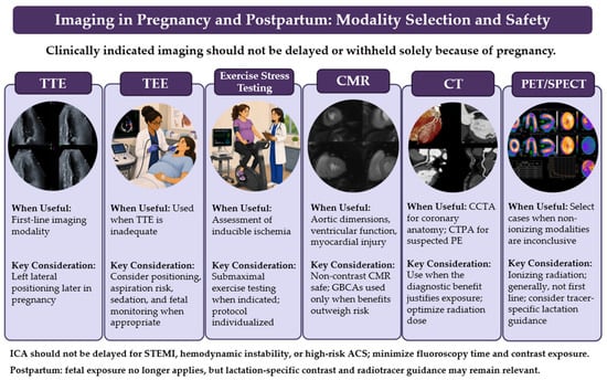
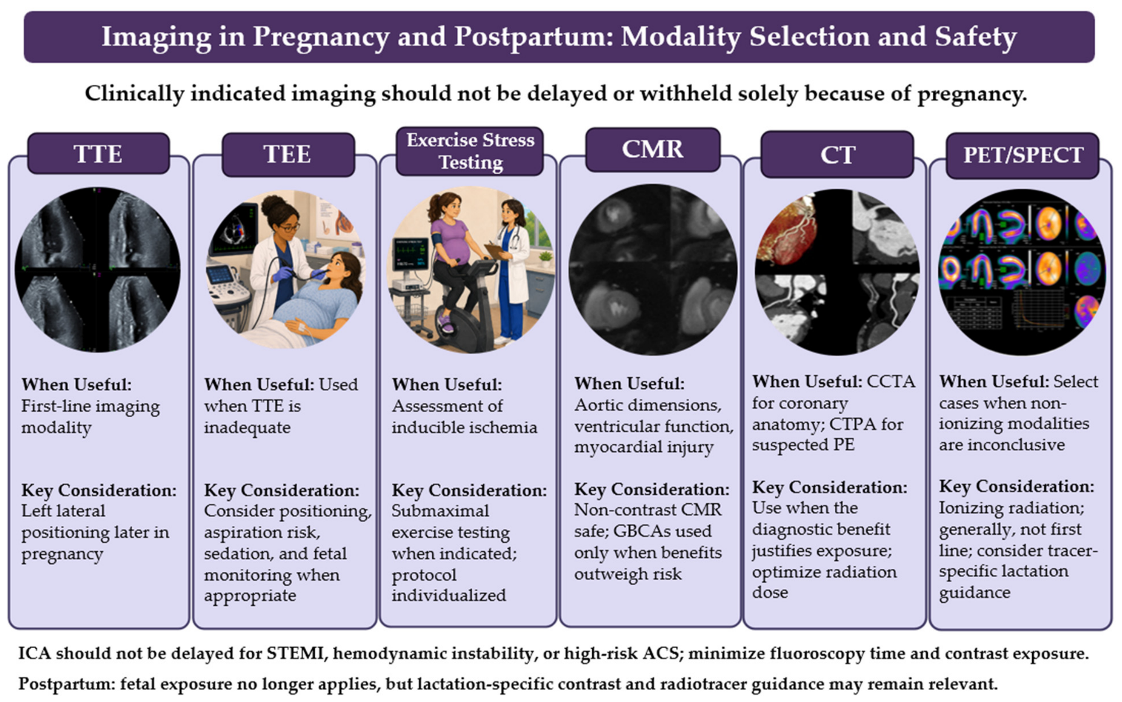
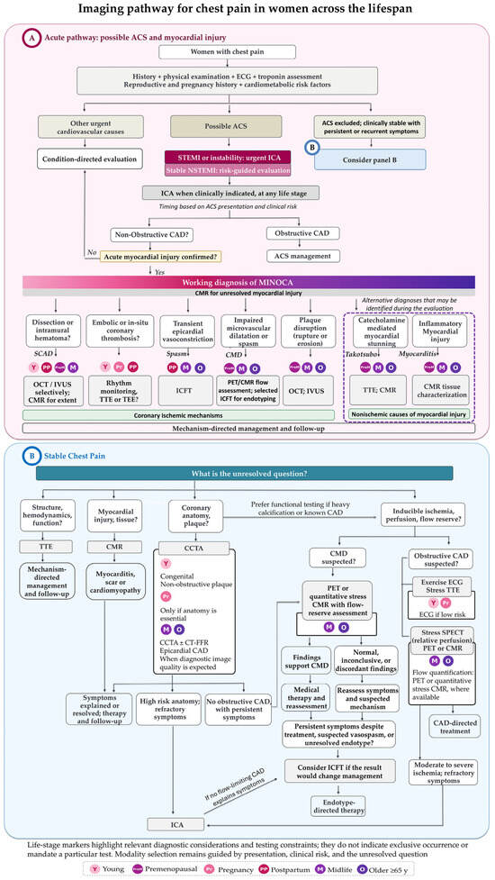
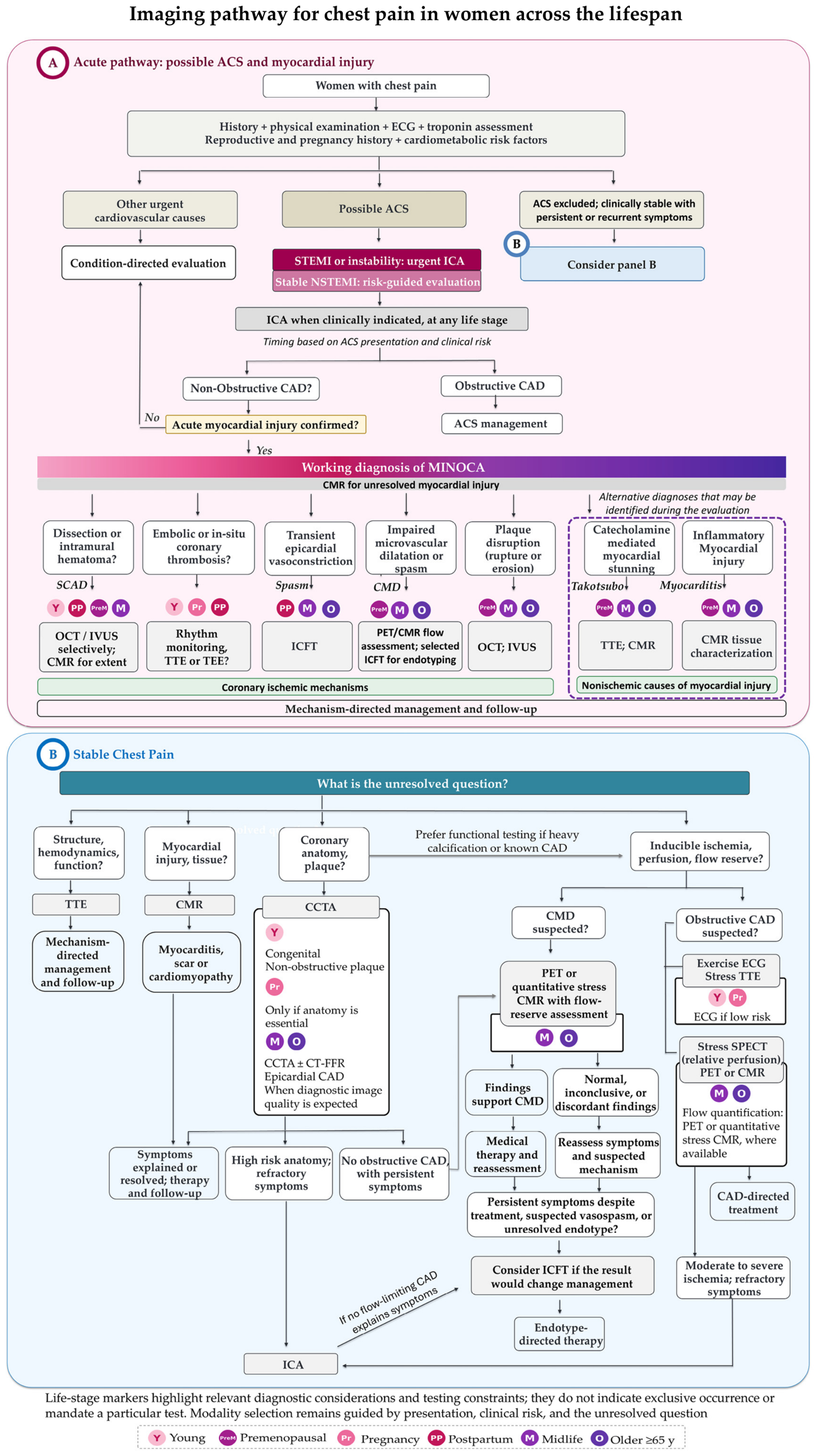



Disclaimer/Publisher’s Note: The statements, opinions and data contained in all publications are solely those of the individual author(s) and contributor(s) and not of MDPI and/or the editor(s). MDPI and/or the editor(s) disclaim responsibility for any injury to people or property resulting from any ideas, methods, instructions or products referred to in the content. |
© 2026 by the authors. Licensee MDPI, Basel, Switzerland. This article is an open access article distributed under the terms and conditions of the Creative Commons Attribution (CC BY) license.