


Abstract
Accurate 3D medical image segmentation requires effective integration of encoder details and decoder semantics. However, their relative contributions vary across decoding stages and spatial locations, and attention based only on the current feature pair may provide limited guidance in ambiguous regions. To address these challenges, we propose Dynamic Cross-Level Contextual Attention U-Net (DCLCA-UNet), which reconstructs cross-level context at each decoding stage from the incoming decoder feature and encoder features at the corresponding and shallower levels. The Cross-Level Contextual Attention Fusion (CLCAF) module uses this context to adaptively weight encoder and decoder features before fusion. The Context-Guided Feature Modulation (CGFM) module then reuses the context to refine the fused representation through scaling and additive correction. Experiments on Synapse, BTCV, and BraTS2023 yield average Dice scores of 88.70%, 84.13%, and 92.34%, respectively, with the lowest corresponding average Hausdorff distance (HD) or 95th-percentile Hausdorff distance (HD95) among the compared methods. These improvements are accompanied by increased computational cost. Repeated experiments and evaluation on AMOS22 provide additional evidence of stability and cross-dataset generalization. Ablation studies further assess the contributions of contextual fusion, modulation, and stage-specific context reconstruction. Together, these results support the effectiveness of contextual guidance in volumetric segmentation.
1. Introduction
Automatic segmentation of anatomical structures and pathological regions in three-dimensional medical images supports quantitative analysis for diagnosis, treatment planning, and disease assessment [1]. U-shaped encoder–decoder architectures, represented by U-Net [2] and its volumetric extension, 3D U-Net [3], are widely used for medical image segmentation. Their encoders extract hierarchical semantic representations, while their decoders progressively restore spatial resolution. Skip connections link these two processes by transferring encoder features to the decoder, helping recover anatomical details weakened by downsampling. Recent architectures, including 3D UX-Net [4], SwinUNETR [5], and U-Mamba [6], have further advanced volumetric representation learning through convolutional, Transformer-based, and state-space designs. Despite these developments, accurate spatial reconstruction continues to depend on effective encoder–decoder interaction.
Encoder and decoder features provide complementary information, but their contributions vary throughout reconstruction. Encoder features preserve local appearance and spatial detail, whereas decoder features carry semantic information accumulated from deeper layers. The balance between these sources depends on both the decoding stage and the spatial region: coarse reconstruction requires sufficient semantic guidance, while finer reconstruction increasingly depends on accurate structural detail. Dense skip pathways [7,8], attention gates [9], cross-scale aggregation [10], and learnable encoder–decoder connections [11,12] have therefore been investigated to improve feature transfer and selection.
Two challenges remain in this interaction. First, direct concatenation and static weighting do not explicitly account for spatial and stage-dependent differences in the usefulness of encoder and decoder features. Second, attention computed only from the corresponding feature pair has limited access to complementary evidence when both representations are ambiguous in the same region. Adjusting their relative weights may redistribute existing responses without supplying the additional information needed to refine them.
Cross-level encoder features offer a potential source of such information. Representations at different depths contain complementary structural and semantic cues that can support feature selection during decoding. However, simply aggregating these representations may introduce redundant or stage-irrelevant information. The resulting guidance should therefore depend on the current decoder state, so that contextual information is selected and used according to the needs of progressive reconstruction.
Motivated by these observations, we propose Dynamic Cross-Level Contextual Attention U-Net (DCLCA-UNet), which introduces decoder-conditioned cross-level context to guide both feature fusion and subsequent refinement. The context is reconstructed at each decoding stage, allowing the guidance to evolve with the decoder representation. The main contributions are as follows:
- To accommodate the changing requirements of progressive reconstruction, we propose a dynamic contextual decoding strategy that combines the current decoder state with encoder information from the corresponding and shallower levels, providing stage-specific guidance for feature integration.
- We design the Cross-Level Contextual Attention Fusion (CLCAF) module to incorporate contextual evidence beyond the current encoder–decoder pair. It generates spatial attention maps that adaptively regulate the contributions of the two feature sources before fusion.
- To further refine responses that remain incomplete or ambiguous after fusion, we introduce the Context-Guided Feature Modulation (CGFM) module. It uses the same context to generate multiplicative and additive modulation, enabling contextual refinement beyond relative feature weighting.
- Experiments using the Synapse and BTCV protocols [13] and the BraTS2023 benchmark [14] show improved aggregate segmentation performance over the compared methods. Repeated experiments and cross-dataset evaluation further support the method’s stability and cross-dataset generalizability. Ablation studies assess the contributions of contextual fusion, feature modulation, and dynamic context reconstruction.
2. Related Work
2.1. U-Shaped Architectures for Volumetric Segmentation
U-shaped networks provide a well-established framework for medical image segmentation. Building on U-Net [2], 3D U-Net [3] extends convolutional encoding and decoding to volumetric inputs. nnU-Net [15] further demonstrates the importance of adapting preprocessing, network configuration, and training to dataset characteristics through a self-configuring pipeline. Its residual-encoder variants [16] strengthen hierarchical feature extraction and serve as strong baselines for 3D segmentation.
Subsequent work has explored different ways to improve the representations learned within this framework. Among convolutional methods, 3D UX-Net [4] uses large-kernel depth-wise convolutions to expand the receptive field, while MedNeXt [17] adapts ConvNeXt-inspired designs [18] and scales network depth, width, and kernel size. Transformer-based methods emphasize long-range interactions: UNETR [19] employs a Transformer encoder for volumetric representation learning, and SwinUNETR [5] constructs hierarchical features using shifted-window attention.
State-space and diffusion-based approaches offer additional modeling strategies. U-Mamba [6] combines convolutional processing with state-space modeling, while EM-Net [20] incorporates channel-aware selection and frequency-domain learning. Diff-UNet [21] explores diffusion modeling with boundary and uncertainty information. These advances primarily expand how features are extracted or refined within the network. DCLCA-UNet focuses on the complementary problem of using hierarchical features to guide encoder–decoder interaction during reconstruction.
2.2. Skip Connections and Contextual Feature Fusion
The original U-Net transfers encoder features directly to decoder stages at matching resolutions. Although this preserves spatial information, it leaves the subsequent fusion layers to reconcile differences in feature content and semantic abstraction. UNet++ [7] introduces nested dense pathways to reduce these differences, whereas UNet3+ [8] uses full-scale skip connections to combine features from multiple resolutions.
Alongside changes to skip-connection pathways, attention mechanisms have been used to enhance medical image features by selectively emphasizing informative channels or spatial regions [22,23]. Attention U-Net [9] applies decoder-guided gates to suppress irrelevant encoder responses during skip feature transfer.
Feature selection can also take into account relationships among multiple feature levels. MCTrans [10] captures cross-scale dependencies among encoder representations. UCTransNet [11] combines multi-scale encoder fusion with decoder-guided channel recalibration, while UDTransNet [12] models both channel and spatial dependencies to improve encoder–decoder interaction. TA-MoSC [24] uses stage-specific routing over shared experts to adapt aggregated encoder information to different decoding stages. These approaches extend feature integration through explicit dependency modeling and adaptive selection of multi-scale information.
DCLCA-UNet differs in how cross-level guidance is constructed and used during decoding. At each decoding stage, it reconstructs the context by combining the incoming decoder feature with encoder features from the corresponding and shallower levels. This context jointly determines the relative spatial weights of the corresponding encoder feature and the upsampled decoder feature before fusion. The same context is then reused to generate scaling and additive correction after fusion. Thus, feature fusion and subsequent refinement share contextual guidance that evolves with the decoder representation.
3. Methods
3.1. Overall Architecture
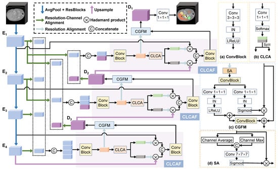
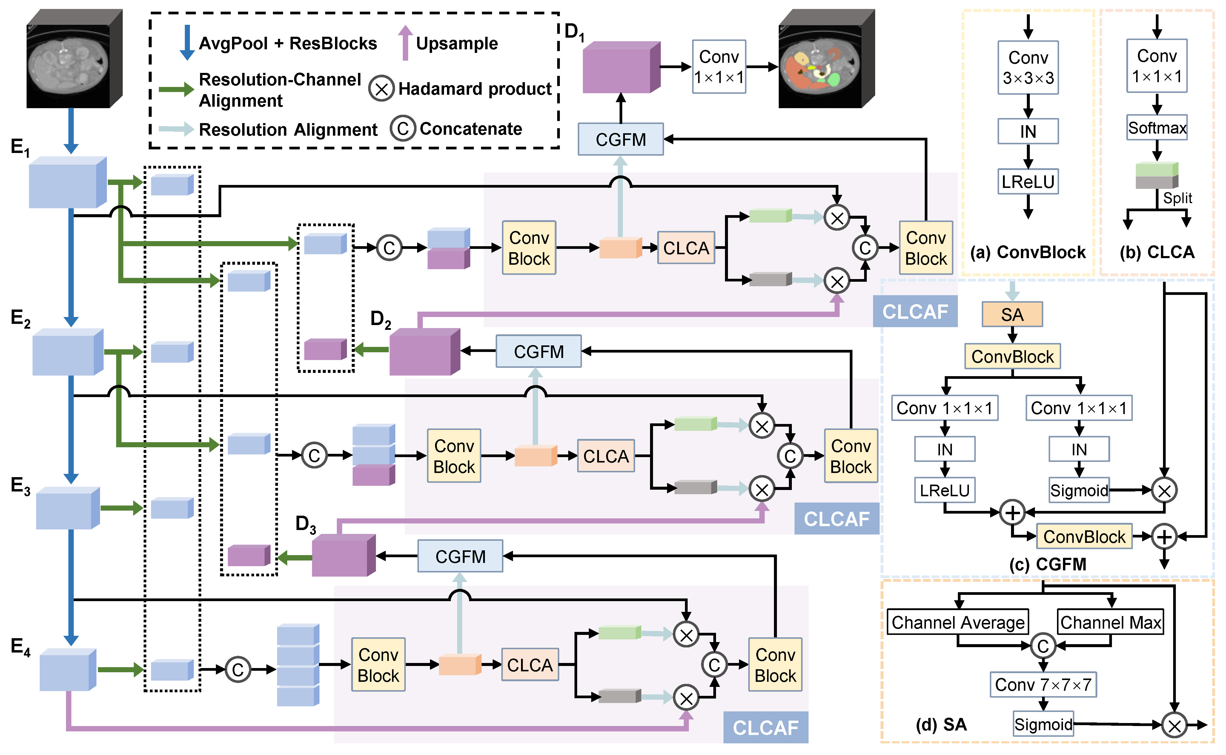
DCLCA-UNet adopts the residual-encoder variant of nnU-Net [15,16], referred to as ResEnc nnU-Net, as its baseline. As illustrated in Figure 1, the network retains a multi-stage U-shaped structure and introduces contextual guidance into the decoder. The encoder comprises six stages with channel dimensions of 32, 64, 128, 256, 320, and 320 from shallow to deep. The decoder comprises five stages, whose output channel dimensions match those of the corresponding encoder stages. For clarity, Figure 1 shows a simplified schematic with four encoder stages and three decoder stages.
Figure 1.
Overview of DCLCA-UNet and its constituent components: (a) Convolutional Block (ConvBlock), (b) Cross-Level Contextual Attention (CLCA), (c) Context-Guided Feature Modulation (CGFM), and (d) Spatial Attention (SA).
Let denote encoder features ordered from shallow to deep, where has the highest spatial resolution and is the bottleneck representation. Decoding proceeds from stage to stage 1. The initial decoder feature is
At stage i, the incoming decoder feature is upsampled to the spatial resolution of the corresponding encoder feature :
where denotes a stage-specific transposed convolution with both kernel size and stride set to , which upsamples the decoder feature and matches the channel dimension of .
At each decoding stage, CLCAF combines with , using context constructed from and . The fused feature is then processed by CGFM to produce . After the final decoding stage, a convolutional prediction layer and subsequent activation generate the segmentation probability maps.
Throughout this section, a ConvBlock consists of a convolution followed by instance normalization [25] and Leaky ReLU activation. DCLCA refers to the overall decoding strategy, while Cross-Level Contextual Attention (CLCA) is the attention-generating component within CLCAF.
3.2. Cross-Level Contextual Attention Fusion
CLCAF uses cross-level information to guide the integration of encoder and decoder features. Its design separates context construction from feature fusion: the contextual representation determines how the fusion inputs should be weighted, while the inputs retain their native feature content.
At stage i, the context incorporates the incoming decoder feature and encoder features . The decoder input carries information accumulated through deeper processing, while the selected encoder features provide complementary cues from the corresponding and shallower levels.
Because these features differ in resolution and channel count, they are spatially aligned to a common context grid and projected to a unified channel dimension.
Considering segmentation accuracy and resource requirements, we align the context inputs to the spatial dimensions of and a common channel count at every decoding stage. The common spatial dimensions are denoted by .
Let denote an input feature to be aligned, where s is its source stage. Let denote the downsampling stride from encoder stage to stage k along spatial axis a. The alignment factor is
where an empty product equals one. Higher-resolution inputs are downsampled by average pooling, with the pooling kernel and stride both set to the alignment factor. Lower-resolution inputs are upsampled by nearest-neighbor interpolation, while inputs already at the target resolution remain unchanged. With , spatial alignment is defined as
where AvgPool3D denotes three-dimensional average pooling and denotes three-dimensional upsampling using nearest-neighbor interpolation.
Channel alignment uses a convolution to project each input to channels while preserving its spatial dimensions:
To reduce intermediate memory usage, downsampling is performed before channel alignment, whereas upsampling is performed after channel alignment. The aligned feature is therefore
Each aligned input has shape .
Denote the aligned representations at stage i by and , where . The context is then constructed as
where denotes channel-wise concatenation and is the context aggregation block. The concatenated representation contains channels, which the aggregation block maps back to channels.
CLCA transforms into spatial attention for the two feature sources. A convolutional mapping produces two channels, followed by softmax normalization across those channels:
The decoder and encoder attention maps are defined as
where and correspond to the decoder and encoder, respectively. The two attention channels determine the relative contributions of the feature sources at each spatial location. The factor of two preserves unit weighting when both channels receive equal attention.
The attention maps are resized to the current decoding resolution and applied to the fusion inputs:
where aligns the context-grid resolution with the current decoding resolution using the same average-pooling or nearest-neighbor upsampling rule described above, and ⊙ denotes element-wise multiplication. Each spatial map is broadcast across the channels of its corresponding feature. The weighted features are concatenated and processed by a fusion ConvBlock:
where denotes the fusion operation.
The context is reconstructed at every decoding stage using the updated decoder input and the encoder features selected for that stage. Thus, a fixed context-grid size does not imply fixed guidance: both the contextual content and the resulting attention maps depend on the current stage and input features.
3.3. Context-Guided Feature Modulation
CLCAF determines the relative contributions of the features entering fusion. However, relative weighting alone may be insufficient when the fused representation remains incomplete or ambiguous. CGFM therefore uses the context to generate feature-dependent scaling and additive correction after fusion.
The context is resized to the spatial resolution of using the same spatial alignment rule as in CLCAF, refined through spatial attention, and transformed by a ConvBlock. This ConvBlock projects the context to the channel dimension of , allowing the subsequent modulation branches to produce parameters with matching spatial and channel dimensions:
where denotes the ConvBlock and is the spatial attention module [26]. Spatial attention combines channel-wise average and maximum responses to estimate spatial weights, which are applied to the resized context.
Two parallel branches generate the modulation parameters:
where and are the scale and offset mappings, respectively, and is the sigmoid function. These parameters are applied to the fused representation through affine modulation [27] and residual refinement:
where denotes the modulation ConvBlock.
The multiplicative term adjusts existing feature responses, while the additive term provides a context-derived contribution that is not restricted to reweighting those responses. The residual connection preserves a direct path for the original fused feature and facilitates its propagation to subsequent decoding stages.
3.4. Loss Function
DCLCA-UNet is trained using a combination of soft Dice loss and voxel-wise classification loss. The specific classification loss depends on the formulation of the segmentation task. Cross-entropy loss is used for mutually exclusive multi-class segmentation, whereas binary cross-entropy loss is used for multi-label segmentation with independently predicted target regions. This objective combines region-overlap optimization with voxel-wise supervision.
Let and denote the predicted probability and ground-truth label for voxel v and output channel k, respectively. The soft Dice loss [28] is
where N is the number of voxels, K is the number of output channels, and is a smoothing constant.
The voxel-wise classification losses are defined as
The final training objective assigns equal weights to the region-overlap and classification terms:
where for mutually exclusive multi-class segmentation and for multi-label segmentation.
4. Experiments
4.1. Datasets and Evaluation Protocols
We evaluate DCLCA-UNet using the Synapse [13] and BTCV [13] abdominal computed tomography (CT) segmentation protocols, the BraTS2023 [14] brain tumor magnetic resonance imaging (MRI) benchmark, and the AMOS22 [29] abdominal multi-organ segmentation dataset. These evaluations cover multi-organ segmentation and tumor-region segmentation across two imaging modalities, with AMOS22 used for cross-dataset generalization evaluation.
Synapse and BTCV are derived from the same abdominal imaging collection but use different target sets. The Synapse protocol evaluates eight structures: the spleen, right kidney, left kidney, gallbladder, liver, stomach, aorta, and pancreas. BTCV evaluates 13 organs and anatomical structures, additionally including the esophagus, inferior vena cava, portal and splenic veins, and both adrenal glands. Following EM-Net [20], the left and right adrenal gland scores are averaged into a single AG (adrenal glands) column for presentation; the overall Dice similarity coefficient (DSC) remains an average across all 13 targets.
For both abdominal CT protocols, we follow the data partitioning adopted by EM-Net [20], using 18 cases for training and 12 cases for testing.
BraTS2023 provides multi-parametric MRI with four modalities: T1, contrast-enhanced T1 (T1ce), T2, and fluid-attenuated inversion recovery (FLAIR). The segmentation targets are whole tumor (WT), tumor core (TC), and enhancing tumor (ET). Following the protocol of Diff-UNet [21], the 1251 cases are divided into training, validation, and test sets in a 70:10:20 ratio.
For AMOS22, we use all 300 CT scans from its 360-case post-challenge release. Evaluation is restricted to the eight anatomical targets shared with the Synapse protocol.
We report DSC and Hausdorff distance (HD) for Synapse, BTCV, and AMOS22, and DSC and the 95th-percentile Hausdorff distance (HD95) for BraTS2023 [30]. Higher DSC indicates better region overlap, while lower HD or HD95 indicates smaller boundary discrepancies. In the multi-seed experiments, normalized surface Dice (NSD) is additionally reported with a tolerance of 1 mm for all evaluated targets. NSD measures surface agreement within the specified tolerance, with higher values indicating better boundary agreement.
4.2. Implementation Details
All models in this study are trained from scratch without pretrained weights. Models are trained for 500 epochs on Synapse and BTCV, using a batch size of 2. The input patch sizes are for Synapse and for BTCV. For BraTS2023, training runs for 1000 epochs with a batch size of 2 and patches of .
Unless otherwise specified, we retain the default nnU-Net v2 settings for preprocessing, online augmentation, weight initialization, and inference [15,16]. Resampling and intensity normalization are determined by the dataset-specific experiment plans. Training augmentation includes rotation, scaling, Gaussian noise, Gaussian blur, brightness and contrast adjustment, simulated low resolution, gamma transformation, and random mirroring. Foreground-aware patch sampling and other data-loading settings retain their default values. During inference, sliding windows match the corresponding training patch size, with a step-size ratio of 0.5. Overlapping predictions are combined using Gaussian weighting, and mirroring-based test-time augmentation is enabled.
All experiments performed in this study are conducted on a single NVIDIA RTX 4090 GPU (NVIDIA Corporation, Santa Clara, CA, USA) with 24 GB of memory. The implementation uses PyTorch 2.6.0 [31], MONAI 1.5.2 [32], and nnU-Net v2.6.2 [15]. Optimization uses stochastic gradient descent with Nesterov momentum of , an initial learning rate of , and polynomial learning-rate decay. DCLCA-UNet and ResEnc nnU-Net use identical preprocessing, augmentation, training, and inference settings for each dataset.
4.3. Comparison with Existing Methods
We compare DCLCA-UNet with representative 3D segmentation methods covering convolutional, Transformer-based, state-space, and diffusion-based architectures. The evaluation includes abdominal multi-organ segmentation on Synapse and BTCV and brain tumor segmentation on BraTS2023. For ResEnc nnU-Net and DCLCA-UNet, we report single-run results from our own training and evaluation. For the remaining methods, the Synapse and BTCV results are taken from EM-Net [20], and the BraTS2023 results from Diff-UNet [21]. Quantitative results are examined together with qualitative examples to assess both region overlap and the spatial characteristics of the predictions.
4.3.1. Results on Synapse
Table 1 presents the quantitative results on Synapse. DCLCA-UNet achieves the highest average DSC of 88.70% and the lowest average HD of 20.83 among the compared methods. Relative to ResEnc nnU-Net, it improves average DSC by 0.74 percentage points and reduces HD from 21.48 to 20.83. It also exceeds the average DSC of EM-Net, the strongest remaining method in this comparison, by 4.75 percentage points.
Table 1.
Quantitative comparison on Synapse. Bold indicates the best results in their respective numerical columns. Upward and downward arrows indicate that higher and lower values are better, respectively.
The organ-wise results show DSC improvements over the baseline for six of the eight targets. The largest gains occur in the stomach and spleen, whose DSC values increase from 89.17% to 91.65% and from 92.47% to 94.64%, respectively. Both kidneys, the aorta, and the pancreas also improve. Although gallbladder and liver DSC decrease slightly relative to the baseline, DCLCA-UNet achieves the best scores for the other six structures, resulting in improved overall overlap.
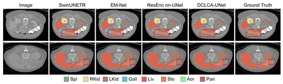
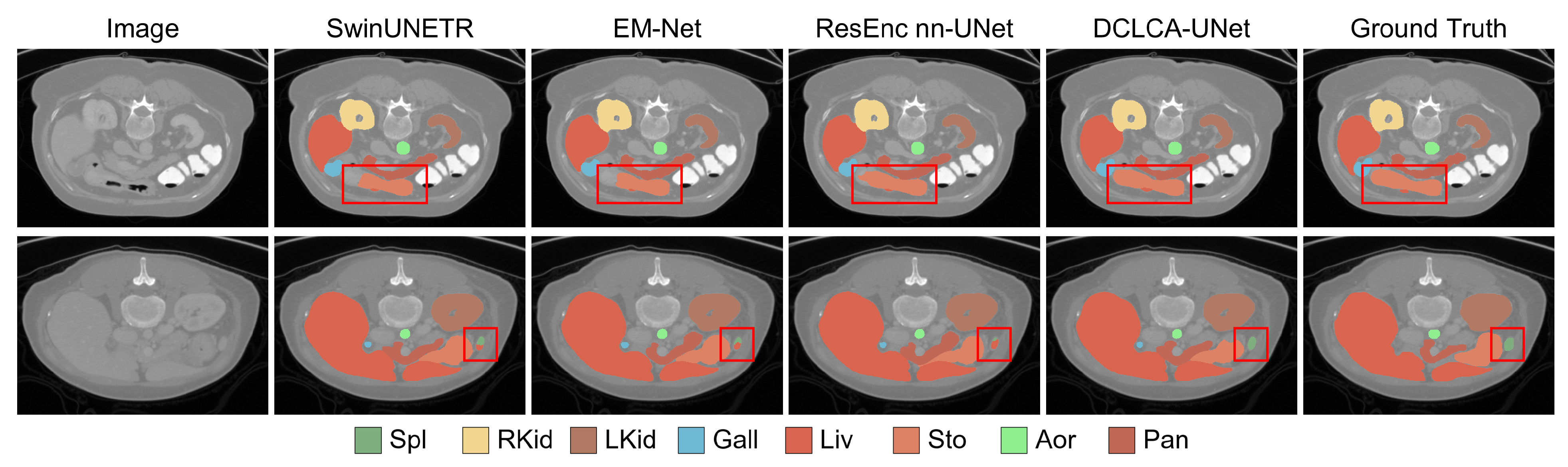
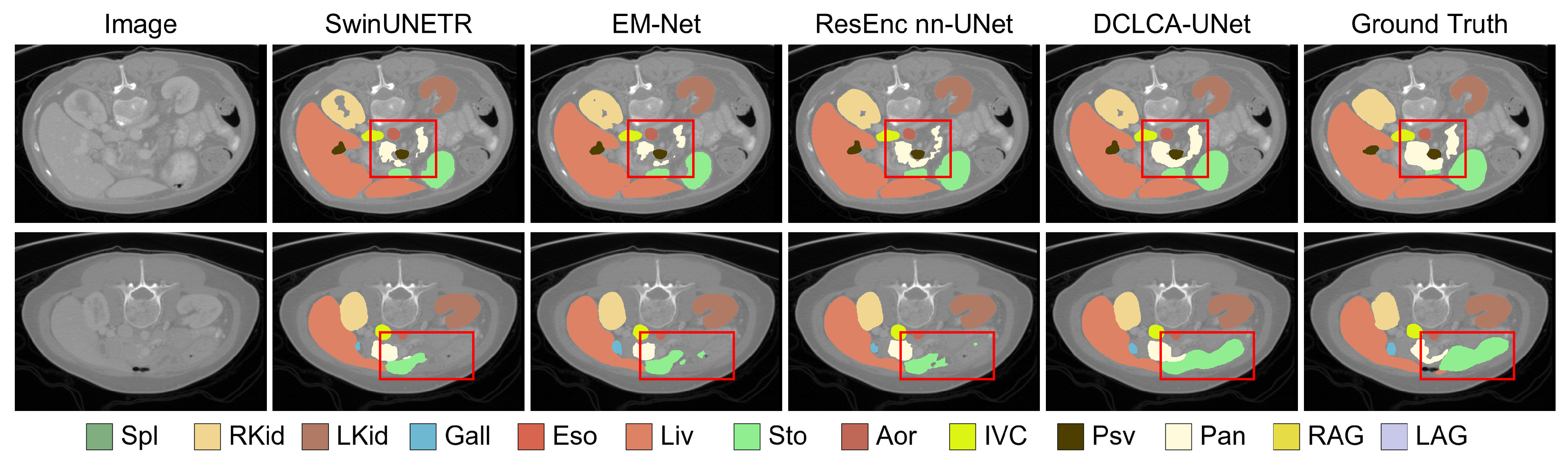
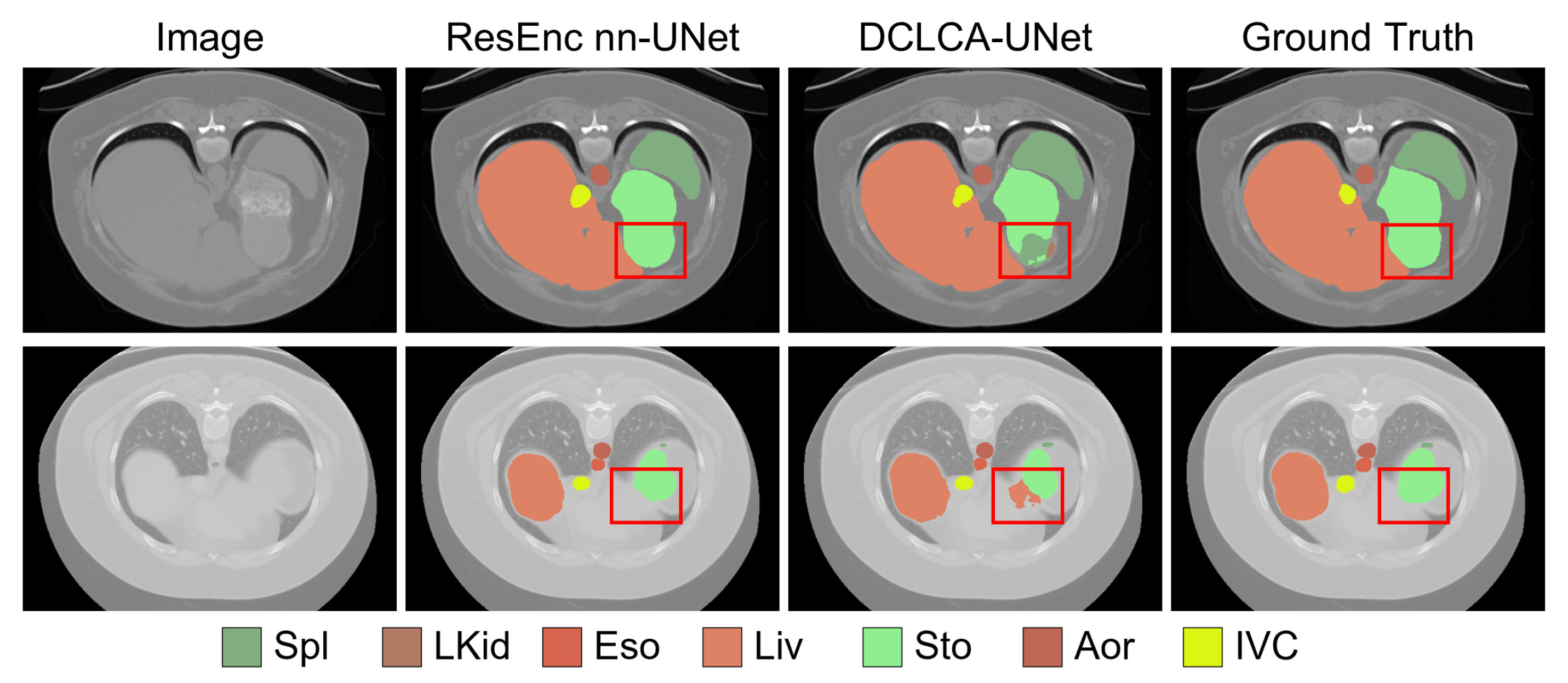
The qualitative examples in Figure 2 provide a more specific view of these differences. In the first case, DCLCA-UNet produces more complete stomach segmentation, consistent with the improvement observed in the stomach DSC. In the second case, it more clearly separates the aorta from the liver, producing a segmentation that more closely matches the ground truth. These examples illustrate improvements in both target completeness and the separation of adjacent anatomical structures.
Figure 2.
Qualitative comparison on Synapse. Red boxes highlight regions with noticeable differences between segmentation results.
4.3.2. Results on BTCV
The BTCV protocol expands the abdominal evaluation to 13 targets, including additional organs and vascular structures. As reported in Table 2, DCLCA-UNet obtains the highest average DSC of 84.13% and the lowest average HD of 23.59. Compared with ResEnc nnU-Net, these results correspond to a DSC increase of 0.68 percentage points and an HD reduction of 0.58. The improvement in both aggregate metrics indicates that the benefit observed under the Synapse protocol is retained with the expanded target set.
Table 2.
Quantitative comparison on BTCV. Bold indicates the best results in their respective numerical columns. Upward and downward arrows indicate that higher and lower values are better, respectively
The stomach again shows the largest DSC gain over the baseline, increasing from 84.17% to 87.25%. Pancreas DSC rises from 82.75% to 84.78%, while the right kidney and gallbladder improve by 1.43 and 1.17 percentage points, respectively. Gains are also observed in the esophagus, aorta, inferior vena cava, and portal and splenic veins. The improvements therefore extend across organ and vascular targets, rather than being restricted to a single anatomical category. Small decreases remain in the spleen, left kidney, liver, and combined adrenal gland score, indicating that the effect varies across structures.
To examine whether performance differences vary with target size, we group the 13 targets according to their mean ground-truth volumes in the BTCV test set. Table 3 reports the mean DSC for the small, medium, and large groups.
Table 3.
Performance stratified by mean target volume on BTCV. Upward arrows indicate that higher values are better.
DCLCA-UNet improves mean DSC by 0.42, 0.78, and 0.92 percentage points for small, medium, and large targets, respectively. Gains are observed in all three size groups, and the group-level DSC improvement increases from small to large targets in this comparison.
The qualitative results in Figure 3 complement these measurements. DCLCA-UNet reduces incomplete segmentation in challenging abdominal regions, yielding predictions that more closely match the reference annotations. Together, the quantitative and qualitative findings support improved reconstruction across several targets within the expanded abdominal segmentation task.
Figure 3.
Qualitative comparison on BTCV. Red boxes highlight regions with noticeable differences between segmentation results.
4.3.3. Results on BraTS2023
To examine performance beyond abdominal CT segmentation, we further evaluate DCLCA-UNet on multi-parametric brain MRI. Table 4 shows that the proposed method achieves the highest average DSC of 92.34% and the lowest average HD95 of 2.50 among the compared methods. Relative to ResEnc nnU-Net, average DSC increases by 0.26 percentage points and HD95 decreases from 2.91 to 2.50. DCLCA-UNet also improves average DSC over Diff-UNet from 91.87% to 92.34%.
Table 4.
Quantitative comparison on BraTS2023. Bold indicate the best results in their respective numerical columns. Upward and downward arrows indicate that higher and lower values are better, respectively.
The region-wise comparison shows improvements over the baseline for all three tumor targets. DCLCA-UNet achieves DSC values of 94.23%, 93.53%, and 89.25% for WT, TC, and ET, respectively, corresponding to gains of 0.19, 0.25, and 0.32 percentage points. The TC and ET scores are the highest among the compared methods, while the WT score remains slightly below that of Diff-UNet. The highest average DSC reflects improved delineation of tumor subregions while maintaining competitive whole-tumor performance.
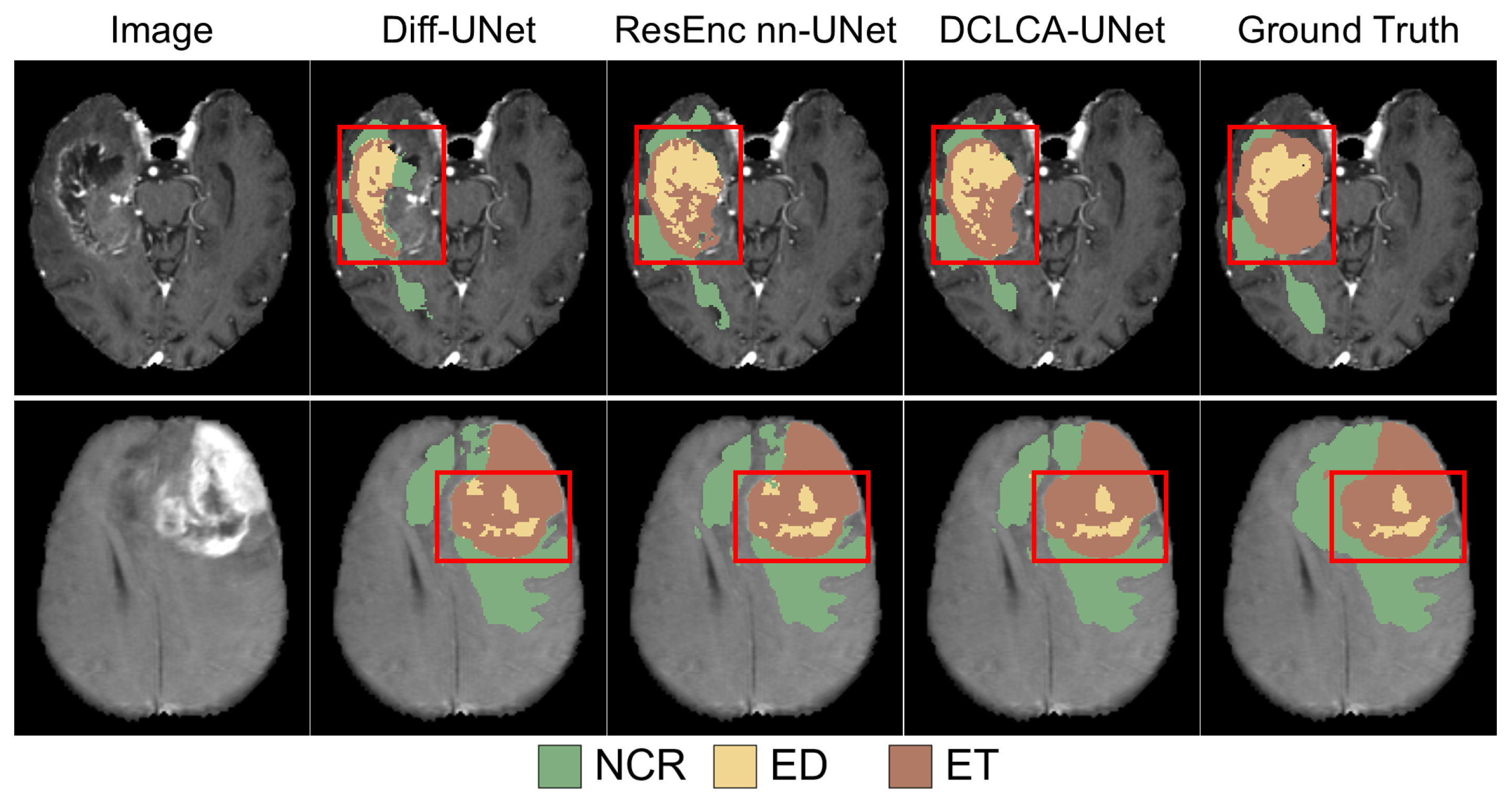
As shown in Figure 4, DCLCA-UNet produces more complete tumor segmentations with fewer false-positive responses in challenging regions. It also localizes small or ambiguous tumor components more accurately. These examples complement the TC and ET results by illustrating the spatial differences associated with tumor-subregion prediction.
Figure 4.
Qualitative comparison on BraTS2023. Red boxes highlight regions with noticeable differences between segmentation results. NCR, ED, and ET denote the necrotic/non-enhancing tumor core, peritumoral edema, and enhancing tumor, respectively. The evaluation regions are defined as and .
4.4. Repeated Experiments and Statistical Analysis
To assess variability across independent training runs, we repeat the experiments for ResEnc nnU-Net and DCLCA-UNet using three random seeds: 42, 123, and 2024. Data partitions and all other training settings are kept unchanged across runs. Table 5 reports the mean and standard deviation of the evaluation metrics over the three runs. For statistical comparison, case-level DSC scores are averaged over the three runs for each method, and a two-sided Wilcoxon signed-rank test is applied to the paired case-level scores. The resulting p-values for Synapse, BTCV, and BraTS2023 are , , and , respectively, all below .
Table 5.
Multi-seed comparison on Synapse, BTCV, and BraTS2023. Upward and downward arrows indicate that higher and lower values are better, respectively
DCLCA-UNet achieves higher mean DSC on all three datasets, with gains of 0.88, 0.71, and 0.31 percentage points on Synapse, BTCV, and BraTS2023, respectively. These improvements are consistent with the single-run comparisons in Section 4.3. The consistent improvements and small standard deviations across the three runs support the stability of the observed performance advantage over the evaluated random seeds.
Mean NSD increases by 0.78, 1.61, and 0.37 percentage points on Synapse, BTCV, and BraTS2023, respectively. Mean HD decreases by 0.32 on Synapse and 0.47 on BTCV, while mean HD95 decreases by 0.44 on BraTS2023. These complementary metrics show that the repeated-run gains are reflected in both volumetric overlap and surface agreement.
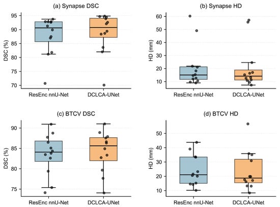
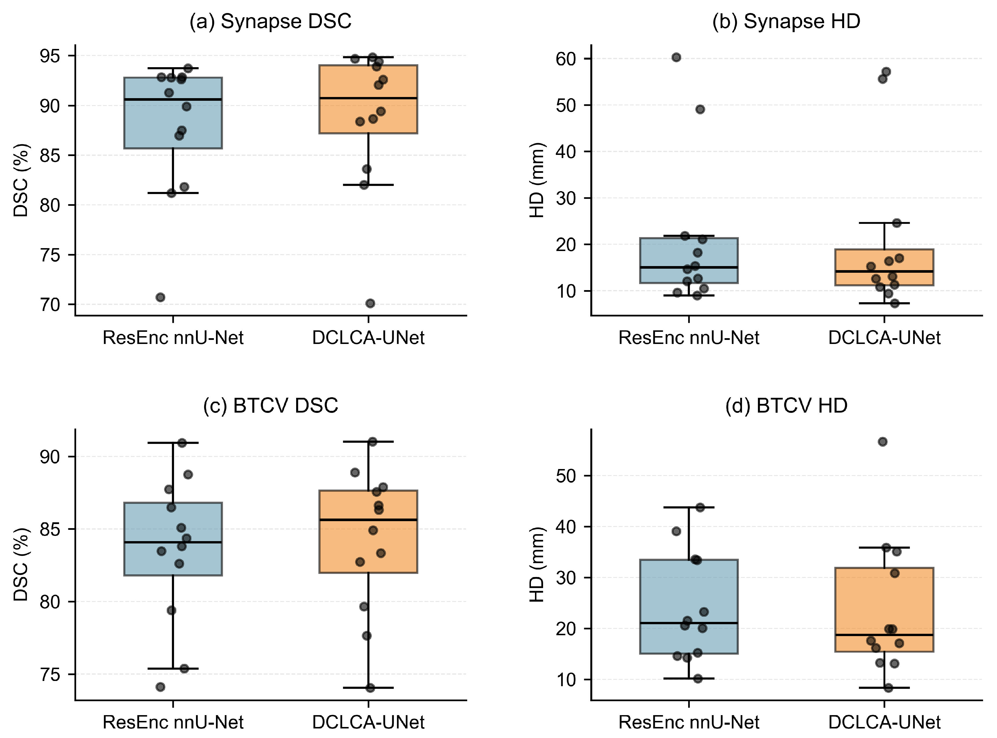
Figure 5 examines how the aggregate differences are reflected in case-level distributions. On Synapse, the DSC medians of the two methods are similar, while the lower and upper quartiles are shifted upward for DCLCA-UNet. On BTCV, DCLCA-UNet shows a higher median DSC together with an upward shift of the interquartile range. These changes indicate favorable shifts within the central portions of the distributions, although low-DSC cases remain on both datasets.
Figure 5.
Case-level DSC and HD distributions of ResEnc nnU-Net and DCLCA-UNet. Each point represents a case’s score averaged over the three runs. Boxes indicate the interquartile range, and central lines denote medians.
HD medians are lower for DCLCA-UNet on both datasets, indicating reduced boundary errors for typical cases. However, large HD values remain for both methods, and DCLCA-UNet exhibits a high-HD outlier on BTCV that exceeds the largest baseline value. The lower median errors therefore coexist with occasional large boundary discrepancies, highlighting case-level variability that is not fully conveyed by the dataset averages.
4.5. Cross-Dataset Evaluation on AMOS22
We directly evaluate ResEnc nnU-Net and DCLCA-UNet on all 300 CT scans from the AMOS22 post-challenge dataset, using the three Synapse-trained models of each method from Section 4.4. Evaluation uses the eight anatomical targets shared with Synapse. No retraining or fine-tuning is performed, and the source-model preprocessing and inference settings are retained. Table 6 reports the mean and standard deviation of the results over the three models.
Table 6.
Cross-dataset generalization from Synapse to AMOS22. Bold indicates the best results in their respective numerical columns. Upward and downward arrows indicate that higher and lower values are better, respectively.
DCLCA-UNet achieves an average DSC of 85.54%, compared with 84.86% for ResEnc nnU-Net, corresponding to a gain of 0.68 percentage points. Average HD decreases from 25.90 to 24.55, indicating improved boundary performance alongside the increase in region overlap. The performance advantage observed on Synapse is therefore retained on the external AMOS22 dataset without adaptation to the target data.
The organ-wise results show DSC improvements for six of the eight targets. The largest gain occurs in the right kidney, increasing from 88.75% to 91.78%, followed by gains of 0.88 and 0.83 percentage points in the gallbladder and stomach, respectively. The spleen, left kidney, and liver also improve. In contrast, aorta and pancreas DSC decrease by 0.61 and 0.14 percentage points, respectively. These differences indicate that the benefit of direct transfer varies across anatomical targets, while the overall advantage over the baseline extends to the external dataset.
4.6. Ablation Studies
Ablation studies on Synapse examine two questions: whether the proposed contextual operations contribute to segmentation performance, and how the context representation size affects accuracy and resource requirements.
4.6.1. Contributions of the Main Components
Table 7 compares the complete model with variants that remove individual operations or change how context is used.
Table 7.
Ablation of the main components on Synapse. Bold indicates the best results in their respective numerical columns. Upward and downward arrows indicate that higher and lower values are better, respectively.
The Context Fusion Only variant concatenates the dynamically constructed context with the encoder and decoder features after spatial alignment, while omitting CLCA weighting and CGFM modulation. It achieves a DSC of 87.82%, slightly below the baseline value of 87.96%, although HD decreases from 21.48 to 21.09. Thus, additional contextual information introduced through direct concatenation does not improve both metrics. This comparison motivates examining how the context is used, rather than assuming that aggregation alone is sufficient.
CLCA uses the cross-level context to spatially weight encoder and decoder features before fusion, complementing the post-fusion modulation performed by CGFM. In the reported ablation, the complete model increases DSC from 88.58% to 88.70% and reduces HD from 21.71 to 20.83 compared with the variant without CLCA. These observations are consistent with a complementary role for CLCA within the overall contextual guidance strategy.
Removing CGFM reduces DSC to 88.19% but improves HD to 20.31, the lowest value in this ablation. CGFM therefore contributes to region overlap in the complete model, but its inclusion does not improve the aggregate boundary-distance metric in this comparison. The results indicate a trade-off between the two evaluation criteria rather than uniform improvement.
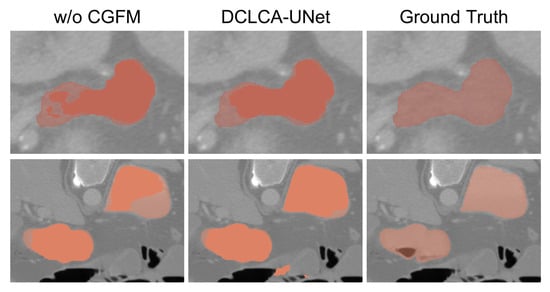
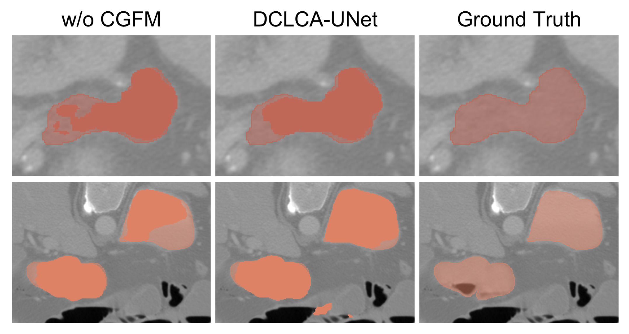
Figure 6 illustrates aspects of this trade-off in pancreas and stomach segmentation. In the pancreas example, the variant without CGFM shows fragmented predictions and missing regions within the target, whereas the complete model recovers more continuous target coverage. In the stomach example, DCLCA-UNet covers more of the annotated target region but also produces small false-positive regions away from the ground truth.
Figure 6.
Qualitative comparison with and without CGFM on Synapse. The upper and lower rows show pancreas and stomach segmentation, respectively. The colored regions represent the segmentation masks of the pancreas and stomach.
CGFM uses context-dependent scaling and additive correction to refine the fused features. This may help recover weak or missing target responses, while modulation based on inaccurate contextual cues may also strengthen spurious responses outside the target. Because HD is sensitive to distant false-positive regions [30], these errors can increase boundary distance even when the main target region is more completely recovered. This provides a possible explanation for the coexistence of improved DSC and worse HD in the current ablation.
The variant without dynamic updating uses fixed contextual guidance instead of reconstructing it across decoding stages. Its DSC of 88.09% and HD of 21.43 are both worse than those of the complete model, supporting the use of context that adapts to the evolving decoder representation.
These comparisons support the use of stage-specific context for both fusion and refinement, while showing that the contribution of CGFM differs between overlap and boundary-distance metrics.
4.6.2. Context Representation Configuration
The context representation introduces a choice between spatial detail, channel capacity, and computational cost. Table 8 examines five configurations, with representation size expressed as .
Table 8.
Effect of context representation size on Synapse. Bold indicates the best results in their respective numerical columns. Upward and downward arrows indicate that higher and lower values are better, respectively.
With 64 context channels, the grid in Variant B provides the highest DSC and lowest HD among Variants A–C. Increasing the grid to raises FLOPs and memory usage while reducing performance. Compressing it to lowers resource requirements but also reduces DSC and increases HD relative to Variant B. These results show that a finer context grid does not necessarily produce more useful guidance.
At the resolution, increasing the channel count from 64 to 128 raises DSC from 88.46% to 88.70%, while increasing HD from 20.53 to 20.83. Relative to Variant B, Variant D requires an additional 8.18 million parameters, approximately 33.3% more FLOPs, and 25.2% more memory. Reducing the spatial resolution of the 128-channel representation, as in Variant E, lowers cost but worsens both segmentation metrics relative to Variant D.
Variant D is selected for the final model because it achieves the highest DSC, reflecting an explicit preference for region overlap in the accuracy–resource trade-off. The selected context has 128 channels and is aligned to the third encoder stage, corresponding to in the Synapse configuration. Variant B provides a reasonable alternative when computational resources, memory usage, or boundary distance are prioritized, as it achieves better HD at lower cost.
The context-configuration study uses the same Synapse data split and training protocol as the preceding experiments. The selected configuration is applied unchanged to Synapse, BTCV, and BraTS2023. No dataset-specific retuning of this context configuration is performed.
4.7. Computational Cost and Runtime Performance
Table 9 compares the parameter counts and computational complexity of representative segmentation methods. DCLCA-UNet contains 119.84 million parameters and requires 1496.42 GFLOPs.
Table 9.
Comparison of model parameters and computational complexity. Bold indicates the best results in their respective numerical columns. Downward arrows indicate that lower values are better.
DCLCA-UNet requires a similar number of FLOPs to SwinUNETR but has more parameters. EM-Net uses fewer parameters and FLOPs, while ResEnc nnU-Net has the lowest FLOPs among the methods listed. Compared with ResEnc nnU-Net, DCLCA-UNet adds 17.48 million parameters and 723.22 GFLOPs, corresponding to increases of 17.08% and 93.54%, respectively. The additional contextual operations therefore introduce a substantial computational overhead despite the smaller relative increase in parameter count.
To assess the practical implications of this overhead, Table 10 compares peak GPU memory, average inference time per case, and training speed against the direct baseline on Synapse. Measurements follow the experimental settings described in Section 4.2.
Table 10.
Runtime and resource comparison with ResEnc nnU-Net on Synapse. Upward and downward arrows indicate that higher and lower values are better, respectively.
The increase in computational complexity is accompanied by higher memory requirements and longer inference time. Relative to the baseline, peak GPU memory and average inference time increase by 93.80% and 91.08%, respectively, while training speed decreases by 48.46%. Thus, the contextual decoding strategy nearly doubles memory usage and inference time and approximately halves training throughput under the evaluated settings.
These costs should be considered alongside the segmentation improvements reported in Section 4.3 and Section 4.4. DCLCA-UNet provides an accuracy-oriented extension of ResEnc nnU-Net, with improved segmentation performance at a substantial resource cost. When memory capacity or processing speed is prioritized, the baseline remains a reasonable choice; when segmentation accuracy is prioritized and sufficient resources are available, the proposed contextual decoding strategy offers an alternative.
5. Discussion
In 3D medical image segmentation, accurate spatial reconstruction depends on the effective integration of encoder details and decoder semantics. DCLCA-UNet uses cross-level context to guide this integration throughout decoding. The reported comparisons on Synapse, BTCV, and BraTS2023 show that DCLCA-UNet achieves the highest average DSC and the lowest corresponding average HD or HD95 among the compared methods. The repeated experiments show the same direction of improvement over ResEnc nnU-Net, providing additional support for the performance advantage of the complete decoding strategy.
The improvements over ResEnc nnU-Net suggest that guiding decoder fusion and refinement with cross-level context can improve segmentation while retaining the same residual encoder architecture. However, the overlap–boundary trade-off observed for CGFM shows that recovering more of a target region does not necessarily reduce extreme boundary errors.
In the cross-dataset evaluation from Synapse to AMOS22, DCLCA-UNet retains its performance advantage over ResEnc nnU-Net. This supports the applicability of the learned contextual decoding strategy beyond the original abdominal cohort, although the organ-wise results indicate that the transfer benefit varies among anatomical targets.
However, several limitations remain. First, the effectiveness of contextual guidance depends on the accuracy of the contextual representation. If this representation is inaccurate, the resulting weighting and modulation may suppress useful target responses or reinforce incorrect ones. As illustrated in Figure 7, parts of the stomach are incorrectly classified as spleen and left kidney in Case 1, while a false-positive liver region appears outside the annotated stomach in Case 2. ResEnc nnU-Net more closely follows the ground truth in the highlighted regions. Future work could explore mechanisms for assessing contextual reliability and constraining anatomically inconsistent predictions.
Figure 7.
Representative failure cases of DCLCA-UNet compared with ResEnc nnU-Net. Red boxes highlight regions with segmentation errors.
Second, the segmentation improvements come with substantial memory and runtime costs. DCLCA-UNet should therefore be considered an accuracy-oriented extension whose suitability depends on available resources and segmentation requirements. More selective feature aggregation and lightweight contextual modulation may help reduce this overhead.
Finally, the current evidence is drawn from public benchmarks. The Synapse and BTCV experiments use the same abdominal CT cases and differ in the organs evaluated. Both use 18 cases for training and 12 for testing. The small cohort and fixed data split limit the assessment of variability across patients. Although AMOS22 provides independent external CT evidence, broader clinical generalizability remains to be established. The DSC gains on BraTS2023 are also modest. Further evaluation across institutions, scanners, acquisition protocols, and patient populations is needed to assess robustness and determine whether the metric improvements translate into meaningful benefits for specific clinical tasks.
6. Conclusions
This work presents DCLCA-UNet, a contextual decoding framework for 3D medical image segmentation that improves the integration of encoder details and decoder semantics. The framework combines CLCAF and CGFM to guide adaptive feature fusion and subsequent refinement using shared cross-level context. Reconstructing this context with the incoming decoder feature allows the guidance to evolve throughout spatial reconstruction. The reported comparisons demonstrate improved aggregate segmentation performance on the evaluated public benchmarks, while the ablation studies support the coordinated use of contextual fusion, modulation, and stage-specific context reconstruction. Future work will focus on improving contextual reliability and computational efficiency. Broader clinical validation is also needed to establish generalizability and the practical significance of the segmentation improvements.
Author Contributions
Methodology, H.Y.; Software, H.Y.; Investigation, H.Y.; Writing—original draft preparation, H.Y.; Supervision, G.L.; Writing—review and editing, G.L. All authors have read and agreed to the published version of the manuscript.
Funding
This work was supported by the Hubei Design and Research Center of Cultural and Creative Industry Open Fund (No. HBCY2402) and the Joint Funds for the Innovation of Science and Technology, Fujian Province (No. 2024Y9265).
Institutional Review Board Statement
Not applicable.
Informed Consent Statement
Not applicable.
Data Availability Statement
Publicly available datasets were analyzed in this study. These data can be found at the following links: Synapse/BTCV, https://www.synapse.org/Synapse:syn3193805/wiki/217789 (accessed on 15 October 2026); BraTS2023, https://www.synapse.org/Synapse:syn51156910/wiki/627000 (accessed on 15 October 2026); and AMOS22, https://amos22.grand-challenge.org/ (accessed on 15 October 2026). Access is subject to the respective providers’ requirements.
Conflicts of Interest
The authors declare no conflicts of interest.
References
- Ma, J.; He, Y.; Li, F.; Han, L.; You, C.; Wang, B. Segment Anything in Medical Images. Nat. Commun. 2024, 15, 654. [Google Scholar] [CrossRef] [Scilit] [PubMed]
- Ronneberger, O.; Fischer, P.; Brox, T. U-Net: Convolutional Networks for Biomedical Image Segmentation. In Medical Image Computing and Computer-Assisted Intervention—MICCAI 2015; Lecture Notes in Computer Science; Springer: Cham, Switzerland, 2015; Volume 9351, pp. 234–241. [Google Scholar] [CrossRef] [Scilit]
- Çiçek, Ö.; Abdulkadir, A.; Lienkamp, S.S.; Brox, T.; Ronneberger, O. 3D U-Net: Learning Dense Volumetric Segmentation From Sparse Annotation. In Medical Image Computing and Computer-Assisted Intervention—MICCAI 2016; Lecture Notes in Computer Science; Springer: Cham, Switzerland, 2016; Volume 9901, pp. 424–432. [Google Scholar] [CrossRef] [Scilit]
- Lee, H.H.; Bao, S.; Huo, Y.; Landman, B.A. 3D UX-Net: A Large Kernel Volumetric ConvNet Modernizing Hierarchical Transformer for Medical Image Segmentation. In Proceedings of the International Conference on Learning Representations (ICLR), Kigali, Rwanda, 1–5 May 2023. [Google Scholar]
- Hatamizadeh, A.; Nath, V.; Tang, Y.; Yang, D.; Roth, H.R.; Xu, D. Swin UNETR: Swin Transformers for Semantic Segmentation of Brain Tumors in MRI Images. In Brainlesion: Glioma, Multiple Sclerosis, Stroke and Traumatic Brain Injuries; Lecture Notes in Computer Science; Springer: Cham, Switzerland, 2022; Volume 12962, pp. 272–284. [Google Scholar] [CrossRef] [Scilit]
- Ma, J.; Li, F.; Wang, B. U-Mamba: Enhancing Long-Range Dependency for Biomedical Image Segmentation. arXiv 2024, arXiv:2401.04722. [Google Scholar] [CrossRef] [Scilit]
- Zhou, Z.; Rahman Siddiquee, M.M.; Tajbakhsh, N.; Liang, J. UNet++: A Nested U-Net Architecture for Medical Image Segmentation. In Deep Learning in Medical Image Analysis and Multimodal Learning for Clinical Decision Support; Lecture Notes in Computer Science; Springer: Cham, Switzerland, 2018; Volume 11045, pp. 3–11. [Google Scholar] [CrossRef] [Scilit] [PubMed]
- Huang, H.; Lin, L.; Tong, R.; Hu, H.; Zhang, Q.; Iwamoto, Y.; Han, X.; Chen, Y.W.; Wu, J. UNet 3+: A Full-Scale Connected UNet for Medical Image Segmentation. In Proceedings of the IEEE International Conference on Acoustics, Speech and Signal Processing (ICASSP), Barcelona, Spain, 4–8 May 2020; pp. 1055–1059. [Google Scholar] [CrossRef] [Scilit]
- Schlemper, J.; Oktay, O.; Schaap, M.; Heinrich, M.; Kainz, B.; Glocker, B.; Rueckert, D. Attention Gated Networks: Learning to Leverage Salient Regions in Medical Images. Med. Image Anal. 2019, 53, 197–207. [Google Scholar] [CrossRef] [Scilit] [PubMed]
- Ji, Y.; Zhang, R.; Wang, H.; Li, Z.; Wu, L.; Zhang, S.; Luo, P. Multi-Compound Transformer for Accurate Biomedical Image Segmentation. In Medical Image Computing and Computer-Assisted Intervention—MICCAI 2021; Lecture Notes in Computer Science; Springer: Cham, Switzerland, 2021; Volume 12901, pp. 326–336. [Google Scholar] [CrossRef] [Scilit]
- Wang, H.; Cao, P.; Wang, J.; Zaiane, O.R. UCTransNet: Rethinking the Skip Connections in U-Net From a Channel-Wise Perspective With Transformer. Proc. AAAI Conf. Artif. Intell. 2022, 36, 2441–2449. [Google Scholar] [CrossRef] [Scilit]
- Wang, H.; Cao, P.; Yang, J.; Zaiane, O.R. Narrowing the Semantic Gaps in U-Net With Learnable Skip Connections: The Case of Medical Image Segmentation. Neural Netw. 2024, 178, 106546. [Google Scholar] [CrossRef] [Scilit] [PubMed]
- Landman, B.A.; Xu, Z.; Iglesias, J.E.; Styner, M.; Langerak, T.R.; Klein, A. MICCAI Multi-Atlas Labeling Beyond the Cranial Vault—Workshop and Challenge. Synapse. 2015. Available online: https://www.synapse.org/Synapse:syn3193805/wiki/89480 (accessed on 28 October 2026).
- BraTS. BraTS 2023 Challenge. Synapse. 2023. Available online: https://www.synapse.org/Synapse:syn51156910/wiki/621282 (accessed on 16 July 2026).
- Isensee, F.; Jaeger, P.F.; Kohl, S.A.A.; Petersen, J.; Maier-Hein, K.H. nnU-Net: A Self-Configuring Method for Deep Learning-Based Biomedical Image Segmentation. Nat. Methods 2021, 18, 203–211. [Google Scholar] [CrossRef] [Scilit] [PubMed]
- Isensee, F.; Wald, T.; Ulrich, C.; Baumgartner, M.; Roy, S.; Maier-Hein, K.H.; Jäger, P.F. nnU-Net Revisited: A Call for Rigorous Validation in 3D Medical Image Segmentation. In Medical Image Computing and Computer-Assisted Intervention—MICCAI 2024; Lecture Notes in Computer Science; Springer: Cham, Switzerland, 2024; Volume 15009, pp. 488–498. [Google Scholar] [CrossRef] [Scilit]
- Roy, S.; Koehler, G.; Ulrich, C.; Baumgartner, M.; Petersen, J.; Isensee, F.; Jäger, P.F.; Maier-Hein, K.H. MedNeXt: Transformer-Driven Scaling of ConvNets for Medical Image Segmentation. In Medical Image Computing and Computer-Assisted Intervention—MICCAI 2023; Lecture Notes in Computer Science; Springer: Cham, Switzerland, 2023; Volume 14223, pp. 405–415. [Google Scholar] [CrossRef] [Scilit]
- Liu, Z.; Mao, H.; Wu, C.Y.; Feichtenhofer, C.; Darrell, T.; Xie, S. A ConvNet for the 2020s. In Proceedings of the IEEE/CVF Conference on Computer Vision and Pattern Recognition (CVPR), New Orleans, LA, USA, 18–24 June 2022; pp. 11966–11976. [Google Scholar] [CrossRef] [Scilit]
- Hatamizadeh, A.; Tang, Y.; Nath, V.; Yang, D.; Myronenko, A.; Landman, B.; Roth, H.R.; Xu, D. UNETR: Transformers for 3D Medical Image Segmentation. In Proceedings of the IEEE/CVF Winter Conference on Applications of Computer Vision (WACV), Waikoloa, HI, USA, 3–8 January 2022; pp. 1748–1758. [Google Scholar] [CrossRef] [Scilit]
- Chang, A.; Zeng, J.; Huang, R.; Ni, D. EM-Net: Efficient Channel and Frequency Learning With Mamba for 3D Medical Image Segmentation. In Medical Image Computing and Computer-Assisted Intervention—MICCAI 2024; Lecture Notes in Computer Science; Springer: Cham, Switzerland, 2024; Volume 15009, pp. 266–275. [Google Scholar] [CrossRef] [Scilit]
- Xing, Z.; Wan, L.; Fu, H.; Yang, G.; Yang, Y.; Yu, L.; Lei, B.; Zhu, L. Diff-UNet: A Diffusion Embedded Network for Robust 3D Medical Image Segmentation. Med. Image Anal. 2025, 105, 103654. [Google Scholar] [CrossRef] [Scilit] [PubMed]
- Ullah, Z.; Usman, M.; Latif, S.; Gwak, J. Densely attention mechanism based network for COVID-19 detection in chest X-rays. Sci. Rep. 2023, 13, 261. [Google Scholar] [CrossRef] [Scilit] [PubMed]
- Ullah, Z.; Hong, M.; Mahmood, T.; Kim, J. Systematic Integration of Attention Modules into CNNs for Accurate and Generalizable Medical Image Classification. Mathematics 2025, 13, 3728. [Google Scholar] [CrossRef] [Scilit]
- Luo, Z.; Zhu, X.; Zhang, L.; Sun, B. Rethinking U-Net: Task-Adaptive Mixture of Skip Connections for Enhanced Medical Image Segmentation. Proc. AAAI Conf. Artif. Intell. 2025, 39, 5874–5882. [Google Scholar] [CrossRef] [Scilit]
- Ulyanov, D.; Vedaldi, A.; Lempitsky, V. Improved Texture Networks: Maximizing Quality and Diversity in Feed-Forward Stylization and Texture Synthesis. In Proceedings of the IEEE Conference on Computer Vision and Pattern Recognition (CVPR), Honolulu, HI, USA, 21–26 July 2017; pp. 4105–4113. [Google Scholar] [CrossRef] [Scilit]
- Woo, S.; Park, J.; Lee, J.Y.; Kweon, I.S. CBAM: Convolutional Block Attention Module. In Computer Vision—ECCV 2018; Lecture Notes in Computer Science; Springer: Cham, Switzerland, 2018; Volume 11211, pp. 3–19. [Google Scholar] [CrossRef] [Scilit]
- Perez, E.; Strub, F.; de Vries, H.; Dumoulin, V.; Courville, A. FiLM: Visual Reasoning With a General Conditioning Layer. Proc. AAAI Conf. Artif. Intell. 2018, 32, 3942–3951. [Google Scholar] [CrossRef] [Scilit]
- Milletari, F.; Navab, N.; Ahmadi, S.A. V-Net: Fully Convolutional Neural Networks for Volumetric Medical Image Segmentation. In Proceedings of the Fourth International Conference on 3D Vision (3DV), Stanford, CA, USA, 25–28 October 2016; pp. 565–571. [Google Scholar] [CrossRef] [Scilit]
- Ji, Y.; Bai, H.; Ge, C.; Yang, J.; Zhu, Y.; Zhang, R.; Li, Z.; Zhang, L.; Ma, W.; Wan, X.; et al. AMOS: A Large-Scale Abdominal Multi-Organ Benchmark for Versatile Medical Image Segmentation. In Advances in Neural Information Processing Systems; Curran Associates, Inc.: Red Hook, NY, USA, 2022; Volume 35, pp. 36722–36732. [Google Scholar] [CrossRef] [Scilit]
- Taha, A.A.; Hanbury, A. Metrics for Evaluating 3D Medical Image Segmentation: Analysis, Selection, and Tool. BMC Med. Imaging 2015, 15, 29. [Google Scholar] [CrossRef] [Scilit] [PubMed]
- Paszke, A.; Gross, S.; Massa, F.; Lerer, A.; Bradbury, J.; Chanan, G.; Killeen, T.; Lin, Z.; Gimelshein, N.; Antiga, L.; et al. PyTorch: An Imperative Style, High-Performance Deep Learning Library. In Advances in Neural Information Processing Systems 32; Curran Associates, Inc.: Red Hook, NY, USA, 2019; pp. 8024–8035. [Google Scholar]
- MONAI Consortium. MONAI: Medical Open Network for AI, version 1.5.2; Zenodo: Geneva, Switzerland, 2026. [CrossRef]
- Chen, J.; Mei, J.; Li, X.; Lu, Y.; Yu, Q.; Wei, Q.; Luo, X.; Xie, Y.; Adeli, E.; Wang, Y.; et al. TransUNet: Rethinking the U-Net Architecture Design for Medical Image Segmentation Through the Lens of Transformers. Med. Image Anal. 2024, 97, 103280. [Google Scholar] [CrossRef] [Scilit] [PubMed]
- He, Y.; Nath, V.; Yang, D.; Tang, Y.; Myronenko, A.; Xu, D. SwinUNETR-V2: Stronger Swin Transformers With Stagewise Convolutions for 3D Medical Image Segmentation. In Medical Image Computing and Computer-Assisted Intervention—MICCAI 2023; Lecture Notes in Computer Science; Springer: Cham, Switzerland, 2023; Volume 14223, pp. 416–426. [Google Scholar] [CrossRef] [Scilit]
- Zhou, H.Y.; Guo, J.; Zhang, Y.; Han, X.; Yu, L.; Wang, L.; Yu, Y. nnFormer: Volumetric Medical Image Segmentation via a 3D Transformer. IEEE Trans. Image Process. 2023, 32, 4036–4045. [Google Scholar] [CrossRef] [Scilit] [PubMed]

Disclaimer/Publisher’s Note: The statements, opinions and data contained in all publications are solely those of the individual author(s) and contributor(s) and not of MDPI and/or the editor(s). MDPI and/or the editor(s) disclaim responsibility for any injury to people or property resulting from any ideas, methods, instructions or products referred to in the content. |
© 2026 by the authors. Licensee MDPI, Basel, Switzerland. This article is an open access article distributed under the terms and conditions of the Creative Commons Attribution (CC BY) license.