


Abstract
Magnetic cell assembly has emerged as a powerful biofabrication strategy that uses externally applied magnetic fields to manipulate and organize living cells with spatial control, enabling the fabrication of scaffold-free multicellular constructs while preserving cell viability and function. Advances in magnetic nanoparticles, cell labeling techniques, and magnetic field engineering have expanded its applications from rapid spheroid formation to the assembly of complex, spatially organized tissues. This review provides a comprehensive overview of the fundamental principles governing magnetic cell assembly, including the generation of magnetically responsive cells, magnetic force-mediated manipulation, and the biological processes driving tissue formation after magnetic assembly. We discuss the major assembly strategies, including magnetic aggregation, levitation, patterning, alignment, and modular tissue assembly, highlighting their underlying mechanisms, representative studies, engineering advantages, and current limitations. Recent progress in musculoskeletal, cardiovascular, neural, and vascular tissue engineering, as well as organoid and assembloid technologies, disease modeling, and drug discovery, is critically evaluated with an emphasis on experimental outcomes and remaining challenges. Particular attention is given to how magnetic cell assembly has evolved from a technique for manipulating individual cells into a programmable platform for organizing living building blocks with increasing structural and biological complexity. Finally, we discuss the key obstacles to clinical translation, including vascularization, tissue maturation, scalability, reproducibility, and standardization, together with future opportunities arising from the integration of magnetic cell assembly with bioprinting, stem cell engineering, microphysiological systems, and artificial intelligence. This review highlights the potential of magnetic cell assembly as an enabling technology that bridges magnetism and biofabrication to engineer next-generation living tissue models.
1. Introduction
Biofabrication has undergone a profound transformation over the past two decades, evolving from the fabrication of cell-laden biomaterial constructs toward the engineering of biomimetic living tissues [1,2]. While scaffold-based strategies have significantly advanced tissue engineering and regenerative medicine by providing structural support for cell growth and tissue formation, their reliance on artificial biomaterials often limits the faithful replication of the native cellular microenvironment [3,4]. Biomaterial scaffolds may interfere with direct cell–cell interactions, alter extracellular matrix (ECM) remodeling, and introduce challenges associated with degradation kinetics, immunogenicity, and long-term tissue integration [4,5,6]. Consequently, the objective of next-generation biofabrication is shifting beyond the fabrication of biomaterial architectures toward the precise organization of living cells into functional biological structures that more closely resemble native tissues [7,8,9].
This paradigm shift has transformed living cells from passive components embedded within biomaterials into active building blocks capable of self-organization, tissue fusion, ECM deposition, and functional maturation [9]. Living cells are recognized as the fundamental structural and functional units of engineered tissues, extending beyond their traditional role as cellular payloads [8,9,10,11,12,13,14]. Consequently, scaffold-free biofabrication has emerged as a promising strategy that harnesses the intrinsic biological properties of cells to fabricate complex tissues without relying on permanent supporting biomaterials [2]. Within this framework, a diverse range of living building blocks, including multicellular spheroids, organoids, cell sheets, tissue strands, assembloids, and other modular cellular aggregates, has been developed to enable bottom-up tissue fabrication [15,16]. These living building blocks exhibit enhanced cell–cell communication, tissue-specific self-organization, and improved ECM production, making them highly attractive for applications in tissue engineering, regenerative medicine, disease modeling, and drug discovery [17,18].
Despite these remarkable advances, the controlled assembly of living cells into predefined three-dimensional (3D) architectures remains one of the defining challenges in modern biofabrication [19,20]. Successful tissue formation requires precise regulation of cellular positioning, aggregation, alignment, fusion, and maturation while simultaneously preserving cell viability, phenotype, and biological functionality [19,20]. Conventional assembly techniques—including hanging-drop culture, low-adhesion microwells, centrifugation-induced aggregation, acoustic manipulation, dielectrophoresis, and microfluidic confinement—have enabled the generation of multicellular aggregates and engineered tissues, although individual approaches may face limitations related to scalability, spatial control, throughput, or compatibility with large and heterogeneous tissue constructs [21,22]. Furthermore, many of these approaches require extensive handling procedures or specialized culture systems that may compromise reproducibility and hinder their translation toward clinically relevant tissue biomanufacturing [20]. Therefore, there remains a pressing need for assembly technologies capable of organizing living cells rapidly, reproducibly, non-invasively, and with high spatial precision [2].
Among the emerging technologies addressing these challenges, magnetic cell assembly has attracted considerable attention as a versatile platform for engineering living cellular architectures [23]. Following magnetic labeling with biocompatible magnetic nanoparticles, which typically exhibit superparamagnetic behavior and therefore enable reversible magnetic manipulation without residual magnetization, living cells can be remotely manipulated and assembled into predefined 3D configurations under externally applied magnetic fields without direct physical contact [24]. Compared with conventional assembly techniques, magnetic approaches enable remote and contact-free control over cellular positioning and organization without requiring permanent supporting scaffolds [21]. Depending on the magnetic field configuration, cells can be assembled into multicellular spheroids, levitated tissue constructs, patterned cellular architectures, aligned cellular networks, fused tissue modules, organoid-like structures, and other living building blocks with tunable geometry and cellular composition [21,25]. These capabilities have expanded the design space of scaffold-free biofabrication and enabled the generation of complex in vitro tissue models for regenerative medicine, disease modeling, pharmaceutical screening, and fundamental biological research [19,26].
The rapid development of magnetic nanoparticle engineering, magnetic field generation systems, and biofabrication technologies has considerably accelerated research in magnetic cell manipulation over the last decade [21]. Studies over the past decade have shown the successful application of magnetic strategies for engineering bone, cartilage, cardiac, skeletal muscle, vascular, hepatic, neural, and tumor tissue models, highlighting the versatility of magnetic assembly across diverse biological systems [23,27]. Simultaneously, advances in magnetic levitation, programmable magnetic field systems, magnetically assisted bioprinting, and microfabricated magnetic platforms have expanded the architectural complexity and throughput of magnetic assembly platforms [21,27]. These developments suggest that magnetic cell assembly is progressing beyond a cell manipulation technique toward a broader engineering platform for biomanufacturing increasingly complex living tissue models [21].
Although several review articles have summarized magnetic nanoparticles for biomedical applications, magnetic biomaterials, magnetic tissue engineering, or magnetic bioprinting technologies [21,27,28], the field remains conceptually fragmented. Existing reviews predominantly classify the literature according to nanoparticle composition, magnetic materials, fabrication technologies, or specific tissue applications [21,27]. While these perspectives have significantly advanced the field, they often overlook the common engineering principle that underlies these diverse technologies: the controlled assembly of living cells into modular biological architectures. As a result, magnetic cell assembly has rarely been recognized as the central engineering strategy responsible for generating the living building blocks that underpin scaffold-free biofabrication [21,27]. The relationships among magnetic aggregation, levitation, patterning, alignment, tissue fusion, and modular tissue formation have therefore not been systematically integrated within a broader engineering perspective. Addressing this gap is essential for understanding how magnetic forces can be rationally exploited to engineer increasingly complex living tissue models and to accelerate the development of next-generation biofabrication technologies.
In this review article, we examine the fundamental principles and emerging strategies of magnetic cell assembly and discuss their roles in engineering living building blocks for scaffold-free biofabrication. By bringing together advances spanning magnetic cell manipulation, living building block engineering, and functional tissue fabrication, we aim to provide a comprehensive perspective on how magnetic assembly technologies are reshaping modern biofabrication. We further discuss how these approaches have been translated into applications in tissue engineering, regenerative medicine, disease modeling, and drug discovery. Finally, we highlight the major scientific and technological challenges that continue to limit the field and discuss future opportunities for developing scalable, programmable, and clinically translatable magnetic biofabrication technologies (see Figure 1).
Figure 1.
Overview of magnetic cell assembly for engineering living building blocks. Cells are first rendered magnetically responsive through intracellular nanoparticle uptake, membrane attachment, or magnetic microcarrier-based approaches. External magnetic fields then direct cell translation, alignment, and aggregation to establish predefined cellular architectures. Following assembly, cell–cell adhesion, cytoskeletal remodeling, ECM deposition, tissue fusion, and functional maturation progressively stabilize the construct, giving rise to living building blocks, including spheroids, tissue strands, patterned multicellular constructs, and functional tissues. Magnetic guidance defines the initial cellular organization, whereas long-term tissue development is driven by biological self-organization.
2. Fundamentals of Magnetic Cell Assembly
2.1. Endowing Living Cells with Magnetic Responsiveness
The successful implementation of magnetic cell assembly relies on a fundamental prerequisite: living cells must first acquire sufficient magnetic responsiveness to become controllable under externally applied magnetic fields [29]. Because most mammalian cells are intrinsically diamagnetic and exhibit extremely low magnetic susceptibility, they experience negligible magnetic forces under conventional magnetic field strengths and gradients [30]. Accordingly, the engineering of magnetic responsiveness has become the cornerstone of virtually all magnetic cell assembly technologies, enabling the remote manipulation, positioning, and organization of living cells into predefined biological architectures [21].
To achieve this, cells are commonly labeled with biocompatible magnetic nanoparticles, most notably superparamagnetic iron oxide nanoparticles (SPIONs), although alternative magnetic materials and composite particles have also been investigated [21,27,31]. Among these materials, SPIONs have become the preferred platform owing to their excellent biocompatibility, high saturation magnetization, chemical stability, and established biomedical safety profile [21,28]. Importantly, their superparamagnetic behavior enables nanoparticles to exhibit strong magnetic responses only in the presence of an external magnetic field while displaying negligible residual magnetization after field removal [21,32]. This property minimizes irreversible particle aggregation and unwanted magnetic interactions between labeled cells, thereby allowing reversible and highly controllable magnetic assembly processes [33,34].
Magnetic responsiveness can be introduced through several labeling strategies, each offering distinct advantages depending on the intended biofabrication application [21,27]. The most widely adopted approach relies on the cellular internalization of nanoparticles through endocytic pathways, resulting in intracellular magnetic labeling while largely preserving normal cellular morphology and function [33,35]. Alternatively, nanoparticles may be immobilized on the cell membrane through electrostatic interactions, receptor-mediated binding, or bioorthogonal surface conjugation, providing magnetic responsiveness without substantial intracellular uptake [36,37,38]. More recently, magnetic microcarriers, magnetically responsive hydrogels, and genetically engineered biomineralization systems have emerged as alternative approaches for imparting magnetic properties to living cells and tissues [21,38,39].
Beyond differences in cellular localization, surface-bound and intracellular magnetic labeling can generate distinct modes of force transmission during magnetic actuation. When magnetic particles are retained at the plasma membrane, magnetic forces are applied locally to the membrane and, when particles are coupled to mechanosensitive adhesion receptors such as integrins or cadherins, can be transmitted to the actin cytoskeleton and associated signaling machinery [40,41]. In contrast, internalized nanoparticles predominantly accumulate within endosomal and lysosomal compartments, where magnetomechanical actuation can induce intracellular organelle displacement and perturbation of cytoskeletal networks, including microtubule reorganization [42]. Thus, the labeling strategy may influence not only magnetic responsiveness but also where and how mechanically generated stresses are transmitted within the cell. Surface labeling may additionally perturb native cell–cell interactions if nanoparticles sterically interfere with adhesion molecules or directly engage mechanosensitive junctional proteins; however, such interference is not an inherent consequence of membrane labeling and depends on nanoparticle size, surface density, coating chemistry, and molecular targeting. Indeed, appropriately engineered surface labeling has been shown to preserve cellular adhesion and cytoskeletal organization and to support subsequent formation of multicellular sheets and spheroids [43], although direct preservation of cadherin-mediated adhesion has not been systematically established across surface-labeling platforms. Conversely, when magnetic particles directly engage cadherins, applied forces can activate cadherin-associated mechanotransduction and induce cytoskeletal remodeling [41]. Therefore, preservation of native cadherin-mediated adhesion and junctional mechanics should be considered when designing surface-labeling strategies for multicellular magnetic assembly.
The efficiency of magnetic labeling is governed by multiple physicochemical parameters, including nanoparticle size, shape, composition, surface chemistry, coating material, surface charge, and colloidal stability [21,44]. These parameters directly influence cellular uptake efficiency, intracellular distribution, magnetic moment, and long-term retention, while simultaneously affecting cytocompatibility and biological performance [45,46,47]. Surface coatings such as dextran, polyethylene glycol (PEG), silica, poly-L-lysine, and other biocompatible polymers have therefore been extensively employed to enhance nanoparticle stability, reduce aggregation, improve cellular internalization, and minimize cytotoxicity [48,49]. Optimizing these properties is essential because insufficient magnetic loading limits magnetic responsiveness, whereas excessive nanoparticle accumulation may interfere with cellular metabolism, oxidative homeostasis, proliferation, or differentiation [21,25,50]. Cell-to-cell variability in nanoparticle uptake can further produce heterogeneous magnetic responsiveness within an otherwise uniformly treated population [51]. Consequently, more strongly labeled cells may exhibit greater magnetophoretic mobility and be recruited more rapidly under a given magnetic field gradient, potentially introducing variability in assembly kinetics and initial construct compaction. Controlling and characterizing the distribution, rather than only the mean level, of nanoparticle loading is therefore important for reproducible magnetic assembly.
Magnetic labeling should not be viewed merely as a method for attaching nanoparticles to cells but rather as a bioengineering strategy for endowing living cells with controllable physical properties while preserving their biological identity [21,25]. Numerous studies have demonstrated that appropriately optimized labeling protocols maintain high cell viability and preserve proliferation, migration, stemness, and lineage-specific differentiation across a wide variety of cell types, including mesenchymal stem cells, induced pluripotent stem cells, endothelial cells (ECs), fibroblasts, chondrocytes, myoblasts, and cancer cells [21,25,35]. Nevertheless, labeling efficiency and biological responses remain highly dependent on nanoparticle characteristics, exposure conditions, and cell type, emphasizing the need for standardized labeling strategies tailored to specific biofabrication applications [21,35].
An additional consideration is that magnetic responsiveness is not necessarily maintained indefinitely after cell labeling. Following internalization, SPIONs are predominantly trafficked to endosomal and lysosomal compartments, where progressive dissolution of the iron oxide core can occur under acidic conditions. The released iron can subsequently enter endogenous iron-handling pathways, including sequestration within ferritin and cellular iron export [52,53]. Long-term studies have demonstrated substantial intracellular degradation and biotransformation of iron oxide nanoparticles over several weeks, with the kinetics of these processes influenced by the cellular context, including cell density and spatial culture configuration [52,54]. In parallel, cell proliferation progressively dilutes the intracellular nanoparticle load through its partitioning among daughter cells, thereby decreasing the magnetic load per cell and potentially reducing cellular responsiveness to externally applied magnetic fields [55]. Consequently, both intracellular biodegradation and proliferation-dependent dilution should be considered when prolonged magnetic manipulation is required. Importantly, however, a reduction in magnetic responsiveness does not necessarily imply loss of construct integrity. Once magnetic forces have established the desired cellular organization, subsequent cell–cell adhesion, extracellular matrix deposition, tissue fusion, and maturation can progressively stabilize the assembled architecture, reducing the need for persistent magnetic responsiveness. Thus, magnetic labeling may function primarily as a transient physical handle for directing initial cellular organization rather than as a permanent property required throughout tissue maturation.
Ultimately, the ability to impart reversible magnetic responsiveness to living cells establishes the physical foundation upon which all subsequent magnetic assembly strategies are built. Once magnetically responsive, cells can be manipulated through externally generated magnetic fields to undergo controlled aggregation, levitation, alignment, patterning, fusion, and hierarchical assembly into living building blocks. These physical principles governing magnetic manipulation are discussed in the following section.
2.2. Physical Principles Governing Magnetic Cell Assembly
Once living cells acquire magnetic responsiveness, their behavior becomes governed by the interaction between magnetic properties of the labeling particles and externally applied magnetic fields [21,30]. Unlike unlabeled mammalian cells, which experience negligible magnetic forces because of their weak diamagnetic nature, magnetically labeled cells respond predictably to magnetic field gradients, enabling their controlled positioning, transport, and assembly within 3D environments [21,25]. The magnitude and direction of these magnetic forces ultimately determine how cells migrate, aggregate, align, and organize into functional biological architectures [30,56].
The driving force responsible for magnetic cell assembly is not the magnetic field itself but the magnetic field gradient, which generates a translational force on magnetically responsive cells [21,30]. As a result, cells migrate toward regions of higher magnetic field strength under positive magnetophoresis, allowing precise spatial control over cell positioning and assembly [30]. Increasing either the magnetic field strength or the field gradient generally enhances assembly kinetics by accelerating cell migration and reducing the time required for multicellular aggregate formation [21,29,30]. However, excessively strong magnetic forces may compromise spatial precision, promote uncontrolled aggregation, or introduce localized mechanical stresses that influence cellular behavior [27,30]. Therefore, successful magnetic cell assembly requires careful optimization of magnetic field characteristics in conjunction with biological parameters [21,30].
For a SPION-labeled cell, the magnetic force can be expressed in the general dipole form as
where is the effective magnetic dipole moment of the labeled cell and B is the magnetic flux density. In the linear, unsaturated regime, this force can be approximated as
where is the effective cell volume, is the effective magnetic susceptibility difference between the labeled cell and the surrounding medium, and μ0 is the permeability of free space [29,30]. This relationship highlights that magnetic translation depends not only on field strength but also critically on the spatial magnetic field gradient.
In a fluid environment, cell motion is opposed by hydrodynamic drag. For an approximately spherical cell under low-Reynolds-number conditions, the drag force can be described by Stokes’ law:
where is the dynamic viscosity of the medium, is the hydrodynamic radius of the cell, is the cell velocity, and is the local fluid velocity. The effective gravitational force, including buoyancy, is given by
Because magnetic cell manipulation generally occurs at low Reynolds numbers, inertial effects are negligible, and the motion rapidly approaches a quasi-steady force balance. During lateral magnetic aggregation or patterning, hydrodynamic drag is typically the principal resistance to magnetophoretic motion, whereas during vertical magnetic levitation, stable equilibrium is reached when the magnetic force balances the effective gravitational and buoyant forces [29,30].
The magnetic response of labeled cells is strongly influenced by the intrinsic magnetic properties of the nanoparticles employed for labeling [21,30]. Superparamagnetic nanoparticles exhibit magnetic moments only while an external magnetic field is present and display negligible remanent magnetization after field removal due to thermal randomization of magnetic domains [45,57]. This reversible behavior represents one of the key advantages of superparamagnetic systems, as it minimizes irreversible particle aggregation and prevents permanent magnetic interactions between neighboring cells [30,57]. Thus, magnetic actuation can be initiated and dynamically regulated by controlling the external field, providing flexibility for biofabrication applications [21,30].
Beyond translational motion, magnetic fields can also generate rotational forces and magnetic torques that influence cellular orientation and tissue organization [21,30]. These effects become particularly important in anisotropic tissues, where cellular alignment directly affects tissue function, including skeletal muscle, cardiac muscle, tendon, ligament, and neural tissues [58,59]. Dynamic magnetic fields, rotating magnets, and programmable electromagnetic systems have therefore been explored to regulate not only cell positioning but also collective cellular orientation during tissue maturation [21,60]. Such approaches demonstrate that magnetic manipulation extends beyond simple cell transport and provides opportunities to engineer tissue architecture across multiple length scales [21,30].
An additional characteristic distinguishing magnetic cell assembly from many conventional biofabrication techniques is its contact-free nature [21,30]. Because magnetic forces are generated remotely, cells can be manipulated without direct mechanical contact, minimizing physical damage associated with pipetting, mechanical gripping, or compression-based assembly methods [21,30]. Furthermore, magnetic manipulation can be performed within standard culture vessels, hydrogel matrices, microfluidic devices, or bioreactors, allowing seamless integration with diverse biofabrication platforms [21,29,30]. These features have contributed to the growing adoption of magnetic assembly strategies in scaffold-free tissue engineering and organoid fabrication [21,30].
Magnetic parameters determine how cells are assembled, but the development of functional tissues depends on the interplay between physical guidance and subsequent biological responses [21,61]. Although stronger magnetic fields generally accelerate cell assembly, long-term tissue formation is driven by cell–cell adhesion, cytoskeletal remodeling, ECM deposition, and tissue fusion [62,63]. In practice, magnetic forces primarily determine where and how rapidly cells assemble, whereas the long-term structural integrity and functionality of engineered tissues are governed by intrinsic cellular activities that occur after assembly [61,63]. The principal engineering parameters that govern magnetic cell assembly and their physical and biological implications are summarized in Table 1. Understanding this balance between magnetic guidance and biological self-organization is fundamental for designing reproducible and physiologically relevant living building blocks [21,23].
Table 1.
Major engineering parameters affecting magnetic cell assembly. The table summarizes the key physical variables that regulate magnetic manipulation together with their biological impact and practical considerations for tissue biofabrication.
These physical principles underpin magnetic cell assembly by explaining how externally applied magnetic fields can be translated into predictable cellular organization. Building on this foundation, the following section explores the magnetic field generation systems developed to achieve precise, programmable, and application-specific control over living cell assembly.
2.3. Engineering Magnetic Fields for Cell Assembly
While magnetic responsiveness determines whether cells can be manipulated, the behavior of magnetically labeled cells is ultimately dictated by how magnetic fields are generated, spatially distributed, and dynamically controlled [73]. The design of magnetic field systems therefore represents a critical engineering component of magnetic cell assembly, directly influencing assembly kinetics, spatial precision, construct geometry, and the complexity of the resulting living tissues [74]. Modern magnetic systems have evolved beyond simple sources of magnetic attraction into programmable platforms for regulating cellular organization across multiple spatial and temporal scales [21,30].
The simplest magnetic cell assembly platforms rely on permanent magnets positioned beneath or adjacent to cell culture vessels [21,30]. Owing to their simplicity, low cost, and ease of implementation, permanent magnets have become the most widely adopted systems for generating multicellular spheroids, tissue aggregates, and magnetically levitated constructs [21,30]. Depending on magnet geometry and positioning, magnetic field gradients can be tailored to induce rapid cellular aggregation, promote tissue compaction, or generate defined 3D cellular architectures [23,25,73]. These systems have therefore become valuable tools for routine scaffold-free tissue fabrication and high-throughput cell assembly applications [21,30,75].
Although permanent magnets remain highly effective for static assembly, many biological processes require dynamic regulation of cellular organization during tissue development [21,76,77]. To address this limitation, electromagnets and programmable electromagnetic systems have been utilized to generate magnetic fields whose magnitude, direction, and temporal characteristics can be precisely adjusted [30,78]. Unlike permanent magnets, these systems permit real-time modulation of magnetic forces, enabling sequential control over cell migration, aggregation, alignment, and tissue remodeling throughout the assembly process [30,79]. Such flexibility has expanded the capability of magnetic biofabrication by allowing engineered tissues to evolve under continuously changing physical environments [21,80].
The use of dynamically varying magnetic fields also introduces potential thermal considerations that should be distinguished from the effects of spatial field gradients. Static or slowly varying high-gradient fields primarily generate translational forces on magnetically responsive cells or particles and do not, by themselves, define a hyperthermic regime. In contrast, sufficiently rapid alternating magnetic fields (AMFs) can generate heat through nanoparticle-associated Néel and Brownian relaxation and, depending on particle properties, hysteretic losses, while eddy currents may additionally contribute to non-specific heating in conductive biological media [81]. No universal combination of frequency, field amplitude, and spatial gradient defines a sharp boundary between non-thermal magnetic assembly and thermally damaging exposure, as heating depends on nanoparticle composition, size, concentration, aggregation state, field waveform, exposure duration, sample geometry, and heat dissipation. For AMF exposure, the product of magnetic field amplitude and frequency (H·f) is frequently used as an empirical safety metric; the classical Atkinson–Brezovich criterion corresponds to H·f ≈ 4.85 × 10⁸ A m−1 s−1, whereas Hergt and Dutz later discussed a less restrictive value of approximately 5 × 10⁹ A m−1 s−1 [82,83]. These values should not be interpreted as universal thresholds for cellular thermal damage but rather as practical exposure guidelines whose applicability depends on the experimental configuration. Accordingly, when rapidly varying fields are employed for magnetic cell assembly, direct temperature monitoring together with reporting of field amplitude, frequency, exposure duration, nanoparticle concentration, and sample geometry is advisable to distinguish magnetomechanical effects from thermally mediated cellular responses.
Recent advances in magnetic field engineering have further increased the spatial complexity achievable during magnetic cell assembly [21,25]. Arrays of permanent magnets, microfabricated magnetic elements, rotating magnetic systems, and programmable electromagnetic platforms have enabled the generation of highly localized and dynamically reconfigurable magnetic field gradients capable of directing cellular organization with increased precision [27,84]. Advances in magnetic field engineering have extended magnetic assembly beyond simple spheroid formation to the generation of anisotropic, multilayered, and spatially heterogeneous tissue architectures [56,85]. This increased level of spatial control enables the fabrication of multicellular interfaces, patterned organoid systems, and other structurally organized living building blocks [85,86].
The choice of magnetic field generation strategy is closely linked to the intended biofabrication objective [69,80]. Static magnetic fields are generally sufficient for rapid cell aggregation and spheroid formation, whereas rotating or dynamically programmable fields are better suited for inducing cellular alignment, directional assembly, and complex tissue patterning [60,80,87]. Likewise, localized magnetic field gradients facilitate high-resolution positioning of individual cellular modules, while larger-scale magnetic systems can enable the simultaneous manipulation of large numbers of living building blocks, providing a potential route toward higher-throughput tissue fabrication [23,88]. Thus, magnetic field engineering should be viewed not as a separate technological component but as an integral design variable that determines the architecture and functionality of engineered tissues [23,80].
Importantly, the evolution of magnetic field generation has paralleled a broader transition in biofabrication from assembling isolated cell aggregates to programming multicellular organization with increasing precision [80,89]. Modern magnetic systems no longer serve solely to collect magnetically labeled cells at predefined locations; instead, they can provide control over the spatial and, in dynamically actuated systems, temporal organization of living cells during the initial stages of biofabrication [80,90]. This transition has transformed magnetic fields from passive manipulation tools into programmable engineering platforms capable of directing the formation of 3D living building blocks [91,92].
As magnetic field engineering continues to evolve, the primary challenge is shifting from simply moving cells toward precisely controlling how they interact, organize, and mature into functional tissues [23,80,93]. Achieving this objective requires not only accurate magnetic manipulation but also a comprehensive understanding of the biological responses that follow magnetic assembly. These cellular responses are discussed in the following section.
2.4. Biological Responses to Magnetic Cell Assembly
Magnetic cell assembly provides precise control over the initial spatial organization of living cells [94]. The resulting constructs subsequently undergo cell–cell adhesion, cytoskeletal remodeling, extracellular matrix deposition, and tissue fusion, processes that collectively drive tissue morphogenesis and functional maturation [95]. In this sense, magnetic assembly defines the initial tissue architecture, whereas the cells themselves generate the biological complexity required for mature tissue formation [80].
One of the earliest biological events following magnetic assembly is the rapid establishment of cell–cell adhesion [96]. As magnetically responsive cells are brought into close proximity, membrane adhesion molecules, particularly cadherins and associated junctional proteins, promote stable intercellular contacts that progressively replace the external magnetic force as the dominant mechanism maintaining tissue integrity [97,98]. The transition from magnetically guided aggregation to biologically stabilized tissue formation represents a defining feature of scaffold-free biofabrication, allowing engineered constructs to maintain their structural organization even after removal of the magnetic field [75].
The formation of stable cell–cell contacts is followed by extensive cytoskeletal remodeling, during which actin filaments, microtubules, and intermediate filaments reorganize in response to changes in cell geometry and mechanical interactions with neighboring cells [99,100]. These structural adaptations regulate cellular contractility, collective migration, and tissue compaction while simultaneously influencing intracellular signaling pathways that govern tissue development [99,101]. Therefore, magnetic assembly not only determines where cells are positioned but also indirectly shapes how they mechanically interact during the earliest stages of tissue formation [87].
Mechanical interactions established during assembly further activate multiple mechanotransduction pathways that regulate cell fate and tissue maturation [76]. Signaling networks involving integrins, focal adhesion kinase (FAK), Rho-associated kinase (ROCK), and the Hippo-associated transcriptional regulators YAP and TAZ have all been implicated in translating changes in cellular architecture into biochemical responses [102,103]. These pathways influence proliferation, differentiation, ECM synthesis, and collective cellular behavior, thereby coupling the physical organization generated during magnetic assembly to long-term biological function [102,103]. Importantly, the activation of these signaling mechanisms depends not only on magnetic manipulation itself but also on the geometry, density, and mechanical environment of the assembled cellular construct [61,104].
As tissue development progresses, ECM deposition gradually becomes the principal structural component responsible for maintaining construct stability [61,105]. Magnetically assembled cells actively synthesize collagen, fibronectin, laminin, and other matrix proteins that reinforce intercellular cohesion while establishing tissue-specific mechanical properties [23,87]. Simultaneously, cellular contractility drives tissue compaction and remodeling, resulting in highly dense and mechanically integrated living constructs [106]. These processes closely resemble aspects of embryonic tissue morphogenesis and wound healing, where collective cellular organization precedes ECM maturation [76].
Beyond controlling cell positioning, magnetic assembly provides a simple route for combining independently generated living building blocks into unified tissues [25,74]. Following assembly, spheroids, tissue strands, organoids, and other multicellular modules gradually fuse through coordinated cell migration, junction remodeling, and extracellular matrix reorganization [107]. As fusion progresses, the boundaries between individual modules disappear, giving rise to larger tissue constructs with increasing structural and functional complexity [8,87].
These biological responses show that magnetic cell assembly is not simply a method for positioning cells. Although magnetic forces determine the initial arrangement of cells, tissue organization, mechanical properties, and functional maturation are ultimately shaped by the cells themselves through processes such as cell–cell interactions, matrix remodeling, and self-organization [87]. For this reason, successful tissue engineering depends not only on precise magnetic guidance but also on understanding how cells respond to and remodel their microenvironment over time. This balance between external control and intrinsic biological behavior is central to the design of magnetic biofabrication strategies for generating reproducible and functional tissue constructs [106].
3. Strategies for Magnetic Cell Assembly
Advances in magnetic cell assembly have given rise to a diverse set of fabrication strategies that differ in their methods of cellular manipulation and the architectures they produce [108]. Despite these technical differences, the underlying concept remains the same: magnetic forces are used to control the spatial organization of living cells [92]. The resulting approaches provide complementary solutions for engineering biological structures ranging from simple cellular assemblies to complex multicellular tissues [109].
Depending on how magnetic fields are generated and applied, magnetic cell assembly can promote rapid cellular aggregation, 3D levitation, spatial patterning, directional alignment, or the hierarchical fusion of modular tissue units (Figure 2). Each strategy offers distinct advantages for engineering specific tissue architectures and biological functions while addressing different challenges associated with scaffold-free biofabrication [75,92]. The development of these approaches has enabled the construction of living building blocks with greater structural and functional complexity [91].
Figure 2.
Major strategies for magnetic cell assembly. Magnetic cell assembly encompasses five complementary approaches for engineering living building blocks. (A) Magnetic aggregation rapidly concentrates dispersed cells into multicellular spheroids. (B) Magnetic levitation enables scaffold-free 3D assembly by suspending cells within the culture medium. (C) Magnetic patterning spatially organizes cells into predefined architectures using localized magnetic field gradients. (D) Magnetic alignment directs cellular orientation to generate anisotropic tissue structures. (E) Magnetic fusion and modular assembly position preformed living building blocks to promote biological fusion and hierarchical tissue formation.
In the following sections, magnetic cell assembly strategies are discussed according to their primary mechanism of cellular organization, beginning with magnetic aggregation, which represents the most widely adopted and experimentally accessible approach for engineering multicellular living building blocks.
3.1. Magnetic Aggregation
Magnetic aggregation is the simplest and most widely utilized strategy for assembling magnetically responsive cells into 3D living building blocks [109]. By exposing magnetically labeled cells to static magnetic field gradients, individual cells are rapidly driven toward a common focal region where repeated cell–cell contacts initiate multicellular aggregation [87,110]. Unlike conventional spheroid formation techniques that rely primarily on spontaneous sedimentation or gravity-driven self-assembly, magnetic aggregation actively accelerates cellular encounters, reducing the time required for spheroid formation while improving reproducibility and throughput [108,111].
The simplicity of magnetic aggregation has made it one of the most broadly adopted approaches for generating multicellular spheroids across a wide variety of cell types, including stem cells, ECs, fibroblasts, hepatocytes, chondrocytes, myoblasts, neural cells, and cancer cells [108,112]. Because magnetic forces can be applied simultaneously to large cell populations, numerous aggregates with relatively uniform size and morphology can be generated in parallel, making magnetic aggregation particularly attractive for high-throughput tissue engineering and drug screening applications [25].
An important advantage of magnetic aggregation is the ability to regulate spheroid characteristics by controlling both biological and magnetic parameters [108]. Initial cell number, magnetic loading, field strength, exposure duration, and culture conditions collectively influence aggregate diameter, cellular density, compaction, and long-term maturation [88]. This level of control provides greater experimental reproducibility than many passive aggregation methods, where spheroid formation depends largely on stochastic cellular interactions [75,107]. As a result, magnetic aggregation has become an effective platform for producing relatively uniform living building blocks suitable for downstream biofabrication processes [113].
Beyond simply accelerating spheroid formation, magnetic aggregation also influences the biological evolution of engineered tissues [80]. Rapid establishment of cell–cell contacts promotes cadherin-mediated adhesion, cytoskeletal remodeling, ECM secretion, and progressive tissue compaction, all of which contribute to the formation of biologically stable multicellular constructs [113]. As these endogenous processes gradually replace the initial magnetic guidance, spheroids transition from physically assembled cellular clusters into self-organized living tissues capable of sustained growth and functional maturation [80].
The versatility of magnetic aggregation has enabled its application across numerous areas of regenerative medicine and disease modeling [114,115]. Magnetically assembled spheroids have been employed to engineer bone, cartilage, cardiac, skeletal muscle, liver, vascular, neural, and tumor tissue models, as well as multicellular platforms for pharmaceutical screening and personalized medicine [113,116]. In many applications, spheroids function not as the final tissue construct but as modular living building blocks that can subsequently be fused, patterned, or incorporated into bioprinted constructs to generate tissues with increasing structural complexity [9,117]. Magnetic aggregation therefore represents not only an efficient strategy for spheroid fabrication but also a foundational technology that underpins many subsequent magnetic biofabrication approaches [71].
3.2. Magnetic Levitation
Magnetic levitation has emerged as one of the most distinctive strategies for scaffold-free biofabrication by enabling the assembly of living cells in a gravity-independent manner [91,118]. Unlike magnetic aggregation, where cells are collected toward a localized magnetic field gradient, magnetic levitation suspends magnetically responsive cells within the culture environment, allowing them to self-organize freely into 3D tissue-like structures [74]. By eliminating physical contact with culture substrates, magnetic levitation more closely reproduces the 3D cellular interactions found in native tissues and has therefore become an attractive platform for engineering physiologically relevant in vitro models [75,92].
Magnetic levitation maintains magnetically labeled cells in suspension through a balance between gravitational and magnetic forces [92,119]. Cells therefore assemble in a substrate-free environment, where cell–cell interactions dominate over cell–material interactions [26]. This shift favors isotropic multicellular organization while reducing the influence of substrate-derived mechanical cues on tissue development [76,120].
In label-free magnetic levitation, intrinsically diamagnetic cells can be suspended in a paramagnetic medium, with the equilibrium levitation height determined by the balance between magnetic and effective gravitational forces. For a cell with magnetic susceptibility and density suspended in a medium with susceptibility and density , the equilibrium position zeq is defined by:
evaluated at z = zeq, where B is the magnetic flux density, μ0 is the permeability of free space, and is gravitational acceleration. Thus, the equilibrium levitation height is governed by the susceptibility contrast between the medium and the cell, their density difference, and the spatial magnetic-field profile. Changing the susceptibility of the paramagnetic medium therefore shifts the position at which magnetic and effective gravitational forces balance [121].
One of the major advantages of magnetic levitation is its ability to accelerate tissue self-organization while preserving high cellular viability [122]. Because levitated cells remain in continuous contact throughout the assembly process, they rapidly establish intercellular junctions, remodel their cytoskeleton, and initiate ECM deposition, leading to the spontaneous formation of compact 3D tissues [26,75]. The absence of supporting scaffolds further allows cells to remodel their surrounding microenvironment through intrinsic biological processes, thereby generating constructs that more closely resemble native tissue architecture [71].
Magnetic levitation has been widely adopted for the fabrication of multicellular spheroids, organoids, tumor models, vascularized tissues, and complex co-culture systems [108,123]. In particular, cancer biology has benefited substantially from levitation-based approaches, as suspended multicellular tumor models better reproduce hypoxic gradients, nutrient diffusion, ECM remodeling, and therapeutic responses than conventional monolayer cultures [124,125]. Similarly, levitation has facilitated the generation of liver, cardiac, skeletal muscle, and neural tissue models with enhanced structural organization and physiological relevance [126,127,128].
Recent developments have further expanded magnetic levitation beyond simple tissue suspension. Dynamic levitation systems employing programmable electromagnets, rotating magnetic fields, and spatially controlled magnetic gradients have enabled the fabrication of complex tissue architectures, including layered constructs, multicellular interfaces, and heterogeneous organoid systems [23,72,128]. These advances demonstrate that magnetic levitation is evolving from a simple scaffold-free culture technique into a versatile engineering platform for directing 3D tissue morphogenesis [23].
Despite its numerous advantages, magnetic levitation also presents several challenges [74]. The geometry of levitated constructs remains strongly influenced by magnetic field distribution, while increasing construct size may introduce limitations associated with oxygen transport, nutrient diffusion, and waste removal [75,129]. Furthermore, maintaining stable levitation over extended culture periods often requires careful optimization of magnetic loading, field configuration, and culture conditions to ensure reproducible tissue maturation [122]. Addressing these challenges will be essential for extending magnetic levitation toward larger and more physiologically complex engineered tissues [130].
Overall, magnetic levitation provides a useful strategy for engineering living building blocks under scaffold-free 3D culture conditions. By combining contact-free manipulation with intrinsic cellular self-organization, levitation can support the generation of tissue models with greater 3D organization and biological complexity than conventional monolayer cultures, making it an important strategy within magnetic biofabrication [74,75].
3.3. Magnetic Patterning
Magnetic patterning is not defined by a physical mechanism entirely distinct from magnetic aggregation or levitation. Depending on the experimental configuration, pattern formation may involve localized aggregation toward spatially defined field maxima or the positioning of suspended cellular structures. Its distinguishing feature is the deliberate spatial organization of cells or cellular building blocks into predefined geometries through controlled magnetic field distributions. Native tissues are characterized not only by the presence of multiple cell types but also by their precise arrangement into defined architectures, gradients, interfaces, and functional microenvironments [20]. The ability to control where individual cells or cellular building blocks are positioned has emerged as a central objective of modern biofabrication. Magnetic patterning addresses this challenge by enabling programmable spatial organization of living cells while minimizing physical disturbance [108,128].
Magnetic patterning exploits spatially defined magnetic field gradients to direct the migration and positioning of magnetically responsive cells into predetermined configurations [88]. Unlike simple aggregation, where cells converge toward a single focal point, patterned magnetic fields generate multiple regions of controlled magnetic attraction that guide cells into complex two-dimensional or 3D arrangements [94]. Depending on the design of the magnetic system, cells can be organized into linear arrays, concentric structures, branched geometries, multilayered constructs, or heterogeneous multicellular assemblies that mimic key architectural features of native tissues [73,131].
The ability to engineer spatial organization is particularly important for tissues in which biological function depends on the relative positioning of different cell populations [128]. Vascularized tissues require coordinated interactions between ECs and supporting stromal cells, osteochondral interfaces rely on distinct yet interconnected cartilage and bone compartments, and neural tissues exhibit highly organized networks composed of multiple neuronal and glial cell types [132]. Magnetic patterning provides a versatile strategy for recreating these spatial relationships by positioning different cellular components within defined spatial arrangements before endogenous tissue maturation begins [131].
Programmable magnetic systems have expanded the architectural possibilities of magnetic cell assembly [94]. Permanent magnet arrays, microfabricated magnetic substrates, programmable electromagnets, and dynamic magnetic fields enable multiple cell populations to be positioned with controlled spatial resolution within the same construct [122]. The assembled structures continue to evolve through self-organization, ECM deposition, and tissue fusion, allowing increasingly complex tissues to emerge from an initially defined cellular arrangement [25].
The spatial resolution of magnetic patterning is not defined by a single intrinsic feature-size limit but depends on magnetic field-gradient geometry, magnet–cell distance, and cellular magnetic responsiveness. Macroscale field-modulation systems have produced cellular patterns with pitches of approximately 200–600 μm, whereas microfabricated magnetic elements can enable single-cell-scale positioning, illustrating the strong dependence of resolution on field-generation architecture [133,134]. In comparison, conventional extrusion bioprinting is generally constrained by nozzle dimensions, bioink rheology, and deposition mechanics, whereas light-based bioprinting can provide finer feature definition through spatially controlled photopolymerization [132,135,136]. Pattern fidelity before cell–cell adhesion or ECM consolidation is also dynamic. Brownian diffusion of whole mammalian cells is relatively limited because of their micrometer-scale dimensions; thus, insufficient magnetic confinement, fluid motion, sedimentation, and active cell migration are likely to contribute more substantially to positional drift and pattern remodeling over time.
An important advantage of magnetic patterning is its compatibility with other biofabrication technologies [118]. Patterned cellular assemblies can be integrated with extrusion-based bioprinting, microfluidic systems, hydrogel encapsulation, organ-on-a-chip (OoC) platforms, and modular tissue engineering approaches to build complex in vitro models [16,23]. In many cases, magnetic patterning serves as an intermediate assembly step that defines tissue architecture before additional fabrication or maturation processes are applied, thereby combining the precision of magnetic manipulation with the versatility of complementary biofabrication techniques [94].
Beyond tissue engineering, magnetic patterning has become a useful tool for investigating fundamental biological questions [70,71]. Controlled spatial organization enables researchers to examine how cellular arrangement influences cell–cell communication, morphogen diffusion, collective migration, developmental pattern formation, and tissue morphogenesis under highly reproducible experimental conditions [137]. Such capabilities have expanded the role of magnetic patterning from a fabrication technique to an experimental platform for studying the principles governing multicellular organization.
Programmable magnetic patterning extends magnetic cell assembly beyond the generation of multicellular aggregates by enabling the controlled organization of living building blocks into predefined tissue architectures [74,75]. The spatial organization established during assembly serves as an initial blueprint for subsequent tissue development, influencing cell–cell interactions, tissue remodeling, and functional maturation [117]. Continued advances in magnetic field engineering are expanding the precision and versatility of this approach, enabling increasingly sophisticated tissue architectures for biofabrication applications [74].
3.4. Magnetic Alignment
While magnetic patterning determines where cells are positioned within an engineered tissue, many native biological structures also require precise control over cellular orientation [138]. The structural organization of skeletal muscle, myocardium, tendon, ligament, peripheral nerve, and vascular tissues depends on highly aligned cells that coordinate mechanical loading, electrical signal propagation, or directional cell migration [139]. Reproducing this anisotropic architecture is therefore essential for generating engineered tissues with physiologically relevant structure and function. Magnetic alignment addresses this need by guiding the orientation of living cells and multicellular building blocks before tissue maturation begins [140].
Magnetic alignment is achieved by exposing magnetically responsive cells or cellular building blocks to directional magnetic fields that generate controlled magnetic forces or torques [140]. Torque-driven alignment, however, requires magnetic or structural anisotropy and should not be considered an intrinsic rotational response of ideal, isolated, magnetically isotropic spherical SPIONs. For such particles, the induced magnetic moment follows the applied field without defining a preferred particle orientation. Effective alignment can instead arise from intrinsic magnetic anisotropy, field-induced chaining or clustering of spherical nanoparticles, or anisotropic magnetic structures such as rods, elongated microcarriers, and high-aspect-ratio magnetic fibers [141,142]. These anisotropic structures provide a preferred magnetic or geometric axis and can therefore experience an aligning torque under an applied field. Instead of drawing cells toward a single location, these magnetic cues promote organization along a preferred axis, resulting in uniformly oriented cellular assemblies [143].
Cellular orientation plays a fundamental role in regulating tissue development and function [137]. Alignment influences cytoskeletal organization, focal adhesion formation, ECM deposition, and mechanotransduction pathways that collectively regulate cell phenotype and tissue maturation [144,145]. In skeletal muscle, aligned myoblasts fuse more efficiently to form elongated myotubes with improved contractile properties [146]. In cardiac tissues, anisotropic alignment facilitates coordinated electrical conduction and synchronized contraction between neighboring cardiomyocytes [147]. Similarly, aligned fibroblasts direct collagen deposition during tendon and ligament formation, while oriented neuronal and glial cells promote axonal guidance and neural network organization [148,149].
Compared with conventional alignment strategies based on micropatterned substrates, electrospun fibers, or mechanically constrained culture systems, magnetic alignment offers several practical advantages [75,117]. Because magnetic fields act remotely, cellular orientation can be achieved without permanent physical templates or direct mechanical manipulation [150,151]. This provides greater flexibility during tissue fabrication and allows alignment to be performed under scaffold-free conditions or in combination with hydrogels, OoC platforms, and bioprinting technologies [150,151]. Furthermore, magnetic fields can be adjusted throughout the culture period, enabling researchers to regulate tissue organization dynamically as development progresses.
Recent advances in magnetic field engineering have significantly expanded the complexity of tissues that can be generated through magnetic alignment [75]. Programmable electromagnets, rotating magnetic fields, and microfabricated magnetic systems enable dynamic control over the direction and timing of alignment, allowing different regions of the same construct to be organized independently [152]. Such capabilities are particularly valuable for engineering tissues with heterogeneous fiber orientation, including myocardium [153], skeletal muscle [154], tendon-to-bone interfaces [155], and other hierarchical biological structures in which regional organization determines tissue function [154].
Magnetic alignment is rarely used as a stand-alone strategy. Instead, it is typically integrated with other magnetic assembly approaches to progressively increase tissue complexity [75]. Cells may first be assembled into spheroids through magnetic aggregation, positioned into predefined architectures by magnetic patterning, and subsequently aligned to establish tissue-specific organization before long-term maturation [71,156]. Combining these complementary strategies enables simultaneous control over cellular position, orientation, and multicellular organization, bringing engineered tissues closer to the structural complexity observed in vivo.
As engineered tissues become more sophisticated, controlling cellular orientation is becoming as important as controlling cellular position [145]. Magnetic alignment provides a practical approach for reproducing the anisotropic organization that defines the function of many native tissues. By guiding the orientation of cells and multicellular building blocks before tissue maturation, this strategy improves structural organization while creating a favorable environment for coordinated cellular remodeling, ECM deposition, and tissue-specific function [140,156]. Thus, magnetic alignment provides a useful approach within biofabrication strategies aimed at constructing physiologically relevant skeletal muscle, cardiac, tendon, ligament, and neural tissues [2].
3.5. Magnetic Fusion and Modular Tissue Assembly
Although individual spheroids, organoids, tissue strands, and other multicellular aggregates possess many characteristics of native tissues, their relatively small size limits their direct use for engineering clinically relevant tissue constructs [117,157]. A major challenge in scaffold-free biofabrication is therefore not only generating living building blocks but also integrating them into larger, structurally organized tissues while preserving cellular viability and biological function [2]. Magnetic fusion and modular tissue assembly address this challenge by enabling controlled interactions between independently fabricated living building blocks, allowing progressively larger and more complex tissues to be constructed through bottom-up assembly [9,75].
Modular tissue assembly exploits the natural capacity of living building blocks to fuse after physical contact is established [9]. Following magnetic assembly, neighboring spheroids, organoids, and tissue strands progressively integrate as cells migrate across their interfaces, intercellular junctions reorganize, and newly deposited extracellular matrix connects adjacent modules [117]. Individual boundaries become less distinct during culture, giving rise to continuous tissue structures with increasing structural and functional integration [107]. Magnetic forces not only position adjacent building blocks but can also accelerate spheroid fusion by maintaining sustained contact and increasing compaction at the spheroid–spheroid interface. However, this external force does not replace differential adhesion-driven tissue rearrangement; rather, it facilitates interfacial contact while cell migration, cell–cell adhesion, cytoskeletal remodeling, and ECM reorganization govern subsequent biological fusion. Although accelerated fusion has been observed under magnetic assembly, whether continuous magnetic compaction specifically accelerates or restricts cell migration across the interface remains insufficiently resolved [158].
Compared with conventional manual assembly approaches, magnetic manipulation enables greater control over the spatial arrangement of living building blocks [75,117]. Individual modules can be positioned with defined orientation, spacing, and sequence before fusion occurs, allowing researchers to engineer tissues with predetermined geometry and cellular composition [117]. This level of control is particularly valuable for constructing heterogeneous tissues composed of multiple building block types, where the relative position of each module directly influences tissue development and function [2].
An important potential advantage of modular assembly is its capacity to support scale-up through the integration of multiple preformed living building blocks [157]. Instead of attempting to fabricate an entire tissue in a single manufacturing step, large constructs can be generated by combining multiple smaller living building blocks that have already undergone partial maturation [117]. This modular strategy may facilitate quality control and manufacturing reproducibility by allowing individual building blocks to be characterized before assembly, although transport limitations remain as the dimensions of the final construct increase [117]. Furthermore, modular assembly offers considerable flexibility, as individual building blocks can be fabricated under different biological conditions before being integrated into a common tissue construct [159].
Magnetic assembly makes it possible to organize living building blocks into hierarchical tissue architectures before fusion occurs [117]. Sequential placement of spheroids, organoids, or tissue strands establishes defined spatial relationships between distinct cellular compartments, allowing these arrangements to be preserved as the constructs mature [75]. This strategy has been used to build vascularized tissues and multicellular tumor models [71], where neighboring tissue compartments must communicate while retaining their individual biological identities [160]. The resulting constructs exhibit structural and functional heterogeneity that would be difficult to obtain through spontaneous tissue fusion alone [161].
As living building blocks become complex, modular assembly is expanding beyond the fusion of identical spheroids toward the integration of diverse tissue modules with complementary biological functions [2,9]. Advances in organoid technology, stem cell engineering, and multicellular co-culture systems have created opportunities to assemble neural, vascular, epithelial, stromal, and immune tissue components within a single construct [162]. Magnetic assembly provides a versatile platform for organizing these complex modules while preserving their spatial relationships throughout early tissue development [75]. Such capabilities are particularly relevant for engineering organoid networks, tissue interfaces, and multi-organ systems that more closely reproduce the complexity of human physiology [163].
The field is steadily moving from the assembly of individual cells toward the assembly of living building blocks [1,9]. The major magnetic cell assembly strategies discussed in this section are compared in Table 2. As modular tissue engineering matures, spheroids, organoids, tissue strands, and other multicellular modules are increasingly being treated as modular building units for constructing complex tissues with defined architecture [9,164]. In this setting, magnetic technologies provide a practical means of bringing independently engineered modules together with controlled spatial organization, creating the conditions required for their subsequent integration and maturation [2]. This shift has broadened the scope of scaffold-free biofabrication, making it possible to engineer tissues with levels of structural organization that were previously difficult to achieve.
Table 2.
Comparison of the major magnetic cell assembly strategies used for biofabrication. The table summarizes the underlying physical principles, representative living building blocks, principal advantages, current limitations, typical biomedical applications, and representative studies for each strategy.
4. Engineering Living Building Blocks
While magnetic cell assembly is often described in terms of the physical mechanisms used to manipulate cells, its ultimate objective extends far beyond cellular positioning. The true value of these technologies lies in their ability to generate living building blocks that can serve as the fundamental units of engineered tissues. Once cells are brought together under magnetic guidance, biological processes including cell–cell adhesion, ECM deposition, tissue compaction, and functional maturation gradually replace magnetic forces as the primary drivers of tissue development [9]. Thus, the quality of a magnetically assembled construct is determined not only by how efficiently cells are assembled, but also by how effectively the resulting structure recapitulates the biological and functional characteristics of native tissues [2].
Figure 3 highlights the diversity of living building blocks and multicellular architectures that can be generated or organized through magnetic cell assembly. Spheroids, organoids, cell sheets, and spatially patterned multicellular constructs represent major classes of magnetically engineered systems, while tissue strands and related preformed modules can serve as building units for higher-order modular assembly. The choice of building block or architecture depends on the biological question or engineering objective, ranging from disease modeling and drug screening to regenerative medicine and the construction of complex tissue architectures [8,9,11,12,13,14,171,172].
Figure 3.
From magnetic cell assembly to engineered tissues. Magnetic cell assembly supports the fabrication of diverse living building blocks, including multicellular spheroids, organoids, cell sheets, tissue strands, and spatially organized multicellular constructs. These building blocks provide different levels of biological organization and can be assembled into more advanced tissue models through cell–cell interactions, ECM deposition, tissue fusion, and functional maturation. This approach enables the development of engineered tissues for regenerative medicine, disease modeling, drug discovery, and other biofabrication applications.
Recent advances have also shifted the focus of magnetic biofabrication. Early studies primarily sought to demonstrate that magnetic forces could rapidly assemble viable 3D cell aggregates [71]. More recent work, however, emphasizes controlling cellular composition, spatial organization, and tissue functionality, reflecting a broader transition from simply producing 3D cultures toward engineering biologically defined multicellular systems [74]. The following sections focus on the principal classes of magnetically engineered living systems that have been most extensively investigated, highlighting how magnetic assembly contributes to their formation, what advantages it offers over conventional approaches, and which challenges remain before their broader biological and translational potential can be realized.
4.1. Multicellular Spheroids
Multicellular spheroids represent the most established and widely adopted living building blocks generated through magnetic cell assembly [71]. Their widespread use reflects their experimental simplicity, biological relevance, and compatibility with high-throughput workflows. Compared with conventional monolayer cultures, spheroids better recapitulate the 3D microenvironment of native tissues by promoting extensive cell–cell interactions, ECM deposition, and the formation of physiologically relevant gradients of oxygen, nutrients, metabolites, and signaling molecules [173,174]. As illustrated in Figure 3, these characteristics position spheroids as versatile living modules that can function both as stand-alone tissue models and as building blocks for more complex multicellular constructs.
Magnetic assembly was initially developed to overcome practical limitations of conventional spheroid culture, including hanging-drop methods [175], low-adhesion plates [176], and spinner flasks [176]. These approaches are often time-consuming and can produce spheroids with substantial variability in size and morphology [176]. Magnetic aggregation and levitation accelerate the initial encounter between cells, providing a rapid and reproducible route to multicellular assembly [177]. Subsequent tissue maturation proceeds through endogenous biological mechanisms, including cadherin-mediated adhesion, cytoskeletal remodeling, extracellular matrix deposition, and collective cell dynamics [157].
As magnetic assembly technologies matured, the emphasis gradually shifted from simply generating spheroids to engineering their biological composition and functionality. Early studies demonstrated that magnetically assembled spheroids could be reproducibly generated from various cell types, establishing magnetic manipulation as a practical alternative to conventional aggregation methods [71]. More recent investigations have focused on heterotypic spheroids containing multiple cell populations, allowing researchers to reproduce key aspects of tissue-specific microenvironments, including stromal support, endothelial interactions, and tumor–stromal crosstalk [71]. These studies highlight an important advantage of magnetic assembly: the ability to rapidly control the initial cellular composition while preserving the spontaneous self-organization that follows during tissue maturation.
Several challenges remain before magnetically assembled spheroids can be used more consistently across biological applications. Spheroids with similar diameters do not necessarily exhibit comparable biological behavior, as cellular composition, nanoparticle uptake, ECM deposition, and internal organization all influence tissue function [166,178]. Differences in magnetic labeling efficiency and local magnetic field distribution can further contribute to experimental variability, yet these parameters are often insufficiently characterized [70]. Greater attention to standardized structural and functional characterization will improve reproducibility and facilitate comparisons between studies. As the field evolves, spheroids are increasingly used as modular living building blocks that can be assembled, patterned, or fused into larger and more sophisticated tissue constructs (Figure 3).
The first magnetic assembly studies addressed a practical challenge: producing multicellular spheroids rapidly and reproducibly. Kim et al. demonstrated that magnetic pin arrays could efficiently concentrate magnetically labeled cells in standard multiwell plates, increasing the speed and throughput of spheroid formation [165]. Souza et al. expanded this concept with magnetic levitation, allowing cells to assemble in suspension and develop within a substrate-free 3D environment that supported extracellular matrix deposition and tissue organization [71]. Together, these studies laid the experimental foundation for magnetic spheroid assembly across a broad range of cell types.
The scope of magnetic spheroid assembly soon expanded beyond homogeneous cell populations to multicellular models that better capture the complexity of native tissues [71]. Jaganathan et al., for example, combined breast cancer cells with fibroblasts and endothelial cells to generate heterotypic tumor spheroids, showing that stromal cells altered tissue architecture and cellular behavior relative to monocultures [179]. Similar strategies have since been applied to a wide range of disease models, where magnetic assembly defines the initial cellular composition while the internal tissue organization emerges through subsequent self-organization [179]. These multicellular systems have become valuable platforms for investigating cell–cell communication, tumor–stroma interactions, and responses to therapeutic intervention.
The growing use of magnetic spheroids in drug discovery further illustrates this transition from proof-of-concept assembly toward functional biological applications. Because magnetic manipulation enables rapid and reproducible generation of large numbers of similarly sized spheroids, the technology is well suited for high-content screening and automated workflows [71]. Nevertheless, spheroid uniformity alone should not be interpreted as an indicator of biological equivalence. Constructs with comparable diameters may differ in cellular organization, ECM composition, hypoxic core formation, or therapeutic response, emphasizing the importance of functional characterization in addition to morphological assessment [178]. Magnetic compaction can increase spheroid density and potentially influence oxygen and nutrient transport; however, it does not necessarily accelerate core necrosis. Indeed, magnetically compressed tumor spheroids approaching 1 mm in diameter remained free of detectable central necrosis during early culture, indicating that necrotic core formation depends on construct size, cell type, and metabolic demand in addition to magnetic compaction [180]. Therefore, future studies should prioritize standardized reporting of magnetic labeling conditions, field parameters, cellular composition, and tissue-specific functional outcomes to facilitate meaningful comparisons between studies.
Over the past decade, the scope of magnetic cell assembly has expanded considerably. Early studies focused primarily on accelerating spheroid formation, whereas more recent work has demonstrated that the same principles can be applied to engineer multicellular building blocks with defined cellular composition and improved reproducibility [71,75]. At the same time, the utility of magnetic assembly extends well beyond spheroid fabrication. Once assembled, spheroids can be positioned, patterned, fused, or combined with other tissue modules to generate more sophisticated multicellular constructs that are difficult to produce using conventional spheroid culture alone [75]. Despite this progress, further work is still needed to establish standardized methods for characterizing these systems and to distinguish improvements in fabrication efficiency from meaningful gains in biological performance [1].
Magnetic biofabrication is increasingly applied to tissue models with greater structural and functional organization than conventional multicellular aggregates. Organoids and assembloids illustrate this progression by combining stem cell self-organization with the spatial control of magnetic assembly (Figure 3). The initial arrangement of cellular components can therefore be defined experimentally, while tissue development continues through endogenous biological processes [16,181]. These systems provide a powerful platform for examining how tissue architecture influences development, homeostasis, and disease.
4.2. Organoids and Assembloids
Unlike conventional spheroids, organoids are defined not simply by their 3D form, but by their capacity for tissue-specific differentiation and self-organization [179,182]. This distinction changes the role of magnetic cell assembly. In spheroid culture, magnetic forces are commonly used to accelerate the aggregation of dispersed cells [183]. In organoid systems, by contrast, their more distinctive contribution lies in manipulating preformed and structurally organized tissue modules without disrupting their internal cytoarchitecture. Within the design space presented in Figure 3, magnetic technologies therefore act less as drivers of organoid development and more as tools for positioning, combining, and spatially programming living organoid building blocks.
Magnetic levitation was first used to assemble primary cells into glandular and organotypic 3D tissues under scaffold-free conditions. Salivary gland cells formed secretory gland-like structures, whereas adipose-derived cells assembled into tissue-like constructs with adipogenic and vascular characteristics [184]. These studies demonstrated that magnetic assembly could support tissue-specific organization in primary cell cultures. The terminology, however, has evolved. Many constructs described as organoids in these reports would now be classified as primary cell-derived organotypic microtissues, reflecting the current definition of stem cell-derived organoids.
A more direct demonstration of magnetic organoid assembly was provided by the Spatially Patterned Organoid Transfer (SPOT) platform [16]. In this approach, organoids were temporarily coated with an iron oxide nanoparticle-laden cellulose nanofiber hydrogel and manipulated using a magnetized bioprinting system [16]. SPOT enabled dorsal and ventral forebrain organoids to be positioned in defined linear, ring-like, and multilayer arrangements before fusion. The resulting assembloids preserved their regional identities and supported interneuron migration across the tissue interface. The same platform was also used to combine patient-derived glioma organoids with regionalized neural organoids, enabling controlled analysis of tumor infiltration and context-dependent drug responses [16]. The MNP-laden cellulose nanofiber (CNF) coating preserved organoid cytoarchitecture; however, its effects on morphogen-gradient signaling, nutrient diffusion, and spontaneous branching were not directly quantified and remain to be evaluated [16].
Magnetic assembly is particularly well suited to controlling how independently generated organoids are brought together. Differentiation, maturation, and functional development remain intrinsic properties of the organoids, whereas magnetic positioning defines their relative orientation, points of contact, and overall spatial organization. Although this concept is promising, its application to stem cell-derived organoids is still limited. Only a small number of studies have combined bona fide organoids with magnetic positioning, and the long-term effects of magnetic coatings, residual nanoparticles, support materials, and repeated manipulation have yet to be systematically evaluated [75]. An important next step will be to determine whether precise control over tissue geometry provides biological insight beyond conventional manual fusion.
4.3. Cell Sheets
Cell sheets differ from other living building blocks in that they are organized as continuous planar tissues rather than 3D cellular aggregates. Preserved cell–cell junctions, extracellular matrix, and adhesion proteins allow intact sheets to be transferred without enzymatic harvesting [185,186]. Magnetic assembly extends this approach by facilitating rapid multilayer formation while also providing a non-contact strategy for harvesting, transferring, and stacking engineered tissues (Figure 3).
The earliest demonstrations of magnetic force-based tissue engineering used magnetite-loaded liposomes to render cells responsive to external magnetic fields. Magnetically labeled keratinocytes could be concentrated on low-adhesion culture surfaces to form multilayered sheets and then recovered by repositioning the magnet, eliminating the need for proteolytic detachment [187]. A related strategy was subsequently applied to retinal pigment epithelial cells, where magnetic forces supported both sheet formation and delivery onto a target substrate [188]. These studies established an important distinction from temperature-responsive culture systems: magnetic forces were not only used to release a pre-existing monolayer, but also actively determined its initial cellular density, thickness, and handling trajectory.
Magnetic cell-sheet engineering has moved beyond sheet fabrication toward regenerative applications. Mesenchymal stromal cell sheets assembled by magnetic force promoted neovascularization after implantation into ischemic tissue, whereas iPSC-derived Flk-1-positive sheets improved blood flow recovery and angiogenic factor expression compared with conventional cell delivery [189]. Magnetic assembly enables dense multilayered tissues to be generated rapidly while reducing the cell loss associated with injection-based transplantation. More recent studies have applied the same approach to genetically engineered angiogenic sheets, prevascularized multilayers, tendon-derived cell sheets, and magnetically responsive tendon patches, integrating therapeutic function directly into the engineered tissue during assembly [190].
Magnetic biofabrication is particularly attractive when several sheets must be combined. Because the labeled tissues remain responsive after formation, individual layers can be lifted, repositioned, and stacked without forceps or direct aspiration. This has enabled the production of thicker and compositionally heterogeneous constructs, including prevascularized sheets containing both stromal and endothelial populations [191]. The engineering advantage is therefore not simply faster sheet production. It lies in maintaining control over the tissue from initial condensation through harvesting and multilayer assembly, potentially reducing direct physical manipulation of the tissue during handling. However, although magnetic cell sheets can be harvested intact and mechanical properties such as Young’s modulus have been reported, direct quantitative comparisons of tensile strength or Young’s modulus with thermoresponsive-surface-harvested sheets remain limited. Thus, mechanical equivalence between magnetic and conventional cell-sheet harvesting strategies has not yet been firmly established [192].
This same increase in thickness, however, exposes the principal biological limitation of cell-sheet assembly. Magnetic forces can bring additional layers together, but they cannot resolve the diffusion constraints that emerge as cellular density and construct thickness increase. Multilayered sheets remain vulnerable to internal hypoxia, apoptosis, and incomplete vascular integration unless perfusable networks or rapid host anastomosis are established [193]. Magnetic labeling also introduces variability related to nanoparticle uptake, intracellular persistence, cell-type-dependent toxicity, and field gradients [115]. Future systems should therefore treat magnetic responsiveness as a temporary manufacturing property rather than a sufficient indicator of tissue quality. The most meaningful advances will combine controlled multilayer assembly with prevascular organization, tissue-specific maturation, and standardized assessment of residual magnetic materials.
5. From Cell Assembly to Functional Tissues
5.1. Cartilage
Articular cartilage has become one of the most extensively investigated targets for magnetic cell assembly because its intrinsically limited regenerative capacity makes the fabrication of functional cartilage particularly challenging [167,172]. Although scaffold-based strategies have advanced cartilage tissue engineering, artificial biomaterials may alter cell–cell communication, interfere with ECM remodeling, and restrict the formation of native hyaline cartilage [194,195]. Altogether, scaffold-free approaches that exploit direct cellular interactions have attracted increasing interest as more biomimetic alternatives for cartilage regeneration.
Magnetic cell assembly provides a strategy for initiating cartilage morphogenesis by rapidly organizing magnetically labeled cells into densely packed 3D constructs without the need for permanent supporting biomaterials. Rather than functioning solely as a cell manipulation technique, magnetic forces recreate the high cellular density and extensive cell–cell interactions that characterize mesenchymal condensation during embryonic chondrogenesis [194,195]. This developmental biomimicry promotes endogenous ECM deposition and supports the formation of cartilage-like tissues through the cells’ intrinsic self-organizing capacity.
Recent studies have demonstrated this concept, as reported by Van de Walle et al., who combined magnetic stem cell aggregation with dynamic bioreactor culture to engineer scaffold-free cartilage constructs [183,196]. Human mesenchymal stromal cells labeled with superparamagnetic nanoparticles were rapidly assembled into multicellular spheroids using external magnetic fields and subsequently matured under perfusion conditions. The resulting tissues exhibited enhanced expression of chondrogenic markers, including COL2A1 and ACAN, abundant deposition of sulfated glycosaminoglycans and type II collagen, while maintaining high cell viability throughout the culture period. The enhanced chondrogenic response is consistent with increased cellular condensation and cell–cell contact, both of which promote chondrogenesis. Although magnetomechanical stimulation may also contribute, direct involvement of integrin- or Piezo-mediated pathways has not been demonstrated in these aggregation studies [183,197]. Importantly, dynamic bioreactor maturation alleviated diffusion limitations commonly associated with large spheroids and promoted more homogeneous matrix formation, demonstrating that magnetic assembly can effectively establish the initial tissue architecture while subsequent dynamic culture supports tissue maturation.
Subsequent studies have expanded magnetic cartilage engineering beyond in vitro construct fabrication toward translational applications. Magnetic targeting has been used to enhance the retention of magnetically labeled mesenchymal stromal cells at cartilage defects following minimally invasive intra-articular injection, thereby improving local cell delivery without requiring open implantation. In the first clinical study of this approach, Kamei et al. treated patients with focal articular cartilage defects using autologous magnetically labeled bone marrow-derived MSCs guided by an external 1.0 T magnetic field. No serious adverse events were observed, MRI demonstrated substantial filling of cartilage defects with cartilage-like tissue, and both structural and clinical outcome scores improved during follow-up, providing the first clinical evidence supporting the feasibility and safety of magnetic cell targeting for cartilage repair [198]. Although this study provides important clinical evidence for magnetic guidance in cartilage repair, the approach relies on magnetic targeting of labeled cells in vivo rather than the ex vivo magnetic assembly of a tissue construct. Thus, its translational significance should be interpreted as evidence for the clinical feasibility of magnetic cell guidance, rather than direct clinical validation of magnetic tissue assembly.
Despite these encouraging advances, important challenges remain before widespread clinical translation can be achieved. The fabrication of clinically relevant cartilage volumes continues to be constrained by nutrient transport and long-term tissue maturation, while standardized protocols for magnetic labeling, construct manufacturing, and quality control are still lacking [199]. Standardization should encompass the major variables that influence both magnetic assembly and subsequent cartilage maturation. For magnetic labeling, these include nanoparticle dose, cellular magnetic loading, labeling efficiency and retention, and the preservation of cell viability and chondrogenic potential. Manufacturing protocols should further define initial cell number, magnetic field strength and gradient, assembly time, construct geometry, and post-assembly culture conditions. Quality control should combine manufacturing-related criteria, including construct dimensions, structural uniformity, and batch-to-batch reproducibility, with biological and functional readouts such as cell viability, chondrogenic marker expression, glycosaminoglycan and type II collagen deposition, and mechanical properties. Establishing such standardized reporting and acceptance criteria will be important for comparing magnetic cartilage engineering platforms across laboratories and for developing reproducible manufacturing workflows. Future progress will likely depend on integrating magnetic cell assembly with advanced bioreactor systems, bioprinting technologies, and patient-specific stem cell platforms to generate mechanically robust cartilage constructs with greater translational potential.
5.2. Bone
Bone tissue engineering represents one of the most clinically significant applications of magnetic cell assembly because successful regeneration of critical-sized defects requires not only osteogenic differentiation but also the formation of spatially organized, vascularized, and mechanically competent tissues. Although scaffold-based biomaterials have advanced bone regeneration, permanent matrices may interfere with tissue remodeling, generate degradation by-products, and limit homogeneous cell distribution and vascular infiltration within large constructs. These challenges have stimulated growing interest in scaffold-free biofabrication strategies that exploit direct cell–cell interactions and endogenous ECM deposition to recapitulate early stages of bone development [3,200].
Magnetic cell assembly provides a unique platform for initiating osteogenesis by rapidly organizing magnetically labeled mesenchymal stromal cells into densely packed 3D living constructs. By recreating the cellular condensation events that precede intramembranous ossification, magnetic assembly promotes extensive cell–cell communication and matrix production without relying on permanent biomaterial scaffolds. Beyond simple cellular positioning, accumulating evidence indicates that magnetic stimulation can further enhance osteogenic differentiation through mechanotransduction pathways involving focal adhesion kinase (FAK), MAPK/ERK, Rho/ROCK, and YAP/TAZ signaling, ultimately stimulating RUNX2 activation and matrix mineralization [201,202].
Ito and colleagues introduced the concept of Magnetic Force-Based Tissue Engineering (Mag-TE), demonstrating that cells labeled with superparamagnetic iron oxide nanoparticles could be assembled into multilayered sheets and 3D tissue constructs using external magnetic fields [203,204]. Cell viability and osteogenic potential were maintained throughout the assembly process, indicating that magnetic forces could replace temporary scaffold support during the earliest stages of tissue formation. This work established a cell-driven strategy for scaffold-free tissue engineering, where tissue formation begins with the controlled assembly of living cells instead of permanent biomaterial scaffolds.
Subsequent studies have expanded this concept by integrating magnetic assembly with osteogenic induction, cyclic magnetic stimulation, and dynamic culture systems. Compared with non-magnetic controls, magnetically assembled constructs consistently exhibit increased alkaline phosphatase activity, enhanced expression of osteogenic markers including RUNX2, COL1A1, ALP, osteocalcin, and osteopontin, together with accelerated calcium mineralization. Recent studies further suggest that magnetic stimulation may coordinate osteogenesis and angiogenesis by modulating angiogenic signaling pathways, thereby addressing one of the principal limitations of engineered bone tissues—the establishment of a functional vascular network [205]. Prevascularization can be promoted by co-assembling endothelial and osteogenic cells [12]; however, whether such networks can rapidly anastomose with the host vasculature and prevent core necrosis in large magnetically assembled bone constructs remains to be established [12].
An emerging direction is the convergence of magnetic cell assembly with living building block-based biofabrication. Rather than assembling individual cells alone, magnetic manipulation can be applied to osteogenic spheroids or other multicellular modules to construct complex bone tissues through bottom-up assembly. This strategy could combine the spatial control provided by magnetic guidance with the biological advantages of pre-matured multicellular building blocks, including enhanced cell–cell interactions, endogenous ECM production, and tissue maturation [191].
Despite these advances, significant translational challenges remain. Engineering clinically relevant bone grafts will require standardized magnetic labeling protocols, reproducible manufacturing workflows, rapid vascular integration, and long-term functional remodeling following implantation. Nevertheless, the integration of magnetic cell assembly with stem cell engineering, developmental biofabrication, bioprinting, and perfusion bioreactors is expected to accelerate the development of patient-specific, scaffold-free bone grafts capable of repairing large skeletal defects.
5.3. Cardiac Tissue
Cardiac tissue places stringent demands on cellular organization, requiring dense cell packing, electrical coupling, anisotropic alignment, and synchronized contraction. These structural features are well suited to magnetic assembly. Shimizu and colleagues used magnetically labeled neonatal rat cardiomyocytes to generate scaffold-free multilayered cell sheets that expressed connexin-43 and supported electrical signal propagation [206]. The study showed that magnetic assembly could produce structurally dense cardiac tissues without compromising functional cell–cell communication.
Akiyama et al. later extended this approach to 3D geometries by fabricating cardiac tissue rings around a cylindrical template. Magnetically accumulated cardiomyocytes remodeled into continuous ring-shaped tissues that exhibited spontaneous contraction and generated measurable force under electrical stimulation. This study showed that magnetic fields could define the initial tissue geometry, while subsequent cellular remodeling produced a mechanically functional cardiac construct [207].
More recent studies have moved beyond tissue assembly toward magnetic regulation of cardiac maturation. Shin et al. applied rotational magnetic torque to human stem cell-derived cardiovascular organoids and reported increased expression of cardiac maturation markers, including TNNT2, GJA1, MYH7, and KCNJ2, together with vascular markers such as PECAM1, VWF, PDGFRB, and ACTA2. The response was associated with increased activation of mechanotransduction-related proteins, including FAK, cofilin, and MLC2, indicating that magnetic stimulation can promote both structural maturation and vascular development in cardiac organoids [170]. Although earlier magnetically organized cardiac constructs demonstrated synchronous contraction and gap-junction formation, direct comparisons of conduction velocity (CV) or action potential duration (APD) between aligned and unaligned constructs remain limited. More recently, MNP-treated hiPSC-CM syncytia exposed to a Halbach magnetic field exhibited a direction-dependent increase in CV of up to 25%, providing quantitative evidence that magnetic-field-induced anisotropy can modulate cardiac electrical propagation [208].
Together, these studies illustrate the progression of magnetic cardiac biofabrication from scaffold-free cell sheets and contractile tissue rings to remotely stimulated human cardiac organoids. However, clinically relevant myocardium will still require greater tissue thickness, stable anisotropy, perfusable vascular networks, adult-like electrophysiology, and long-term control over nanoparticle fate. Future systems will therefore need to combine magnetic assembly with human pluripotent stem cell-derived cardiac building blocks, ECs, perfusion, and electrical conditioning.
5.4. Skeletal Muscle
Skeletal muscle is defined by its highly organized architecture, where the alignment of multinucleated myofibers directly determines force generation and contractile performance. Reproducing this anisotropic organization remains one of the major challenges in skeletal muscle tissue engineering, making magnetic cell assembly an attractive strategy for guiding tissue formation [209].
Mag-TE was subsequently applied to skeletal muscle engineering. Magnetically-labeled C2C12 myoblasts were assembled into aligned muscle-like constructs using external magnetic fields [210]. Fujita and colleagues further developed this strategy by patterning myoblasts on fibroblast cell sheets to generate scaffold-free skeletal muscle tissues. During differentiation, the cells fused into aligned multinucleated myotubes with organized sarcomeric structures, and the engineered tissues responded to electrical stimulation by generating contractile force [168]. These studies demonstrated that magnetic assembly supports the development of structurally organized and functionally active skeletal muscle tissues.
These early studies were later complemented by functional analyses showing progressive improvements in muscle-specific protein expression, electrical excitability, and force generation during culture, further supporting the ability of magnetic assembly to promote skeletal muscle maturation [156]. These studies demonstrate the potential of magnetic cell assembly to engineer densely packed and structurally organized muscle tissues.
More recently, magnetic biofabrication has progressed beyond static tissue assembly toward the active regulation of tissue maturation. Demri et al. developed a scaffold-free magnetic bioprinting platform capable of producing shape-controlled skeletal muscle tissues from both C2C12 and human induced pluripotent stem cell-derived myoblasts. By magnetically anchoring the constructs to magnetic needles and applying controlled stretching during culture, they enhanced tissue anisotropy, myoblast fusion, and contractile maturation, demonstrating that magnetic actuation can guide tissue maturation after assembly rather than serving solely as a tool for initial tissue fabrication [151]. No universal optimal frequency–strain window has been established for magnetic muscle actuation; the reported response depends strongly on construct design and loading regimen, and excessive cyclic strain may impair myotube formation [151].
Despite these advances, several challenges remain before clinical translation becomes feasible, including vascularization, innervation, volumetric scaling, and long-term functional integration with host tissue [154,211,212]. Future studies integrating magnetic cell assembly with stem cell-derived muscle progenitors, multicellular tissue models, and physiologically relevant bioreactor systems are expected to further expand the potential of magnetic biofabrication for regenerative medicine and skeletal muscle-on-chip applications.
5.5. Vascularized Tissues
The formation of functional vascular networks remains a major challenge in tissue engineering, as engineered tissues require rapid endothelialization to support oxygen and nutrient transport [132]. Magnetic cell assembly has been investigated as a means of controlling EC localization, enabling rapid cell attachment to vascular surfaces without prolonged culture or complex fabrication procedures [213].
Pislaru et al. provided one of the earliest demonstrations of this approach by magnetically labeling ECs and capturing them onto the luminal surface of synthetic vascular grafts. Uniform endothelial coverage was achieved within minutes, while the attached cells remained stable under physiological flow conditions and after implantation in a porcine carotid artery model, demonstrating the feasibility of magnetic endothelialization for vascular graft engineering [214].
A decade later, Vosen et al. applied magnetic cell assembly to vascular repair by positioning magnetic nanoparticle-labeled ECs circumferentially along injured vessels using a tailored magnetic field. The magnetically guided cells restored endothelial function through enhanced eNOS activity and improved vascular recovery under flow conditions, illustrating that magnetic cell assembly can contribute not only to EC positioning but also to functional vascular regeneration [215].
Although applications remain limited, these studies demonstrate that magnetic cell assembly can support rapid endothelial organization and vascular repair. Future efforts will likely focus on integrating this approach with perfusable tissue models, vascularized organoids, and OoC systems to generate more physiologically relevant vascularized tissues [216,217].
5.6. Neural Tissue
The precise spatial organization of neurons is fundamental to the formation of functional neural circuits. Unlike many other tissues, neural function depends not only on cell survival but also on the establishment of highly organized axonal networks and synaptic connections, making spatial control a central challenge in neural tissue engineering [218,219]. In this context, magnetic cell assembly has emerged as a non-contact strategy for directing neuronal organization through externally applied magnetic fields. By enabling remote control over cell positioning and neurite extension, magnetic approaches provide an additional level of spatial regulation that complements conventional biochemical and scaffold-based guidance strategies [60,220].
Magnetic guidance of neuronal growth was first demonstrated by Riggio and colleagues using magnetic nanoparticles and externally applied magnetic fields []. The applied magnetic field guided the direction of neuronal outgrowth, providing spatial control during neural regeneration. Marcus and colleagues later showed that magnetic nanoparticle labeling preserved neuronal morphology, viability, and electrophysiological activity while enabling controlled cell positioning and directed cellular growth [221]. These studies showed that neuronal organization can be guided magnetically while preserving neuronal viability and function.
Subsequent work shifted from demonstrating neuronal guidance toward understanding how magnetic forces regulate neuronal development. Jin et al. introduced receptor-targeted magnetic nanoparticles to steer axonal navigation in reprogrammed neurons, enabling spatial control over axon trajectory and the formation of directed neuronal connections [222]. More recently, Falconieri et al. demonstrated that prolonged magnetic nano-pulling actively remodels the neuronal cytoskeleton through increased microtubule assembly, enhanced organelle transport, and elevated local protein translation, ultimately promoting axonal elongation and neuronal maturation [223]. The applied nano-pulling force is estimated to be on the order of ~10 pN per axon, substantially lower than endogenous growth cone traction forces that can reach hundreds of piconewtons, indicating that nano-pulling acts through low-magnitude, sustained mechanical stimulation rather than force matching [224]. These findings suggest that magnetic stimulation is not merely a positioning tool but can directly influence the biological processes governing neural development.
Although applications of magnetic cell assembly in neural tissue engineering remain less developed than those reported for musculoskeletal or cardiovascular tissues, recent advances indicate increasing efforts to move beyond simple cell guidance toward more functionally organized neural architectures. Future efforts integrating magnetic assembly with stem cell-derived neural tissues, brain organoids, and bioelectronic interfaces may provide spatially controlled in vitro models for studying neural development, regeneration, and neurological disorders while enabling greater experimental control over tissue organization [181,225,226,227].
Altogether, these studies demonstrate the broad applicability of magnetic cell assembly across diverse tissue systems. A comparative overview of representative applications and their current development stage is provided in Table 3.
Table 3.
Representative applications of magnetic cell assembly across different tissue engineering and disease models. The table summarizes the primary assembly strategy, key biological outcomes, current development stage, and representative studies for each application.
6. Challenges and Future Perspectives
Magnetic cell assembly has evolved from a proof-of-concept technology into a versatile platform for engineering living tissues. Nevertheless, several issues must be addressed before it can be adopted more broadly in biomedical research and regenerative medicine. Among these, the interaction between magnetic nanoparticles and cells remains one of the most important considerations [21]. Although iron oxide nanoparticles have shown favorable biocompatibility in many experimental systems, their intracellular fate, degradation behavior, and long-term influence on cell phenotype are not yet fully understood. These questions become relevant as magnetic assembly is applied to long-term tissue culture, stem cell-derived models, and therapeutic constructs intended for transplantation [229]. The major translational challenges and enabling technologies that are expected to accelerate clinical implementation are summarized in Figure 4.
Figure 4.
Translational roadmap for advancing magnetic cell assembly from laboratory-scale proof-of-concept studies to clinical application.
Another challenge lies in achieving consistent and reproducible tissue assembly. The final architecture of a magnetic construct depends on multiple experimental variables, including nanoparticle labeling efficiency, magnetic field geometry, cell density, and assembly time. Small variations in any of these parameters may influence tissue morphology and ultimately affect biological function.
As increasingly complex tissues are engineered, greater emphasis should be placed on standardized labeling protocols, quantitative quality-control methods, and reporting practices that improve reproducibility across laboratories. Quantitative quality control could be implemented at three levels. First, magnetic labeling can be characterized by labeling efficiency and the distribution of magnetophoretic mobility within the cell population, providing quantitative measures of functional magnetic responsiveness and cell-to-cell heterogeneity [230]. Second, assembly performance can be evaluated by measuring assembly time, positioning accuracy, construct dimensions, geometric fidelity relative to the intended architecture, spatial uniformity, and variability across replicate constructs. Quantitative image analysis of cell distributions, pattern dimensions, and spatial heterogeneity provides a practical approach for assessing the reproducibility of magnetically generated architectures [231]. Third, post-assembly quality control should include cell viability, spatial cellular organization, extracellular matrix deposition, and tissue-specific structural or functional readouts. These measurements should be reported together with key process parameters, including magnetic field strength and gradient, exposure duration, cell density, nanoparticle concentration, and culture conditions, to enable meaningful comparison of magnetic assembly platforms across laboratories.
Clinical translation also introduces specific Good Manufacturing Practice (GMP) challenges for magnetic nanoparticles, particularly when they are used during cell processing but remain associated with the final therapeutic product. Key requirements include qualified raw-material sourcing, batch-to-batch control of particle size, coating and magnetic properties, sterility and endotoxin testing, and validated quantification of cellular loading and residual nanoparticles. When nanoparticles persist in the final product, their fate and potential effects on product safety and potency should also be incorporated into quality-control and release strategies [232,233].
A further limitation is the rapid attenuation of magnetic field gradients with distance, which restricts uniform manipulation within thick tissues. Optimized magnet arrays, dynamic electromagnetic control, and locally incorporated magnetizable elements may extend the effective manipulation depth; however, homogeneous magnetic assembly across centimeter-scale tissues remains an unresolved translational challenge [234].
Future progress will depend not only on improvements in magnetic materials but also on advances in biofabrication technologies. Automated manufacturing systems, robotic handling platforms, and real-time imaging may improve process consistency, spatial control, and throughput while reducing operator-dependent variability [21]. At the same time, computational approaches, including artificial intelligence and machine learning, are beginning to influence biofabrication through process optimization and predictive modeling [203]. Currently, real-time AI-enabled control is more established in magnetic microrobotics than in magnetic cell assembly. Demonstrated inputs include microscopic image-based position tracking and magnetic-sensor feedback, coupled with PID/Kalman filtering, deep neural networks, or reinforcement-learning controllers to adjust electromagnetic actuation [235,236]. Comparable closed-loop systems for living-cell assembly remain largely undeveloped.
Magnetic cell assembly is also likely to converge with emerging tissue engineering platforms. The combination of magnetic manipulation with organoids, assembloids, OoC technologies, and in situ biofabrication could provide new ways to organize cells with greater spatial precision while preserving tissue function [21,237]. Looking ahead, the success of magnetic cell assembly will depend less on demonstrating that cells can be manipulated by magnetic fields and more on integrating this capability into robust, scalable, and clinically relevant biofabrication strategies. Figure 4 summarizes the major translational bottlenecks and enabling technologies that will shape the future development of magnetic cell assembly. Reaching that stage will require close collaboration between materials scientists, engineers, biologists, and clinicians to translate promising laboratory demonstrations into reproducible manufacturing technologies and, ultimately, therapeutic applications. As the field continues to mature, the true value of magnetic cell assembly will lie not in controlling the position of cells with magnetic fields, but in using that control to build functional tissues that advance both fundamental biology and regenerative medicine.
7. Conclusions
Magnetic cell assembly provides a distinctive approach to biofabrication by using magnetic forces to establish the initial position, organization, and geometry of living cells and multicellular building blocks. The studies reviewed here demonstrate its ability to generate spheroids, cell sheets, organoids, aligned cellular structures, and modular tissue assemblies across a range of biological applications. However, the ability to position cells or rapidly generate a desired architecture should not by itself be considered evidence of successful tissue engineering. Magnetic fields establish the initial spatial organization, whereas cell–cell adhesion, extracellular matrix deposition, tissue fusion, cytoskeletal remodeling, and maturation determine whether the assembled structure develops into a stable and functional tissue. A major limitation is that many studies remain at the proof-of-concept stage, with outcomes dependent on nanoparticle labeling, magnetic field strength and gradient, cell density, assembly conditions, and culture parameters that are not consistently standardized or quantitatively compared across studies. Long-term nanoparticle fate, proliferation-dependent changes in magnetic responsiveness, scale-up, mass transport, and vascularization introduce additional challenges as construct complexity increases. Progress in the field will therefore require a shift from demonstrating magnetic manipulation toward standardized, reproducible, and function-oriented manufacturing, supported by defined process parameters, quantitative assessment of magnetic responsiveness and assembly fidelity, batch-to-batch reproducibility, and tissue-specific functional validation. Automation, real-time imaging, and closed-loop magnetic control may further improve manufacturing consistency and scalability. Magnetic assembly may therefore be most valuable as part of broader biofabrication workflows, where remote spatial organization is integrated with biological self-organization, maturation, and complementary technologies such as organoid and assembloid engineering, microphysiological systems, and bioprinting. Establishing a robust link between physical control, manufacturing reproducibility, and biological function will be critical for moving magnetic cell assembly beyond experimental manipulation toward the engineering of functional living tissues.
Author Contributions
Conceptualization, I.D. and B.A.; writing—original draft preparation, I.D.; review and editing, B.A.; supervision, B.A. All authors have read and agreed to the published version of the manuscript.
Funding
This research received no external funding.
Institutional Review Board Statement
Not applicable.
Informed Consent Statement
Not applicable.
Data Availability Statement
No new data were created or analyzed in this study. Data sharing is not applicable to this article.
Acknowledgments
The authors thank Alp Deniz Yüzbaşıç from METU BIOMATEN for preparing the figures and for fruitful discussions. B.A. gratefully acknowledges the Young Investigator Award received from Ankara Güven Hospital.
Conflicts of Interest
The authors declare no conflicts of interest.
Abbreviations
The following abbreviations are used in this manuscript:
| AI | Artificial Intelligence |
| EC | Endothelial Cell |
| ECM | Extracellular Matrix |
| FAK | Focal Adhesion Kinase |
| iPSC | Induced Pluripotent Stem Cell |
| MSC | Mesenchymal Stem/Stromal Cell |
| PEG | Poly(ethylene glycol) |
| ROCK | Rho-associated protein kinase |
| SPION | Superparamagnetic Iron Oxide Nanoparticle |
| OoC | Organ-on-a-Chip |
| YAP | Yes-associated Protein |
| TAZ | Transcriptional Co-activator with PDZ-binding Motif |
References
- Murphy, S.V.; Atala, A. 3D Bioprinting of Tissues and Organs. Nat. Biotechnol. 2014, 32, 773–785. [Google Scholar] [CrossRef] [Scilit] [PubMed]
- Groll, J.; Boland, T.; Blunk, T.; Burdick, J.A.; Cho, D.-W.; Dalton, P.D.; Derby, B.; Forgacs, G.; Li, Q.; Mironov, V.A.; et al. Biofabrication: Reappraising the Definition of an Evolving Field. Biofabrication 2016, 8, 013001. [Google Scholar] [CrossRef] [Scilit] [PubMed]
- O’Brien, F.J. Biomaterials & Scaffolds for Tissue Engineering. Mater. Today 2011, 14, 88–95. [Google Scholar] [CrossRef] [Scilit]
- Place, E.S.; Evans, N.D.; Stevens, M.M. Complexity in Biomaterials for Tissue Engineering. Nat. Mater. 2009, 8, 457–470. [Google Scholar] [CrossRef] [Scilit] [PubMed]
- Lutolf, M.P.; Hubbell, J.A. Synthetic Biomaterials as Instructive Extracellular Microenvironments for Morphogenesis in Tissue Engineering. Nat. Biotechnol. 2005, 23, 47–55. [Google Scholar] [CrossRef] [Scilit] [PubMed]
- Hollister, S.J. Porous Scaffold Design for Tissue Engineering. Nat. Mater. 2005, 4, 518–524. [Google Scholar] [CrossRef] [Scilit] [PubMed]
- Skylar-Scott, M.A.; Uzel, S.G.M.; Nam, L.L.; Ahrens, J.H.; Truby, R.L.; Damaraju, S.; Lewis, J.A. Biomanufacturing of Organ-Specific Tissues with High Cellular Density and Embedded Vascular Channels. Sci. Adv. 2019, 5, eaaw2459. [Google Scholar] [CrossRef] [Scilit] [PubMed]
- Ayan, B.; Celik, N.; Zhang, Z.; Zhou, K.; Kim, M.H.; Banerjee, D.; Wu, Y.; Costanzo, F.; Ozbolat, I.T. Aspiration-Assisted Freeform Bioprinting of Pre-Fabricated Tissue Spheroids in a Yield-Stress Gel. Commun. Phys. 2020, 3, 183. [Google Scholar] [CrossRef] [Scilit] [PubMed]
- Mironov, V.; Visconti, R.P.; Kasyanov, V.; Forgacs, G.; Drake, C.J.; Markwald, R.R. Organ Printing: Tissue Spheroids as Building Blocks. Biomaterials 2009, 30, 2164–2174. [Google Scholar] [CrossRef] [Scilit] [PubMed]
- Norotte, C.; Marga, F.S.; Niklason, L.E.; Forgacs, G. Scaffold-Free Vascular Tissue Engineering Using Bioprinting. Biomaterials 2009, 30, 5910–5917. [Google Scholar] [CrossRef] [Scilit] [PubMed]
- Ayan, B.; Heo, D.N.; Zhang, Z.; Dey, M.; Povilianskas, A.; Drapaca, C.; Ozbolat, I.T. Aspiration-Assisted Bioprinting for Precise Positioning of Biologics. Sci. Adv. 2020, 6, eaaw5111. [Google Scholar] [CrossRef] [Scilit] [PubMed]
- Heo, D.N.; Ayan, B.; Dey, M.; Banerjee, D.; Wee, H.; Lewis, G.S.; Ozbolat, I.T. Aspiration-Assisted Bioprinting of Co-Cultured Osteogenic Spheroids for Bone Tissue Engineering. Biofabrication 2021, 13, 015013. [Google Scholar] [CrossRef] [Scilit] [PubMed]
- Ayan, B.; Wu, Y.; Karuppagounder, V.; Kamal, F.; Ozbolat, I.T. Aspiration-Assisted Bioprinting of the Osteochondral Interface. Sci. Rep. 2020, 10, 13148. [Google Scholar] [CrossRef] [Scilit] [PubMed]
- Ayan, B.; Chen, G.; Jain, I.; Chen, S.; Chiang, G.; Hu, C.; Reyes, R.; Oropeza, B.P.; Huang, N.F. Geometrically Tunable Scaffold-Free Muscle Bioconstructs for Treating Volumetric Muscle Loss. Adv. Healthc. Mater. 2026, 15, e01887. [Google Scholar] [CrossRef] [Scilit] [PubMed]
- Lancaster, M.A.; Knoblich, J.A. Organogenesis in a Dish: Modeling Development and Disease Using Organoid Technologies. Science 2014, 345, 1247125. [Google Scholar] [CrossRef] [Scilit] [PubMed]
- Roth, J.G.; Brunel, L.G.; Huang, M.S.; Liu, Y.; Cai, B.; Sinha, S.; Yang, F.; Pașca, S.P.; Shin, S.; Heilshorn, S.C. Spatially Controlled Construction of Assembloids Using Bioprinting. Nat. Commun. 2023, 14, 4346. [Google Scholar] [CrossRef] [Scilit] [PubMed]
- Fatehullah, A.; Tan, S.H.; Barker, N. Organoids as an in Vitro Model of Human Development and Disease. Nat. Cell Biol. 2016, 18, 246–254. [Google Scholar] [CrossRef] [Scilit] [PubMed]
- Rossi, G.; Manfrin, A.; Lutolf, M.P. Progress and Potential in Organoid Research. Nat. Rev. Genet. 2018, 19, 671–687. [Google Scholar] [CrossRef] [Scilit] [PubMed]
- Groll, J.; Burdick, J.A.; Cho, D.-W.; Derby, B.; Gelinsky, M.; Heilshorn, S.C.; Jüngst, T.; Malda, J.; Mironov, V.A.; Nakayama, K.; et al. A Definition of Bioinks and Their Distinction from Biomaterial Inks. Biofabrication 2018, 11, 013001. [Google Scholar] [CrossRef] [Scilit] [PubMed]
- Moroni, L.; Burdick, J.A.; Highley, C.; Lee, S.J.; Morimoto, Y.; Takeuchi, S.; Yoo, J.J. Biofabrication Strategies for 3D in Vitro Models and Regenerative Medicine. Nat. Rev. Mater. 2018, 3, 21–37. [Google Scholar] [CrossRef] [Scilit] [PubMed]
- Choi, H.; Jang, J. Magnetic-Based Tissue Engineering: Principles, Applications, and Future Prospects in Biofabrication. Biomater. Sci. 2025, 13, 6215–6238. [Google Scholar] [CrossRef] [Scilit] [PubMed]
- Tevlek, A.; Kecili, S.; Ozcelik, O.S.; Kulah, H.; Tekin, H.C. Spheroid Engineering in Microfluidic Devices. ACS Omega 2023, 8, 3630–3649. [Google Scholar] [CrossRef] [Scilit] [PubMed]
- Tocchio, A.; Durmus, N.G.; Sridhar, K.; Mani, V.; Coskun, B.; El Assal, R.; Demirci, U. Magnetically Guided Self-Assembly and Coding of 3D Living Architectures. Adv. Mater. 2018, 30, 1705034. [Google Scholar] [CrossRef] [Scilit] [PubMed]
- Chen, Y.; Hou, S. Application of Magnetic Nanoparticles in Cell Therapy. Stem Cell Res. Ther. 2022, 13, 135. [Google Scholar] [CrossRef] [Scilit] [PubMed]
- Byun, H.; Lee, S.; Jang, G.N.; Lee, H.; Park, S.; Shin, H. Magnetism-Controlled Assembly of Composite Stem Cell Spheroids for the Biofabrication of Contraction-Modulatory 3D Tissue. Biofabrication 2022, 14, 015007. [Google Scholar] [CrossRef] [Scilit] [PubMed]
- De Pieri, A.; Rochev, Y.; Zeugolis, D.I. Scaffold-Free Cell-Based Tissue Engineering Therapies: Advances, Shortfalls and Forecast. npj Regen. Med. 2021, 6, 18. [Google Scholar] [CrossRef] [Scilit] [PubMed]
- Pardo, A.; Gómez-Florit, M.; Barbosa, S.; Taboada, P.; Domingues, R.M.A.; Gomes, M.E. Magnetic Nanocomposite Hydrogels for Tissue Engineering: Design Concepts and Remote Actuation Strategies to Control Cell Fate. ACS Nano 2021, 15, 175–209. [Google Scholar] [CrossRef] [Scilit] [PubMed]
- Wu, K.; Su, D.; Liu, J.; Saha, R.; Wang, J.-P. Magnetic Nanoparticles in Nanomedicine: A Review of Recent Advances. Nanotechnology 2019, 30, 502003. [Google Scholar] [CrossRef] [Scilit] [PubMed]
- Yaman, S.; Anil-Inevi, M.; Ozcivici, E.; Tekin, H.C. Magnetic Force-Based Microfluidic Techniques for Cellular and Tissue Bioengineering. Front. Bioeng. Biotechnol. 2018, 6, 192. [Google Scholar] [CrossRef] [Scilit] [PubMed]
- Hu, H.; Krishaa, L.; Fong, E.L.S. Magnetic Force-Based Cell Manipulation for in Vitro Tissue Engineering. APL Bioeng. 2023, 7, 031504. [Google Scholar] [CrossRef] [Scilit] [PubMed]
- Dadfar, S.M.; Roemhild, K.; Drude, N.I.; von Stillfried, S.; Knüchel, R.; Kiessling, F.; Lammers, T. Iron Oxide Nanoparticles: Diagnostic, Therapeutic and Theranostic Applications. Adv. Drug Deliv. Rev. 2019, 138, 302–325. [Google Scholar] [CrossRef] [Scilit] [PubMed]
- Li, Y.; Barmin, R.A.; Zhang, R.; Kiessling, F.; Lammers, T.; Pallares, R.M. Clinical Translation and Landscape of Superparamagnetic Iron Oxide Nanoparticles. Adv. Drug Deliv. Rev. 2026, 229, 115756. [Google Scholar] [CrossRef] [Scilit] [PubMed]
- Wei, H.; Hu, Y.; Wang, J.; Gao, X.; Qian, X.; Tang, M. Superparamagnetic Iron Oxide Nanoparticles: Cytotoxicity, Metabolism, and Cellular Behavior in Biomedicine Applications. Int. J. Nanomed. 2021, 16, 6097–6113. [Google Scholar] [CrossRef] [Scilit] [PubMed]
- Liu, X.; Tian, Y.; Jiang, L. Manipulating Dispersions of Magnetic Nanoparticles. Nano Lett. 2021, 21, 2699–2708. [Google Scholar] [CrossRef] [Scilit] [PubMed]
- Yun, W.S.; Aryal, S.; Ahn, Y.J.; Seo, Y.J.; Key, J. Engineered Iron Oxide Nanoparticles to Improve Regenerative Effects of Mesenchymal Stem Cells. Biomed. Eng. Lett. 2020, 10, 259–273. [Google Scholar] [CrossRef] [Scilit] [PubMed]
- Wang, Y.; Li, Z.; Mo, F.; Chen-Mayfield, T.-J.; Saini, A.; LaMere, A.M.; Hu, Q. Chemically Engineering Cells for Precision Medicine. Chem. Soc. Rev. 2023, 52, 1068–1102. [Google Scholar] [CrossRef] [Scilit] [PubMed]
- Yi, W.; Xiao, P.; Liu, X.; Zhao, Z.; Sun, X.; Wang, J.; Zhou, L.; Wang, G.; Cao, H.; Wang, D.; et al. Recent Advances in Developing Active Targeting and Multi-Functional Drug Delivery Systems via Bioorthogonal Chemistry. Signal Transduct. Target. Ther. 2022, 7, 386. [Google Scholar] [CrossRef] [Scilit] [PubMed]
- Idiago-López, J.; Ferreira, D.; Asín, L.; Moros, M.; Armenia, I.; Grazú, V.; Fernandes, A.R.; de la Fuente, J.M.; Baptista, P.V.; Fratila, R.M. Membrane-Localized Magnetic Hyperthermia Promotes Intracellular Delivery of Cell-Impermeant Probes. Nanoscale 2024, 16, 15176–15195. [Google Scholar] [CrossRef] [Scilit] [PubMed]
- Li, T.L.; Wang, Z.; You, H.; Ong, Q.; Varanasi, V.J.; Dong, M.; Lu, B.; Paşca, S.P.; Cui, B. Engineering a Genetically Encoded Magnetic Protein Crystal. Nano Lett. 2019, 19, 6955–6963. [Google Scholar] [CrossRef] [Scilit] [PubMed]
- Roca-Cusachs, P.; Gauthier, N.C.; del Rio, A.; Sheetz, M.P. Clustering of A5β1 Integrins Determines Adhesion Strength Whereas Avβ3 and Talin Enable Mechanotransduction. Proc. Natl. Acad. Sci. USA 2009, 106, 16245–16250. [Google Scholar] [CrossRef] [Scilit] [PubMed]
- Marjoram, R.J.; Guilluy, C.; Burridge, K. Using Magnets and Magnetic Beads to Dissect Signaling Pathways Activated by Mechanical Tension Applied to Cells. Methods 2016, 94, 19–26. [Google Scholar] [CrossRef] [Scilit] [PubMed]
- Hillion, A.; Hallali, N.; Clerc, P.; Lopez, S.; Lalatonne, Y.; Noûs, C.; Motte, L.; Gigoux, V.; Carrey, J. Real-Time Observation and Analysis of Magnetomechanical Actuation of Magnetic Nanoparticles in Cells. Nano Lett. 2022, 22, 1986–1991. [Google Scholar] [CrossRef] [Scilit] [PubMed]
- Dzamukova, M.R.; Naumenko, E.A.; Rozhina, E.V.; Trifonov, A.A.; Fakhrullin, R.F. Cell Surface Engineering with Polyelectrolyte-Stabilized Magnetic Nanoparticles: A Facile Approach for Fabrication of Artificial Multicellular Tissue-Mimicking Clusters. Nano Res. 2015, 8, 2515–2532. [Google Scholar] [CrossRef] [Scilit]
- Vasić, K.; Knez, Ž.; Leitgeb, M. Multifunctional Iron Oxide Nanoparticles as Promising Magnetic Biomaterials in Drug Delivery: A Review. J. Funct. Biomater. 2024, 15, 227. [Google Scholar] [CrossRef] [Scilit] [PubMed]
- Martins, P.M.; Lima, A.C.; Ribeiro, S.; Lanceros-Mendez, S.; Martins, P. Magnetic Nanoparticles for Biomedical Applications: From the Soul of the Earth to the Deep History of Ourselves. ACS Appl. Bio Mater. 2021, 4, 5839–5870. [Google Scholar] [CrossRef] [Scilit] [PubMed]
- Rezaei, B.; Yari, P.; Sanders, S.M.; Wang, H.; Chugh, V.K.; Liang, S.; Mostufa, S.; Xu, K.; Wang, J.; Gómez-Pastora, J.; et al. Magnetic Nanoparticles: A Review on Synthesis, Characterization, Functionalization, and Biomedical Applications. Small 2024, 20, e2304848. [Google Scholar] [CrossRef] [Scilit] [PubMed]
- Sabourian, P.; Yazdani, G.; Ashraf, S.S.; Frounchi, M.; Mashayekhan, S.; Kiani, S.; Kakkar, A. Effect of Physico-Chemical Properties of Nanoparticles on Their Intracellular Uptake. Int. J. Mol. Sci. 2020, 21, 8019. [Google Scholar] [CrossRef] [Scilit] [PubMed]
- Mitchell, M.J.; Billingsley, M.M.; Haley, R.M.; Wechsler, M.E.; Peppas, N.A.; Langer, R. Engineering Precision Nanoparticles for Drug Delivery. Nat. Rev. Drug Discov. 2021, 20, 101–124. [Google Scholar] [CrossRef] [Scilit] [PubMed]
- Sun, H.; Jiang, C.; Wu, L.; Bai, X.; Zhai, S. Cytotoxicity-Related Bioeffects Induced by Nanoparticles: The Role of Surface Chemistry. Front. Bioeng. Biotechnol. 2019, 7, 414. [Google Scholar] [CrossRef] [Scilit] [PubMed]
- Nowak-Jary, J.; Machnicka, B. Comprehensive Analysis of the Potential Toxicity of Magnetic Iron Oxide Nanoparticles for Medical Applications: Cellular Mechanisms and Systemic Effects. Int. J. Mol. Sci. 2024, 25, 12013. [Google Scholar] [CrossRef] [Scilit] [PubMed]
- Jing, Y.; Mal, N.; Williams, P.S.; Mayorga, M.; Penn, M.S.; Chalmers, J.J.; Zborowski, M. Quantitative Intracellular Magnetic Nanoparticle Uptake Measured by Live Cell Magnetophoresis. FASEB J. 2008, 22, 4239–4247. [Google Scholar] [CrossRef] [Scilit] [PubMed]
- Mazuel, F.; Espinosa, A.; Luciani, N.; Reffay, M.; Le Borgne, R.; Motte, L.; Desboeufs, K.; Michel, A.; Pellegrino, T.; Lalatonne, Y.; et al. Massive Intracellular Biodegradation of Iron Oxide Nanoparticles Evidenced Magnetically at Single-Endosome and Tissue Levels. ACS Nano 2016, 10, 7627–7638. [Google Scholar] [CrossRef] [Scilit] [PubMed]
- Volatron, J.; Carn, F.; Kolosnjaj-Tabi, J.; Javed, Y.; Vuong, Q.L.; Gossuin, Y.; Ménager, C.; Luciani, N.; Charron, G.; Hémadi, M.; et al. Ferritin Protein Regulates the Degradation of Iron Oxide Nanoparticles. Small 2017, 13, 1602030. [Google Scholar] [CrossRef] [Scilit] [PubMed]
- Curcio, A.; Curé, G.; Espinosa, A.; Menguy, N.; Galarreta-Rodriguez, I.; Abou-Hassan, A.; Piquet, B.; Motte, L.; Lalatonne, Y.; Wilhelm, C.; et al. Elucidating the Dynamics of Biodegradation and Biosynthesis of Magnetic Nanoparticles in Human Stem Cells. Small 2024, 20, e2407034. [Google Scholar] [CrossRef] [Scilit] [PubMed]
- Neri, M.; Maderna, C.; Cavazzin, C.; Deidda-Vigoriti, V.; Politi, L.S.; Scotti, G.; Marzola, P.; Sbarbati, A.; Vescovi, A.L.; Gritti, A. Efficient In Vitro Labeling of Human Neural Precursor Cells with Superparamagnetic Iron Oxide Particles: Relevance for In Vivo Cell Tracking. Stem Cells 2008, 26, 505–516. [Google Scholar] [CrossRef] [Scilit] [PubMed]
- Pardo, A.; Bakht, S.M.; Gomez-Florit, M.; Rial, R.; Monteiro, R.F.; Teixeira, S.P.B.; Taboada, P.; Reis, R.L.; Domingues, R.M.A.; Gomes, M.E. Magnetically-Assisted 3D Bioprinting of Anisotropic Tissue-Mimetic Constructs. Adv. Funct. Mater. 2022, 32, 2208940. [Google Scholar] [CrossRef] [Scilit]
- Dulińska-Litewka, J.; Łazarczyk, A.; Hałubiec, P.; Szafrański, O.; Karnas, K.; Karewicz, A. Superparamagnetic Iron Oxide Nanoparticles—Current and Prospective Medical Applications. Materials 2019, 12, 617. [Google Scholar] [CrossRef] [Scilit] [PubMed]
- Babu, S.; Albertino, F.; Omidinia Anarkoli, A.; De Laporte, L. Controlling Structure with Injectable Biomaterials to Better Mimic Tissue Heterogeneity and Anisotropy. Adv. Healthc. Mater. 2021, 10, e2002221. [Google Scholar] [CrossRef] [Scilit] [PubMed]
- Abalymov, A.; Pinchasik, B.-E.; Akasov, R.A.; Lomova, M.; Parakhonskiy, B.V. Strategies for Anisotropic Fibrillar Hydrogels: Design, Cell Alignment, and Applications in Tissue Engineering. Biomacromolecules 2023, 24, 4532–4552. [Google Scholar] [CrossRef] [Scilit] [PubMed]
- Antman-Passig, M.; Shefi, O. Remote Magnetic Orientation of 3D Collagen Hydrogels for Directed Neuronal Regeneration. Nano Lett. 2016, 16, 2567–2573. [Google Scholar] [CrossRef] [Scilit] [PubMed]
- Özkale, B.; Sakar, M.S.; Mooney, D.J. Active Biomaterials for Mechanobiology. Biomaterials 2021, 267, 120497. [Google Scholar] [CrossRef] [Scilit] [PubMed]
- Saraswathibhatla, A.; Indana, D.; Chaudhuri, O. Cell–Extracellular Matrix Mechanotransduction in 3D. Nat. Rev. Mol. Cell Biol. 2023, 24, 495–516. [Google Scholar] [CrossRef] [Scilit] [PubMed]
- Campàs, O.; Noordstra, I.; Yap, A.S. Adherens Junctions as Molecular Regulators of Emergent Tissue Mechanics. Nat. Rev. Mol. Cell Biol. 2024, 25, 252–269. [Google Scholar] [CrossRef] [Scilit] [PubMed]
- Farinha, P.; Coelho, J.M.P.; Reis, C.P.; Gaspar, M.M. A Comprehensive Updated Review on Magnetic Nanoparticles in Diagnostics. Nanomaterials 2021, 11, 3432. [Google Scholar] [CrossRef] [Scilit] [PubMed]
- Li, Z.; Xue, L.; Wang, P.; Ren, X.; Zhang, Y.; Wang, C.; Sun, J. Biological Scaffolds Assembled with Magnetic Nanoparticles for Bone Tissue Engineering: A Review. Materials 2023, 16, 1429. [Google Scholar] [CrossRef] [Scilit] [PubMed]
- Sharifi, S.; Behzadi, S.; Laurent, S.; Laird Forrest, M.; Stroeve, P.; Mahmoudi, M. Toxicity of Nanomaterials. Chem. Soc. Rev. 2012, 41, 2323–2343. [Google Scholar] [CrossRef] [Scilit] [PubMed]
- Van de Walle, A.; Perez, J.E.; Abou-Hassan, A.; Hémadi, M.; Luciani, N.; Wilhelm, C. Magnetic Nanoparticles in Regenerative Medicine: What of Their Fate and Impact in Stem Cells? Mater. Today Nano 2020, 11, 100084. [Google Scholar] [CrossRef] [Scilit]
- Cinar SanSegundo, E.; Mirzaali, M.J.; Fratila-Apachitei, L.E.; Zadpoor, A.A. Magnetized Cell-Scaffold Constructs for Bone Tissue Engineering: Advances in Fabrication and Magnetic Stimulation. Adv. Sci. 2025, 12, e10094. [Google Scholar] [CrossRef] [Scilit] [PubMed]
- Blachowicz, T.; Ehrmann, A. Magnetic Substrates for Tissue Engineering—A Review. Magnetochemistry 2024, 10, 52. [Google Scholar] [CrossRef] [Scilit]
- Haisler, W.L.; Timm, D.M.; Gage, J.A.; Tseng, H.; Killian, T.C.; Souza, G.R. Three-Dimensional Cell Culturing by Magnetic Levitation. Nat. Protoc. 2013, 8, 1940–1949. [Google Scholar] [CrossRef] [Scilit] [PubMed]
- Souza, G.R.; Molina, J.R.; Raphael, R.M.; Ozawa, M.G.; Stark, D.J.; Levin, C.S.; Bronk, L.F.; Ananta, J.S.; Mandelin, J.; Georgescu, M.-M.; et al. Three-Dimensional Tissue Culture Based on Magnetic Cell Levitation. Nat. Nanotechnol. 2010, 5, 291–296. [Google Scholar] [CrossRef] [Scilit] [PubMed]
- Tseng, H.; Gage, J.A.; Raphael, R.M.; Moore, R.H.; Killian, T.C.; Grande-Allen, K.J.; Souza, G.R. Assembly of a Three-Dimensional Multitype Bronchiole Coculture Model Using Magnetic Levitation. Tissue Eng. Part C Methods 2013, 19, 665–675. [Google Scholar] [CrossRef] [Scilit] [PubMed]
- Gupta, T.; Aithal, S.; Mishriki, S.; Sahu, R.P.; Geng, F.; Puri, I.K. Label-Free Magnetic-Field-Assisted Assembly of Layer-on-Layer Cellular Structures. ACS Biomater. Sci. Eng. 2020, 6, 4294–4303. [Google Scholar] [CrossRef] [Scilit] [PubMed]
- Turker, E.; Arslan-Yildiz, A. Recent Advances in Magnetic Levitation: A Biological Approach from Diagnostics to Tissue Engineering. ACS Biomater. Sci. Eng. 2018, 4, 787–799. [Google Scholar] [CrossRef] [Scilit] [PubMed]
- Parfenov, V.A.; Petrov, S.V.; Pereira, F.D.A.S.; Levin, A.A.; Koudan, E.V.; Nezhurina, E.K.; Karalkin, P.A.; Vasiliev, M.M.; Petrov, O.F.; Komlev, V.S.; et al. Scaffold-Free, Label-Free, and Nozzle-Free Magnetic Levitational Bioassembler for Rapid Formative Biofabrication of 3D Tissues and Organs. Int. J. Bioprint. 2020, 6, 304. [Google Scholar] [CrossRef] [Scilit] [PubMed]
- Vining, K.H.; Mooney, D.J. Mechanical Forces Direct Stem Cell Behaviour in Development and Regeneration. Nat. Rev. Mol. Cell Biol. 2017, 18, 728–742. [Google Scholar] [CrossRef] [Scilit] [PubMed]
- Zwi-Dantsis, L.; Wang, B.; Marijon, C.; Zonetti, S.; Ferrini, A.; Massi, L.; Stuckey, D.J.; Terracciano, C.M.; Stevens, M.M. Remote Magnetic Nanoparticle Manipulation Enables the Dynamic Patterning of Cardiac Tissues. Adv. Mater. 2020, 32, e1904598. [Google Scholar] [CrossRef] [Scilit] [PubMed]
- Ebrahimi, N.; Bi, C.; Cappelleri, D.J.; Ciuti, G.; Conn, A.T.; Faivre, D.; Habibi, N.; Hošovský, A.; Iacovacci, V.; Khalil, I.S.M.; et al. Magnetic Actuation Methods in Bio/Soft Robotics. Adv. Funct. Mater. 2021, 31, 2005137. [Google Scholar] [CrossRef] [Scilit]
- Abdel Fattah, A.R.; Kolaitis, N.; Van Daele, K.; Daza, B.; Rustandi, A.G.; Ranga, A. Targeted Mechanical Stimulation via Magnetic Nanoparticles Guides in Vitro Tissue Development. Nat. Commun. 2023, 14, 5281. [Google Scholar] [CrossRef] [Scilit] [PubMed]
- Anand, S.; Müller, C.A.; Nørrehvedde Jensen, B.; Chen, M. Embracing Remote Fields as the Fourth Dimension of Tissue Biofabrication. Adv. Funct. Mater. 2024, 34, 2401654. [Google Scholar] [CrossRef] [Scilit]
- Suriyanto; Ng, E.Y.K.; Kumar, S.D. Physical Mechanism and Modeling of Heat Generation and Transfer in Magnetic Fluid Hyperthermia through Néelian and Brownian Relaxation: A Review. Biomed. Eng. Online 2017, 16, 36. [Google Scholar] [CrossRef] [Scilit] [PubMed]
- Atkinson, W.J.; Brezovich, I.A.; Chakraborty, D.P. Usable Frequencies in Hyperthermia with Thermal Seeds. IEEE Trans. Biomed. Eng. 1984, BME-31, 70–75. [Google Scholar] [CrossRef] [Scilit] [PubMed]
- Hergt, R.; Dutz, S. Magnetic Particle Hyperthermia—Biophysical Limitations of a Visionary Tumour Therapy. J. Magn. Magn. Mater. 2007, 311, 187–192. [Google Scholar] [CrossRef] [Scilit]
- Mary, G.; Van de Walle, A.; Perez, J.E.; Ukai, T.; Maekawa, T.; Luciani, N.; Wilhelm, C. High-Throughput Differentiation of Embryonic Stem Cells into Cardiomyocytes with a Microfabricated Magnetic Pattern and Cyclic Stimulation. Adv. Funct. Mater. 2020, 30, 2002541. [Google Scholar] [CrossRef] [Scilit]
- Hao, L.; Mao, H. Magnetically Anisotropic Hydrogels for Tissue Engineering. Biomater. Sci. 2023, 11, 6384–6402. [Google Scholar] [CrossRef] [Scilit] [PubMed]
- Daly, A.C.; Davidson, M.D.; Burdick, J.A. 3D Bioprinting of High Cell-Density Heterogeneous Tissue Models through Spheroid Fusion within Self-Healing Hydrogels. Nat. Commun. 2021, 12, 753. [Google Scholar] [CrossRef] [Scilit] [PubMed]
- Du, V.; Luciani, N.; Richard, S.; Mary, G.; Gay, C.; Mazuel, F.; Reffay, M.; Menasché, P.; Agbulut, O.; Wilhelm, C. A 3D Magnetic Tissue Stretcher for Remote Mechanical Control of Embryonic Stem Cell Differentiation. Nat. Commun. 2017, 8, 400. [Google Scholar] [CrossRef] [Scilit] [PubMed]
- Jafari, J.; Han, X.; Palmer, J.; Tran, P.A.; O’Connor, A.J. Remote Control in Formation of 3D Multicellular Assemblies Using Magnetic Forces. ACS Biomater. Sci. Eng. 2019, 5, 2532–2542. [Google Scholar] [CrossRef] [Scilit] [PubMed]
- Blatchley, M.R.; Anseth, K.S. Middle-out Methods for Spatiotemporal Tissue Engineering of Organoids. Nat. Rev. Bioeng. 2023, 1, 329–345. [Google Scholar] [CrossRef] [Scilit] [PubMed]
- Chansoria, P.; Liu, H.; Christiansen, M.G.; Schürle-Finke, S.; Zenobi-Wong, M. Untethered: Using Remote Magnetic Fields for Regenerative Medicine. Trends Biotechnol. 2023, 41, 615–631. [Google Scholar] [CrossRef] [Scilit] [PubMed]
- Anil-Inevi, M.; Delikoyun, K.; Mese, G.; Tekin, H.C.; Ozcivici, E. Magnetic Levitation Assisted Biofabrication, Culture, and Manipulation of 3D Cellular Structures Using a Ring Magnet Based Setup. Biotechnol. Bioeng. 2021, 118, 4771–4785. [Google Scholar] [CrossRef] [Scilit] [PubMed]
- Caleffi, J.T.; Aal, M.C.E.; Gallindo, H.D.O.M.; Caxali, G.H.; Crulhas, B.P.; Ribeiro, A.O.; Souza, G.R.; Delella, F.K. Magnetic 3D Cell Culture: State of the Art and Current Advances. Life Sci. 2021, 286, 120028. [Google Scholar] [CrossRef] [Scilit] [PubMed]
- Lu, Y.; Yu, C.-H.; Yang, G.; Sun, N.; Jiang, F.; Zhou, M.; Wu, X.; Luo, J.; Huang, C.; Zhang, W.; et al. A Rapidly Magnetically Assembled Stem Cell Microtissue with “Hamburger” Architecture and Enhanced Vascularization Capacity. Bioact. Mater. 2021, 6, 3756–3765. [Google Scholar] [CrossRef] [Scilit] [PubMed]
- Ren, T.; Maitusong, M.; Zhou, X.; Hong, X.; Cheng, S.; Lin, Y.; Xue, J.; Xu, D.; Chen, J.; Qian, Y.; et al. Programing Cell Assembly via Ink-Free, Label-Free Magneto-Archimedes Based Strategy. ACS Nano 2023, 17, 12072–12086. [Google Scholar] [CrossRef] [Scilit] [PubMed]
- Burdis, R.; Kelly, D.J. Biofabrication and Bioprinting Using Cellular Aggregates, Microtissues and Organoids for the Engineering of Musculoskeletal Tissues. Acta Biomater. 2021, 126, 1–14. [Google Scholar] [CrossRef] [Scilit] [PubMed]
- Urbanczyk, M.; Zbinden, A.; Layland, S.L.; Duffy, G.; Schenke-Layland, K. Controlled Heterotypic Pseudo-Islet Assembly of Human β-Cells and Human Umbilical Vein Endothelial Cells Using Magnetic Levitation. Tissue Eng. Part A 2020, 26, 387–399. [Google Scholar] [CrossRef] [Scilit] [PubMed]
- Passanha, F.R.; Geuens, T.; LaPointe, V.L.S. Sticking Together: Harnessing Cadherin Biology for Tissue Engineering. Acta Biomater. 2021, 134, 107–115. [Google Scholar] [CrossRef] [Scilit] [PubMed]
- Ghosh, S.; Kumar, S.R.P.; Puri, I.K.; Elankumaran, S. Magnetic Assembly of 3D Cell Clusters: Visualizing the Formation of an Engineered Tissue. Cell Prolif. 2016, 49, 134–144. [Google Scholar] [CrossRef] [Scilit] [PubMed]
- Ladoux, B.; Mège, R.-M. Mechanobiology of Collective Cell Behaviours. Nat. Rev. Mol. Cell Biol. 2017, 18, 743–757. [Google Scholar] [CrossRef] [Scilit] [PubMed]
- Seetharaman, S.; Etienne-Manneville, S. Cytoskeletal Crosstalk in Cell Migration. Trends Cell Biol. 2020, 30, 720–735. [Google Scholar] [CrossRef] [Scilit] [PubMed]
- Hannezo, E.; Heisenberg, C.-P. Mechanochemical Feedback Loops in Development and Disease. Cell 2019, 178, 12–25. [Google Scholar] [CrossRef] [Scilit] [PubMed]
- Romani, P.; Valcarcel-Jimenez, L.; Frezza, C.; Dupont, S. Crosstalk between Mechanotransduction and Metabolism. Nat. Rev. Mol. Cell Biol. 2021, 22, 22–38. [Google Scholar] [CrossRef] [Scilit] [PubMed]
- Katoh, K. Integrin and Its Associated Proteins as a Mediator for Mechano-Signal Transduction. Biomolecules 2025, 15, 166. [Google Scholar] [CrossRef] [Scilit] [PubMed]
- Shou, Y.; Teo, X.Y.; Li, X.; Zhicheng, L.; Liu, L.; Sun, X.; Jonhson, W.; Ding, J.; Lim, C.T.; Tay, A. Dynamic Magneto-Softening of 3D Hydrogel Reverses Malignant Transformation of Cancer Cells and Enhances Drug Efficacy. ACS Nano 2023, 17, 2851–2867. [Google Scholar] [CrossRef] [Scilit] [PubMed]
- Urciuolo, F.; Imparato, G.; Netti, P.A. In Vitro Strategies for Mimicking Dynamic Cell–ECM Reciprocity in 3D Culture Models. Front. Bioeng. Biotechnol. 2023, 11, 1197075. [Google Scholar] [CrossRef] [Scilit] [PubMed]
- Eichinger, J.F.; Paukner, D.; Aydin, R.C.; Wall, W.A.; Humphrey, J.D.; Cyron, C.J. What Do Cells Regulate in Soft Tissues on Short Time Scales? Acta Biomater. 2021, 134, 348–356. [Google Scholar] [CrossRef] [Scilit] [PubMed]
- Di Caprio, N.; Burdick, J.A. Engineered Biomaterials to Guide Spheroid Formation, Function, and Fabrication into 3D Tissue Constructs. Acta Biomater. 2023, 165, 4–18. [Google Scholar] [CrossRef] [Scilit] [PubMed]
- Marques, I.A.; Fernandes, C.; Tavares, N.T.; Pires, A.S.; Abrantes, A.M.; Botelho, M.F. Magnetic-Based Human Tissue 3D Cell Culture: A Systematic Review. Int. J. Mol. Sci. 2022, 23, 12681. [Google Scholar] [CrossRef] [Scilit] [PubMed]
- Gonçalves, A.I.; Gomes, M.E. Outlook in Tissue-Engineered Magnetic Systems and Biomagnetic Control. Curr. Opin. Biomed. Eng. 2023, 25, 100431. [Google Scholar] [CrossRef] [Scilit]
- Wang, X.; Wang, Z.; Zhai, W.; Wang, F.; Ge, Z.; Yu, H.; Yang, W. Engineering Biological Tissues from the Bottom-Up: Recent Advances and Future Prospects. Micromachines 2021, 13, 75. [Google Scholar] [CrossRef] [Scilit] [PubMed]
- Decarli, M.C.; Amaral, R.; Santos, D.P.D.; Tofani, L.B.; Katayama, E.; Rezende, R.A.; Silva, J.V.L.D.; Swiech, K.; Suazo, C.A.T.; Mota, C.; et al. Cell Spheroids as a Versatile Research Platform: Formation Mechanisms, High Throughput Production, Characterization and Applications. Biofabrication 2021, 13, 032002. [Google Scholar] [CrossRef] [Scilit] [PubMed]
- Eke, G.; Vaysse, L.; Yao, X.; Escudero, M.; Carrière, A.; Trevisiol, E.; Vieu, C.; Dani, C.; Casteilla, L.; Malaquin, L. Cell Aggregate Assembly through Microengineering for Functional Tissue Emergence. Cells 2022, 11, 1394. [Google Scholar] [CrossRef] [Scilit] [PubMed]
- Almeida-Pinto, J.; Moura, B.S.; Gaspar, V.M.; Mano, J.F. Advances in Cell-Rich Inks for Biofabricating Living Architectures. Adv. Mater. 2024, 36, e2313776. [Google Scholar] [CrossRef] [Scilit] [PubMed]
- Byun, H.; Lee, S.; Shin, H. Bioassembly of Multicellular Spheroids to Mimic Complex Tissue Structure Using Surface-Modified Magnetized Nanofibers. Biofabrication 2024, 16, 025006. [Google Scholar] [CrossRef] [Scilit] [PubMed]
- Guan, W.; Gao, H.; Liu, Y.; Sun, S.; Li, G. Application of Magnetism in Tissue Regeneration: Recent Progress and Future Prospects. Regen. Biomater. 2024, 11, rbae048. [Google Scholar] [CrossRef] [Scilit] [PubMed]
- Kim, W.; Gwon, Y.; Park, S.; Kim, H.; Kim, J. Therapeutic Strategies of Three-Dimensional Stem Cell Spheroids and Organoids for Tissue Repair and Regeneration. Bioact. Mater. 2023, 19, 50–74. [Google Scholar] [CrossRef] [Scilit] [PubMed]
- Ouyang, L.; Armstrong, J.P.K.; Salmeron-Sanchez, M.; Stevens, M.M. Assembling Living Building Blocks to Engineer Complex Tissues. Adv. Funct. Mater. 2020, 30, 1909009. [Google Scholar] [CrossRef] [Scilit] [PubMed]
- Sarigil, O.; Anil-Inevi, M.; Firatligil-Yildirir, B.; Unal, Y.C.; Yalcin-Ozuysal, O.; Mese, G.; Tekin, H.C.; Ozcivici, E. Scaffold-free Biofabrication of Adipocyte Structures with Magnetic Levitation. Biotechnol. Bioeng. 2021, 118, 1127–1140. [Google Scholar] [CrossRef] [Scilit] [PubMed]
- Dabbagh, S.R.; Alseed, M.M.; Saadat, M.; Sitti, M.; Tasoglu, S. Biomedical Applications of Magnetic Levitation. Adv. Nanobiomed Res. 2022, 2, 2100103. [Google Scholar] [CrossRef] [Scilit]
- Chaudhuri, O.; Cooper-White, J.; Janmey, P.A.; Mooney, D.J.; Shenoy, V.B. Effects of Extracellular Matrix Viscoelasticity on Cellular Behaviour. Nature 2020, 584, 535–546. [Google Scholar] [CrossRef] [Scilit] [PubMed]
- Durmus, N.G.; Tekin, H.C.; Guven, S.; Sridhar, K.; Arslan Yildiz, A.; Calibasi, G.; Ghiran, I.; Davis, R.W.; Steinmetz, L.M.; Demirci, U. Magnetic Levitation of Single Cells. Proc. Natl. Acad. Sci. USA 2015, 112, E3661–E3668. [Google Scholar] [CrossRef] [Scilit] [PubMed]
- Anil-Inevi, M.; Yaman, S.; Yildiz, A.A.; Mese, G.; Yalcin-Ozuysal, O.; Tekin, H.C.; Ozcivici, E. Biofabrication of in Situ Self Assembled 3D Cell Cultures in a Weightlessness Environment Generated Using Magnetic Levitation. Sci. Rep. 2018, 8, 7239. [Google Scholar] [CrossRef] [Scilit] [PubMed]
- Libring, S.; Enríquez, Á.; Lee, H.; Solorio, L. In Vitro Magnetic Techniques for Investigating Cancer Progression. Cancers 2021, 13, 4440. [Google Scholar] [CrossRef] [Scilit] [PubMed]
- Rodrigues, T.; Kundu, B.; Silva-Correia, J.; Kundu, S.C.; Oliveira, J.M.; Reis, R.L.; Correlo, V.M. Emerging Tumor Spheroids Technologies for 3D in Vitro Cancer Modeling. Pharmacol. Ther. 2018, 184, 201–211. [Google Scholar] [CrossRef] [Scilit] [PubMed]
- Katt, M.E.; Placone, A.L.; Wong, A.D.; Xu, Z.S.; Searson, P.C. In Vitro Tumor Models: Advantages, Disadvantages, Variables, and Selecting the Right Platform. Front. Bioeng. Biotechnol. 2016, 4, 12. [Google Scholar] [CrossRef] [Scilit] [PubMed]
- Bouyer, C.; Chen, P.; Güven, S.; Demirtaş, T.T.; Nieland, T.J.F.; Padilla, F.; Demirci, U. A Bio-Acoustic Levitational (BAL) Assembly Method for Engineering of Multilayered, 3D Brain-Like Constructs, Using Human Embryonic Stem Cell Derived Neuro-Progenitors. Adv. Mater. 2016, 28, 161–167. [Google Scholar] [CrossRef] [Scilit] [PubMed]
- Xie, R.; Pal, V.; Yu, Y.; Lu, X.; Gao, M.; Liang, S.; Huang, M.; Peng, W.; Ozbolat, I.T. A Comprehensive Review on 3D Tissue Models: Biofabrication Technologies and Preclinical Applications. Biomaterials 2024, 304, 122408. [Google Scholar] [CrossRef] [Scilit] [PubMed]
- Armstrong, J.P.K.; Stevens, M.M. Using Remote Fields for Complex Tissue Engineering. Trends Biotechnol. 2020, 38, 254–263. [Google Scholar] [CrossRef] [Scilit] [PubMed]
- Rouwkema, J.; Khademhosseini, A. Vascularization and Angiogenesis in Tissue Engineering: Beyond Creating Static Networks. Trends Biotechnol. 2016, 34, 733–745. [Google Scholar] [CrossRef] [Scilit] [PubMed]
- Santos, L.F.; Patrício, S.G.; Silva, A.S.; Mano, J.F. Freestanding Magnetic Microtissues for Tissue Engineering Applications. Adv. Healthc. Mater. 2022, 11, e2101532. [Google Scholar] [CrossRef] [Scilit] [PubMed]
- Abdel Fattah, A.R.; Mishriki, S.; Kammann, T.; Sahu, R.P.; Geng, F.; Puri, I.K. 3D Cellular Structures and Co-Cultures Formed through the Contactless Magnetic Manipulation of Cells on Adherent Surfaces. Biomater. Sci. 2018, 6, 683–694. [Google Scholar] [CrossRef] [Scilit] [PubMed]
- Datta, P.; Ayan, B.; Ozbolat, I.T. Bioprinting for Vascular and Vascularized Tissue Biofabrication. Acta Biomater. 2017, 51, 1–20. [Google Scholar] [CrossRef] [Scilit] [PubMed]
- Kimura, T.; Sato, Y.; Kimura, F.; Iwasaka, M.; Ueno, S. Micropatterning of Cells Using Modulated Magnetic Fields. Langmuir 2005, 21, 830–832. [Google Scholar] [CrossRef] [Scilit] [PubMed]
- Shields, C.W.; Livingston, C.E.; Yellen, B.B.; López, G.P.; Murdoch, D.M. Magnetographic Array for the Capture and Enumeration of Single Cells and Cell Pairs. Biomicrofluidics 2014, 8, 041101. [Google Scholar] [CrossRef] [Scilit] [PubMed]
- Umur, E.; Bayrak, E.; Arslan, F.; Bulut, S.B.; Baysoy, E.; Kaleli-Can, G.; Ayan, B. Advances in Three Dimensional Bioprinting for Wound Healing: A Comprehensive Review. Appl. Sci. 2023, 13, 10269. [Google Scholar] [CrossRef] [Scilit]
- Peng, W.; Datta, P.; Ayan, B.; Ozbolat, V.; Sosnoski, D.; Ozbolat, I.T. 3D Bioprinting for Drug Discovery and Development in Pharmaceutics. Acta Biomater. 2017, 57, 26–46. [Google Scholar] [CrossRef] [Scilit] [PubMed]
- Heisenberg, C.-P.; Bellaïche, Y. Forces in Tissue Morphogenesis and Patterning. Cell 2013, 153, 948–962. [Google Scholar] [CrossRef] [Scilit] [PubMed]
- Lecuit, T.; Lenne, P.-F. Cell Surface Mechanics and the Control of Cell Shape, Tissue Patterns and Morphogenesis. Nat. Rev. Mol. Cell Biol. 2007, 8, 633–644. [Google Scholar] [CrossRef] [Scilit] [PubMed]
- Demri, N.; Descroix, S.; Wilhelm, C. Engineering Anisotropic Tissues: From Structured Scaffolds to Magnetic Actuation. Mater. Today Bio 2026, 38, 103191. [Google Scholar] [CrossRef] [Scilit] [PubMed]
- Demri, N.; Dumas, S.; Nguyen, M.; Gropplero, G.; Abou-Hassan, A.; Descroix, S.; Wilhelm, C. Remote Magnetic Microengineering and Alignment of Spheroids into 3D Cellular Fibers. Adv. Funct. Mater. 2022, 32, 2204850. [Google Scholar] [CrossRef] [Scilit]
- Erb, R.M.; Libanori, R.; Rothfuchs, N.; Studart, A.R. Composites Reinforced in Three Dimensions by Using Low Magnetic Fields. Science 2012, 335, 199–204. [Google Scholar] [CrossRef] [Scilit] [PubMed]
- Rose, J.C.; Cámara-Torres, M.; Rahimi, K.; Köhler, J.; Möller, M.; De Laporte, L. Nerve Cells Decide to Orient inside an Injectable Hydrogel with Minimal Structural Guidance. Nano Lett. 2017, 17, 3782–3791. [Google Scholar] [CrossRef] [Scilit] [PubMed]
- Shimizu, K.; Ito, A.; Yoshida, T.; Yamada, Y.; Ueda, M.; Honda, H. Bone Tissue Engineering with Human Mesenchymal Stem Cell Sheets Constructed Using Magnetite Nanoparticles and Magnetic Force. J. Biomed. Mater. Res. B Appl. Biomater. 2007, 82B, 471–480. [Google Scholar] [CrossRef] [Scilit] [PubMed]
- Dupont, S.; Morsut, L.; Aragona, M.; Enzo, E.; Giulitti, S.; Cordenonsi, M.; Zanconato, F.; Le Digabel, J.; Forcato, M.; Bicciato, S.; et al. Role of YAP/TAZ in Mechanotransduction. Nature 2011, 474, 179–183. [Google Scholar] [CrossRef] [Scilit] [PubMed]
- Discher, D.E.; Janmey, P.; Wang, Y. Tissue Cells Feel and Respond to the Stiffness of Their Substrate. Science 2005, 310, 1139–1143. [Google Scholar] [CrossRef] [Scilit] [PubMed]
- Hu, C.; Ayan, B.; Chiang, G.; Chan, A.H.P.; Rando, T.A.; Huang, N.F. Comparative Effects of Basic Fibroblast Growth Factor Delivery or Voluntary Exercise on Muscle Regeneration after Volumetric Muscle Loss. Bioengineering 2022, 9, 37. [Google Scholar] [CrossRef] [Scilit] [PubMed]
- Khanna, A.; Ayan, B.; Undieh, A.A.; Yang, Y.P.; Huang, N.F. Advances in Three-Dimensional Bioprinted Stem Cell-Based Tissue Engineering for Cardiovascular Regeneration. J. Mol. Cell. Cardiol. 2022, 169, 13–27. [Google Scholar] [CrossRef] [Scilit] [PubMed]
- Delaine-Smith, R.M.; Green, N.H.; Matcher, S.J.; MacNeil, S.; Reilly, G.C. Monitoring Fibrous Scaffold Guidance of Three-Dimensional Collagen Organisation Using Minimally-Invasive Second Harmonic Generation. PLoS ONE 2014, 9, e89761. [Google Scholar] [CrossRef] [Scilit] [PubMed]
- Thompson, D.M.; Buettner, H.M. Neurite Outgrowth Is Directed by Schwann Cell Alignment in the Absence of Other Guidance Cues. Ann. Biomed. Eng. 2006, 34, 161–168. [Google Scholar] [CrossRef] [Scilit] [PubMed]
- Richard, S.; Silva, A.K.A.; Mary, G.; Ragot, H.; Perez, J.E.; Ménager, C.; Gazeau, F.; Boucenna, I.; Agbulut, O.; Wilhelm, C. 3D Magnetic Alignment of Cardiac Cells in Hydrogels. ACS Appl. Bio Mater. 2020, 3, 6802–6810. [Google Scholar] [CrossRef] [Scilit] [PubMed]
- Demri, N.; Morizur, L.; Dumas, S.; Gropplero, G.; Martinat, C.; Descroix, S.; Wilhelm, C. Magnetic Bioprinting and Actuation of Stretchable Muscle Tissue. Adv. Healthc. Mater. 2026, 15, e03035. [Google Scholar] [CrossRef] [Scilit] [PubMed]
- Tasoglu, S.; Yu, C.H.; Liaudanskaya, V.; Guven, S.; Migliaresi, C.; Demirci, U. Magnetic Levitational Assembly for Living Material Fabrication. Adv. Healthc. Mater. 2015, 4, 1469–1476. [Google Scholar] [CrossRef] [Scilit] [PubMed]
- Zimmermann, W.-H.; Melnychenko, I.; Wasmeier, G.; Didié, M.; Naito, H.; Nixdorff, U.; Hess, A.; Budinsky, L.; Brune, K.; Michaelis, B.; et al. Engineered Heart Tissue Grafts Improve Systolic and Diastolic Function in Infarcted Rat Hearts. Nat. Med. 2006, 12, 452–458. [Google Scholar] [CrossRef] [Scilit] [PubMed]
- Juhas, M.; Bursac, N. Engineering Skeletal Muscle Repair. Curr. Opin. Biotechnol. 2013, 24, 880–886. [Google Scholar] [CrossRef] [Scilit] [PubMed]
- Fratzl, P.; Weinkamer, R. Nature’s Hierarchical Materials. Prog. Mater. Sci. 2007, 52, 1263–1334. [Google Scholar] [CrossRef] [Scilit]
- Yamamoto, Y.; Ito, A.; Fujita, H.; Nagamori, E.; Kawabe, Y.; Kamihira, M. Functional Evaluation of Artificial Skeletal Muscle Tissue Constructs Fabricated by a Magnetic Force-Based Tissue Engineering Technique. Tissue Eng. Part A 2011, 17, 107–114. [Google Scholar] [CrossRef] [Scilit] [PubMed]
- Guven, S.; Chen, P.; Inci, F.; Tasoglu, S.; Erkmen, B.; Demirci, U. Multiscale Assembly for Tissue Engineering and Regenerative Medicine. Trends Biotechnol. 2015, 33, 269–279. [Google Scholar] [CrossRef] [Scilit] [PubMed]
- Olsen, T.R.; Mattix, B.; Casco, M.; Herbst, A.; Williams, C.; Tarasidis, A.; Simionescu, D.; Visconti, R.P.; Alexis, F. Manipulation of Cellular Spheroid Composition and the Effects on Vascular Tissue Fusion. Acta Biomater. 2015, 13, 188–198. [Google Scholar] [CrossRef] [Scilit] [PubMed]
- Nichol, J.W.; Khademhosseini, A. Modular Tissue Engineering: Engineering Biological Tissues from the Bottom Up. Soft Matter 2009, 5, 1312. [Google Scholar] [CrossRef] [Scilit] [PubMed]
- Landau, S.; Okhovatian, S.; Zhao, Y.; Liu, C.; Shakeri, A.; Wang, Y.; Ramsay, K.; Kieda, J.; Jiang, R.; Radisic, M. Bioengineering Vascularization. Development 2024, 151, dev204455. [Google Scholar] [CrossRef] [Scilit] [PubMed]
- Lopez-Vince, E.; Wilhelm, C.; Simon-Yarza, T. Vascularized Tumor Models for the Evaluation of Drug Delivery Systems: A Paradigm Shift. Drug Deliv. Transl. Res. 2024, 14, 2216–2241. [Google Scholar] [CrossRef] [Scilit] [PubMed]
- Paşca, S.P. Assembling Human Brain Organoids. Science 2019, 363, 126–127. [Google Scholar] [CrossRef] [Scilit] [PubMed]
- Bhatia, S.N.; Ingber, D.E. Microfluidic Organs-on-Chips. Nat. Biotechnol. 2014, 32, 760–772. [Google Scholar] [CrossRef] [Scilit] [PubMed]
- Moldovan, N.I.; Hibino, N.; Nakayama, K. Principles of the Kenzan Method for Robotic Cell Spheroid-Based Three-Dimensional Bioprinting. Tissue Eng. Part B Rev. 2017, 23, 237–244. [Google Scholar] [CrossRef] [Scilit] [PubMed]
- Kim, J.A.; Choi, J.-H.; Kim, M.; Rhee, W.J.; Son, B.; Jung, H.-K.; Park, T.H. High-Throughput Generation of Spheroids Using Magnetic Nanoparticles for Three-Dimensional Cell Culture. Biomaterials 2013, 34, 8555–8563. [Google Scholar] [CrossRef] [Scilit] [PubMed]
- Lin, R.; Chang, H. Recent Advances in Three-dimensional Multicellular Spheroid Culture for Biomedical Research. Biotechnol. J. 2008, 3, 1172–1184. [Google Scholar] [CrossRef] [Scilit] [PubMed]
- Lee, J.K.; Link, J.M.; Hu, J.C.Y.; Athanasiou, K.A. The Self-Assembling Process and Applications in Tissue Engineering. Cold Spring Harb. Perspect. Med. 2017, 7, a025668. [Google Scholar] [CrossRef] [Scilit] [PubMed]
- Dey, M.; Ayan, B.; Yurieva, M.; Unutmaz, D.; Ozbolat, I.T. Studying Tumor Angiogenesis and Cancer Invasion in a Three-Dimensional Vascularized Breast Cancer Micro-Environment. Adv. Biol. 2021, 5, e2100090. [Google Scholar] [CrossRef] [Scilit] [PubMed]
- Wu, Y.; Ayan, B.; Moncal, K.K.; Kang, Y.; Dhawan, A.; Koduru, S.V.; Ravnic, D.J.; Kamal, F.; Ozbolat, I.T. Hybrid Bioprinting of Zonally Stratified Human Articular Cartilage Using Scaffold-Free Tissue Strands as Building Blocks. Adv. Healthc. Mater 2020, 9, e2001657. [Google Scholar]
- Riggio, C.; Calatayud, M.P.; Giannaccini, M.; Sanz, B.; Torres, T.E.; Fernández-Pacheco, R.; Ripoli, A.; Ibarra, M.R.; Dente, L.; Cuschieri, A. The Orientation of the Neuronal Growth Process Can Be Directed via Magnetic Nanoparticles under an Applied Magnetic Field. Nanomedicine 2014, 10, 1549–1558. [Google Scholar] [CrossRef] [Scilit] [PubMed]
- Mattix, B.; Olsen, T.R.; Gu, Y.; Casco, M.; Herbst, A.; Simionescu, D.T.; Visconti, R.P.; Kornev, K.G.; Alexis, F. Biological Magnetic Cellular Spheroids as Building Blocks for Tissue Engineering. Acta Biomater. 2014, 10, 623–629. [Google Scholar] [CrossRef] [Scilit] [PubMed]
- Ho, V.H.B.; Müller, K.H.; Barcza, A.; Chen, R.; Slater, N.K.H. Generation and Manipulation of Magnetic Multicellular Spheroids. Biomaterials 2010, 31, 3095–3102. [Google Scholar] [CrossRef] [Scilit] [PubMed]
- Foty, R. A Simple Hanging Drop Cell Culture Protocol for Generation of 3D Spheroids. J. Vis. Exp. 2011, 51. [Google Scholar] [CrossRef] [Scilit] [PubMed]
- Lee, N.-H.; Bayaraa, O.; Zechu, Z.; Kim, H.S. Biomaterials-Assisted Spheroid Engineering for Regenerative Therapy. BMB Rep. 2021, 54, 356–367. [Google Scholar] [CrossRef] [Scilit] [PubMed]
- Onbas, R.; Arslan Yildiz, A. Fabrication of Tunable 3D Cellular Structures in High Volume Using Magnetic Levitation Guided Assembly. ACS Appl. Bio Mater. 2021, 4, 1794–1802. [Google Scholar] [CrossRef] [Scilit] [PubMed]
- Friedrich, J.; Seidel, C.; Ebner, R.; Kunz-Schughart, L.A. Spheroid-Based Drug Screen: Considerations and Practical Approach. Nat. Protoc. 2009, 4, 309–324. [Google Scholar] [CrossRef] [Scilit] [PubMed]
- Jaganathan, H.; Gage, J.; Leonard, F.; Srinivasan, S.; Souza, G.R.; Dave, B.; Godin, B. Three-Dimensional In Vitro Co-Culture Model of Breast Tumor Using Magnetic Levitation. Sci. Rep. 2014, 4, 6468. [Google Scholar] [CrossRef] [Scilit] [PubMed]
- Mary, G.; Malgras, B.; Perez, J.E.; Nagle, I.; Luciani, N.; Pimpie, C.; Asnacios, A.; Pocard, M.; Reffay, M.; Wilhelm, C. Magnetic Compression of Tumor Spheroids Increases Cell Proliferation In Vitro and Cancer Progression In Vivo. Cancers 2022, 14, 366. [Google Scholar] [CrossRef] [Scilit] [PubMed]
- Miura, Y.; Li, M.-Y.; Revah, O.; Yoon, S.-J.; Narazaki, G.; Pașca, S.P. Engineering Brain Assembloids to Interrogate Human Neural Circuits. Nat. Protoc. 2022, 17, 15–35. [Google Scholar] [CrossRef] [Scilit] [PubMed]
- Huang, Y.; Huang, Z.; Tang, Z.; Chen, Y.; Huang, M.; Liu, H.; Huang, W.; Ye, Q.; Jia, B. Research Progress, Challenges, and Breakthroughs of Organoids as Disease Models. Front. Cell Dev. Biol. 2021, 9, 740574. [Google Scholar] [CrossRef] [Scilit] [PubMed]
- Fayol, D.; Frasca, G.; Le Visage, C.; Gazeau, F.; Luciani, N.; Wilhelm, C. Use of Magnetic Forces to Promote Stem Cell Aggregation During Differentiation, and Cartilage Tissue Modeling. Adv. Mater. 2013, 25, 2611–2616. [Google Scholar] [CrossRef] [Scilit] [PubMed]
- Ferreira, J.N.; Hasan, R.; Urkasemsin, G.; Ng, K.K.; Adine, C.; Muthumariappan, S.; Souza, G.R. A Magnetic Three-dimensional Levitated Primary Cell Culture System for the Development of Secretory Salivary Gland-like Organoids. J. Tissue Eng. Regen. Med. 2019, 13, 495–508. [Google Scholar] [CrossRef] [Scilit] [PubMed]
- Yang, J.; Yamato, M.; Kohno, C.; Nishimoto, A.; Sekine, H.; Fukai, F.; Okano, T. Cell Sheet Engineering: Recreating Tissues without Biodegradable Scaffolds. Biomaterials 2005, 26, 6415–6422. [Google Scholar] [CrossRef] [Scilit] [PubMed]
- Owaki, T.; Shimizu, T.; Yamato, M.; Okano, T. Cell Sheet Engineering for Regenerative Medicine: Current Challenges and Strategies. Biotechnol. J. 2014, 9, 904–914. [Google Scholar] [CrossRef] [Scilit] [PubMed]
- Ito, A.; Hayashida, M.; Honda, H.; Hata, K.-I.; Kagami, H.; Ueda, M.; Kobayashi, T. Construction and Harvest of Multilayered Keratinocyte Sheets Using Magnetite Nanoparticles and Magnetic Force. Tissue Eng. 2004, 10, 873–880. [Google Scholar] [CrossRef] [Scilit] [PubMed]
- Ito, A.; Hibino, E.; Kobayashi, C.; Terasaki, H.; Kagami, H.; Ueda, M.; Kobayashi, T.; Honda, H. Construction and Delivery of Tissue-Engineered Human Retinal Pigment Epithelial Cell Sheets, Using Magnetite Nanoparticles and Magnetic Force. Tissue Eng. 2005, 11, 489–496. [Google Scholar] [CrossRef] [Scilit] [PubMed]
- Ishii, M.; Shibata, R.; Numaguchi, Y.; Kito, T.; Suzuki, H.; Shimizu, K.; Ito, A.; Honda, H.; Murohara, T. Enhanced Angiogenesis by Transplantation of Mesenchymal Stem Cell Sheet Created by a Novel Magnetic Tissue Engineering Method. Arterioscler. Thromb. Vasc. Biol. 2011, 31, 2210–2215. [Google Scholar] [CrossRef] [Scilit] [PubMed]
- Gonçalves, A.I.; Rodrigues, M.T.; Gomes, M.E. Tissue-Engineered Magnetic Cell Sheet Patches for Advanced Strategies in Tendon Regeneration. Acta Biomater. 2017, 63, 110–122. [Google Scholar] [CrossRef] [Scilit] [PubMed]
- Silva, A.S.; Santos, L.F.; Mendes, M.C.; Mano, J.F. Multi-Layer Pre-Vascularized Magnetic Cell Sheets for Bone Regeneration. Biomaterials 2020, 231, 119664. [Google Scholar] [CrossRef] [Scilit] [PubMed]
- Santos, L.F.; Sofia Silva, A.; Mano, J.F. Complex-Shaped Magnetic 3D Cell-Based Structures for Tissue Engineering. Acta Biomater. 2020, 118, 18–31. [Google Scholar] [CrossRef] [Scilit] [PubMed]
- Imashiro, C.; Shimizu, T. Fundamental Technologies and Recent Advances of Cell-Sheet-Based Tissue Engineering. Int. J. Mol. Sci. 2021, 22, 425. [Google Scholar] [CrossRef] [Scilit] [PubMed]
- Hall, B.K.; Miyake, T. All for One and One for All: Condensations and the Initiation of Skeletal Development. BioEssays 2000, 22, 138–147. [Google Scholar] [CrossRef]
- DeLise, A.M.; Fischer, L.; Tuan, R.S. Cellular Interactions and Signaling in Cartilage Development. Osteoarthr. Cartil. 2000, 8, 309–334. [Google Scholar] [CrossRef] [Scilit] [PubMed]
- Van de Walle, A.; Wilhelm, C.; Luciani, N. 3D Magnetic Stem Cell Aggregation and Bioreactor Maturation for Cartilage Regeneration. J. Vis. Exp. 2017, 122, e55221. [Google Scholar] [CrossRef] [Scilit] [PubMed]
- Son, B.; Kim, H.D.; Kim, M.; Kim, J.A.; Lee, J.; Shin, H.; Hwang, N.S.; Park, T.H. Physical Stimuli-Induced Chondrogenic Differentiation of Mesenchymal Stem Cells Using Magnetic Nanoparticles. Adv. Healthc. Mater. 2015, 4, 1339–1347. [Google Scholar] [CrossRef] [Scilit] [PubMed]
- Kamei, N.; Ochi, M.; Adachi, N.; Ishikawa, M.; Yanada, S.; Levin, L.S.; Kamei, G.; Kobayashi, T. The Safety and Efficacy of Magnetic Targeting Using Autologous Mesenchymal Stem Cells for Cartilage Repair. Knee Surg. Sports Traumatol. Arthrosc. 2018, 26, 3626–3635. [Google Scholar] [CrossRef] [Scilit] [PubMed]
- Martin, I.; Wendt, D.; Heberer, M. The Role of Bioreactors in Tissue Engineering. Trends Biotechnol. 2004, 22, 80–86. [Google Scholar] [CrossRef] [Scilit] [PubMed]
- Peng, J.; Zhao, J.; Long, Y.; Xie, Y.; Nie, J.; Chen, L. Magnetic Materials in Promoting Bone Regeneration. Front. Mater. 2019, 6, 268. [Google Scholar] [CrossRef] [Scilit]
- Wang, Q.; Chen, B.; Cao, M.; Sun, J.; Wu, H.; Zhao, P.; Xing, J.; Yang, Y.; Zhang, X.; Ji, M.; et al. Response of MAPK Pathway to Iron Oxide Nanoparticles in Vitro Treatment Promotes Osteogenic Differentiation of HBMSCs. Biomaterials 2016, 86, 11–20. [Google Scholar] [CrossRef] [Scilit] [PubMed]
- Yang, J.; Wu, J.; Guo, Z.; Zhang, G.; Zhang, H. Iron Oxide Nanoparticles Combined with Static Magnetic Fields in Bone Remodeling. Cells 2022, 11, 3298. [Google Scholar] [CrossRef] [Scilit] [PubMed]
- Santos, L.F.; Silva, A.S.; Mano, J.F. Magnetic-Based Strategies for Regenerative Medicine and Tissue Engineering. Adv. Healthc. Mater. 2023, 12, e2300605. [Google Scholar] [CrossRef] [Scilit] [PubMed]
- Santos, L.F.; Mendes, M.C.; Pereira, J.A.; Dias, I.R.; Viegas, C.A.; Amorim, C.O.; Amaral, J.S.; Nogueira, L.P.; Haugen, H.J.; Silva, A.S.; et al. Remote-Controlled Magnetic Stimulation of Cell-Based Bioengineered Tissues for In Situ Bone Regeneration. Adv. Mater. 2025, 37, 2500657. [Google Scholar] [CrossRef] [Scilit] [PubMed]
- Wu, D.; Chang, X.; Tian, J.; Kang, L.; Wu, Y.; Liu, J.; Wu, X.; Huang, Y.; Gao, B.; Wang, H.; et al. Bone Mesenchymal Stem Cells Stimulation by Magnetic Nanoparticles and a Static Magnetic Field: Release of Exosomal MiR-1260a Improves Osteogenesis and Angiogenesis. J. Nanobiotechnol. 2021, 19, 209. [Google Scholar] [CrossRef] [Scilit] [PubMed]
- Shimizu, K.; Ito, A.; Lee, J.; Yoshida, T.; Miwa, K.; Ishiguro, H.; Numaguchi, Y.; Murohara, T.; Kodama, I.; Honda, H. Construction of Multi-layered Cardiomyocyte Sheets Using Magnetite Nanoparticles and Magnetic Force. Biotechnol. Bioeng. 2007, 96, 803–809. [Google Scholar] [CrossRef] [Scilit] [PubMed]
- Akiyama, H.; Ito, A.; Sato, M.; Kawabe, Y.; Kamihira, M. Construction of Cardiac Tissue Rings Using a Magnetic Tissue Fabrication Technique. Int. J. Mol. Sci. 2010, 11, 2910–2920. [Google Scholar] [CrossRef] [Scilit] [PubMed]
- Shin, T.H.; Noh, J.-M.; Choi, S.-C.; Song, M.; Kang, M.; Song, M.-H.; Heo, R.; Jeon, Y.K.; Kim, S.J.; Kim, S.-J.; et al. Three-Dimensional Magnetic Torque Stimulation Enhances Functional Structural Maturation in Developing Human Cardiac Organoids. Acta Biomater. 2025, 208, 350–361. [Google Scholar] [CrossRef] [Scilit] [PubMed]
- Pozo, M.R.; Heinson, Y.W.; Chua, C.J.; Entcheva, E. Control of Cardiac Waves in Human IPSC-CM Syncytia by a Halbach Array and Magnetic Nanoparticles. Biophys. J. 2025, 124, 1273–1284. [Google Scholar] [CrossRef] [Scilit] [PubMed]
- Sicherer, S.T.; Haque, N.; Parikh, Y.; Grasman, J.M. Current Methodologies for Inducing Aligned Myofibers in Tissue Constructs for Skeletal Muscle Tissue Regeneration. Adv. Wound Care 2025, 14, 114–131. [Google Scholar] [CrossRef] [Scilit] [PubMed]
- Arifuzzaman, M.; Ito, A.; Ikeda, K.; Kawabe, Y.; Kamihira, M. Fabricating Muscle–Neuron Constructs with Improved Contractile Force Generation. Tissue Eng. Part A 2019, 25, 563–574. [Google Scholar] [CrossRef] [Scilit] [PubMed]
- Fujita, H.; Shimizu, K.; Yamamoto, Y.; Ito, A.; Kamihira, M.; Nagamori, E. Fabrication of Scaffold-Free Contractile Skeletal Muscle Tissue Using Magnetite-Incorporated Myogenic C2C12 Cells. J. Tissue Eng. Regen. Med. 2010, 4, 437–443. [Google Scholar] [CrossRef] [Scilit] [PubMed]
- Ostrovidov, S.; Hosseini, V.; Ahadian, S.; Fujie, T.; Parthiban, S.P.; Ramalingam, M.; Bae, H.; Kaji, H.; Khademhosseini, A. Skeletal Muscle Tissue Engineering: Methods to Form Skeletal Myotubes and Their Applications. Tissue Eng. Part B Rev. 2014, 20, 403–436. [Google Scholar] [CrossRef] [Scilit] [PubMed]
- Madden, L.; Juhas, M.; Kraus, W.E.; Truskey, G.A.; Bursac, N. Bioengineered Human Myobundles Mimic Clinical Responses of Skeletal Muscle to Drugs. eLife 2015, 4, e04885. [Google Scholar] [CrossRef] [Scilit] [PubMed]
- Thomas, J.; Jones, D.; Moldovan, L.; Anghelina, M.; Gooch, K.J.; Moldovan, N.I. Labeling of Endothelial Cells with Magnetic Microbeads by Angiophagy. Biotechnol. Lett. 2018, 40, 1189–1200. [Google Scholar] [CrossRef] [Scilit] [PubMed]
- Pislaru, S.V.; Harbuzariu, A.; Agarwal, G.; Witt, T.; Gulati, R.; Sandhu, N.P.; Mueske, C.; Kalra, M.; Simari, R.D.; Sandhu, G.S. Magnetic Forces Enable Rapid Endothelialization of Synthetic Vascular Grafts. Circulation 2006, 114, I314–I318. [Google Scholar] [CrossRef] [Scilit] [PubMed]
- Vosen, S.; Rieck, S.; Heidsieck, A.; Mykhaylyk, O.; Zimmermann, K.; Bloch, W.; Eberbeck, D.; Plank, C.; Gleich, B.; Pfeifer, A.; et al. Vascular Repair by Circumferential Cell Therapy Using Magnetic Nanoparticles and Tailored Magnets. ACS Nano 2016, 10, 369–376. [Google Scholar] [CrossRef] [Scilit] [PubMed]
- Quintard, C.; Tubbs, E.; Jonsson, G.; Jiao, J.; Wang, J.; Werschler, N.; Laporte, C.; Pitaval, A.; Bah, T.-S.; Pomeranz, G.; et al. A Microfluidic Platform Integrating Functional Vascularized Organoids-on-Chip. Nat. Commun. 2024, 15, 1452. [Google Scholar] [CrossRef] [Scilit] [PubMed]
- Zhao, Y.; Landau, S.; Okhovatian, S.; Liu, C.; Lu, R.X.Z.; Lai, B.F.L.; Wu, Q.; Kieda, J.; Cheung, K.; Rajasekar, S.; et al. Integrating Organoids and Organ-on-a-Chip Devices. Nat. Rev. Bioeng. 2024, 2, 588–608. [Google Scholar] [CrossRef] [Scilit]
- Franze, K.; Janmey, P.A.; Guck, J. Mechanics in Neuronal Development and Repair. Annu. Rev. Biomed. Eng. 2013, 15, 227–251. [Google Scholar] [CrossRef] [Scilit] [PubMed]
- Struzyna, L.A.; Wolf, J.A.; Mietus, C.J.; Adewole, D.O.; Chen, H.I.; Smith, D.H.; Cullen, D.K. Rebuilding Brain Circuitry with Living Micro-Tissue Engineered Neural Networks. Tissue Eng. Part A 2015, 21, 2744–2756. [Google Scholar] [CrossRef] [Scilit] [PubMed]
- Riggio, C.; Calatayud, M.P.; Hoskins, C.; Pinkernelle, J.; Sanz, B.; Torres, T.E.; Ibarra, M.R.; Wang, L.; Keilhoff, G.; Goya, G.F.; et al. Poly-l-Lysine-Coated Magnetic Nanoparticles as Intracellular Actuators for Neural Guidance. Int. J. Nanomed. 2012, 7, 3155–3166. [Google Scholar] [CrossRef] [Scilit] [PubMed]
- Marcus, M.; Karni, M.; Baranes, K.; Levy, I.; Alon, N.; Margel, S.; Shefi, O. Iron Oxide Nanoparticles for Neuronal Cell Applications: Uptake Study and Magnetic Manipulations. J. Nanobiotechnol. 2016, 14, 37. [Google Scholar] [CrossRef] [Scilit] [PubMed]
- Jin, Y.; Lee, J.; Chung, E.; Yang, K.; Kim, J.; Kim, J.; Lee, J.S.; Cho, A.-N.; Oh, T.; Lee, J.-H.; et al. Magnetic Control of Axon Navigation in Reprogrammed Neurons. Nano Lett. 2019, 19, 6517–6523. [Google Scholar] [CrossRef] [Scilit] [PubMed]
- Falconieri, A.; Folino, P.; Da Palmata, L.; Raffa, V. Nano-Pulling Stimulates Axon Regeneration in Dorsal Root Ganglia by Inducing Stabilization of Axonal Microtubules and Activation of Local Translation. Front. Mol. Neurosci. 2024, 17, 1340958. [Google Scholar] [CrossRef] [Scilit] [PubMed]
- Falconieri, A.; De Vincentiis, S.; Cappello, V.; Convertino, D.; Das, R.; Ghignoli, S.; Figoli, S.; Luin, S.; Català-Castro, F.; Marchetti, L.; et al. Axonal Plasticity in Response to Active Forces Generated through Magnetic Nano-Pulling. Cell Rep. 2023, 42, 111912. [Google Scholar] [CrossRef] [Scilit] [PubMed]
- Pașca, S.P. The Rise of Three-Dimensional Human Brain Cultures. Nature 2018, 553, 437–445. [Google Scholar] [CrossRef] [Scilit] [PubMed]
- Feiner, R.; Dvir, T. Tissue–Electronics Interfaces: From Implantable Devices to Engineered Tissues. Nat. Rev. Mater. 2017, 3, 17076. [Google Scholar] [CrossRef] [Scilit]
- Park, S.E.; Georgescu, A.; Huh, D. Organoids-on-a-Chip. Science 2019, 364, 960–965. [Google Scholar] [CrossRef] [Scilit] [PubMed]
- Ho, V.H.B.; Guo, W.M.; Huang, C.L.; Ho, S.F.; Chaw, S.Y.; Tan, E.Y.; Ng, K.W.; Loo, J.S.C. Manipulating Magnetic 3D Spheroids in Hanging Drops for Applications in Tissue Engineering and Drug Screening. Adv. Healthc. Mater. 2013, 2, 1430–1434. [Google Scholar] [CrossRef] [Scilit] [PubMed]
- Goranov, V. Biomaterials Functionalized with Magnetic Nanoparticles for Tissue Engineering: Between Advantages and Challenges. Biomater. Biosyst. 2024, 15, 100100. [Google Scholar] [CrossRef] [Scilit] [PubMed]
- McCloskey, K.E.; Chalmers, J.J.; Zborowski, M. Magnetic Cell Separation: Characterization of Magnetophoretic Mobility. Anal. Chem. 2003, 75, 6868–6874. [Google Scholar] [CrossRef] [Scilit] [PubMed]
- Cagan-Algan, M.; Anil-Inevi, M.; Kecili, S.; Inal, E.; Tekin, H.C.; Mese, G.; Ozcivici, E. Negative Magnetophoresis Guided Unidirectional Cell Patterning on Culture Surface. Biomed. Eng. Adv. 2025, 9, 100169. [Google Scholar] [CrossRef] [Scilit]
- Spielvogel, H.; Schreiber, E.; Siegert, T.; Alexiou, C. GMP-Compliant Batch Manufacturing of Dextran-Coated Iron Oxide Nanoparticles: A Process Development Case Study. Biochem. Biophys. Res. Commun. 2026, 805, 153336. [Google Scholar] [CrossRef] [Scilit] [PubMed]
- Harrison, R.; Lugo Leija, H.A.; Strohbuecker, S.; Crutchley, J.; Marsh, S.; Denning, C.; El Haj, A.; Sottile, V. Development and Validation of Broad-Spectrum Magnetic Particle Labelling Processes for Cell Therapy Manufacturing. Stem Cell Res. Ther. 2018, 9, 248. [Google Scholar] [CrossRef] [Scilit] [PubMed]
- Barnsley, L.C.; Carugo, D.; Owen, J.; Stride, E. Halbach Arrays Consisting of Cubic Elements Optimised for High Field Gradients in Magnetic Drug Targeting Applications. Phys. Med. Biol. 2015, 60, 8303–8327. [Google Scholar] [CrossRef] [Scilit] [PubMed]
- Tang, X.; Li, Y.; Liu, X.; Liu, D.; Chen, Z.; Arai, T. Vision-Based Automated Control of Magnetic Microrobots. Micromachines 2022, 13, 337. [Google Scholar] [CrossRef] [Scilit] [PubMed]
- Lee, S.; Duygu, Y.C.; Rogowski, L.W.; Kim, M.J. Non-Vision-Based Localization and Feedback Control of Untethered Magnetic Microrobots with Hall-Effect Magnetic Sensors under Actuation Field Interference. Sens. Actuators A Phys. 2026, 403, 117688. [Google Scholar] [CrossRef] [Scilit]
- Skardal, A. Grand Challenges in Organoid and Organ-on-a-Chip Technologies. Front. Bioeng. Biotechnol. 2024, 12, 1366280. [Google Scholar] [CrossRef] [Scilit] [PubMed]
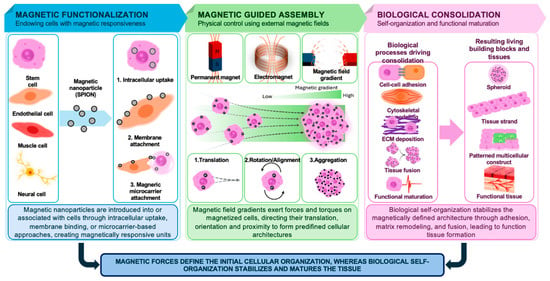
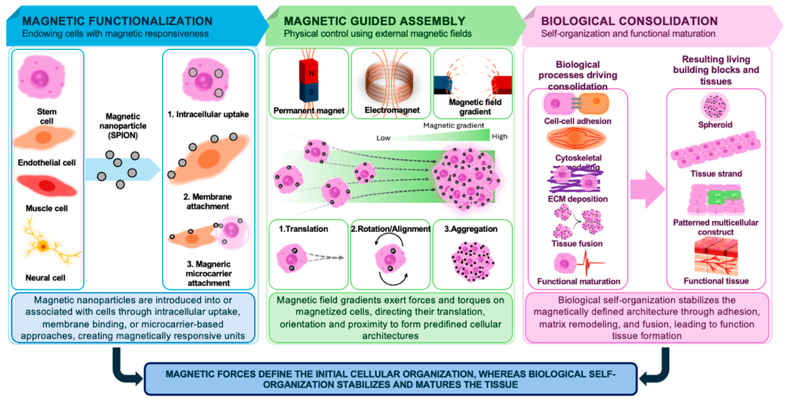
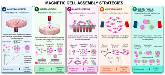
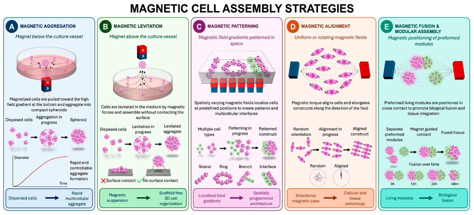
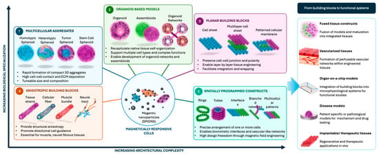
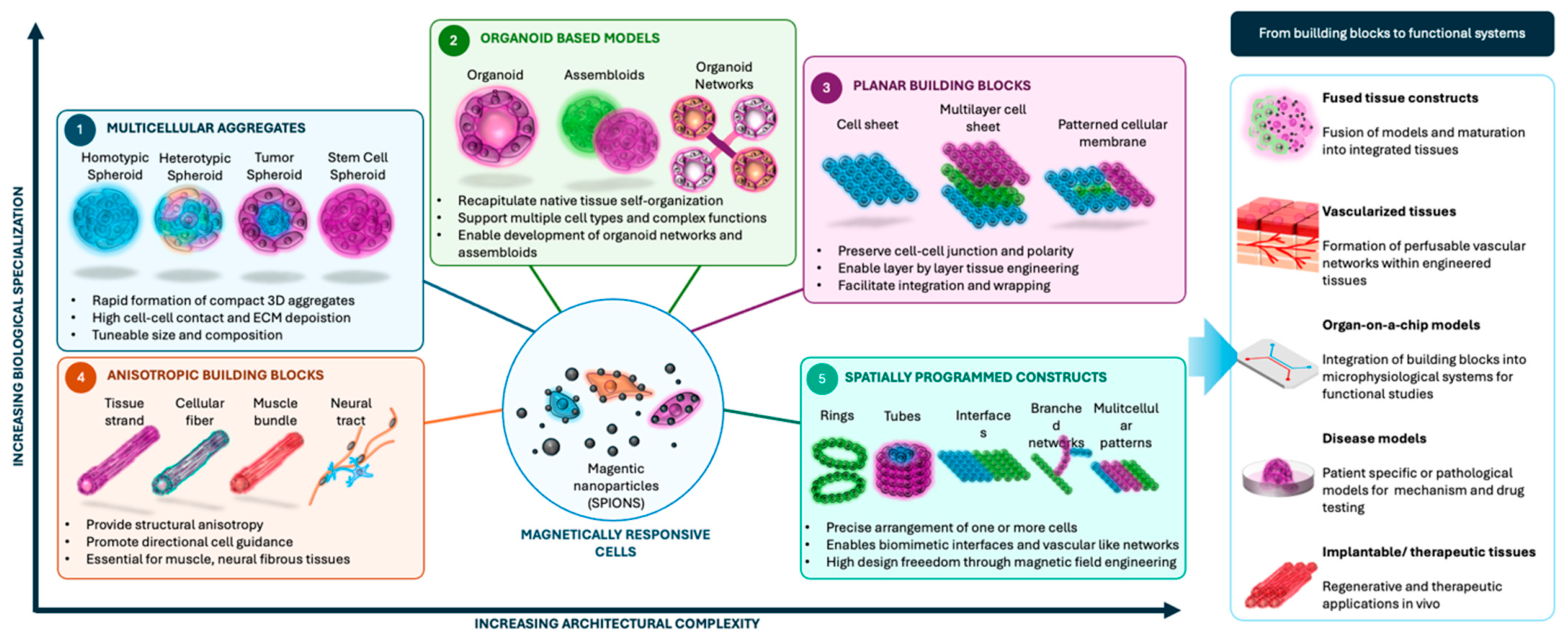
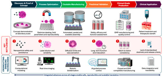
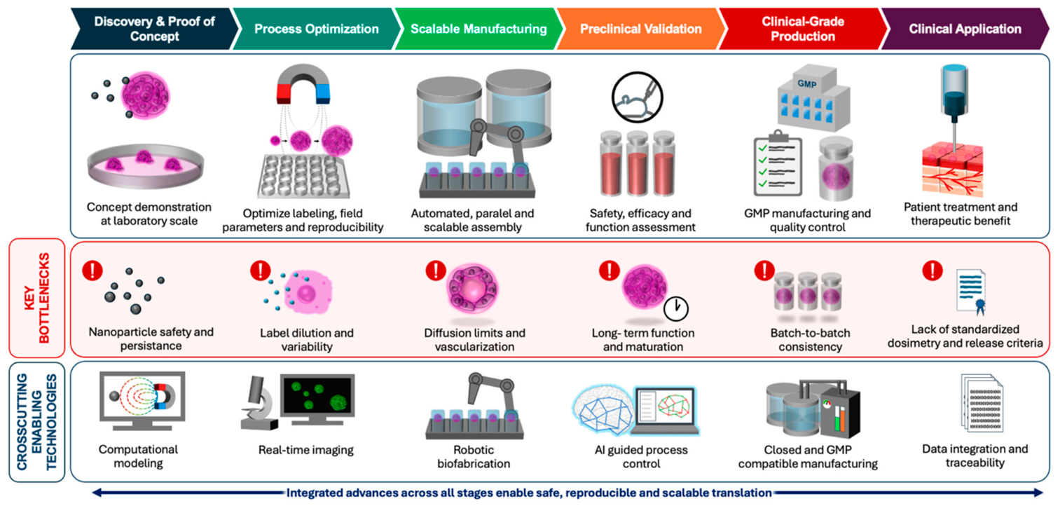

Disclaimer/Publisher’s Note: The statements, opinions and data contained in all publications are solely those of the individual author(s) and contributor(s) and not of MDPI and/or the editor(s). MDPI and/or the editor(s) disclaim responsibility for any injury to people or property resulting from any ideas, methods, instructions or products referred to in the content. |
© 2026 by the authors. Licensee MDPI, Basel, Switzerland. This article is an open access article distributed under the terms and conditions of the Creative Commons Attribution (CC BY) license.