


Highlights
What are the main findings?
- Strain Y107 colonized the rhizosphere; improved soil nutrients, root biomass, tissue water, and antioxidant capacity; and promoted lingonberry growth.
- The upregulation of photosynthesis and C-N metabolism genes was detected in the transcriptome.
What are the implications of the main findings?
- In this study, strain Y107 promoted growth under sterile conditions, and a new strain of Ericaceae PGPR was added.
- It reveals mechanisms via colonization, nutrient cycling, and gene regulation.
Abstract
Lingonberry (Vaccinium vitis-idaea L.) has a weak root system and limited nutrient acquisition capacity. Plant growth-promoting rhizobacteria (PGPR) can promote plant growth by regulating rhizosphere nutrient cycling and host metabolism, but the underlying mechanisms in Ericaceae remain unclear. In this study, the strain Microbacterium sp. Y107 was isolated from the rhizosphere of lingonberry. Using growth-promoting assays (with 9 independent biological replicates, each consisting of 3 plants per bottle as the experimental unit, an inoculum density of OD600 = 1.0, a cultivation period of 60 days, and whole-plant harvesting), GC–MS-based metabolite analysis, axenic coculture, analyses of the rhizosphere environment and gene expression, transcriptome sequencing (3 biological replicates per treatment), and qRT–PCR validation, we systematically evaluated the effects of strain Y107 on lingonberry growth, rhizosphere conditions, and host gene expression. CK (control) plants were defined as those inoculated with sterile LB liquid medium. Pearson correlation analysis was also used to assess the relationships among plant growth, soil traits, and physiological indices. Compared with the CK treatment, strain Y107 significantly promoted lingonberry growth (p < 0.05), increasing the fresh weight of the roots by 157.65% and the whole-plant fresh weight by 33.85%, respectively, and increasing peroxidase (POD) activity by 61.11%, while reducing the contents of reducing sugars and proline. Strain Y107 stably colonized the lingonberry root surface and formed biofilms. Y107 treatment resulted in detectable levels of soil microbial biomass carbon and nitrogen, indicating successful microbial colonization. The ammonium nitrogen content significantly increased, increasing nitrogen availability and promoting root nutrient uptake. Transcriptome analysis revealed that the expression of genes enriched in photosynthesis-related pathways, as well as those involved in carbon and nitrogen metabolism and redox regulation, was altered in strain Y107, which upregulated key photosynthesis-related genes such as psbP and ndhU and significantly enriched the cytosolic ribosomal large subunit gene set in a negative direction. Correlation analysis further revealed that strain Y107 optimized the association between lingonberry growth and soil nutrients. Overall, strain Y107 promoted lingonberry growth through stable colonization, enhanced nutrient cycling, promoted lingonberry growth, and altered the expression patterns of genes involved in photosynthesis-related pathways.
1. Introduction
Lingonberry (Vaccinium vitis-idaea L.) is a perennial shrub of the family Ericaceae and is widely distributed in coniferous forests across the cold temperate regions of the Northern Hemisphere [1]. Its fruits are rich in various bioactive compounds, including anthocyanins and flavonoids [2], and possess high nutritional value and health-promoting properties, indicating broad application prospects in the development of food, pharmaceutical, and functional products [3]. With the increasing demand for natural foods, the exploitation of lingonberry resources and its artificial cultivation have attracted increasing attention. However, lingonberry has a sparse and shallow root system with limited nutrient uptake capacity, and agronomic guidance on soil pH and fertility management during artificial cultivation remains insufficient, constraining the expansion of its planting scale [4]. Therefore, exploring novel green technologies to promote lingonberry growth has become important.
Rhizosphere microorganisms are essential components for maintaining plant growth and ecosystem functions and serve as key links between plants and soil. Among them, plant growth-promoting rhizobacteria (PGPR) can promote plant growth through multiple mechanisms, including phosphate solubilization, potassium solubilization, nitrogen fixation, and the secretion of siderophores and phytohormones. They can also increase plant environmental adaptability and stress resistance [5]. In recent years, with the rapid development of green agriculture and microbial fertilizers, PGPR have been widely applied in the production of food crops, economic crops, and horticultural plants [6]. However, the growth-promoting effects of PGPR are host-dependent. Therefore, screening functional strains that meet the growth requirements of specific plant species remains fundamental to the development of microbial resources [7].
Bacteria of the genus Microbacterium are widely distributed in various environments, including soil and plant rhizosphere soil, and represent important components of plant-associated microbial communities [8]. Previous studies have shown that some Microbacterium sp. strains can produce various secreted compounds, such as indole-3-acetic acid (IAA) and siderophores, thereby enhancing plant stress resistance and promoting plant growth through multiple mechanisms [9]. In addition, strains of this genus exhibit strong environmental adaptability and colonization ability, enabling them to survive under drought, heavy metal contamination, and salt stress conditions while effectively exerting growth-promoting functions, indicating substantial application potential. However, current studies on Microbacterium have focused mainly on food crops and model plants, whereas studies on their interaction mechanisms with Ericaceae plants are relatively limited [10].
Lingonberry usually grows in acidic environments with low nitrogen and phosphorus availability, where the composition and activity of its rhizosphere microorganisms (including ericoid mycorrhizal fungi and associated bacterial communities) are closely associated with plant nutrient status and ecological adaptation [11]. However, previous studies have focused mainly on the roles of ericoid mycorrhizal fungi in nutrient uptake and growth promotion in lingonberry, whereas how functional bacteria regulate rhizosphere nutrient cycling and host metabolism in lingonberry remains unclear [12]. Consequently, the development and utilization of rhizosphere growth-promoting bacterial resources for lingonberry remain relatively insufficient.
In this context, in the present study, a potential plant growth-promoting (PGP) bacterial strain, Microbacterium sp. Y107 (Y107), was isolated from the rhizosphere soil of lingonberry. The strain was characterized through morphological observation, phylogenetic analysis, and functional assays. Subsequently, axenic coculture experiments, soil nutrient profiling, and transcriptome sequencing were employed to test the following hypotheses: (i) Y107 significantly enhances lingonberry growth, as evidenced by increased biomass; (ii) Y107 modulates multiple rhizosphere soil nutrient parameters; and (iii) Y107 elicits reproducible transcriptional responses in the host. This study was designed to establish a theoretical foundation and provide microbial resource support for the development of functional bioinoculants tailored for lingonberry and for the broader utilization of microbial resources in Ericaceae plants.
2. Materials and Methods
2.1. Study Materials
Lingonberry seeds were obtained from fruits collected in September 2024 in the Da Xing’an Mountains (52°10′29″ N, 123°15′27″ E) forest region of China. The seeds were subsequently washed, naturally air-dried, and stored at 4 °C before use. To minimize genetic heterogeneity among wild-derived seedlings, seeds from multiple mother plants were pooled and randomly assigned to treatments. Before sowing, the seeds were vernalized at 4 °C for 7–14 days. After vernalization, the seeds were sequentially disinfected with 75% (v/v) ethanol for 30 s, followed by 2% (w/v) sodium hypochlorite for 3 min, rinsed 5 times with sterile distilled water, and then cultured at 22 ± 2 °C under a 16 h light/8 h dark photoperiod with a light intensity of 5000 lx on 1/2 MS medium (pH 5.8) solidified with 0.8% agar [13]. The soil used for subsequent cultivation was collected from the rhizosphere of wild lingonberry plants.
The wild lingonberry rhizosphere soil samples used for bacterial isolation were collected from the same location. Soil was collected from a depth of 0–20 cm at approximately 10 independent sampling points and composited into a single pooled sample. The collected soil was immediately stored at 4 °C and processed within 48 h for bacterial isolation. Briefly, 1 g of rhizosphere soil was weighed and transferred to sterile saline containing glass beads at a soil-to-water ratio of 1:10. The mixture was shaken at 180 rpm for 10 min and then allowed to stand for 5 min to obtain a soil suspension. Under aseptic conditions, the suspension was serially diluted with sterile water to 10−4, 10−5, and 10−6. Afterward, 100 μL of each dilution was spread onto TSA solid medium, with three replicates for each dilution. The plates were incubated at 30 °C for 48 h. Approximately 23 representative single colonies were selected according to differences in colony morphology and color and subsequently purified. All the purified isolates were subjected to preliminary screening for plant growth-promoting traits (including IAA production, nitrogen fixation, and phosphate solubilization). Among them, strain Y107 exhibited the greatest comprehensive growth-promoting potential and was therefore selected as the candidate strain for this study. The purified strain Y107 was stored in glycerol tubes at −80 °C for subsequent experiments [14].
2.2. Strain Identification
Colony PCR was performed using the activated liquid culture of strain Y107 directly as the template, with the universal 16S rRNA gene primers 27F (5′-AGAGTTTGATCCTGGCTCAG-3′) and 1492R (5′-TACGGTTACCTTGTTACGACTT-3′) [15]. An aliquot of 1 μL of the activated Y107 liquid culture was used as the template. The PCR mixture (20 μL total volume) consisted of 10 μL of 2× Taq PCR MasterMix (containing Taq DNA polymerase, dNTPs, Mg2+, and reaction buffer), 1 μL of bacterial liquid culture template, 0.8 μL of each primer (10 μM, final concentration 0.4 μM each), and 7.4 μL of nuclease-free water. Sterile nuclease-free water replacing the template was used as a negative control. The amplification program was as follows: initial denaturation at 94 °C for 2 min; 30 cycles of denaturation at 94 °C for 30 s, annealing at 58 °C for 30 s, and extension at 72 °C for 1 min; and a final extension at 72 °C for 3 min. The PCR products were verified by electrophoresis on a 1% (w/v) agarose gel stained with nucleic acid dye, purified using a gel extraction kit, and sequenced by Sanger sequencing.
The obtained 16S rDNA gene sequence of strain Y107 was submitted to NCBI GenBank under accession number PV630865.1. BLAST alignment against the NCBI (National Center for Biotechnology Information (https://www.ncbi.nlm.nih.gov/), Bethesda, MD, USA) database revealed that strain Y107 shared the highest sequence identity (99.9%) with the type strain of Microbacterium azadirachtae (KF150485.1). Sequence alignment was performed using MUSCLE with default parameters. A phylogenetic tree was constructed using the neighbor-joining method with the Kimura 2-parameter substitution model and 1000 bootstrap replicates in MEGA12 software (Molecular Evolutionary Genetics Analysis version 12, MEGA Software, St. Paul, MN, USA) [16]. Since the 16S rRNA gene sequence alone cannot reliably distinguish closely related species within the genus Microbacterium, we tentatively identified strain Y107 as a Microbacterium sp., pending the confirmation of its exact species identity by whole-genome analysis.
2.3. Strain Morphology and Plant Growth-Promoting Traits
The activated strain Y107 was observed under a stereomicroscope, and its colony morphology, color, gloss, and other characteristics were recorded. Gram staining was performed, and the cells were observed under a microscope. Each functional assay was conducted with three independent biological replicates. Strain Y107 (OD600 adjusted to 1.0) was spot-inoculated (1 μL) onto CAS agar medium, PKO agar medium, Aleksandrov agar medium, and nitrogen-free Ashby solid medium and incubated at 30 °C for 5–7 days (48 h for CAS plates). Siderophore production was qualitatively evaluated by observing orange halos on CAS plates and was quantitatively determined using the CAS liquid assay with the following formula:
where Ar is the absorbance of the uninoculated CAS solution at 630 nm and As is the absorbance of the sample. Phosphate solubilization and potassium solubilization were qualitatively assessed by the formation of clear hydrolysis halos on PKO and Aleksandrov agar media, respectively, indicating potential phosphate and potassium solubilization activities. Nitrogen fixation potential was evaluated by growth on nitrogen-free Ashby medium, indicating potential nitrogen fixation activity. For IAA production, strain Y107 was cultured in LB liquid medium supplemented with 0.1% (w/v) L-tryptophan at 30 °C for 48 h with shaking at 180 rpm. The culture supernatant was collected by centrifugation, and the IAA concentration was determined using the Salkowski colorimetric method [17] by measuring the absorbance at 530 nm and comparing it against a standard curve (0–100 μg/mL). Uninoculated medium served as a negative control for all the assays [17].
siderophore units (%) = [(Ar − As)/Ar] × 100%,
2.4. Colonization Ability and Metabolite Production of the Strain
Strain Y107 was cultured in LB broth at 30 °C with shaking at 180 rpm to the logarithmic phase (OD600 ≈ 1.0) as the seed culture, with three independent cultures and three technical measurements for all assays. The cell-free supernatant obtained by centrifugation at 8000× g for 10 min at 4 °C and filtration through a 0.22 μm membrane was used for extracellular product analyses. Cell surface hydrophobicity was assessed by the MATH method, in which the bacterial suspension (OD600 = 0.6–0.8, recorded as A1) was mixed with xylene (5:1, v/v), incubated at 30 °C for 10 min, and vortexed for 20 min, after which the absorbance of the aqueous phase at 400 nm (A2) was measured, and the hydrophobicity was calculated as follows:
with sterile PBS as the blank. Biofilm formation was quantified by crystal violet staining in 96-well plates: Y107 was inoculated into TSA medium and incubated statically at 28 °C for 24 h (sterile TSA as a negative control), followed by washing, fixation with 2.5% glutaraldehyde for 15 min, staining with 1% crystal violet for 20 min, and dissolution of the bound dye in 95% ethanol for absorbance measurement at 600 nm. Extracellular soluble sugars were quantified by the DNS method, where 2 mL of the supernatant was heated at 80 °C for 30 min, reacted with DNS reagent by boiling for 5 min, and measured at 540 nm against a glucose standard (0–100 μg/mL), with the results expressed as glucose equivalents and sterile deionized water as the blank. Extracellular soluble proteins were determined by the Bradford method using BSA as the standard at 595 nm and sterile deionized water as the blank. For motility characterization, bacterial suspensions adjusted to OD600 = 0.6–0.8 were used: swarming motility was assessed by spotting 3 μL onto 0.7% LB soft agar, while twitching motility was evaluated by stabbing a sterile needle through 1% LB agar to the agar–plastic interface. In both assays, the plates were incubated at 28 °C, with uninoculated plates used as negative controls, and the expansion diameter was recorded every 6 h (up to 24 h).
hydrophobicity (%) = (1 − A2/A1) × 100%,
Furthermore, metabolite analysis of Y107 was conducted. Y107 was inoculated into LB liquid medium and cultured with shaking at 30 °C and 180 rpm for 24 h, with three biological replicates and uninoculated LB medium as the blank control. After cultivation, the bacterial suspension was centrifuged at 8000× g for 10 min at 4 °C to obtain the cell-free supernatant, which was then sterilized by filtration through a 0.22 μm membrane. The cell-free supernatant was freeze-dried at −40 °C, and the resulting dry powder was dissolved in methanol (5 mL). Samples were analyzed by GC–MS (Thermo Fisher Trace GC 2000 DSQII, Thermo Fisher Scientific, Waltham, MA, USA) equipped with a BP-5MS column (30 m × 0.25 mm × 0.25 μm) using helium as the carrier gas with splitless injection [18]. The temperature program was as follows: initial temperature 50 °C, ramped to 250 °C at 10 °C/min and held for 15 min; the injector, transfer line, and ion source temperatures were all set to 250 °C. The ion source was EI, with a scan range of 35–453 m/z. Metabolites were identified by matching against the NIST library. Compounds detected in the blank were excluded, and only metabolites present in all replicates with a >2-fold change relative to the blank were retained.
2.5. Inoculation of Lingonberry with Strain Y107
The axenic coculture experiment included two treatments: a blank control group (CK) and an experimental group supplemented with 2 mL of bacterial suspension at an inoculation rate of 2% (v/w). The substrate was prepared by mixing soil and vermiculite at a volume ratio of 2:1. Each bottle was filled with 0.1 kg of substrate and sterilized at high temperature (121 °C for 90 min). To confirm sterility, spread-plate assays were performed after sterilization, and no colony growth was observed. The sterilized substrate was then used for subsequent experiments. Each treatment consisted of 30 replicate bottles, with three seedlings planted per bottle. Strain Y107 was inoculated into TSA medium and cultured at 28 °C with shaking at 180 rpm to the logarithmic phase, after which the bacterial suspension was adjusted to an OD600 = 1.0. For the control group, an equal volume of sterile TSA medium was added instead of the bacterial suspension. Lingonberry seedlings that had been cultivated under the same axenic conditions for 2 months (22 ± 2 °C, 16 h/8 h photoperiod) and whose growth was uniform were selected, and three seedlings were transplanted into each bottle. At the beginning of the experiment, 30 mL of sterile water was added to each bottle, and thereafter, depending on the moisture condition of the substrate, 5 mL of sterile water was supplemented weekly as needed to maintain its moisture, with no additional water added between scheduled irrigations. The culture bottles were then maintained under the original culture conditions for an additional 60 days. All procedures were performed under aseptic conditions.
At harvest, three different culture bottles were selected from the 30 bottles per treatment for sampling. Within each selected bottle, the overall growth status of the three seedlings was first examined; after any visibly abnormal individuals (e.g., chlorotic, stunted, or mechanically damaged) were excluded, one seedling whose growth status was closest to the average level of the three seedlings was selected for phenotypic measurements, physiological/biochemical [19]. assays and RNA extraction. Additionally, soil samples were also collected from the same three bottles, with one soil sample taken independently from each bottle. Thus, three independent biological replicates (n = 3) were obtained per treatment, with each replicate representing an individual culture bottle.
2.6. Measurement of Lingonberry Phenotypic, Physiological, and Biochemical Parameters
After the cultivation experiment, the height, total leaf number, basal diameter, and branch number of the lingonberry plants were measured to analyze their growth and morphological characteristics. After sampling, the seedlings were divided into above-ground (shoot) and root parts and weighed using an analytical balance with an accuracy of 0.0001 g to determine whole-plant fresh weight, shoot fresh weight, and root fresh weight. Each part was then dried in an oven at 65 °C to a constant weight, after which the shoot dry weight, root dry weight, and whole-plant dry weight were determined, and the water content was calculated.
The proline content was determined using the ninhydrin-sulfosalicylic acid method [20]: fresh leaf tissue (0.01 g) was extracted with 3% sulfosalicylic acid and centrifuged, after which the supernatant was reacted with ninhydrin reagent and extracted with toluene, after which the absorbance of the toluene phase was measured at 520 nm, and the results were expressed as μg/g fresh weight. The reducing sugar content was measured using the 3,5-dinitrosalicylic acid method [21]: fresh leaf tissue (0.02 g) was ground and reacted in a boiling water bath for 5 min, after which the absorbance was measured at 540 nm, and the content was calculated against a glucose standard curve and expressed as a percentage (%). The malondialdehyde (MDA) content was determined using the thiobarbituric acid (TBA) method [22]: fresh leaf tissue (0.02 g) was extracted with 10% trichloroacetic acid, mixed with TBA, heated in a boiling water bath for 40 min, and absorbances were measured at 450 nm, 532 nm, and 600 nm, with the content calculated using the corresponding formula and expressed as μmol/g fresh weight. Peroxidase (POD) activity was measured using the guaiacol method: fresh leaf tissue (0.02 g) was ground in acetate buffer (pH 5.5) containing 1 mmol/L PEG 6000, 4% PVPP, and 1% Triton X-100 to obtain the crude enzyme extract. The reaction mixture contained the crude enzyme extract, guaiacol, and H2O2. The absorbance at 470 nm was recorded starting at 15 s of reaction and then every 1 min for six consecutive readings, and POD activity was calculated using the standard formula and expressed as U/g fresh weight. All extraction and measurement steps were performed under ice-bath conditions, and the results were calculated on a fresh weight basis [23].
2.7. Determination of Soil Physicochemical Properties
After the coculture experiment, the lingonberry rhizosphere soil was collected. Fresh soil subsamples designated for the determination of ammonium nitrogen, nitrate nitrogen, microbial biomass carbon, and microbial biomass nitrogen were immediately stored at 4 °C and processed within 48 h. The soil water content was determined gravimetrically by weighing fresh soil samples, drying them at 105 °C to constant weight, and calculating the percentage of water loss. The remaining soil was air-dried at room temperature, ground, and passed through a 0.2 mm sieve for other analyses. Soil pH was measured using a pH meter: 5.0 g of air-dried soil was mixed with deionized water at a soil-to-water ratio of 1:2.5 (m/v) and shaken at 180 rpm for 30 min, after which the pH was determined with a calibrated pH meter. The total carbon, total nitrogen, total phosphorus, total magnesium, and total potassium contents were determined using laser-induced breakdown spectroscopy (LIBS) [24]. Air-dried and sieved fine soil was pressed into pellets using a tablet press and analyzed with a laser-induced breakdown spectroscopy elemental analyzer (J200, Applied Spectra, Inc., West Sacramento, CA, USA). The instrument was factory-calibrated using certified reference materials, and the results represent the total concentrations of the elements in the soil. Ammonium nitrogen and nitrate nitrogen were extracted with 2 mol/L KCl at a soil-to-solution ratio of 1:10 (shaken for 30 min, then filtered) and determined using a continuous flow analyzer, with ammonium nitrogen measured by the indophenol blue method and nitrate nitrogen measured by the cadmium reduction-diazotization method [25]. The results are expressed as mg/L. Soil microbial biomass carbon and nitrogen were determined by the chloroform fumigation-extraction method. Fresh soil was fumigated with ethanol-free chloroform for 24 h and then extracted with 0.5 mol/L K2SO4 at a soil-to-solution ratio of 1:4 (shaken for 30 min and then filtered), with nonfumigated soil serving as the control. Organic carbon and total nitrogen in the extracts were measured using a continuous flow analyzer, and microbial biomass carbon and nitrogen were calculated using conversion factors of kEC = 0.45 and kEN = 0.54, respectively. Microbial biomass carbon, microbial biomass nitrogen, and total element concentrations were expressed on a soil dry weight basis [26].
2.8. RNA Extraction, Library Construction, and Sequencing of Lingonberry
Stems and leaves were collected from three different culture bottles per treatment (Y107-inoculated and uninoculated CK), with one seedling taken from each bottle, and immediately stored at −80 °C, resulting in three independent biological replicates (from three different bottles). The frozen samples were sent to Shanghai Majorbio Biopharm Technology Co., Ltd. (Shanghai, China), for total RNA extraction, cDNA library construction, and sequencing.
In brief, total RNA was extracted from each sample. RNA concentration and purity were assessed using a Nanodrop 2000 spectrophotometer (Thermo Fisher Scientific, Waltham, MA, USA), and RNA integrity was evaluated by agarose gel electrophoresis. The raw data were filtered using fastp v0.19.5. Approximately 39.9–46.7 million clean reads were obtained per sample (Q30 > 95.24%). Clean reads were aligned to the V. vitis-idaea ssp. minus the reference genome using HISAT2 v2.1.0, with mapping rates of 89.79–91.33%. Gene expression levels were quantified using RSEM v1.3.3, which generated both raw integer counts and TPM values. Raw counts were used for differential expression analysis with DESeq2 v1.10.1, and p values were adjusted using the Benjamini–Hochberg method. Genes with an adjusted p value < 0.05 and |log2FC| ≥ 1 were considered significantly differentially expressed. TPM values were used for gene set enrichment analysis (GSEA) and expression visualization. GO enrichment analysis was performed using goatools v0.6.5 with Fisher’s exact test, and a p value < 0.05 was considered to indicate statistical significance. KEGG pathway enrichment analysis was performed using the same calculation principle as that used for the GO enrichment analysis, and pathways for which p was adjusted to <0.05 were considered significantly enriched. In addition, GSEA was performed using the Signal2Noise metric as the ranking statistic, with gene sets derived from GO and KEGG annotations. Significance was assessed by 1000 permutations, and pathways with |NES| > 1 and p adjusted < 0.25 were considered significantly enriched.
Total RNA was extracted from lingonberry stems and leaves using a Universal Plant Total RNA Extraction Kit (Beijing Bioteke Biotechnology Co., Ltd., Beijing, China, Cat. No. RP3302). On the basis of the phenotypic differences observed under Y107 treatment, we focused on candidate genes associated with photosynthesis and flavone and flavonol biosynthesis pathways for qRT-PCR validation. Gene selection was based on KEGG Orthology (KO) annotations, prioritizing genes assigned to key enzymes in these pathways. Three DEGs were ultimately selected: STRG.9502 (K02717, annotated as photosystem II oxygen-evolving enhancer protein 2), STRG.15131 (K23052, annotated as chloroplastic NAD(p)H-quinone oxidoreductase subunit U), and STRG.35646 (K13269, annotated as kaempferol 3-O-beta-D-galactosyltransferase). All qRT-PCRs were performed in 96-well plates using a LightCycler 480 real-time PCR system (Roche Diagnostics GmbH, Rotkreuz, Switzerlandd). The sequences of primers used were as follows: Actin (internal reference gene, ID: CL5740. Contig2_All, encoding actin) forward primer 5′-ACCTTTTGGATTGTGGGCTAGA-3′, reverse primer 5′-AGCTCCACTTGCACTTTTCCTT-3′ [27]; STRG.9502 forward primer 5′-TCTCTCTCTCTCTCTCTCTCTCTCTCT-3′, reverse primer 5′-CTCCTCCTCGATGAATGGAAAC-3′; STRG.15131 forward primer 5′-CTTAGCCTCCAATCCTGTCTTC-3′, reverse primer 5′-TGCTCCAAGGTCCCAATATG-3′; STRG.35646 forward primer 5′-CTACTTTGAGGCGGGATGTTTA-3′, reverse primer 5′-CCCATGGTTGTGTAGGAATAGG-3′. The qPCR mixture contained 7.5 μL of THUNDERBIRD® Next SYBR® qPCR mix (Toyobo Co., Ltd., Osaka, Japan), 0.6 μL of each forward and reverse primer, and 6.3 μL of cDNA template, for a total volume of 15 μL. No-template and no-reverse transcription controls were included and produced no detectable signal. The thermal cycling conditions were as follows: initial denaturation at 95 °C for 5 min, followed by 40 cycles of denaturation at 95 °C for 30 s, annealing at 55 °C for 30 s, and extension at 72 °C for 30 s. For each sample, three biological replicates (from three different culture bottles) and three technical replicates per biological replicate were used. Actin was validated as a stably expressed reference gene under Y107 treatment conditions [28]. The relative expression levels of the candidate genes were calculated using the 2^(−ΔΔCT) method, and statistical significance was assessed using Student’s t-test (p < 0.05).
2.9. Statistical Analysis
All the data are expressed as the mean (). Data processing was performed using Microsoft Excel 2021 and SPSS 27.0, and data visualization was conducted using Origin 2024 (OriginLab Corporation, Northampton, MA, USA). Differences among treatment groups were evaluated by one-way analysis of variance (ANOVA) followed by Tukey’s multiple comparison test, with the significance level set at p < 0.05. Relationships among variables were analyzed using Pearson correlation coefficients. All the statistical analyses were performed in SPSS 27.0 and R 4.5.1.
3. Results
3.1. Identification, Plant Growth-Promoting Traits, and Colonization Ability of Strain Y107
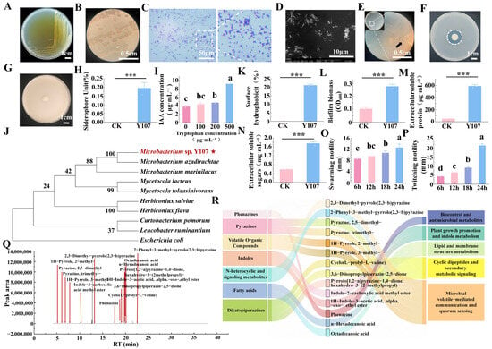
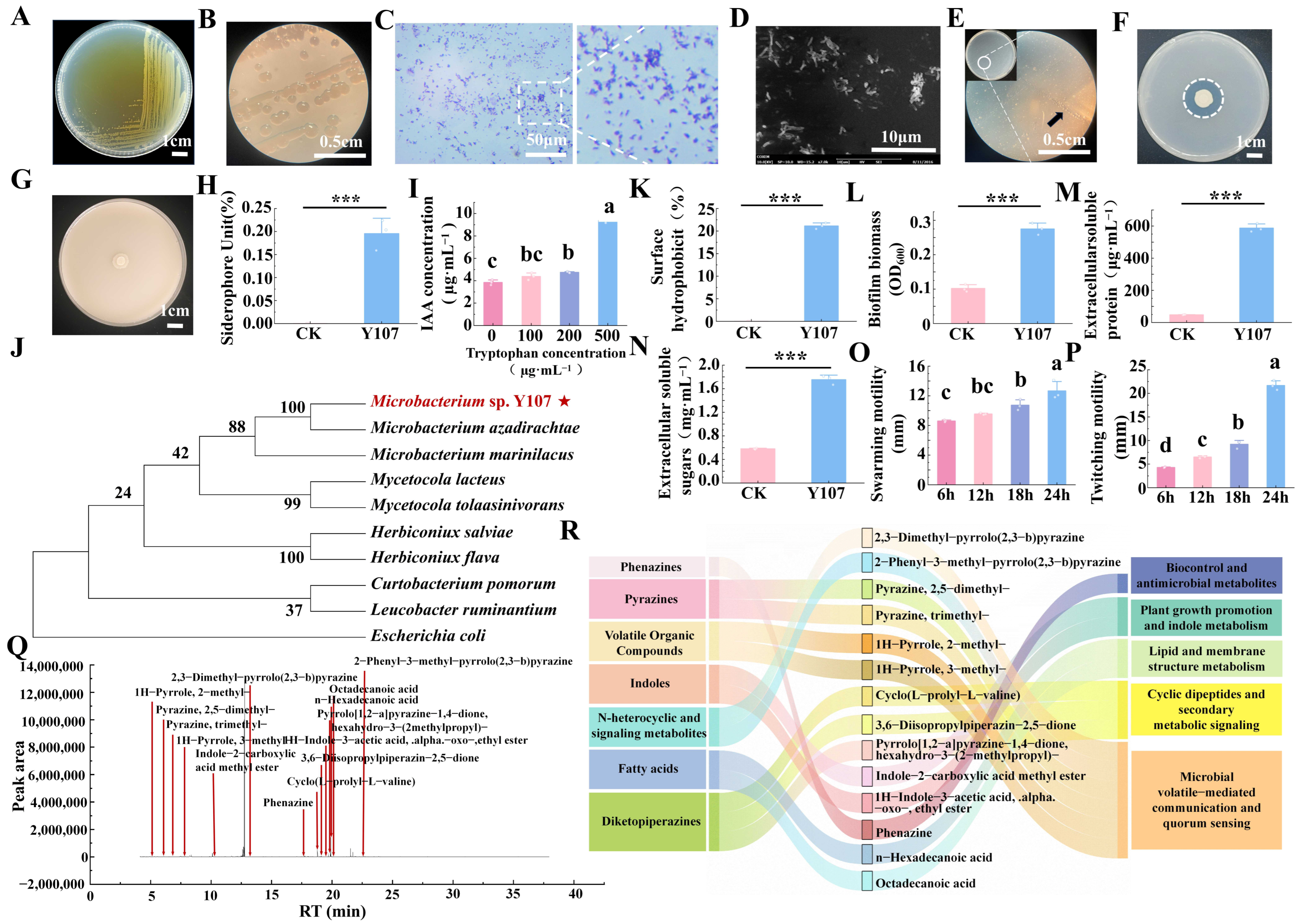
We systematically identified the isolated and purified strain Y107. Morphological observation revealed that the colonies were regular and circular with smooth surfaces (Figure 1A). Observations under a stereomicroscope revealed milky-white, circular, raised colonies with a translucent appearance (Figure 1B). Gram staining revealed that the cells were blue–purple (Figure 1C). Further observation by scanning electron microscopy indicated that the strain consisted of rod-shaped cells with uniform morphology, no branching, bluntly rounded ends, and a smooth surface. The cells were approximately 0.6 μm wide and 1.3 μm long (Figure 1D). To preliminarily evaluate the plant growth-promoting functions of strain Y107, we performed screening using functional media. The results revealed that strain Y107 grew on Ashby nitrogen-free medium (Figure 1E) and formed a distinct clear halo on PKO medium (Figure 1F); however, no clear halo was observed on potassium feldspar powder medium (Figure 1G). The siderophore activity of strain Y107 was 19.55% (Figure 1H). Strain Y107 produced IAA in the absence of tryptophan, and its IAA concentration increased significantly with increasing exogenous tryptophan concentration (p < 0.05) (Figure 1I). The 16S rDNA sequence of strain Y107 was uploaded to the NCBI database and assigned the accession number PV630865.1. Sequence alignment and phylogenetic analysis revealed that strain Y107 clustered on the same branch as Microbacterium azadirachtae. On the basis of its morphological characteristics and phylogenetic tree, the strain was identified as Microbacterium sp. Y107 (Figure 1J).
Figure 1.
Identification, plant growth-promoting traits, colonization ability, and metabolite analysis of strain Y107. (A) Colony morphology of strain Y107 on TSA medium, scale bar = 1 cm; (B) colony morphology observed under a stereomicroscope, scale bar = 0.5 cm; (C) Gram staining results (×400), scale bar = 100 μm; (D) scanning electron microscopy image, scale bar = 1 μm; (E–G) verification of nitrogen fixation, phosphate solubilization, and potassium solubilization abilities of strain Y107, scale bar = 1 cm; (H) relative siderophore content of strain Y107; (I) IAA production by strain Y107 under different tryptophan concentrations; (J) phylogenetic tree constructed on the basis of 16S rDNA sequences; (K) cell surface hydrophobicity of strain Y107; (L) biofilm formation ability of strain Y107; (M) extracellular soluble protein content of strain Y107; (N) extracellular soluble sugar content of strain Y107; (O) swarming motility of strain Y107; (P) twitching motility of strain Y107; (Q) GC–MS metabolite profile of strain Y107; (R) Sankey diagram showing the classification, composition, and potential ecological functions of metabolites secreted by strain Y107. Quantitative data are presented as the mean (n = 3). Different lowercase letters indicate significant differences among treatments after one-way ANOVA followed by Tukey’s multiple comparison test (p < 0.05); asterisks denote significant differences compared with the control group (CK) according to Student’s t-test (*** p < 0.001); and ns indicates no significant difference.
To further evaluate the potential root colonization ability of strain Y107, we measured indicators related to environmental colonization. The results revealed that the hydrophobicity of strain Y107 was 21.13%, indicating a moderate level of hydrophobicity (Figure 1K). Crystal violet quantitative analysis revealed that the biofilm-forming ability of strain Y107 was significantly stronger than that of the CK (p < 0.001), with a biofilm biomass, expressed as the OD600, of 0.275 (Figure 1L). The secretion levels of extracellular soluble sugars and extracellular soluble proteins were also determined, and both significantly increased. The extracellular soluble protein and soluble sugar contents increased to 587.39 μg·mL−1 (p < 0.001) (Figure 1M) and 1.75 mg·mL−1 (p < 0.001) (Figure 1N), respectively. The results of the swarming motility assay revealed that strain Y107 formed a radial bacterial film with a diameter of 12.67 mm on LB soft agar medium after 24 h (Figure 1O). The twitching motility assay results further revealed that strain Y107 formed a migration zone with a diameter of 21.67 mm on the medium surface after 24 h of incubation (Figure 1P).
To further characterize the composition of functional metabolites produced by strain Y107, its secreted metabolites were analyzed using gas chromatography–mass spectrometry (GC–MS) (Table S1), which lists detailed information, including compound names, retention times, and peak areas of the identified metabolites. A total of 14 metabolites with potential plant growth-promoting functions were detected (Figure 1Q). Furthermore, a Sankey diagram was constructed to illustrate the composition of the strains’ secretions and their putative ecological functions inferred from the literature (Figure 1R). The detected metabolites were mainly pyrazines, diketopiperazines, indoles, fatty acids, and phenazines and were closely associated with microbial communication, quorum sensing, plant growth promotion, and biocontrol functions. These functional assignments are primarily based on the literature, and their exact ecological functions require further experimental validation in subsequent studies.
3.2. Effects of Strain Y107 on the Growth, Phenotypic, Physiological, and Biochemical Characteristics of Lingonberry
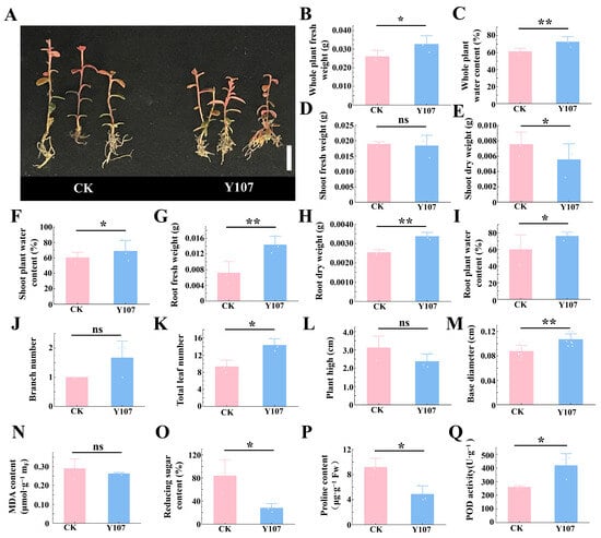
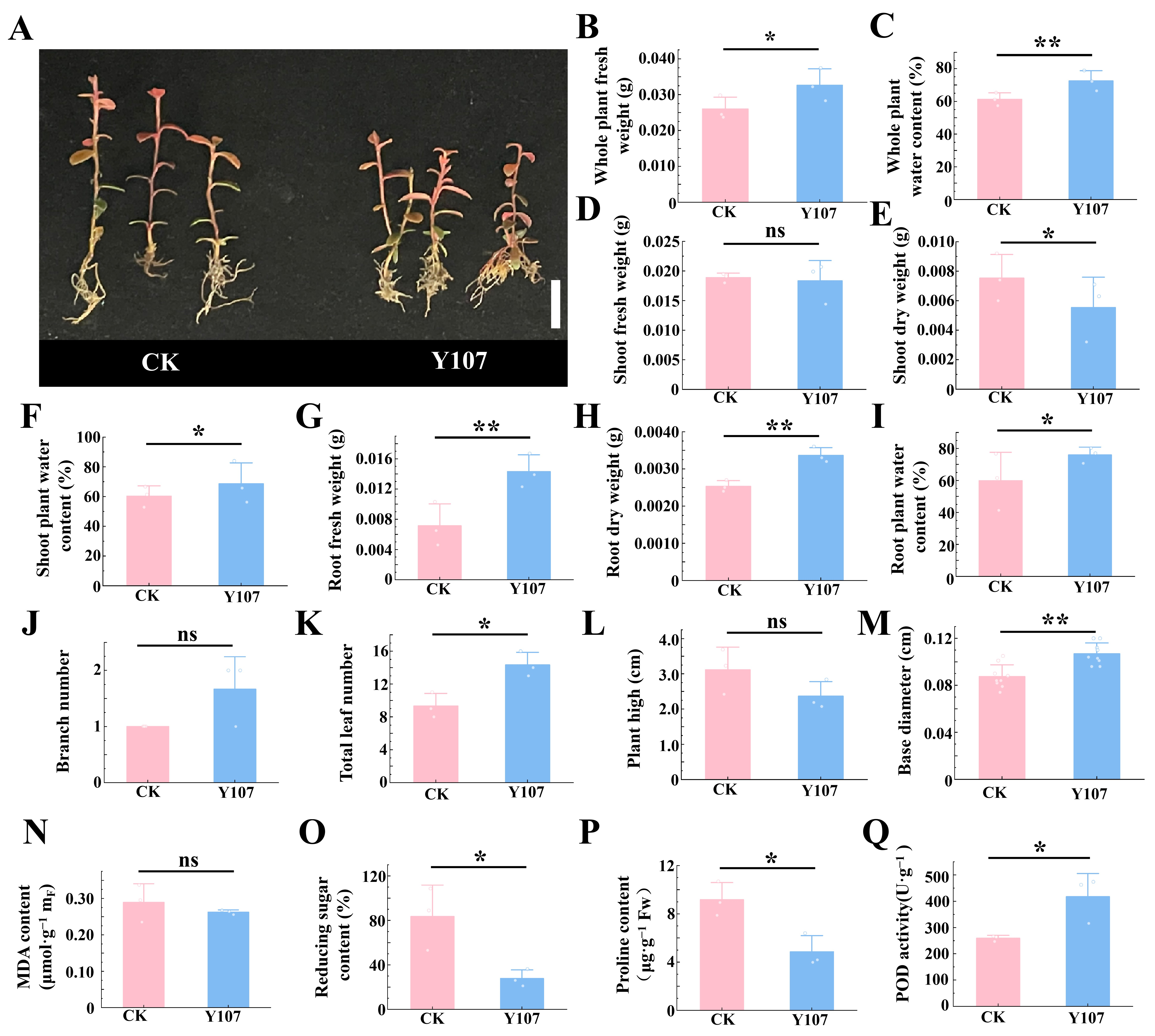
On the basis of the growth-promoting characteristics of strain Y107, we inoculated it onto lingonberry plants to verify its potential to promote plant growth. Compared with the CK plants, the lingonberry plants inoculated with Y107 exhibited clear morphological differences (Figure 2A). The total leaf number (Figure 2K) and basal diameter (Figure 2M) of the lingonberry plants in the Y107 treatment group increased significantly. Specifically, the total leaf number increased from 9.33 to 14.33, representing an increase of 53.57% (p < 0.05), whereas the base diameter increased from 0.08 cm to 0.11 cm, representing an increase of 34.65% (p < 0.01). On the basis of the observed phenotypic differences, biomass measurements were further conducted to evaluate the growth-promoting effects of Y107 on lingonberry. Compared with those in the CK treatment, whole-plant fresh weight (Figure 2B), root fresh weight (Figure 2G), and root dry weight (Figure 2H) increased significantly. The whole-plant fresh weight increased from 0.026 g to 0.035 g, representing an increase of 33.85% (p < 0.05). The fresh weight and dry weight of the roots increased from 0.006 g and 0.0025 g to 0.014 g and 0.0033 g, corresponding to increases of 157.65% and 32.89%, respectively (p < 0.01). In contrast, the shoot dry weight (Figure 2E) decreased from 0.0083 g to 0.0067 g, representing a decrease of 19.28% (p < 0.01). The whole-plant dry weight did not significantly differ between the two treatments (Figure S1), whereas the root-to-shoot ratio (Figure S2) significantly increased (p < 0.05) from 0.31 to 0.50, representing an increase of 61.29%.
Figure 2.
Effects of strain Y107 on the growth phenotypes and physiological and biochemical indices of lingonberry. (A) Phenotypic comparison of lingonberry plants inoculated with Y107 and the control (CK); scale bar = 1 cm; (B) whole-plant fresh weight; (C) whole-plant water content; (D) aboveground fresh weight; (E) aboveground dry weight; (F) aboveground water content; (G) root fresh weight; (H) root dry weight; (I), root water content; (J) branch number; (K) total leaf number; (L) plant height; (M) basal diameter; (N) malondialdehyde (MDA) content; (O) reducing sugar content; (P) proline content; (Q) POD activity. The data are expressed as the mean ± standard deviation (n = 3). Asterisks indicate significant differences compared with the CK group (* p < 0.05, ** p < 0.01), and ns indicates no significant difference. The error bars represent the standard errors.
We further elucidated the growth-promoting effects of Y107 at the physiological and biochemical levels. Compared with those in the CK group, the whole-plant water content (Figure 2C), shoot water content (Figure 2F), and root water content (Figure 2I) in the Y107 group significantly increased. Specifically, the whole-plant water content increased from 58.13% to 70.97%, representing a year-on-year increase of 22.10%. (p < 0.01). The shoot water content and root water content increased from 59.07% and 53.20% to 67.30% and 75.98%, respectively, representing year-over-year increases of 13.93% and 42.83% (p < 0.05), respectively, and the reducing sugar content (Figure 2O) and proline content (Figure 2P) decreased significantly, from 8.36% and 9.17 μg·g−1 FW to 2.78% and 4.86 μg·g−1 FW, respectively, representing decreases of 66.79% and 47.07% (p < 0.05), respectively. Antioxidant index measurements revealed that POD activity (Figure 2Q) increased significantly, from 259.15 U·g−1 to 417.51 U·g−1, representing an increase of 61.11% (p < 0.05).
3.3. Effects of Y107 Inoculation on the Rhizosphere Microenvironment and Nutrient Cycling of Lingonberry
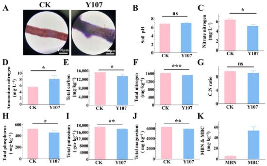
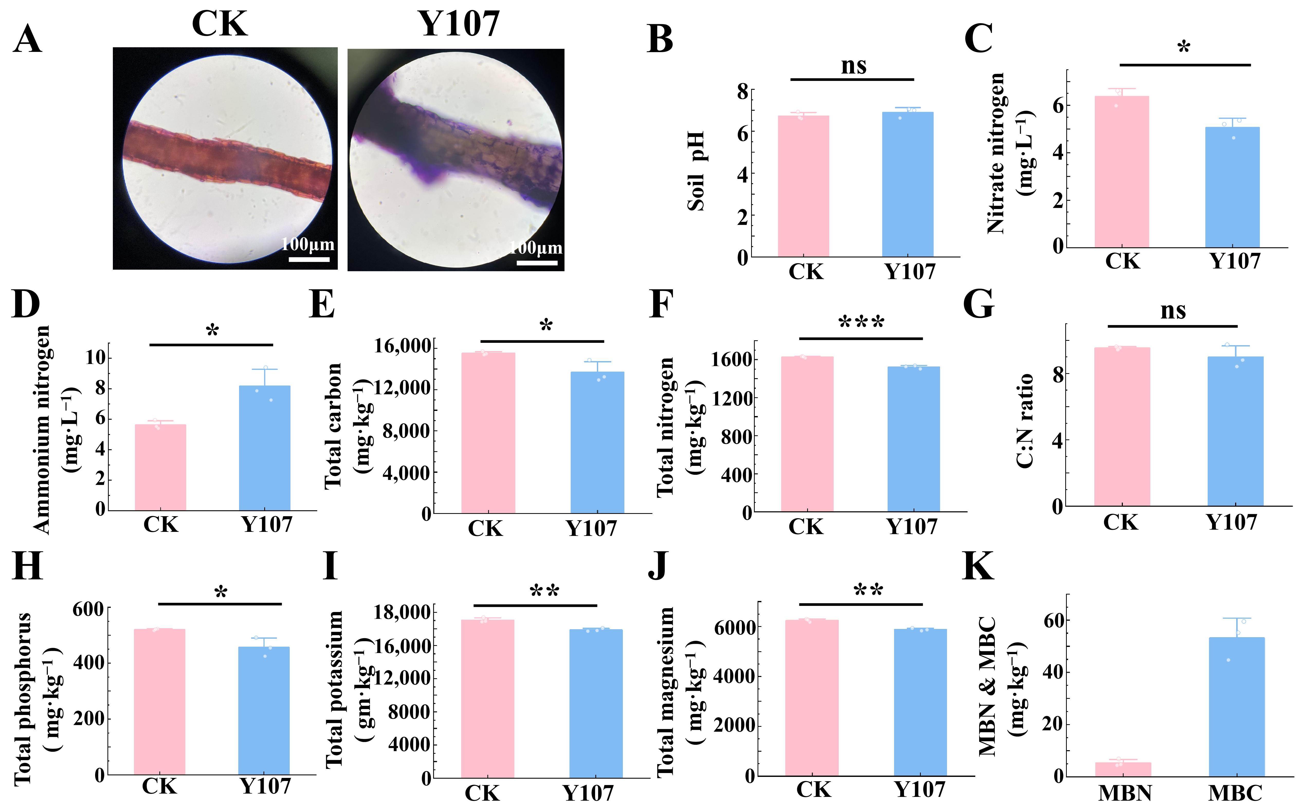
Given the marked changes in the lingonberry phenotype and related physiological indices following Y107 inoculation, we employed crystal violet staining to observe root surface attachment. The results revealed that the crystal violet staining signal on the root surface was significantly enhanced in the Y107-inoculated group, whereas the CK group exhibited weak background staining (Figure 3A), indicating a substantial increase in root surface attachment after inoculation.
Figure 3.
Colonization of lingonberry roots by strain Y107 and its effects on soil physicochemical properties. (A) Biofilm formation of strain Y107 on lingonberry roots observed by crystal violet staining. (B) Soil pH. (C) Soil nitrate nitrogen content. (D) Soil ammonium nitrogen content. (E) Soil total carbon content. (F) Soil total nitrogen content. (G) Soil carbon-to-nitrogen ratio (C/N). (H) Soil total phosphorus content. (I) Soil total potassium content. (J) Soil total magnesium content. (K) Soil microbial biomass carbon (MBC) and microbial biomass nitrogen (MBN). The data are expressed as the mean ± standard deviation (n = 3). Asterisks indicate significant differences compared with the CK group (* p < 0.05, ** p < 0.01, *** p < 0.001), and ns indicates no significant difference. The error bars represent the standard errors.
We measured soil physicochemical properties and nutrient indices to investigate the effects of Y107 inoculation on the rhizosphere microenvironment of lingonberry. The results revealed that compared with those in the CK group, the soil moisture content (Figure 3A) and pH (Figure 3B) did not significantly change, whereas the total soil nutrient element content changed significantly. The soil total carbon content (Figure 3E) decreased from 15,516.37 to 13,676.71 mg·kg−1, representing a decrease of 11.82% (p < 0.05). The total nitrogen content (Figure 3F) decreased from 1627.20 mg·kg−1 to 1521.28 mg·kg−1, representing a decrease of 6.51% (p < 0.001). The total phosphorus content (Figure 3H) decreased from 519.76 mg·kg−1 to 456.68 mg·kg−1, representing a decrease of 12.14% (p < 0.05). The total potassium (Figure 3I) and total magnesium (Figure 3J) contents decreased from 19,045.53 mg·kg−1 and 6427.22 mg·kg−1 to 17,864 mg·kg−1 and 5881.29 mg·kg−1, corresponding to decreases of 6.20% and 5.86%, respectively (p < 0.05).
To further evaluate the effect of the Y107 treatment on soil nitrogen availability, we analyzed the contents of ammonium nitrogen (Figure 3D) and nitrate nitrogen (Figure 3C) in the soil. Compared with the CK treatment, the ammonium nitrogen content increased significantly from 5.62 mg·L−1 to 8.17 mg·L−1, representing an increase of 45.40% (p < 0.05). In contrast, the nitrate nitrogen content decreased significantly from 6.37 mg·L−1 to 5.06 mg·L−1, representing a decrease of 20.44% (p < 0.05). Since this study was conducted using a sterilized substrate, we measured microbial biomass carbon (MBC) and microbial biomass nitrogen (MBN) in a culture system to evaluate changes in microbial biomass following Y107 inoculation. The results revealed that both MBC and MBN remained relatively high after the Y107 treatment, reaching 53.15 mg·L−1 and 5.35 mg·L−1, respectively.
3.4. Transcriptome Analysis Reveals the Molecular Mechanisms by Which Strain Y107 Regulates Photosynthesis and Metabolism in Lingonberry
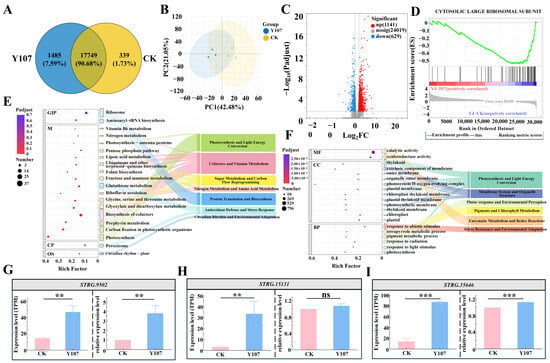
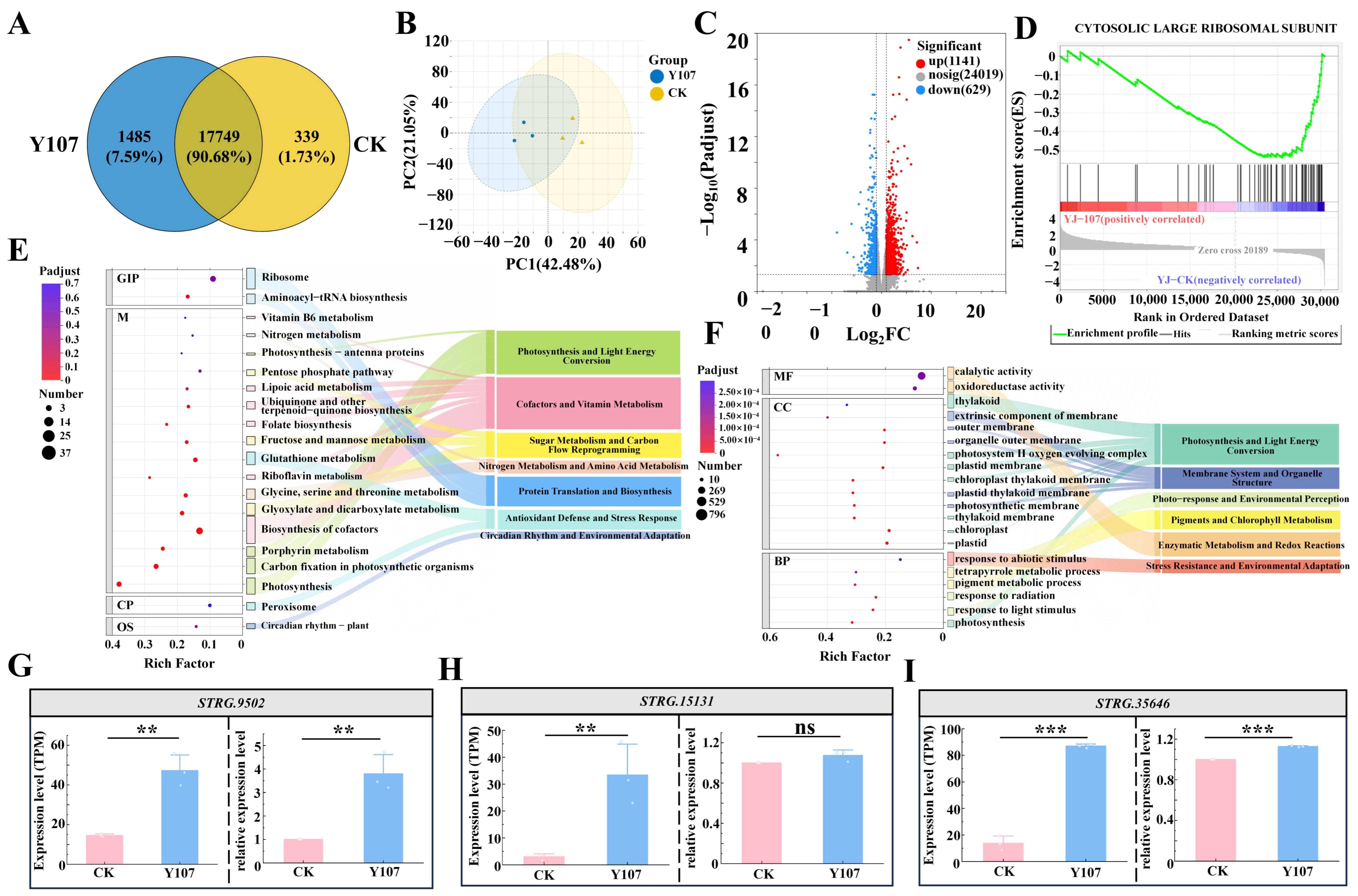
Transcriptome sequencing was performed to elucidate the molecular mechanisms through which strain Y107 affects plant growth and the rhizosphere environment. A Venn diagram of the samples (Figure 4A) illustrates the DEG sets obtained from different comparison combinations, revealing 339, 1485, and 17,749 DEGs unique to each comparison and shared across comparisons, respectively. The PCA results (Figure 4B) revealed that PC1 and PC2 explained 42.48% and 21.05% of the total variation, respectively, indicating tight clustering among all biological replicates and ensuring the reliability of the subsequent differential expression analysis.
Figure 4.
Differential expression analysis and key gene validation of the lingonberry transcriptome regulated by strain Y107. (A) Venn diagram illustrating the unique and shared differentially expressed genes (DEGs) obtained from different comparisons. (B) Principal component analysis (PCA) showing the overall distribution of samples from the two groups and the clustering of biological replicates within each group. (C) Volcano plot showing the expression changes and significance levels of all genes, with red and blue indicating significantly upregulated and downregulated genes, respectively. (D) Gene set enrichment analysis (GSEA) showing significant negative enrichment of the cytosolic large ribosomal subunit-related gene set in the Y107 treatment group. (E) Bubble–Sankey diagram of the results of the GO enrichment analysis showing significantly enriched GO terms and their functional categories. (F) Bubble–Sankey diagram of the results of the KEGG pathway enrichment analysis showing significantly enriched metabolic pathways and their corresponding functional modules. (G–I) Comparison of the expression levels of three key genes (STRG.9502, STRG.15131, and STRG.35646) between the transcriptome data (TPM) and the qPCR results (relative expression levels). Asterisks indicate significant differences compared with the CK group (** p < 0.01, *** p < 0.001). The error bars represent the standard errors.
To systematically identify the transcriptomic changes induced by Y107 treatment, differential expression analysis was performed. A volcano plot (Figure 4C) visually displays the expression changes and significance levels of all the genes. A total of 1770 DEGs were identified, including 1141 significantly upregulated genes and 629 significantly downregulated genes.
After key differentially expressed gene sets were identified, GO and KEGG enrichment analyses were performed, and the results were integrated and visualized using a combined bubble–Sankey diagram to systematically interpret their biological significance. GO enrichment analysis (Figure 4E) revealed that the most enriched GO terms in the Y107 treatment group were associated with photosynthesis and light energy conversion pathways (7 GO terms), membrane systems and organelle structural modules (5 GO terms), and pigment and chlorophyll metabolism modules (2 GO terms). KEGG pathway analysis (Figure 4F) further provided pathway-level support for these functions. The enriched pathways of the DEGs were associated mainly with cofactor and vitamin metabolism modules (6 pathways) and photosynthesis and light energy conversion modules (4 pathways).
To further characterize the host transcriptomic response after Y107 inoculation, gene set enrichment analysis (GSEA) was performed on the basis of the ranked list of differentially expressed genes (Figure 4D). The results revealed that the CYTOSOLIC LARGE RIBOSOMAL SUBUNIT gene set was significantly negatively enriched in the Y107 treatment group. Most of the core enriched genes were concentrated in the CK-associated region, indicating that the overall expression levels of genes related to the cytosolic large ribosomal subunit decreased after Y107 treatment.
To verify the reliability of the transcriptome sequencing data, three candidate genes related to photosynthesis and metabolic regulation (STRG.9502, STRG.15131, and STRG.35646) were selected from the DEGs on the basis of the phenotypic results, which revealed that strain Y107 promoted lingonberry growth. Their expression levels in the Y107 and CK groups were independently validated using RT–qPCR. The results revealed that the expression trends of STRG.9502 (Figure 4G) and STRG.35646 (Figure 4I) were significantly similar between the RNA-seq and RT–qPCR results (p < 0.05). Although the expression trend of STRG.15131 (Figure 4H) was consistent with that observed by RNA-seq, the RT–qPCR results did not significantly differ.
3.5. Correlation Analysis and Mechanistic Insights into the Plant Growth-Promoting Effects of Strain Y107
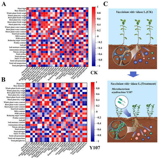
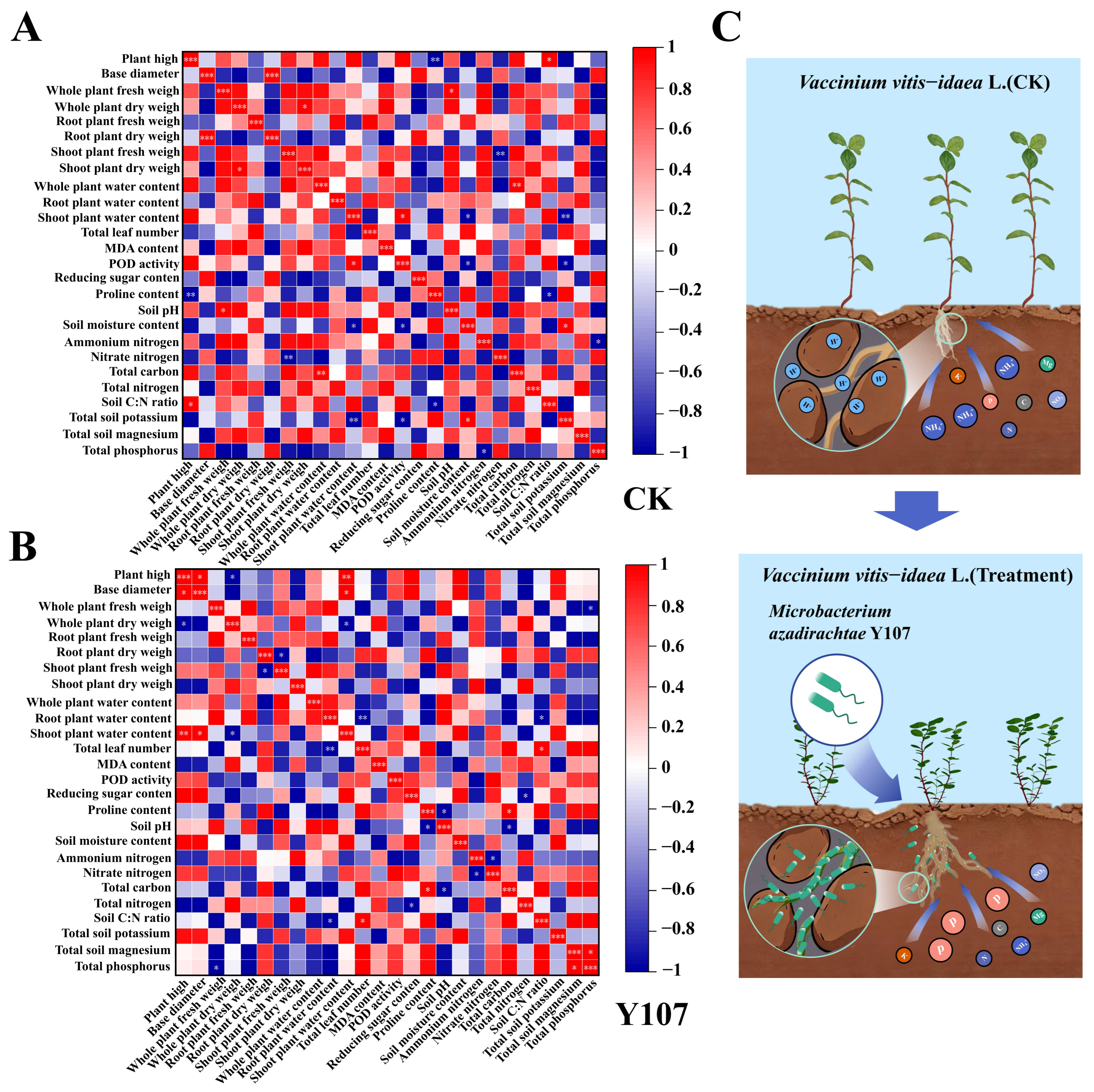
To further investigate the correlations between lingonberry and soil indices under different treatments, the data were analyzed using the Pearson model (Figure 5A,B). In the CK group, whole-plant fresh weight was significantly positively correlated with soil pH (p < 0.05), and whole-plant dry weight was significantly positively correlated with shoot dry weight (p < 0.05). In contrast, shoot plant fresh weight was significantly negatively correlated with soil ammonium nitrogen content (p < 0.01). After Y107 treatment, whole-plant fresh weight was significantly negatively correlated with the soil total phosphorus content (p < 0.05), whereas whole-plant dry weight was significantly negatively correlated with the shoot plant ground water content (p < 0.05).
Figure 5.
Correlation heatmaps of lingonberry growth, physiological, and soil indices and a proposed hypothetical model of Y107-mediated growth promotion. (A,B) Pearson correlation heatmaps showing pairwise correlation coefficients among lingonberry growth indices, physiological indices, and soil factors. Red and blue indicate positive and negative correlations, respectively, and color intensity represents the strength of the correlation. Asterisks indicate significant correlations (* p < 0.05; ** p < 0.01; *** p < 0.001). Pearson correlation analysis was performed on the basis of three independent biological replicates (n = 3). (C) Hypothetical model illustrating the potential physiological and molecular responses of lingonberry to inoculation with Microbacterium sp. Y107, constructed on the basis of the observations obtained in this study.
On the basis of the results of the above correlation analysis, we constructed a working model for the Y107-mediated promotion of biomass accumulation in lingonberry (Figure 5C). Strain Y107 improved rhizosphere nutrient availability, particularly phosphorus activation and uptake, thereby increasing the efficiency of soil nutrient utilization by the plants. As plants absorb more nutrients, the residual nutrient content in the soil decreases, resulting in a negative correlation between the biomass indices and soil nutrient contents. Moreover, the negative correlation between whole-plant dry weight and water content indicated that dry matter accumulation occurred preferentially over water storage under the Y107 treatment, further supporting its growth-promoting effect.
4. Discussion
With the continuous advancement of research on the development and utilization of plant rhizosphere microbial resources, screening functional strains with high growth-promoting capacity has become important for the development of novel microbial inoculants [29]. The results of the present study demonstrated that strain Y107 possesses multiple plant growth-promoting traits, including phosphate solubilization, nitrogen fixation, siderophore production, and IAA secretion, indicating its potential to promote plant growth through diverse complementary mechanisms. Previous studies have shown that the synergistic action of multiple growth-promoting functions is generally more effective than a single mechanism in enhancing plant nutrient acquisition, promoting root development, and improving plant adaptability to environmental stress [30]. In this study, strain Y107 was identified as Microbacterium sp. Y107 on the basis of its morphological characteristics and 16S rDNA sequence analysis. However, taxonomic identification based on 16S rRNA gene sequences has certain limitations in distinguishing closely related species, and further genome-level analysis is needed to determine its precise taxonomic position. GC–MS analysis revealed that strain Y107 can secrete diketopiperazines (DKPs), pyrazines, pyrroles, and other compounds, which have been reported to be involved in microbial communication, quorum sensing, and biofilm formation [31] and thus may facilitate the stable colonization of lingonberry roots by Y107. We broadly categorized these metabolites on the basis of their generally recognized biochemical properties, intending to offer an exploratory visual perspective on the potential functional repertoire of Y107-derived metabolites. While this classification may not be regarded as a definitive ecological functional annotation, it might still provide a useful reference for hypothesis generation in future studies, laying a preliminary foundation for investigating the possible involvement of specific metabolites in rhizosphere interactions. Furthermore, this study revealed that Y107 has a biofilm-forming ability, extracellular polymeric substance secretion capacity, and certain motility. Crystal violet staining of roots also revealed increased staining intensity on the root surface after Y107 inoculation. These results suggest that Y107 possesses potential root-surface adhesion and rhizosphere interaction capabilities. However, since strain-specific labeling methods were not used in this study to track colonization dynamics, the long-term stable colonization ability of Y107 on lingonberry roots cannot be directly confirmed [32].
Moreover, strain Y107 was also found to secrete soluble proteins and soluble sugars, which may be involved in the survival of the strain itself and in rhizosphere interaction processes [33]. However, since the present study did not further determine the activities of relevant extracellular enzymes or their effects on nutrient transformation processes, the direct role of these secretions in soil nutrient cycling remains unclear. Notably, we employed the 3,5-dinitrosalicylic acid (DNS) method to quantify extracellular soluble sugars. This method primarily detects reducing sugars, whereas nonreducing sugars (e.g., sucrose) and polysaccharides are not effectively measured. Therefore, the total amount and composition of soluble sugars secreted by Y107 may have been underestimated. Future studies should adopt complementary analytical approaches, such as high-performance liquid chromatography (HPLC) or enzymatic assays, to obtain a more comprehensive profile of secreted carbohydrates. Nevertheless, owing to the lack of further determination of relevant extracellular enzyme activities and their influence on nutrient transformation processes in this study, the direct role of these secretions in soil nutrient cycling cannot be ascertained. Despite these limitations, inoculation with Y107 led to changes in the concentrations of some soil nutrient elements, accompanied by increased plant growth and root development [34], indicating that Y107 alters the environmental characteristics of the lingonberry rhizosphere and may affect the interactions between plants and soil nutrients. However, given the absence of plant tissue nutrient content and nutrient balance analyses, a decrease in soil nutrient content cannot be directly equated with an improvement in plant nutrient use efficiency, as it may also be related to factors such as increased nutrient uptake during plant growth and changes in microbial activity [35].
After inoculation with strain Y107, the whole-plant fresh weight of the lingonberry plants increased, whereas the total dry weight did not significantly change. Moreover, the root-to-shoot ratio significantly increased, indicating that Y107 treatment altered the biomass allocation pattern of the plants, leading to a trend toward greater biomass accumulation being allocated to belowground parts, thereby promoting root development. This change may also reflect a trade-off in resource allocation within the plant. Given that carbon assimilation products are typically involved in root growth and carbon storage, this phenomenon may be related to the allocation of photosynthates to the root system [36]. However, since the transport process of assimilates was not directly measured in this study, this speculation still requires further verification. The increase in basal diameter reflects an improvement in plant structural growth, which may provide a structural basis for sustained root growth and resource exchange. Nevertheless, the material accumulation process of plants requires a relatively stable internal environment. Y107 treatment significantly increased POD activity in lingonberry, whereas the MDA content did not significantly change, indicating that Y107 induced redox-related responses in the plant and may have enhanced its ability to cope with changes in the oxidative environment [37]. However, elevated POD activity may also reflect a defensive response of the plant upon receiving certain oxidative stimuli; therefore, it cannot be taken alone as direct evidence of enhanced antioxidant capacity. Since ROS accumulation levels, SOD and CAT activities, electrolyte leakage, and photosynthetic parameters were not measured in this study, the specific effects of Y107 on alleviating oxidative damage and regulating photosynthetic function still require further investigation [38]. In addition, the increase in plant water content after Y107 treatment, accompanied by a decrease in proline content, suggests that Y107 may affect plant water status and osmoregulatory processes [39]. However, the direct causal relationship between these two observations remains to be verified. A higher plant water status may help maintain normal cellular physiological functions and provide favorable conditions for plant growth [40].
In summary, strain Y107 promoted the growth of lingonberry and induced multilayered transcriptional responses. To further elucidate the molecular response characteristics of lingonberry after Y107 treatment, we analyzed changes in gene expression using transcriptomic analysis. GO and KEGG enrichment analyses revealed that the DEGs were involved mainly in processes such as photosynthesis, carbon and nitrogen metabolism, redox reactions, and environmental responses, indicating that the expression patterns of metabolism-related genes in lingonberry were altered after Y107 treatment [41]. The enrichment of photosynthesis-related genes and the upregulation of key genes such as psbP and ndhU suggest that strain Y107 may regulate molecular processes associated with photosynthesis in lingonberry [42]. The changes in the expression of genes related to carbon and nitrogen metabolism suggest that strain Y107 may regulate the plant’s responses associated with carbon and nitrogen metabolism [43]. Changes in redox- and environmental response-related genes may be associated with the physiological adaptation of the plant. Further GSEA revealed that the gene set related to the cytosolic large ribosomal subunit was significantly negatively enriched after Y107 treatment, indicating that Y107 affects the ribosome-related expression pattern in lingonberry [44]. Ribosome-related processes are involved in the regulation of protein synthesis in plants, suggesting that the translation process in lingonberry may change after treatment with strain Y107. However, the specific physiological significance of these changes requires further experimental validation. The qRT–PCR results were consistent with the RNA–seq expression trends, further confirming the reliability of the transcriptomic data. The significant upregulation of key photosynthesis-related genes such as psbP and ndhU further supports that strain Y107 affects the expression of photosynthesis-related genes in lingonberry [45]. These molecular-level changes are consistent with the aforementioned results, including enhanced root development, altered soil nutrient utilization characteristics, increased biomass accumulation, and increased antioxidant capacity, collectively revealing the potential regulatory mechanisms underlying the physiological and molecular responses by which strain Y107 promotes lingonberry growth.
Furthermore, correlation analysis revealed changes in the associations between lingonberry growth parameters and soil nutrient factors following Y107 treatment. In the CK treatment, lingonberry growth parameters were significantly correlated with certain soil environmental factors, indicating that plant growth may be influenced by rhizosphere environmental conditions [46]. Following Y107 treatment, whole-plant fresh weight was significantly negatively correlated with soil total phosphorus content, accompanied by increased root development and decreased soil total phosphorus content, indicating that Y107 treatment may have altered the association pattern between plant growth and soil nutrient status [47]. However, since direct measurements of plant tissue nutrient contents were not conducted in this study, the above results indicate only that strain Y107 may influence rhizosphere nutrient dynamics and plant growth responses, and its role in specific nutrient uptake processes requires further investigation. These findings are consistent with the changes in the expression of genes related to carbon and nitrogen metabolism observed in the transcriptomic analysis, collectively suggesting that Y107 may be involved in regulating the metabolic response processes of lingonberry. The correlation results obtained in this study reflect preliminary trends in the associations among variables, which may serve as directional references for future investigations, and their consistency warrants further validation under broader experimental conditions.
5. Conclusions
This study demonstrated that Microbacterium sp. Y107 promotes lingonberry growth and influences soil nutrient characteristics as well as host gene expression responses. Y107 inoculation significantly increased root development and biomass accumulation, with the fresh weight of roots increasing by 157.65% and the dry weight of roots increasing by 32.89%. It also improved the plant’s antioxidant defense capacity (POD activity increased by 61.11%) and tissue water content. Furthermore, strain Y107 formed biofilms on the surface of lingonberry roots and altered the content profiles of multiple nutrient indices in the rhizosphere soil, indicating its ability to modulate the nutritional structure of the root microenvironment. Transcriptomic analysis revealed that strain Y107 induced changes in the expression of genes associated with photosynthesis and carbon–nitrogen metabolism and resulted in negative enrichment of the cytoplasmic ribosome large subunit gene set, suggesting that strain Y107 may influence the metabolic regulatory network and transcriptional response processes of lingonberry, thereby promoting plant growth. Correlation analysis further indicated that strain Y107 altered the association patterns between lingonberry growth parameters and soil nutrient factors. Overall, this study elucidates the physiological and molecular response characteristics underlying the growth-promoting effects of Microbacterium sp. Y107 on lingonberry, providing a theoretical basis for functional microbial resources in Ericaceae plants. Although the growth-promoting effects of strain Y107 were validated in an axenic system in this study, its long-term colonization ability, ecological adaptability, and field application efficacy in complex soil environments remain to be further investigated.
Supplementary Materials
The following supporting information can be downloaded at: https://www.mdpi.com/article/10.3390/horticulturae12081027/s1, Figure S1: Whole-plant dry weight; Figure S2: Root-to-shoot ratio; Figure S3: Soil moisture content. Table S1: GC–MS identification of secreted metabolites produced by strain Y107.
Author Contributions
E.Y.: Investigation, Formal analysis, Data curation, Writing—original draft, Visualization. Y.W.: Investigation, Formal analysis, Data curation, Writing—original draft, Visualization. Y.L.: Methodology, Investigation. J.L.: Methodology, Data curation. Z.L.: Methodology, Investigation. F.B.: Investigation, Data curation. H.P.: Methodology. X.W.: Data curation. D.T.: Investigation. H.L.: Conceptualization, Methodology, Writing—review & editing, Supervision, Funding acquisition. J.Z.: Conceptualization, Methodology, Writing—review & editing, Supervision, Funding acquisition. All authors have read and agreed to the published version of the manuscript.
Funding
This research was funded by the National Natural Science Foundation of China (32271921 and 32401583) and the Natural Science Foundation of Heilongjiang Province (PL2025C072).
Data Availability Statement
The original contributions presented in this study are included in the article. Further inquiries can be directed to the corresponding authors.
Acknowledgments
The authors would like to acknowledge the invaluable support of Northeast Forestry University in this study.
Conflicts of Interest
The authors declare no conflicts of interest.
References
- Baldwin, T.; Ahlquist, L.; Pop, E.; Parker La Puma, I.; Starr, G.; Oberbauer, S.; Khorsand Rosa, R. Plant phenological responses to a long-term experimental extension of growing season and soil warming in the tussock tundra of alaska. Glob. Change Biol. 2015, 21, 4520–4532. [Google Scholar] [CrossRef] [Scilit] [PubMed]
- Kowalska, K. Lingonberry (Vaccinium vitis-idaea L.) Fruit as a source of bioactive compounds with health-promoting effects—A review. Int. J. Mol. Sci. 2021, 22, 5126. [Google Scholar] [CrossRef] [Scilit] [PubMed]
- Pärnänen, P.; Niikko, S.; Lähteenmäki, H.; Räisänen, I.T.; Tervahartiala, T.; Sorsa, T.; Ranki, A. Lingonberry (Vaccinium vitis-idaea L.) Fruit phenolic bioactivities—A review of in vitro and in vivo human studies. Microorganisms 2024, 12, 1850. [Google Scholar] [CrossRef] [Scilit] [PubMed]
- Sharifi, M.; Debnath, S.C.; Hajiaghaei-Kamrani, M.; Rabie, B.; Forsyth, J. Growing media ph and nutrient concentrations for fostering the propagation and production of lingonberry (Vaccinium vitis-idaea L.). Agronomy 2024, 14, 2553. [Google Scholar] [CrossRef] [Scilit]
- Zhang, S.; Li, F.; Chang, L.; Zhang, Q.; Zhang, Z.; Wu, T.; Wu, Y.; Wang, Z.; Su, Y.; Yang, X.; et al. Broad-spectrum applications of plant growth-promoting rhizobacteria (PGPR) across diverse crops and intricate planting systems. Microbiol. Spectr. 2025, 13, e0187924. [Google Scholar] [CrossRef] [Scilit] [PubMed]
- Chieb, M.; Gachomo, E.W. The role of plant growth promoting rhizobacteria in plant drought stress responses. BMC Plant Biol. 2023, 23, 407. [Google Scholar] [CrossRef] [Scilit] [PubMed]
- Wahab, A.; Batool, F.; Abdi, G.; Muhammad, M.; Ullah, S.; Zaman, W. Role of plant growth-promoting rhizobacteria in sustainable agriculture: Addressing environmental and biological challenges. J. Plant Physiol. 2025, 307, 154455. [Google Scholar] [CrossRef] [Scilit] [PubMed]
- An, P.; Yang, C.; Li, W.; Zhao, D.; Xiang, H. The isolation and characterization of a novel psychrotolerant cellulolytic bacterium, Microbacterium sp. QXD-8t. Microorganisms 2024, 12, 303. [Google Scholar] [CrossRef] [Scilit] [PubMed]
- Corretto, E.; Antonielli, L.; Sessitsch, A.; Höfer, C.; Puschenreiter, M.; Widhalm, S.; Swarnalakshmi, K.; Brader, G. Comparative genomics of Microbacterium species to reveal diversity, potential for secondary metabolites and heavy metal resistance. Front. Microbiol. 2020, 11, 2020. [Google Scholar] [CrossRef] [Scilit] [PubMed]
- Zheng, J.; Liao, Y.; Li, Y.; Li, D.; Sun, Y.; Xiao, Z. Description of Microbacterium dauci sp. Nov., A plant growth hormone indoleacetic acid-producing and nitrogen-fixing bacterium isolated from carrot rhizosphere soil. Arch. Microbiol. 2024, 206, 102. [Google Scholar] [CrossRef] [Scilit] [PubMed]
- Kubov, M.; Fleischer, P.; Tomes, J.; Mukarram, M.; Janík, R.; Turyasingura, B.; Fleischer, P.; Schieber, B. Differential responses of bilberry (Vaccinium myrtillus) phenology and density to a changing environment: A study from western carpathians. Plants 2024, 13, 2406. [Google Scholar] [CrossRef] [Scilit] [PubMed]
- Lou, H.; Cai, H.; Fu, R.; Guo, C.; Fan, B.; Hu, H.; Zhang, J.; Sun, L. Effects of wildfire disturbance on forest soil microbes and colonization of ericoid mycorrhizal fungi in northern China. Environ. Res. 2023, 231, 116220. [Google Scholar] [CrossRef] [Scilit] [PubMed]
- Costa-Catala, J.; Bori, J.; Veciana-Nogués, M.T.; Latorre-Moratalla, M.L.; Vidal-Carou, M.C.; Comas-Basté, O. Influence of seed disinfection treatments on the germination rate and histamine-degrading activity of legume sprouts. Foods 2024, 13, 4105. [Google Scholar] [CrossRef] [Scilit] [PubMed]
- Duc, H.D.; Oanh, N.T. Composition of bacterial community and isolation of bacteria responsible for diuron degradation in sediment and soil under anaerobic condition. Arch. Microbiol. 2022, 204, 418. [Google Scholar] [CrossRef] [Scilit] [PubMed]
- Kumaishi, K.; Usui, E.; Suzuki, K.; Kobori, S.; Sato, T.; Toda, Y.; Takanashi, H.; Shinozaki, S.; Noda, M.; Takakura, A.; et al. High throughput method of 16 s rRNA gene sequencing library preparation for plant root microbial community profiling. Sci. Rep. 2022, 12, 19289. [Google Scholar] [CrossRef] [Scilit] [PubMed]
- Dutta, J.; Thakur, D. Diversity of culturable bacteria endowed with antifungal metabolites biosynthetic characteristics associated with tea rhizosphere soil of assam, india. BMC Microbiol. 2021, 21, 216. [Google Scholar] [CrossRef] [Scilit] [PubMed]
- Sritongon, N.; Boonlue, S.; Mongkolthanaruk, W.; Jogloy, S.; Riddech, N. The combination of multiple plant growth promotion and hydrolytic enzyme producing rhizobacteria and their effect on jerusalem artichoke growth improvement. Sci. Rep. 2023, 13, 5917. [Google Scholar] [CrossRef] [Scilit] [PubMed]
- Ma, Y.; Gao, Y.; Xu, Y.; Zhou, H.; Zhou, K.; Li, C.; Xu, B. Microbiota dynamics and volatile metabolite generation during sausage fermentation. Food Chem. 2023, 423, 136297. [Google Scholar] [CrossRef] [Scilit] [PubMed]
- Yu, Q.; Liu, S.; Yu, L.; Xiao, Y.; Zhang, S.; Wang, X.; Xu, Y.; Yu, H.; Li, Y.; Yang, J.; et al. RNA demethylation increases the yield and biomass of rice and potato plants in field trials. Nat. Biotechnol. 2021, 39, 1581–1588. [Google Scholar] [CrossRef] [Scilit] [PubMed]
- Forlani, G.; Funck, D. A specific and sensitive enzymatic assay for the quantitation of l-proline. Front. Plant Sci. 2020, 11. [Google Scholar] [CrossRef] [Scilit] [PubMed]
- King, B.C.; Donnelly, M.K.; Bergstrom, G.C.; Walker, L.P.; Gibson, D.M. An optimized microplate assay system for quantitative evaluation of plant cell wall-degrading enzyme activity of fungal culture extracts. Biotechnol. Bioeng. 2009, 102, 1033–1044. [Google Scholar] [CrossRef] [Scilit] [PubMed]
- Sobahan, M.A.; Akter, N.; Karim, M.M.; Badhon, M.M.I.; Khan, S.N.; Alam, S.; Prasad, P.V.V.; Hasanuzzaman, M. Bioaugmentation with plant growth-promoting rhizobacteria alleviates chromium and salt stress in rice through the improvement of physiology, ion homeostasis, and antioxidant defense. Microorganisms 2025, 13, 1462. [Google Scholar] [CrossRef] [Scilit] [PubMed]
- Mishra, N.; Jiang, C.; Chen, L.; Paul, A.; Chatterjee, A.; Shen, G. Achieving abiotic stress tolerance in plants through antioxidative defense mechanisms. Front. Plant Sci. 2023, 14, 1110622. [Google Scholar] [CrossRef] [Scilit] [PubMed]
- De Morais, C.P.; Mcmeekin, K.; Nault, C. Scalable solution for agricultural soil organic carbon measurements using laser-induced breakdown spectroscopy. Sci. Rep. 2024, 14, 15272. [Google Scholar] [CrossRef] [Scilit] [PubMed]
- Qin, G.; Zou, K.; He, F.; Shao, J.; Zuo, B.; Liu, J.; Liu, R.; Yang, B.; Zhao, G. Simultaneous determination of volatile phenol, cyanide, anionic surfactant, and ammonia nitrogen in drinking water by a continuous flow analyzer. Sci. Rep. 2023, 13, 1829. [Google Scholar] [CrossRef] [Scilit] [PubMed]
- Han, B.; Gao, W.; Feng, J.; Iroshan, A.; Yang, J.; Chen, G.; Zhang, Y.; Aizezi, N.; Liu, Y. Laser-induced breakdown spectroscopy for imaging and distribution analysis of heavy metal elements in soil. J. Hazard. Mater. 2025, 496, 139284. [Google Scholar] [CrossRef] [Scilit] [PubMed]
- Kundu, S.; Majumder, S.; Barua, R.; Marshall, H.D.; Igamberdiev, A.U.; Debnath, S.C. From disorder to recovery: Unraveling hyperhydricity insights in Vaccinium vitis-idaea L. Through phytochemical and gene expression studies. Vitr. Cell. Dev. Biol.-Plant 2026, 62, 418–432. [Google Scholar] [CrossRef] [Scilit]
- Bielecka, M.; Pencakowski, B.; Stafiniak, M.; Kozłowska, W.; Dziwak, M.; Nowis, K.; Łaczmański, Ł.; Matkowski, A. Reference gene stability in Agrostemma githago using quantitative real-time PCR. Int. J. Mol. Sci. 2026, 27, 889. [Google Scholar] [CrossRef] [Scilit] [PubMed]
- Swarnalakshmi, K.; Yadav, V.; Tyagi, D.; Dhar, D.W.; Kannepalli, A.; Kumar, S. Significance of plant growth promoting rhizobacteria in grain legumes: Growth promotion and crop production. Plants 2020, 9, 1596. [Google Scholar] [CrossRef] [Scilit] [PubMed]
- Kulkova, I.; Dobrzyński, J.; Kowalczyk, P.; Bełżecki, G.; Kramkowski, K. Plant growth promotion using Bacillus cereus. Int. J. Mol. Sci. 2023, 24, 9759. [Google Scholar] [CrossRef] [Scilit] [PubMed]
- Huang, Z.; Yu, X.; Ma, Y.; Wang, J.; Zheng, Y. Biodegradation of polycyclic aromatic hydrocarbons enhanced by a newly isolated Acinetobacter sp. YY-1 and exogenous quorum sensing signaling molecule n-acyl-homoserine lactones. Bioresour. Technol. 2025, 433, 132718. [Google Scholar] [CrossRef] [Scilit] [PubMed]
- Corral, J.; Sebastià, P.; Coll, N.S.; Barbé, J.; Aranda, J.; Valls, M.; Bowman, G.R. Twitching and swimming motility play a role in Ralstonia solanacearum pathogenicity. mSphere 2020, 5, e00740–19. [Google Scholar] [CrossRef] [Scilit] [PubMed]
- Yang, M.; Qin, X.; Yue, C.; Wu, Q.; Tian, P.; Wu, Z.; Wei, X.; Wu, L. Phosphate-solubilizing and polymerizing bacteria enhance phosphorus availability and growth of rice. Front. Microbiol. 2025, 16, 2025. [Google Scholar] [CrossRef] [Scilit] [PubMed]
- Rawat, S.S.; Laxmi, A. Rooted in communication: Exploring auxin–salicylic acid nexus in root growth and development. Plant Cell Environ. 2025, 48, 4140–4160. [Google Scholar] [CrossRef] [Scilit] [PubMed]
- Gupta, R.; Khan, F.; Alqahtani, F.M.; Hashem, M.; Ahmad, F. Plant growth–promoting rhizobacteria (PGPR) assisted bioremediation of heavy metal toxicity. Appl. Biochem. Biotechnol. 2024, 196, 2928–2956. [Google Scholar] [CrossRef] [Scilit] [PubMed]
- Sun, J.; Zhang, Z.; Zhang, S.; Dan, Y.; Sun, H.; Wu, Y.; Luan, G.; Lu, X. Engineering cyanobacterial cell factories for photosynthetic production of fructose. ACS Synth. Biol. 2023, 12, 3008–3019. [Google Scholar] [CrossRef] [Scilit] [PubMed]
- Waheed, Z.; Iqbal, S.; Irfan, M.; Jabeen, K.; Umar, A.; Aljowaie, R.M.; Almutairi, S.M.; Gancarz, M. Pseudochrobactrum asaccharolyticum mitigates arsenic induced oxidative stress of maize plant by enhancing water status and antioxidant defense system. BMC Plant Biol. 2024, 24, 832. [Google Scholar] [CrossRef] [Scilit] [PubMed]
- Antoniou, C.; Chatzimichail, G.; Xenofontos, R.; Pavlou, J.J.; Panagiotou, E.; Christou, A.; Fotopoulos, V. Melatonin systemically ameliorates drought stress-induced damage in Medicago sativa plants by modulating nitro-oxidative homeostasis and proline metabolism. J. Pineal Res. 2017, 62, e12401. [Google Scholar] [CrossRef] [Scilit] [PubMed]
- Zegaoui, Z.; Planchais, S.; Cabassa, C.; Djebbar, R.; Belbachir, O.A.; Carol, P. Variation in relative water content, proline accumulation and stress gene expression in two cowpea landraces under drought. J. Plant Physiol. 2017, 218, 26–34. [Google Scholar] [CrossRef] [Scilit] [PubMed]
- Salvi, A.M.; Smith, D.D.; Adams, M.A.; Mcculloh, K.A.; Givnish, T.J. Mesophyll photosynthetic sensitivity to leaf water potential in eucalyptus: A new dimension of plant adaptation to native moisture supply. New Phytol. 2021, 230, 1844–1855. [Google Scholar] [CrossRef] [Scilit] [PubMed]
- Zhou, Z.; Tran, P.Q.; Cowley, E.S.; Trembath-Reichert, E.; Anantharaman, K. Diversity and ecology of microbial sulfur metabolism. Nat. Rev. Microbiol. 2025, 23, 122–140. [Google Scholar] [CrossRef] [Scilit] [PubMed]
- Leschevin, M.; Ksas, B.; Baltenweck, R.; Hugueney, P.; Caffarri, S.; Havaux, M. Photosystem rearrangements, photosynthetic efficiency, and plant growth in far red-enriched light. Plant J. 2024, 120, 2536–2552. [Google Scholar] [CrossRef] [Scilit] [PubMed]
- Zuo, H.; Shen, H.; Dong, S.; Guo, Q.; Yamori, W.; Liu, J.; Zheng, H.; He, F.; Zhang, Y.; Zhang, R.; et al. Photosynthetic carbon reallocation to nitrogen metabolism confers adaptation advantage of leymus secalinus under elevated nitrogen deposition in alpine grassland. Plant Cell Environ. 2026, 49, 2116–2133. [Google Scholar] [CrossRef] [Scilit] [PubMed]
- Siodmak, A.; Martinez-Seidel, F.; Rayapuram, N.; Bazin, J.; Alhoraibi, H.; Gentry-Torfer, D.; Tabassum, N.; Sheikh, A.H.; Kise, J.K.G.; Blilou, I.; et al. Dynamics of ribosome composition and ribosomal protein phosphorylation in immune signaling in Arabidopsis thaliana. Nucleic Acids Res. 2023, 51, 11876–11892. [Google Scholar] [CrossRef] [Scilit] [PubMed]
- Ido, K.; Nield, J.; Fukao, Y.; Nishimura, T.; Sato, F.; Ifuku, K. Cross-linking evidence for multiple interactions of the PsbP and PsbQ proteins in a higher plant photosystem II supercomplex. J. Biol. Chem. 2014, 289, 20150–20157. [Google Scholar] [CrossRef] [Scilit] [PubMed]
- Zhu, C.; Wu, Y.; Ding, Z.; Wu, J.; Zhang, L.; Liu, C. Electrochemical strategy based on iron oxide promotes in situ nitrate reduction to ammonium for nitrogen uptake by rice. J. Hazard. Mater. 2026, 511, 142067. [Google Scholar] [CrossRef] [Scilit] [PubMed]
- Aslam, M.M.; Pueyo, J.J.; Pang, J.; Yang, J.; Chen, W.; Chen, H.; Waseem, M.; Li, Y.; Zhang, J.; Xu, W. Root acid phosphatases and rhizobacteria synergistically enhance white lupin and rice phosphorus acquisition. Plant Physiol. 2022, 190, 2449–2465. [Google Scholar] [CrossRef] [Scilit] [PubMed]
Disclaimer/Publisher’s Note: The statements, opinions and data contained in all publications are solely those of the individual author(s) and contributor(s) and not of MDPI and/or the editor(s). MDPI and/or the editor(s) disclaim responsibility for any injury to people or property resulting from any ideas, methods, instructions or products referred to in the content. |
© 2026 by the authors. Licensee MDPI, Basel, Switzerland. This article is an open access article distributed under the terms and conditions of the Creative Commons Attribution (CC BY) license.