


Abstract
Grapes are among the most widely cultivated fruit crops globally, yet their growth and yield are severely compromised by Colletotrichum viniferum, which causes a devastating disease that affects grape berries. The wall-associated kinase (WAK) gene family, a unique subfamily of receptor-like-kinases (RLKs), plays important roles in mediating plant responses to both abiotic and biotic stresses. However, the expression patterns and biological functions of grape WAKs in response to C. viniferum infection remain largely uncharacterized. In this study, a total of 57 VdWAK genes were identified and phylogenetically classified into twelve subgroups. Chromosomal localization and collinearity analyses further revealed that tandem duplication and segmental duplication events contributed to the expansion of the VdWAK gene family. Transcriptomic profiling identified VdWAK19 as a key responsive gene. It was predominantly expressed in mature berries but transcriptionally repressed upon C. viniferum infection. Virus-induced gene silencing assays in grape berries demonstrated that knockdown of VdWAK19 significantly reduced fruit resistance to C. viniferum infection. Overall, these findings advance our understanding of the functional roles of VdWAK genes during C. viniferum infection and provide a theoretical basis for the potential application of VdWAK19 in breeding grape varieties with enhanced resistance to ripe rot.
1. Introduction
Wall-associated kinases (WAKs) represent a distinct subfamily of receptor-like kinases (RLKs). Endowed with a unique structure that enables direct binding to cell wall components, WAKs act as critical signaling “bridges” linking the cell wall and cytoplasm, playing essential roles in regulating cell growth and mediating stress defense responses. WAKs typically consist of three parts: an extracellular ligand-binding domain, a hydrophobic transmembrane domain, and a cytoplasmic kinase catalytic domain [1]. The Arabidopsis thaliana AtWAK family comprises 33 members in total; their sequence divergence is mainly reflected in the extracellular domains, where amino acid homology ranges from 40% to 64%, whereas the homology of intracellular kinase domains is as high as 86% [2]. This divergence indicates that extracellular ligand-binding domains may bind to distinct ligands to transduce diverse environmental signals.
To date, genes of the plant WAK family have been identified in various economic crops, including apple [3], poplar [4] and rice [5]. The roles of WAKs in plant responses to fungal infection have also been well demonstrated. For instance, the tomato SlWak1 protein forms a functional complex with the pattern recognition receptors FlS2 and FlS3, exerting a significant regulatory role during the subsequent phases of pattern-triggered immunity (PTI) in the apoplastic compartment [6]. In cotton, GhWAK7A positively regulates the response to fungal wilt infection by forming a complex with chitin receptors [7]. In rice, OsWAK1 and OsWAK91 positively regulated defense against rice blast and sheath blight, respectively [8,9]. GmWAK1 in soybean plays a positive role in resistance to Phytophthora sojae, potentially via the salicylic acid pathway [10]. In maize, ZmWAK02 confers strong resistance to gray leaf spot and has been confirmed to hold great value in maize breeding [11]. These studies suggest that WAK genes are essential for plant defense against pathogen infection.
Grape is one of the most widely cultivated fruits globally, and its growth and yield are highly susceptible to Colletotrichum spp., the causal agent of grape ripe rot [12]. This pathogen exists in nearly all vineyards and can cause yield losses of up to 20% or even more [13]. Grape ripe rot, triggered by C. viniferum, ranks among the most important fungal diseases affecting grape fruit quality; it can also result in sunken necrotic lesions on stems, flowers, and leaf clusters [14]. Fruit defense against pathogenic infection is a complex biological process involving the coordinated regulation of multiple signaling pathways and disease-resistant genes [15]. As key mediators of cell wall-environment interactions, the WAK genes play crucial roles in initiating defense responses, yet the specific functions of WAK genes in grape responses to C. viniferum infection remain unclear. In this study, we systematically analyzed the conserved protein domains, gene structures, phylogenetic relationships, collinearity patterns, and promoter cis-acting elements of the VdWAK gene family in Vitis davidii. Furthermore, we investigated the expression patterns of VdWAK genes across different tissues and during C. viniferum infection using transcriptome data and real-time quantitative PCR (RT-qPCR) assays. Through these analyses, VdWAK19 was identified as a candidate gene that exhibits significant differential expression in fruit tissues and in response to pathogen infection. Finally, the virus-induced gene silencing (VIGS) technology was employed to silence VdWAK19 in grape fruits, thereby verifying its function in disease resistance. These findings provide a theoretical basis for elucidating the function of WAKs underlying grape resistance to C. viniferum infection, and lay a foundation of genetic resources for breeding new disease-resistant grape varieties.
2. Materials and Methods
2.1. Identification of the VdWAK Family in Vitis davidii
The genomic data of V. davidii were obtained from Figshare public database (DOI: 10.6084/m9.figshare.31841497, Supplementary Text S1). The WAK protein domain GUB_WAK_bind (PF13947) was obtained from the PFAM database. HMMER3.0 was used to search for putative WAK proteins encoded by the V. davidii genome. Additionally, a BLAST (v2.16.0) comparison was performed against Arabidopsis thaliana WAK family members, and grape genes with an e-value < 1 × 10⁻⁵ were retained. The union of the HMMER search results and BLAST results was then constructed. Finally, the complete GUB_WAK_bind domain was verified using the SMART database (http://smart.embl-heidelberg.de/, accessed on 5 December 2025), the Pfam database (http://pfam.xfam.org/, accessed on 8 December 2025), InterPro, and NCBI CDD [16,17]. The physicochemical properties of VdWAK proteins (isoelectric point and molecular weight) were determined using the ProtParam (https://web.expasy.org/protparam/, accessed on 16 December 2025) [18]. The CELLO program (http://cello.life.nctu.edu.tw/cello2go/, accessed on 31 December 2025) was used to predict the subcellular localization of each VdWAK protein [19].
2.2. Phylogenetic, Conserved Motif, and Gene Structure Analyses
For phylogenetic analysis, the WAK proteins of V. davidii, Oryza sativa, and Aarabidopsis thaliana were aligned. A phylogenetic tree was then reconstructed using MEGA 7.0 via the neighbor-joining method with 1000 bootstrap replicates. Conserved motifs of VdWAK proteins were identified with the online MEME tool (https://meme-suite.org/meme/, accessed on 16 December 2025) by setting the number of motifs to 10, and the results were visualized with the TBtools (V2) software. The gene structures of VdWAKs were analyzed based on the genome annotation file. The 2000 bp upstream sequences of VdWAKs were extracted from the genome and then submitted to the PlantCARE website (http://bioinformatics.psb.ugent.be/webtools/plantcare/html/, accessed on 22 December 2025) to predict their cis-acting elements [20]. Genome annotation files and whole-genome sequences for A. thaliana, Ananas comosus, Malus domestica, and O. sativa were downloaded from the JGI database. MCScanX software (1.0.0) was used to construct collinearity relationships between VdWAK genes and their orthologs in the aforementioned species, and TBtools was used for visualization [21,22].
2.3. Expression Profiles of VdWAK Genes
The expression profiles of VdWAK genes across different tissues (mature fruit, stem, tendril, young leaf, and mature leaf) and at different time points post C. viniferum infection (0 dpi, 1 dpi, 2 dpi, 4 dpi, 6 dpi) were determined by analyzing our previous RNA-seq data (NCBI BioProject ID. PRJNA1414223 and BioProject ID. PRJNA952825) [23]. Briefly, raw transcriptome reads were pruned with Trim Galore (0.6.10), followed by alignment to the V. davidii reference genome with Hisat2 [24]. To analyze the expression profiles of VdWAK genes in various tissues, differential expression analysis was conducted in edgeR by applying a fixed dispersion value of 0.1, as no biological replicates were available. Raw counts were obtained via the prepDE.py script of StringTie (v2.1.5). Differential expression analysis of VdWAK genes during C. viniferum infection was performed using DESeq2 (v1.46.0) in R project (R 4.4.0). p-values were adjusted using the Benjamini–Hochberg method for multiple testing correction. The DEGs were identified by |log2(fold change)| ≥ 1.5 and padj ≤ 0.05. Heatmaps illustrating the expression modes of VdWAK genes were generated using TBtools with TPM (Transcripts Per Million) values [22].
2.4. Expression Validation of VdWAKs via Real-Time Quantitative PCR
Total RNA was isolated from grape berries using Biozol RNA Reagent (Magen, Guangzhou, China). First-strand cDNA was generated from 1 μg of total RNA using the PrimeScript RT Reagent Kit with gDNA Eraser (Takara, Dalian, China). Real-time quantitative PCR (RT-qPCR) analysis was performed on a Bio-Rad IQ™5 Real-Time PCR System (Bio-Rad, Hercules, CA, USA). The VdActin gene was used as the internal reference gene, and the relative expression levels of VdWAK genes were normalized to those of VdActin. All experiments were carried out with three biological repeats. The primers used in this study are listed in Supplementary Table S1.
2.5. Silencing of VdWAK19 in Grapes
A specific fragment of VdWAK19 was amplified by PCR from the cDNA library of V. davidii ‘Fuan’. Meanwhile, the homologous VvWAK19 sequence was cloned from the cDNA library of V. labrusca × vinifera ‘Kyoho’, and the sequence identity between VdWAK19 and VvWAK19 was determined. VdWAK19 was then subcloned into the pDONR207 vector, and subsequently recombined into the PYL279 vector using the Gateway system (Invitrogen, Carlsbad, CA, USA). The virus-induced gene silencing (VIGS) assay was conducted following the standardized protocol established by Wang et al. [23]. In brief, the TRV::VdWAK19 and TRV::00 constructs were separately transformed into Agrobacterium tumefaciens (GV3101), and then, mixed individually with the PYL192 construct. Then, different combinations of Agrobacterium were co-infiltrated into nearly mature ‘Kyoho’ grape berries under vacuum conditions (0.05 MPa, 10 min) using a vacuum pump. Fruits infiltrated with TRV::00 (empty vector) served as the negative control.
2.6. Statistical Analysis
All statistical analyses in this work were performed using SPSS Statistics 24.0 software (IBM, New York, NY, USA). Student’s t-test was used to determine the statistical significance of differences between groups.
3. Results
3.1. Genome-Wide Identification of the VdWAK Family Genes in V. davidii
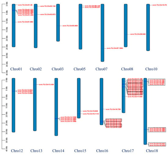
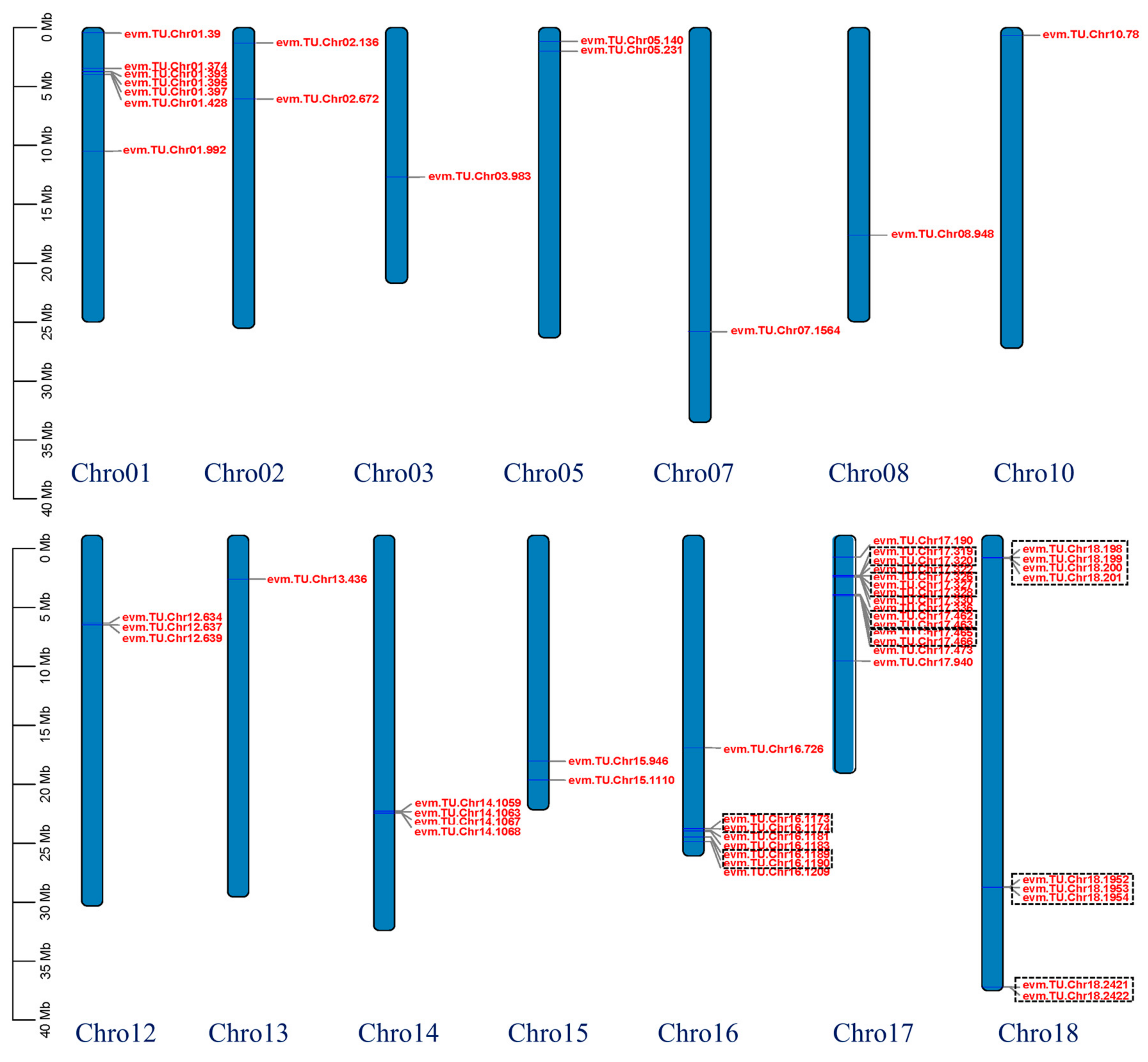
In this study, genome-wide identification and bioinformatics analysis revealed 57 WAK family members in V. davidii (Table 1), which were named according to their chromosomal distribution. Specifically, the coding sequence lengths of VdWAK genes varied from 591 to 3441 bp, encoding proteins of 196 to 1146 amino acids. The molecular weights and isoelectric points of VdWAK proteins varied from 22.12 to 127.49 kDa and 4.44 to 9.53, respectively. Subcellular localization prediction indicated that VdWAK proteins are distributed across multiple subcellular compartments, including the plasma membrane, vacuole, extracellular space, chloroplast, nucleus, endoplasmic reticulum, golgi apparatus, and cytoplasm. As shown in Figure 1, the 57 VdWAK genes exhibited uneven chromosomal distribution across 14 of the 19 chromosomes in the V. davidii genome. Chromosome 17 harbored the largest number of VdWAK genes (15 members), followed by chromosomes 18 and 16 with 9 and 8 members, respectively. The remaining VdWAK genes were distributed across the other 11 chromosomes. Furthermore, the VdWAK gene family contains a cluster of ten tandemly arrayed paralogs. Specifically, one, two, four, and three VdWAK tandem duplication clusters were identified on chromosomes 14, 16, 17, and 18, respectively.
Table 1.
The detailed information of VdWAK genes in Vitis davidii.
Figure 1.
Chromosomal localization of VdWAK genes in Vitis davidii. The red box represented a tandem repeat cluster.
3.2. Phylogenetic Analyses of VdWAKs
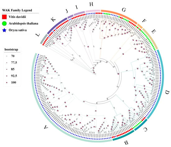
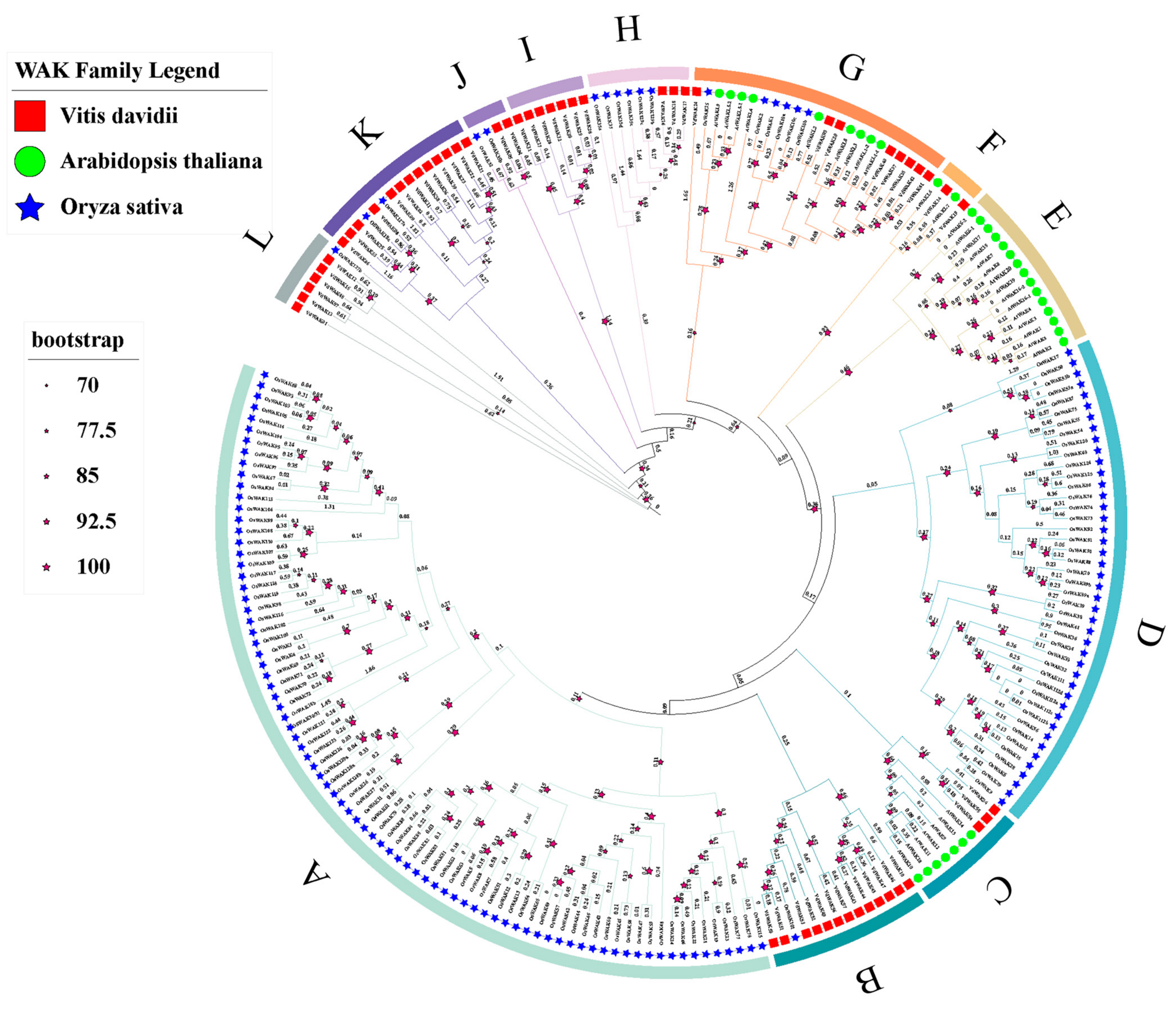
To investigate the evolution relationships of WAKs among V. davidii, O. sativa, and A. thaliana, we reconstructed a phylogenetic tree using the neighbor-joining method with 57 VdWAKs, 129 OsWAKs and 33 AtWAKs (Figure 2). As shown in Figure 2, all 214 WAKs were classified into 12 groups (A to L). The 57 VdWAKs were unevenly clustered into nine groups (B, C, F, G, H, I, J, K, L). Group A and Group D exclusively contained rice OsWAKs, while group E solely included Arabidopsis AtWAKs, and group I harbored only grape VdWAKs. This distribution suggests that these WAK genes have undergone species-specific expansion.
Figure 2.
Phylogenetic analysis of WAK gene families from grape, rice, and Arabidopsis. The phylogenetic tree was constructed with MEGA 7.0 via the Neighbor-Joining (NJ) method with 1000 bootstrap replicates, based on multiple sequence alignments of the full-length amino acid sequences of 57 VdWAK genes from V. davidii, 129 OsWAK genes from rice and 33 AtWAK genes from Arabidopsis.
3.3. Conserved Motifs and Exon-Intron Structures of VdWAK Genes
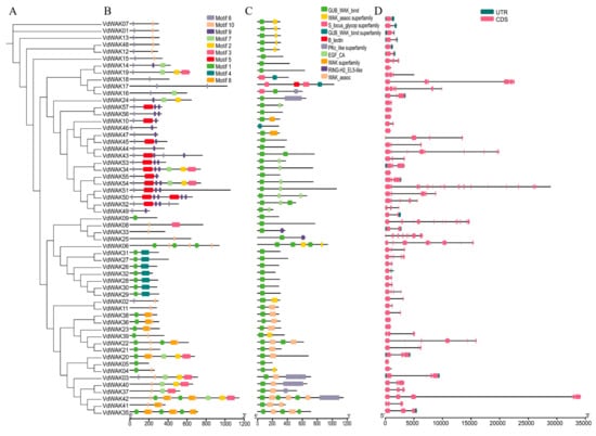
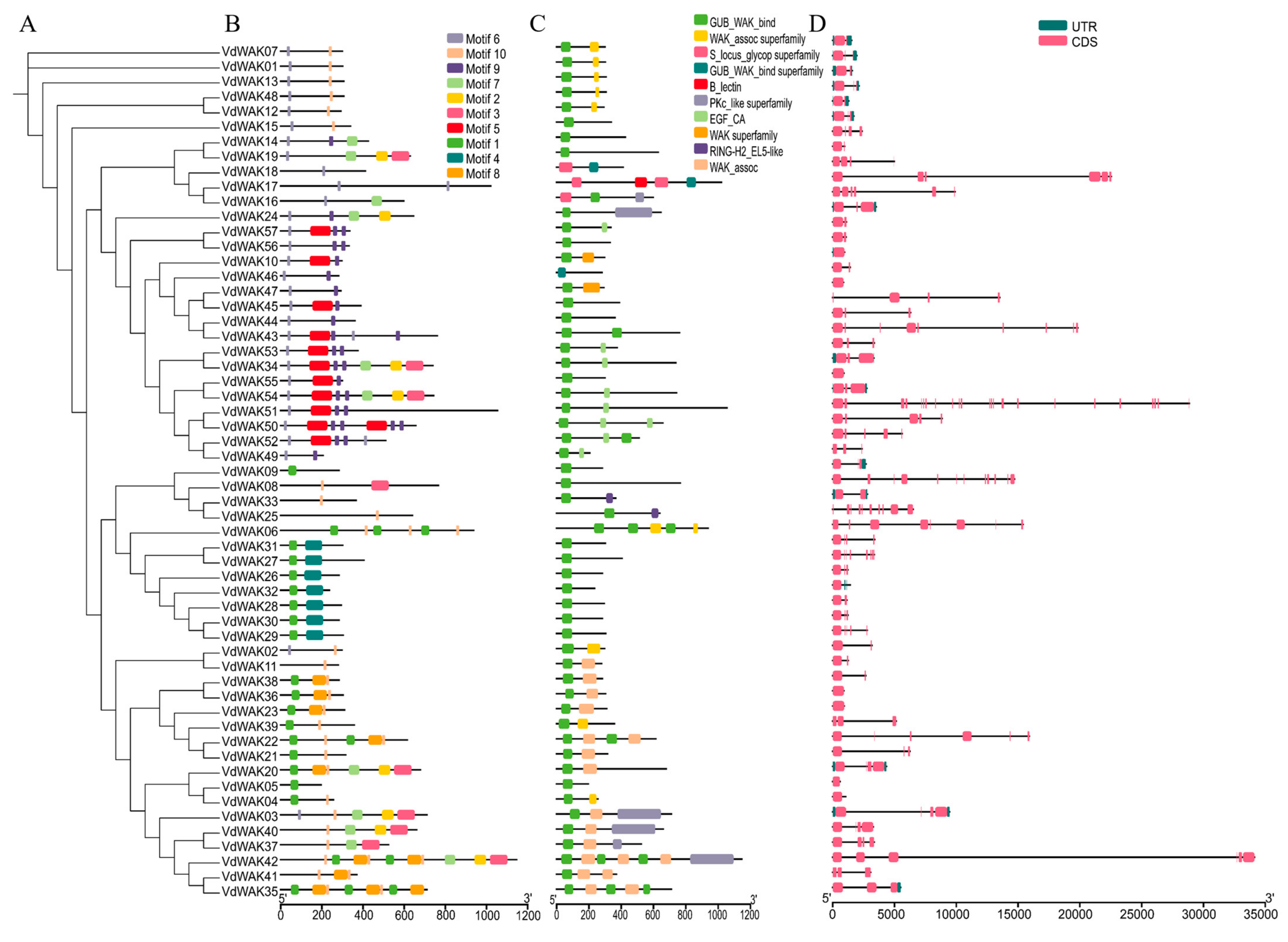
To elucidate the function characteristics of VdWAK proteins, conserved motifs analysis showed that motifs 6 and 10 were widely distributed among most VdWAK proteins, with the exception of VdWAK05 and VdWAK09 (Figure 3B). These two motifs were annotated as core motifs of the plant WAK family. Furthermore, despite variations in motif composition and distribution across the entire family, proteins clustered within the same phylogenetic group exhibited high conservation in their motif profiles.
Figure 3.
Phylogenetic relationship, conserved motifs, functional domains, and gene structures of VdWAK genes. (A) Phylogenetic tree of VdWAKs. (B) Conserved motifs of VdWAK proteins. Each motif is represented by a colored box. (C) Functional domains of VdWAKs. (D) Exon-intron structures of VdWAK genes. Exons and introns are indicated by pink boxes and single black lines, respectively.
Analysis of conserved structural domains revealed that nearly all VdWAK members possess at least one GUB_WAK_bind domain, with the exception of three members (VdWAK17, VdWAK18, and VdWAK46) (Figure 3C). Additionally, the exon and intron composition analysis showed that VdWAK genes within the same phylogenetic group tend to display similar numbers of exons and introns (Figure 3D).
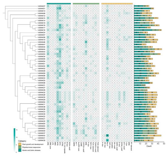
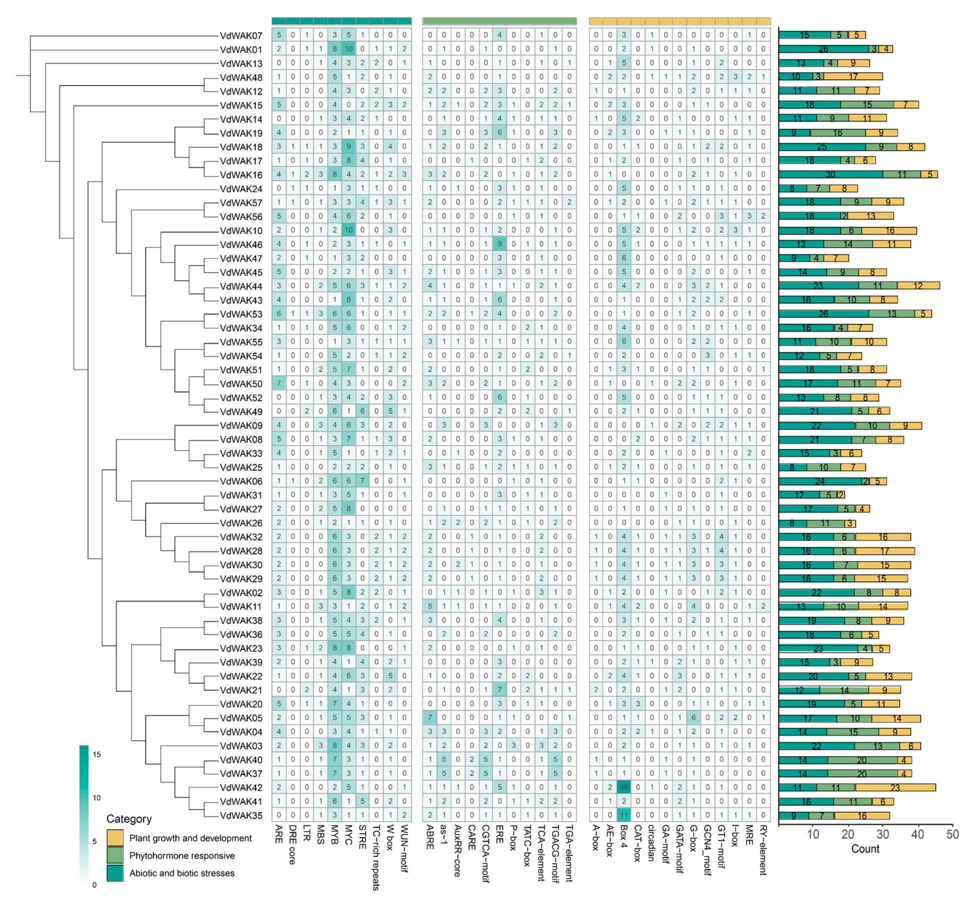
3.4. Cis-Acting Elements Analysis
The PlantCARE website was used to analyse 2 Kb upstream promoter regions of the VdWAK genes for the identification of cis-acting elements (CAE, Figure 4). Most CAEs identified in VdWAK promoters were associated with abiotic and biotic stresses, phytohormone responses, and plant growth and development processes. Several CAEs were highly abundant across VdWAK promoters, such as MYB and MYC binding sites. For closely clustered VdWAK genes, the distribution of CAEs in their promoters was relatively conserved, exhibiting similarities in both the number and type of elements; representative examples include VdWAK28, VdWAK29, VdWAK30, and VdWAK32.
Figure 4.
The distribution of cis-acting elements in the promoters of VdWAK family.
3.5. Syntenic Analysis
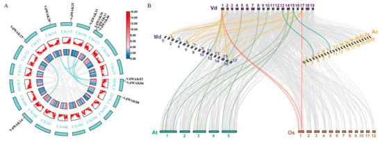
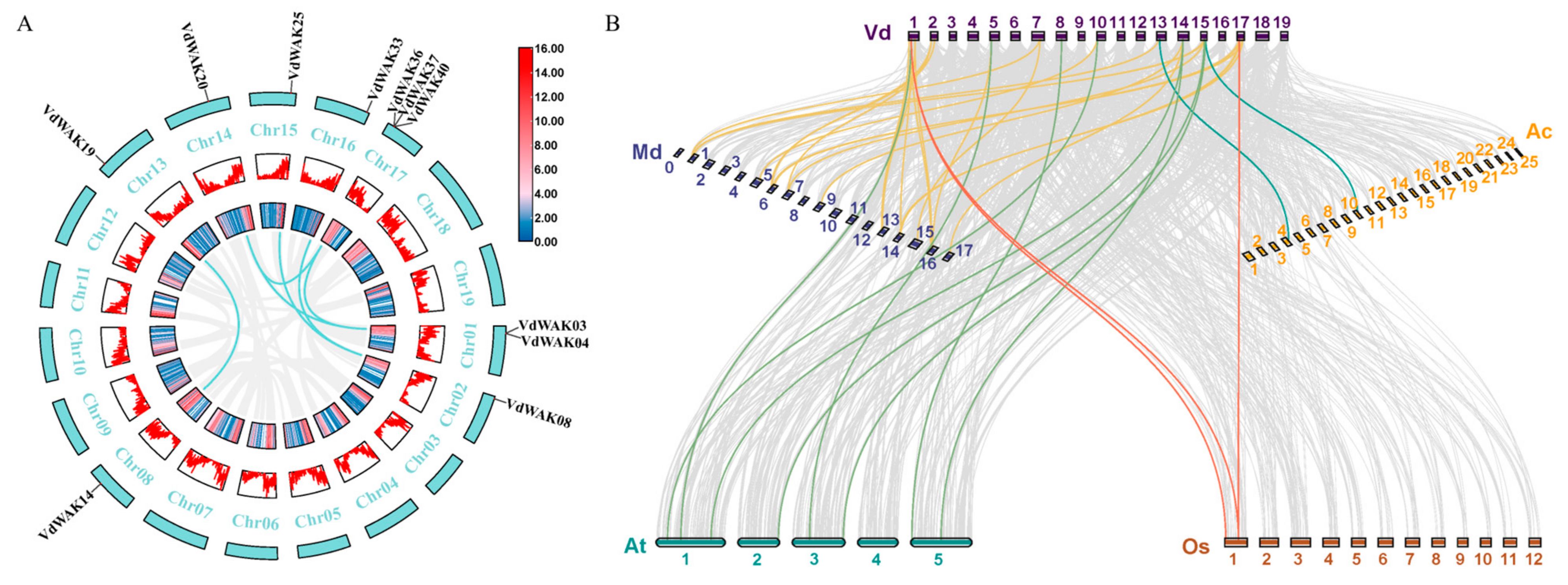
To investigate the homology and evolutionary relationships of VdWAK genes, genomic intraspecific and interspecific collinearity analyses were performed. As shown in Figure 5A, six pairs of syntenic genes were identified within the VdWAK family, indicating that segmental duplication events have driven the evolutionary expansion of this gene family (Supplementary Table S2). Additionally, interspecific collinearity analysis was performed between V. davidii and four other species (A. thaliana, A. comosus, M. domestica, O. sativa) (Figure 5B and Supplementary Table S3). A total of 42 collinear gene pair events were found between V. davidii and four species, including 2 orthologous pairs between V. davidii and A. comosus, 3 between V. davidii and O. sativa, 8 between V. davidii and A. thaliana, and 29 between V. davidii and M. domestica. These results indicate that collinearity relationships between the VdWAK gene family of V. davidii and those of the other examined species exhibited a certain degree of evolutionary divergence.
Figure 5.
Synteny analysis of VdWAK genes. (A) Intraspecific collinearity of the VdWAK gene family. (B) Interspecific collinearity of the V. davidii and four other species. The gray lines in the background represented collinear blocks between V. davidii and four other plant species (A. thaliana, A. comosus, M. domestica, O. sativa), while the colored lines indicated syntenic WAK gene pairs.
3.6. Expression of VdWAK Genes in Different Tissues and During C. viniferum Infection
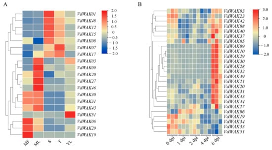
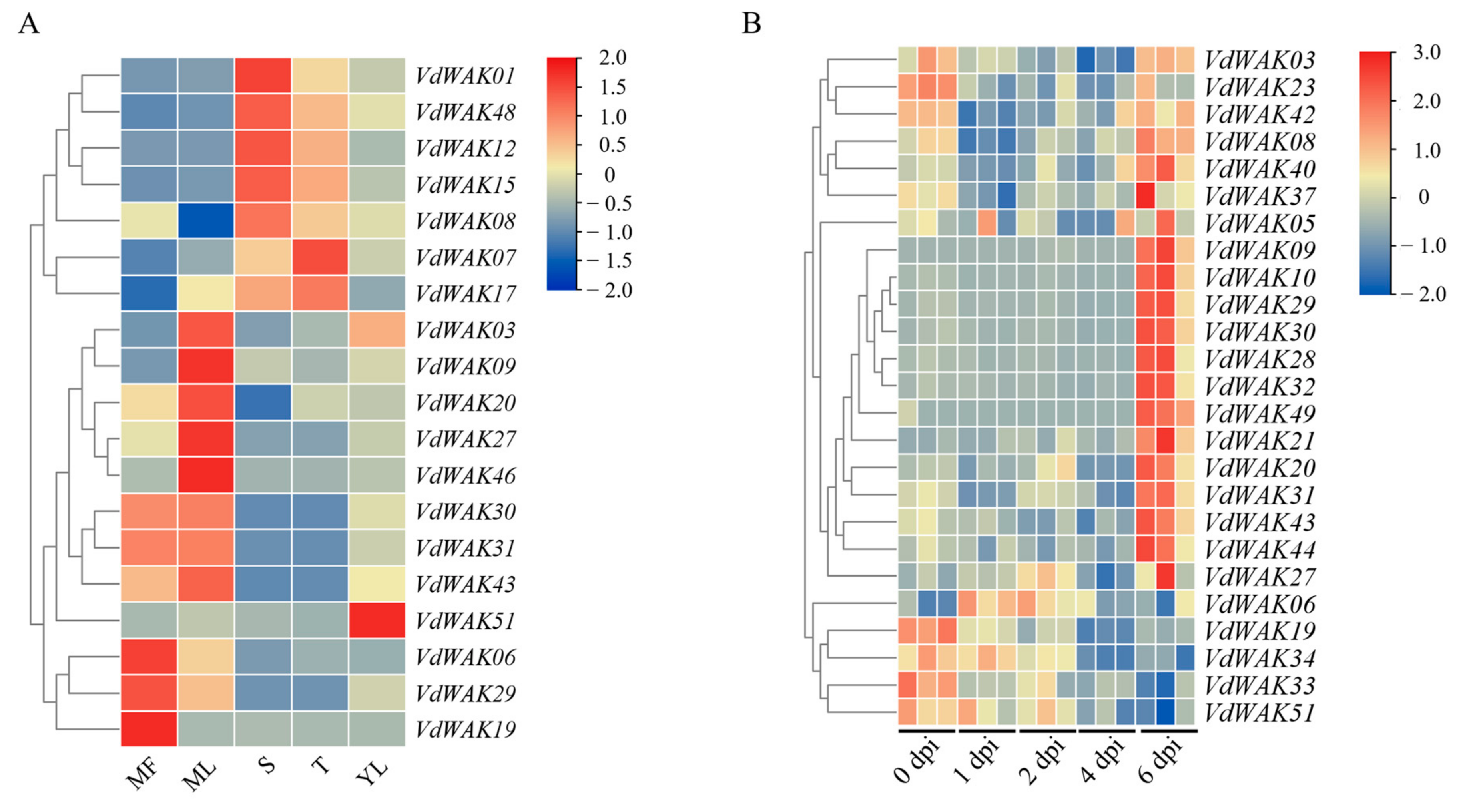
The transcript levels analysis of VdWAK genes across five distinct tissues (mature fruit, stem, tendril, young leaf, and mature leaf) revealed that 42 out of 57 VdWAK genes exhibited extremely low expression or were not expressed in nearly all tested tissues, indicating that these genes have limited transcriptional activity under the detected tissues or have tissue-specific expression patterns restricted to unexamined tissues. Notably, VdWAK06, VdWAK19, and VdWAK29 were highly expressed in mature fruits, whereas the remaining 14 genes were expressed exclusively in tissues other than mature fruit (Figure 6A and Supplementary Table S4).
Figure 6.
Expression profiles of VdWAK genes in different tissues (A) and during C. viniferum infection (B) based on RNA-seq data. VdWAK gene expression levels were quantified as TPM values. Gene with |log2(fold change)| ≥ 1.5 and padj ≤ 0.05 were defined as DEGs. The abbreviations used are defined as follows: MF (mature fruit), S (stem), T (tendril), YL (young leaf), and ML (mature leaf), and dpi (days post inoculation).
Transcriptome data at five time points post C. viniferum infection revealed that 25 members of the VdWAK family were detected to be differentially expressed. Hierarchical clustering analysis further categorized these genes into four distinct groups (Figure 6B and Supplementary Table S5). Specifically, five members (VdWAK03/08/37/40/42) exhibited a down-regulated expression trend prior to 4 dpi, followed by up-regulation at 6 dpi. Another fourteen members (VdWAK05/09/10/20/21/27/28/29/30/31/32/43/44/49) were up-regulated at 6 dpi. In contrast, VdWAK06 showed an initial up-regulation before 2 dpi, which was then followed by a gradual down-regulation until 6 dpi. Notably, the expression of five additional members (VdWAK19/23/33/34/51) was down-regulated following C. viniferum infection.
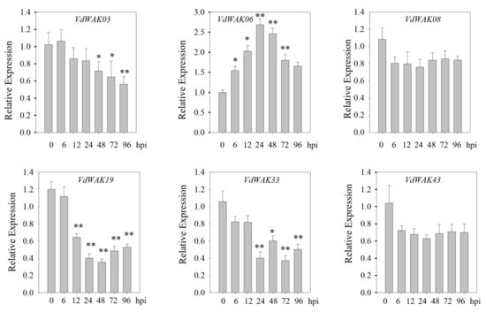
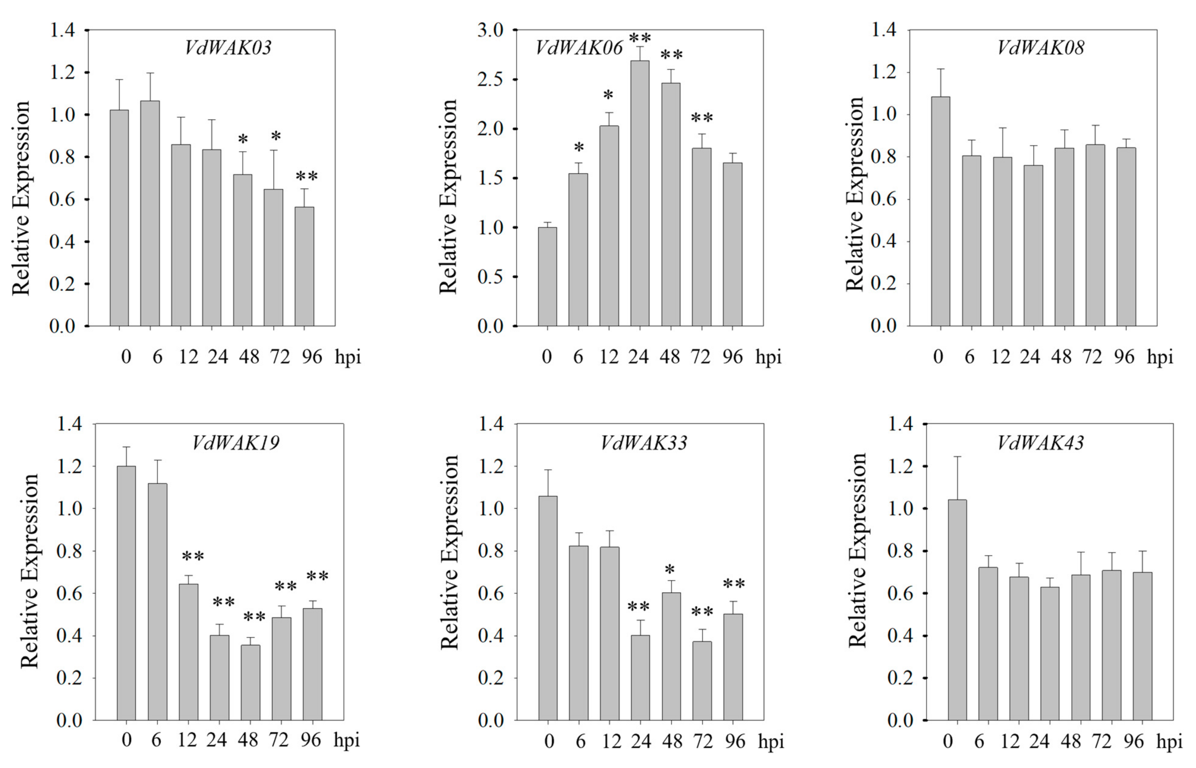
To validate the expression profiles, six VdWAK genes were randomly selected for RT-qPCR assays over a 96 h period of C. viniferum infection. As shown in Figure 7, the expression of VdWAK06 was significantly up-regulated post-infection with C. viniferum, whereas the other five VdWAK genes (VdWAK03, VdWAK08, VdWAK19, VdWAK33, and VdWAK43) were markedly down-regulated. These RT-qPCR results were consistent with the transcriptome findings.
Figure 7.
RT-qPCR verification of VdWAK genes under C. viniferum infection. The Y-axis indicates the relative expression level, and the X-axis represents different time points post C. viniferum infection. The data are presented as the mean ± standard error of the mean (SEM) from three replications (n = 3). Each time point was compared to the control group, and student’s t-tests were used to calculate statistical significance: * indicates p < 0.05, and ** indicates p < 0.01.
3.7. Silencing of VdWAK19 Decreases the Tolerance of Grape Berries to C. viniferum Infection
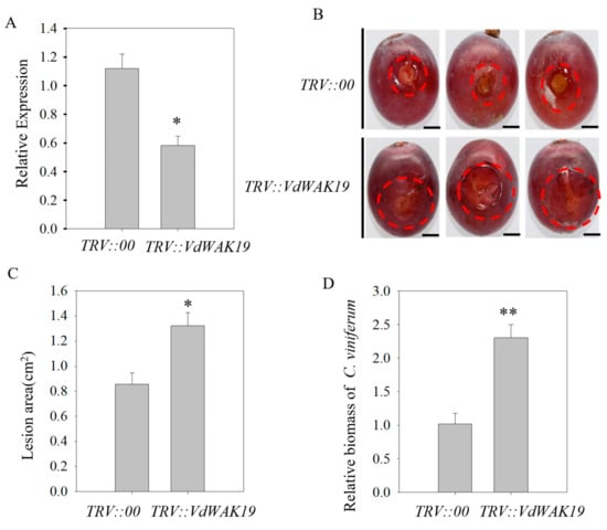
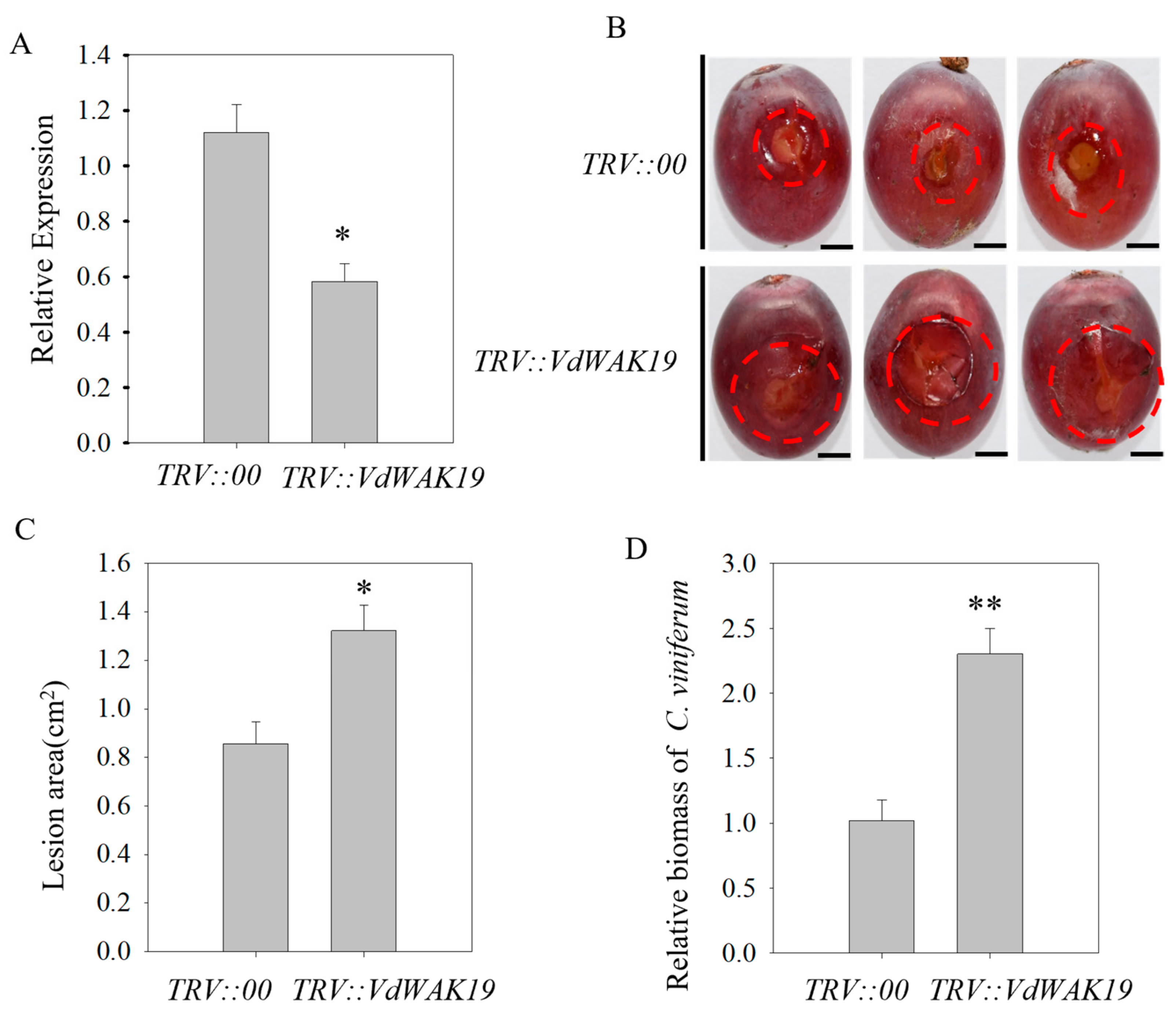
As previously demonstrated, VdWAK19 was highly expressed in mature berries and downregulated under C. viniferum inoculation. To further verify the function of VdWAK19 in grape, VIGS was used to silence VdWAK19 in ‘Kyoho’ berries. Based on comparative nucleotide sequence analysis, the silencing fragment shared 100% sequence identity between V. davidii ‘Fuan’ and V. labrusca × vinifera ‘Kyoho’ (Supplementary Figure S1). The efficacy of silencing was confirmed by RT-qPCR (Figure 8A). Berries with VdWAK19 gene silencing exhibited significantly larger lesions compared with the control group (Figure 8B), which was further supported by quantitative analyses of lesion area and pathogen biomass (Figure 8C, D). These results indicate that VdWAK19 acts as a positive regulator in grape defense response against C. viniferum infection.
Figure 8.
Functional analysis of VdWAK19 via gene silencing in grape berries. (A), RT-qPCR analysis of VdWAK19 in silenced berries. (B), Lesion phenotypes of TRV::00 and TRV::VdWAK19 berries at 3 dpi post C. viniferum infection. Scale bar = 1 cm. (C), Lesion area of TRV::00 and TRV::VdWAK19 berries at 3 dpi post C. viniferum infection. (D), pathogen biomass in TRV::00 and TRV::VdWAK19 berries at 3 dpi post C. viniferum infection. All assays were performed with three biological replicates. The data are presented as the mean ± standard error of the mean (SEM) from three replications (n = 3). The student’s t-tests were used to calculate statistical significance: * indicates p < 0.05, and ** indicates p < 0.01. Red circles indicate the areas where lesions have spread.
4. Discussion
Grapes, one of the oldest fruit crops, are economically valuable and widely cultivated across the globe [25]. The first complete genome sequence of ‘Heirbiro’ was published in 2007, followed by genomics sequencing of wild grape varieties [26,27]. With the availability of these genome sequences, a series of genome-wide identification studies on gene families in grape have been carried out in recent years, such as the B-box and GATA families [28,29]. In this study, a total of 57 VdWAK genes were identified from the V. davidii genome (Table 1). There exists significant interspecific divergence in the size of the WAK gene family across different plant species. For example, the WAK gene family comprises 33 members in A. thaliana, 35 in Malus domestica, 36 in Fragaria vesca, 36 in Prunus persica, and 129 in O. sativa [3]. However, the genome size of Arabidopsis is only 125 Mb [30], that of rice is more than 400 Mb [31], and that of V. davidii is approximately 510 Mb. This data indicates that the size of the WAK gene family in plants is not correlated with their genome size, implying no association between WAK gene family expansion and genome complexity. Chromosomal localization and intraspecific collinearity analyses revealed ten tandem duplication events and six segmental duplication events in the VdWAK gene family of V. davidii, demonstrating that these two duplication types are the major driving forces underlying the expansion of the VdWAK gene family. This expansion pattern is analogous to that reported for WAK family members in Juglans regia [32] and cotton [33]. Additionally, previous phylogenetic analysis of WAK proteins in Arabidopsis, rice and cotton have showed that WAK proteins tend to form species-specific clusters [34,35,36]. Consistent with our findings, species-specific clusters were also observed in the phylogenetic tree constructed with WAK proteins from V. davidii, O. sativa, and A. thaliana, suggesting that different plant species have undergone relatively independent evolutionary processes with respect to the WAK gene family.
Cis-elements in promoter regions enable plants to respond to environmental cues by modulating the transcriptional regulation of downstream genes [37]. A variety of plant hormone-responsive elements (e.g., SA, ETH, and JA responsive elements) were identified in the promoter regions of VdWAK genes (Figure 4). Accumulating evidence has established a regulatory link between plant hormone signaling and the plant defense response against C. viniferum infections [38,39]. For instance, exogenous SA treatment has been shown to enhance grape resistance to C. viniferum infection [40]. In tea plants, the JA signaling pathway, mediated by the CsJAZ1-CsMYC2.2 module, regulates the expression of CsGSTU45, which in turn promotes H2O2 accumulation and modulates plant susceptibility to Colletotrichum camelliae [41]. By contrast, ethylene has been found to enhance spore germination of Colletotrichum asianum and the formation of multiple appressoria in the wild-type TYC-2 [42]. The enrichment of these hormone-responsive cis-elements in the promoter regions of VdWAK genes highlights their potential critical role in mediating the adaptive responses of V. davidii to environmental stresses. Additionally, WAK genes in tea and persimmon have been investigated for their responses to Colletotrichum infection [16,40], which is consistent with our findings in V. davidii. However, the specific cis-acting elements involved in this response require further investigation using approaches such as promoter sequence truncation assays.
Tissue-specific expression patterns and stage-dependent transcriptional regulation of genes are tightly correlated with their biological functions [43]. For instance, Saccharum ScWAK1 is predominantly expressed in leaves and roots, and its transcription is significantly downregulated in response to Sporisorium scitamineum infection. Moreover, transient overexpression of ScWAK1 in Nicotiana benthamiana enhanced the susceptibility of tobacco plants to Ralstonia solanacearum and Fusarium solani var. coeruleum [16]. In maize, the ZmWAK gene, which is specifically expressed in the mesocotyl of seedlings, confers resistance to head smut [44]. In the present study, VdWAK19 was highly expressed in mature V. davidii fruits, and its expression was markedly down-regulated following C. viniferum infection (Figure 6). VIGS has developed into an indispensable approach to gene function analysis in a wide array of species, many of which are not amenable to stable genetic transformation [45]. To address potential off-target effects, sequence similarity between the VIGS fragment of VdWAK19 and other VdWAK genes was analyzed. The fragment showed high specificity to VdWAK19 and low homology with other family members, thus reducing the risk of cross-silencing in the expanded WAK family. Compared with the control group, VdWAK19-silenced grape berries exhibited more severe disease symptoms, which was further corroborated by quantitative analyses of lesion area and pathogen biomass (Figure 8B–D). Collectively, these findings demonstrate that VdWAK19 functions as a positive regulator of grape immunity against ripe rot caused by C. viniferum.
The down-regulation of VdWAK19 following pathogen infection, together with its role as a positive regulator confirmed by silencing, represents a typical phenomenon of host–pathogen coevolution. This discrepancy can be interpreted from multiple regulatory layers: firstly, the pathogen may actively suppress VdWAK19 transcription to weaken host immunity, such as StGOX4, whose expression was downregulated during P. infestans infection but positively regulates plant immunity against P. infestan [46]; Similar results have also been found in pepper [47]. Secondly, temporal expression dynamics imply that this gene may be induced at the early infection stage and subsequently repressed via negative feedback to balance growth-defense trade-offs; thirdly, in the context of hormone signaling, the transcriptional repression of VdWAK19 is likely coordinated with the dynamic crosstalk of salicylic acid (SA), jasmonic acid (JA) and ethylene (ET) pathways during compatible interactions, through which the pathogen manipulates host hormone balance to dampen positive immune responses.
5. Conclusions
In this study, 57 putative VdWAK genes with complete coding sequences were systematically identified and functionally annotated using integrated bioinformatics approaches. Expression profiles of these genes were investigated across various growth and developmental stages, and in response to C. viniferum infection. Transcriptomic analysis identified VdWAK19 as a key responsive gene, which is highly expressed in mature berries but significantly downregulated following C. viniferum infection. VIGS assays in grape berries further confirmed that silencing of VdWAK19 significantly impaired the resistance of grape berries to C. viniferum. Collectively, our findings provide a systematic characterization of the WAK gene family in V. davidii and reveal its involvement in defense responses against C. viniferum infection.
Supplementary Materials
The following supporting information can be downloaded at: https://www.mdpi.com/article/10.3390/horticulturae12050524/s1, Figure S1: Alignment of the silencing sequence of V. davidii WAK19 and the nucleotide sequence of Vitis vinifera cv. Kyoho WAK; Text S1: Amino acid sequence of WAK from V. davidii; Supplementary Table S1: Primers used in this study; Supplementary Table S2: Syntenic genes within the VdWAK family; Supplementary Table S3: Synteny analysis of WAKs between Vitis davidii and four other plant species (Arabidopsis thaliana, Ananas comosus, Malus domestica, Oryza sativa); Supplementary Table S4: TMP-based expression profiles of WAKs in different tissues; Supplementary Table S5: TMP-based expression profiles of WAKs at various time points following C. viniferum infection.
Author Contributions
J.L., X.F., J.H., H.G. and Y.L. conceived and designed the experiments and obtained the funding. J.L., X.F., X.L., Y.J. and T.C. performed the experiments and analyzed the data. J.L. and X.F. drafted the manuscript. J.H. and Y.L. contributed to the revision of the manuscript. All authors have read and agreed to the published version of the manuscript.
Funding
This work was supported by Fundamental Research Project of Provincial Public Welfare Research Institutes (2023R1026005), Central Guidance for Local Science and Technology Development Funds (2023L3025) and China Agriculture Research System (CARS-29).
Data Availability Statement
The data that support the findings will be available in Figshare public database at DOI: 10.6084/m9.figshare.31841497 following an embargo from the date of publication to allow for commercialization of research findings.
Acknowledgments
Thanks to Lili Huang from Northwest Agricultural and Forestry University for her valuable suggestions on the experimental design and manuscript revision.
Conflicts of Interest
The authors declare no conflicts of interest. The funders had no role in the design of the study, in the collection, analyses or interpretation of data, in the writing of the manuscript, or in the decision to publish the results.
References
- Tai, Y.; Li, M.; Chen, G.; Zhou, M.; Fan, Y.; Lei, M.; Tang, R.; Ye, J.; Li, D.; Wang, Y. Battle beneath the wall: Modulating cell Wall-Associated Kinase (WAK) and WAK-Likes (WAKLs) to cope with biotic and abiotic stresses in plants. Plant Cell Environ. 2025, 48, 8326–8340. [Google Scholar] [CrossRef] [Scilit] [PubMed]
- Herold, L.; Ordon, J.; Hua, C.; Kohorn, B.D.; Nürnberger, T.; DeFalco, T.A.; Zipfel, C. Arabidopsis WALL-ASSOCIATED KINASES are not required for oligogalacturonide-induced signaling and immunity. Plant Cell 2024, 37, koae317. [Google Scholar] [CrossRef] [Scilit]
- Wang, Z.C.; Ma, Y.; Chen, M.; Da, L.L.; Su, Z.; Zhang, Z.; Liu, X.T. Comparative genomics analysis of WAK/WAKL family in Rosaceae identify candidate WAKs involved in the resistance to Botrytis cinerea. BMC Genom. 2023, 24, 337. [Google Scholar] [CrossRef] [Scilit] [PubMed]
- Rizzo, M.; Bernardi, R.; Salvini, M.; Nali, C.; Lorenzini, G.; Durante, M. Identification of differentially expressed genes induced by ozone stress in sensitive and tolerant poplar hybrids. J. Plant Physiol. 2007, 164, 945–949. [Google Scholar] [CrossRef] [Scilit] [PubMed]
- de Oliveira, L.F.V.; Christoff, A.P.; de Lima, J.C.; de Ross, B.C.F.; Sachetto-Martins, G.; Margis-Pinheiro, M.; Margis, R. The Wall-associated Kinase gene family in rice genomes. Plant Sci. Int. J. Exp. Plant Biol. 2014, 229, 181–192. [Google Scholar] [CrossRef] [Scilit]
- Zhang, N.; Pombo, M.A.; Rosli, H.G.; Martin, G.B. Tomato wall-associated kinase SlWak1 depends on Fls2/Fls3 to promote apoplastic immune responses to Pseudomonas syringae. Plant Physiol. 2020, 183, 1869–1882. [Google Scholar] [CrossRef] [Scilit]
- Wang, P.; Zhou, L.; Jamieson, P.; Zhang, L.; Zhao, Z.X.; Babilonia, K.; Shao, W.Y.; Wu, L.Z.; Mustafa, R.; Amin, I.; et al. The cotton wall-associated kinase GhWAK7A mediates responses to fungal wilt pathogens by complexing with the chitin sensory receptors. Plant Cell 2020, 32, 3978–4001. [Google Scholar] [CrossRef] [Scilit]
- Li, H.; Zhou, S.Y.; Zhao, W.S.; Su, S.C.; Peng, Y.L. A novel wall-associated receptor-like protein kinase gene, OsWAK1, plays important roles in rice blast disease resistance. Plant Mol. Biol. 2009, 69, 337–346. [Google Scholar] [CrossRef] [Scilit]
- Al-Bader, N.; Meier, A.; Geniza, M.; Gongora, Y.S.; Oard, J.; Jaiswal, P. Loss of a premature stop codon in the rice wall-associated kinase 91 (WAK91) gene is a candidate for improving leaf sheath blight disease resistance. Genes 2023, 14, 1673. [Google Scholar] [CrossRef] [Scilit]
- Zhao, M.; Li, N.H.; Chen, S.M.; Wu, J.J.; He, S.F.; Zhao, Y.X.; Wang, X.R.; Chen, X.Y.; Zhang, C.Z.; Fang, X.; et al. GmWAK1, novel wall-associated protein kinase, positively regulates response of soybean to Phytophthora sojae infection. Int. J. Mol. Sci. 2023, 24, 798. [Google Scholar] [CrossRef] [Scilit]
- Dai, Z.K.; Pi, Q.Y.; Liu, Y.T.; Hu, L.; Li, B.C.; Zhang, B.; Wang, Y.B.; Jiang, M.; Qi, X.; Li, W.Q.; et al. ZmWAK02 encoding an RD-WAK protein confers maize resistance against gray leaf spot. New Phytol. 2024, 241, 1780–1793. [Google Scholar] [CrossRef] [Scilit] [PubMed]
- Fabian, M.L.; Zhang, C.; Sun, J.; Price, N.P.; Chen, P.; Clarke, C.R.; Jones, R.W.; Stommel, J.R. Steroidal glycoalkaloids contribute to anthracnose resistance in Solanum lycopersicum. J. Exp. Bot. 2023, 74, 3700–3713. [Google Scholar] [CrossRef] [Scilit]
- Hsieh, T.F.; Shen, Y.M.; Huang, J.H.; Tsai, J.N.; Lu, M.T.; Lin, C.P. Insights into grape ripe rot: A focus on the Colletotrichum gloeosporioides species complex and its management strategies. Plants 2023, 12, 2873. [Google Scholar] [CrossRef] [Scilit] [PubMed]
- Law, C.X.; Hashim, N.; Ismail, S.I.; Jahari, M.; Al Riza, D.F. A review on anthracnose disease caused by Colletotrichum spp. in fruits and advances in control strategies. Int. J. Food Microbiol. 2025, 442, 111397. [Google Scholar] [CrossRef] [Scilit] [PubMed]
- Forlani, S.; Masiero, S.; Mizzotti, C. Fruit ripening: The role of hormones, cell wall modifications, and their relationship with pathogens. J. Exp. Bot. 2019, 70, 2993–3006. [Google Scholar] [CrossRef] [Scilit]
- Wang, D.J.; Qin, L.Q.; Wu, M.X.; Zou, W.H.; Zang, S.J.; Zhao, Z.N.; Lin, P.X.; Guo, J.L.; Wang, H.B.; Que, Y.X. Identification and characterization of WAK gene family in Saccharum and the negative roles of ScWAK1 under the pathogen stress. Int. J. Biol. Macromol. 2023, 224, 1–19. [Google Scholar] [CrossRef] [Scilit]
- Zhang, Z.Q.; Ma, W.Y.; Wang, H.J.; Ren, Z.Y.; Liu, Y.G.; He, K.L.; Zhang, F.; Ye, W.; Huo, W.W.; Li, W.Q.; et al. Characterization of the wall-associated kinase (WAK) gene family in Gossypium barbadense reveals the positive role of GbWAK5 in salt tolerance. Plant Cell Rep. 2024, 44, 18. [Google Scholar] [CrossRef] [Scilit]
- Garg, V.K.; Avashthi, H.; Tiwari, A.; Jain, P.A.; Ramkete, P.W.; Kayastha, A.M.; Singh, V.K. MFPPI—Multi FASTA ProtParam interface. Bioinformation 2016, 12, 74–77. [Google Scholar] [CrossRef] [Scilit]
- Yu, C.S.; Cheng, C.W.; Su, W.C.; Chang, K.C.; Huang, S.W.; Hwang, J.K.; Lu, C.H. CELLO2GO: A web server for protein subCELlular LOcalization prediction with functional gene ontology annotation. PLoS ONE 2014, 9, e99368. [Google Scholar] [CrossRef] [Scilit]
- Lescot, M.; Déhais, P.; Thijs, G.; Marchal, K.; Moreau, Y.; Van de Peer, Y.; Rouzé, P.; Rombauts, S. PlantCARE, a database of plant cis-acting regulatory elements and a portal to tools for in silico analysis of promoter sequences. Nucleic Acids Res. 2002, 30, 325–327. [Google Scholar] [CrossRef] [Scilit]
- Wang, Y.P.; Tang, H.B.; Debarry, J.D.; Tan, X.; Li, J.P.; Wang, X.Y.; Lee, T.H.; Jin, H.Z.; Marler, B.; Guo, H.; et al. MCScanX: A toolkit for detection and evolutionary analysis of gene synteny and collinearity. Nucleic Acids Res. 2012, 40, e49. [Google Scholar] [CrossRef] [Scilit]
- Chen, C.; Chen, H.; Zhang, Y.; Thomas, H.R.; Frank, M.H.; He, Y.; Xia, R. TBtools: An integrative toolkit developed for interactive analyses of big biological data. Mol. Plant 2020, 13, 1194–1202. [Google Scholar] [CrossRef] [Scilit]
- Wang, D.; Jiang, X.L.; Zhang, W.B.; Cao, D.D.; Ye, G.P.; Chen, J.J.; Lei, Y.; Wei, X.Y. Transcriptomic and metabolomic analyses reveal mechanisms underpinning resistance of Chinese wild grape to Colletotrichum viniferum. Plant Physiol. Biochem. 2024, 215, 108851. [Google Scholar] [CrossRef] [Scilit] [PubMed]
- Kim, D.; Paggi, J.M.; Park, C.; Bennett, C.; Salzberg, S.L. Graph-based genome alignment and genotyping with HISAT2 and HISAT-genotype. Nat. Biotechnol. 2019, 37, 907–915. [Google Scholar] [CrossRef] [Scilit] [PubMed]
- Venkitasamy, C.; Zhao, L.M.; Zhang, R.H.; Pan, Z.L. Grapes. In Integrated Processing Technologies for Food and Agricultural By-Products; Academic Press: Cambridge, MA, USA, 2019; pp. 133–163. [Google Scholar] [CrossRef] [Scilit]
- Jaillon, O.; Aury, J.M.; Noel, B.; Policriti, A.; Clepet, C.; Casagrande, A.; Choisne, N.; Aubourg, S.; Vitulo, N.; Jubin, C.; et al. The grapevine genome sequence suggests ancestral hexaploidization in major angiosperm phyla. Nature 2007, 449, 463–467. [Google Scholar] [CrossRef] [Scilit] [PubMed]
- Shi, X.Y.; Cao, S.; Wang, X.; Huang, S.Y.; Wang, Y.; Liu, Z.J.; Liu, W.W.; Leng, X.P.; Peng, Y.L.; Wang, N.W.; et al. The complete reference genome for grapevine (Vitis vinifera L.) genetics and breeding. Hortic. Res. 2023, 10, uhad061. [Google Scholar] [CrossRef] [Scilit]
- Zhang, X.M.; Zhang, L.; Ji, M.M.; Wu, Y.F.; Zhang, S.L.; Zhu, Y.X.; Yao, J.; Li, Z.; Gao, H.; Wang, X.P. Genome-wide identification and expression analysis of the B-box transcription factor gene family in grapevine (Vitis vinifera L.). BMC Genom. 2021, 22, 221. [Google Scholar] [CrossRef] [Scilit]
- Zhang, X.M.; Ma, J.H.; Yang, S.J.; Yao, W.K.; Zhang, N.B.; Hao, X.Y.; Xu, W.R. Analysis of GATA transcription factors and their expression patterns under abiotic stress in grapevine (Vitis vinifera L.). BMC Plant Biol. 2023, 23, 611. [Google Scholar] [CrossRef] [Scilit]
- The Arabidopsis Genome Initiative. Analysis of the genome sequence of the flowering plant Arabidopsis thaliana. Nature 2000, 408, 796–815. [Google Scholar] [CrossRef] [Scilit]
- Shang, L.; Li, X.; He, H.; Yuan, Q.; Song, Y.; Wei, Z.; Lin, H.; Hu, M.; Zhao, F.; Zhang, C.; et al. A super pan-genomic landscape of rice. Cell Res. 2022, 32, 878–896. [Google Scholar] [CrossRef] [Scilit]
- Li, M.; Ma, J.; Liu, H.; Ou, M.; Ye, H.; Zhao, P. Identification and characterization of Wall-Associated Kinase (WAK) and WAK-like (WAKL) gene family in Juglans regia and its wild related species Juglans mandshurica. Genes 2022, 13, 134. [Google Scholar] [CrossRef] [Scilit]
- Zhang, Z.; Ma, W.; Ren, Z.; Wang, X.; Zhao, J.; Pei, X.; Liu, Y.; He, K.; Zhang, F.; Huo, W.; et al. Characterization and expression analysis of wall-associated kinase (WAK) and WAK-like family in cotton. Int. J. Biol. Macromol. 2021, 187, 867–879. [Google Scholar] [CrossRef] [Scilit]
- Kohorn, B.D. WAKs; cell wall associated kinases. Curr. Opin. Cell Biol. 2001, 13, 529–533. [Google Scholar] [CrossRef] [Scilit] [PubMed]
- Zhang, S.; Chen, C.; Li, L.; Meng, L.; Singh, J.; Jiang, N.; Deng, X.W.; He, Z.H.; Lemaux, P.G. Evolutionary expansion, gene structure, and expression of the rice wall-associated kinase gene family. Plant Physiol. 2005, 139, 1107–1124. [Google Scholar] [CrossRef] [Scilit] [PubMed]
- Dou, L.; Li, Z.; Shen, Q.; Shi, H.; Li, H.; Wang, W.; Zou, C.; Shang, H.; Li, H.; Xiao, G. Genome-wide characterization of the WAK gene family and expression analysis under plant hormone treatment in cotton. BMC Genom. 2021, 22, 85. [Google Scholar] [CrossRef] [Scilit] [PubMed]
- Sazegari, S.; Niazi, A.; Ahmadi, F.S. A study on the regulatory network with promoter analysis for Arabidopsis DREB-genes. Bioinformation 2015, 11, 101–106. [Google Scholar] [CrossRef] [Scilit]
- Fang, X.; Chai, W.; Li, S.; Zhang, L.; Yu, H.; Shen, J.; Xiao, W.; Liu, A.; Zhou, B.; Zhang, X. HSP17.4 mediates salicylic acid and jasmonic acid pathways in the regulation of resistance to Colletotrichum gloeosporioides in strawberry. Mol. Plant Pathol. 2021, 22, 817–828. [Google Scholar] [CrossRef] [Scilit]
- Liu, C.; Liu, Z.; Li, X.; Chen, Y.; Sun, R.; Li, P.; Feng, Q.; Wang, Y.; Ren, J.; Li, Q.; et al. Jasmonate modulates strawberry susceptibility to anthracnose by activating SnRK2.1 to regulate the WRKY50-JAZ5 module. Plant Biotechnol. J. 2025, 24, 2350–2371. [Google Scholar] [CrossRef] [Scilit]
- Lin, J.; Chen, T.; Liu, X.; Chen, Z.; Lei, Y. Salicylic acid represses VdMYB31 expression to enhance grape resistance to Colletotrichum viniferum. Int. J. Biol. Macromol. 2025, 288, 138731. [Google Scholar] [CrossRef] [Scilit]
- Lv, W.; Jiang, H.; Cao, Q.; Ren, H.; Wang, X.; Wang, Y. A tau class glutathione S-transferase in tea plant, CsGSTU45, facilitates tea plant susceptibility to Colletotrichum camelliae infection mediated by jasmonate signaling pathway. Plant J. Cell Mol. Biol. 2024, 117, 1356–1376. [Google Scholar] [CrossRef] [Scilit]
- Kao, C.Y.; Wu, C.T.; Lin, H.C.; Hsieh, D.K.; Lin, H.L.; Lee, M.H. The G protein subunit α1, CaGα1, mediates ethylene sensing of mango anthracnose pathogen Colletotrichum asianum to regulate fungal development and virulence and mediates surface sensing for spore germination. Front. Microbiol. 2022, 13, 1048447. [Google Scholar] [CrossRef] [Scilit]
- He, Z.H.; Cheeseman, I.; He, D.; Kohorn, B.D. A cluster of five cell wall-associated receptor kinase genes, Wak1-5, are expressed in specific organs of Arabidopsis. Plant Mol. Biol. 1999, 39, 1189–1196. [Google Scholar] [CrossRef] [Scilit]
- Zuo, W.; Chao, Q.; Zhang, N.; Ye, J.; Tan, G.; Li, B.; Xing, Y.; Zhang, B.; Liu, H.; Fengler, K.A.; et al. A maize wall-associated kinase confers quantitative resistance to head smut. Nat. Genet. 2015, 47, 151–157. [Google Scholar] [CrossRef] [Scilit]
- Rössner, C.; Lotz, D.; Becker, A. VIGS Goes Viral: How VIGS Transforms Our Understanding of Plant Science. Annu. Rev. Plant Biol. 2022, 73, 703–728. [Google Scholar] [CrossRef] [Scilit]
- Zhang, P.L.; Li, J.Y.; Gou, X.H.; Zhu, L.; Yang, Y.; Li, Y.L.; Zhang, Y.Q.; Ding, L.W.; Ansabayeva, A.; Meng, Y.L.; et al. The Phytophthora infestans effector Pi05910 suppresses and destabilizes host glycolate oxidase StGOX4 to promote plant susceptibility. Mol. Plant Pathol. 2024, 25, e70021. [Google Scholar] [CrossRef] [Scilit]
- Dang, F.F.; Wang, Y.N.; She, J.J.; Lei, Y.F.; Liu, Z.Q.; Eulgem, T.; Lai, Y.; Lin, J.; Yu, L.; Lei, D.; et al. Overexpression of CaWRKY27, a subgroup IIe WRKY transcription factor of Capsicum annuum, positively regulates tobacco resistance to Ralstonia solanacearum infection. Physiol. Plant. 2014, 150, 397–411. [Google Scholar] [CrossRef] [Scilit]

Disclaimer/Publisher’s Note: The statements, opinions and data contained in all publications are solely those of the individual author(s) and contributor(s) and not of MDPI and/or the editor(s). MDPI and/or the editor(s) disclaim responsibility for any injury to people or property resulting from any ideas, methods, instructions or products referred to in the content. |
© 2026 by the authors. Licensee MDPI, Basel, Switzerland. This article is an open access article distributed under the terms and conditions of the Creative Commons Attribution (CC BY) license.