


Abstract
NEAT1 is among the most extensively studied lncRNAs, classically recognized as the architectural scaffold of nuclear paraspeckles (PSs), membranelles nuclear bodies that assemble through liquid–liquid phase separation to sequester RNA-binding proteins and edited transcripts. Within this framework, NEAT1 was thought to regulate gene expression mainly indirectly, by trapping essential gene regulatory factors within these condensates. Here, we aim to review growing evidence that repositions NEAT1 from a scaffold to an active, highly context-dependent chromatin regulator that finely contributes to genome surveillance and cellular homeostasis. NEAT1 engages chromatin directly by occupying gene regulatory elements and DNA:RNA hybrids, recruiting epigenetic regulators and cooperating with lineage-specific transcription factors. We further examine how such a regulatory NEAT1 activity varies across a broad spectrum of cancerous and non-cancerous diseases. The evidence discussed reframes NEAT1 as an adaptor molecule in transcriptional machinery whose function is deeply influenced by cellular stimuli, interacting partners and chromatin targets accessible in each context. This conceptual shift not only raises new mechanistic questions but also underscores the challenges of therapeutically targeting NEAT1.
1. Introduction
The discovery of non-coding RNAs (ncRNAs) has revolutionized our understanding of gene expression regulation, revealing how these molecules finely tune precise molecular mechanisms that sustain essential physiological and pathological processes [1,2,3]. NcRNAs are defined as transcripts with low or no translational potential, which represent up to 98% of the human transcriptome. Historically, these molecules have been classified into two broad functional categories: structural and regulatory ncRNAs [4]. The structural ncRNA category comprises major constituents of post-transcriptional and protein synthesis machineries, including small nuclear RNAs (snRNAs), small nucleolar RNAs (snoRNAs), transfer RNAs (tRNAs), and ribosomal RNAs (rRNAs). Regulatory ncRNAs are a wider and more heterogeneous class of transcripts mostly involved in the homeostatic maintenance of cellular functions [3,5]. Furthermore, ncRNAs have been categorized into short ncRNAs and long ncRNAs (lncRNAs), based on a cutoff length of 200 nt. For a long period of time, lncRNAs have been described for what they are not. This misconception was largely fueled by their poor sequence conservation and highly specific, dynamic and low level of expression [1]. Conversely, accumulating research evidence posits lncRNAs as functional macromolecules rather than merely transcriptional noise byproducts. Often, their cellular localization determines their functions [6]. LncRNAs display conservation of discrete structural domains that mediate specific protein interactions [7], enabling them to regulate protein activity by acting as guides that direct interacting proteins to specific targets or as scaffolds that spatially organize multiple proteins within nuclear compartments [8]. Different from mRNAs, lncRNAs are often retained in the nucleus and associated with chromatin [9,10]. One of the best examples is represented by the lncRNA NEAT1, which was primarily characterized by its ability to sequester RNA-binding proteins (RBPs) and trap edited mRNAs within nuclear organelles named paraspeckles (PSs) [11]. Beyond this well-annotated function, accumulating evidence indicates that NEAT1 acts as more than a structural hub within PSs, directly influencing both chromatin architecture and function. Here, we discuss the mechanisms through which NEAT1 directly interfaces with chromatin to influence genome organization, transcription, and cellular homeostasis across physiological and pathological states. We particularly focus on the molecular determinants that dictate where and how NEAT1 acts, which partners it engages, and how these interactions shape its function, reframing NEAT1 as an adaptable regulator rather than a merely structural scaffold.
2. NEAT1 Biogenesis, Transcriptional Control, Role in PS Assembly
2.1. NEAT1 Biogenesis and Transcriptional Activation
NEAT1 is a mono-exonic lncRNA that originates from the Multiple Endocrine Neoplasia (MEN) type I locus, localized on the human chromosome 11q13. It is transcribed by RNA polymerase II (RNAP II) in two different variants that share an identical 5′ terminus: NEAT1_1, a shorter polyadenylated isoform of 3.7 kb, and NEAT1_2, a longer isoform of 22.7 kb, lacking the polyA tail but with a triple helix structure at its 3′ terminus that protects the transcript from 3′–5′ exonuclease activity [11]. Transcriptional activation of NEAT1 is driven by diverse cellular stressors, including DNA damage and oncogenic replicative stress, viral infection, hypoxia, and proteotoxicity. Indeed, NEAT1 transcription is highly coordinated by several stress-responsive transcription factors (TFs). Among them, TP53 induces the expression of NEAT1 in response to DNA damage and replicative stress [12,13], whereas HIF-2α promotes NEAT1 transcription by binding the HRE element on the NEAT1 promoter region in response to hypoxic stimuli [14]. Also, under proteotoxic stress, HSF1 starts to accumulate in cell nuclei, enhancing the transcription of NEAT1 [15]. Activation of NEAT1 has been related to innate immunity activation and inflammasome assembly, driving inflammation-related outcomes [16,17,18,19]. Specifically, upon inflammatory stimuli from pathogens or damaged tissues, NF-KB [20], STAT3 [21] and YY1 [22] TFs trigger NEAT1 expression.
Beyond transcriptional control, NEAT1 abundance is extensively regulated at the post-transcriptional level, primarily through competing 3′-end processing pathways that dictate isoform choice [19]. TAR DNA-binding protein 43 (TDP-43) promotes recognition of the proximal polyadenylation site within the NEAT1 locus, favoring 3′-end cleavage that yields the short, polyadenylated NEAT1_1 isoform while concomitantly reducing read-through into the long, non-polyadenylated NEAT1_2 [23]. Conversely, the Cleavage and Polyadenylation Specific Factor 6 (CPSF6), Nudix hydrolase 21 (NUDT21/CFIm25), and heterogeneous nuclear ribonucleoprotein K (hnRNPK) modulate 3′ cleavage and polyadenylation in a manner that shifts the balance toward NEAT1_2 biogenesis [24].
Although both isoforms are transcribed from a shared promoter, their steady-state expression is uncoupled: NEAT1_1 is expressed ubiquitously across tissues, whereas NEAT1_2 displays marked cell- and tissue-type specificity [25,26]. Moreover, NEAT1_2 is selectively induced under cellular stress [27], reinforcing the emerging notion that the two isoforms subserve distinct biological functions.
2.2. NEAT1 Role in PSs Assembly
The long NEAT1_2 isoform is known to act as an essential structural scaffold for PSs, membranelles, sub-nuclear organelles involved in a variety of molecular processes. The biogenesis of PSs is strictly coupled with both NEAT1_2 transcription and the lncRNA’s architectural capacity, which relies on the physical phenomenon of liquid–liquid phase separation (LLPS) [11,28]. CRISPR-Cas9 deletion experiments revealed that the central domain of NEAT1, between 8 and 16.6 kb, contains modular and functionally redundant subdomains, termed C1, C2 and C3. These act as multivalent docking sites for proteins belonging to the Drosophila Behavior Human Splicing protein (DBHS) family, including NONO, SFPQ, PSPC1 and proteins with prion-like domains (PLDs), among which are FUS and RBM14 [25]. The recruitment of NONO dimers initiates massive protein oligomerization, driving the condensation of the ribonucleoprotein complex into a highly ordered, phase-separated core–shell spheroidal structure, where the middle domain of NEAT1_2 forms the dense core while its 5′ and 3′ ends form the peripheral shell. More than 40 proteins have been identified as paraspeckle-associated proteins (PSPs). Among them, seven essential PSPs have been classified into two functional categories according to their sequential roles in PS biogenesis: Category 1A, comprising NONO, SFPQ, RBM14, and hnRNP K, which initiate NEAT1_2–RNP complex formation, and Category 1B, including FUS, DAZAP1, and hnRNP H3, which contribute to the subsequent assembly of mature PSs. The proper PS assembly requires a precise hierarchical interaction between categories 1A and 1B, mediated by the SWI/SNF chromatin remodeling complex [24,29]. PSs are not constitutively expressed since their abundance is highly dynamic and cell-type- and context-specific. The main function associated with PSs relies on the regulation of gene expression at different layers [28,30]. They can indirectly influence gene transcription through the sequestration of known TFs, such as NONO, and transcriptional regulators (TRs), such as RBM14 [28].
PSs also act post-transcriptionally, mainly through A-to-I RNA editing. The NONO/SFPQ heterodimer binds A-to-I-edited transcripts and retains them in the nucleus, therefore blocking their translation [31,32].
PSs also affect pre-mRNA splicing. In part, this occurs by sequestering splicing factors such as SFPQ [33]. Paraspeckles can also influence splicing indirectly, through their interaction with the SWI/SNF subunit ARID1B, which controls several splicing-related genes [34]. Beyond splicing, PSs have been reported to influence gene expression by modulating pri-miRNAs processing. This activity depends largely on NEAT1, whose 3′ terminus contains hairpin structures resembling pri-miRNAs. These pseudo pri-miRNAs elements facilitate the recruitment of the Microprocessor complex to PSs, while secondary structures along NEAT1 provide the binding platform for NONO and SFPQ. Thus, NEAT1 mediates interactions between the heterodimer NONO-SFPQ and Microprocessor components, particularly the DGCR8 subunit, thereby enhancing global pri-miRNA processing by Drosha-DGCR8 [35].
The main function of paraspeckles, however, is in the DNA-damage response (DDR) [36,37,38,39,40]. The essential paraspeckle proteins (PSPs) coordinate NHEJ, homologous recombination (HR), single-strand break (SSB) repair, and base excision repair (BER) [41]. Paraspeckles also contribute to cell fate. Their assembly marks the loss of pluripotency in embryonic stem cells, and they are needed for myoblast differentiation into myotubes [23,42]. Finally, they take part in the response to viral infection [43]. Together, these roles make paraspeckles relevant to normal cell physiology and potential drivers of disease.
Different from the long NEAT1_2, the function of the short NEAT1_1 isoform remains elusive. This is mainly due to the sequence overlap with the long NEAT1_2 that shares the same 5′ region, which poses severe technical challenges in disentangling the individual roles of the two isoforms. In addition, NEAT1_1 expression is highly dynamic and variable throughout different phases of the cell cycle, as compared to NEAT1_2, whose expression remains stable [12]. Different lines of evidence indicate that the polyadenylated NEAT1_1 is not involved in the generation of nuclear PSs, as it is mainly localized outside PS loci, where it accumulates in nucleoplasmic foci termed microspeckles, whose function and composition are still under debate [25,44,45]. However, this appears to be a controversial issue since other studies suggest that the short isoform can be recruited within PSs to support their stability and/or function. Another interesting recent study points out, for the first time, the ability of nuclear NEAT1_1 to destabilize the TIRR/53BP1 complex, which plays a key role in regulating the choice of DNA DSB repair pathway engaged by cells [46].
3. Regulation of Chromatin Structure, Topology, and Epigenetic Function
While PSs are traditionally viewed as independent nuclear bodies residing in the interchromatin space, a profound conceptual shift has occurred with the discovery that NEAT1 dynamically and physically alters chromatin architecture, function and epigenetic landscapes (Table 1).
Table 1.
Context-dependent chromatin and transcriptional functions of NEAT1.
3.1. NEAT1 Directly Interacts with Epigenetic Complexes
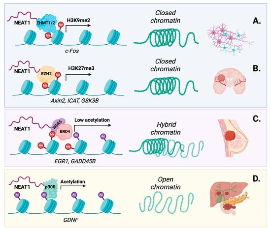
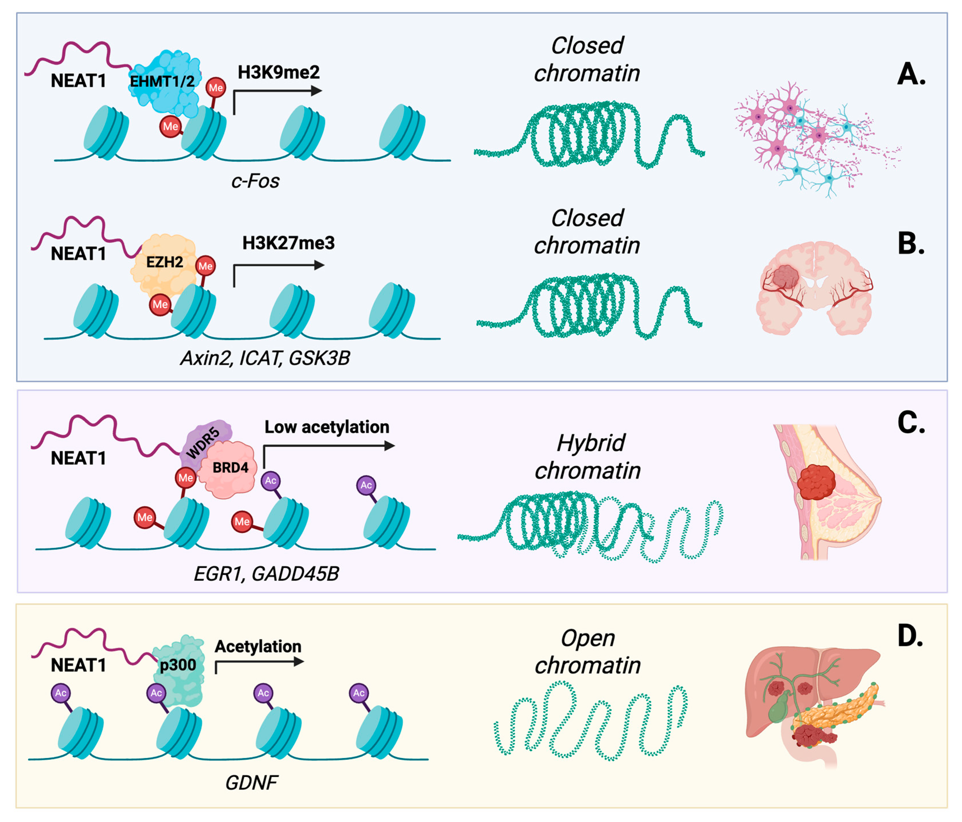
NEAT1 has been described as able to bind chromatin regulators in a PS-dependent and independent manner. In a PS-dependent way, NEAT1 can indirectly influence the chromatin landscape by mediating interaction between PSs and the ARID1B protein of the cBAF-type SWI/SNF complex, as discussed above. The SWI/SNF is known to extensively bind several enhancer and promoter regions, directly influencing transcription initiation, elongation and RNA splicing [34]. However, most of the evidence connecting NEAT1 to chromatin biology supports mechanisms that operate independently of PSs. These interactions can be broadly divided into repressive and activating functions, depending on the chromatin regulators engaged by NEAT1 (Figure 1).
Figure 1.
NEAT1 regulates chromatin landscape independently of paraspeckle formation through the recruitment of epigenetic factors. (A) In age-related disorders, NEAT1 recruits the EHMT1/2 methyltransferase complex to the c-Fos promoter, promoting H3K9me2 deposition, chromatin compaction and transcriptional repression, with consequences for neuronal plasticity and memory. (B) In glioblastoma, NEAT1 binds EZH2 and promotes H3K27me3 deposition at the Axin2, ICAT, and GSK3B tumor-suppressor loci, contributing to their repression and WNT/β-catenin pathway activation. (C) In breast cancer, NEAT1 forms a complex with BRD4 and WDR5, maintaining a low-acetyltransferase state associated with repression of target genes, including EGR1 and GADD45B. (D) In pancreatic cancer, NEAT1 recruits the histone acetyltransferase p300 to the GDNF promoter, promoting histone acetylation and transcriptional activation associated with perineural invasion. Me, methylation; Ac, acetylation.
In the context of transcriptional repression, NEAT1 can recruit or sequester chromatin-modifying factors to alter local chromatin states. In age-related disorders, NEAT1 interacts with the EHMT1/2 complex, favoring the deposition of H3K9me2 repressor marks in the c-Fos promoter, thereby affecting c-Fos transcription and leading to neuronal plasticity defects and age-related memory impairment [47]. In occlusive vascular disease, NEAT1 sequesters key chromatin modifiers such as WDR5 from specific SM loci, leading to epigenetic silencing of smooth muscle contractile genes and contributing to the switch from a contractile to a proliferative phenotype, which is linked to vascular injuries [48]. In glioblastoma, NEAT1 directly binds the repressor EZH2, which introduces the repressive H3K27me3 mark at the Axin2, ICAT and GSK3B tumor suppressor gene loci, leading to WNT/β-catenin pathway activation [49]. In breast cancer, NEAT1 can form a complex with BRD4 and WDR5, holding the complex in a low-acetyltransferase-activity state, affecting target gene expression [50].
Conversely, NEAT1 can also promote transcriptional activation by interacting with chromatin co-activators and facilitating an open chromatin state. Among the best-characterized examples is the interaction between NEAT1 and the histone acetyltransferase p300, which has been reported across multiple physiological and pathological contexts. NEAT1 can recruit the p300 protein to the promoter region of the GDNF gene, which is involved in perineural invasion of pancreatic cancer [51]. In Alzheimer’s disease, NEAT1 has been shown to exert a protective function by recruiting the histone acetyltransferase p300 to the FZD3 promoter, where it promotes H3K27 acetylation and sustains FZD3 transcription, supporting microtubule stabilization [52]. More recently, NEAT1 has also emerged as a pro-hypertrophic lncRNA that recruits, via direct binding, the co-activator p300 on the promoter region of the GRK2 gene, an essential regulator of cardiac hypertrophy [53,54]. In colorectal cancer (CRC) cells, NEAT1 knockdown leads to widespread changes in chromatin accessibility, affecting chromatin remodeling. Although the detailed mechanisms remain to be fully elucidated, high NEAT1 expression levels lead to increased histone acetylation in the promoter region of ALDH1 and c-MYC, which results in enhanced expression of stemness-related genes, contributing to 5-Fu resistance [55].
3.2. NEAT1 Chromatin Occupancy and Regulation of Tridimensional Genome Architecture
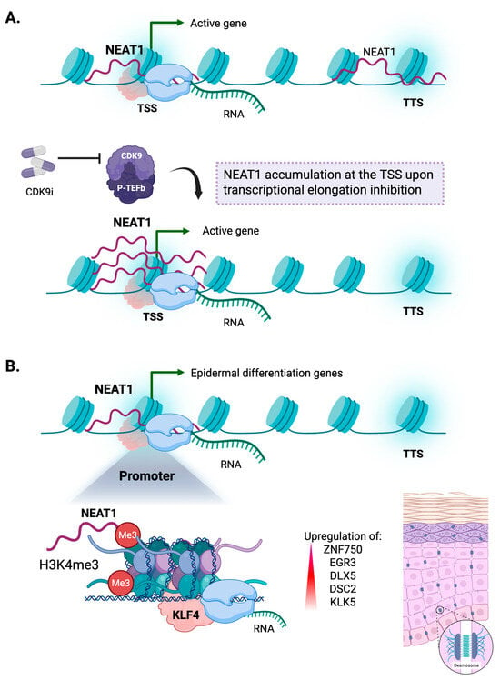
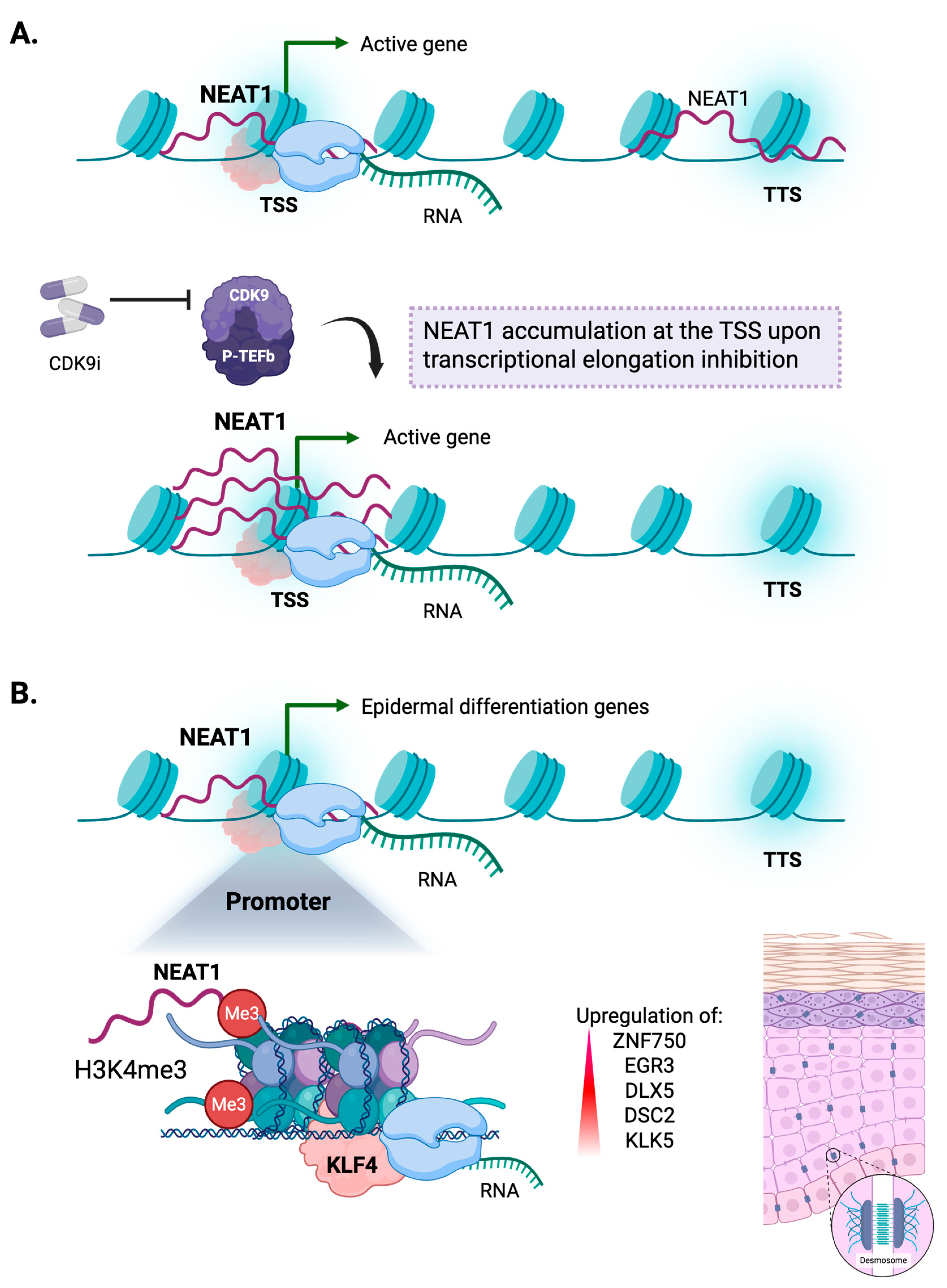
Growing evidence points to the direct binding of NEAT1 to chromatin and its potential role in regulating genome homeostasis through higher-order genome organization (Figure 2), although the underlying mechanisms remain incompletely understood. The pioneering work of West J.A. et al. demonstrates that in breast cancer, NEAT1 and MALAT1 associate with discrete genomic locations [56] and since both lncRNAs show a short half-life, the nature of these interactions might be transient and specifically driven by cellular stimuli. Both lncRNAs preferentially bind active transcribed regions, and although they can have common binding sites, they differ in genomic localization. NEAT1 binds preferentially to transcriptional start sites (TSS) and transcriptional termination sites (TTS), while MALAT1 is most enriched at the gene bodies and TTS. Crucially, NEAT1’s localization on chromatin is highly dynamic and responsive to transcriptional status, as inhibition of transcriptional elongation through disruption of the PTEFb/CDK9 complex leads to a strong enrichment of NEAT1 at TSSs [56]. These results identify, for the first time, a dynamic coupling between NEAT1 recruitment at targeted gene loci and transcriptional elongation, suggesting that NEAT1 may function as a transcriptionally responsive regulator of RNA polymerase II (RNAP II) activity, participating in complex regulatory feedback. In line with this model, accumulating evidence indicates that NEAT1 can be selectively recruited to regulatory regions of genes belonging to cell-type-specific transcriptional programs, where it cooperates with transcription factors to modulate gene expression. ChIRP-seq experiments reveal that it is physically tethered to the promoters of critical epidermal differentiation genes, at H3K4me3 marks, where it cooperates with key epidermal TFs such as KLF4. The NEAT1-KLF4 cooperation sustains the transcription of late-stage epithelial genes, including ZNF750, EGR3, DLX5, and structural desmosomal components (DSC2, KLK5), making NEAT1 an indispensable driver of epidermal tissue stratification [57].
Figure 2.
NEAT1 chromatin occupancy is dynamically coupled to transcription and to cell-type-specific gene-expression programs. (A) Chromatin-bound NEAT1 preferentially localizes to transcription start sites (TSSs) and transcription termination sites (TTSs) of actively transcribed genes. Inhibition of transcriptional elongation through CDK9/P-TEFb disruption leads to NEAT1 accumulation at TSSs, supporting a coupling between NEAT1 chromatin occupancy and RNA polymerase II activity. (B) NEAT1 associates with H3K4me3-marked promoters of epidermal differentiation genes and cooperates with KLF4 to promote expression of late-stage epithelial genes, including ZNF750, EGR3, DLX5, DSC2, and KLK5, contributing to epidermal differentiation and stratification.
NEAT1 is down-regulated in patients suffering from schizophrenia, and the transcriptomic analyses on NEAT1-deficient mouse models revealed that its depletion significantly impacts the oligodendrocyte differentiation transcriptional program, accompanied by severe de-regulation of myelin-related gene expression. Consistent with a direct chromatin-associated function, transcriptomic profiling of NEAT-associated chromatin fractions, purified by ChIRP-RNA-Seq, revealed an enrichment of oligodendrocyte transcripts in NEAT1-bound regions, suggesting that it may contribute to the modulation of the lineage-specific transcriptional program supportive of oligodendrocyte differentiation [58].
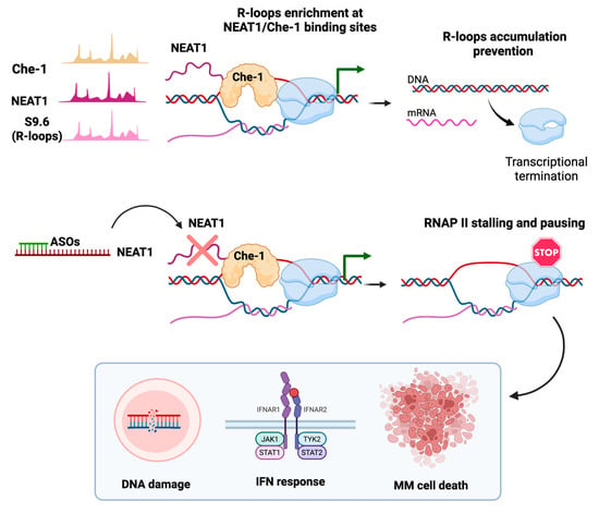
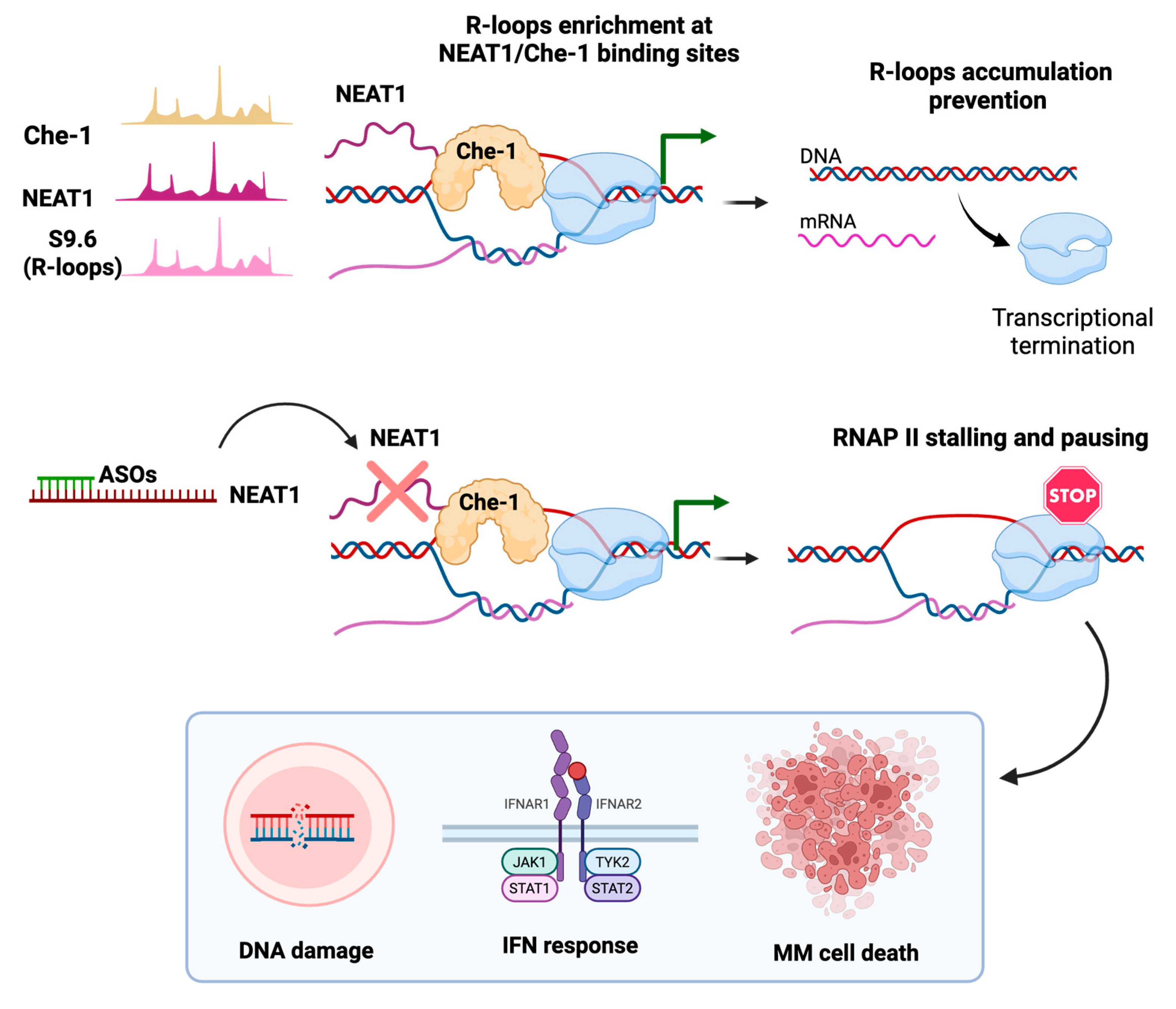
NEAT1 also exerts control over transcription by modulating secondary DNA:RNA hybrid structures as R-loops, arising naturally during transcription [59]. Most of these structures play a physiological role in cellular processes, such as during immunoglobulin class-switch recombination or mitochondrial DNA replication [60]. However, such structures, if unscheduled or persistent, may become pathological, leading to transcriptional-replication conflicts (TRCs), DNA damage, and compromised genome integrity and function [61]. In multiple myeloma (MM), a disease characterized by profound alterations in the ncRNA landscape [62,63], NEAT1 physically anchors to R-loop structures, mainly localized at TSSs. Notably, although NEAT1 binding to DNA:RNA hybrids is independent of intermediary proteins, it can act as a scaffold, bringing the regulatory protein Che-1, alongside SFPQ and NONO, directly to R-loops. In detail, NEAT1 knockdown experiments in MM demonstrated that the NEAT1-mediated recruitment of Che-1 is necessary to promote R-loop resolution. Indeed, NEAT1 depletion strongly correlates with the occurrence of DNA:RNA hybrids, RNA polymerase II (RNAP II) stalling, and aberrant activation of interferon-response genes [59] (Figure 3). These findings identified NEAT1, for the first time, as an essential coordinator of both transcription and co-transcriptional RNA processing.
Figure 3.
NEAT-1 prevents R-loop accumulation in multiple myeloma. Genome-wide profiling of NEAT1 revealed overlapping Che-1 binding sites in proximity to R-loops at the TSSs of actively transcribed genes (green arrow). NEAT1 scaffolds the recruitment of Che-1, participating in R-loop resolution and allowing transcriptional termination. NEAT1 KD results in R-loop accumulation, RNAP II stalling, and evidence of DNA damage, activation of the IFN response and myeloma cell death.
4. Novel Emerging Roles of NEAT1 in Pathophysiology
The mechanisms described above provide a new perspective on why NEAT1 has been implicated in many pathological conditions. NEAT1 expression is induced by specific cellular stimuli like hypoxia, proteotoxic stress, damage and inflammation, but the functional consequences may differ in each setting. Its activity appears to be dependent on the isoform expressed, subcellular localization, and is deeply influenced by wide molecular interactions and environmental cues. These findings have raised interest in considering NEAT1 as a potential diagnostic and prognostic biomarker but also as a potential targetable vulnerability in several diseases.
4.1. NEAT1 in Cancer
NEAT1 is consistently deregulated in several cancers and predominantly exerts pro-oncogenic functions, with its stress-induced expression potentially promoting tumor cell adaptation and survival and contributing to aggressive disease and unfavorable clinical outcomes. A meta-analysis of 12 studies comprising 3262 cancer patients reported that high NEAT1 expression is associated with poorer survival across different cancers [64]. Interestingly, many of these described the association of NEAT1 with different aspects of tumor-cell adaptation [64]. As discussed, NEAT1 has been implicated in the regulation of transcriptional and epigenetic programs involved in proliferation and invasion by working in concert with chromatin and transcriptional regulators.
NEAT1 has also been implicated in metabolic rewiring, promoting glycolytic activity in breast cancer and glioma by favoring PGK1 activity [65,66]. NEAT1 up-regulation has also been correlated with chemotherapy resistance. In both non-small cell lung cancer (NSCLC) and endometrial cancer, NEAT1 has been demonstrated to promote paclitaxel resistance by sustaining AKT/mTOR signaling and inhibiting apoptosis-related proteins and ABC transporter expression, respectively [67]. In ovarian cancer, NEAT1 positively correlates with homologous recombination associated proteins RAD51 and FOXM1, and its depletion sensitizes cancer cells to platinum-based chemotherapy, suggesting that NEAT1 may contribute to platinum resistance by promoting HR-mediated DNA repair [68].
A relevant example of how NEAT1 can influence tumor biology and disease evolution is represented by MM, where it is emerging as a potential biomarker of therapy resistance [69,70,71]. We demonstrated that NEAT1 expression is significantly higher in MM and overt stages of the disease, compared with healthy individuals and with the asymptomatic conditions, MGUS and SMM [72]. Notably, although NEAT1 does not directly affect patients’ prognosis, it supports key adaptive programs like DDR, mitosis, and the unfolded protein response (UPR) [72,73,74]. Consistent with its role in R-loop homeostasis [59], NEAT1 depletion in MM results in a significant reduction in DDR effectors, both involved either in HR (RAD51B, RAD51D, CHK1, CHK2, RPA32 and BRCA1) [73] or in NHEJ (DNA-PKcs) [27], suggesting that NEAT1 helps PCs tolerate high transcriptional and replication-associated stress. Furthermore, we have shown that NEAT1 supports transcriptional adaptation in MM by cooperating with AURKA and regulating its allosteric regulator TPX2, thereby influencing the mitotic transcriptional program and providing the rationale for combinatorial AURKA/NEAT1 targeting approaches [74]. Beyond these cell-intrinsic programs, exosomal NEAT1 inhibits NK-cell activity through the EZH2/PBX1 axis, promoting MM immune escape [75].
Colon-rectal cancer (CRC) provides a further significant example of how NEAT1 can influence tumor biology, with its expression recently emerging as a prognostic indicator of unfavorable clinical outcomes of the disease [76]. Besides its role in driving 5-Fu resistance [55], as described above, the recent work of Li et al. linked NEAT1 expression with tumor immune microenvironment remodeling in CRC, broadening the functional relevance of NEAT1 in this disease context. In detail, immune-infiltration analysis in CRC patients revealed a positive correlation between NEAT1 expression and memory T-cell subsets, and a negative correlation between NEAT1 expression and cytotoxic and NK-cell populations [76]. While the underlying mechanisms remain to be fully elucidated, these observations suggest that NEAT1 may reshape the tumor microenvironment through the up-regulation of SIRT1 [76], a deacetylase that acts on chromatin organization and transcription at multiple levels and has been implicated in the control of T-cell activation [77].
4.2. NEAT1 in Non-Cancer-Related Disorders
The pathological role of NEAT1 has also been well documented in non-malignant diseases. In amyotrophic lateral sclerosis (ALS), the expression of NEAT1_2 is up-regulated in motor neurons during early phases, while it progressively decreases during disease progression. During the early stages, NEAT1 exerts neuroprotective effects, whereas at later stages, the loss of TDP-43 allows the reduction in NEAT1 expression, thereby promoting the accumulation of pathological protein aggregates associated with the occurrence of neurodegeneration [78]. NEAT1 has likewise been implicated in Huntington’s disease, where huntingtin itself was identified as an RNA-binding protein that associates with NEAT1 and localizes within PSs [79]. Beyond protein-aggregation disorders, NEAT1 has also been implicated in the regulation of neuronal excitability, with its dysregulation associated with increased susceptibility to seizures. In this context, NEAT1 interacts with the ion-channel-regulatory proteins KCNAB2 and KCNIP1, which contribute to limiting neuronal excitability and protecting neurons from excessive activity. Although the detailed mechanisms underlying these interactions need to be elucidated, transcriptomic profiling of NEAT1-depleted neurons reveals altered expression of neuronal activity-related genes and enrichment of cellular stress response pathways, further supporting a role for NEAT1 in maintaining neuronal homeostasis [80].
In addition, NEAT1 is a potent modulator of innate immunity and inflammation. In macrophages, upon stimulation with inflammasome-activating signals, NEAT1 dissociates from PSs and translocates to the cytoplasm, interacting with NLRP3, NLRC4 and AIM2 inflammasomes to enhance their assembly, along with activation of caspase-1 processing, IL-1β production and pyroptosis [18]. In Sjögren’s syndrome, an autoimmune disorder, NEAT1 promotes activation of the MAPK pathway, as evidenced by increased phosphorylation and consequent activation of p38 and ERK1/2 [81].
5. Preclinical Strategies for Targeting NEAT1 In Vivo
The multiple mechanisms through which NEAT1 sustains tumor adaptation have motivated the efforts to develop targeting modalities. To date, the most efficient modality to knockdown chromatin-associated lncRNAs, especially in in vivo settings, is antisense oligonucleotides (ASOs) [82,83,84]. Successive generations of backbone and sugar chemistry-phosphorothioate linkages (first generation), 2-O-methoxyethyl substitutions (second generation) and constrained-ethyl or locked nucleic acid (LNA) modifications (generation 2.5) have progressively improved nuclease resistance and target affinity [85]. Mechanistically, ASOs can act through two distinct modes. LNA-gapmer ASOs comprise a central DNA domain flanked by chemically modified wings. The central DNA domain forms an RNA:DNA duplex with the target, which recruits RNAseH1 to cleave the nascent transcript, while the wings confer increased stability. Steric-blocker ASOs instead occlude functional sequences to disrupt RNA-protein and RNA-RNA interactions, without inducing the degradation of the transcript [85].
We have demonstrated proof of concept for gapmer ASOs, especially LNA-gapmers, both for the lncRNAs NEAT1 [73] and MALAT1 [86] in preclinical models of MM. We found that LNA-gapmers against NEAT1 and MALAT1, delivered as “naked” sequences by systemic injection, robustly reduce these transcripts in in vivo MM models, impairing myeloma cell proliferation and viability [73,86]. Combining NEAT1 inhibition with gold-standard MM therapies as proteasome inhibitors strongly restrains MM cell growth [73]. However, the principal liability that limits the application of this method is the occurrence of late hepatic and renal toxicity, which constrains the therapeutic window [87,88].
More recent strategies to target non-coding transcripts rely on the use of ribonuclease-targeting chimeras (RIBOTACs), small molecules that combine an RNA-binding moiety with an RNAse L-activating domain, enabling selective isoform targeting [82,89]. A key advantage of RIBOTACs is their polar nature, which facilitates cellular uptake through passive diffusion or active transport, while their chemical structure can be readily modified to enhance on-target activity [90]. Strategies such as RIBOTACs are increasingly being extended to target long-considered “undruggable” targets, including dysfunctional ncRNAs. An interesting example of RIBOTAC strategy applied to lncRNAs has been provided for the generation of a new molecule that selectively binds the G-quadruplex structure of the telomeric lncRNA TERRA, allowing the recruitment of RNAse L to trigger its degradation, impairing telomere function in alternative lengthening of telomeres (ALT) cancer cells [91].
Overall, whether such new approaches can be tuned to the specificity and delivery requirements for chromatin-associated lncRNAs in vivo remains to be established, but outlines a promising direction.
6. Conclusions
Since its discovery, NEAT1 has been considered almost exclusively as the architectural scaffold of nuclear PSs, and implicit in this framework, there was the concept that it could indirectly regulate gene expression by trapping essential regulators within these condensates. Now, there is increasing evidence that suggests that NEAT1 functions extend beyond the organization of these nuclear bodies and that it engages chromatin directly, participating in the deposition and removal of epigenetic marks, associating with regulatory elements and DNA:RNA hybrids, binding TFs, contributing to the preservation of genome integrity during transcription and RNA processing. Importantly, these activities exhibit a striking degree of context dependence. A purely architectural role would be expected to produce concordant phenotypic outcomes across biological contexts. Instead, NEAT1 can exert divergent functions depending on the cellular and genomic landscape. It acts as an oncogenic driver in one setting and a protective factor in another, as a mediator of epigenetic silencing at some loci and of transcriptional activation at others, scarcely tenable under a strictly architectural interpretation. For this reason, NEAT1 is now regarded as an adaptor molecule whose activity is determined by its available interacting partners and its accessible chromatin targets within a given cellular state.
As our understanding of NEAT1’s gene-regulatory functions expands, several fundamental questions remain to be addressed. First, much of the literature continues to refer to NEAT1 as a single transcript, notwithstanding that the regulatory functions increasingly attributed to the locus may be isoform-specific. The extensive sequence overlap between the two isoforms has hindered their functional dissection and has left this question open longer. The second concerns its genome-binding specificity. Although NEAT1 accumulates at specific loci and interacts with multiple protein partners in response to different cellular stimuli, the mechanisms underlying this context-dependent recruitment remain poorly understood. To date, no sequence motifs have been defined, and the limited interspecies conservation of NEAT1 secondary structure argues against recruitment specificity exclusively determined by its sequence or structural features [92]. Instead, accumulating evidence suggests that NEAT1 recruitment is closely linked to local transcriptional activity and chromatin state, with its association with genomic loci coupled with transcriptional elongation state and RNAP II processivity. Given the pleiotropic nature of NEAT1 in controlling gene expression in specific contexts, its recruitment at chromatin target loci and its protein-binding capacity are unlikely to be dependent on a single determinant. Rather, its target specificity may result from the functional interplay between the local transcriptional environment, chromatin features, and the availability of specific molecular interactors, including RNA-binding proteins and factors associated with DNA hybrids.
The third aspect that remains to be elucidated is whether NEAT1 is simply recruited to chromatin in response to changes in transcription, or whether it can contribute actively to establishing and maintaining a precise topological state.
From a translational perspective, a lot of efforts have been made to develop lncRNA-targeting strategies, also in vivo pathological models. However, the nature of NEAT1 itself and its ability to orchestrate multiple gene-regulatory networks raise an important challenge: whether NEAT1 can be therapeutically targeted while preserving essential cellular functions. Further studies are therefore needed to define the consequences of NEAT1 targeting and develop novel strategies that can be safely translated into therapeutic applications.
Author Contributions
N.P. and A.N. conceived the idea, analyzed the literature and wrote the manuscript. F.T., N.A. and A.C. analyzed the literature, discussed the manuscript content and provided editing. All authors have read and agreed to the published version of the manuscript.
Funding
This study was partially supported by the Italian Ministry of Health—Ricerca Corrente Annual Program 2027. The research leading to these results has received funding from AIRC under IG 2020—ID. 24365—P.I. Neri Antonino. Also, NA is supported by AIRC-IG-24449 and AC is supported by AIRC-IG-2024-ID.30870.
Institutional Review Board Statement
Not applicable.
Informed Consent Statement
Not applicable.
Data Availability Statement
No new data were created or analyzed in this study. Data sharing is not applicable to this article.
Conflicts of Interest
The authors declare no conflicts of interest.
Abbreviations
The following abbreviations are used in this manuscript:
| 5-FU | 5-fluorouracil |
| ALS | amyotrophic lateral sclerosis |
| ALT | alternative lengthening of telomeres |
| ASO | antisense oligonucleotide |
| BER | base excision repair |
| CDK9 | cyclin-dependent kinase 9 |
| ChIRP | chromatin isolation by RNA purification |
| CPSF6 | cleavage and polyadenylation specific factor CRC |
| CRC | colon-rectal cancer |
| DBHS | Drosophila behavior human splicing |
| DDR | DNA-damage response |
| DNA-PKcs | DNA-dependent protein kinase catalytic subunit |
| H3K27me3 | histone H3 lysine 27 trimethylation |
| H3K4me3 | histone H3 lysine 4 trimethylation |
| H3K9me2 | histone H3 lysine 9 dimethylation |
| HR | homologous recombination |
| IFN | interferon |
| LLPS | liquid–liquid phase separation |
| LNA | locked nucleic acid |
| lncRNA | long non-coding RNA |
| MEN | multiple endocrine neoplasia |
| MGUS | monoclonal gammopathy of undetermined significance |
| MM | multiple myeloma |
| ncRNA | non-coding RNA |
| NHEJ | non-homologous end joining |
| NUDT21 | nudix hydrolase 21 (CFIm25) |
| P-TEFb | positive transcription elongation factor b |
| PLD | prion-like domain |
| PS | paraspeckle |
| PSP | paraspeckle protein |
| RBP | RNA-binding protein |
| RIBOTAC | ribonuclease-targeting chimera |
| RNAP II | RNA polymerase II |
| SM | smooth muscle |
| SSB | single-strand break |
| TDP-43 | TAR DNA-binding protein 43 |
| TF | transcription factor |
| TRC | transcriptional-replication conflict |
| TSS | transcription start site |
| TTS | transcription termination site |
| UPR | unfolded protein response |
| WNT | wingless-related integration site |
References
- Statello, L.; Guo, C.J.; Chen, L.L.; Huarte, M. Gene regulation by long non-coding RNAs and its biological functions. Nat. Rev. Mol. Cell Biol. 2021, 22, 96–118, Correction in Nat. Rev. Mol. Cell Biol. 2021, 22, 159. [Google Scholar] [CrossRef] [Scilit] [PubMed]
- Ferrer, J.; Dimitrova, N. Transcription regulation by long non-coding RNAs: Mechanisms and disease relevance. Nat. Rev. Mol. Cell Biol. 2024, 25, 396–415. [Google Scholar] [CrossRef] [Scilit] [PubMed]
- Hombach, S.; Kretz, M. Non-coding RNAs: Classification, Biology and Functioning. Adv. Exp. Med. Biol. 2016, 937, 3–17. [Google Scholar] [CrossRef] [Scilit] [PubMed]
- Mattick, J.S.; Makunin, I.V. Non-coding RNA. Hum. Mol. Genet. 2006, 15, R17–R29. [Google Scholar] [CrossRef] [Scilit] [PubMed]
- Gallo Cantafio, M.E.; Torcasio, R.; Viglietto, G.; Amodio, N. Non-Coding RNA-Dependent Regulation of Mitochondrial Dynamics in Cancer Pathophysiology. Non-Coding RNA 2023, 9, 16. [Google Scholar] [CrossRef] [Scilit] [PubMed]
- Sarropoulos, I.; Marin, R.; Cardoso-Moreira, M.; Kaessmann, H. Developmental dynamics of lncRNAs across mammalian organs and species. Nature 2019, 571, 510–514. [Google Scholar] [CrossRef] [Scilit] [PubMed]
- Guttman, M.; Rinn, J.L. Modular regulatory principles of large non-coding RNAs. Nature 2012, 482, 339–346. [Google Scholar] [CrossRef] [Scilit] [PubMed]
- Bridges, M.C.; Daulagala, A.C.; Kourtidis, A. LNCcation: lncRNA localization and function. J. Cell Biol. 2021, 220, e202009045. [Google Scholar] [CrossRef] [Scilit] [PubMed]
- Melé, M.; Mattioli, K.; Mallard, W.; Shechner, D.M.; Gerhardinger, C.; Rinn, J.L. Chromatin environment, transcriptional regulation, and splicing distinguish lincRNAs and mRNAs. Genome Res. 2017, 27, 27–37. [Google Scholar] [CrossRef] [Scilit] [PubMed]
- Zuckerman, B.; Ulitsky, I. Predictive models of subcellular localization of long RNAs. RNA 2019, 25, 557–572. [Google Scholar] [CrossRef] [Scilit] [PubMed]
- Clemson, C.M.; Hutchinson, J.N.; Sara, S.A.; Ensminger, A.W.; Fox, A.H.; Chess, A.; Lawrence, J.B. An architectural role for a nuclear noncoding RNA: NEAT1 RNA is essential for the structure of paraspeckles. Mol. Cell 2009, 33, 717–726. [Google Scholar] [CrossRef] [Scilit] [PubMed]
- Adriaens, C.; Standaert, L.; Barra, J.; Latil, M.; Verfaillie, A.; Kalev, P.; Boeckx, B.; Wijnhoven, P.W.; Radaelli, E.; Vermi, W.; et al. p53 induces formation of NEAT1 lncRNA-containing paraspeckles that modulate replication stress response and chemosensitivity. Nat. Med. 2016, 22, 861–868, Correction in Nat. Med. 2024, 30, 1506. [Google Scholar] [CrossRef] [Scilit] [PubMed]
- Idogawa, M.; Ohashi, T.; Sasaki, Y.; Nakase, H.; Tokino, T. Long non-coding RNA NEAT1 is a transcriptional target of p53 and modulates p53-induced transactivation and tumor-suppressor function. Int. J. Cancer 2017, 140, 2785–2791. [Google Scholar] [CrossRef] [Scilit] [PubMed]
- Choudhry, H.; Albukhari, A.; Morotti, M.; Haider, S.; Moralli, D.; Smythies, J.; Schödel, J.; Green, C.M.; Camps, C.; Buffa, F.; et al. Tumor hypoxia induces nuclear paraspeckle formation through HIF-2α dependent transcriptional activation of NEAT1 leading to cancer cell survival. Oncogene 2015, 34, 4482–4490, Erratum in Oncogene 2015, 34, 4546. [Google Scholar] [CrossRef] [Scilit] [PubMed]
- Lellahi, S.M.; Rosenlund, I.A.; Hedberg, A.; Kiaer, L.T.; Mikkola, I.; Knutsen, E.; Perander, M. The long noncoding RNA NEAT1 and nuclear paraspeckles are up-regulated by the transcription factor HSF1 in the heat shock response. J. Biol. Chem. 2018, 293, 18965–18976. [Google Scholar] [CrossRef] [Scilit] [PubMed]
- Medzhitov, R. The spectrum of inflammatory responses. Science 2021, 374, 1070–1075. [Google Scholar] [CrossRef] [Scilit] [PubMed]
- Pan, Y.; Wang, T.; Zhao, Z.; Wei, W.; Yang, X.; Wang, X.; Xin, W. Novel Insights into the Emerging Role of Neat1 and Its Effects Downstream in the Regulation of Inflammation. J. Inflamm. Res. 2022, 15, 557–571. [Google Scholar] [CrossRef] [Scilit] [PubMed]
- Zhang, P.; Cao, L.; Zhou, R.; Yang, X.; Wu, M. The lncRNA Neat1 promotes activation of inflammasomes in macrophages. Nat. Commun. 2019, 10, 1495. [Google Scholar] [CrossRef] [Scilit] [PubMed]
- De Domenico, S.; La Banca, V.; D’Amico, S.; Nicolai, S.; Peschiaroli, A. Defining the transcriptional routes controlling lncRNA NEAT1 expression: Implications in cellular stress response, inflammation, and differentiation. Discov. Oncol. 2025, 16, 768. [Google Scholar] [CrossRef] [Scilit] [PubMed]
- Zhou, W.; Chen, X.; Hu, Q.; Chen, X.; Chen, Y.; Huang, L. Galectin-3 activates TLR4/NF-κB signaling to promote lung adenocarcinoma cell proliferation through activating lncRNA-NEAT1 expression. BMC Cancer 2018, 18, 580. [Google Scholar] [CrossRef] [Scilit] [PubMed]
- Cai, B.; Yang, B.; Huang, D.; Wang, D.; Tian, J.; Chen, F.; Wang, X. STAT3-induced up-regulation of lncRNA NEAT1 as a ceRNA facilitates abdominal aortic aneurysm formation by elevating TULP3. Biosci. Rep. 2020, 40, BSR20193299. [Google Scholar] [CrossRef] [Scilit] [PubMed]
- Han, D.; Zhou, Y. YY1-induced upregulation of lncRNA NEAT1 contributes to OGD/R injury-induced inflammatory response in cerebral microglial cells via Wnt/β-catenin signaling pathway. In Vitro Cell. Dev. Biol. Anim. 2019, 55, 501–511. [Google Scholar] [CrossRef] [Scilit] [PubMed]
- Modic, M.; Grosch, M.; Rot, G.; Schirge, S.; Lepko, T.; Yamazaki, T.; Lee, F.C.Y.; Rusha, E.; Shaposhnikov, D.; Palo, M.; et al. Cross-Regulation between TDP-43 and Paraspeckles Promotes Pluripotency-Differentiation Transition. Mol. Cell 2019, 74, 951–965.e13. [Google Scholar] [CrossRef] [Scilit] [PubMed]
- Naganuma, T.; Nakagawa, S.; Tanigawa, A.; Sasaki, Y.F.; Goshima, N.; Hirose, T. Alternative 3′-end processing of long noncoding RNA initiates construction of nuclear paraspeckles. EMBO J. 2012, 31, 4020–4034. [Google Scholar] [CrossRef] [Scilit] [PubMed]
- Li, R.; Harvey, A.R.; Hodgetts, S.I.; Fox, A.H. Functional dissection of NEAT1 using genome editing reveals substantial localization of the NEAT1_1 isoform outside paraspeckles. RNA 2017, 23, 872–881. [Google Scholar] [CrossRef] [Scilit] [PubMed]
- Nakagawa, S.; Shimada, M.; Yanaka, K.; Mito, M.; Arai, T.; Takahashi, E.; Fujita, Y.; Fujimori, T.; Standaert, L.; Marine, J.C.; et al. The lncRNA Neat1 is required for corpus luteum formation and the establishment of pregnancy in a subpopulation of mice. Development 2014, 141, 4618–4627. [Google Scholar] [CrossRef] [Scilit] [PubMed]
- Taiana, E.; Bandini, C.; Favasuli, V.K.; Ronchetti, D.; Silvestris, I.; Puccio, N.; Todoerti, K.; Erratico, S.; Giannandrea, D.; Bolli, N.; et al. Activation of long non-coding RNA NEAT1 leads to survival advantage of multiple myeloma cells by supporting a positive regulatory loop with DNA repair proteins. Haematologica 2023, 108, 219–233. [Google Scholar] [CrossRef] [Scilit] [PubMed]
- Hirose, T.; Virnicchi, G.; Tanigawa, A.; Naganuma, T.; Li, R.; Kimura, H.; Yokoi, T.; Nakagawa, S.; Bénard, M.; Fox, A.H.; et al. NEAT1 long noncoding RNA regulates transcription via protein sequestration within subnuclear bodies. Mol. Biol. Cell 2014, 25, 169–183. [Google Scholar] [CrossRef] [Scilit] [PubMed]
- Kawaguchi, T.; Tanigawa, A.; Naganuma, T.; Ohkawa, Y.; Souquere, S.; Pierron, G.; Hirose, T. SWI/SNF chromatin-remodeling complexes function in noncoding RNA-dependent assembly of nuclear bodies. Proc. Natl. Acad. Sci. USA 2015, 112, 4304–4309. [Google Scholar] [CrossRef] [Scilit] [PubMed]
- Ingram, H.B.; Fox, A.H. Unveiling the intricacies of paraspeckle formation and function. Curr. Opin. Cell Biol. 2024, 90, 102399. [Google Scholar] [CrossRef] [Scilit] [PubMed]
- Fox, A.H.; Bond, C.S.; Lamond, A.I. P54nrb forms a heterodimer with PSP1 that localizes to paraspeckles in an RNA-dependent manner. Mol. Biol. Cell 2005, 16, 5304–5315. [Google Scholar] [CrossRef] [Scilit] [PubMed]
- Pisani, G.; Baron, B. Nuclear paraspeckles function in mediating gene regulatory and apoptotic pathways. Non-Coding RNA Res. 2019, 4, 128–134, Erratum in Non-Coding RNA Res. 2020, 5, 219. [Google Scholar] [CrossRef] [Scilit] [PubMed]
- Nakagawa, S.; Yamazaki, T.; Hirose, T. Molecular dissection of nuclear paraspeckles: Towards understanding the emerging world of the RNP milieu. Open Biol. 2018, 8, 180150. [Google Scholar] [CrossRef] [Scilit] [PubMed]
- Reddy, D.; Bhattacharya, S.; Levy, M.; Zhang, Y.; Gogol, M.; Li, H.; Florens, L.; Workman, J.L. Paraspeckles interact with SWI/SNF subunit ARID1B to regulate transcription and splicing. EMBO Rep. 2023, 24, e55345. [Google Scholar] [CrossRef] [Scilit] [PubMed]
- Jiang, L.; Shao, C.; Wu, Q.J.; Chen, G.; Zhou, J.; Yang, B.; Li, H.; Gou, L.T.; Zhang, Y.; Wang, Y.; et al. NEAT1 scaffolds RNA-binding proteins and the Microprocessor to globally enhance pri-miRNA processing. Nat. Struct. Mol. Biol. 2017, 24, 816–824. [Google Scholar] [CrossRef] [Scilit] [PubMed]
- Alfano, L.; Costa, C.; Caporaso, A.; Altieri, A.; Indovina, P.; Macaluso, M.; Giordano, A.; Pentimalli, F. NONO regulates the intra-S-phase checkpoint in response to UV radiation. Oncogene 2016, 35, 567–576. [Google Scholar] [CrossRef] [Scilit] [PubMed]
- Jaafar, L.; Li, Z.; Li, S.; Dynan, W.S. SFPQ•NONO and XLF function separately and together to promote DNA double-strand break repair via canonical nonhomologous end joining. Nucleic Acids Res. 2017, 45, 1848–1859. [Google Scholar] [CrossRef] [Scilit] [PubMed]
- Salton, M.; Lerenthal, Y.; Wang, S.Y.; Chen, D.J.; Shiloh, Y. Involvement of Matrin 3 and SFPQ/NONO in the DNA damage response. Cell Cycle 2010, 9, 1568–1576. [Google Scholar] [CrossRef] [Scilit] [PubMed]
- Simon, N.E.; Yuan, M.; Kai, M. RNA-binding protein RBM14 regulates dissociation and association of non-homologous end joining proteins. Cell Cycle 2017, 16, 1175–1180. [Google Scholar] [CrossRef] [Scilit] [PubMed]
- Taiana, E.; Ronchetti, D.; Todoerti, K.; Nobili, L.; Tassone, P.; Amodio, N.; Neri, A. LncRNA NEAT1 in Paraspeckles: A Structural Scaffold for Cellular DNA Damage Response Systems? Non-Coding RNA 2020, 6, 26. [Google Scholar] [CrossRef] [Scilit] [PubMed]
- Krietsch, J.; Caron, M.C.; Gagné, J.P.; Ethier, C.; Vignard, J.; Vincent, M.; Rouleau, M.; Hendzel, M.J.; Poirier, G.G.; Masson, J.Y. PARP activation regulates the RNA-binding protein NONO in the DNA damage response to DNA double-strand breaks. Nucleic Acids Res. 2012, 40, 10287–10301. [Google Scholar] [CrossRef] [Scilit] [PubMed]
- Sunwoo, H.; Dinger, M.E.; Wilusz, J.E.; Amaral, P.P.; Mattick, J.S.; Spector, D.L. MEN epsilon/beta nuclear- retained non-coding RNAs are up-regulated upon muscle differentiation and are essential components of paraspeckles. Genome Res. 2009, 19, 347–359. [Google Scholar] [CrossRef] [Scilit] [PubMed]
- Milcamps, R.; Michiels, T. Involvement of paraspeckle components in viral infections. Nucleus 2024, 15, 2350178. [Google Scholar] [CrossRef] [Scilit] [PubMed]
- Adriaens, C.; Rambow, F.; Bervoets, G.; Silla, T.; Mito, M.; Chiba, T.; Asahara, H.; Hirose, T.; Nakagawa, S.; Jensen, T.H.; et al. The long noncoding RNA NEAT1_1 is seemingly dispensable for normal tissue homeostasis and cancer cell growth. RNA 2019, 25, 1681–1695. [Google Scholar] [CrossRef] [Scilit] [PubMed]
- Wang, Y.; Chen, L.L. Organization and function of paraspeckles. Essays Biochem. 2020, 64, 875–882. [Google Scholar] [CrossRef] [Scilit] [PubMed]
- Kilgas, S.; Syed, A.; Toolan-Kerr, P.; Swift, M.L.; Roychoudhury, S.; Sarkar, A.; Wilkins, S.; Quigley, M.; Poetsch, A.R.; Botuyan, M.V.; et al. NEAT1 modulates the TIRR/53BP1 complex to maintain genome integrity. Nat. Commun. 2024, 15, 8438. [Google Scholar] [CrossRef] [Scilit] [PubMed]
- Butler, A.A.; Johnston, D.R.; Kaur, S.; Lubin, F.D. Long noncoding RNA NEAT1 mediates neuronal histone methylation and age-related memory impairment. Sci. Signal. 2019, 12, eaaw9277. [Google Scholar] [CrossRef] [Scilit] [PubMed]
- Ahmed, A.S.I.; Dong, K.; Liu, J.; Wen, T.; Yu, L.; Xu, F.; Kang, X.; Osman, I.; Hu, G.; Bunting, K.M.; et al. Long noncoding RNA NEAT1 (nuclear paraspeckle assembly transcript 1) is critical for phenotypic switching of vascular smooth muscle cells. Proc. Natl. Acad. Sci. USA 2018, 115, E8660–E8667. [Google Scholar] [CrossRef] [Scilit] [PubMed]
- Chen, Q.; Cai, J.; Wang, Q.; Wang, Y.; Liu, M.; Yang, J.; Zhou, J.; Kang, C.-S.; Li, M.; Jiang, C. Long noncoding RNA NEAT1, regulated by the EGFR pathway, contributes to glioblastoma progression through the WNT/β-catenin pathway by scaffolding EZH2. Clin. Cancer Res. 2018, 24, 684–695. [Google Scholar] [CrossRef] [Scilit] [PubMed]
- Pistoni, M.; Rossi, T.; Donati, B.; Torricelli, F.; Polano, M.; Ciarrocchi, A. Long Noncoding RNA NEAT1 Acts as a Molecular Switch for BRD4 Transcriptional Activity and Mediates Repression of BRD4/WDR5 Target Genes. Mol. Cancer Res. 2021, 19, 799–811. [Google Scholar] [CrossRef] [Scilit] [PubMed]
- Gu, J.; Wang, Q.; Mo, J.; Qin, T.; Qian, W.; Duan, W.; Han, L.; Wang, Z.; Ma, Q.; Ma, J. NEAT1 promotes the perineural invasion of pancreatic cancer via the E2F1/GDNF axis. Cancer Lett. 2025, 613, 217497. [Google Scholar] [CrossRef] [Scilit] [PubMed]
- Zhao, Y.; Wang, Z.; Mao, Y.; Li, B.; Zhu, Y.; Zhang, S.; Wang, S.; Jiang, Y.; Xu, N.; Xie, Y.; et al. NEAT1 regulates microtubule stabilization via FZD3/GSK3β/P-tau pathway in SH-SY5Y cells and APP/PS1 mice. Aging 2020, 12, 23233–23250. [Google Scholar] [CrossRef] [Scilit] [PubMed]
- Pant, P.; Sivakumar, P.; Nanda, D.; Ram, N.S.; Dhandapany, P.; Thum, T.; Kumarswamy, R. The long non-coding RNA NEAT1 regulates the transcriptional landscape of cardiomyocytes. bioRxiv 2024. [Google Scholar] [CrossRef] [Scilit]
- Jiang, X.; Zhang, M. The roles of long noncoding RNA NEAT1 in cardiovascular diseases. Hypertens. Res. 2024, 47, 735–746. [Google Scholar] [CrossRef] [Scilit] [PubMed]
- Zhu, Y.; Hu, H.; Yuan, Z.; Zhang, Q.; Xiong, H.; Hu, Z.; Wu, H.; Huang, R.; Wang, G.; Tang, Q. LncRNA NEAT1 remodels chromatin to promote the 5-Fu resistance by maintaining colorectal cancer stemness. Cell Death Dis. 2020, 11, 962. [Google Scholar] [CrossRef] [Scilit] [PubMed]
- West, J.A.; Davis, C.P.; Sunwoo, H.; Simon, M.D.; Sadreyev, R.I.; Wang, P.I.; Tolstorukov, M.Y.; Kingston, R.E. The long noncoding RNAs NEAT1 and MALAT1 bind active chromatin sites. Mol. Cell 2014, 55, 791–802. [Google Scholar] [CrossRef] [Scilit] [PubMed]
- Fierro, C.; Gatti, V.; La Banca, V.; De Domenico, S.; Scalera, S.; Corleone, G.; Fanciulli, M.; De Nicola, F.; Mauriello, A.; Montanaro, M.; et al. The long non-coding RNA NEAT1 is a ΔNp63 target gene modulating epidermal differentiation. Nat. Commun. 2023, 14, 3795. [Google Scholar] [CrossRef] [Scilit] [PubMed]
- Katsel, P.; Roussos, P.; Fam, P.; Khan, S.; Tan, W.; Hirose, T.; Nakagawa, S.; Pletnikov, M.V.; Haroutunian, V. The expression of long noncoding RNA NEAT1 is reduced in schizophrenia and modulates oligodendrocytes transcription. npj Schizophr. 2019, 5, 3. [Google Scholar] [CrossRef] [Scilit] [PubMed]
- Bruno, T.; Corleone, G.; Catena, V.; Cortile, C.; De Nicola, F.; Fabretti, F.; Gumenyuk, S.; Pisani, F.; Mengarelli, A.; Passananti, C.; et al. AATF/Che-1 localizes to paraspeckles and suppresses R-loops accumulation and interferon activation in Multiple Myeloma. EMBO J. 2022, 41, e109711. [Google Scholar] [CrossRef] [Scilit] [PubMed]
- Santos-Pereira, J.; Aguilera, A. R loops: New modulators of genome dynamics and function. Nat. Rev. Genet. 2015, 16, 583–597. [Google Scholar] [CrossRef] [Scilit] [PubMed]
- Aguilera, P.; Aguilera, A. R-loop homeostasis in genome dynamics, gene expression and development. Curr. Opin. Genet. Dev. 2025, 92, 102325. [Google Scholar] [CrossRef] [Scilit] [PubMed]
- Morelli, E.; Gullà, A.; Rocca, R.; Federico, C.; Raimondi, L.; Malvestiti, S.; Agosti, V.; Rossi, M.; Costa, G.; Giavaresi, G.; et al. The Non-Coding RNA Landscape of Plasma Cell Dyscrasias. Cancers 2020, 12, 320. [Google Scholar] [CrossRef] [Scilit] [PubMed]
- Taiana, E.; Gallo Cantafio, M.E.; Favasuli, V.K.; Bandini, C.; Viglietto, G.; Piva, R.; Neri, A.; Amodio, N. Genomic Instability in Multiple Myeloma: A ‘Non-Coding RNA’ Perspective. Cancers 2021, 13, 2127. [Google Scholar] [CrossRef] [Scilit] [PubMed] [PubMed Central]
- Zhang, Y.; Lun, L.; Li, H.; Wang, Q.; Lin, J.; Tian, R.; Pan, H.; Zhang, H.; Chen, X. The Value of lncRNA NEAT1 as a Prognostic Factor for Survival of Cancer Outcome: A Meta-Analysis. Sci. Rep. 2017, 7, 13080. [Google Scholar] [CrossRef] [Scilit] [PubMed]
- Park, M.K.; Zhang, L.; Min, K.W.; Cho, J.H.; Yeh, C.C.; Moon, H.; Hormaechea-Agulla, D.; Mun, H.; Ko, S.; Lee, J.W.; et al. NEAT1 is essential for metabolic changes that promote breast cancer growth and metastasis. Cell Metab. 2021, 33, 2380–2397.e9. [Google Scholar] [CrossRef] [Scilit] [PubMed]
- Liang, J.; Liu, C.; Xu, D.; Xie, K.; Li, A. LncRNA NEAT1 facilitates glioma progression via stabilizing PGK1. J. Transl. Med. 2022, 20, 80. [Google Scholar] [CrossRef] [Scilit] [PubMed]
- Zang, F.; Rao, Y.; Zhu, X.; Wu, Z.; Jiang, H. Shikonin suppresses NEAT1 and Akt signaling in treating paclitaxel-resistant non-small cell of lung cancer. Mol. Med. 2020, 26, 28. [Google Scholar] [CrossRef] [Scilit] [PubMed]
- Liu, Y.; Liu, G. Targeting NEAT1 Affects the Sensitivity to PARPi in Serous Ovarian Cancer by Regulating the Homologous Recombination Repair Pathway. J. Cancer 2024, 15, 1397–1413. [Google Scholar] [CrossRef] [Scilit] [PubMed]
- Farzaneh, M.; Masoodi, T.; Ghaedrahmati, F.; Radoszkiewicz, K.; Anbiyaiee, A.; Sheykhi-Sabzehpoush, M.; Rad, N.K.; Uddin, S.; Jooybari, S.P.M.; Khoshnam, S.E.; et al. An updated review of contribution of long noncoding RNA-NEAT1 to the progression of human cancers. Pathol. Res. Pract. 2023, 245, 154380. [Google Scholar] [CrossRef] [Scilit] [PubMed]
- Long, F.; Li, X.; Pan, J.; Ye, H.; Di, C.; Huang, Y.; Li, J.; Zhou, X.; Yi, H.; Huang, Q.; et al. The role of lncRNA NEAT1 in human cancer chemoresistance. Cancer Cell Int. 2024, 24, 236. [Google Scholar] [CrossRef] [Scilit] [PubMed]
- Ren, Y.; Liu, Y.; He, W.; Zhao, W.; Pan, J.; Gao, H.; Li, Y.; Zhang, Y.; Wang, W. Expression of NEAT1 can be used as a predictor for Dex resistance in multiple myeloma patients. BMC Cancer 2023, 23, 630. [Google Scholar] [CrossRef] [Scilit] [PubMed]
- Taiana, E.; Ronchetti, D.; Favasuli, V.; Todoerti, K.; Manzoni, M.; Amodio, N.; Tassone, P.; Agnelli, L.; Neri, A. Long non-coding RNA NEAT1 shows high expression unrelated to molecular features and clinical outcome in multiple myeloma. Haematologica 2019, 104, e72–e76. [Google Scholar] [CrossRef] [Scilit] [PubMed]
- Taiana, E.; Favasuli, V.; Ronchetti, D.; Todoerti, K.; Pelizzoni, F.; Manzoni, M.; Barbieri, M.; Fabris, S.; Silvestris, I.; Gallo Cantafio, M.E.; et al. Long non-coding RNA NEAT1 targeting impairs the DNA repair machinery and triggers anti-tumor activity in multiple myeloma. Leukemia 2020, 34, 234–244. [Google Scholar] [CrossRef] [Scilit] [PubMed]
- Puccio, N.; Manzotti, G.; Mereu, E.; Torricelli, F.; Ronchetti, D.; Cumerlato, M.; Craparotta, I.; Di Rito, L.; Bolis, M.; Traini, V.; et al. Combinatorial strategies targeting NEAT1 and AURKA as new potential therapeutic options for multiple myeloma. Haematologica 2024, 109, 4040–4055. [Google Scholar] [CrossRef] [Scilit] [PubMed]
- Wang, Q.M.; Lian, G.Y.; Sheng, S.M.; Xu, J.; Ye, L.L.; Min, C.; Guo, S.F. Exosomal lncRNA NEAT1 Inhibits NK-Cell Activity to Promote Multiple Myeloma Cell Immune Escape via an EZH2/PBX1 Axis. Mol. Cancer Res. 2024, 22, 125–136. [Google Scholar] [CrossRef] [Scilit] [PubMed]
- Li, Y.; Xu, Y.; Yu, X.; Su, S.; Wu, B.; Su, Y.; Guo, L. Long non-coding RNA NEAT1 promotes colorectal cancer progression via interacting with SIRT1. Sci. Rep. 2025, 15, 5673. [Google Scholar] [CrossRef] [Scilit] [PubMed]
- Zhang, J.; Lee, S.M.; Shannon, S.; Gao, B.; Chen, W.; Chen, A.; Divekar, R.; McBurney, M.W.; Braley-Mullen, H.; Zaghouani, H.; et al. The type III histone deacetylase Sirt1 is essential for maintenance of T cell tolerance in mice. J. Clin. Investig. 2009, 119, 3048–3058. [Google Scholar] [CrossRef] [Scilit] [PubMed]
- Kawakami, Y.; Iguchi, Y.; Li, J.; Amakusa, Y.; Yoshimura, T.; Chikuchi, R.; Yokoi, S.; Iida, M.; Riku, Y.; Iwasaki, Y.; et al. Downregulation of NEAT1 due to loss of TDP-43 function exacerbates motor neuron degeneration in amyotrophic lateral sclerosis. Brain Commun. 2025, 7, fcaf261. [Google Scholar] [CrossRef] [Scilit] [PubMed]
- Yadav, M.; Harding, R.J.; Li, T.; Xu, X.; Gall-Duncan, T.; Khan, M.; Bardile, C.F.; Sequiera, G.L.; Duan, S.; Chandrasekaran, R.; et al. Huntingtin is an RNA binding protein and participates in NEAT1-mediated paraspeckles. Sci. Adv. 2024, 10, eado5264. [Google Scholar] [CrossRef] [Scilit] [PubMed]
- Barry, G.; Briggs, J.A.; Hwang, D.W.; Nayler, S.P.; Fortuna, P.R.; Jonkhout, N.; Dachet, F.; Maag, J.L.; Mestdagh, P.; Singh, E.M.; et al. The long non-coding RNA NEAT1 is responsive to neuronal activity and is associated with hyperexcitability states. Sci. Rep. 2017, 7, 40127. [Google Scholar] [CrossRef] [Scilit] [PubMed]
- Ye, L.; Shi, H.; Yu, C.; Fu, J.; Chen, C.; Wu, S.; Zhan, T.; Wang, B.; Zheng, L. LncRNA Neat1 positively regulates MAPK signaling and is involved in the pathogenesis of Sjögren’s syndrome. Int. Immunopharmacol. 2020, 88, 106992. [Google Scholar] [CrossRef] [Scilit] [PubMed]
- Coan, M.; Haefliger, S.; Ounzain, S.; Johnson, R. Targeting and engineering long non-coding RNAs for cancer therapy. Nat. Rev. Genet. 2024, 25, 578–595. [Google Scholar] [CrossRef] [Scilit] [PubMed]
- Ghafouri-Fard, S.; Taheri, M. Nuclear Enriched Abundant Transcript 1 (NEAT1): A long non-coding RNA with diverse functions in tumorigenesis. Biomed. Pharmacother. 2019, 111, 51–59. [Google Scholar] [CrossRef] [Scilit] [PubMed]
- Arun, G.; Diermeier, S.; Akerman, M.; Chang, K.C.; Wilkinson, J.E.; Hearn, S.; Kim, Y.; MacLeod, A.R.; Krainer, A.R.; Norton, L.; et al. Differentiation of mammary tumors and reduction in metastasis upon Malat1 lncRNA loss. Genes Dev. 2016, 30, 34–51. [Google Scholar] [CrossRef] [Scilit] [PubMed]
- Crooke, S.T.; Baker, B.F.; Crooke, R.M.; Liang, X.H. Antisense technology: An overview and prospectus. Nat. Rev. Drug Discov. 2021, 20, 427–453. [Google Scholar] [CrossRef] [Scilit] [PubMed]
- Amodio, N.; Stamato, M.A.; Juli, G.; Morelli, E.; Fulciniti, M.; Manzoni, M.; Taiana, E.; Agnelli, L.; Cantafio, M.E.G.; Romeo, E.; et al. Drugging the lncRNA MALAT1 via LNA gapmeR ASO inhibits gene expression of proteasome subunits and triggers anti-multiple myeloma activity. Leukemia 2018, 32, 1948–1957. [Google Scholar] [CrossRef] [Scilit] [PubMed]
- Burel, S.A.; Hart, C.E.; Cauntay, P.; Hsiao, J.; Machemer, T.; Katz, M.; Watt, A.; Bui, H.H.; Younis, H.; Sabripour, M.; et al. Hepatotoxicity of high affinity gapmer antisense oligonucleotides is mediated by RNase H1 dependent promiscuous reduction of very long pre-mRNA transcripts. Nucleic Acids Res. 2016, 44, 2093–2109. [Google Scholar] [CrossRef] [Scilit] [PubMed]
- Alhamadani, F.; Zhang, K.; Parikh, R.; Wu, H.; Rasmussen, T.P.; Bahal, R.; Zhong, X.B.; Manautou, J.E. Adverse Drug Reactions and Toxicity of the Food and Drug Administration-Approved Antisense Oligonucleotide Drugs. Drug Metab. Dispos. 2022, 50, 879–887. [Google Scholar] [CrossRef] [Scilit] [PubMed]
- Costales, M.G.; Matsumoto, Y.; Velagapudi, S.P.; Disney, M.D. Small Molecule Targeted Recruitment of a Nuclease to RNA. J. Am. Chem. Soc. 2018, 140, 6741–6744. [Google Scholar] [CrossRef] [Scilit] [PubMed]
- Rusciano, D.; Gagliano, C.; Avitabile, A.; Maya-Vetencourt, J.F. Targeted RNA Degradation by RIBOTACs: A Novel Therapeutic Avenue for Ophthalmic Diseases. Int. J. Mol. Sci. 2026, 27, 1493. [Google Scholar] [CrossRef] [Scilit] [PubMed]
- Khaskia, E.; Dahatonde, D.; Benhamou, R.I. RNA G-Quadruplex RIBOTAC-Mediated Targeted Degradation of lncRNA TERRA. Adv. Sci. 2025, 12, e12715. [Google Scholar] [CrossRef] [Scilit] [PubMed]
- Lin, Y.; Schmidt, B.F.; Bruchez, M.P.; McManus, C.J. Structural analyses of NEAT1 lncRNAs suggest long-range RNA interactions that may contribute to paraspeckle architecture. Nucleic Acids Res. 2018, 46, 3742–3752. [Google Scholar] [CrossRef] [Scilit] [PubMed]

Disclaimer/Publisher’s Note: The statements, opinions and data contained in all publications are solely those of the individual author(s) and contributor(s) and not of MDPI and/or the editor(s). MDPI and/or the editor(s) disclaim responsibility for any injury to people or property resulting from any ideas, methods, instructions or products referred to in the content. |
© 2026 by the authors. Licensee MDPI, Basel, Switzerland. This article is an open access article distributed under the terms and conditions of the Creative Commons Attribution (CC BY) license.