


Abstract
Background/Objectives: Intestinal mucosal function relies on the integrated contribution of multiple cell lineages, including secretory cells. Long noncoding RNA H19 modulates many cell processes essential for human pathologies. Here, we investigated the role of H19 in development and function of secretory lineage cells and elucidated the implication of altered ATOH1 by H19 in dysfunction of the secretory cells. Methods: Studies were conducted in H19-transgenic (H19-Tg) and H19-knockout (H19−/−) mice, intestinal organoids, and Caco-2 cells. Secretory lineage cells were examined by fluorescent immunostaining with their specific cell markers, including Lysozyme, Mucin2, DCLK1, and Chromogranin A. Intestinal mucosal growth was measured by BrdU incorporation, and intestinal permeability was detected by paracellular tracer flux assay using FITC-Dextran. Single cell RNA-sequence, quantitative real-time PCR, and Western immunoblotting analyses were used to monitor changes in gene expression. Results: Tissue-specific transgenic expression of H19 in the intestinal epithelium caused defects in Paneth, goblet, tuft, and enteroendocrine cells in mice, whereas targeted deletion of the H19 gene increased the function of secretory lineage cells. Locally increasing the levels of H19 in the epithelium impaired gut barrier function in H19-Tg mice, although it failed to alter mucosal growth. The intestinal mucosal tissues of H19-Tg mice exhibited a remarkable decrease in the levels of differentiation-associated transcription factors including ATOH1. Ectopically expressed ATOH1 in the H19-enriched intestinal organoids derived from H19-Tg mice rescued development of the secretory lineage cells. Mechanistically, H19 inhibited ATOH1 expression via inhibition of its translation. Conclusions: These findings indicate that H19 functions as a negative regulator of differentiation and function of secretory lineage cells in the intestinal mucosa at least partially via the inhibition of ATOH1 translation.
1. Introduction
The mammalian small intestinal epithelium facilitates selective uptake of nutrients and functions as a physical barrier protecting the subepithelial tissue against luminal noxious substances and microbial pathogens. The epithelium undergoes a rapid and continual self-renewal that is primarily driven by intestinal stem cells (ISCs) located at the bottom of crypts [1,2]. The intestinal epithelium comprises different cell lineages that can be divided into two broad categories: absorptive cells and secretory cells [3,4,5]. Enterocytes account for the majority (~80%) of all differentiated cells and are efficient nutrient- and water-absorbing epithelial cells. Secretory cells include Paneth cells that produce antibacterial compounds and niche factors for ISCs, mucus-secreting goblet cells, chemosensory tuft cells, and hormone-producing enteroendocrine cells; all these lineages are essential for the gut epithelium-host defense, constant epithelial renewal, mucosal immunity, and gut barrier function [3,6,7]. Rapid proliferation of ISCs in the crypts generates transit-amplifying progenitor cells that divide two to five times before maturing into enterocytes, while secretory cells may differentiate directly from a stem cell [8,9]. Differentiation of the secretory lineage cells in the intestine is tightly controlled by numerous transcription factors including the key transcription factor ATOH1 in prosecretory cells, its repressor HES1, and other transcription factors such as SOX9, SPDEF, POU2F3 and neuroG3 that are crucial for the maturation of specific secretory lineages [3,10]. Dysfunction of secretory lineage cells in the intestinal mucosa occurs commonly in various pathologies, leading to defects in the epithelial integrity and gut barrier function [11,12,13].
The majority of the mammalian genome (>96%) is transcribed into a large array of noncoding RNAs (ncRNAs) that have emerged as important players and regulators in diverse cellular processes and human diseases [14,15]. Conversely, protein-coding sequences are a minority (<4%) of the transcriptional output [16]. Long ncRNAs (lncRNAs) are defined as transcripts spanning >200 nucleotides in length and are expressed in cell type- and differentiation stage-dependent manners in various tissues and organs [17,18]. LncRNAs function as molecular scaffolds, decoys, or signals to regulate virtually every level of gene expression and cellular processes, and their deregulation is intimately involved in diverse human disorders [15,16,17,18]. LncRNAs work jointly with microRNAs (miRNAs), RNA-binding proteins (RBPs), and other gene regulators to govern gene transcription, mRNA stability or/and translation [16,17,18,19]. Our previous work has revealed several gut mucosal enriched lncRNAs, including SPRY4-IT1, uc.173, uc.230, and GAS5, which are shown to play an important role in maintaining the epithelial homeostasis via control of constant mucosal renewal, wound healing, and gut permeability in stressful environments [20,21,22,23]. For instance, increased levels of SPRY4-IT1 strengthens the intestinal barrier function via interacting with RBP HuR [23], while uc.173 stimulates regeneration of the intestinal mucosa and enhances gut barrier function by modulating mitochondrial metabolism and tight junctions’ expression [23,24,25]. Uc.230 protects the intestinal epithelium against apoptosis and promotes injury-induced epithelial regeneration by regulating CUG-binding protein 1 (CUGBP1) via sequestering miR-503 [26]. Recently, increased GAS5 was found to induce enteric neuroplasticity through its interplay with the miR-23/NMDA NR2b axis [27] and to disrupt the gut barrier function by stimulating the expression of small noncoding vault RNAs [28].
LncRNA H19 is transcribed from the conserved imprinted H19/Igf2 gene cluster located on human chromosome 11p5.5 and affects many cellular processes central to human pathologies [29,30]. During embryogenesis, H19 is highly expressed in most tissues, but its levels decline dramatically after birth [30,31]. Increased H19 represses embryonic placental growth and alters the expression of imprinted genes during fetal development. Targeted deletion of the mouse H19 locus results in an overgrowth phenotype with an increase in body weight, which is abolished by the transgenic re-expression of the H19 gene [30]. The increased expression of H19 is commonly observed in a broad spectrum of pathophysiological conditions, such as various cancers [31,32], exposure to hypoxia or estrogens [33,34], sepsis [35], and the inflamed intestinal mucosa [35,36]. However, there has not been any report on major shifts in the canonical genomic imprinting status of H19 in non-malignant conditions where the intestinal epithelial integrity is damaged. Emerging evidence indicates that H19 regulates distinct cellular processes and is implicated in various human diseases by either acting as primary miRNA template for miR-675 [37], working as a molecular sponge for target miRNAs including let-7 and miR-145-5p [36,38], or interacting with RBPs.
Our previous study shows that ectopically expressed H19 in cultured intestinal epithelial cells (IECs) disrupts the epithelial barrier function in an in vitro model via release of miR-675 embedded in H19 exon 1 [21]. In an in vivo study, we demonstrate that ablating H19 gene in mice protects the intestinal barrier against septic stress by enhancing function of Paneth and goblet cells and promoting autophagy [35]. Here, we investigated the role of H19 in the regulation of development and function of secretory lineage cells in the small intestinal epithelium and identified H19 as an inhibitor of secretory cell differentiation by inhibiting ATOH1 translation.
2. Results
2.1. H19 Deletion Alters Cell Type-Specific Gene Expression Profiles in the Intestinal Mucosa
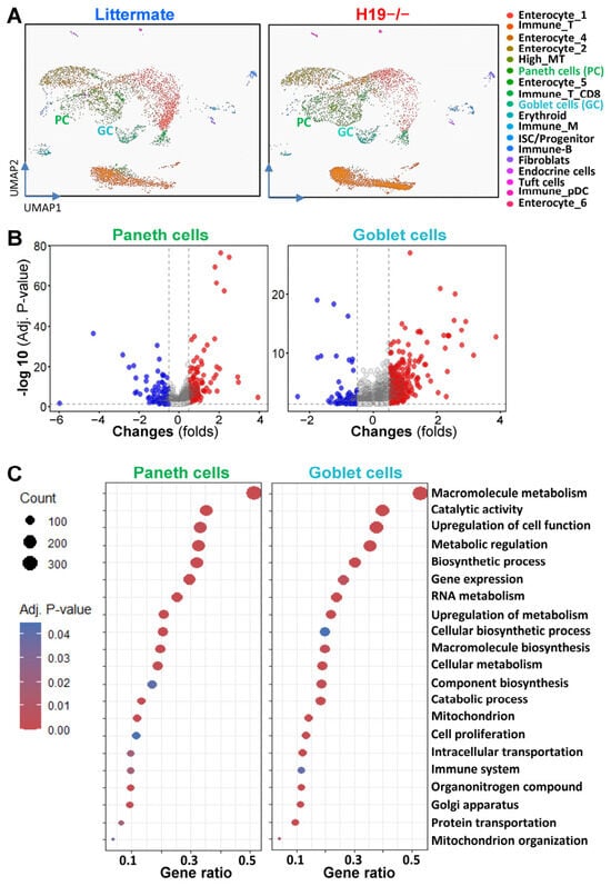
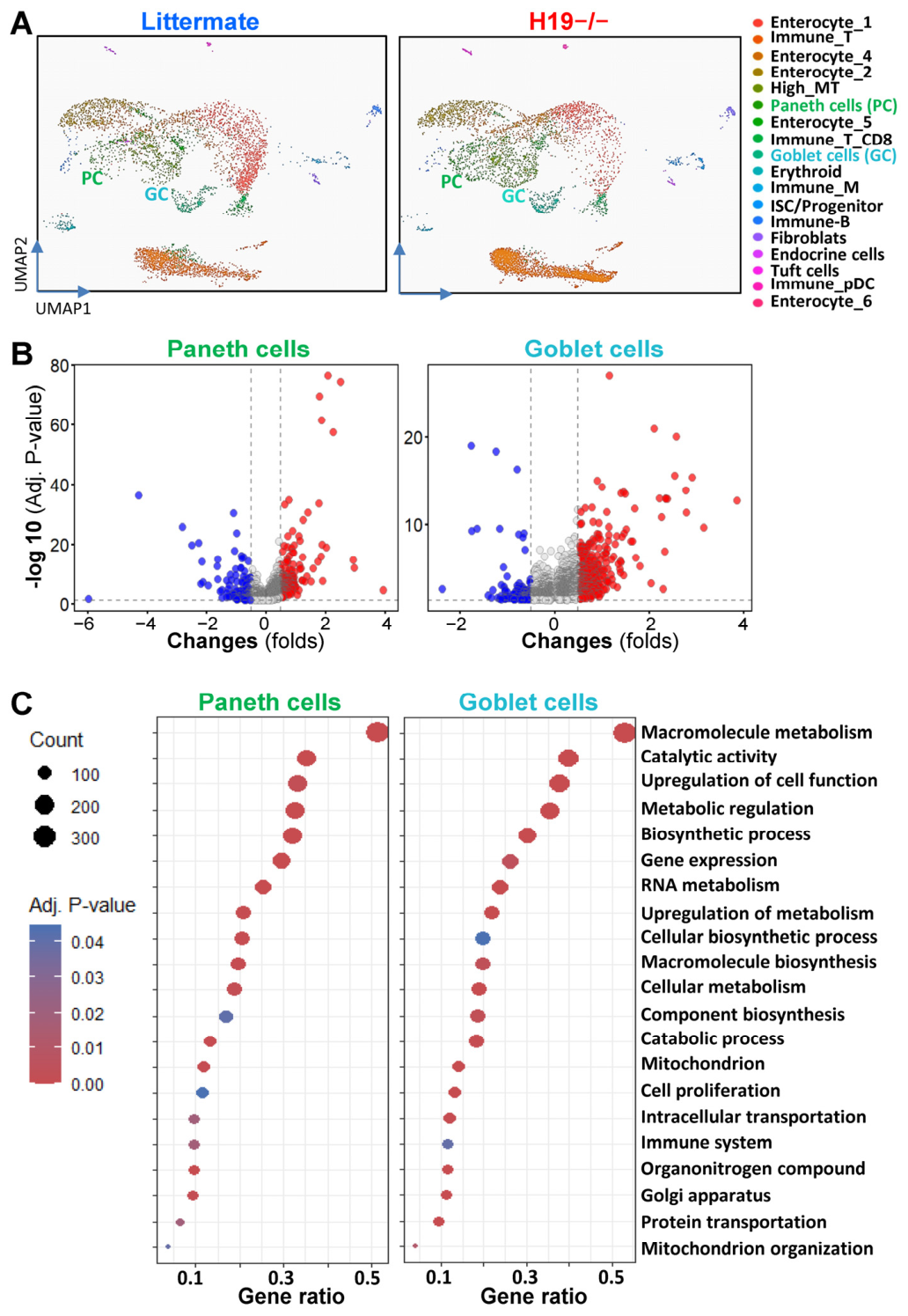
To investigate the role of H19 in the intestinal epithelial cell-specific transcriptome and cell lineage, single-cell RNA-sequencing (scRNA-seq) analysis was performed on the fresh tissues of small intestinal mucosa harvested from H19∆Exon1 (H19−/−) mice carrying a maternal deletion of exon 1 in the H19 gene [35,39] and control littermates. About 40,000 live cells were captured, as quality characterized and validated by flow-cytometry analysis as described previously [12,23], and grouped into 18 clusters that expressed classic cell type markers for the lineages of absorptive and secretory cells, aligned with published single-cell transcriptomic signatures [40]. Specifically, the secretory cells in the epithelial compartment of the small intestine consisted of goblet cells, tuft cells, Paneth cells, and enteroendocrine cells in both groups, while the absorptive cells included various enterocytes (Figure 1A). Of note, goblet cells expressed high levels of transcripts including Myo5c, Atoh1, spink4, Muc2Zg16, and Tff3, and Paneth cells had marked expression of Reg3a and Defa5 mRNAs, similar to others reports [12,40,41]. Enteroendocrine cells were marked by Chgb, Chga, Cck, Sct, Ghrl, Pyy, and Ambp; tuft cells were marked by Trpm5, Dclk1, Sox9, Sucnr1, Sh2d6, and Rgs13, while ISCs were marked by transcripts including Olfm4, Rgmb, Ascl2, Lgr5, and Smoc2 mRNAs. Cell cluster analysis suggested that the numbers of goblet cells and Paneth cells were increased in H19−/− mice compared to littermates, which were consistent with the findings obtained from studies using immunostaining assays in our previous work [35]. However, there was no significant difference in the numbers of enteroendocrine cells, tuft cells, and ISCs in the small intestinal mucosa between H19−/− mice and control littermates.
Figure 1.
Single-cell transcriptional profiles of Paneth cells and goblet cells in the small intestinal epithelium after deletion of the H19 in mice. (A) Uniform manifold approximation and projection of key epithelial cell types of the small intestinal mucosa from control littermate (left) and H19−/− mice (right). Annotated clusters of Paneth cells are shown in green; goblet cells are shown in light-green. (B) Scatter plot depictions of genes expressed differentially in Paneth cells (left) and goblet cells (right) in the small intestinal mucosa of littermate and H19−/− mice, as measured by scRNA-seq analysis. Values are the means from two separate experiments. (C) Causal analysis of most altered pathways involved in gene transcription and various cellular processes in Paneth cells (left) and goblet cells (right) after H19 deletion in mice.
The cell type-specific gene expression profiling demonstrated the existence of transcriptionally distinct Paneth cells and goblet cells in the small intestinal epithelium of H19−/− mice. We compared the transcriptomic profiles from H19−/− mice to control littermates and found that ~348 RNAs were differentially expressed in the Paneth cells, with 128 genes less abundant and 220 genes more abundant after H19 deletion (Figure 1B, left). In goblet cells, ~480 RNAs were differentially expressed, 128 RNAs were lower and 352 RNAs were higher in H19−/− mice compared to the mucosa of control littermates (Figure 1B, right). Causal analysis to screen for function-associated gene expressions in our single-cell sequencing data originated from Paneth cells and goblet cells further suggested that targeted deletion of the H19 gene in mice increased the activity of Paneth cells and goblet cells, as supported by significantly increased expression of mRNAs encoding proteins that are in charge of essential cellular processes and metabolism (Figure 1C). These activated pathways in the H19-deficient epithelium included the processes involved in macromolecular metabolism, catalytic activity, cellular biosynthesis, gene expression, RNA metabolism, and mitochondrial structure and function. Among these differentially abundant mRNAs are also many encoding transcription factors and regulatory proteins that are necessary for differentiation and function of secretory lineage cells in the intestinal epithelium. Since these cellular processes and transcriptional factors are essential for the function and effectiveness of Paneth cells and goblet cells [3,4], their activation by decreasing the levels of cellular H19 is implicated in maintaining the intestinal epithelium homeostasis in stressful environments. In summary, the data from our single-cell RNA sequencing clearly pointed out transcriptomic and cellular responses to H19 deletion in the small intestinal mucosa, and suggested an essential role of H19 in regulating Paneth and goblet cells in the intestinal epithelium.
2.2. Transgenic Expression of H19 in Mice Results in Defects in Secretory Lineage Cells
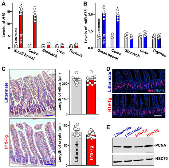
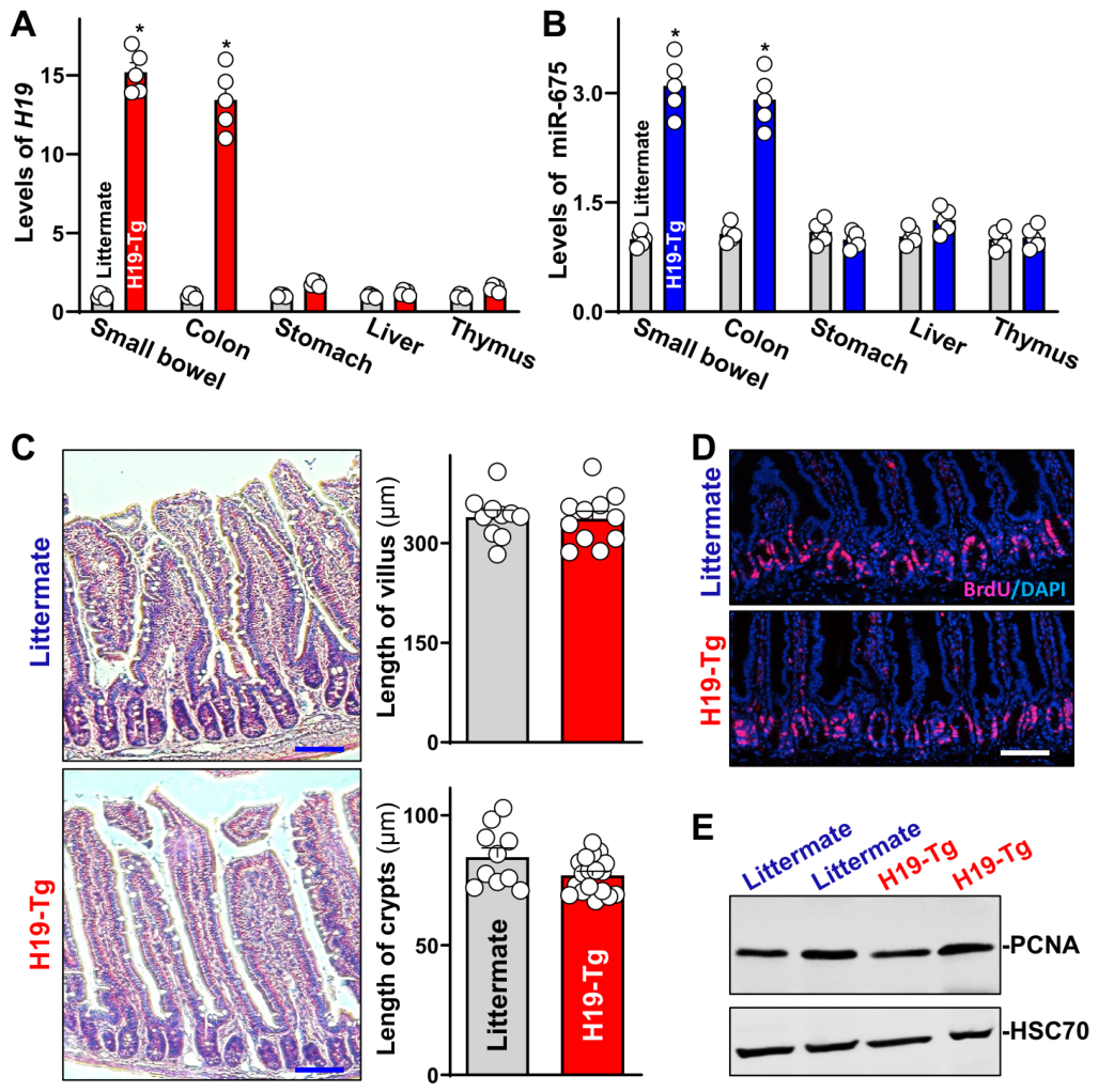
The basal level of H19 in the intestinal mucosa is relatively low [21,35]. To further investigate the role of H19 in the maturation of secretory lineage cells in vivo, we created intestinal epithelial tissue-specific H19 transgenic (H19-Tg) mice by using the A33 promoter, as described previously [42]. Age-matched H19-Tg mice and littermate controls (6–9 weeks old, both male and female) were used for phenotype comparison. H19-Tg mice exhibited a specific overexpression of H19 in the mucosal tissues of the small intestine and colon, but not in gastric mucosa, liver, and thymus (Figure 2A), as measured by quantitative real-time PCR (qRCR) analysis [43,44]. The levels of mucosal H19 in H19-Tg mice increased by ~15-fold in the small intestine and ~13-fold in colon, respectively, when compared with those observed in control littermates. Transgenic overexpression of H19 also specifically induced the expression levels of miRNA-675 (Figure 2B) that is embedded in H19 exon 1. However, overexpression of H19 did not change the levels of gene expression of other miRNAs such as miR-29b and miR-222. H19-Tg mice were generally normal and healthy, with no significant differences in appearance, body weight, gastrointestinal gross morphology, or reproductivity compared to control littermates.
Figure 2.
Characterization of intestinal epithelium-specific H19 transgenic (H19-Tg) mice. (A) Levels of tissue H19 in the small intestinal mucosa, colon, stomach, liver, and thymus as measured by RT-qPCR analysis. Total RNA was isolated and used in RT-qPCR reactions, and equal loading was monitored by assessing Gapdh mRNA levels. Values are the means ± SEM (n = 5). * p < 0.05 compared with control littermate mice. (B) Levels of mucosal miR-675 in various tissues as described in (A). Values are the means ± SEM (n = 5). * p < 0.05 compared with littermates. (C) Photomicrographs of hematoxylin and eosin (left) and changes in the length of villi and crypts (right) of the intestinal mucosa described in (A) (n = 10). (D) Proliferating cells in small intestinal crypts in control littermate and H19-Tg mice as measured by BrdU immunostaining. Red, Ki67; Blue, DAPI. Scale bars: 50 μm. (E) Immunoblots of growth-associated protein PCNA in the small intestinal mucosa. Whole-cell lysates were harvested from littermate and H19-Tg mice and prepared for Western immunoblotting analysis. HSC70 immunoblotting was performed as an internal control for equal loading. In (A–C), statistical significance was analyzed using unpaired, two-tailed Student’s t test. In (D,E), experiments were repeated three times and showed similar results.
H19 transgenic expression in the intestinal epithelium did not impact the renewal of the intestinal mucosa in vivo. There were no significant alterations in histological features of the small intestinal mucosa (Figure 2C, left), the lengths of villi and crypts (Figure 2C, right), the proliferating crypt cell population marked by BrdU incorporation (Figure 2D), and the levels of proliferation marker PCNA (Figure 2E) between H19-Tg mice and control littermates. In addition, H19 overexpression did not directly induce apoptosis in the intestinal epithelium, since there was no significant increase in cell death observed in H19-Tg mice, as examined by TUNEL staining [42]. These results indicate that H19-Tg mouse is an excellent tissue-specific gain-of-function model of H19 for studying maturation and function of the secretory lineage cells in the intestinal epithelium.
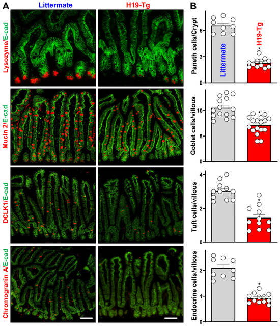
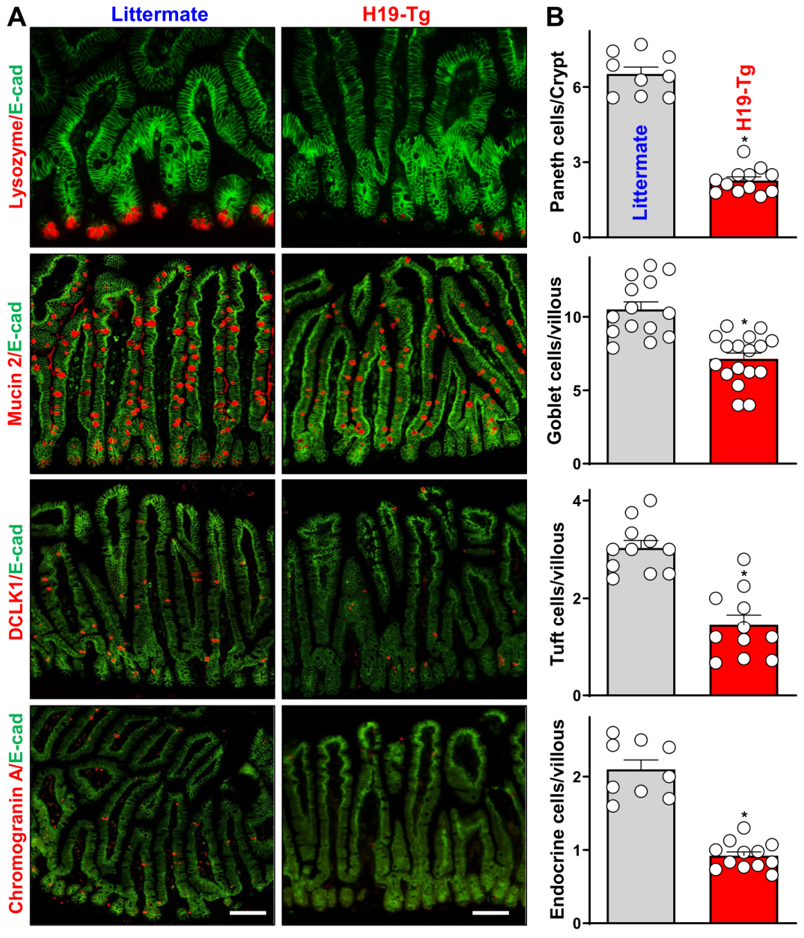
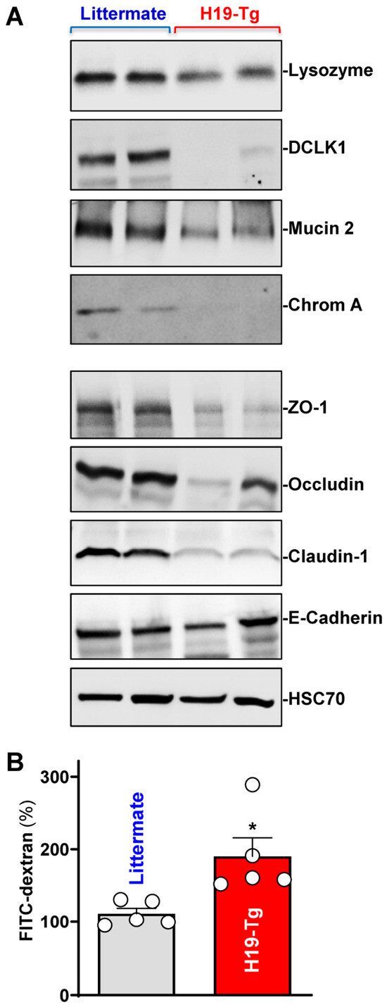
Tissue-specific transgenic expression of H19 in the intestinal epithelium caused defects in secretory lineage cells, such as Paneth cells, goblet cells, tuft cells, and enteroendocrine cells in the small intestinal mucosa of mice, as indicated by immunofluorescence staining [11,35]. The tissue staining of the small intestine suggested that in control littermates, lysozyme-positive cells (Paneth cells) were located at the base of the intestinal crypts, but mucin 2-positive cells (goblet cells), double cortin-like kinase 1 (DCLK1)-positive cells (tuft cells), and chromogranin A-positive cells (enteroendocrine cells) were distributed at both villous and crypt regions (Figure 3A, left). The numbers of Paneth cells, goblet cells, tuft cells, and enteroendocrine cells in the small intestinal mucosa were markedly reduced in H19-Tg mice compared to control littermates (Figure 3A, right). Quantification of this reduction indicated that the numbers of intestinal Paneth cells, goblet cells, tuft cells and enteroendocrine cells in H19-Tg mice decreased by ~65%, ~33%, ~52%, and ~56%, respectively, compared with control littermate mice (Figure 3B). Moreover, the levels of marker proteins for each secretory cells such as lysozyme, mucin 2, DCLK1 and chromogranin A in the small intestinal mucosa also decreased in H19-Tg mice (Figure 4A, top), as measured by immunoblotting analysis [43]. However, transgenic H19 overexpression failed to alter enterocyte differentiation in the small intestinal mucosa, as examined by villin immunostaining analysis.
Figure 3.
Transgenic expression of H19 in mice inhibits development of secretory lineage cells in the small intestinal epithelium. (A) Immunostaining of Paneth (lysozyme-positive), goblet (mucin 2-positive), tuft (DCLK1-positive), and enteroendocrine (chromogranin A-positive) cells in the small intestinal mucosa of littermate and H19-Tg mice. Red, lysozyme, mucin 2, DCLK1 or chromogranin A; and green, E-cadherin (E-cad). Scale bars: 50 μm. (B) Summarized results from studies described in (A). Values are the means ± SEM (n = 10). * p < 0.05 compared with control littermate mice. In (B), statistical significance was analyzed using unpaired, two-tailed Student’s t test.
Figure 4.
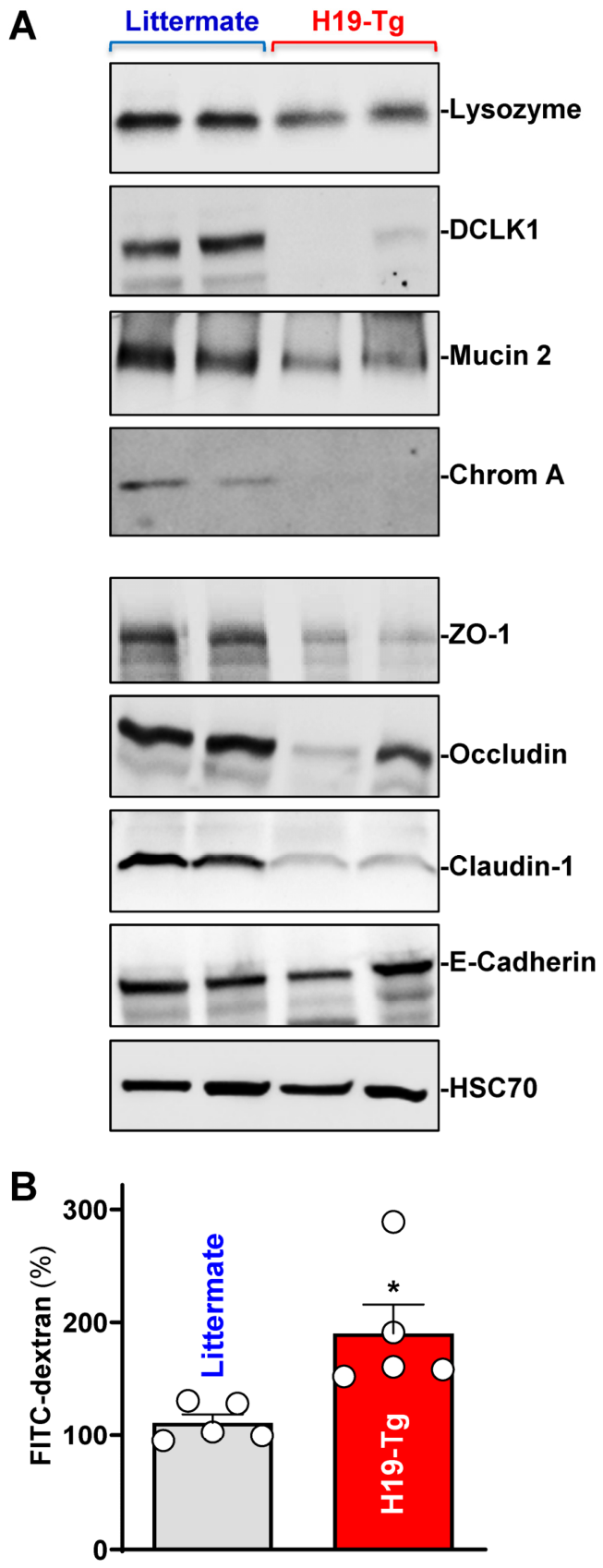
Transgenic H19 expression decreases the levels of marker proteins of secretory lineage cells and reduces the abundance of tight junction proteins in the small intestinal mucosa. (A) Immunoblots of lysozyme, mucin 2, DCLK1, chromogranin A (Chrom A), and tight junction proteins in the small intestinal mucosa of littermate and H19-Tg mice. HSC70 immunoblotting was performed as an internal control for equal loading. (B) Gut permeability in mice described in (A). FITC dextran was given orally, and blood samples were collected 4 h thereafter for measurement. Values are the means ± SEM (n = 5). * p < 0.05 compared with control littermates. In (B), unpaired, two-tailed Student’s t test was used for statistical analysis.
As expected, transgenic H19 overexpression in the intestinal epithelium disrupted the epithelial barrier function, as evidenced by decreased levels of tight junction proteins ZO-1, occludin, and claudin-1 in H19-Tg mice relative to control littermates (Figure 4A, bottom). Consistent with its effect on tight junction expression, H19-Tg mice exhibited increased basal levels of gut permeability compared to that observed in control littermates (Figure 4B), as measured by FITC-conjugated dextran assays [42]. On the other hand, increased H19 did not affect the level of adherens junction protein E-cadherin in the small intestinal mucosa. Another line of H19-Tg mice generated from an independent founder was also examined and displayed similar defects in secretory lineage cells and gut barrier dysfunction.
2.3. H19 Decreases Levels of Transcriptional Factors Necessary for Secretory Cell Differentiation
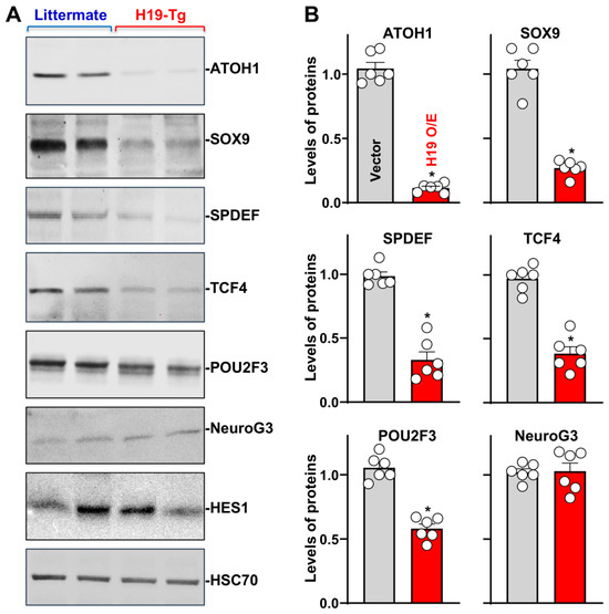
To investigate the mechanism underlying H19 in the regulation of secretory lineage cell maturation, we examined the expression levels of various transcription factors that are necessary for the differentiation of secretory lineage cells in the small intestinal epithelium. As shown in Figure 5 (top), the intestinal mucosa of H19-Tg mice exhibited a dramatical decrease in the levels of ATOH1, SOX9, SPDEF, TCF4, and POU2F3 proteins when compared with those observed in control littermates. Quantification analysis showed that the levels of tissue ATOH1, SOX9, SPDEF, TCF4, and POU2F3 proteins in the intestinal mucosa of H19-Tg mice decreased by ~88%, ~75%, ~68%, ~60%, and ~45%, respectively (Figure 5B). In contrast, there were no significant differences in the mucosal abundances of NeuroG3 and HES1 proteins in the small intestine between H19-Tg mice and control littermates (Figure 5A, bottom). These results indicate that locally increasing the levels of H19 in the intestinal epithelium decreases the expression levels of several differentiation-associated transcription factors, including ATOH1, along with defects in secretory lineage cells.
Figure 5.
Transgenic H19 expression decreases the levels of ATOH1 and other differentiation-associated transcription factors in the small intestinal mucosa. (A) Immunoblots of ATOH1, SOX9, SPDEF, Tcf4, POU2F3, NeuroG3, and HES1 in the small intestinal mucosa of littermate and H19-Tg mice. HSC70 immunoblotting was performed as an internal control for equal loading. (B) Summarized data from studies as described in (A). Values are the means ± SEM (n = 6). * p < 0.05 compared with control littermates. In (B), statistical significance was analyzed using unpaired, two-tailed Student’s t test.
2.4. Increased ATOH1 Rescues Maturation of Secretory Cells in H19-Enriched Intestinal Organoids
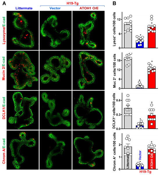
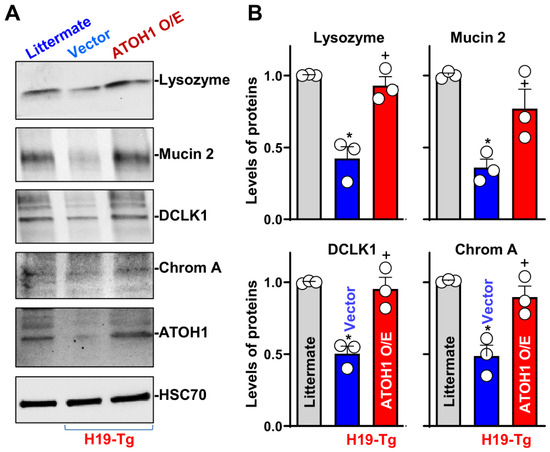
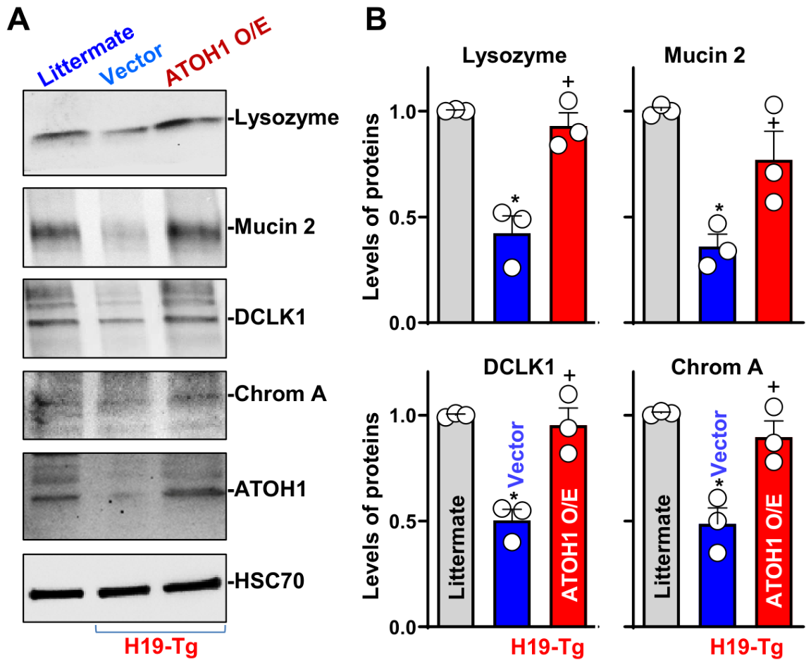
Since ATOH1 is the key prosecretory basic helix-loop-helix transcription factor and its loss leads to depletion of the secretory lineages in the intestinal epithelium [1,3], we elucidated the role of altered ATOH1 in H19-mediated defects in the secretory lineage cells in an ex vivo model using primarily cultured intestinal organoids. As described previously [35,45], intestinal organoids were derived from proliferating crypts of the small intestinal mucosa of H19-Tg and control littermate mice. By 5 days after initial culture, the structures of intestinal organoids from both H19-Tg and littermate mice consisted of multiple buds and cells. As expected, there were no significant differences in the growth of intestinal organoids between the two groups, as examined by the organoid surface area and BrdU incorporation. In agreement with the observations in vivo, intestinal organoids derived from H19-Tg mice exhibited decreased numbers of secretory lineage cells, including Paneth cells, goblet cells, tuft cells and enteroendocrine cells, when compared with those observed in the organoids generated from littermate mice (Figure 6A, middle). Quantification of these results showed that the numbers of Paneth cells, goblet cells, tuft cells, and enteroendocrine cells in the organoids derived from H19-Tg mice decreased by ~68%, ~90%, ~84%, and ~82%, respectively, compared to those observed in the organoids from littermate mice (Figure 6B, middle). Consistently, the levels of lysozyme, mucin 2, DCLK1, chromogranin A, and ATOH1 proteins in the intestinal organoids derived from H19-Tg mice were also decreased (Figure 7), as measured by immunoblotting analysis. These results indicate that defects in secretory lineage cells in H19-Tg mice results primarily from a cell-autonomous regulatory effect of this lncRNA but not from the effect of secreted factors in the gut.
Figure 6.
Ectopic overexpression of ATOH1 restores differentiation of secretory lineage cells in intestinal organoids derived from H19-Tg mice. (A) Immunostainings of Paneth cells (lysozyme-positive cells), goblet cells (mucin 2-positive cells), tuft cells (DCLK1-positive cells), and endocrine cells (chrom A-positive cells) in intestinal organoids 48 h after transfection with an ATOH1 expression vector or a control empty vector. (B) Quantitative data of these secretory lineage cells in intestinal organoids described in panel (A). Values are the means ± SEM (n = 10). * p < 0.05 compared with control littermates; + p < 0.05 compared with empty vector-transfected organoids derived from H19-Tg mice. In (B), statistical significance was analyzed using one-way ANOVA with Tukey’s post hoc test.
Figure 7.
Ectopic overexpression of ATOH1 prevents a decrease in the levels of marker proteins of different secretory cells in intestinal organoids derived from H19-Tg mice. (A) Immunoblots of lysozyme, mucin 2, DCLK1 and chromogranin A (chrom A) proteins in intestinal organoids 48 h after transfection with control or ATOH1 expression vector. HSC70 immunoblotting was performed as an internal control for equal loading. (B) Summarized data from studies as described in (A). Values are the means ± SEM (n = 3). *,+ p < 0.05 compared with littermates and H19-overexpressed organoids transfected with control vector, respectively. In (B), statistical significance was analyzed using one-way ANOVA with Tukey’s post hoc test.
To examine the effect of increasing the levels of ATOH1 on differentiation of the secretory lineage cells in intestinal organoids isolated from H19-Tg mice, we constructed a plasmid vector expressing ATOH1 under control of the pCMV promoter as reported previously [46]. Ectopically expressed ATOH1 in the H19-enriched organoids derived from H19-Tg mice improved development of the secretory lineage cells, as evidenced by a significant increase in the numbers of Paneth cells, goblet cells, tuft cells, and enteroendocrine cells in the organoids transfected with the ATOH1 expression vector relative to those transfected with the empty control vector (Figure 6A,B, right). Consistently, the levels of lysozyme, mucin 2, DCLK1, and chromogranin A proteins were also returned to near normal levels in the organoids derived from H19-Tg mice after ATOH1 overexpression (Figure 7). These data indicate that defects in development and differentiation of secretory lineage cells in the intestinal organoids derived from H19-Tg mice result primarily from the inhibition of ATOH1.
2.5. H19 Inhibits ATOH1 Expression via Repression of Its Translation
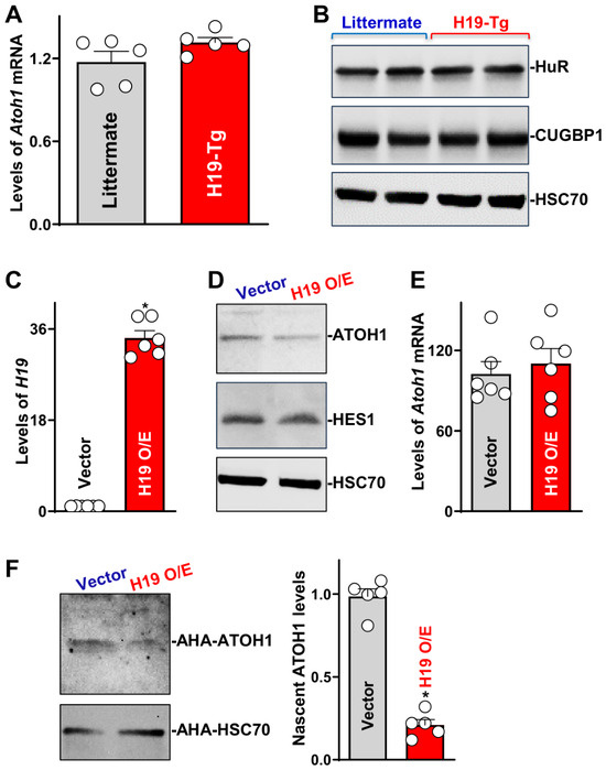
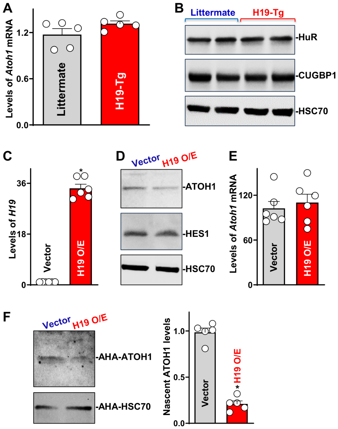
To examine the mechanism by which H19 regulates ATOH1 expression, we examined levels of the Atoh1 mRNA in the intestinal mucosa and demonstrated that increasing the levels of H19 did not alter transcription of the Atoh1 gene, since there were no significant differences in the levels of Atoh1 mRNA between H19-Tg mice and control littermates (Figure 8A). To determine the involvement of RBPs in this process, we examined changes in the levels of mucosal HuR and CUG-binding protein 1 (CUGBP1) in the small intestine of H19-Tg mice. Our results revealed that transgenic expression of H19 failed to affect expression levels of RBPs, since the levels of tissue HuR and CUGBP1 in the intestinal mucosa of H19-Tg mice were indistinguishable from those observed in control littermate mice (Figure 8B).
Figure 8.
Ectopic H19 overexpression inhibits translation of ATOH1 in cultured IECs. (A) Levels of the Atoh1 mRNA in the small intestinal mucosa of littermate and H19-Tg mice. Total RNAs were isolated from the tissue and examined by RT-qPCR analysis. Values are the means ± SEM (n = 5). (B) Immunoblots of RNA-binding proteins HuR and CUGBP1 in the mucosal tissue described in (A). HSC70 immunoblotting was performed as an internal control for equal loading. (C) Levels of cellular H19 in Caco-2 cells 48 h after transfection with the H19 expression vector. Values are the means ± SEM (n = 6). * p < 0.05 compared with control vector. (D) Immunoblots of ATOH1 and HES1 proteins in cells described in (C). (E) Levels of the Atoh1 mRNA in cells described in (C) (n = 6). (F) Newly synthesized ATOH1 protein as measured by L-azidohomoalaine (AHA) incorporation assays in cells described in (C). Values are the means ± SEM (n = 5). * p < 0.05 compared with control vector. In (A,C,E,F), statistically significant was analyzed using unpaired, two-tailed Student’s t test. In (B,D), experiments were repeated three times and showed similar results.
In cultured IECs, ectopically expressed H19, by transfection with its expression vector (Figure 8C), decreased the levels of ATOH1 protein (Figure 8D), although it did not alter level of the Atoh1 mRNA (Figure 8E). Consistent with the observation in H19-Tg mice, H19 overexpression did not reduce the abundance of cellular HES1 protein in IECs. However, ectopically expressed H19 inhibits ATOH1 translation, since the levels of newly synthesized ATOH1 protein decreased dramatically in cells overexpressing H19 relative to cells transfected with control vector (Figure 8F). The translational rate of ATOH1 in H19-transfected cells decreased by ~79%, compared with those from cells transfected with control vector. These results strongly suggest that H19 inhibits ATOH1 expression primarily by decreasing Atoh1 mRNA translation in the intestinal epithelium.
3. Discussion
In response to different stresses, activation and effectiveness of secretory lineage cells in the intestine are essential for sustaining the epithelial homeostasis by enhancing the epithelium-host defense, constant mucosal renewal, immune-response, and gut barrier function [3,4,10]. Our previous study showed that targeted deletion of H19 in mice increases the function of Paneth and goblet cells, thus promoting gut barrier function [35], but the exact mechanism underlying the control of secretory lineage-cell function by H19 remains unknown. The present study provides genetic evidence demonstrating the importance of H19 in regulating the development and maturation of secretory lineage cells via the posttranscriptional control of transcription factor ATOH1. Intestinal epithelial tissue-specific transgenic expression of H19 in mice decreased the levels of ATOH1 protein and led to defects in Paneth cells, goblet cells, tuft cells, and enteroendocrine cells. In contrast, ectopically expressed ATOH1 in primarily cultured intestinal organoids derived from H19-Tg mice rescued the development of secretory lineage cells ex vivo. These findings advance our knowledge about the maturation of secretory lineage cells in the intestinal mucosa and reveal the important role of the H19/ATOH1 axis in the regulation of secretory lineage cell function and the epithelial integrity, which in turn influences mucosal injury, inflammation, and gut permeability in various pathological conditions.
Our previous study has shown that the mucosal levels of H19 in the intestine increase remarkably in mice exposed to septic stress induced by cecal ligation and puncture (CLP) and in patients with sepsis, along with defective secretory lineage cells, whereas H19 knockout enhances the function of Paneth and goblet cells and protects the gut barrier against CLP in H19−/− mice [35]. The present study further reveals that there were significant differences in cell type-specific gene expression profiles of secretory lineage cells between H19−/− mice and control littermates. H19-deficient Paneth and goblet cells in the small intestine of H19−/− mice exhibited increased expression levels of many genes involved in various cellular processes including macromolecule metabolism, biosynthetic process, gene expression, and RNA metabolism. Conversely, locally increasing the levels of H19 in the intestinal epithelium of H19-Tg mice caused defects in secretory lineage cells, as evidenced by a significant decrease in the numbers of Paneth cells, goblet cells, tuft cells, and enteroendocrine cells, although increased levels of H19 failed to affect mucosal growth in the small intestine. Importantly, transgenic expression of H19 in the intestinal epithelium also disrupts gut barrier function, since increasing the levels of tissue H19 not only decreased levels of tight junction proteins but also increased basal level of gut permeability in H19-Tg mice. These findings clearly demonstrate the essential role of H19 in regulating development and function of the secretory lineage cells in the intestinal epithelium and subsequent gut barrier function.
The results presented here also indicate that H19 represses the development of secretory lineage cells at least partially by inhibiting ATOH1 expression. ATOH1 is a master transcription factor in prosecretory cells by activating the expression of other transcription factors that induce differentiation of specific secretory cells [3,47,48]. In this regard, SOX9 and SPDEF are necessary for differentiation of Paneth cells and goblet cells, respectively, and TCF4 controls the balance between Paneth and goblet cells [8,43,49]. POU2F3 induces tuft cell maturation, while neuroG3 is involved in the generation of enteroendocrine cells [3,50]. Activation of ATOH1 by overexpression of its transgene or ablation of its natural repressor HES1 causes excessive differentiation of prosecretory cells into all secretory cells [51,52]. In contrast, targeted deletion of the Atoh1 gene leads to depletion of whole secretory lineages in the intestinal epithelium [53]. Although transgenic expression of H19 also decreased the levels of SOX9, SPDEF, TCF4, and POU2F3 in the intestinal mucosa, defects in the secretory lineage cells in H19-Tg mice may result primarily from reduction in the levels of tissue ATOH1. Ectopically expressed ATOH1 in the H19-enriched intestinal organoids derived from H19-Tg mice restored development of all secretory cell lineages in an ex vivo model. Since neither increased H19 in H19-Tg mice nor ATOH1 overexpression in intestinal organoids altered growth of the intestinal mucosa, the regulation of secretory lineage cells by H19 via ATOH1 results from the effect of H19 on cell differentiation rather than on ISC proliferation and mucosal turnover.
Our results further show that H19 regulates ATOH1 expression in the intestinal epithelium through control of ATOH1 translation without effect on its gene transcription. Increased H19 did not affect the levels of Atoh1 mRNA in vivo nor in vitro but it inhibited the rate of newly synthesized ATOH1 protein in cultured IECs. This inhibitory effect of H19 on ATOH1 expression is specific, since H19 overexpression failed to alter the levels of HES1 protein. H19 has multiple cellular functions by acting on specific targets via different molecular mechanisms, particularly through the H19/miRNA/mRNA-competing endogenous RNA network, where H19 functions as a mRNA sponge [21,54,55]. H19 modulates skeletal muscle metabolism, differentiation and regeneration by acting as an upstream regulator of the miRNA let-7 that inhibits expression of critical metabolic genes such as Insr and Lpl [56,57]. H19 also functions as an RNA decoy for miR-34b and let-7, two microRNAs involved in controlling the expression of p53 and other apoptosis-associated proteins [32,36]. H19 controls expression of the imprinted network genes by recruiting repressive histone markers to their differentially methylated regions and it inhibits placental growth through the processing of the embedded miR-675 [37,58]. H19 interacts with the RBP HuR that decreases miR-675 levels by inhibiting its processing from H19 [37]. Our previous study reveals that H19 represses expression of intercellular junction proteins ZO-1 and E-cadherin and impairs the epithelial barrier function in cultured IECs by acting as a regulatable reservoir of miR-675 [21]. However, the exact mechanism by which H19 inhibits ATOH1 translation remains unknown and should be fully investigated in a future study.
In summary, our results from studies of mice, intestinal organoids, and IECs indicate that by down-regulating ATOH1 translation, H19 inhibits development and function of secretory lineage cells in the small intestinal epithelium. Tissue-specific transgenic expression of H19 in mice decreased the levels of differentiation-associated transcription factors including ATOH1, which was associated with defects in the secretory lineage cells in the mucosa. Importantly, ectopically expressed ATOH1, by transfection with its expression vector, restored development of all secretory cells in the H19-enriched organoids derived from H19-Tg mice. Moreover, H19 inhibits ATOH1 expression at the translation level, and this regulation of ATOH1 by H19 is critical for maintaining secretory cell development and function. The present study strongly supports a novel model whereby rapidly altered levels of mucosal H19 in stressful environments functionally regulate ATOH1 expression to affect maturation and function of secretory lineage cells, thus contributing to the epithelium-host defense and effectiveness of the gut barrier function. However, there are only limited results showing the molecular mechanism underlying the control of ATOH1 translation by H19 in this study, and the exact role of altered H19 in the pathogenesis of IBD and sepsis-induced gut mucosal disruption remains largely unknown. Since the levels of tissue H19 increase dramatically in patients with critical illnesses such as sepsis and gut mucosal inflammation [35,36], these findings provide a better understanding of control of the secretory cell function in pathological conditions and identify the H19/ATOH1 axis as a potential therapeutic target for interventions to preserve the intestinal epithelial integrity and gut barrier function in the clinical setting.
4. Materials and Methods
4.1. Animal Studies
All animal experiments were performed in accordance with NIH guidelines and were approved by the Institutional Animal Care and Use Committee of University Maryland School of Medicine and Baltimore VA hospital. Wild-type C57BL/6J mice (male and female, 6 to 9 weeks old) were purchased from The Jackson Laboratory (Bar Harbor, ME, USA). H19−/− mice were kindly provided by Dr. Karl Pfeifer, NIH [39], and they carried a 1-kb deletion of H19 exon 1 that removes almost the entire exon 1, including the region encoding miR-675, and reduces levels of even the partial H19 transcript by >100-fold globally [35,39,59].
H19-Tg mice were generated by using A33-promoter and following the strategy as we used in the generation of miR-195, miR-222, and vtRNA1-1 transgenic mice [42,60,61]. The 2.2-kb fragment flanking the mouse H19 locus (including exon1) was cloned into the pIRES-AcGFP1-Nuc vector. Then, the final H19 expression vector, A33-H19/GFP, was injected into fertilized murine eggs [61,62]. Harvard University’s Genome Modification Facility established transgenic founder mice on pure C57BL/6J background by pronuclear injection. The first generation of recombinant mice with H19/GFP bicistronic RNA was identified by PCR genotyping of DNA extracted from tail clippings. Two founder mice were further characterized to subsequently establish transgenic colonies. To include proper littermate control mice, we crossed male Tg mice with wild-type female mice to generate wild-type and H19 transgene-positive mice. To exclude the possible effect of the A33-GFP transgene, A33-GFP mice were used as pseudo-transgene controls. All H19−/−, H19-Tg, and control littermate mice were housed in a specific pathogen-free breeding barrier and cared for by trained technicians and veterinarians.
Animals were deprived of food but allowed free access to tap water for 24 h before experiments. To examine gut mucosal growth in transgenic and control mice, BrdU was incorporated into intestinal mucosa via intraperitoneal (i.p.) injection of 2 mg BrdU (Sigma, St. Louis, MO, USA) diluted in phosphate-buffered saline. Two portions of the small intestine 0.5 cm distal to the ligament of Trietz were removed. One segment was used for histological examination, while the other segment was reserved for extraction of protein and RNA. The mucosa was scraped with a sterile cell scraper for various measurements as described previously [24,43]. Representative results from two independent founder mice were reported and compared with data obtained from control littermate mice.
4.2. Cell and Intestinal Organoid Culture
Human colorectal carcinoma Caco-2 cells originally sourced from the American Type Culture Collection (Manassas, VA, USA) were maintained under standard culture conditions [20,24]. The basal medium and fetal bovine serum were purchased from Invitrogen (Carlsbad, CA, USA), and biochemical reagents were purchased from Sigma (St. Louis, MO, USA). Isolation and culture of primary enterocytes were conducted following the protocol provided by STEMCELL Technologies (Cambridge, MA, USA) with minor modifications as described previously [35,45]. To briefly summarize, primary intestinal crypts were first isolated from murine small intestinal tissue segments. Then, isolated intestinal crypt fractions were assessed, suspended in Matrigel (Corning, Tewksbury, MA, USA), and cultured in mouse IntestiCult™ organoid growth medium (STEMCELL technology, Vancouver, BC, Canada). The growth of enteroids was examined under phase-contrast microscopy as described.
4.3. Plasmid Construction
An expression vector containing a full-length of cDNA encoding ATOH1 under the control of pCMV promoter was purchased from Origene (Rockville, MD, USA) and used to increase the ATOH1 levels in intestinal organoids as described previously [21,46]. Transient transfections were performed using the Lipofectamine reagent following the manufacturer’s recommendations (Invitrogen). Forty-eight hours after transfection, organoids were harvested for analysis.
4.4. Isolation of Intestinal Epithelial Cells and scRNA-Seq Analysis
The small intestinal mucosal tissues harvested from littermate and H19 knockout mice were weighed, washed in cold D-PBS and sectioned with a scalpel, as described previously [23]. The washed mucosal tissue segments were incubated in chelation medium (20 mM EDTA-DPBS) at 37 °C for 90 min with gentle agitation. Then, villus-rich supernatant was collected and filtered through a 40 μm filter. The pellet is further dissociated with TrypLE for 10 min at 37 °C (crypt-rich cells) and later combined with villus-rich fragment. The epithelial single-cell suspension was washed in DPBS with 1% BSA and passed through 40 μm filters. The purity of epithelial populations was confirmed by flow-cytometry analysis before proceeding to scRNA-sequencing. Once viability and EPCAM purity were confirmed, these high-quality intestinal epithelial cells were directly loaded for droplet-based scRNA-sequencing according to the manufacturer’s protocol for the Chromium Single Cell Platform (10X Genomics, 3’V2) to obtain ~10,000 cells per reaction. The single-cell sequencing library preparation was carried out according to the manufacturer’s protocol. Using the Cell Ranger software v.9.0.1 and mm10-2020-A transcriptome as a reference, 10× Genomics scRNA-seq gene expression raw sequencing data were processed. Following previous established methods [40,41], the dimensionality reduction, Leiden clustering, differential abundance analysis, cell-type composition analysis, gene enrichment analysis, and mitochondrion Gene Ontology Term (GO: 0005739) were performed with these samples. The threshold used for identifying differentially expressed genes is p-value < 0.05 and log2(Fold-change) > 0.25.
4.5. Histology and Immunofluorescence Staining1
Dissected and opened intestines were mounted onto a solid surface and fixed in formalin and paraffin. Sections of 5 μm thickness were stained with H&E for general histology. As described previously [46], the overall length of villus and crypts of each intestinal section was measured by using a calibrated micrometer eyepiece. Immunofluorescence staining was performed according to established protocols [11,12]. For experiments analyzing murine mucosal tissue samples, more than five 5 μm-thick slides (5 μm thickness section) per tissue sample were prepared for immunofluorescence staining. For studies utilizing cultured intestinal organoids, slides were fixed in 3.7% formaldehyde in PBS and rehydrated. All slides were incubated overnight with a primary antibody in blocking buffer recognizing lysozyme, mucin 2, DCLK1, chromogranin A, or E-cadherin, then incubated with secondary antibody conjugated with Alexa Fluor-594 (Cat No. A32754, Molecular Probes, Eugene, OR, USA) for 2 h at room temperature. After rinsing 3 times, the slides were incubated with DAPI (Cat No. D1306, Molecular Probes, Thermo Fisher Scientific, Eugene, OR, USA) to stain cell nuclei at a concentration of 1 μM for 10 min. Finally, the tissue slides were washed, mounted, and imaged through a Zeiss confocal microscope (model LSM700, Carl Zeiss Microscopy GmbH, Jena, Germany). To ensure unbiased results, the slides were examined while coded, and they were decoded only after data collection was completed. Images were processed using Photoshop software version 25.0 (Adobe, San Jose, CA, USA).
4.6. Measurement of Gut Permeability
FITC-conjugated dextran diluted in sterile 1x phosphate-buffered saline (Cat No. FD4, Sigma; 4KD; 600 mg/kg) was administered to mice via oral gavage as described [42]. After 4 h, blood was collected via cardiac puncture. The serum concentration of the FITC-dextran suspension was examined using a fluorescence plate reader set at an excitation wavelength of 490 nm and an emission wavelength of 530 nm. The concentration of FITC-dextran in sera was determined by comparing the values to the FITC-dextran standard curve.
4.7. Reverse Transcription and Q-PCR Analysis
Total RNA was extracted by using RNeasy mini kit (Qiagen, Valencia, CA, USA), and reverse transcription and subsequent PCR amplification reactions were performed as described [23]. After performing RT, quantitative PCR (Q-PCR) analysis was conducted using 7500-Fast Real-Time PCR Systems with specific target primers, probes, and software (Applied Biosystems, Foster City, CA, USA). The levels of Gapdh PCR product were measured in RT- quantitative (q) PCR samples to account for potential variations in RNA input. For miRNA studies, miR-675 levels were also quantified by Q-PCR following the Taqman MicroRNA assay. Small nuclear RNA (snRNA) U6 levels were measured as an endogenous control. All Q-PCR primers were purchased from Thermo Fisher Scientific (Waltham, MA, USA).
4.8. Western Immunoblotting and Newly Synthesized Protein Assays
Whole-cell lysates were prepared by using the RIPA buffer (50 mM Tris pH 7.4, 150 mM NaCl) containing 1% sodium dodecyl sulfate (SDS), sonicated, and centrifuged at 4 °C for 15 min [35]. The supernatants were boiled and size-fractionated by SDS-PAGE. After the blots were incubated with primary antibody and then secondary antibodies, immunocomplexes were developed by using chemiluminescence. Relative protein levels were analyzed by using Biorad Chemidoc (Bio-Rad Laboratories, Inc., Hercules, CA, USA) and XRS system equipped with Image lab software (version 4.1), and the values were normalized with internal loading control HSC70.
In addition, new protein synthesis was measured by Click-iT Protein Analysis Detection Kit (Life technologies, Grand Island, NY, USA) following the manufacturer’s manual. First, Caco-2 cells were incubated with methionine-free medium. Then, to label nascent protein synthesis, cells were exposed to and metabolically incorporated azide-modified amino acid L-azidohomoalanine (AHA). Next, the cell lysates were combined with the reaction buffer and incubated for 20 min. Following incubation, paramagnetic Streptavidin-conjugated Dynabeads (Invitrogen, Thermo Fisher Scientific, Waltham, MA, USA) were used to pull down biotin-alkyne/azide-modified protein complexes. Finally, the precipitated samples were resolubilized in 10% SDS-PAGE and analyzed by Western immunoblotting analysis using antibodies against ATOH1 or HSC70 as a loading control.
4.9. Chemicals and Antibodies
Biochemicals were purchased from Sigma (St. Louis, MO, USA), and culture medium and fetal bovine serum were from Invitrogen (Carlsbad, CA, USA). Antibodies recognizing lysozyme (sc-518012), DCLK1 (Cat No. ab31704), mucin 2 (Cat No. ab272692), chromogranin A (Cat No. PA5-77917), ZO-1 (Cat No. 5406S), occludin (Cat No. 91131S), claudin-1 (Cat No. 37-4900), E-cadherin (Cat No. 610182), ATOH1 (Cat No. PA5-29392), SOX9 (Cat No. 82630S), SPDEF (Cat No. 11467-1-AP), TCF4 (Cat No. 2569T), POU2F3 (Cat No. HPA019652), neuroG3 (Cat No. 703206), HES1 (Cat No. 11988S), PCNA (Cat No. 2586S), HuR (Cat No. SC5261), CUGBP1 (sc-5261), and HSC70 (MABE1120) were obtained from Cell Signaling Technology (Danvers, MA, USA), Invitrogen, Santa Cruz Biotechnology, BD Biosciences (Sparks, MD, USA), Proteintech (Rosemont, IL, USA), and Atlas Antibodies (Stockholm, Sweden). The secondary antibody conjugated to horseradish peroxidase was purchased from Sigma. All antibodies utilized in this study were validated for species specificity. Antibody dilutions used for Western immunoblotting analysis were 1:800 or 1000 (first Ab) and 1:2000 (second AB), respectively, whereas antibody dilutions for immunostaining were 1:200 (first) and 1:2000 (second).
4.10. Statistical Analysis
All data values were presented as the means ± SEM, derived from five animals or three separate experiments. When applicable, an unpaired, two-tailed Student’s t-test was used to determine statistical significance p < 0.05. For multiple group comparisons, one-way ANOVA was utilized with Tukey’s post hoc test [63]. Non-parametric rank comparisons were evaluated using the Kruskal–Wallis test. All statistical analyses were conducted in GraphPad Prism 9.0 (San Diego, CA, USA). For non-parametric analysis rank comparison, the Kruskal–Wallis test was conducted.
Author Contributions
M.S.K. performed most experiments and summarized data. H.K.C. generated H19-Tg mice; H.C., A.V., B.W., H.Z., P.A., and E.T. participated in experiments using mice and experiments conducted in intestinal organoids and cultured IECs. D.J.T. and L.X. participated in data analysis and edited the manuscript. J.-Y.W. designed experiments, analyzed data, prepared figures, and drafted the manuscript. All authors have read and agreed to the published version of the manuscript.
Funding
This work is the result of NIH funding, in whole or in part, and is subject to the NIH Public Access Policy. Through acceptance of this federal funding, the NIH has been given a right to make the work publicly available in PubMed Central. This work was supported by Merit Review Award (BX000332 to J.Y.W.) from the US Department of Veterans Affairs; and grants from the National Institutes of Health (NIH) (DK57819, DK68491 to J.Y.W.) and (AG084613 to L.X.).
Institutional Review Board Statement
The animal study protocol was approved by the Institutional Review Board at University of Maryland Baltimore under protocol AUP-00000858 approved on 15 March 2024.
Informed Consent Statement
Not applicable.
Data Availability Statement
All data are available upon request due to the process of data deposition.
Conflicts of Interest
This author discloses the following: Jian-Ying Wang is a Senior Research Career Scientist (IK6BX004213) at the Biomedical Laboratory Research and Development Service, US Department of Veterans Affairs. The remaining authors disclose no conflicts.
References
- Bankaitis, E.D.; Ha, A.; Kuo, C.J.; Magness, S.T. Reserve stem cells in intestinal homeostasis and injury. Gastroenterology 2018, 155, 1348–1361. [Google Scholar] [CrossRef] [Scilit] [PubMed]
- Capdevila, C.; Miller, J.; Cheng, L.; Kornberg, A.; George, J.J.; Lee, H.; Botella, T.; Moon, C.S.; Murray, J.W.; Lam, S.; et al. Time-resolved fate mapping identifies the intestinal upper crypt zone as an origin of Lgr5+ crypt base columnar cells. Cell 2024, 187, 3039–3055.e14. [Google Scholar] [CrossRef] [Scilit] [PubMed]
- Beumer, J.; Clevers, H. Cell fate specification and differentiation in the adult mammalian intestine. Nat. Rev. Mol. Cell Biol. 2021, 22, 39–53. [Google Scholar] [CrossRef] [Scilit] [PubMed]
- Wallaeys, C.; Garcia-Gonzalez, N.; Libert, C. Paneth cells as the cornerstones of intestinal and organismal health: A primer. EMBO Mol. Med. 2023, 15, e16427. [Google Scholar] [CrossRef] [Scilit] [PubMed]
- Chege, N.; Finney, C.A.M. Mechanistic insights into intestinal stem cell disruption during infection. Am. J. Physiol. Gastrointest. Liver Physiol. 2025, 329, G371–G389. [Google Scholar] [CrossRef] [Scilit] [PubMed]
- Goga, A.; Yagabasan, B.; Herrmanns, K.; Godbersen, S.; Silva, P.N.; Denzler, R.; Zund, M.; Furter, M.; Schwank, G.; Sunagawa, S.; et al. miR-802 regulates Paneth cell function and enterocyte differentiation in the mouse small intestine. Nat. Commun. 2021, 12, 3339. [Google Scholar] [CrossRef] [Scilit] [PubMed]
- Zhang, Z.; Wan, Q.; Zhu, Y.; Ye, J.; Fu, F.; Fan, X.; Chen, H.; Ren, G.; Jiang, W.; Guo, X.; et al. Lipoic acid functions in Paneth cells to prevent human intestinal stem cell aging. Nat. Commun. 2025, 16, 6016. [Google Scholar] [CrossRef] [Scilit] [PubMed]
- Cray, P.; Sheahan, B.J.; Dekaney, C.M. Secretory sorcery: Paneth cell control of intestinal repair and homeostasis. Cell. Mol. Gastroenterol. Hepatol. 2021, 12, 1239–1250. [Google Scholar] [CrossRef] [Scilit] [PubMed]
- Malagola, E.; Vasciaveo, A.; Ochiai, Y.; Kim, W.; Zheng, B.; Zanella, L.; Wang, A.L.E.; Middelhoff, M.; Nienhuser, H.; Deng, L.; et al. Isthmus progenitor cells contribute to homeostatic cellular turnover and support regeneration following intestinal injury. Cell 2024, 187, 3056–3071.e17. [Google Scholar] [CrossRef] [Scilit] [PubMed]
- Bel, S.; Pendse, M.; Wang, Y.; Li, Y.; Ruhn, K.A.; Hassell, B.; Leal, T.; Winter, S.E.; Xavier, R.J.; Hooper, L.V. Paneth cells secrete lysozyme via secretory autophagy during bacterial infection of the intestine. Science 2017, 357, 1047–1052. [Google Scholar] [CrossRef] [Scilit] [PubMed]
- Xiao, L.; Li, X.X.; Chung, H.K.; Kalakonda, S.; Cai, J.Z.; Cao, S.; Chen, N.; Liu, Y.; Rao, J.N.; Wang, H.Y.; et al. RNA-binding protein HuR regulates Paneth cell function by altering membrane localization of TLR2 via post-transcriptional control of CNPY3. Gastroenterology 2019, 157, 731–743. [Google Scholar] [CrossRef] [Scilit] [PubMed]
- Xiao, L.; Warner, B.; Mallard, C.G.; Chung, H.K.; Shetty, A.; Brantner, C.A.; Rao, J.N.; Yochum, G.S.; Koltun, W.A.; To, K.B.; et al. Control of Paneth cell function by HuR regulates gut mucosal growth by altering stem cell activity. Life Sci. Alliance 2023, 6, e202302152. [Google Scholar] [CrossRef] [Scilit] [PubMed]
- Yu, S.; Balasubramanian, I.; Laubitz, D.; Tong, K.; Bandyopadhyay, S.; Lin, X.; Flores, J.; Singh, R.; Liu, Y.; Macazana, C.; et al. Paneth cell-derived lysozyme defines the composition of mucolytic microbiota and the inflammatory tone of the intestine. Immunity 2020, 53, 398–416.e8. [Google Scholar] [CrossRef] [Scilit] [PubMed]
- Guttman, M.; Rinn, J.L. Modular regulatory principles of large non-coding RNAs. Nature 2012, 482, 339–346. [Google Scholar] [CrossRef] [Scilit] [PubMed]
- Slack, F.J.; Chinnaiyan, A.M. The role of non-coding RNAs in oncology. Cell 2019, 179, 1033–1055. [Google Scholar] [CrossRef] [Scilit] [PubMed]
- Mercer, T.R.; Dinger, M.E.; Mattick, J.S. Long non-coding RNAs: Insights into functions. Nat. Rev. Genet. 2009, 10, 155–159. [Google Scholar] [CrossRef] [Scilit] [PubMed]
- Batista, P.J.; Chang, H.Y. Long noncoding RNAs: Cellular address codes in development and disease. Cell 2013, 152, 1298–1307. [Google Scholar] [CrossRef] [Scilit] [PubMed]
- Morris, K.V.; Mattick, J.S. The rise of regulatory RNA. Nat. Rev. Genet. 2014, 15, 423–437. [Google Scholar] [CrossRef] [Scilit] [PubMed]
- Wang, K.C.; Chang, H.Y. Molecular mechanisms of long noncoding RNAs. Mol. Cell 2011, 43, 904–914. [Google Scholar] [CrossRef] [Scilit] [PubMed]
- Wang, J.Y.; Cui, Y.H.; Xiao, L.; Chung, H.K.; Zhang, Y.; Rao, J.N.; Gorospe, M.; Wang, J.Y. Regulation of intestinal epithelial barrier function by long noncoding RNA uc.173 through interaction with microRNA 29b. Mol. Cell. Biol. 2018, 38, e00010-18. [Google Scholar] [CrossRef] [Scilit] [PubMed]
- Zou, T.; Jaladanki, S.K.; Liu, L.; Xiao, L.; Chung, H.K.; Wang, J.Y.; Xu, Y.; Gorospe, M.; Wang, J.Y. H19 long noncoding RNA regulates intestinal epithelial barrier function via microRNA 675 by interacting with RNA-binding protein HuR. Mol. Cell. Biol. 2016, 36, 1332–1341. [Google Scholar] [CrossRef] [Scilit] [PubMed]
- Xiao, L.; Gorospe, M.; Wang, J.Y. Long noncoding RNAs in intestinal epithelium homeostasis. Am. J. Physiol. Cell Physiol. 2019, 317, C93–C100. [Google Scholar] [CrossRef] [Scilit] [PubMed]
- Xiao, L.; Rao, J.N.; Cao, S.; Liu, L.; Chung, H.K.; Zhang, Y.; Zhang, J.; Liu, Y.; Gorospe, M.; Wang, J.Y. Long noncoding RNA SPRY4-IT1 regulates intestinal epithelial barrier function by modulating the expression levels of tight junction proteins. Mol. Biol. Cell 2016, 27, 617–626. [Google Scholar] [CrossRef] [Scilit] [PubMed]
- Xiao, L.; Wu, J.; Wang, J.Y.; Chung, H.K.; Kalakonda, S.; Rao, J.N.; Gorospe, M.; Wang, J.Y. Long noncoding RNA uc.173 promotes renewal of the intestinal mucosa by inducing degradation of microRNA 195. Gastroenterology 2018, 154, 599–611. [Google Scholar] [CrossRef] [Scilit] [PubMed]
- Xiao, L.; Chae, S.A.; Yoo, D.; Chung, H.K.; Kwon, M.S.; VanderStoep, A.; Yu, T.X.; Warner, B.; Gorospe, M.; Wang, J.Y. Long noncoding RNA uc173 is a novel regulator of mitochondrial metabolism driving intestinal mucosal growth. Cell. Mol. Gastroenterol. Hepatol. 2026, 20, 101653. [Google Scholar] [CrossRef] [Scilit] [PubMed]
- Yu, T.X.; Kalakonda, S.; Liu, X.; Han, N.; Chung, H.K.; Xiao, L.; Rao, J.N.; He, T.C.; Raufman, J.P.; Wang, J.Y. Long noncoding RNA uc.230/CUG-binding protein 1 axis sustains intestinal epithelial homeostasis and response to tissue injury. JCI Insight 2022, 7, e156612. [Google Scholar] [CrossRef] [Scilit] [PubMed]
- Zhou, Q.; Yang, L.; Verne, Z.T.; Zhang, B.B.; Fields, J.Z.; Thacker, A.T.; Verne, G.N. Human colonic EVs induce murine enteric neuroplasticity via the lncRNA GAS5/miR-23/NMDA NR2B axis. JCI Insight 2025, 10, e178631. [Google Scholar] [CrossRef] [Scilit] [PubMed]
- Yu, T.X.; Chung, H.K.; VanderStoep, A.; Warner, B.; Chen, H.; Zhao, H.; Cunningham, A.S.; Kozar, R.; Gorospe, M.; Xiao, L.; et al. Long noncoding RNA GAS5 disrupts intestinal epithelial barrier function by increasing small vault RNA levels. JCI Insight 2026, 11, e198593. [Google Scholar] [CrossRef] [Scilit] [PubMed]
- Juan, V.; Crain, C.; Wilson, C. Evidence for evolutionarily conserved secondary structure in the H19 tumor suppressor RNA. Nucleic Acids Res. 2000, 28, 1221–1227. [Google Scholar] [CrossRef] [Scilit] [PubMed]
- Leighton, P.A.; Ingram, R.S.; Eggenschwiler, J.; Efstratiadis, A.; Tilghman, S.M. Disruption of imprinting caused by deletion of the H19 gene region in mice. Nature 1995, 375, 34–39. [Google Scholar] [CrossRef] [Scilit] [PubMed]
- Yan, L.; Zhou, J.; Gao, Y.; Ghazal, S.; Lu, L.; Bellone, S.; Yang, Y.; Liu, N.; Zhao, X.; Santin, A.D.; et al. Regulation of tumor cell migration and invasion by the H19/let-7 axis is antagonized by metformin-induced DNA methylation. Oncogene 2015, 34, 3076–3084. [Google Scholar] [CrossRef] [Scilit] [PubMed]
- Chowdhury, P.R.; Salvamani, S.; Gunasekaran, B.; Peng, H.B.; Ulaganathan, V. H19: An oncogenic long non-coding RNA in colorectal cancer. Yale J. Biol. Med. 2023, 96, 495–509. [Google Scholar] [CrossRef] [Scilit] [PubMed]
- Ghazal, S.; McKinnon, B.; Zhou, J.; Mueller, M.; Men, Y.; Yang, L.; Mueller, M.; Flannery, C.; Huang, Y.; Taylor, H.S. H19 lncRNA alters stromal cell growth via IGF signaling in the endometrium of women with endometriosis. EMBO Mol. Med. 2015, 7, 996–1003. [Google Scholar] [CrossRef] [Scilit] [PubMed]
- Matouk, I.J.; DeGroot, N.; Mezan, S.; Ayesh, S.; Abu-lail, R.; Hochberg, A.; Galun, E. The H19 non-coding RNA is essential for human tumor growth. PLoS ONE 2007, 2, e845. [Google Scholar] [CrossRef] [Scilit] [PubMed]
- Yu, T.X.; Chung, H.K.; Xiao, L.; Piao, J.J.; Lan, S.; Jaladanki, S.K.; Turner, D.J.; Raufman, J.P.; Gorospe, M.; Wang, J.Y. Long noncoding RNA H19 impairs the intestinal barrier by suppressing autophagy and lowering Paneth and goblet cell function. Cell. Mol. Gastroenterol. Hepatol. 2020, 9, 611–625. [Google Scholar] [CrossRef] [Scilit] [PubMed]
- Geng, H.; Bu, H.F.; Liu, F.; Wu, L.; Pfeifer, K.; Chou, P.M.; Wang, X.; Sun, J.; Lu, L.; Pandey, A.; et al. In inflamed intestinal tissues and epithelial cells, interleukin 22 signaling increases expression of H19 long noncoding RNA, which promotes mucosal regeneration. Gastroenterology 2018, 155, 144–155. [Google Scholar] [CrossRef] [Scilit] [PubMed]
- Keniry, A.; Oxley, D.; Monnier, P.; Kyba, M.; Dandolo, L.; Smits, G.; Reik, W. The H19 lincRNA is a developmental reservoir of miR-675 that suppresses growth and Igf1r. Nat. Cell Biol. 2012, 14, 659–665. [Google Scholar] [CrossRef] [Scilit] [PubMed]
- Zhao, C.L.; Zhang, Y.; Guo, J.K.; Wang, J.; Wang, C.Y.; Song, Z.Q.; You, Y. Long non-coding RNA H19 contributes to M1 macrophage polarization mediated inflammation via miR-145-5p-PAI-1 axis in systemic lupus erythematosus. Autoimmunity 2026, 59, 2649582. [Google Scholar] [CrossRef] [Scilit] [PubMed]
- Park, K.Y.; Sellars, E.A.; Grinberg, A.; Huang, S.P.; Pfeifer, K. The H19 differentially methylated region marks the parental origin of a heterologous locus without gametic DNA methylation. Mol. Cell. Biol. 2004, 24, 3588–3595. [Google Scholar] [CrossRef] [Scilit] [PubMed]
- Elmentaite, R.; Kumasaka, N.; Roberts, K.; Fleming, A.; Dann, E.; King, H.W.; Kleshchevnikov, V.; Dabrowska, M.; Pritchard, S.; Bolt, L.; et al. Cells of the human intestinal tract mapped across space and time. Nature 2021, 597, 250–255. [Google Scholar] [CrossRef] [Scilit] [PubMed]
- Islam, M.; Chen, B.; Spraggins, J.M.; Kelly, R.T.; Lau, K.S. Use of single-cell-omic technologies to study the gastrointestinal tract and diseases: From single cell identities to patient features. Gastroenterology 2020, 159, 453–466 e451. [Google Scholar] [CrossRef] [Scilit] [PubMed]
- Kwon, M.S.; Chung, H.K.; Xiao, L.; Yu, T.X.; Wang, S.R.; Piao, J.J.; Rao, J.N.; Gorospe, M.; Wang, J.Y. MicroRNA-195 regulates Tuft cell function in the intestinal epithelium by altering translation of DCLK1. Am. J. Physiol. Cell Physiol. 2021, 320, C1042–C1054. [Google Scholar] [CrossRef] [Scilit] [PubMed]
- Kwon, M.S.; Chung, H.K.; Xiao, L.; Yu, T.X.; Sharma, S.; Cairns, C.M.; Chen, T.; Chae, S.; Turner, D.J.; Wang, J.Y. Interaction between microRNA-195 and HuR regulates Paneth cell function in the intestinal epithelium by altering SOX9 translation. Am. J. Physiol. Cell Physiol. 2024, 327, C817–C829. [Google Scholar] [CrossRef] [Scilit] [PubMed]
- Yu, T.X.; Wang, P.Y.; Rao, J.N.; Zou, T.; Liu, L.; Xiao, L.; Gorospe, M.; Wang, J.Y. Chk2-dependent HuR phosphorylation regulates occludin mRNA translation and epithelial barrier function. Nucleic Acids Res. 2011, 39, 8472–8487. [Google Scholar] [CrossRef] [Scilit] [PubMed]
- Lindemans, C.A.; Calafiore, M.; Mertelsmann, A.M.; O’Connor, M.H.; Dudakov, J.A.; Jenq, R.R.; Velardi, E.; Young, L.F.; Smith, O.M.; Lawrence, G.; et al. Interleukin-22 promotes intestinal-stem-cell-mediated epithelial regeneration. Nature 2015, 528, 560–564. [Google Scholar] [CrossRef] [Scilit] [PubMed]
- Xiao, L.; Rao, J.N.; Zou, T.; Liu, L.; Cao, S.; Martindale, J.L.; Su, W.; Chung, H.K.; Gorospe, M.; Wang, J.Y. miR-29b represses intestinal mucosal growth by inhibiting translation of cyclin-dependent kinase 2. Mol. Biol. Cell 2013, 24, 3038–3046. [Google Scholar] [CrossRef] [Scilit] [PubMed]
- Zhang, H.; Wang, Z.; Wang, X.; Yu, W.; Zhang, G.; Zhang, H.; Lu, Y.; Sun, Y.; Lu, T.; Li, X.; et al. VGLL4 modulates Paneth cells and sustains intestinal homeostasis. EMBO Rep. 2026, 27, 1353–1382. [Google Scholar] [CrossRef] [Scilit] [PubMed]
- Choi, I.V.; Zwick, R.K. To play Paneth or goblet: Shapeshifting secretory cells read the room. Cell Stem Cell 2025, 32, 1183–1184. [Google Scholar] [CrossRef] [Scilit] [PubMed]
- Noah, T.K.; Kazanjian, A.; Whitsett, J.; Shroyer, N.F. SAM pointed domain ETS factor (SPDEF) regulates terminal differentiation and maturation of intestinal goblet cells. Exp. Cell Res. 2010, 316, 452–465. [Google Scholar] [CrossRef] [Scilit] [PubMed]
- Gerbe, F.; Sidot, E.; Smyth, D.J.; Ohmoto, M.; Matsumoto, I.; Dardalhon, V.; Cesses, P.; Garnier, L.; Pouzolles, M.; Brulin, B.; et al. Intestinal epithelial tuft cells initiate type 2 mucosal immunity to helminth parasites. Nature 2016, 529, 226–230. [Google Scholar] [CrossRef] [Scilit] [PubMed]
- Jensen, J.; Pedersen, E.E.; Galante, P.; Hald, J.; Heller, R.S.; Ishibashi, M.; Kageyama, R.; Guillemot, F.; Serup, P.; Madsen, O.D. Control of endodermal endocrine development by Hes-1. Nat. Genet. 2000, 24, 36–44. [Google Scholar] [CrossRef] [Scilit] [PubMed]
- Suzuki, K.; Fukui, H.; Kayahara, T.; Sawada, M.; Seno, H.; Hiai, H.; Kageyama, R.; Okano, H.; Chiba, T. Hes1-deficient mice show precocious differentiation of Paneth cells in the small intestine. Biochem. Biophys. Res. Commun. 2005, 328, 348–352. [Google Scholar] [CrossRef] [Scilit] [PubMed]
- Yang, Q.; Bermingham, N.A.; Finegold, M.J.; Zoghbi, H.Y. Requirement of Math1 for secretory cell lineage commitment in the mouse intestine. Science 2001, 294, 2155–2158. [Google Scholar] [CrossRef] [Scilit] [PubMed]
- Zhu, M.; Chen, Q.; Liu, X.; Sun, Q.; Zhao, X.; Deng, R.; Wang, Y.; Huang, J.; Xu, M.; Yan, J.; et al. lncRNA H19/miR-675 axis represses prostate cancer metastasis by targeting TGFBI. FEBS J. 2014, 281, 3766–3775. [Google Scholar] [CrossRef] [Scilit] [PubMed]
- Zhou, W.; Ye, X.L.; Xu, J.; Cao, M.G.; Fang, Z.Y.; Li, L.Y.; Guan, G.H.; Liu, Q.; Qian, Y.H.; Xie, D. The lncRNA H19 mediates breast cancer cell plasticity during EMT and MET plasticity by differentially sponging miR-200b/c and let-7b. Sci. Signal. 2017, 10, eaak9557. [Google Scholar] [CrossRef] [Scilit] [PubMed]
- Islam, S.; Du, M. H19 lncRNA in programming prenatal development and DOHaD due to maternal obesity. Life Sci. 2026, 394, 124360. [Google Scholar] [CrossRef] [Scilit] [PubMed]
- Thorvaldsen, J.L.; Juan, A.M.; Lan, Y.; Krapp, C.; Bartolomei, M.S. Igf2 adult-specific skeletal muscle enhancer activity revealed in mice with intergenic CTCF boundary deletion. PLoS Genet. 2025, 21, e1011834. [Google Scholar] [CrossRef] [Scilit] [PubMed]
- Monnier, P.; Martinet, C.; Pontis, J.; Stancheva, I.; Ait-Si-Ali, S.; Dandolo, L. H19 lncRNA controls gene expression of the Imprinted Gene Network by recruiting MBD1. Proc. Natl. Acad. Sci. USA 2013, 110, 20693–20698. [Google Scholar] [CrossRef] [Scilit] [PubMed]
- Katz, D.J.; Beer, M.A.; Levorse, J.M.; Tilghman, S.M. Functional characterization of a novel Ku70/80 pause site at the H19/Igf2 imprinting control region. Mol. Cell. Biol. 2005, 25, 3855–3863. [Google Scholar] [CrossRef] [Scilit] [PubMed]
- Sharma, S.; Xiao, L.; Chung, H.K.; Chen, T.; Mallard, C.G.; Warner, B.; Yu, T.-X.; Kwon, M.S.; Chae, S.; Raufman, J.-P. Noncoding vault RNA1-1 impairs intestinal epithelial renewal and barrier function by interacting with CUG-binding protein 1. Cell. Mol. Gastroenterol. Hepatol. 2025, 19, 101410. [Google Scholar] [CrossRef] [Scilit] [PubMed]
- Chung, H.K.; Chen, Y.; Rao, J.N.; Liu, L.; Xiao, L.; Turner, D.J.; Yang, P.; Gorospe, M.; Wang, J.Y. Transgenic expression of miR-222 disrupts intestinal epithelial regeneration by targeting multiple genes including Frizzled-7. Mol. Med. 2015, 21, 676–687. [Google Scholar] [CrossRef] [Scilit] [PubMed]
- Flentjar, N.; Chu, P.Y.; Ng, A.Y.; Johnstone, C.N.; Heath, J.K.; Ernst, M.; Hertzog, P.J.; Pritchard, M.A. TGF-betaRII rescues development of small intestinal epithelial cells in Elf3-deficient mice. Gastroenterology 2007, 132, 1410–1419. [Google Scholar] [CrossRef] [Scilit] [PubMed]
- Harter, H.L. Critical values for Duncan’s new multiple range test. Biometrics 1960, 16, 671–685. [Google Scholar] [CrossRef] [Scilit]
Disclaimer/Publisher’s Note: The statements, opinions and data contained in all publications are solely those of the individual author(s) and contributor(s) and not of MDPI and/or the editor(s). MDPI and/or the editor(s) disclaim responsibility for any injury to people or property resulting from any ideas, methods, instructions or products referred to in the content. |
© 2026 by the authors. Licensee MDPI, Basel, Switzerland. This article is an open access article distributed under the terms and conditions of the Creative Commons Attribution (CC BY) license.