


Abstract
Recent evidence has highlighted the crucial role of non-coding genetic elements in regulating gene expression and has been linked to a broad range of biological functions. Notably, dysregulation of long non-coding RNAs has been strongly associated with tumorigenesis and cancer progression. Background/Objectives: This study aimed to investigate the potential association between the H19 rs3741219 T>C, MEG3 rs7158663 G>A, POLR2E rs3787016 T>C, and ANRIL rs10757274 A>G variants and Breast Cancer (BC) susceptibility, as well as their relationship with clinicopathological characteristics in Mexican patients. Methods: DNA was obtained from peripheral blood samples of 505 women (254 patients and 251 control females). Genotyping was performed by polymerase chain reaction restriction fragment length polymorphism (PCR-RFLP) methodology. Associations were calculated using odds ratios (ORs), with p-values adjusted by the Bonferroni test (p < 0.012). In silico analyses were conducted to predict the functional impact of the variants associated. Results: Patients carrying the C/C genotypes in H19 rs3741219 and POLR2E rs3787016 variants showed increased susceptibility to developing BC and with clinical and pathological characteristics (age at diagnosis, TNM stage, histologic type and molecular subtype) (p < 0.001). Conclusions: The results suggest that H19 rs3741219 and POLR2E rs3787016 variants significantly influence BC risk.
1. Introduction
Breast cancer comprises a biological and molecular group of heterogeneous diseases that originate in breast tissue due to dysregulation of pathways controlling cell proliferation and apoptosis. These alterations promote a complex multistep carcinogenic process leading to malignant transformation [1,2]. Globally, BC continues to represent a major public health challenge. Data from GLOBOCAN 2022 indicate that it accounted for 2,296,840 newly diagnosed cases and 666,103 deaths, making BC the fifth leading cause of cancer-related mortality and the most diagnosed malignancy in women [3]. In Mexico, 29,929 new cases and 7931 deaths were reported in the same year, making BC the most prevalent malignancy and the primary cause of cancer-related mortality among Mexican women [4].
The development and clinical behavior of BC results from a complex interplay of genetic and environmental factors [5]. Beyond well-established breast cancer susceptibility genes such as BRCA1, BRCA2, PALB2, CHEK2, and TP53, accumulating evidence indicates that non-coding regulatory elements, particularly long non-coding RNAs (lncRNAs), also contribute to breast cancer susceptibility and tumor heterogeneity. Recent large-scale genomic studies have highlighted that many breast cancer risk variants are enriched within non-coding regions, where they may modulate gene expression, chromatin dynamics, and oncogenic pathways [6,7,8].
LncRNAs are transcripts longer than 200 nt that are transcribed like mRNAs but are not processed into protein [9]. Recently, they have gained attention due to their emerging role in gene regulation and tumorigenesis. LncRNAs have been implicated in key cellular processes such as transcriptional and translational regulation, chromatin modification, nuclear organization, and cytoplasmic signaling [9,10,11]. Through these mechanisms, lncRNAs have been implicated in breast carcinogenesis, disease progression, and therapeutic response, highlighting their potential utility [6]. Nevertheless, the precise molecular mechanisms by which lncRNAs contribute to BC development remain incompletely understood [12,13].
Among the most studied lncRNAs is H19, a 2.3 kb oncofetal transcript located on chromosome 11p15.5 that is maternally expressed and paternally imprinted. This gene is approximately 35 kb in size and contains five exons. H19 is overexpressed and implicated in malignant conditions, including BC, lung cancer, glioma, and cholangiocarcinoma [14,15,16,17,18]. H19 expression varies according to clinical and histological subtype, suggesting a role that is influenced by tumor context [16]. It contributes to BC tumorigenesis and progression through diverse mechanisms, including encoding microRNA-675, regulating gene expression as a competing endogenous RNA (ceRNA) sequestering other microRNAs, and interacting with oncogenic regulators such as MYC, thereby affecting cell proliferation and survival [19]. The rs3741219 A>G variant in the H19 gene may create a binding site for hsa-miR-1539 and alter the stability and secondary structure of the lncRNA, potentially affecting miRNA-lncRNA interactions [16]. Notably, the rs3741219 A>G variant demonstrates cancer type-specific associations [17,20,21,22,23]. In hepatocellular and ovarian cancer, it has been linked to an increased risk and poorer prognosis, whereas in glioma, the same variant appears to exert a protective effect [21,22]. However, in other cancers such as breast cancer [24], gastric [25], colorectal [24], lung [26], cervical [20], and bladder [24], results remain inconclusive.
Another relevant lncRNA is ANRIL (Antisense Non-Coding RNA in the INK4 Locus), also known as CDKN2B-AS, located in the CDKN2A/B gene cluster on chromosome 9p21.3. This gene comprises 19–21 exons and spans approximately 196 kb. ANRIL regulates gene expression through chromatin modification, repressing the expression of neighboring tumor suppressor genes CDKN2A and CDKN2B, which increases tumorigenesis [27]. Recent evidence has shown an elevated ANRIL expression in BC patients compared to healthy controls. ANRIL demonstrates variable expression according to BC subtype and indicates a potential role in disease progression and its utility as a biomarker for clinicopathological staging [10,28,29]. Variants of ANRIL, such as rs10757274 A>G, have been analyzed in cardiovascular disease and several types of cancer, including BC [27,30,31,32]. Although no association was observed in BC [31], some haplotypes, including rs10757274, have been associated with increased susceptibility to BC risk [30].
The polymerase II polypeptide E (POLR2E) gene is located on chromosome 19p13.3, contains nine exons and encodes the fifth-largest subunit of RNA polymerase II [33]. POLR2E has been associated with increased susceptibility in several cancers, including prostate, esophageal, papillary thyroid, and BC [34]. The rs3787016 C>T variant is an intronic SNV located within intron 4 of POLR2E and has been associated with increased susceptibility to various malignancies, including thyroid [35], gastric [36], prostate [37], and BC [34,38].
On the other hand, MEG3 (Maternally Expressed Gene 3), located on chromosome 14q32.3 within the DLK1-MEG3 locus and spanning approximately 1.6 kb, is regarded as a tumor suppressor gene [39,40]. Its proposed effect is exerted primarily through the activation of the p53 pathway, often via hypermethylation [39,41]. Reduced expression of MEG3 has been documented in various cancers, including gliomas, hepatocarcinoma, colorectal, cervical, and BC [39]. Bioinformatic analysis revealed that the rs7158663 G>A variant alters the secondary structure of MEG3, potentially disrupting its regulatory interactions. Thus, rs7158663 may represent a functional regulatory SNP capable of modulating MEG3 expression and contributing to cancer susceptibility [41]. The rs7158663 G>A variant has been associated with altered MEG3 expression and increased susceptibility to several cancers, including BC [39,42,43].
LncRNAs have been extensively studied because they do not overlap with protein-coding regions. However, SNVs located in lncRNAs that overlap protein-coding genes may simultaneously impact both lncRNA regulatory activity and protein function. Moreover, recent studies suggest that these SNVs can disrupt RNA folding and alter miRNA or protein binding, thereby affecting cancer-related pathways [34,35]. Although the clinical application of these variants remains to be fully established, identifying lncRNA-related variants associated with BC susceptibility may improve our understanding of genetic risk factors and contribute to future personalized screening and prevention strategies [6].
In this context, this study aimed to investigate, for the first time, the association of H19 rs3741219 T>C, ANRIL rs10757274 A>G, POLR2E rs3787016 C>T, and MEG3 rs7158663 G>A variants with breast cancer susceptibility in Mexican patients. These variants have shown potential functional relevance and associations with cancer susceptibility in different populations; however, their contribution to BC risk remains inconsistent across ethnic groups and has been poorly explored in Latin American populations. Additionally, this study evaluated their potential relationship with clinicopathological features in Mexican patients.
2. Results
2.1. Characteristics of the Subjects Included in the Study
Table 1 shows a comparative analysis of demographic and clinicopathological characteristics of BC patients and the control group. The mean age observed was 47.15 and 46.60 years for the BC and control group, respectively. In the age-stratified analysis, a statistically significant difference was observed between the groups (p < 0.001). Alcohol consumption was not associated (p = 0.831), while tobacco consumption showed an association with breast cancer (p < 0.001). The BMI observed in the BC group was overweight (27.29). According to clinicopathological characteristics observed in the BC group, 65.4% were in advanced stages III-IV, 94.9% had unilateral tumors, 87.4% had ductal type cancer, 56.3% were luminal type A, and 30.7% were lymph node-positive.
Table 1.
Clinicopathological data of BC patients.
2.2. Genotypic and Allelic Frequencies Analysis of the Variants Studied
Table 2 shows the comparative analysis of the genotypic and allelic frequencies for the variants MEG3 rs7158663, POLR2E rs3787016, ANRIL rs10757274 and H19 rs37741219 in BC and the control group. In the control group, the four analyzed SNVs were observed in Hardy–Weinberg equilibrium. Statistical significance was observed for the H19 rs3741219 (T>C) and POLR2E rs3787016 (T>C) variants in the BC patients. For the MEG3 rs7158663 (G>A) and ANRIL rs10757274 (A>G) variants, no statistically significant differences were observed. For the H19 rs3741219 (T>C) variant, we observed that the patients carriers of C/C genotype showed an increased susceptibility for developing breast cancer (OR = 2.41; 95% CI = 1.42–4.09, p = 0.001); this association was also evident under the dominant model of inheritance (T/C + C/C vs. T/T) (OR = 1.84; 95% CI = 1.26–2.69, p = 0.001). Allelic frequencies were also significantly different, demonstrating that carriers of the C allele have increased susceptibility for developing BC (OR = 1.55; 95% CI = 1.21–2.00, p = 0.001). Regarding the POLR2E rs3787016 (T>C) variant, patients carriers of C/C genotype shown an increased susceptibility for developing BC (OR = 2.08; 95% CI = 1.21–3.58, p = 0.019); this association was also evident under the dominant model of inheritance (T/C + C/C vs. T/T) (OR = 1.54; 95% CI = 1.06–2.22, p = 0.025). Also, allelic frequencies were significantly different, demonstrating that carriers of the C allele have increased susceptibility for developing breast cancer (OR = 1.42; 95% CI = 1.10–1.83, p = 0.007).
Table 2.
Distribution of genotypes and allelic frequencies of H19 rs3741219, POLR2E rs3787016, MEG3 rs7158663, and ANRIL rs10757274 variants in breast cancer and control group.
2.3. Association of H19, POLR2E, ANRIL and MEG3 Genotypes with Demographic and Clinicopathological Characteristics
The stratification analysis by age, smoking, alcohol consumption, TNM stage, histologic type, and histologic–molecular subtype for the H19 rs3741219, POLR2E rs3787016, ANRIL rs10757274, and MEG3 rs7158663 variants is shown in Table 3, Table 4, Table 5 and Table 6. Concerning the H19 rs3741219 variant, we observed that patients who were carriers of the C/C genotype and under 50 years old showed an increased susceptibility to developing BC (OR 2.87; 95% CI = 1.44–5.70, p = 0.003) (Table 3).
Table 3.
Association of the H19 rs3741219 T>C variant with demographic and clinical variables.
Table 4.
Association of the POLR2E rs3787016 T>C variant with demographic and clinical variables.
Table 5.
Association of the MEG3 rs7158663 G>A variant with demographic and clinical variables.
Table 6.
Association of the ANRIL rs10757274 A>G variant with demographic and clinical variables.
In the stratification analysis by TNM stage, histologic type, and histologic–molecular subtype, in patients with the H19 rs374119 variant, we found that patients with an early TNM stage (I + II) and carriers of the C/C genotype showed an increased susceptibility (OR 3.36; 95% CI 1.66–6.79, p = 0.001). Regarding the ductal histologic type, we observed that patient carriers of T/C and C/C genotypes show an increased susceptibility to develop breast cancer (OR = 1.95; 95% CI = 1.28–2.98, p = 0.002 and OR = 2.88; 95% CI = 1.66–4.99, p = 0.001), respectively. For the histological–molecular subtypes, an increased susceptibility was observed in patients with luminal A and B subtypes and carrying C/C genotypes (OR = 2.26; 95% CI = 1.22–4.18, p = 0.013 and OR = 3.18; 95% CI = 1.32–7.64, p = 0.014), respectively (Table 3).
Regarding the POLR2E rs3787016 variant, we observed that patient carriers of the C/C genotype and over 50 years old showed an increased susceptibility to develop BC (OR 2.66; 95% CI = 1.28–5.54, p = 0.013) (Table 4). In the analysis by TNM stage, histologic type, and histologic–molecular subtype, we found that the patients with early TNM stage and carriers of C/C genotype showed an increased susceptibility (OR 3.15; 95% CI = 1.65–5.99, p = 0.001). In the ductal histologic type, carriers of C/C genotype show an increased susceptibility to developing breast cancer (OR = 2.61; 95% CI = 1.61–4.21, p = 300.001), while for the histologic–molecular subtypes, statistical significance was observed for luminal A subtype in the patients carrying the C/C genotype (OR = 2.00; 95% CI = 1.07–3.71, p = 0.039) (Table 4).
2.4. Multivariable Logistic Regression Analysis with Variables Associated
The results of the multiple logistic regression analysis are presented in Table 7. Statistical significance was observed for age (>50 years) and tobacco consumption in the presence of two variants associated with H19 rs3741219 and POLR2E rs3787016 (OR = 1.78; 95% CI = 1.20–2.63; p = 0.004 and OR = 1.52; 95% CI = 1.04–2.22; p = 0.030), respectively, suggesting that those variables increase the risk and susceptibility to developing BC.
Table 7.
Logistic regression analysis for the H19 and POLR2E variants analyzed with variables associated.
2.5. In Silico Analysis of Variants H19 rs3741219 and POLR2E rs3787016
According to the CADD analysis, the H19 rs3741219 variant received a PHRED score of 9.365 and a raw score of 0.9108, suggesting that the variant may possibly have a deleterious effect. The GERP conservation score of 5.24 further supports its evolutionary conservation, suggesting functional relevance.
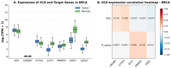
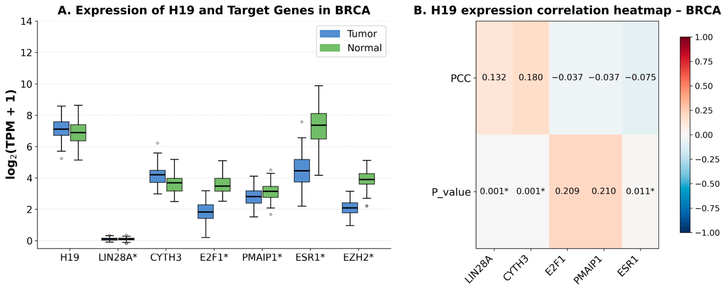
As illustrated in Table 8, H19 exhibits an intricate interaction with various genes associated with BC, including ESR1, EZH2, PMAIP1, HOTAIR, 91H, E2F1, CYTH3, LIN28A, CBL, and DNMT. Figure 1 presents the expression profiles of H19, along with the predicted targets of H19, and a heatmap illustrating the correlation coefficients for each H19–target pair. Among the genes evaluated, five targets were identified. As demonstrated in Figure 1A, LIN28A, E2F1, PMAIP1, ESR1, and EZH2 exhibited statistically significant differential expressions. As demonstrated by the correlation heatmap, there were statistically significant associations for LIN28A, CYTH3, and ESR1. LIN28A and CYTH3 displayed weak positive correlations with H19, whereas ESR1 exhibited a slight negative correlation (see Figure 1B).
Table 8.
Regulatory targets of H19 and its associated ceRNAs and miRNAs.
Figure 1.
Expression and correlation analysis of H19 and predicted target genes in BRCA. (A). Boxplots showing the expression levels (log2[TPM+1]) of H19 and its predicted target genes in BRCA tumor and normal tissues. Genes marked with an asterisk (*) exhibit statistically significant differential expression. Circles (○) represent outliers (B). Heatmap illustrates the Pearson correlation coefficients (PCCs) and corresponding p-values between H19 and selected target genes in BRCA. * p < 0.05. Color intensity ranges from negative/weak correlation to red positive/strong correlation.
Regarding the POLR2E rs3787016 variant, CADD analysis showed that this variant has a PHRED score of 0.289 and a raw score of –0.331. The GERP conservation score was 1.73, and the variant was annotated within a transcription factor binding site (TFBS). Figure S1 shows the identification and characterization of candidate transcription factors that can bind to the POLR2E variant site.
Figure S1A illustrates the genomic mapping where the variant overlaps with the binding coordinates of AGO2, RBFOX2, and POLR2A. Figure S1B demonstrates the signal intensity values (relative binding strength), with POLR2A exhibiting the most substantial enrichment (109), followed by RBFOX2 (8.6 × 10²) and AGO2 (2.1 × 10²). Figure S1C describes the tissue or cell line origin of each transcription factor: POLR2A in spleen tissue, RBFOX2 in the K562 cell line (blood), and AGO2 in the HepG2 cell line (liver).
Immunohistochemistry data retrieved from The Human Protein Atlas (an independent external cohort not characterized for the SNPs of interest in our study) were reviewed to assess the protein expression of the transcription factors identified through RegulomeDB. RBFOX2 was detected in 67% (8/12) of BC samples, predominantly in ductal carcinoma (89%) with low staining intensity. AGO2 was detected in all cases (12/12), with high intensity in 25% (3/12, all lobular), moderate in 67% (8/12; 88% lobular, 12% ductal), and low in 8% (1/12, lobular). POLR2A immunohistochemistry data were not available (Table S1). These results provide orthogonal evidence of biological plausibility at the protein level rather than direct evidence of a genotype–phenotype association in our population.
2.6. In Silico Analysis of H19 and POLR2E Gene Expression
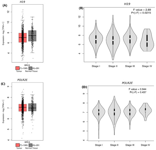
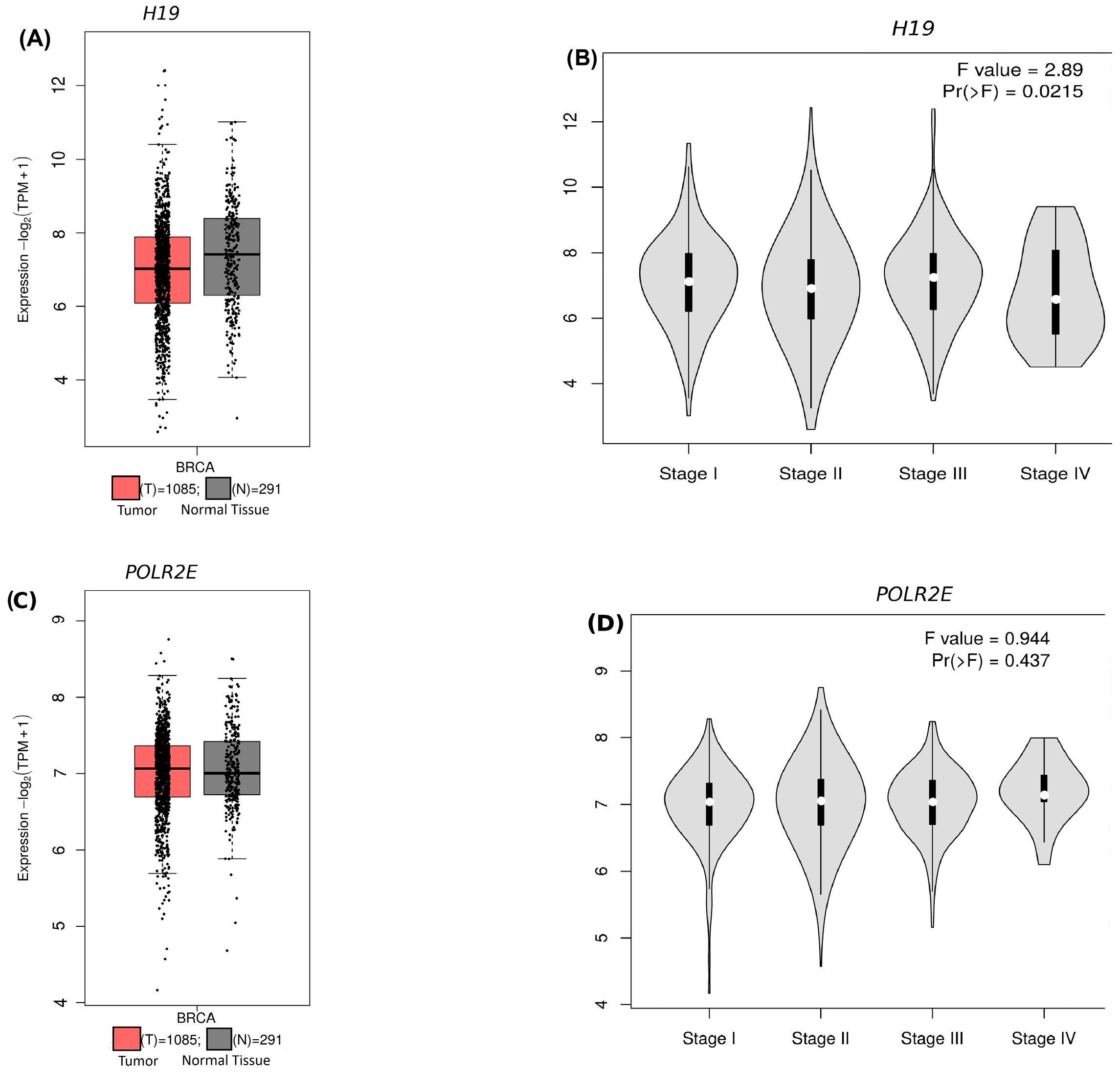
Gene expression profiling was performed, integrating RNA-Seq expression data from repositories, to evaluate the behavior of these genes in breast cancer tissue from patients. Figure 2 illustrates the gene expression profiling of H19 and POLR2E among tissue types and tumoral stages. Figure 2A presents the differential expression analysis, where H19 exhibited no statistically significant differences between tumor and normal breast tissue, and Figure 2B shows that stage-specific expression analysis of H19 demonstrated significant variability across clinical stages (F = 2.89; p = 0.02), with underexpression observed in stage IV. Finally, Figure 2C shows differential expression analysis of POLR2E, which showed no statistically significant differential expression between tumor and normal breast tissue. Figure 2D indicates that stage-specific expression analysis of POLR2E detected no considerable variation in expression levels (F = 0.944; p = 0.43).
Figure 2.
Expression analysis of H19 and POLR2E in BC and across clinical stages (GEPIA2). (A,C) Boxplots showing expression levels (log2[TPM+1]) of H19 and POLR2E, respectively, comparing breast cancer tumor samples with normal breast tissue. (B,D) Violin plots illustrating stage-specific expression patterns for H19 and POLR2E across BC stages I, II, III, IV.
2.7. In Silico Analysis of Survival of H19 and POLR2E
Kaplan–Meier survival analyses were performed using RNA-seq data from online repositories to evaluate the association of H19 and POLR2E expression with overall survival in BC patients.
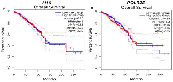
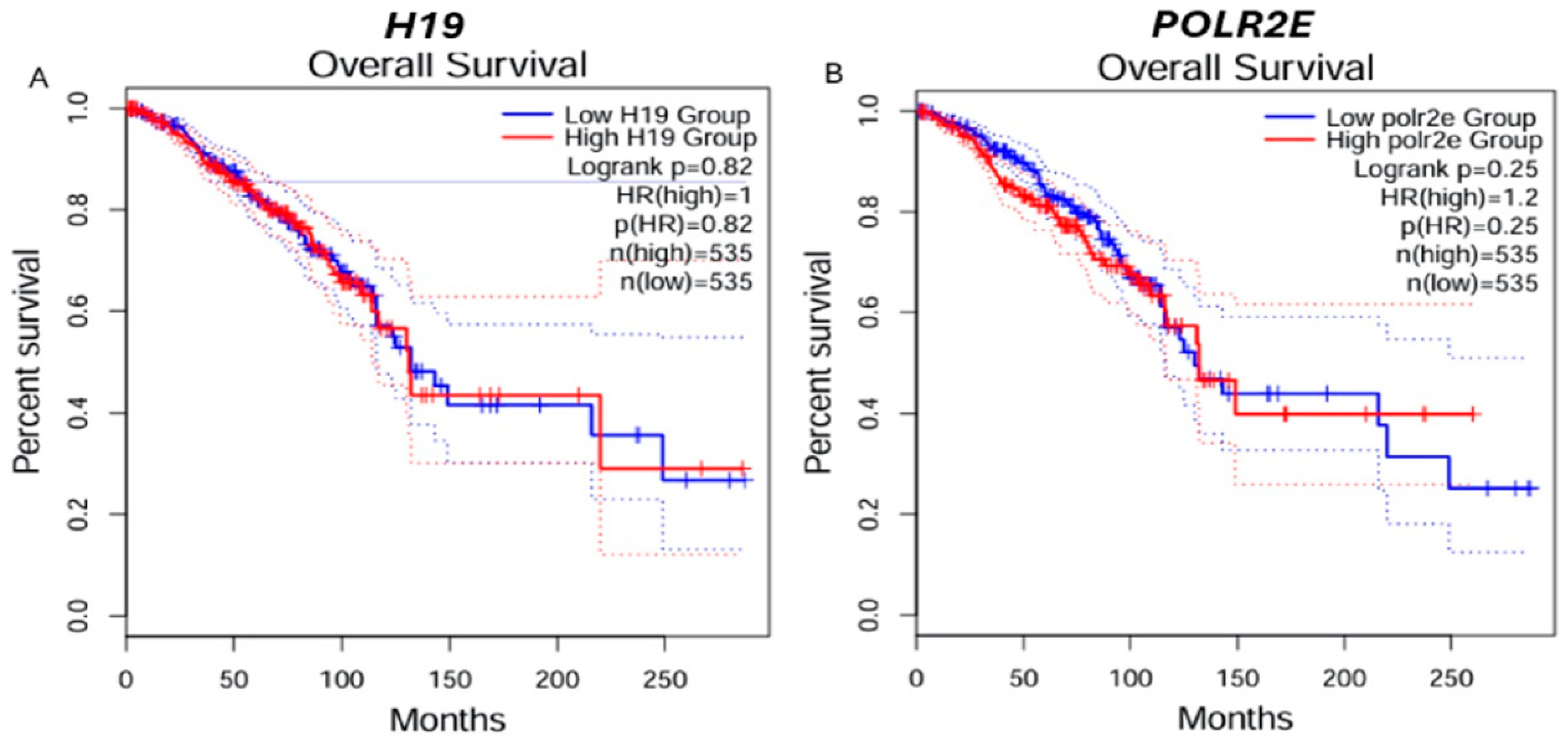
Figure 3 presents the Kaplan–Meier survival analysis performed based on H19 and POLR2E expression. Figure 3A shows that in overall survival in breast cancer for H19, this gene has no statistically significant differences in overall survival between patients with high and low expression of this gene (log rank p = 0.82; HR = 1.00; p(HR) = 0.82). Finally, Figure 3B indicates overall survival for POLR2E; this survival curve revealed no statistically significant differences between high and low expression groups of this gene (log rank p = 0.25; HR = 1.20; p(HR) = 0.25).
Figure 3.
Overall survival according to H19 and POLR2E expression levels in breast cancer (GEPIA2). (A,B). Kaplan–Meier curves comparing overall survival between patients with low (blue) and high (red) expression of H19 (left) and POLR2E (right). Each panel displays the number of patients per group (n), the log-rank p-value, and the hazard ratio (HR) with its corresponding p-value for the high-expression group.
3. Discussion
In the present study, the association between four genetic variants located in long non-coding RNAs (H19 rs3741219, MEG3 rs7158663, POLR2E rs3787016, and ANRIL rs10757274) and BC susceptibility was evaluated in Mexican mestizo women, integrating the clinicopathological characterization of the cohort with in silico functional analyses. Two main findings can be highlighted; on one hand, we observed that the C/C genotypes of H19 rs3741219 and POLR2E rs3787016 were significantly associated with increased BC susceptibility, while specific clinicopathological associations were also found, with early-onset disease and luminal B subtype for H19 and late-onset disease and luminal A subtype for POLR2E. On the other hand, the in silico analyses showed that both variants are located within regions that have regulatory relevance, although with different levels of predicted intrinsic impact, so that biological plausibility for their phenotypic associations is supported. For the MEG3 rs7158663 and ANRIL rs10757274 variants, no significant association with BC susceptibility was observed in our population. To our knowledge, this is the first work in which these four lncRNA variants are characterized in an admixed Latin American population, as population-specific data in the literature is mostly derived from Asian and European cohorts, and a recognized gap in the genetic architecture of BC susceptibility in Latin American women is addressed.
3.1. Demographic and Clinicopathological Context
In our cohort, BC incidence was statistically more frequent in patients aged 50 years and above, which is consistent with the international and national guidelines (ESMO, NICE, NCCN, NOM), in which age is considered a major risk factor for BC [44,45,46]. This pattern can be explained by the cumulative accrual of genetic damage in breast tissue throughout life, the prolonged hormonal exposure to estrogen and progesterone during reproductive years, and the age-related decline in immune surveillance [47,48]. Additionally, a statistically significant association with cigarette smoking was observed, which is in line with the IMSS guidelines and the previous evidence on the carcinogenic effects of the chemical compounds present in tobacco smoke, including polycyclic hydrocarbons, nitrosamines, and aromatic amines [49,50]. Thus, the demographic and behavioral patterns observed in our population provide the clinical context in which the genetic associations described below should be interpreted.
3.2. H19 rs3741219 Variant: Results in Context
Our results show that patients carrying the T/C or C/C genotypes of H19 rs3741219 exhibited an increased susceptibility to developing BC, particularly with early-onset disease and luminal A and luminal B subtypes. These findings contrast with reports by Xia et al. (2016) [51] and Abdollahzadeh et al. (2019) [52], who found no association in Chinese and Iranian populations, and with meta-analyses by Li et al. (2022) [53], Yuan et al. (2022) [54], and Yang et al. (2023) [22] reporting no overall association with cancer risk. The discrepancies likely reflect genetic heterogeneity across populations and differences in environmental exposures.
The functional plausibility of H19 rs3741219 is supported by its location within exon 1, a region essential for the regulatory activity of the transcript, together with a high GERP conservation score (5.24) and a modestly elevated CADD PHRED score, indicating that this position is under evolutionary constraint and may harbor regulatory relevance. Among the H19 targets retrieved from LncTarD, only LIN28A, CYTH3, and ESR1 showed significant expression correlations in TCGA-BRCA data, with ESR1 displaying an inverse association. This pattern is consistent with our observation of increased susceptibility in luminal A and luminal B carriers of the C/C genotype, as H19 has been described as an estrogen-responsive transcript whose expression is induced by ERα in luminal progenitor cells [54]. Within this framework, the rs3741219 variant may modulate H19-mediated regulation of hormone-responsive programs, providing a biologically coherent, though not definitive, explanation for the subtype-specific signal observed in our cohort. Other reported functions of H19, including its roles in EMT, stemness, and endocrine resistance, were not assessed in the present study and therefore cannot be linked directly to our findings.
3.3. POLR2E rs3787016 Variant: Results in Context
Patients carrying the C/C genotype of POLR2E rs3787016 showed increased BC susceptibility, with the strongest signal in women > 50 years and in luminal A tumors. These results are consistent with those reported by Xu et al. (2017) [38] and Chen et al. [34], although the subtype-specific association differs from Xu et al., who reported T/T as the susceptibility genotype for ER/PR-positive tumors in a different population. Multivariate analysis confirmed age as an independent susceptibility factor in our cohort, in line with established age-related mechanisms in BC pathogenesis [45,46].
In contrast to H19 rs3741219, in silico prediction scores for POLR2E rs3787016 did not indicate a strong intrinsic functional impact (PHRED 0.289; GERP 1.73), suggesting that if this variant is causal, its effect is likely subtle and context-dependent. The most parsimonious interpretation compatible with our data is that rs3787016 overlaps a transcription factor binding region with documented occupancy by POLR2A, RBFOX2, and AGO2, proteins involved in transcriptional and post-transcriptional regulation, and RBFOX2 and AGO2 were detectable in breast tissue by immunohistochemistry in The Human Protein Atlas, supporting the biological plausibility of a regulatory effect at this locus. An alternative possibility, which our study cannot exclude, is that rs3787016 is not the causal variant but rather a marker in linkage disequilibrium with a yet-unidentified functional variant. Taken together, the evidence supports a context-dependent role for rs3787016 in BC susceptibility, most apparent in women over 50 years of age and in luminal A tumors, rather than a strong, generalized functional effect.
3.4. MEG3 rs7158663 and ANRIL rs10757274: Absence of Association
For the MEG3 rs7158663 and ANRIL rs10757274 variants, no significant associations with BC susceptibility were observed in our population. Regarding ANRIL rs10757274, this negative result is consistent with what has been previously reported in BC, where no clear association has been found despite the well-documented role of ANRIL in the epigenetic regulation of the CDKN2A/CDKN2B tumor-suppressor locus [27,29,30,31]. For MEG3 rs7158663, however, prior evidence has been mixed, while some studies have suggested an association with reduced MEG3 expression and increased cancer susceptibility [39,41,43]. The absence of association in our cohort may be explained by insufficient statistical power to detect modest effects, ancestry-specific allele frequencies, or a genuine lack of association in Mexican mestizo women, so that replication in larger Latin American cohorts is needed to clarify the role of these variants in our population.
3.5. Clinical Relevance and Translational Considerations
Although our findings identify H19 rs3741219 and POLR2E rs3787016 as susceptibility variants in Mexican mestizo women, several considerations should be taken into account before their clinical translation can be considered. The effect sizes observed in our analyses (OR ~1.5–2.4) are characteristic of low-to-moderate penetrance variants, so that limited discriminatory power would be provided if used as standalone biomarkers. A more realistic clinical incorporation would occur through their integration into population-specific polygenic risk scores (PRS), in combination with high- and moderate-penetrance gene panels (BRCA1, BRCA2, PALB2, ATM, CHEK2, TP53) and the established non-genetic risk factors that are routinely used in the clinical setting, such as age, BMI, reproductive history, and family history of cancer.
This integration is particularly relevant in Latin American populations, in which the PRS (Polygenic Risk Score) models that are currently available, mostly developed in European-ancestry cohorts, have shown a substantially poorer performance when applied to admixed populations. Recent evaluations of breast cancer PRS panels in Hispanic/Latinx women have shown lower discriminatory accuracy (AUC ~0.61–0.63) when compared to European-ancestry cohorts, while the performance was found to vary according to the degree of Indigenous American ancestry of the participants [55,56]. Additionally, multi-ancestry PRS (MA-PRS) approaches have shown improved performance in Hispanic women, particularly for the prediction of early-onset and triple-negative BC, although these approaches still require further validation before being incorporated into routine clinical use [57]. Therefore, the identification of ancestry-relevant variants like those characterized in the present study represents one step towards closing this gap.
Several barriers should still be overcome before any clinical implementation can be considered, including the independent validation of these results in additional Mexican and broader Latin American cohorts; the functional characterization of the regulatory consequences of each variant through experimental approaches such as allele-specific expression assays, reporter constructs, and CRISPR-based perturbations; and the cost-effectiveness analyses needed for any population-level screening strategy. Additionally, the development of clinical-grade genotyping platforms that incorporate ancestry-informative markers represents another step required for a meaningful implementation.
3.6. Limitations and Future Directions
Several limitations should be considered when interpreting these findings. The associations identified correspond to a case–control comparison and reflect susceptibility to breast cancer development; they do not imply causality and should not be interpreted as prognostic markers. Consistent with this distinction, H19 and POLR2E expression did not differ significantly between tumor and normal tissue in the TCGA-BRCA cohort, and neither was associated with overall survival—an expected result if the susceptibility signal operates at the level of disease initiation rather than tumor progression. Moreover, bulk transcriptomic analyses average expression across heterogeneous cell populations and may dilute cell type- or subtype-specific regulatory effects that are particularly relevant for lncRNAs, whose expression is often spatially and temporally restricted. The absence of patient-matched expression data within our own cohort, together with the lack of survival follow-up, also limits the ability to integrate genetic and transcriptional findings in the same individuals; this should be regarded as a constraint of the present study. Functional validation in larger, ethnically diverse cohorts and in subtype-specific cellular models will be needed to clarify the mechanistic contribution of these variants to BC susceptibility.
4. Materials and Methods
4.1. Subjects
A total of 505 women were recruited, including 254 patients with a clinical and histological diagnosis of BC, based on the criteria of the UMAE Hospital de Gineco-Obstetricia of the Instituto Mexicano del Seguro Social (IMSS) in Guadalajara, Mexico. BC was stratified according to the tumor–node–metastasis (TNM) classification. The study was approved by the National Committee for Scientific Research of the Mexican Institute of Social Security (IMSS) (R-2020-785-130) with approval date August 2020 and conducted following national and international ethical standards. The patient group comprised women aged 18 years or older with any stage of BC, regardless of treatment status or therapeutic stage. The control group included 251 unrelated healthy women donors aged 18 years or older from the general Mexican population, matched for age with the patient group. Patients and healthy women were enrolled in a non-probabilistic consecutive manner during consultation at this institution between 2020 and 2023. All the participants provided written informed consent for participation in this study. In addition, this study was performed in accordance with the Declaration of Helsinki and its latest amendments. A standard epidemiological questionnaire allowed us to collect personal data, including age, drinking and smoking status, and familial history for all participants. Women with a history of previous cancer were excluded from the control group, while those who had undergone chemotherapy or radiotherapy were excluded from the patient group. Information about the clinical and pathological characteristics of patients was obtained from hospital records.
4.2. Genotyping
Genomic DNA from peripheral blood lymphocytes was isolated by the salting-out method [58]. The variants H19 rs3741219 (T>C), POLR2E rs3787016 (T>C), MEG3 rs7158663 (G>A), and ANRIL rs10757274 (A>G) were genotyped using the polymerase chain reaction–restriction fragment length polymorphism (PCR-RFLP) methodology with the following primer pairs: for the H19 rs3741219 variant, the forward primer F: 5′-CCC CCT GCG GCG GAC GGT TGA-3′ and reverse primer R: 5′-GGC GTA ATG GAA TGC TTG AA-3′ [59]; for the POLR2E rs3787016 variant F: 5′-CAT CAA CAT CAC GCA GCA CG-3′ and reverse primer R: 5′-CCC TGT CCT CCA AGC ACT CAT-3 [38]; for ANRIL rs10757274 F: 5′-GTT TCT GCA CAT GGT GAT GG-3′ and reverse primer 5′-CTGCCTCACTCTCCAGTTCC-3′ [60]; and for the MEG3 rs7158663 variant, the forward primer F: 5′-GGT TCT TTA GTT CTG CGA TGC T-3′ and the reverse primer 5′-TTG GGA GTC ACA AGA GGA GG-3′ were used [61].
PCR for the MEG3 rs7158663, POLR2E rs3787016, ANRIL rs10757274 and H19 rs37741219 variants was performed for 35 cycles in a 10 μL volume containing 100 ng DNA, 10X buffer (500 mM KCl, 100 mM Tris- HCl, and 0.1% Triton TMX-100) (Invitrogen, Carlsbad, CA, USA), 2.0 mM MgCl2, 200 μM dNTPs, 5 pM of each primer, and 2 U Taq DNA Polymerase. Denaturation was carried out at 95 °C, annealing temperature was set at 60 °C for rs7158663, 59 °C for rs3787016, 60 °C for rs10757274, and 62 °C for rs3741219, and elongation at 72 °C for 2 min each. All PCR reactions were performed in a thermocycler T-100 Biorad (Biorad, California, USA). Five microliters of the PCR product were digested with 4U of BtsCI, NlaIII, BsmAI, and HhaI, restriction enzymes (New England Bio-Labs Inc; Beverly, MA, USA), respectively. The digested products were separated into 8% polyacrylamide gels. For MEG3 rs7158663, the fragment sizes observed by electrophoresis corresponded to 233 and 68 bp for the polymorphic allele (A) and 301 bp for the wild-type allele (G). For the POLR2E rs3787016 variant, the fragment sizes observed by electrophoresis corresponded to 127 and 20 bp for the polymorphic allele (C) and 147 bp for the wild-type allele (T); for the ANRIL rs10757274 variant, the fragment sizes observed were 250 bp for the polymorphic allele (G) and 172 and 78 bp for the wild-type allele (A); and finally, for the H19 rs3741219 variant, the fragment sizes observed were 301, 92 and 41 bp for the polymorphic allele (C) and 342 and 92 bp for the wild-type allele (T).
Quality control for these assays was assessed in 20% of randomly selected samples that were re-genotyped by an independent technician. The concordance among genotype assays was 100%.
4.3. Statistical Analysis
Allele and genotype frequencies were estimated by direct counting in both groups. The chi-square test was used to assess the Hardy–Weinberg equilibrium (HWE). The chi-square test was used to examine the differences in allele and genotype distributions and clinical characteristics between patients and controls. To measure the association of alleles or genotypes with clinicopathological characteristics, we performed a stratified analysis including age, smoking and alcohol status, TNM stage, and histologic–molecular subtype, and the odds ratio (OR) with corresponding 95% confidence intervals (CIs) using SPSS v17.0 software package (SPSS, Inc., Chicago, IL, USA). Multivariable logistic regression analysis was performed to evaluate confounding variables. p-values were adjusted using the Bonferroni method. Statistical significance was defined as p < 0.012 after adjustment.
4.4. In Silico Analysis of Genetic Variants
The estimated functional impact of the analyzed variants was assessed using complementary in silico tools, applied exclusively to the two variants that showed statistically significant association with breast cancer susceptibility in our experimental analysis. As a first step, CADD v1.7 (https://cadd.gs.washington.edu, accessed on 15 March 2026) [62,63] was used as the principal marker of deleteriousness, since it integrates more than sixty annotation features into a single PHRED-scaled score, including evolutionary conservation metrics, regulatory elements, transcription factor binding sites, chromatin marks, and splicing predictions; thus, it remains applicable to non-coding variants whose operational characteristics differ from those of coding variants. Based on the regulatory annotations retrieved from CADD, complementary tools were applied selectively according to the functional context of each variant. For variants overlapping a transcription factor binding site, RegulomeDB (https://regulomedb.org, accessed on 15 March 2026) [64] was queried to identify transcription factors with experimental ChIP-seq evidence at the locus, while the Human Protein Atlas (https://proteinatlas.org, accessed on 16 March 2026) [65] was consulted to confirm expression of these regulators in breast tissue. For variants located within a long non-coding RNA gene with documented target activity, curated lncRNA–target interactions in breast cancer were retrieved from LncTarD 2.0 (https://lnctard.bio-database.com, accessed on 17 March 2026) [66]. Custom figures derived from these analyses, including locus maps, correlation heatmaps, and grouped boxplots, were generated in R version 4.3.2 using the ggplot2 and ggpubr packages and were exported in TIFF format at 300 dpi.
4.5. In Silico Gene Expression and Survival Analysis
To support the in silico analysis with publicly available expression data, we utilized the GEPIA2 platform (http://gepia2.cancer-pku.cn accessed on 15 March 2026) [67], which integrates RNA-seq data from TCGA and GTEx. We used the TCGA-BRCA cohort (1089 primary breast tumor samples and 113 matched normal breast tissue samples; RNA-seq, log2[TPM+1]-transformed) as the original reference dataset to compare expression levels between tumor and normal tissue and across clinical stages. Boxplots, violin plots, and Kaplan–Meier curves indicated in the figures were created on the GEPIA2 platform. All GEPIA2 analyses were conducted in March 2026.
5. Conclusions
In conclusion, our results indicate that the C/C genotypes of H19 rs3741219 and POLR2E rs3787016 are associated with an increased susceptibility to develop breast cancer in Mexican mestizo women, and distinct clinicopathological patterns were observed for each variant, such that H19 was associated with early-onset disease and luminal A and luminal B subtypes, while POLR2E was associated with late-onset disease and luminal A subtype. No significant association was observed for MEG3 rs7158663 and ANRIL rs10757274 in our population. The in silico analyses support the biological plausibility of these associations, with stronger evidence for H19, given its higher evolutionary conservation and its documented regulatory interactions with luminal-related targets, while the contribution of POLR2E rs3787016 appears to be more context-dependent and likely mediated through transcription factors binding at this locus. These findings provide the first characterization of these variants in an admixed Latin American population and represent candidate markers for future incorporation into ancestry-informed risk stratification frameworks. Functional validation in larger cohorts and in subtype-specific cellular models will be necessary to confirm the mechanistic role of these variants and to evaluate their potential clinical utility.
Supplementary Materials
The following supporting information can be downloaded at: https://www.mdpi.com/article/10.3390/ncrna12030019/s1, Figure S1: Transcription factor binding evidence at the POLR2E locus identified by RegulomeDB; Table S1: Immunohistochemical expression of RBFOX2 and AGO2 in breast cancer according to histological subtype.
Author Contributions
Research design and preparation of the main manuscript: M.A.R.-R. and C.d.J.T.-J. Data analysis and statistical analysis: C.d.J.T.-J., A.M.S.-S., C.I.J.-V., M.A.R.-R. and M.P.G.-A. Provided blood samples and clinical data: R.A.G.-S., K.G.M.-V., G.A.T.-S., E.S.-G. and J.E.G.-O. Prepared clinical samples and performed the laboratory work: R.A.G.-S., K.G.M.-V. and C.d.J.T.-J. Responsible for patient data and follow-up: E.S.-G. and J.E.G.-O. Review and revision: M.A.R.-R., A.M.S.-S., C.I.J.-V., C.d.J.T.-J., R.A.G.-S., K.G.M.-V., G.A.T.-S., J.E.G.-O., E.S.-G. and M.P.G.-A. All authors have read and agreed to the published version of the manuscript.
Funding
This work was supported by grants from the “Instituto Mexicano del Seguro Social” (R-2020-785-130).
Institutional Review Board Statement
The study was approved by the National Committee for Scientific Research of the Mexican Institute of Social Security (IMSS) (R-2020-785-130) and conducted in accordance with national and international ethical standards.
Informed Consent Statement
Informed consent was obtained from all subjects involved in the study.
Data Availability Statement
The original contributions presented in this study are included in the article and its Supplementary Material. Further inquiries can be directed to the corresponding author.
Conflicts of Interest
The authors declare no conflicts of interest.
Abbreviations
The following abbreviations are used in this manuscript:
| AGO2 | Argonaute-2 |
| ANRIL | Antisense Non-Coding RNA in the INK4 Locus |
| ANOVA | Analysis of variance |
| BC | Breast cancer |
| BMI | Body Mass Index |
| bp | Base pairs |
| BRCA | Breast cancer dataset (TCGA) |
| CADD | Combined Annotation Dependent Depletion |
| ceRNA | Competing endogenous RNA |
| ChIP-seq | Chromatin immunoprecipitation sequencing |
| CI | Confidence interval |
| DC | Ductal carcinoma |
| DNA | Deoxyribonucleic acid |
| dNTPs | Deoxyribonucleotide triphosphates |
| EMT | Epithelial–Mesenchymal Transition |
| ER | Estrogen receptor |
| ERα | Estrogen receptor alpha |
| ESMO | European Society for Medical Oncology |
| GEPIA2 | Gene Expression Profiling Interactive Analysis 2 |
| GERP | Genomic Evolutionary Rate Profiling |
| GTEx | Genotype-Tissue Expression |
| GWAS | Genome-wide association studies |
| HR | Hazard ratio |
| HWE | Hardy–Weinberg equilibrium |
| IGF2 | Insulin-like growth factor 2 |
| IHC | Immunohistochemistry |
| IMSS | Instituto Mexicano del Seguro Social |
| kb | Kilobases |
| LB | Lobular carcinoma |
| lncRNA | Long non-coding RNA |
| MEG3 | Maternally Expressed Gene 3 |
| MET | Mesenchymal–Epithelial Transition |
| miRNA | MicroRNA |
| mRNA | Messenger RNA |
| NCCN | National Comprehensive Cancer Network |
| NICE | National Institute for Health and Care Excellence |
| nt | Nucleotides |
| OR | Odds ratio |
| PCC | Pearson correlation coefficient |
| PCR | Polymerase chain reaction |
| PCR-RFLP | Polymerase chain reaction-restriction fragment length polymorphism |
| PHRED | Phred-scaled deleteriousness score (CADD) |
| POLR2E | RNA Polymerase II subunit E |
| PR | Progesterone receptor |
| RBFOX2 | RNA-binding fox-1 homolog 2 |
| RFLP | Restriction fragment length polymorphism |
| RNA | Ribonucleic acid |
| RNA-seq | RNA sequencing |
| SD | Standard deviation |
| SNP | Single-nucleotide polymorphism |
| SNV | Single-nucleotide variant |
| SPSS | Statistical Package for the Social Sciences |
| TCGA | The Cancer Genome Atlas |
| TFBS | Transcription factor binding site |
| TNM | Tumor–node–metastasis |
| TPM | Transcripts per million |
References
- Xiong, X.; Zheng, L.-W.; Ding, Y.; Chen, Y.-F.; Cai, Y.-W.; Wang, L.-P.; Huang, L.; Liu, C.-C.; Shao, Z.-M.; Yu, K.-D. Breast Cancer: Pathogenesis and Treatments. Signal Transduct. Target. Ther. 2025, 10, 49. [Google Scholar] [CrossRef] [Scilit] [PubMed]
- Katsura, C.; Ogunmwonyi, I.; Kankam, H.K.N.; Saha, S. Breast Cancer: Presentation, Investigation and Management. Br. J. Hosp. Med. 2022, 83, 2. [Google Scholar] [CrossRef] [Scilit]
- Ferlay, J.; Ervik, M.; Lam, F.; Laversanne, M.; Colombet, M.; Mery, L.; Piñeros, M.; Znaor, A.; Soerjomataram, I.; Bray, F. Global Cancer Observatory: Cancer Today; International Agency for Research on Cancer: Lyon, France, 2024; Available online: https://gco.iarc.who.int/today (accessed on 15 March 2026).
- Bray, F.; Laversanne, M.; Sung, H.; Ferlay, J.; Siegel, R.L.; Soerjomataram, I.; Jemal, A. Global Cancer Statistics 2022: GLOBOCAN Estimates of Incidence and Mortality Worldwide for 36 Cancers in 185 Countries. CA Cancer J. Clin. 2024, 74, 229–263. [Google Scholar] [CrossRef] [Scilit]
- Jiménez-López, M.R.; Tovar-Jácome, C.d.J.; Palacios-Ramírez, A.; Gallegos-Arreola, M.P.; Aguilar-Macedo, T.G.M.; González-Sánchez, R.A.; Salas-González, E.; García-Ortiz, J.E.; Juárez-Vázquez, C.I.; Rosales-Reynoso, M.A. Genetic Variants in BIRC5 (Rs8073069, Rs17878467, and Rs9904341) Are Associated with Susceptibility in Mexican Patients with Breast Cancer: Clinical Associations and Their Analysis In Silico. Genes 2025, 16, 786. [Google Scholar] [CrossRef] [Scilit]
- Sideris, N.; Dama, P.; Bayraktar, S.; Stiff, T.; Castellano, L. LncRNAs in Breast Cancer: A Link to Future Approaches. Cancer Gene Ther. 2022, 29, 1866–1877. [Google Scholar] [CrossRef] [Scilit]
- Gurioli, G.; Tedaldi, G.; Farolfi, A.; Petracci, E.; Casanova, C.; Comerci, G.; Danesi, R.; Arcangeli, V.; Ravegnani, M.; Calistri, D.; et al. Clinical Impact of Next-Generation Sequencing Multi-Gene Panel Highlighting the Landscape of Germline Alterations in Ovarian Cancer Patients. Int. J. Mol. Sci. 2022, 23, 15789. [Google Scholar] [CrossRef] [Scilit]
- Palleschi, M.; Virga, A.; Scarpi, E.; Fonzi, E.; Musolino, A.; Merloni, F.; Sarti, S.; Danesi, R.; Ravegnani, M.; Casadei, C.; et al. Exploring the Promoter Regions of Cancer Predisposition Genes in Patients with Triple-Negative Breast Cancer Reveals the Presence of Rare Germline Variants. Oncologist 2025, 30, oyaf052. [Google Scholar] [CrossRef] [Scilit] [PubMed]
- Mattick, J.S.; Amaral, P.P.; Carninci, P.; Carpenter, S.; Chang, H.Y.; Chen, L.-L.; Chen, R.; Dean, C.; Dinger, M.E.; Fitzgerald, K.A.; et al. Long Non-Coding RNAs: Definitions, Functions, Challenges and Recommendations. Nat. Rev. Mol. Cell Biol. 2023, 24, 430–447. [Google Scholar] [CrossRef] [Scilit]
- Jiang, M.-C.; Ni, J.-J.; Cui, W.-Y.; Wang, B.-Y.; Zhuo, W. Emerging Roles of LncRNA in Cancer and Therapeutic Opportunities. Am. J. Cancer Res. 2019, 9, 1354–1366. [Google Scholar] [PubMed]
- Alkhathami, A.G.; Hadi, A.; Alfaifi, M.; Alshahrani, M.Y.; Verma, A.K.; Beg, M.M.A. Serum-Based LncRNA ANRIL, TUG1, UCA1, and HIT Expressions in Breast Cancer Patients. Dis. Markers 2022, 2022, 9997212. [Google Scholar] [CrossRef] [Scilit]
- Liu, Q.; Liu, G.; Lin, Z.; Lin, Z.; Tian, N.; Lin, X.; Tan, J.; Huang, B.; Ji, X.; Pi, L.; et al. The Association of LncRNA SNPs and SNPs-Environment Interactions Based on GWAS with HBV-Related HCC Risk and Progression. Mol. Genet. Genom. Med. 2021, 9, e1585. [Google Scholar] [CrossRef] [Scilit]
- Ji, F.; Yao, Z.; Liu, C.; Fu, S.; Ren, B.; Liu, Y.; Ma, L.; Wei, J.; Sun, D. A Novel Lnc-LAMC2-1:1 SNP Promotes Colon Adenocarcinoma Progression by Targeting MiR-216a-3p/HMGB3. Heliyon 2022, 8, e12342. [Google Scholar] [CrossRef] [Scilit]
- Li, L.; Gao, Y.; Yu, B.; Zhang, J.; Ma, G.; Jin, X. Role of LncRNA H19 in Tumor Progression and Treatment. Mol. Cell. Probes 2024, 75, 101961. [Google Scholar] [CrossRef] [Scilit] [PubMed]
- Hernández-Aguilar, A.I.; Luciano-Villa, C.A.; Tello-Flores, V.A.; Beltrán-Anaya, F.O.; Zubillaga-Guerrero, M.I.; Flores-Alfaro, E. Dysregulation of LncRNA-H19 in Cardiometabolic Diseases and the Molecular Mechanism Involved: A Systematic Review. Expert Rev. Mol. Diagn. 2021, 21, 809–821. [Google Scholar] [CrossRef] [Scilit]
- Collette, J.; Le Bourhis, X.; Adriaenssens, E. Regulation of Human Breast Cancer by the Long Non-Coding RNA H19. Int. J. Mol. Sci. 2017, 18, 2319. [Google Scholar] [CrossRef] [Scilit]
- Ghafouri-Fard, S.; Esmaeili, M.; Taheri, M. H19 LncRNA: Roles in Tumorigenesis. Biomed. Pharmacother. 2020, 123, 109774. [Google Scholar] [CrossRef] [Scilit]
- Yang, J.; Qi, M.; Fei, X.; Wang, X.; Wang, K. LncRNA H19: A Novel Oncogene in Multiple Cancers. Int. J. Biol. Sci. 2021, 17, 3188–3208. [Google Scholar] [CrossRef] [Scilit] [PubMed]
- Wang, J.; Sun, J.; Yang, F. The Role of Long Non-Coding RNA H19 in Breast Cancer. Oncol. Lett. 2019, 19, 7–16. [Google Scholar] [CrossRef] [Scilit] [PubMed]
- Huang, M.-C.; Chou, Y.-H.; Shen, H.-P.; Ng, S.-C.; Lee, Y.; Sun, Y.-H.; Hsu, C.-F.; Yang, S.-F.; Wang, P.-H. The Clinicopathological Characteristic Associations of Long Non-Coding RNA Gene H19 Polymorphisms with Uterine Cervical Cancer. J. Cancer 2019, 10, 6191–6198. [Google Scholar] [CrossRef] [Scilit]
- Deng, Y.; Zhou, L.; Yao, J.; Liu, Y.; Zheng, Y.; Yang, S.; Wu, Y.; Li, N.; Xu, P.; Lyu, L.; et al. Associations of LncRNA H19 Polymorphisms at MicroRNA Binding Sites with Glioma Susceptibility and Prognosis. Mol. Ther. Nucleic Acids 2020, 20, 86–96. [Google Scholar] [CrossRef] [Scilit]
- Yang, M.; Zhang, M.; Wang, Q.; Guo, X.; Geng, P.; Gu, J.; Ji, W.; Zhang, L. Six Polymorphisms in the LncRNA H19 Gene and the Risk of Cancer: A Systematic Review and Meta-Analysis. BMC Cancer 2023, 23, 688. [Google Scholar] [CrossRef] [Scilit]
- Mathias, C.; Garcia, L.E.; Teixeira, M.D.; Kohler, A.F.; Marchi, R.D.; Barazetti, J.F.; Gradia, D.F.; de Oliveira, J.C. Polymorphism of LncRNAs in Breast Cancer: Meta-Analysis Shows No Association with Susceptibility. J. Gene Med. 2020, 22, e3271. [Google Scholar] [CrossRef] [Scilit]
- Li, W.; Jiang, X.; Jin, X.; Yan, W.; Liu, Y.; Li, D.; Zhao, Z. Significant Association between Long Non-Coding RNA H19 Polymorphisms and Cancer Susceptibility. Medicine 2020, 99, e19322. [Google Scholar] [CrossRef] [Scilit]
- Yang, C.; Tang, R.; Ma, X.; Wang, Y.; Luo, D.; Xu, Z.; Zhu, Y.; Yang, L. Tag SNPs in Long Non-Coding RNA H19 Contribute to Susceptibility to Gastric Cancer in the Chinese Han Population. Oncotarget 2015, 6, 15311–15320. [Google Scholar] [CrossRef] [Scilit]
- Yin, Z.; Cui, Z.; Li, H.; Li, J.; Zhou, B. Polymorphisms in the H19 Gene and the Risk of Lung Cancer among Female Never Smokers in Shenyang, China. BMC Cancer 2018, 18, 893. [Google Scholar] [CrossRef] [Scilit] [PubMed]
- Kong, Y.; Hsieh, C.-H.; Alonso, L.C. ANRIL: A LncRNA at the CDKN2A/B Locus with Roles in Cancer and Metabolic Disease. Front. Endocrinol. 2018, 9, 405. [Google Scholar] [CrossRef] [Scilit] [PubMed]
- Xu, S.-T.; Xu, J.-H.; Zheng, Z.-R.; Zhao, Q.-Q.; Zeng, X.-S.; Cheng, S.-X.; Liang, Y.-H.; Hu, Q.-F. Long Non-Coding RNA ANRIL Promotes Carcinogenesis via Sponging MiR-199a in Triple-Negative Breast Cancer. Biomed. Pharmacother. 2017, 96, 14–21. [Google Scholar] [CrossRef] [Scilit]
- Ma, J.; Zhao, W.; Zhang, H.; Chu, Z.; Liu, H.; Fang, X.; Tang, D. Long Non-Coding RNA ANRIL Promotes Chemoresistance in Triple-Negative Breast Cancer via Enhancing Aerobic Glycolysis. Life Sci. 2022, 306, 120810. [Google Scholar] [CrossRef] [Scilit] [PubMed]
- Khorshidi, H.R.; Taheri, M.; Noroozi, R.; Sarrafzadeh, S.; Sayad, A.; Ghafouri-Fard, S. ANRIL Genetic Variants in Iranian Breast Cancer Patients. Cell J. 2017, 19, 72–78. [Google Scholar] [CrossRef] [Scilit]
- Xu, L.-B.; Zhang, Y.-Q.; Zhang, N.-N.; Li, B.; Weng, J.-Y.; Li, X.-Y.; Lu, W.-C.; Yu, P.-R.; Wang, X.; Li, Y.; et al. Rs10757274 Gene Polymorphisms in Coronary Artery Disease. Medicine 2020, 99, e18841. [Google Scholar] [CrossRef] [Scilit]
- Asadi-Tarani, M.; Darashti, A.; Javadi, M.; Rezaei, M.; Saravani, M.; Salimi, S. The Effects of ANRIL Polymorphisms on Colorectal Cancer, Tumor Stage, and Tumor Grade among Iranian Population. Mol. Biol. Rep. 2024, 51, 486. [Google Scholar] [CrossRef] [Scilit]
- NIH National Center for Biotechnology Information. POLR2E RNA Polymerase II, I and III Subunit E [Homo sapiens]. NCBI Gene. Available online: https://www.ncbi.nlm.nih.gov/gene/5434 (accessed on 13 March 2026).
- Chen, B.; Wang, S.; Ma, G.; Han, J.; Zhang, J.; Gu, X.; Feng, X. The Association of POLR2E Rs3787016 Polymorphism and Cancer Risk: A Chinese Case-Control Study and Meta-Analysis. Biosci. Rep. 2018, 38, BSR20180853. [Google Scholar] [CrossRef] [Scilit]
- Han, X.; Anthon, C.; Geissler, A.S.; Sabarinathan, R.; Seemann, S.E.; Havgaard, J.H.; Gorodkin, J. Associating Cancer-Related RNA Structure Disrupting SNPs in LincRNAs to Function. BMC Genom. 2025, 26, 1100. [Google Scholar] [CrossRef] [Scilit] [PubMed]
- Chen, B.; Li, J.; Yi, C.; Jiao, Y.; Gu, X.; Feng, X. Long Non-Coding RNA POLR2E Rs3787016 Is Associated with the Risk of Papillary Thyroid Carcinoma in Chinese Population. Pathol. Res. Pract. 2018, 214, 1040–1044. [Google Scholar] [CrossRef] [Scilit] [PubMed]
- Sattarifard, H.; Hashemi, M.; Hassanzarei, S.; Basiri, A.; Narouie, B.; Ghavami, S. Long Non-Coding RNA POLR2E Gene Polymorphisms Increased the Risk of Prostate Cancer in a Sample of the Iranian Population. Nucleosides Nucleotides Nucleic Acids 2019, 38, 1–11. [Google Scholar] [CrossRef] [Scilit]
- Xu, T.; Hu, X.-X.; Liu, X.-X.; Wang, H.-J.; Lin, K.; Pan, Y.-Q.; Sun, H.-L.; Peng, H.-X.; Chen, X.-X.; Wang, S.-K.; et al. Association between SNPs in Long Non-Coding RNAs and the Risk of Female Breast Cancer in a Chinese Population. J. Cancer 2017, 8, 1162–1169. [Google Scholar] [CrossRef] [Scilit] [PubMed]
- Al-Rugeebah, A.; Alanazi, M.; Parine, N.R. MEG3: An Oncogenic Long Non-Coding RNA in Different Cancers. Pathol. Oncol. Res. 2019, 25, 859–874. [Google Scholar] [CrossRef] [Scilit]
- Prasasya, R.; Grotheer, K.V.; Siracusa, L.D.; Bartolomei, M.S. Temple Syndrome and Kagami-Ogata Syndrome: Clinical Presentations, Genotypes, Models and Mechanisms. Hum. Mol. Genet. 2020, 29, R107–R116. [Google Scholar] [CrossRef] [Scilit]
- Hussain, M.S.; Majami, A.A.; Ali, H.; Gupta, G.; Almalki, W.H.; Alzarea, S.I.; Kazmi, I.; Syed, R.U.; Khalifa, N.E.; Bin Break, M.K.; et al. The Complex Role of MEG3: An Emerging Long Non-Coding RNA in Breast Cancer. Pathol. Res. Pract. 2023, 251, 154850. [Google Scholar] [CrossRef] [Scilit]
- Cao, X.; Zhuang, S.; Hu, Y.; Xi, L.; Deng, L.; Sheng, H.; Shen, W. Associations between Polymorphisms of Long Non-Coding RNA MEG3 and Risk of Colorectal Cancer in Chinese. Oncotarget 2016, 7, 19054–19059. [Google Scholar] [CrossRef] [Scilit]
- Gao, X.; Li, X.; Zhang, S.; Wang, X. The Association of MEG3 Gene Rs7158663 Polymorphism with Cancer Susceptibility. Front. Oncol. 2021, 11, 796774. [Google Scholar] [CrossRef] [Scilit] [PubMed]
- Comité Consultivo Nacional de Normalización de Prevención y Control de Enfermedades. NORMA Oficial Mexicana NOM-041-SSA2-2011, Para La Prevención, Diagnóstico, Tratamiento, Control y Vigilancia Epidemiológica Del Cáncer de Mama; Secretaría de Salud: Mexico City, Mexico, 2011. [Google Scholar]
- National Institute for Health and Care Excellence. Familial Breast Cancer: Classification and Care of People at Risk of Familial Breast Cancer and Management of Breast Cancer and Related Risks in People with a Family History of Breast Cancer; NICE Clinical Guideline CG164; NICE: London, UK, 2013. [Google Scholar]
- Paluch-Shimon, S.; Cardoso, F.; Partridge, A.H.; Abulkhair, O.; Azim, H.A.; Bianchi-Micheli, G.; Cardoso, M.J.; Curigliano, G.; Gelmon, K.A.; Gentilini, O.; et al. ESO-ESMO Fifth International Consensus Guidelines for Breast Cancer in Young Women (BCY5). Ann. Oncol. 2022, 33, 1097–1118. [Google Scholar] [CrossRef] [Scilit]
- Azim, H.A.; Partridge, A.H. Biology of Breast Cancer in Young Women. Breast Cancer Res. 2014, 16, 427. [Google Scholar] [CrossRef] [Scilit]
- Partridge, A.H.; Hughes, M.E.; Warner, E.T.; Ottesen, R.A.; Wong, Y.-N.; Edge, S.B.; Theriault, R.L.; Blayney, D.W.; Niland, J.C.; Winer, E.P.; et al. Subtype-Dependent Relationship Between Young Age at Diagnosis and Breast Cancer Survival. J. Clin. Oncol. 2016, 34, 3308–3314. [Google Scholar] [CrossRef] [Scilit] [PubMed]
- Instituto Mexicano del Seguro Social. Protocolo de Atención Integral Cáncer de Mama; IMSS: Mexico City, Mexico, 2023. [Google Scholar]
- Scala, M.; Bosetti, C.; Bagnardi, V.; Possenti, I.; Specchia, C.; Gallus, S.; Lugo, A. Dose-Response Relationships Between Cigarette Smoking and Breast Cancer Risk: A Systematic Review and Meta-Analysis. J. Epidemiol. 2023, 33, JE20220206. [Google Scholar] [CrossRef] [Scilit] [PubMed]
- Xia, Z.; Yan, R.; Duan, F.; Song, C.; Wang, P.; Wang, K. Genetic Polymorphisms in Long Noncoding RNA H19 Are Associated with Susceptibility to Breast Cancer in Chinese Population. Medicine 2016, 95, e2771. [Google Scholar] [CrossRef] [Scilit]
- Abdollahzadeh, S.; Ghorbian, S. Association of the Study between LncRNA-H19 Gene Polymorphisms with the Risk of Breast Cancer. J. Clin. Lab. Anal. 2019, 33, e22826. [Google Scholar] [CrossRef] [Scilit]
- Li, L.; Huang, Q.; Yan, F.; Wei, W.; Li, Z.; Liu, L.; Deng, J. Association between Long Non-Coding RNA H19 Polymorphisms and Breast Cancer Risk: A Meta-Analysis. Women Health 2022, 62, 565–575. [Google Scholar] [CrossRef] [Scilit]
- Yuan, Y.; Wang, Y.; Niu, X.; Han, Y.; Li, W.; Cheng, M.; Li, Z.; Tan, J.; Zhao, Y.; Wang, W. Association of LncRNA H19 Polymorphisms with Cancer Susceptibility: An Updated Meta-Analysis Based on 53 Studies. Front. Genet. 2022, 13, 1051766. [Google Scholar] [CrossRef] [Scilit]
- Shieh, Y.; Fejerman, L.; Lott, P.C.; Marker, K.; Sawyer, S.D.; Hu, D.; Huntsman, S.; Torres, J.; Echeverry, M.; Bohorquez, M.E.; et al. A Polygenic Risk Score for Breast Cancer in U.S. Latinas and Latin American Women. JNCI J. Natl. Cancer Inst. 2020, 112, 590–598. [Google Scholar] [CrossRef] [Scilit]
- Huang, X.; Lott, P.C.; Hu, D.; Zavala, V.A.; Jamal, Z.N.; Vidaurre, T.; Casavilca-Zambrano, S.; Navarro Vásquez, J.; Castañeda, C.A.; Valencia, G.; et al. Evaluation of Multiple Breast Cancer Polygenic Risk Score Panels in Women of Latin American Heritage. Cancer Epidemiol. Biomark. Prev. 2025, 34, 234–245. [Google Scholar] [CrossRef] [Scilit]
- Pederson, H.; Kucera, M.; Hu, E.; Hullinger, B.; Simmons, T.; Hughes, E. Multi-Ancestry Breast Cancer Polygenic Risk Score Performance in Hispanic Women. In Proceedings of the San Antonio Breast Cancer Symposium 2024, San Antonio, TX, USA, 10–13 December 2024. [Google Scholar]
- Miller, S.A.; Dykes, D.D.; Polesky, H.F. A Simple Salting out Procedure for Extracting DNA from Human Nucleated Cells. Nucleic Acids Res. 1988, 16, 1215. [Google Scholar] [CrossRef] [Scilit]
- Beiramzadeh, A.; Heidari, Z.; Norozi, M.; Saravani, M. Association Between HOTAIR Rs920778 and H19 Rs3741219 Polymorphisms with Hashimoto’s Thyroiditis (HT) and Graves’ Disease (GD). Rep. Biochem. Mol. Biol. 2022, 11, 377–385. [Google Scholar] [CrossRef] [Scilit] [PubMed]
- Maruei-Milan, R.; Heidari, Z.; Aryan, A.; Asadi-Tarani, M.; Salimi, S. Long Non-Coding RNA ANRIL Polymorphisms in Papillary Thyroid Cancer and Its Severity. Br. J. Biomed. Sci. 2021, 78, 58–62. [Google Scholar] [CrossRef] [Scilit]
- Ghaedi, H.; Zare, A.; Omrani, M.D.; Doustimotlagh, A.H.; Meshkani, R.; Alipoor, S.; Alipoor, B. Genetic Variants in Long Noncoding RNA H19 and MEG3 Confer Risk of Type 2 Diabetes in an Iranian Population. Gene 2018, 675, 265–271. [Google Scholar] [CrossRef] [Scilit]
- Rentzsch, P.; Witten, D.; Cooper, G.M.; Shendure, J.; Kircher, M. CADD: Predicting the Deleteriousness of Variants throughout the Human Genome. Nucleic Acids Res. 2019, 47, D886–D894. [Google Scholar] [CrossRef] [Scilit] [PubMed]
- Schubach, M.; Maass, T.; Nazaretyan, L.; Röner, S.; Kircher, M. CADD v1.7: Using Protein Language Models, Regulatory CNNs and Other Nucleotide-Level Scores to Improve Genome-Wide Variant Predictions. Nucleic Acids Res. 2024, 52, D1143–D1154. [Google Scholar] [CrossRef] [Scilit]
- Boyle, A.P.; Hong, E.L.; Hariharan, M.; Cheng, Y.; Schaub, M.A.; Kasowski, M.; Karczewski, K.J.; Park, J.; Hitz, B.C.; Weng, S.; et al. Annotation of Functional Variation in Personal Genomes Using RegulomeDB. Genome Res. 2012, 22, 1790–1797. [Google Scholar] [CrossRef] [Scilit] [PubMed]
- The Human Protein Atlas. Available online: https://www.proteinatlas.org (accessed on 16 March 2026).
- Zhao, H.; Yin, X.; Xu, H.; Liu, K.; Liu, W.; Wang, L.; Zhang, C.; Bo, L.; Lan, X.; Lin, S.; et al. LncTarD 2.0: An Updated Comprehensive Database for Experimentally-Supported Functional LncRNA-Target Regulations in Human Diseases. Nucleic Acids Res. 2023, 51, D199–D207. [Google Scholar] [CrossRef] [Scilit]
- Tang, Z.; Kang, B.; Li, C.; Chen, T.; Zhang, Z. GEPIA2: An Enhanced Web Server for Large-Scale Expression Profiling and Interactive Analysis. Nucleic Acids Res. 2019, 47, W556–W560. [Google Scholar] [CrossRef] [Scilit]

Disclaimer/Publisher’s Note: The statements, opinions and data contained in all publications are solely those of the individual author(s) and contributor(s) and not of MDPI and/or the editor(s). MDPI and/or the editor(s) disclaim responsibility for any injury to people or property resulting from any ideas, methods, instructions or products referred to in the content. |
© 2026 by the authors. Licensee MDPI, Basel, Switzerland. This article is an open access article distributed under the terms and conditions of the Creative Commons Attribution (CC BY) license.