


Abstract
Neuropathic pain represents a complex, prolonged pain state arising from lesions within the somatosensory nervous system. Despite significant advances in elucidating its pathophysiology, current therapeutic approaches remain largely symptomatic and frequently inadequate. MicroRNAs, a class of small non-coding RNAs that regulate gene expression post-transcriptionally, have recently emerged as critical modulators of neuronal excitability, neuroinflammation, and synaptic plasticity, which are crucial processes in the development and maintenance of neuropathic pain. This review summarizes the current evidence linking specific miRNAs to the onset and maintenance of neuropathic pain, with an emphasis on their roles in peripheral and central sensitization. The potential of miRNA-based biomarkers for diagnosis and prognostic evaluation is also highlighted. A thorough understanding of the complex miRNA regulatory networks underlying neuropathic pain could facilitate the development of novel, mechanism-based therapies and ultimately improve clinical outcomes.
1. Introduction
According to the International Association for the Study of Pain (IASP), pain is defined as an “unpleasant sensory and emotional experience associated with, or resembling that associated with, actual or potential tissue damage” [1,2]. Pain can be classified into distinct subtypes based on a range of characteristics, including its underlying origin [3]. Nociceptive pain arises from the activation of nociceptors due to actual or potential damage to non-neural tissues [4]. Nociplastic pain is characterized by altered nociceptive processing in the absence of clear evidence for tissue damage, often resulting from pathological lesions within the somatosensory system [5]. Neuropathic pain, in contrast, is attributable to a definitive disease or lesion affecting the somatosensory nervous system.
Neuropathic pain may result from direct injury to components of the nervous system [6]. Clinically, patients report numerous distinctive sensory phenomena, including burning sensations, cold or electric-like dysesthesias, tingling, paresthesia, numbness, and increased somatosensory sensitivity [7]. These manifestations may be accompanied by allodynia and hyperalgesia [8]. The pathophysiology of neuropathic pain involves complex alterations in the peripheral nervous system (PNS) and central nervous system (CNS) [9,10,11]. Peripheral sensitization encompasses hyperexcitability of damaged nerve fibers, increased spontaneous neuronal activity, and heightened responsiveness to several stimuli [9,10]. Central sensitization is characterized by amplification of nociceptive signal transmission and processing within the CNS structures, culminating in enhanced pain perception [9,11].
Neuropathic pain may arise as a secondary consequence of many underlying conditions, like diabetic peripheral neuropathy (DPN), chemotherapy-induced peripheral neuropathy (CIPN), spinal cord injury, postherpetic neuralgia, trigeminal neuralgia, multiple sclerosis, and HIV-associated neuropathy [6,7]. Reported prevalence rates of neuropathic pain have varied substantially across epidemiological studies, largely due to methodological heterogeneity in assessment approaches and population characteristics. Nevertheless, recent large-scale studies provide more robust estimates. For instance, analysis of the UK Biobank cohort estimated the prevalence of neuropathic pain at approximately 9.2% [12]. Additional population-based evidence studies suggest a prevalence range between 6.9% and 10% [13]. In Spain and other European countries, reported prevalence rates are generally within the 6–8% [14,15].
Despite decades of research into its underlying pathophysiological mechanisms, the clinical management of neuropathic pain remains a major therapeutic challenge. Current pharmacological treatments, including anticonvulsants (such as gabapentinoids) [16], antidepressants (like tricyclic antidepressants and serotonin-norepinephrine reuptake inhibitors) [17], and opioids [18] often provide limited analgesic benefits, with clinically meaningful pain reduction achieved in approximately 30–50% of patients [19]. In addition, these treatments are often associated with dose-limiting systemic adverse effects, development of tolerance, and, in the case of opioids, a significant risk of dependence and misuse [20]. These limitations highlight the pressing need for more effective, mechanism-based therapeutic strategies; thus, advancing our understanding of the molecular and cellular pathways underlying peripheral and central sensitization in neuropathic pain is critical for the rational design of targeted interventions that modulate maladaptive pain signaling rather than merely providing symptomatic relief.
Neuropathic pain arises from an intricate interplay of molecular and cellular mechanisms that amplify nociceptive signaling and sustain pathological excitability. Following nerve injury, sensory neurons undergo transcriptional and post-translational modifications in key ion channels, including voltage-gated sodium channels (VGSCs) and voltage-gated calcium channels (VGCCs), leading to lowered activation thresholds and enhanced ectopic firing [21,22]. Concurrently, transient receptor potential (TRP) channels, including TRPV1 and TRPA1, are upregulated at peripheral terminals, heightening thermal and mechanical sensitivity [23]. Activated microglia and astrocytes play an essential role in neuroimmune interactions by releasing several pro-inflammatory cytokines (IL-1β and TNF-α), chemokines (CCL2 and CX3CL1), and reactive oxygen and nitrogen species (ROS and RNS, respectively), which regulate channel phosphorylation, trafficking, and membrane localization, ultimately enhancing nociceptor hyperexcitability [24,25]. In the spinal dorsal horn, these mediators enhance glutamatergic transmission via NMDA receptor phosphorylation, suppress inhibitory GABAergic and glycinergic signaling, and increase local expression of neurotrophic factors, such as NGF and BDNF, which potentiate synaptic plasticity in second-order neurons [26,27]. This creates a self-reinforcing loop where glial-derived inflammatory signals perpetuate both peripheral sensitization and central hyperexcitability. Moreover, sustained neuroinflammation induces epigenetic remodeling in neurons and glia, altering ion channel transcription and receptor density, thereby stabilizing the chronic neuropathic pain state [28].
Over the past decade, non-coding RNAs (ncRNAs), mostly microRNAs (miRNAs), have emerged as pivotal regulators of gene expression and neuronal function, providing novel insights into the molecular mechanisms underlying neuropathic pain [29]. miRNAs are small (~22 nucleotides), conserved non-coding RNAs that regulate gene expression post-transcriptionally by binding to complementary sequences in target messenger RNAs (mRNAs), leading to mRNA degradation [30]. Each miRNA can regulate many mRNAs, and conversely, individual mRNAs can be targeted by multiple miRNAs, forming a complex regulatory network capable of finely tuning cellular processes [31]. Within the nervous system, miRNAs orchestrate key mechanisms such as neuronal excitability, synaptic plasticity, axonal regeneration, and neuroinflammation, processes intimately implicated in the initiation, maintenance, and chronification of neuropathic pain [32,33].
Experimental and clinical studies have demonstrated that dysregulation of specific miRNAs contributes to maladaptive changes in both peripheral and central neural circuits following nerve injury. In the PNS, altered miRNA expression can modulate ion channel function, inflammatory mediator release, and neuronal hyperexcitability, thus promoting peripheral sensitization [34]. Within the CNS, miRNAs modulate synaptic transmission, regulate microglial and astrocyte activation, and facilitate the potentiation of nociceptive pathways, all of which are key contributors to central sensitization [35,36].
Beyond their mechanistic roles, miRNAs have emerged as promising biomarkers and therapeutic targets for neuropathic pain [37]. Their stability in many biological fluids and disease-specific expression patterns facilitates non-invasive diagnostics, prognostics, and monitoring of therapeutic responses [38]. miRNA-based therapeutics (including mimics, inhibitors-antagomiRs-, and gene delivery systems) offer precise modulation of maladaptive molecular pathways, representing a shift toward mechanism-based precision medicine [39].
This review presents a comprehensive synthesis of current evidence implicating miRNAs in the initiation, maintenance, and modulation of neuropathic pain, with a particular focus on their roles in peripheral and central sensitization, neuroinflammation, and synaptic plasticity. It also examines the translational potential of miRNAs as biomarkers and therapeutic targets, highlighting recent advances in miRNA-based strategies and the challenges of clinical implementation. By integrating insights into the complex regulatory networks governed by miRNAs in neuropathic pain, the review aims to inform future research directions and facilitate the development of mechanism-based, precision therapies to mitigate chronic pain and improve patient outcomes.
Importantly, beyond simply cataloguing individual miRNAs linked to neuropathic pain, this review employs an integrative mechanistic framework that systematically connects miRNA dysregulation with molecular and cellular substrates of peripheral and central sensitization. In addition, this review appraises the translational robustness of existing evidence and delineates key methodological and clinical barriers that must be overcome to allow the implementation of miRNA-based precision therapeutics in clinical settings.
2. MicroRNAs: Characteristics and Functions
2.1. Biogenesis of miRNAs
The biogenesis of miRNAs is a regulated, multistep process that integrates nuclear and cytoplasmic events, allowing precise post-transcriptional control of gene expression (Figure 1). MiRNAs are encoded by independent genes, introns of protein-coding genes, or polycistronic clusters, and their transcription is predominantly carried out by RNA polymerase II, which generates capped and polyadenylated primary miRNA transcripts (pri-miRNAs) [40]. A subset of miRNAs, particularly those located within repetitive sequences or specific small RNA loci, may be transcribed by RNA polymerase III [41]. Pri-miRNAs are usually several hundred to thousands of nucleotides in length and contain characteristic stem-loop structures, which are fundamental for recognition by the processing machinery [42]. These transcripts may also contain regulatory elements including enhancer-associated regions, splice sites, or RNA-binding protein motifs that influence their stability, folding, and eventual processing efficiency [43].
Figure 1.
Biogenesis and effector functions of microRNAs. miRNA genes are transcribed by RNA polymerase II into pri-miRNAs, which are cleaved in the nucleus by the Drosha-DGCR8 complex to form pre-miRNAs. Pre-miRNAs are exported to the cytoplasm via Exportin-5/Ran-GTP and processed by Dicer/TRBP into miRNA duplexes. The guide strand is loaded onto AGO to assemble the RISC complex, which mediates translational repression or mRNA decay in P-bodies, depending on the miRNA-mRNA complementarity. Abbreviations: miRNA (microRNA), pri-miRNA (primary microRNA), pre-miRNA (precursor microRNA), DGCR8 (DiGeorge syndrome critical region 8), TRBP (TAR RNA-binding protein), Dicer (RNase III endonuclease), AGO (argonaute), RISC (RNA-induced silencing complex), and mRNA (messenger RNA).
Once synthesized, pri-miRNAs undergo initial nuclear processing by the microprocessor complex, composed of Drosha, an RNase III endonuclease, and DGCR8 (DiGeorge syndrome critical region 8), a double-stranded RNA-binding protein [44]. DGCR8 interacts with the pri-miRNA stem-loop, directing Drosha to the boundary between the single-stranded flanking regions and the double-stranded stem. Drosha then performs site-specific cleavage at the hairpin base, creating a 70-nucleotide pre-miRNA with a 2-nucleotide 3′ overhang [45,46], essential for recognition by nuclear export machinery. Auxiliary proteins (e.g., hnRNPs, Smads, p68/p72 helicases, and other RNA-binding factors) can modulate Drosha cleavage efficiency in response to cellular signals, stress, or developmental cues [47,48,49,50].
Pre-miRNAs are subsequently transported to the cytoplasm by exportin-5 (XPO5), a karyopherin-family nuclear transport receptor, which specifically detects the 3′ overhang and forms a ternary complex with Ran-GTP to provide energy and control, though some studies suggest Ran-GTP-independent export [51,52,53]. Proper regulation of this process is crucial, as disruptions in XPO5 expression or Ran-GTP gradients can impair miRNA maturation and gene regulatory networks [54].
In the cytoplasm, Dicer, an RNase III endonuclease, processes pre-miRNAs near the terminal loop to produce 21–25 nucleotide RNA duplexes containing the mature miRNA and the passenger (miRNA*) strand [55,56]. Dicer function is facilitated by cofactors such as TRBP or PACT, which enhance substrate recognition, stabilize Dicer-pre-miRNA interactions, and influence strand selection [57,58]. Strand selection is a critical regulatory step: the guide strand is incorporated into Argonaute (AGO) within the RNA-induced silencing complex (RISC), while the passenger strand is frequently degraded [59,60]. RISC assembly involves conformational rearrangements and ATP-dependent chaperone activity to ensure proper loading and orientation of the guide strand [61].
The miRNA-RISC complex locates target mRNAs through base-pairing, mainly between nucleotides 2–8 of the miRNA and complementary sequences in the 3′ UTR of target mRNAs [62]. Perfect or near-perfect complementarity typically triggers AGO2-mediated cleavage and degradation, more common in plants but also seen in specific animal targets [63]. Partial complementarity generally induces translational repression, deadenylation, and mRNA destabilization through cofactors including GW182, CCR4-NOT, and decapping enzymes [64,65].
Crucially, the biogenesis and function of miRNAs are context-dependent and highly dynamic, regulated by developmental stage, cellular state, and environmental signals [66]. Post-transcriptional modifications, including RNA editing (e.g., adenosine-to-inosine), 3′ uridylation or adenylation, and methylation, modulate miRNA stability, strand selection, and target specificity [67].
2.2. Mechanisms of Action of miRNAs
Through the mechanisms described in the following section, miRNAs play key roles in a broad spectrum of fundamental biological processes. miRNAs modulate cell proliferation and differentiation by controlling the expression of genes involved in cell cycle progression, lineage specification, and cellular maturation [68]. By coordinating repression or fine-tuning of multiple target mRNAs, miRNAs exert precise temporal and spatial control of gene regulation throughout embryogenesis and adult tissue homeostasis [69]. This regulation is essential for tissue homeostasis, enabling proper balance between self-renewal and differentiation, appropriate responses to developmental cues, and prevention of aberrant proliferative or differentiation programs [70].
In apoptosis, miRNAs act as either pro- or anti-apoptotic regulators by modulating genes involved in cell survival, mitochondrial integrity, and caspase activation [71]. They also regulate cellular stress responses to oxidative stress, hypoxia, endoplasmic reticulum stress, and DNA damage, promoting adaptation and survival under adverse conditions [72]. In immune regulation, miRNAs coordinate immune cell development, cytokine production, and resolution of inflammation, thereby maintaining immune homeostasis [73].
Beyond intracellular functions, miRNAs mediate intercellular communication. They are actively released into biological fluids, such as blood, saliva, urine, cerebrospinal fluid, and breast milk [74,75]. Extracellular miRNAs are highly stable, usually encapsulated in extracellular vesicles like exosomes and microvesicles [76,77,78], or linked to RNA-binding proteins (notably AGO) or lipoprotein complexes [79]. Once internalized by target cells, extracellular miRNAs modulate gene expression and influence cellular behavior, extending their regulatory role beyond the originating cell [80]. This communication is very important in immunosurveillance, tissue remodeling, tumor-stroma interactions, and metastatic niche formation [81,82,83], highlighting miRNAs as systemic signaling molecules [84].
Dysregulation of miRNAs is implicated in various diseases. In cancer, miRNAs may function as oncogenes or tumor suppressors, affecting cell cycle, apoptosis, angiogenesis, and metastasis [85]. Aberrant miRNA expression contributes to neurodegenerative disorders by influencing neuronal survival, synaptic plasticity, and neuroinflammation [86]. In cardiovascular disease, miRNAs regulate cardiac development, vascular integrity, and responses to ischemia [87]. Furthermore, disrupted miRNA-mediated immunomodulation can drive chronic inflammation and autoimmunity [88].
2.3. miRNA-Mediated Gene Regulation in the PNS and CNS: Implications for Neuronal Function
miRNAs in the CNS (Table 1) act as master regulators of neuronal architecture, synaptic plasticity, and intracellular signaling by targeting specific transcripts involved in cytoskeletal remodeling, receptor trafficking, and mitochondrial function [89]. miR-132 regulates dendritic arborization and spine morphogenesis by suppressing the GTPase-activating protein p250GAP, a negative regulator of Rac1 signaling. By reducing p250GAP levels, miR-132 promotes Rac1-mediated actin polymerization, resulting in augmented dendritic branching and spine density [90]. Additionally, miR-132 targets MeCP2, linking activity-dependent transcriptional programs to structural plasticity, thus coupling CREB-mediated transcriptional activation with cytoskeletal reorganization [91]. Through this dual targeting, neurons can integrate extracellular cues such as BDNF into both transcriptional and post-transcriptional programs that shape dendritic morphology and synaptic function.
Table 1.
miRNA-mediated regulation of cytoskeletal dynamics, synaptic plasticity, and axonal development in CNS and PNS. Abbreviations: CNS (central nervous system), PNS (peripheral nervous system), Rac1 (Ras-related C3 botulinum toxin substrate 1), MeCP2 (methyl-CpG-binding protein 2), LIMK1 (LIM domain kinase 1), REST (RE1-silencing transcription factor), SOX9 (SRY-box transcription factor 9), RhoA (Ras homolog family member A), RhoC (Ras homolog family member C), APT1 (acyl-protein thioesterase 1), SIRT1 (sirtuin 1), TLX (Tailless homolog), Phf6 (PHD finger protein 6), Ezh2 (enhancer of zeste homolog 2), PSD-9 (postsynaptic density protein 95), BCL2L2 (B-cell lymphoma 2-like 2), SNCA (alpha-synuclein), SNAP25 (synaptosomal-associated protein 25), ISCU (iron-sulfur cluster assembly enzyme), PTEN (phosphatase and tensin homolog), SPRY2 (sprouty homolog 2), MAPK (mitogen-activated protein kinase), MAP1B (microtubule-associated protein 1B), PTBP1 (polypyrimidine tract-binding protein 1), BACE1 (beta-site amyloid precursor protein cleaving enzyme 1), COX4I1 (cytochrome c oxidase subunit 4I1), and LTP (long-term potentiation).
miR-134, highly enriched in dendritic spines, exerts local translational repression of Limk1, a kinase that phosphorylates cofilin to regulate actin filament dynamics [92]. Fine-tuning Limk1 translation at individual spines allows miR-134 to control spine size and morphology, directly impacting synaptic strength and long-term potentiation (LTP) [93]. Activity-dependent NMDA receptor signaling can relieve miR-134-mediated repression, enabling rapid remodeling of dendritic spines in response to neuronal activity [94]. This mechanism exemplifies how miRNAs spatially restrict protein synthesis to coordinate cytoskeletal rearrangements critical for learning and memory [95]. miR-124 establishes and maintains neuronal identity in the CNS by repressing transcriptional repressors, including REST (RE1-Silencing Transcription factor) and Sox9 [96,97]. Inhibition of REST allows the expression of numerous neuron-specific genes involved in neurotransmission, cytoskeletal organization, and synaptic vesicle trafficking [98]. Furthermore, miR-124 targets RhoA, a regulator of actin dynamics, contributing to dendritic growth and elongation [99]. Through the integration of transcriptional repression and cytoskeletal modulation, this miRNA ensures proper neuronal differentiation and the maintenance of mature neuronal morphology [100].
miR-138 governs dendritic spine morphology and synaptic function by suppressing APT1 (acyl-protein thioesterase 1), which modulates the palmitoylation of several cytoskeletal and synaptic proteins, and RhoC, a member of the Rho GTPase family controlling actin dynamics. By repressing RhoC, miR-138 reduces actomyosin contractility, allowing spine expansion and stabilization [101]. miR-181 targets transcripts involved in dendritic protein translation and synaptic modulation. By repressing SIRT1, a deacetylase affecting transcription factors and cytoskeletal proteins, miR-181 promotes dendritic branching and proper localization of synaptic proteins [102]. miR-9 is crucial for dendritic development and spine formation in CNS neurons. This miRNA inhibits REST and transcription factors such as TLX, facilitating neuronal differentiation and dendritic complexity. By regulating Rho GTPase signaling and cytoskeletal effectors, miR-9 ensures accurate dendritic arbor formation and contributes to activity-dependent plasticity [103].
miR-128 modulates CNS neuronal excitability and dendritic arborization by repressing epigenetic regulators (e.g., Phf6 and Ezh2), which influence cytoskeletal gene expression and synaptic receptor composition [104]. miR-125b regulates dendritic spine density and synaptic stability by controlling the translation of PSD-95 and other postsynaptic density proteins [105]. miR-29a/b contributes to dendritic maintenance and neuronal survival by repressing pro-apoptotic factors including Bcl2l2 and integrins involved in cytoskeletal remodeling, thus preserving dendritic integrity and synaptic stability under stress conditions [106,107]. miR-153 contributes to synaptic homeostasis via repression of α-synuclein (SNCA) and vesicular transport proteins, thereby preventing the accumulation of toxic proteins at synapses and maintaining efficient neurotransmitter release, which supports dendritic spine stability and synaptic plasticity [108,109]. Finally, miR-210 regulates dendritic mitochondrial function by targeting ISCU and additional mitochondrial genes, thus optimizing ATP production within dendritic compartments and modulating spine morphogenesis and activity-dependent structural plasticity [110].
In the PNS (Table 1), miRNAs exert precise control over axonal growth, guidance, regenerative capacity, and glial support via molecular mechanisms that are distinct from those operating in CNS neurons. miR-21 represses PTEN and SPRY2, negative regulators of the PI3K-AKT and MAPK signaling pathways. Targeting these inhibitory biomolecules enhances AKT-dependent cytoskeletal remodeling, promotes growth cone dynamics, and facilitates axonal regeneration [111]. In Schwann cells, miR-21 further modulates the expression of pro-apoptotic and growth-inhibitory genes, thus supporting myelination and remyelination during nerve repair [112]. miR-182 targets Rac1 and Cofilin in PNS neurons, critical regulators of actin filament assembly and disassembly in growth cones. Through modification of Rac1 and Cofilin activity, miR-182 ensures coordinated actin polymerization and microtubule stabilization, essential for accurate axonal guidance and elongation [113]. miR-431 regulates peripheral axonal regeneration by targeting Kremen1 and Sprouty2, inhibitors of Wnt and MAPK signaling, respectively. By blocking these target biomolecules, miR-431 promotes growth cone remodeling and potentiates regenerative signaling pathways. This includes the activation of β-catenin-mediated transcriptional programs that regulate cytoskeletal gene expression, thus facilitating axonal outgrowth and structural reorganization necessary for neuronal regeneration [114].
miR-9 is expressed in PNS neurons and targets MAP1B and Neurofilament L, cytoskeletal components involved in microtubule stabilization and axonal elongation. By fine-tuning MAP1B levels, miR-9 ensures proper axonal caliber and branching patterns, while suppressing excessive neurofilament accumulation that could impair growth cone motility [115]. miR-132 is expressed in developing DRG axons, with knockdown impairing and overexpression enhancing axon extension. It acts locally through the Ras GTPase activator Rasa1, connecting miR-132 to growth cone signaling and axon elongation [116].
miR-124 works in part by targeting Sox9 and PTBP1 in neurons and Schwann cells, promoting differentiation and blocking aberrant glial proliferation [117]. miR-195 promotes PNS regeneration by targeting BACE1, regulator of microtubule stability and local protein degradation, thereby promoting growth cone motility and axonal elongation [118].
Finally, miR-338, localized in CNS dendrites and PNS axons, targets mitochondrial transcripts including COX4I1, which encodes cytochrome c oxidase subunits essential for ATP production [119]. In CNS neurons, miR-338 modulates local dendritic energy supply to support spine morphogenesis and synaptic activity [120], whereas in PNS axons, miR-338 maintains adequate ATP levels for cytoskeletal remodeling and growth cone motility [121].
3. Role of miRNAs in Neuropathic Pain
In recent years, dysregulated miRNA expression has been identified in many experimental models of neuropathic pain. These microRNAs regulate pain signaling through the targeting of genes linked to neuroinflammation, ion channel expression, neurotransmitter release, glial activation, and neuronal excitability. Dysregulation of microRNA expression has been documented in peripheral nerves, dorsal root ganglia, the spinal cord, and supraspinal structures involved in pain processing following nerve injury. Understanding the specific miRNA-target interactions involved in neuropathic pain might provide novel insights into disease mechanisms and facilitate the development of more effective and personalized pain management strategies.
Table 2 provides a summary of all known miRNAs implicated in the development of neuropathic pain. For each miRNA, this table specifies the experimental species in which it was studied, the neuropathic pain model employed, the validated or predicted molecular targets, and the reported effects on pain-related behaviors or signaling pathways.
Table 2.
Comprehensive summary of miRNAs investigated across different species and pain models, their targets, and the resulting effects on neuropathic pain modulation. Given the large number of abbreviations included in this table, a complete list with full definitions is provided in the abbreviations section.
Across the wide range of experimental models summarized in Table 2, a clear mechanistic convergence emerges despite the apparent diversity of dysregulated miRNAs, indicating that miRNA-dependent regulation in neuropathic pain is not random but clustered around a limited number of core biological pathways.
The most prominent and consistently represented mechanism is neuroinflammation. Numerous miRNAs modulate the production of pro-inflammatory cytokines such as IL-1β, IL-6, and TNF-α or regulate glial activation, including miR-15 [124], miR-93 [143], miR-98 [145], miR-124 [153,154], miR-129 [160], miR-140 [173], miR-144 [178], miR-182 [190], miR-183 [192], miR-185 [194], miR-195 [199], miR-200a [202], miR-204 [205], miR-216 [210], miR-221 [212], miR-223 [214], miR-340 [222], miR-381 [225], miR-411 [230], miR-506 [240], miR-544 [244], and miR-1906 [251].
On the other hand, several miRNAs converge on innate immune receptors and adaptor proteins within Toll-like receptor (TLR) signaling cascades. TLR4 and TLR5 are regulated by miR-100 [147], miR-150 [183], miR-154 [185], and miR-1906 [251], while IRAK1 and TRAF6 are targeted by miR-146a [180,181] and miR-125 [155], and MyD88 by miR-185 [194]. Additional convergence is observed at the level of inflammasome activation and danger signals: miR-34c [141], miR-185 [194], and miR-223 [214] regulate NLRP3, whereas miR-129 [160], miR-141 [174], miR-142 [175], miR-193 [197], and miR-381 [225] suppress HMGB1. These regulatory events consistently feed into NF-κB signaling, which emerges as a recurrent downstream hub modulated either directly or indirectly by miR-100 [147], miR-145 [179], miR-200a [202], miR-506 [240], miR-543 [243], miR-665 [247], and miR-1906 [251], highlighting NF-κB-driven transcription as a key integrative node in miRNA-mediated neuropathic pain mechanisms.
Alongside NF-κB activation, the JAK/STAT pathway represents another major signaling node. miR-93 [143], miR-98 [145], miR-218 [211], miR-544 [244], and miR-489 [235] regulate STAT3-dependent transcription, linking cytokine signaling to central sensitization and sustained inflammatory responses. In addition, numerous miRNAs converge on the PI3K/AKT/mTOR axis, including miR-15 [124], miR-20 [128], miR-145 [179], miR-182 [190], miR-183 [191], miR-212 [208], and miR-1906 [251], while MAPK signaling is regulated by miR-101 [148], miR-133a [164], and miR-223 [215]. Wnt/β-catenin (miR-30 [137], miR-92 [142], miR-216 [210]) and Notch signaling (miR-151 [184]) further extend this pattern, supporting the concept that miRNAs function as higher-order modulators integrating inflammatory and plasticity-related cascades.
Beyond inflammatory signaling, a substantial subset of miRNAs regulates neuronal excitability by targeting ion channels and synaptic components. VGSCs are frequently targeted, including Nav1.3/SCN3A by miR-96 [144], miR-212 [207], and miR-384 [228], and Nav1.1/SCN1A by miR-719 [250]. Ca2+ channel subunits are modulated by miR-103 [150], K+ channels (Kv2.1) by miR-323 [218], purinergic receptors (P2X4R by miR-106 [151] and miR-133b [165]) and P2X7R by miR-187 [195], and TRPV1 (by miR-142 [176] and miR-199 [201]).
Importantly, repeated targeting of identical molecular nodes by distinct miRNAs underscores the presence of redundant regulatory layers that stabilize pathological signaling networks. Examples such as AKT3 (miR-15 [124], miR-20 [128], and miR-145 [179]), ZEB1 (miR-28 [135], miR-128 [158], and miR-200b/429 [232]), STAT3 (miR-93 [143], miR-98 [145], and miR-544 [244]), HMGB1 (miR-129 [160], miR-141 [174], miR-142 [175], miR-193 [197], and miR-381 [225]), TRAF6 (miR-146a [180,181], and miR-125 [155]), and RAP1A (miR-202 [203], miR-203 [204], miR-331 [220], miR-340 [222], and miR-590 [246]). This recurrent targeting pattern implies that miRNAs do not act as isolated regulators but rather form an interconnected network that reinforces key pro-nociceptive pathways.
4. miRNAs as Biomarkers of Neuropathic Pain
The utility of miRNAs as biomarkers for neuropathic pain derives from a combination of their intrinsic molecular stability, regulatory specificity, and accessibility in various biological matrices [252]. Unlike most RNA species, miRNAs show remarkable resistance to enzymatic degradation, which is largely attributable to their protection within extracellular vesicles, particularly exosomes and microvesicles, as well as their association with RNA-binding proteins such as AGO2 and high-density lipoproteins (HDLs) [78,253]. This protective encapsulation not only prevents ribonuclease-mediated cleavage but also facilitates long-range intercellular communication, facilitating miRNAs released from affected neurons or glial cells to be detected in peripheral biofluids including plasma, serum, cerebrospinal fluid, and even saliva [254,255]. Consequently, miRNAs are accessible through minimally invasive sampling, enabling longitudinal monitoring of disease progression or therapeutic response, a significant advantage over conventional invasive approaches such as nerve biopsies or repeated cerebrospinal fluid collection [253].
The measurement of miRNAs is achieved via molecular approaches offering exceptional sensitivity. Quantitative real-time polymerase chain reaction (qRT-PCR) allows absolute quantification with sub-femtomolar sensitivity, while high-throughput microarray platforms and next-generation sequencing (NGS) allow profiling of the miRNA transcriptome [256,257]. Library refinements in construction methodologies, normalization strategies, and digital PCR approaches have further enhanced reproducibility, enabling comparative studies with high confidence [258,259]. Disease-specific miRNA signatures have been identified in several neuropathic pain etiologies (Table 3).
Multiple studies have shown that miRNAs in accessible biofluids serve as informative biomarkers for CNS disorders such as Alzheimer’s disease [260], epilepsy [261], and brain tumors [262]. Cerebrospinal fluid miRNA panels differentiate separate pathological from control states, with diagnostic performance often improved by incorporating genetic risk factors, while disease-specific signatures capture underlying pathophysiological processes [263]. Analogously, neuropathic pain exhibits dysregulated miRNAs in some biological fluids (plasma, serum, cerebrospinal fluid, and urine) [264,265,266,267,268,269,270,271,272].
Table 3.
Differentially expressed miRNAs in blood and other fluids as potential biomarkers for diagnosing and monitoring neuropathic pain conditions. Abbreviations: miRNA (microRNA); DPN (diabetic peripheral neuropathy); CIPN (chemotherapy-induced peripheral neuropathy); PHN (post-herpetic neuralgia); CRPS (complex regional pain syndrome); and TN (trigeminal neuralgia).
Definitely, systematic profiling of miRNAs in minimally invasive samples from neuropathic pain patients could identify disease-specific biomarker panels, enabling diagnosis, patient stratification, and treatment monitoring, and translating CNS miRNA insights into clinically actionable indicators of neuropathic pain pathophysiology.
On the other hand, integrating miRNA profiling with other high-dimensional omics modalities (such as transcriptomics, proteomics, and metabolomics) enables the development of multidimensional biomarker panels with upregulated sensitivity, specificity, and predictive performance compared to single-molecule measurements [273]. These signatures allow patient stratification based on disease severity, prediction of treatment responsiveness, and longitudinal monitoring of neuropathic pain progression [274].
Despite the promise of miRNAs as biomarkers, several technical and biological challenges remain. Patient-specific differences in miRNA expression, shaped by demographic factors, genetic background, and comorbidities, pose challenges for the accurate interpretation of circulating miRNA measurements [275]. Pre-analytical variables, including sample collection methods, storage conditions, RNA extraction protocols, and detection platforms, introduce further variability, requiring strict standardization [276]. Moreover, the pleiotropic properties of many miRNAs, which concurrently modulate multiple gene networks across diverse tissues, might reduce the specificity of individual markers for neuropathic pain, potentially limiting their applicability [277]. Advancements in standardized procedures, in silico deconvolution frameworks, cohort studies, and validation in separate populations are progressively alleviating these challenges.
5. Conclusions
miRNAs have emerged as pivotal regulators in neuropathic pain, modulating multiple genes involved in nociceptive signaling, neuroinflammation, and synaptic plasticity. Their capacity to simultaneously target diverse mRNAs allows them to orchestrate complex molecular networks that contribute to the initiation and maintenance of neuropathic pain. Dysregulated expression of some miRNAs in PNS and CNS has been shown to promote nociceptor sensitization, glial activation, and maladaptive neuronal plasticity.
Dysregulated miRNA profiles in neural tissues and biological fluids may function as potential biomarkers for diagnostic and prognostic indicators of neuropathic pain. Due to their biological stability, specificity, and accessibility in serum or cerebrospinal fluid, they make promising tools for non-invasive detection and monitoring of disease progression or therapeutic response. Furthermore, modulation of miRNA activity constitutes a novel therapeutic approach. Preclinical studies employing miRNA mimics or antagomirs have demonstrated significant attenuation of pain-related behaviors and neuroinflammatory responses, highlighting the translational potential of miRNA-based therapeutic strategies. For example, modulation of NRF2, a regulator implicated in the development and maintenance of neuropathic pain, has been linked to reduced nociceptive sensitization [187,237,278].
Despite these promising findings, several limitations should be considered. As with any narrative review, the interpretation of current evidence is limited by the heterogeneity in experimental models, methodologies, and analytical approaches across studies. Differences in species, injury paradigms, tissue sampling, and miRNA detection techniques may contribute to variability in reported miRNA profiles, which limits direct comparisons and the generalization of results. Furthermore, most of the current evidence derives from preclinical animal models, and therefore the translational relevance of specific miRNA signatures to human neuropathic pain conditions remains incompletely understood. Another important limitation lies in the complexity of miRNA-mediated regulation itself. A single miRNA can target multiple genes across different pathways, while individual genes may be regulated by multiple miRNAs. This intricate regulatory network makes it challenging to attribute specific functional outcomes to individual miRNAs and underscores the need for integrative approaches combining transcriptomics, proteomics, and functional validation.
A more comprehensive elucidation of specific miRNA-gene regulatory networks and their dynamic modulation in neuropathic pain will be essential for translating these findings into effective therapeutic strategies. For instance, a deeper understanding of how exercise influences these molecular pathways may provide valuable insight into its capacity to alleviate neuropathic pain symptoms [279]. Overall, miRNAs act as a crucial nexus linking gene regulation, neuroimmune signaling, and neuronal plasticity, providing unique opportunities to uncover pain mechanisms and to facilitate the development of innovative, targeted interventions.
Finally, future research should focus on the systematic characterization of miRNA expression profiles in well-defined clinical cohorts of patients with neuropathic pain, coupled with functional studies to elucidate their biological significance. Longitudinal studies evaluating circulating miRNAs as biomarkers for disease progression and treatment response would also provide valuable translational insights. Additionally, targeted experimental approaches (such as single-cell transcriptomics and CRISPR-based gene regulation) could help clarify the causal role of specific miRNAs within nociceptive circuits. Furthermore, controlled preclinical and clinical studies investigating how interventions such as physical exercise modulate miRNA-gene networks might characterize novel non-pharmacological strategies for neuropathic pain management.
Funding
This research received no external funding.
Data Availability Statement
No new data were created or analyzed in this study. Data sharing is not applicable to this article.
Conflicts of Interest
The author declares no conflicts of interest.
Abbreviations
The following abbreviations are used in this manuscript:
| 3′ UTR | 3′ untranslated region |
| 740 Y-P | 740YPDGFR |
| ACC | Anterior cingulate cortex |
| AGO | Argonaute |
| AGO2 | Argonaute 2 |
| AKT | Protein kinase B |
| AKT3 | RAC-alpha serine/threonine-protein kinase 3 |
| AMPA | α-amino-3-hydroxy-5-methyl-4-isoxazolepropionic acid |
| AMPKα | AMP-activated protein kinase alpha |
| APT1 | Acyl-protein thioesterase 1 |
| AQP4 | Aquaporin 4 |
| ATG14 | Autophagy related 14 |
| ATP | Adenosine triphosphate |
| ASC | Apoptosis-associated speck-like protein containing a CARD |
| BACE1 | β-site amyloid precursor protein cleaving enzyme 1 |
| BAMBI | BMP and activin membrane-bound inhibitor |
| BAX | Bcl-2-like protein 4 |
| BBB | Basso, Beattie and Bresnahan locomotor rating scale |
| bCCI | Bilateral chronic constriction injury |
| BCL2 | B-cell lymphoma 2 |
| BCL2L2 | B-cell lymphoma 2-like 2 |
| BDNF | Brain-derived neurotrophic factor |
| BRD3 | Bromodomain containing 3 |
| BRD4 | Bromodomain containing 4 |
| Ca2+ | Calcium ion |
| Cav1.2 | Voltage-gated calcium channel subunit 1.2 |
| ceRNA | Competing endogenous RNA |
| CCI | Chronic constriction injury |
| CCI-IoN | Chronic constriction injury of infraorbital nerve |
| CCL1 | Chemokine (C-C motif) ligand 1 |
| CCL2 | Chemokine (C-C motif) ligand 2 |
| CCR2 | C-C chemokine receptor type 2 |
| CCR4-NOT | Carbon catabolite repression 4-negative on TATA-less complex |
| CCR8 | C-C chemokine receptor type 8 |
| CDK5 | Cyclin-dependent kinase 5 |
| CIPN | Chemotherapy-induced peripheral neuropathy |
| ciRS | Circular RNA |
| CSF1 | Colony stimulating factor 1 |
| CLDN1 | Claudin 1 |
| CNS | Central nervous system |
| COX-2 | Cyclooxygenase 2 |
| COX4I1 | Cytochrome c oxidase subunit 4 isoform 1 |
| CPSP | Chronic post-surgical pain |
| CREB | cAMP response element-binding protein |
| CRPS | Complex regional pain syndrome |
| Cx43 | Connexin 43 |
| CXCL12 | C-X-C motif chemokine ligand 12 |
| CXCR4 | C-X-C chemokine receptor type 4 |
| CX3CL1 | C-X3-C motif chemokine ligand 1 |
| CYP24A1 | Cytochrome P450 family 24 subfamily A member 1 |
| DGCR5 | DiGeorge syndrome critical region 5 |
| DGCR8 | DiGeorge syndrome critical region 8 |
| Dicer | RNase III endonuclease |
| DNA | Deoxyribonucleic acid |
| DNMT3A | DNA methyltransferase 3 alpha |
| DPN | Diabetic peripheral neuropathy |
| DRG | Dorsal root ganglion |
| DUSP1 | Dual specificity phosphatase 1 |
| DUSP5 | Dual specificity phosphatase 5 |
| EFHD2 | EF-hand domain family member D2 |
| EGR1 | Early growth response 1 |
| ENO1 | Enolase 1 |
| ERK | Extracellular signal-regulated kinase |
| EZH2 | Enhancer of zeste homolog 2 |
| FOXA2 | Forkhead box A2 |
| GABA | Gamma-aminobutyric acid |
| GAD1 | Glutamate decarboxylase 1 |
| GAD67 | Glutamate decarboxylase 67 kDa isoform |
| G6PD | Glucose-6-phosphate dehydrogenase |
| GRK2 | G protein-coupled receptor kinase 2 |
| GRM5 | Glutamate metabotropic receptor 5 |
| GTPase | Guanosine triphosphatase |
| GW182 | Glycine-tryptophan protein of 182 kDa |
| HDL | High-density lipoprotein |
| HMGB1 | High mobility group box 1 |
| hnRNP | Heterogeneous nuclear ribonucleoprotein |
| I-κB | Inhibitor of κB |
| IASP | International Association for the Study of Pain |
| IGF-1 | Insulin-like growth factor 1 |
| IL-1β | Interleukin 1 beta |
| IL-10 | Interleukin 10 |
| IL-12 | Interleukin 12 |
| IL-18 | Interleukin 18 |
| IL-33 | Interleukin 33 |
| IL-6 | Interleukin 6 |
| IL-6R | Interleukin 6 receptor |
| iNOS | Inducible nitric oxide synthase |
| IR | Ischemia–reperfusion |
| IRAK1 | Interleukin-1 receptor-associated kinase 1 |
| ISCU | Iron-sulfur cluster assembly enzyme |
| JNK | c-Jun N-terminal kinase |
| KLF2 | Krüppel-like factor 2 |
| KREMEN1 | Kringle containing transmembrane protein 1 |
| Kv2.1 | Voltage-gated potassium channel subunit 2.1 |
| LASS2 | Longevity assurance homolog 2 |
| LIMK1 | LIM domain kinase 1 |
| lncRNA | Long non-coding RNA |
| LPS | Lipopolysaccharide |
| LTP | Long-term potentiation |
| MAP1B | Microtubule-associated protein 1B |
| MAP3K4 | Mitogen-activated protein kinase kinase kinase 4 |
| MAPK | Mitogen-activated protein kinase |
| MAPK6 | Mitogen-activated protein kinase 6 |
| MeCP2 | Methyl CpG-binding protein 2 |
| MEK | Mitogen-activated protein kinase kinase |
| miRNA | MicroRNA |
| miRNA* | Passenger strand of microRNA duplex |
| MKNK2 | MAP kinase-interacting serine/threonine-protein kinase 2 |
| MKP-1 | MAP kinase phosphatase 1 |
| MKP3 | MAP kinase phosphatase 3 |
| MMP13 | Matrix metallopeptidase 13 |
| MMP9 | Matrix metallopeptidase 9 |
| MNCV | Motor nerve conduction velocity |
| MOR/OPRM1 | Mu opioid receptor |
| mRNA | Messenger RNA |
| MST1 | Mammalian sterile 20-like kinase 1 |
| mTOR | Mechanistic target of rapamycin |
| MyD88 | Myeloid differentiation primary response 88 |
| Nav1.1/SCN1A | Voltage-gated sodium channel alpha subunit 1.1 |
| Nav1.3/SCN3A | Voltage-gated sodium channel alpha subunit 1.3 |
| NCKX2/SLC24A2 | Sodium/calcium/potassium exchanger |
| ncRNA | Non-coding RNA |
| NEAT1 | Nuclear enriched abundant transcript 1 |
| NF-κB | Nuclear factor kappa-light-chain-enhancer of activated B cells |
| NGF | Nerve growth factor |
| NGS | Next-generation sequencing |
| NKAP | NF-κB activating protein |
| NLBP | Neck and low back pain |
| NLRP3 | NOD-, LRR- and pyrin domain-containing protein 3 |
| NMDA | N-Methyl-D-aspartate |
| NRF2 | Nuclear factor erythroid 2-related factor 2 |
| NRG1 | Neuregulin 1 |
| OT | Oxytocin |
| OXTR | Oxytocin receptor |
| p-ERK1/2 | Phospho-ERK1/2 |
| PACHTED1 | PATCHED domain-containing protein 1 |
| p250GAP | GTPase-activating protein p250 |
| P2X4R | P2X4 receptor |
| P2X7R | P2X7 receptor |
| p38 MAPK | p38 mitogen-activated protein kinase |
| p68/p72 | RNA helicases p68 and p72 |
| PACT | Protein activator of the interferon-induced protein kinase |
| PDC4 | Pyruvate decarboxylase 4 |
| PDCD4 | Programmed cell death protein 4 |
| PDK4 | Pyruvate dehydrogenase kinase 4 |
| PHF6 | Plant homeodomain finger protein 6 |
| PHLPP1 | PH domain and leucine-rich repeat protein phosphatase 1 |
| PHN | Post-herpetic neuralgia |
| PIAS3 | Protein inhibitor of activated STAT 3 |
| PI3K | Phosphoinositide 3-kinase |
| PIAS3 | Protein inhibitor of activated STAT3 |
| PNS | Peripheral nervous system |
| pre-miRNA | Precursor microRNA |
| pri-miRNA | Primary microRNA transcripts |
| PSD-95 | Postsynaptic density protein 95 |
| PSL | Partial sciatic ligation |
| pSNL | Partial spinal nerve ligation |
| PTBP1 | Polypyrimidine tract-binding protein 1 |
| PTEN | Phosphatase and tensin homolog |
| PWMT | Paw withdrawal mechanical threshold |
| PWTL | Paw withdrawal thermal latency |
| qRT-PCR | Quantitative real-time polymerase chain reaction |
| RAC1 | Ras-related C3 botulinum toxin substrate 1 |
| RAD23B | RAD23 homolog B, nucleotide excision repair protein |
| Ran-GTP | Ras-related nuclear protein bound to GTP |
| RAP1 | Ras-related protein 1 |
| RAP1A | Ras-related protein 1A |
| RASA1 | RAS p21 protein activator 1 |
| RECK | Reversion-inducing cysteine-rich protein with kazal motifs |
| REEP6 | Receptor accessory protein 6 |
| REST | RE1-silencing transcription factor |
| RhoA | Ras homolog family member A |
| RhoC | Ras homolog family member C |
| RISC | RNA-induced silencing complex |
| RNA | Ribonucleic acid |
| RNS | Reactive nitrogen species |
| ROCK1 | Rho-associated protein kinase 1 |
| ROCK2 | Rho-associated protein kinase 2 |
| ROS | Reactive oxygen species |
| RUNX2 | Runt-related transcription factor 2 |
| S1PR1 | Sphingosine-1-phosphate receptor 1 |
| SCI | Spinal cord injury |
| SCIRI | Spinal cord ischemia–reperfusion |
| SGK3 | Serum/glucocorticoid regulated kinase 3 |
| SIRT1 | Sirtuin 1 |
| Smad | Mothers against decapentaplegic homolog |
| SMURF2 | SMAD ubiquitination regulatory factor 2 |
| SNAP25 | Synaptosome associated protein 25 |
| SNCA | α-synuclein |
| SNHG12 | Small nucleolar RNA host gene 12 |
| SNHG16 | Small nucleolar RNA host gene 16 |
| SNHG4 | Small nucleolar RNA host gene 4 |
| SNCV | Sensory nerve conduction velocity |
| SNI | Spared nerve injury |
| SNL | Spinal nerve ligation |
| SOCS1 | Suppressor of cytokine signaling 1 |
| SOCS3 | Suppressor of cytokine signaling 3 |
| SOX9 | SRY-box transcription factor 9 |
| SOX11 | SRY-box transcription factor 11 |
| SPRY2 | Sprouty homolog 2 |
| STAT3 | Signal transducer and activator of transcription 3 |
| tBPI | Total brachial plexus injury |
| TET1 | Ten-eleven translocation methylcytosine dioxygenase 1 |
| TGF-β | Transforming growth factor beta |
| TGF-β1 | Transforming growth factor beta 1 |
| TGF-βR2 | Transforming growth factor beta receptor 2 |
| TLR | Toll-like receptor |
| TLR4 | Toll-like receptor 4 |
| TLR5 | Toll-like receptor 5 |
| TLX | Tailless homolog |
| TN | Trigeminal neuralgia |
| TNFAIP1 | Tumor necrosis factor alpha-induced protein 1 |
| TNF-α | Tumor necrosis factor alpha |
| TRAF6 | TNF receptor associated factor 6 |
| TRBP | TAR RNA-binding protein |
| TRP | Transient receptor potential |
| TRPV1 | Transient receptor potential vanilloid 1 |
| Treg | Regulatory T cell |
| TWIST1 | Twist family BHLH transcription factor 1 |
| U0126 | 1,4-diamino-2,3-dicyano-1,4-bis(2-aminophenylthio)butadiene |
| USP15 | Ubiquitin carboxyl-terminal hydrolase 15 |
| VAMP-2 | Vesicle-associated membrane protein 2 |
| VEGF | Vascular endothelial growth factor |
| VEGFR-2 | Vascular endothelial growth factor receptor 2 |
| VGCC | Voltage-gated calcium channel |
| VGKC | Voltage-gated potassium channel |
| VGLUT2 | Vesicular glutamate transporter 2 |
| VGSC | Voltage-gated sodium channel |
| VPL | Ventral posterolateral nucleus |
| WNT5A | Wnt family member 5A |
| XIST | X-inactive specific transcript |
| XPO5 | Exportin 5 |
| ZEB1 | Zinc finger E-Box binding homeobox 1 |
References
- Raja, S.N.; Carr, D.B.; Cohen, M.; Finnerup, N.B.; Flor, H.; Gibson, S.; Keefe, F.J.; Mogil, J.S.; Ringkamp, M.; Sluka, K.A.; et al. The revised International Association for the Study of Pain definition of pain: Concepts, challenges, and compromises. Pain 2020, 161, 1976–1982. [Google Scholar] [CrossRef] [Scilit]
- Vader, K.; Bostick, G.P.; Carlesso, L.C.; Hunter, J.; Mesaroli, G.; Perreault, K.; Tousignant-Laflamme, Y.; Tupper, S.; Walton, D.M.; Wideman, T.H.; et al. The Revised IASP Definition of Pain and Accompanying Notes: Considerations for the Physiotherapy Profession. Physiother. Can. 2021, 73, 103–106. [Google Scholar] [CrossRef] [Scilit]
- Bonezzi, C.; Fornasari, D.; Cricelli, C.; Magni, A.; Ventriglia, G. Not All Pain is Created Equal: Basic Definitions and Diagnostic Work-Up. Pain Ther. 2020, 9, 1–15. [Google Scholar] [CrossRef] [Scilit]
- Toda, K. Pure nociceptive pain is very rare. Curr. Med. Res. Opin. 2019, 35, 1991. [Google Scholar] [CrossRef] [Scilit]
- Kaplan, C.M.; Kelleher, E.; Irani, A.; Schrepf, A.; Clauw, D.J.; Harte, S.E. Deciphering nociplastic pain: Clinical features, risk factors and potential mechanisms. Nat. Rev. Neurol. 2024, 20, 347–363. [Google Scholar] [CrossRef] [Scilit] [PubMed]
- Petroianu, G.A.; Aloum, L.; Adem, A. Neuropathic pain: Mechanisms and therapeutic strategies. Front. Cell Dev. Biol. 2023, 11, 1072629. [Google Scholar] [CrossRef] [Scilit] [PubMed]
- Rugnath, R.; Orzechowicz, C.; Newell, C.; Carullo, V.; Rugnath, A. A Literature Review: The Mechanisms and Treatment of Neuropathic Pain-A Brief Discussion. Biomedicines 2024, 12, 204. [Google Scholar] [CrossRef] [Scilit] [PubMed]
- Jensen, T.S.; Finnerup, N.B. Allodynia and hyperalgesia in neuropathic pain: Clinical manifestations and mechanisms. Lancet Neurol. 2014, 13, 924–935. [Google Scholar] [CrossRef] [Scilit]
- Campbell, J.N.; Meyer, R.A. Mechanisms of neuropathic pain. Neuron 2006, 52, 77–92. [Google Scholar] [CrossRef] [Scilit]
- Kocot-Kępska, M.; Zajączkowska, R.; Mika, J.; Wordliczek, J.; Dobrogowski, J.; Przeklasa-Muszyńska, A. Peripheral Mechanisms of Neuropathic Pain-the Role of Neuronal and Non-Neuronal Interactions and Their Implications for Topical Treatment of Neuropathic Pain. Pharmaceuticals 2021, 14, 77. [Google Scholar] [CrossRef] [Scilit]
- Ma, Y.C.; Kang, Z.B.; Shi, Y.Q.; Ji, W.Y.; Zhou, W.M.; Nan, W. The Complexity of Neuropathic Pain and Central Sensitization: Exploring Mechanisms and Therapeutic Prospects. J. Integr. Neurosci. 2024, 23, 89. [Google Scholar] [CrossRef] [Scilit]
- Baskozos, G.; Hébert, H.L.; Pascal, M.M.; Themistocleous, A.C.; Macfarlane, G.J.; Wynick, D.; Bennett, D.L.; Smith, B.H. Epidemiology of neuropathic pain: An analysis of prevalence and associated factors in UK Biobank. Pain Rep. 2023, 8, e1066. [Google Scholar] [CrossRef] [Scilit] [PubMed]
- Bum Chun, S. Etiology and epidemiology of neuropathic pain. J. Korean Med. Assoc. 2021, 64, 461–467. [Google Scholar]
- Carneado-Ruiz, J.; Morera-Guitart, J.; Alfaro-Sáez, A.; Turpín-Fenoll, L.; Serna-Candel, C.; Matías-Guíu, J. Neuropathic pain as the reason for visiting Neurology: An analysis of its frequency. Rev. Neurol. 2005, 41, 643–648. [Google Scholar] [PubMed]
- Montero Homs, J.; Gutiérrez-Rivas, E.; Pardo Fernández, J.; Navarro Darder, C.; PREVADOL. Epidemiological study of prevalence, incidence and neuropathic pain characterization in neurology units. PREVADOL study. Neurologia 2005, 20, 385–389. [Google Scholar]
- Backonja, M.M. Use of anticonvulsants for treatment of neuropathic pain. Neurology 2002, 59, S14–S17. [Google Scholar] [CrossRef] [Scilit]
- Catalisano, G.; Campione, G.M.; Spurio, G.; Galvano, A.N.; di Villalba, C.P.; Giarratano, A.; Alongi, A.; Ippolito, M.; Cortegiani, A. Neuropathic pain, antidepressant drugs, and inflammation: A narrative review. J. Anesth. Analg. Crit. Care 2024, 4, 67. [Google Scholar] [CrossRef] [Scilit]
- Eisenberg, E.; McNicol, E.; Carr, D.B. Opioids for neuropathic pain. Cochrane Database Syst. Rev. 2006, 3, CD006146. [Google Scholar]
- Demartini, L.; Bonezzi, C. Neuropathic pain: Proposal of a mechanism-based treatment. Explor. Neurosci. 2025, 4, 100686. [Google Scholar] [CrossRef] [Scilit]
- Cragg, A.; Hau, J.P.; Woo, S.A.; Kitchen, S.A.; Liu, C.; Doyle-Waters, M.M.; Hohl, C.M. Risk Factors for Misuse of Prescribed Opioids: A Systematic Review and Meta-Analysis. Ann. Emerg. Med. 2019, 74, 634–646. [Google Scholar] [CrossRef] [Scilit]
- Ekberg, J.; Adams, D.J. Neuronal voltage-gated sodium channel subtypes: Key roles in inflammatory and neuropathic pain. Int. J. Biochem. Cell Biol. 2006, 38, 2005–2010. [Google Scholar] [CrossRef] [Scilit] [PubMed]
- Perret, D.; Luo, Z.D. Targeting voltage-gated calcium channels for neuropathic pain management. Neurotherapeutics 2009, 6, 679–692. [Google Scholar] [CrossRef] [Scilit] [PubMed]
- Basso, L.; Altier, C. Transient Receptor Potential Channels in neuropathic pain. Curr. Opin. Pharmacol. 2017, 32, 9–15. [Google Scholar] [CrossRef] [Scilit] [PubMed]
- García-Domínguez, M. Neuroinflammation: Mechanisms, Dual Roles, and Therapeutic Strategies in Neurological Disorders. Curr. Issues Mol. Biol. 2025, 47, 417. [Google Scholar] [CrossRef] [Scilit]
- García-Domínguez, M. Glial Cell Dynamics in Neuroinflammation: Mechanisms, Interactions, and Therapeutic Implications. Biomedicines 2026, 14, 115. [Google Scholar] [CrossRef] [Scilit]
- Ramesh, G.; MacLean, A.G.; Philipp, M.T. Cytokines and chemokines at the crossroads of neuroinflammation, neurodegeneration, and neuropathic pain. Mediat. Inflamm. 2013, 2013, 480739. [Google Scholar] [CrossRef] [Scilit]
- García-Domínguez, M. NGF in Neuropathic Pain: Understanding Its Role and Therapeutic Opportunities. Curr. Issues Mol. Biol. 2025, 47, 93. [Google Scholar] [CrossRef] [Scilit]
- Giallongo, S.; Longhitano, L.; Denaro, S.; D’Aprile, S.; Torrisi, F.; La Spina, E.; Giallongo, C.; Mannino, G.; Lo Furno, D.; Zappalà, A.; et al. The Role of Epigenetics in Neuroinflammatory-Driven Diseases. Int. J. Mol. Sci. 2022, 23, 15218. [Google Scholar] [CrossRef] [Scilit]
- Kovanur Sampath, K.; Belcher, S.; Hales, J.; Thomson, O.P.; Farrell, G.; Gisselman, A.S.; Katare, R.; Tumilty, S. The role of micro-RNAs in neuropathic pain-a scoping review. Pain Rep. 2023, 8, e1108. [Google Scholar] [CrossRef] [Scilit]
- Shang, R.; Lee, S.; Senavirathne, G.; Lai, E.C. microRNAs in action: Biogenesis, function and regulation. Nat. Rev. Genet. 2023, 24, 816–833. [Google Scholar] [CrossRef] [Scilit]
- Vidigal, J.A.; Ventura, A. The biological functions of miRNAs: Lessons from in vivo studies. Trends Cell Biol. 2015, 25, 137–147. [Google Scholar] [CrossRef] [Scilit] [PubMed]
- García-Domínguez, M. Role of ncRNAs in the Development of Chronic Pain. Non-Coding RNA 2025, 11, 51. [Google Scholar] [CrossRef] [Scilit] [PubMed]
- Liu, Y.; Zhang, Y.; Hao, S.; Shi, Y.; Chen, X.; Ye, Z.; Zheng, G.; Kang, L.; Hao, H. Identification of Axonal Regeneration-Related Genes and a Potential Analgesic Drug for Neuropathic Pain. Mol. Neurobiol. 2025, 63, 138. [Google Scholar] [CrossRef] [Scilit] [PubMed]
- Ikuma, Y.; Sakai, A.; Sakamoto, A.; Suzuki, H. Increased extracellular release of microRNAs from dorsal root ganglion cells in a rat model of neuropathic pain caused by peripheral nerve injury. PLoS ONE 2023, 18, e0280425. [Google Scholar] [CrossRef] [Scilit]
- Brandenburger, T.; Castoldi, M.; Brendel, M.; Grievink, H.; Schlösser, L.; Werdehausen, R.; Bauer, I.; Hermanns, H. Expression of spinal cord microRNAs in a rat model of chronic neuropathic pain. Neurosci. Lett. 2012, 506, 281–286. [Google Scholar] [CrossRef] [Scilit]
- Zhang, T.; Liang, W.; Gao, J.; Liu, H.; Li, J.; Chen, J.; Wang, Y.; Li, J. Microglia polarization: A key regulatory mechanism of neuropathic pain. J. Pain Res. 2026, 19, 557153. [Google Scholar] [CrossRef] [Scilit]
- Kovanur-Sampath, K.; Papannarao, J.B.; Roberts, E.; Schwenke, D.; Katare, R. microRNAs involved in neuropathic pain can be measured in saliva. Br. J. Anaesth. 2025, 134, 584–586. [Google Scholar] [CrossRef] [Scilit]
- Ramanathan, S.; Ajit, S.K. MicroRNA-Based Biomarkers in Pain. Adv. Pharmacol. 2016, 75, 35–62. [Google Scholar]
- Wei, Z.; Guo, C.; Zhou, H.; Wu, Y.; Zhou, X.; Chen, J.; Li, F. Exosome-mediated miRNA delivery: A molecular switch for reshaping neuropathic pain therapy. Front. Mol. Neurosci. 2025, 18, 1625943, Correction in Front. Mol. Neurosci. 2025, 18, 1668038. [Google Scholar] [CrossRef] [Scilit]
- Gao, F.; Wang, F.; Chen, Y.; Deng, B.; Yang, F.; Cao, H.; Chen, J.; Chen, H.; Qi, F.; Kapranov, P. The human genome encodes a multitude of novel miRNAs. Nucleic Acids Res. 2025, 53, gkaf070. [Google Scholar] [CrossRef] [Scilit]
- Sun, L.; Chen, M.; Wang, X. RNA Polymerase III-Transcribed RNAs in Health and Disease: Mechanisms, Dysfunction, and Future Directions. Int. J. Mol. Sci. 2025, 26, 5852. [Google Scholar] [CrossRef] [Scilit]
- Adams, L. Non-coding RNA: Pri-miRNA processing: Structure is key. Nat. Rev. Genet. 2017, 18, 145. [Google Scholar] [CrossRef] [Scilit]
- Pawlicki, J.M.; Steitz, J.A. Primary microRNA transcript retention at sites of transcription leads to enhanced microRNA production. J. Cell Biol. 2008, 182, 61–76. [Google Scholar] [CrossRef] [Scilit] [PubMed]
- Partin, A.C.; Zhang, K.; Jeong, B.C.; Herrell, E.; Li, S.; Chiu, W.; Nam, Y. Cryo-EM Structures of Human Drosha and DGCR8 in Complex with Primary MicroRNA. Mol. Cell 2020, 78, 411–422.e4. [Google Scholar] [CrossRef] [Scilit] [PubMed]
- Jin, W.; Wang, J.; Liu, C.P.; Wang, H.W.; Xu, R.M. Structural Basis for pri-miRNA Recognition by Drosha. Mol. Cell 2020, 78, 423–433.e5. [Google Scholar] [CrossRef] [Scilit] [PubMed]
- Nguyen, T.L.; Nguyen, T.D.; Nguyen, T.A. The conserved single-cleavage mechanism of animal DROSHA enzymes. Commun. Biol. 2021, 4, 1332. [Google Scholar] [CrossRef] [Scilit]
- Guil, S.; Cáceres, J.F. The multifunctional RNA-binding protein hnRNP A1 is required for processing of miR-18a. Nat. Struct. Mol. Biol. 2007, 14, 591–596. [Google Scholar] [CrossRef] [Scilit]
- Davis, B.N.; Hilyard, A.C.; Lagna, G.; Hata, A. SMAD proteins control DROSHA-mediated microRNA maturation. Nature 2008, 454, 56–61. [Google Scholar] [CrossRef] [Scilit]
- Janknecht, R. Multi-talented DEAD-box proteins and potential tumor promoters: P68 RNA helicase (DDX5) and its paralog, p72 RNA helicase (DDX17). Am. J. Transl. Res. 2010, 2, 223–234. [Google Scholar]
- Liu, L.; Wang, L.; Wang, Y.; Zhuang, R.; Zhang, Y.; Li, J.; Xie, J.; Yin, W. The RNA-binding protein HuR regulates microRNA biogenesis via increased drosha expression. Exp. Cell Res. 2025, 451, 114686. [Google Scholar] [CrossRef] [Scilit]
- Wu, K.; He, J.; Pu, W.; Peng, Y. The Role of Exportin-5 in MicroRNA Biogenesis and Cancer. Genom. Proteom. Bioinform. 2018, 16, 120–126. [Google Scholar] [CrossRef] [Scilit] [PubMed]
- Bohnsack, M.T.; Czaplinski, K.; Gorlich, D. Exportin 5 is a RanGTP-dependent dsRNA-binding protein that mediates nuclear export of pre-miRNAs. RNA 2004, 10, 185–191. [Google Scholar] [CrossRef] [Scilit] [PubMed]
- Wang, J.; Lee, J.E.; Riemondy, K.; Yu, Y.; Marquez, S.M.; Lai, E.C.; Yi, R. XPO5 promotes primary miRNA processing independently of RanGTP. Nat. Commun. 2020, 11, 1845. [Google Scholar] [CrossRef] [Scilit] [PubMed]
- Shao, Y.; Shen, Y.; Zhao, L.; Guo, X.; Niu, C.; Liu, F. Association of microRNA biosynthesis genes XPO5 and RAN polymorphisms with cancer susceptibility: Bayesian hierarchical meta-analysis. J. Cancer 2020, 11, 2181–2191. [Google Scholar] [CrossRef] [Scilit]
- Yoshida, T.; Asano, Y.; Ui-Tei, K. Modulation of MicroRNA Processing by Dicer via Its Associated dsRNA Binding Proteins. Non-Coding RNA 2021, 7, 57. [Google Scholar] [CrossRef] [Scilit]
- Jouravleva, K.; Golovenko, D.; Demo, G.; Dutcher, R.C.; Hall, T.M.T.; Zamore, P.D.; Korostelev, A.A. Structural basis of microRNA biogenesis by Dicer-1 and its partner protein Loqs-PB. Mol. Cell 2022, 82, 4049–4063.e6. [Google Scholar] [CrossRef] [Scilit]
- Lee, H.Y.; Zhou, K.; Smith, A.M.; Noland, C.L.; Doudna, J.A. Differential roles of human Dicer-binding proteins TRBP and PACT in small RNA processing. Nucleic Acids Res. 2013, 41, 6568–6576. [Google Scholar] [CrossRef] [Scilit]
- Medley, J.C.; Panzade, G.; Zinovyeva, A.Y. microRNA strand selection: Unwinding the rules. Wiley Interdiscip. Rev. RNA 2021, 12, e1627. [Google Scholar] [CrossRef] [Scilit]
- Nakanishi, K. Anatomy of RISC: How do small RNAs and chaperones activate Argonaute proteins? Wiley Interdiscip. Rev. RNA 2016, 7, 637–660. [Google Scholar] [CrossRef] [Scilit]
- Sarshad, A.A.; Juan, A.H.; Muler, A.I.C.; Anastasakis, D.G.; Wang, X.; Genzor, P.; Feng, X.; Tsai, P.F.; Sun, H.W.; Haase, A.D.; et al. Argonaute-miRNA Complexes Silence Target mRNAs in the Nucleus of Mammalian Stem Cells. Mol. Cell 2018, 71, 1040–1050.e8. [Google Scholar] [CrossRef] [Scilit]
- Iwasaki, S.; Kobayashi, M.; Yoda, M.; Sakaguchi, Y.; Katsuma, S.; Suzuki, T.; Tomari, Y. Hsc70/Hsp90 chaperone machinery mediates ATP-dependent RISC loading of small RNA duplexes. Mol. Cell 2010, 39, 292–299. [Google Scholar] [CrossRef] [Scilit] [PubMed]
- van den Berg, A.; Mols, J.; Han, J. RISC-target interaction: Cleavage and translational suppression. Biochim. Biophys. Acta 2008, 1779, 668–677. [Google Scholar] [CrossRef] [Scilit] [PubMed]
- Narasimhan, S.; Erkeland, S.J. Partners in Silencing: Decoding the Mammalian Argonaute Interactome. Non-Coding RNA 2025, 11, 62. [Google Scholar] [CrossRef] [Scilit] [PubMed]
- Tat, T.T.; Maroney, P.A.; Chamnongpol, S.; Coller, J.; Nilsen, T.W. Cotranslational microRNA mediated messenger RNA destabilization. Elife 2016, 5, e12880. [Google Scholar] [CrossRef] [Scilit]
- Behm-Ansmant, I.; Rehwinkel, J.; Doerks, T.; Stark, A.; Bork, P.; Izaurralde, E. mRNA degradation by miRNAs and GW182 requires both CCR4:NOT deadenylase and DCP1:DCP2 decapping complexes. Genes Dev. 2006, 20, 1885–1898. [Google Scholar] [CrossRef] [Scilit]
- Krol, J.; Loedige, I.; Filipowicz, W. The widespread regulation of microRNA biogenesis, function and decay. Nat. Rev. Genet. 2010, 11, 597–610. [Google Scholar] [CrossRef] [Scilit]
- Alaei, A.; Kakumani, P.K. MicroRNA chemical modifications in post-transcriptional gene silencing and human diseases. Mol. Ther. Nucleic Acids 2025, 36, 102745. [Google Scholar] [CrossRef] [Scilit]
- O’Brien, J.; Hayder, H.; Zayed, Y.; Peng, C. Overview of MicroRNA Biogenesis, Mechanisms of Actions, and Circulation. Front. Endocrinol. 2018, 9, 402. [Google Scholar] [CrossRef] [Scilit]
- Bartel, D.P. Metazoan MicroRNAs. Cell 2018, 173, 20–51. [Google Scholar] [CrossRef] [Scilit]
- Crisafulli, L.; Ficara, F. Micro-RNAs: A safety net to protect hematopoietic stem cell self-renewal. Wiley Interdiscip. Rev. RNA 2022, 13, e1693. [Google Scholar] [CrossRef] [Scilit]
- Jang, J.H.; Lee, T.J. The role of microRNAs in cell death pathways. Yeungnam Univ. J. Med. 2021, 38, 107–117. [Google Scholar] [CrossRef] [Scilit]
- Włodarski, A.; Strycharz, J.; Wróblewski, A.; Kasznicki, J.; Drzewoski, J.; Śliwińska, A. The Role of microRNAs in Metabolic Syndrome-Related Oxidative Stress. Int. J. Mol. Sci. 2020, 21, 6902. [Google Scholar] [CrossRef] [Scilit] [PubMed]
- Gaál, Z. Role of microRNAs in Immune Regulation with Translational and Clinical Applications. Int. J. Mol. Sci. 2024, 25, 1942. [Google Scholar] [CrossRef] [Scilit] [PubMed]
- Cortez, M.A.; Bueso-Ramos, C.; Ferdin, J.; Lopez-Berestein, G.; Sood, A.K.; Calin, G.A. MicroRNAs in body fluids--the mix of hormones and biomarkers. Nat. Rev. Clin. Oncol. 2011, 8, 467–477. [Google Scholar] [CrossRef] [Scilit] [PubMed]
- Fei, Q.; Yao, H.; Liang, T.; Chen, S.; Cui, Q. The relationship of human tissue MicroRNAs with those from cerebrospinal fluid, tear, sweat, semen, and saliva. Sci. Rep. 2025, 16, 458. [Google Scholar] [CrossRef] [Scilit]
- Rahimian, N.; Nahand, J.S.; Hamblin, M.R.; Mirzaei, H. Exosomal MicroRNA Profiling. Methods Mol. Biol. 2023, 2595, 13–47. [Google Scholar]
- Shu, Z.; Tan, J.; Miao, Y.; Zhang, Q. The role of microvesicles containing microRNAs in vascular endothelial dysfunction. J. Cell. Mol. Med. 2019, 23, 7933–7945. [Google Scholar] [CrossRef] [Scilit]
- Layne, T.R.; Green, R.A.; Lewis, C.A.; Nogales, F.; Dawson Cruz, T.C.; Zehner, Z.E.; Seashols-Williams, S.J. microRNA Detection in Blood, Urine, Semen, and Saliva Stains After Compromising Treatments. J. Forensic Sci. 2019, 64, 1831–1837. [Google Scholar] [CrossRef] [Scilit]
- Klein, M.; Chandradoss, S.D.; Depken, M.; Joo, C. Why Argonaute is needed to make microRNA target search fast and reliable. Semin. Cell Dev. Biol. 2017, 65, 20–28. [Google Scholar] [CrossRef] [Scilit]
- Mori, M.A.; Ludwig, R.G.; Garcia-Martin, R.; Brandão, B.B.; Kahn, C.R. Extracellular miRNAs: From Biomarkers to Mediators of Physiology and Disease. Cell Metab. 2019, 30, 656–673. [Google Scholar] [CrossRef] [Scilit]
- Turchinovich, A.; Samatov, T.R.; Tonevitsky, A.G.; Burwinkel, B. Circulating miRNAs: Cell-cell communication function? Front. Genet. 2013, 4, 119. [Google Scholar] [CrossRef] [Scilit] [PubMed]
- Yu, X.; Odenthal, M.; Fries, J.W.U. Exosomes as miRNA Carriers: Formation–Function–Future. Int. J. Mol. Sci. 2016, 17, 2028. [Google Scholar] [CrossRef] [Scilit] [PubMed]
- Su, T.; Zhang, P.; Zhao, F.; Zhang, S. Exosomal MicroRNAs Mediating Crosstalk Between Cancer Cells With Cancer-Associated Fibroblasts and Tumor-Associated Macrophages in the Tumor Microenvironment. Front. Oncol. 2021, 11, 631703. [Google Scholar] [CrossRef] [Scilit] [PubMed]
- Bayraktar, R.; Van Roosbroeck, K.; Calin, G.A. Cell-to-cell communication: MicroRNAs as hormones. Mol. Oncol. 2017, 11, 1673–1686. [Google Scholar] [CrossRef] [Scilit]
- Pekarek, L.; Torres-Carranza, D.; Fraile-Martinez, O.; García-Montero, C.; Pekarek, T.; Saez, M.A.; Rueda-Correa, F.; Pimentel-Martinez, C.; Guijarro, L.G.; Diaz-Pedrero, R.; et al. An Overview of the Role of MicroRNAs on Carcinogenesis: A Focus on Cell Cycle, Angiogenesis and Metastasis. Int. J. Mol. Sci. 2023, 24, 7268. [Google Scholar] [CrossRef] [Scilit]
- Li, S.; Lei, Z.; Sun, T. The role of microRNAs in neurodegenerative diseases: A review. Cell Biol. Toxicol. 2023, 39, 53–83. [Google Scholar] [CrossRef] [Scilit]
- Kong, A.S.-Y.; Lai, K.-S.; Lim, S.-H.E.; Sivalingam, S.; Loh, J.-Y.; Maran, S. miRNA in Ischemic Heart Disease and Its Potential as Biomarkers: A Comprehensive Review. Int. J. Mol. Sci. 2022, 23, 9001. [Google Scholar] [CrossRef] [Scilit]
- Nag, S.; Mitra, O.; Tripathi, G.; Samanta, S.; Bhattacharya, B.; Chandane, P.; Mohanto, S.; Sundararajan, V.; Malik, S.; Rustagi, S.; et al. Exploring the theranostic potentials of miRNA and epigenetic networks in autoimmune diseases: A comprehensive review. Immun. Inflamm. Dis. 2023, 11, e1121. [Google Scholar] [CrossRef] [Scilit]
- Portugal, R.; Rodrigues, B.; Leitão, R.A.; Silva, M.; Pinheiro, P.S.; Carvalho, A.L. Shaping the synapse through neuronal activity-regulated miRNAs. Trends Neurosci. 2025, 48, 679–690. [Google Scholar] [CrossRef] [Scilit]
- Impey, S.; Davare, M.; Lesiak, A.; Fortin, D.; Ando, H.; Varlamova, O.; Obrietan, K.; Soderling, T.R.; Goodman, R.H.; Wayman, G.A. An activity-induced microRNA controls dendritic spine formation by regulating Rac1-PAK signaling. Mol. Cell. Neurosci. 2010, 43, 146–156, Corrigendum in Mol. Cell. Neurosci. 2012, 49, 250. [Google Scholar] [CrossRef] [Scilit]
- Chen, D.; Hu, S.; Wu, Z.; Liu, J.; Li, S. The role of miR-132 in regulating neural stem cell proliferation, differentiation and neuronal maturation. Cell. Physiol. Biochem. 2018, 47, 2319–2330. [Google Scholar] [CrossRef] [Scilit]
- Schratt, G.M.; Tuebing, F.; Nigh, E.A.; Kane, C.G.; Sabatini, M.E.; Kiebler, M.; Greenberg, M.E. A brain-specific microRNA regulates dendritic spine development. Nature 2006, 439, 283–289, Corrigendum in Nature 2006, 441, 902. [Google Scholar] [CrossRef] [Scilit] [PubMed]
- Jimenez-Mateos, E.M.; Engel, T.; Merino-Serrais, P.; McKiernan, R.C.; Tanaka, K.; Mouri, G.; Sano, T.; O’Tuathaigh, C.; Waddington, J.L.; Prenter, S.; et al. Silencing microRNA-134 produces neuroprotective and prolonged seizure-suppressive effects. Nat. Med. 2012, 18, 1087–1094. [Google Scholar] [CrossRef] [Scilit]
- Fiore, R.; Rajman, M.; Schwale, C.; Bicker, S.; Antoniou, A.; Bruehl, C.; Draguhn, A.; Schratt, G. miR-134-dependent regulation of Pumilio-2 is necessary for homeostatic synaptic depression. EMBO J. 2014, 33, 2231–2246. [Google Scholar] [CrossRef] [Scilit] [PubMed]
- Alizamir, T.; Jouzdani, A.F.; Attari, F.; Arab, L.; Ashaari, Z.; Komaki, A.; Hassanzadeh, G. Alterations in apoptotic pathways and expression of miR-134, miR-181, and miR-497 induced by Wharton’s jelly-derived mesenchymal stem cells in a rat model of ischemic brain injury. IBRO Neurosci. Rep. 2025, 18, 759–770. [Google Scholar] [CrossRef] [Scilit] [PubMed]
- Visvanathan, J.; Lee, S.; Lee, B.; Lee, J.W.; Lee, S.K. The microRNA miR-124 antagonizes the anti-neural REST/SCP1 pathway during embryonic CNS development. Genes Dev. 2007, 21, 744–749. [Google Scholar] [CrossRef] [Scilit]
- Cheng, L.C.; Pastrana, E.; Tavazoie, M.; Doetsch, F. miR-124 regulates adult neurogenesis in the subventricular zone stem cell niche. Nat. Neurosci. 2009, 12, 399–408. [Google Scholar] [CrossRef] [Scilit]
- Makeyev, E.V.; Zhang, J.; Carrasco, M.A.; Maniatis, T. The microRNA miR-124 promotes neuronal differentiation by triggering brain-specific alternative pre-mRNA splicing. Mol. Cell 2007, 27, 435–448. [Google Scholar] [CrossRef] [Scilit]
- Yu, J.Y.; Chung, K.H.; Deo, M.; Thompson, R.C.; Turner, D.L. MicroRNA miR-124 regulates neurite outgrowth during neuronal differentiation. Exp. Cell Res. 2008, 314, 2618–2633. [Google Scholar] [CrossRef] [Scilit]
- Olde Loohuis, N.F.; Kos, A.; Martens, G.J.; van Bokhoven, H.; Nadif Kasri, N.; Aschrafi, A. MicroRNA networks direct neuronal development and plasticity. Cell. Mol. Life Sci. 2012, 69, 89–102. [Google Scholar] [CrossRef] [Scilit]
- Siegel, G.; Obernosterer, G.; Fiore, R.; Oehmen, M.; Bicker, S.; Christensen, M.; Khudayberdiev, S.; Leuschner, P.F.; Busch, C.J.; Kane, C.; et al. A functional screen implicates microRNA-138-dependent regulation of the depalmitoylation enzyme APT1 in dendritic spine morphogenesis. Nat. Cell Biol. 2009, 11, 705–716. [Google Scholar] [CrossRef] [Scilit] [PubMed]
- Rodriguez-Ortiz, C.J.; Baglietto-Vargas, D.; Martinez-Coria, H.; LaFerla, F.M.; Kitazawa, M. Upregulation of miR-181 decreases c-Fos and SIRT-1 in the hippocampus of 3xTg-AD mice. J. Alzheimers Dis. 2014, 42, 1229–1238. [Google Scholar] [CrossRef] [Scilit] [PubMed]
- Giusti, S.A.; Vogl, A.M.; Brockmann, M.M.; Vercelli, C.A.; Rein, M.L.; Trümbach, D.; Wurst, W.; Cazalla, D.; Stein, V.; Deussing, J.M.; et al. MicroRNA-9 controls dendritic development by targeting REST. Elife 2014, 3, e02755. [Google Scholar] [CrossRef] [Scilit] [PubMed]
- Franzoni, E.; Booker, S.A.; Parthasarathy, S.; Rehfeld, F.; Grosser, S.; Srivatsa, S.; Fuchs, H.R.; Tarabykin, V.; Vida, I.; Wulczyn, F.G. miR-128 regulates neuronal migration, outgrowth and intrinsic excitability via the intellectual disability gene Phf6. Elife 2015, 4, e04263. [Google Scholar] [CrossRef] [Scilit]
- Edbauer, D.; Neilson, J.R.; Foster, K.A.; Wang, C.F.; Seeburg, D.P.; Batterton, M.N.; Tada, T.; Dolan, B.M.; Sharp, P.A.; Sheng, M. Regulation of synaptic structure and function by FMRP-associated microRNAs miR-125b and miR-132. Neuron 2010, 65, 373–384. [Google Scholar] [CrossRef] [Scilit]
- Lippi, G.; Steinert, J.R.; Marczylo, E.L.; D’Oro, S.; Fiore, R.; Forsythe, I.D.; Schratt, G.; Zoli, M.; Nicotera, P.; Young, K.W. Targeting of the Arpc3 actin nucleation factor by miR-29a/b regulates dendritic spine morphology. J. Cell Biol. 2011, 194, 889–904. [Google Scholar] [CrossRef] [Scilit]
- Roshan, R.; Shridhar, S.; Sarangdhar, M.A.; Banik, A.; Chawla, M.; Garg, M.; Singh, V.P.; Pillai, B. Brain-specific knockdown of miR-29 results in neuronal cell death and ataxia in mice. RNA 2014, 20, 1287–1297. [Google Scholar] [CrossRef] [Scilit]
- Doxakis, E. Post-transcriptional regulation of alpha-synuclein expression by mir-7 and mir-153. J. Biol. Chem. 2010, 285, 12726–12734. [Google Scholar] [CrossRef] [Scilit]
- Wei, C.; Thatcher, E.J.; Olena, A.F.; Cha, D.J.; Perdigoto, A.L.; Marshall, A.F.; Carter, B.D.; Broadie, K.; Patton, J.G. miR-153 regulates SNAP-25, synaptic transmission, and neuronal development. PLoS ONE 2013, 8, e57080. [Google Scholar] [CrossRef] [Scilit]
- Ma, Q.; Dasgupta, C.; Li, Y.; Huang, L.; Zhang, L. MicroRNA-210 downregulates ISCU and induces mitochondrial dysfunction and neuronal death in neonatal hypoxic-ischemic brain injury. Mol. Neurobiol. 2019, 56, 5608–5625. [Google Scholar] [CrossRef] [Scilit]
- Strickland, I.T.; Richards, L.; Holmes, F.E.; Wynick, D.; Uney, J.B.; Wong, L.F. Axotomy-induced miR-21 promotes axon growth in adult dorsal root ganglion neurons. PLoS ONE 2011, 6, e23423. [Google Scholar] [CrossRef] [Scilit]
- Ning, X.J.; Lu, X.H.; Luo, J.C.; Chen, C.; Gao, Q.; Li, Z.Y.; Wang, H. Molecular mechanism of microRNA-21 promoting Schwann cell proliferation and axon regeneration during injured nerve repair. RNA Biol. 2020, 17, 1508–1519. [Google Scholar] [CrossRef] [Scilit]
- Wang, W.M.; Lu, G.; Su, X.W.; Lyu, H.; Poon, W.S. MicroRNA-182 regulates neurite outgrowth involving the PTEN/AKT pathway. Front. Cell. Neurosci. 2017, 11, 96. [Google Scholar] [CrossRef] [Scilit]
- Wu, D.; Murashov, A.K. MicroRNA-431 regulates axon regeneration in mature sensory neurons by targeting the Wnt antagonist Kremen1. Front. Mol. Neurosci. 2013, 6, 35. [Google Scholar] [CrossRef] [Scilit] [PubMed]
- Dajas-Bailador, F.; Bonev, B.; Garcez, P.; Stanley, P.; Guillemot, F.; Papalopulu, N. microRNA-9 regulates axon extension and branching by targeting Map1b in mouse cortical neurons. Nat. Neurosci. 2012, 15, 697–699. [Google Scholar] [CrossRef] [Scilit] [PubMed]
- Hancock, M.L.; Preitner, N.; Quan, J.; Flanagan, J.G. MicroRNA-132 is enriched in developing axons, locally regulates Rasa1 mRNA, and promotes axon extension. J. Neurosci. 2014, 34, 66–78. [Google Scholar] [CrossRef] [Scilit] [PubMed]
- Mokabber, H.; Najafzadeh, N.; Mohammadzadeh Vardin, M. miR-124 promotes neural differentiation in mouse bulge stem cells by repressing Ptbp1 and Sox9. J. Cell. Physiol. 2019, 234, 8941–8950. [Google Scholar] [CrossRef] [Scilit]
- Zhu, H.C.; Wang, L.M.; Wang, M.; Song, B.; Tan, S.; Teng, J.F.; Duan, D.X. MicroRNA-195 downregulates Alzheimer’s disease amyloid-β production by targeting BACE1. Brain Res. Bull. 2012, 88, 596–601. [Google Scholar] [CrossRef] [Scilit]
- Li, L.; Voloboueva, L.; Griffiths, B.B.; Xu, L.; Giffard, R.G.; Stary, C.M. MicroRNA-338 inhibition protects against focal cerebral ischemia and preserves mitochondrial function in vitro in astrocytes and neurons via COX4I1. Mitochondrion 2021, 59, 105–112. [Google Scholar] [CrossRef] [Scilit]
- Kos, A.; Klein-Gunnewiek, T.; Meinhardt, J.; Loohuis, N.F.M.O.; van Bokhoven, H.; Kaplan, B.B.; Martens, G.J.; Kolk, S.M.; Aschrafi, A. MicroRNA-338 attenuates cortical neuronal outgrowth by modulating the expression of axon guidance genes. Mol. Neurobiol. 2017, 54, 3439–3452. [Google Scholar] [CrossRef] [Scilit]
- Aschrafi, A.; Schwechter, A.D.; Mameza, M.G.; Natera-Naranjo, O.; Gioio, A.E.; Kaplan, B.B. MicroRNA-338 regulates local cytochrome c oxidase IV mRNA levels and oxidative phosphorylation in the axons of sympathetic neurons. J. Neurosci. 2008, 28, 12581–12590. [Google Scholar] [CrossRef] [Scilit]
- Neumann, E.; Hermanns, H.; Barthel, F.; Werdehausen, R.; Brandenburger, T. Expression changes of microRNA-1 and its targets Connexin 43 and brain-derived neurotrophic factor in the peripheral nervous system of chronic neuropathic rats. Mol. Pain 2015, 11, 39. [Google Scholar] [CrossRef] [Scilit]
- Sakai, A.; Saitow, F.; Miyake, N.; Miyake, K.; Shimada, T.; Suzuki, H. miR-7a alleviates the maintenance of neuropathic pain through regulation of neuronal excitability. Brain 2013, 136, 2738–2750. [Google Scholar] [CrossRef] [Scilit]
- Cai, L.; Liu, X.; Guo, Q.; Huang, Q.; Zhang, Q.; Cao, Z. MiR-15a attenuates peripheral nerve injury-induced neuropathic pain by targeting AKT3 to regulate autophagy. Genes Genom. 2020, 42, 77–85. [Google Scholar] [CrossRef] [Scilit]
- Ito, N.; Sakai, A.; Miyake, N.; Maruyama, M.; Iwasaki, H.; Miyake, K.; Okada, T.; Sakamoto, A.; Suzuki, H. miR-15b mediates oxaliplatin-induced chronic neuropathic pain through BACE1 down-regulation. Br. J. Pharmacol. 2017, 174, 386–395. [Google Scholar] [CrossRef] [Scilit]
- Li, T.; Wan, Y.; Sun, L.; Tao, S.; Chen, P.; Liu, C.; Wang, K.; Zhou, C.; Zhao, G. Inhibition of MicroRNA-15a/16 Expression Alleviates Neuropathic Pain Development through Upregulation of G Protein-Coupled Receptor Kinase 2. Biomol. Ther. 2019, 27, 414–422. [Google Scholar] [CrossRef] [Scilit]
- Sakai, A.; Saitow, F.; Maruyama, M.; Miyake, N.; Miyake, K.; Shimada, T.; Okada, T.; Suzuki, H. MicroRNA cluster miR-17-92 regulates multiple functionally related voltage-gated potassium channels in chronic neuropathic pain. Nat. Commun. 2017, 8, 16079. [Google Scholar] [CrossRef] [Scilit]
- You, H.; Zhang, L.; Chen, Z.; Liu, W.; Wang, H.; He, H. MiR-20b-5p relieves neuropathic pain by targeting Akt3 in a chronic constriction injury rat model. Synapse 2019, 73, e22125. [Google Scholar] [CrossRef] [Scilit]
- Zeboudj, L.; Sideris-Lampretsas, G.; Silva, R.; Al-Mudaris, S.; Picco, F.; Fox, S.; Chambers, D.; Malcangio, M. Silencing miR-21-5p in sensory neurons reverses neuropathic allodynia via activation of TGF-β-related pathway in macrophages. J. Clin. Investig. 2023, 133, e164472. [Google Scholar] [CrossRef] [Scilit]
- Reinhold, A.K.; Krug, S.M.; Salvador, E.; Sauer, R.S.; Karl-Schöller, F.; Malcangio, M.; Sommer, C.; Rittner, H.L. MicroRNA-21-5p functions via RECK/MMP9 as a proalgesic regulator of the blood nerve barrier in nerve injury. Ann. N. Y. Acad. Sci. 2022, 1515, 184–195. [Google Scholar] [CrossRef] [Scilit] [PubMed]
- Li, L.; Luo, Y.; Zhang, Y.; Wei, M.; Zhang, M.; Liu, H.; Su, Z. CircZNF609 aggravates neuropathic pain via miR-22-3p/ENO1 axis in CCI rat models. Gene 2020, 763, 145069. [Google Scholar] [CrossRef] [Scilit] [PubMed]
- Pan, Z.; Shan, Q.; Gu, P.; Wang, X.M.; Tai, L.W.; Sun, M.; Luo, X.; Sun, L.; Cheung, C.W. miRNA-23a/CXCR4 regulates neuropathic pain via directly targeting TXNIP/NLRP3 inflammasome axis. J. Neuroinflamm. 2018, 15, 29. [Google Scholar] [CrossRef] [Scilit] [PubMed]
- Hao, S.-J.; Zhao, Y.-Y.; Wu, Z.-J.; An, M.-Y.; Song, C.-C.; Li, J. MiR-23b regulation of metabolites in neuropathic pain: A novel approach. Behav. Brain Res. 2025, 495, 115768. [Google Scholar] [CrossRef] [Scilit] [PubMed]
- Zhang, Y.; Su, Z.; Liu, H.L.; Li, L.; Wei, M.; Ge, D.J.; Zhang, Z.J. Effects of miR-26a-5p on neuropathic pain development by targeting MAPK6 in in CCI rat models. Biomed. Pharmacother. 2018, 107, 644–649. [Google Scholar] [CrossRef] [Scilit]
- Bao, Y.; Wang, S.; Xie, Y.; Jin, K.; Bai, Y.; Shan, S. MiR-28-5p relieves neuropathic pain by targeting Zeb1 in CCI rat models. J. Cell. Biochem. 2018, 119, 8555–8563. [Google Scholar] [CrossRef] [Scilit]
- Li, C.; Wang, X.; Zhang, G.; Zhang, Y.; Xia, F.; Xu, S.; Shen, X. Downregulation of microRNA-29c reduces pain after child delivery by activating the oxytocin-GABA pathway. Mol. Med. Rep. 2020, 22, 1921–1931. [Google Scholar] [CrossRef] [Scilit]
- Liao, J.; Liu, J.; Long, G.; Lv, X. MiR-30b-5p attenuates neuropathic pain by the CYP24A1-Wnt/β-catenin signaling in CCI rats. Exp. Brain Res. 2022, 240, 263–277. [Google Scholar] [CrossRef] [Scilit]
- Liu, Y.; Wang, L.; Zhou, C.; Yuan, Y.; Fang, B.; Lu, K.; Xu, F.; Chen, L.; Huang, L. MiR-31-5p regulates the neuroinflammatory response via TRAF6 in neuropathic pain. Biol. Direct 2024, 19, 10. [Google Scholar] [CrossRef] [Scilit]
- Yan, T.; Zhang, F.; Sun, C.; Sun, J.; Wang, Y.; Xu, X.; Shi, J.; Shi, G. miR-32-5p-mediated Dusp5 downregulation contributes to neuropathic pain. Biochem. Biophys. Res. Commun. 2018, 495, 506–511. [Google Scholar] [CrossRef] [Scilit]
- Brandenburger, T.; Johannsen, L.; Prassek, V.; Kuebart, A.; Raile, J.; Wohlfromm, S.; Köhrer, K.; Huhn, R.; Hollmann, M.W.; Hermanns, H. MiR-34a is differentially expressed in dorsal root ganglia in a rat model of chronic neuropathic pain. Neurosci. Lett. 2019, 708, 134365. [Google Scholar] [CrossRef] [Scilit]
- Xu, L.; Wang, Q.; Jiang, W.; Yu, S.; Zhang, S. MiR-34c Ameliorates Neuropathic Pain by Targeting NLRP3 in a Mouse Model of Chronic Constriction Injury. Neuroscience 2019, 399, 125–134. [Google Scholar] [CrossRef] [Scilit] [PubMed]
- Geng, X.; Guo, X.; Wang, T.; Xu, J.; Jiang, L. miR-92a-3p regulates neuropathic pain and neuroinflammation by regulating the expression of WNT5A. J. Neuroimmunol. 2025, 407, 578695. [Google Scholar] [CrossRef] [Scilit] [PubMed]
- Yan, X.T.; Ji, L.J.; Wang, Z.; Wu, X.; Wang, Q.; Sun, S.; Lu, J.M.; Zhang, Y. MicroRNA-93 alleviates neuropathic pain through targeting signal transducer and activator of transcription 3. Int. Immunopharmacol. 2017, 46, 156–162. [Google Scholar] [CrossRef] [Scilit] [PubMed]
- Chen, H.P.; Zhou, W.; Kang, L.M.; Yan, H.; Zhang, L.; Xu, B.H.; Cai, W.H. Intrathecal miR-96 inhibits Nav1.3 expression and alleviates neuropathic pain in rat following chronic constriction injury. Neurochem. Res. 2014, 39, 76–83. [Google Scholar] [CrossRef] [Scilit]
- Zhong, L.; Fu, K.; Xiao, W.; Wang, F.; Shen, L.L. Overexpression of miR-98 attenuates neuropathic pain development via targeting STAT3 in CCI rat models. J. Cell. Biochem. 2019, 120, 7989–7997. [Google Scholar] [CrossRef] [Scilit]
- Gao, X.; Gao, L.F.; Zhang, Z.Y.; Jia, S.; Meng, C.Y. miR-99b-3p/Mmp13 axis regulates NLRP3 inflammasome-dependent microglial pyroptosis and alleviates neuropathic pain via the promotion of autophagy. Int. Immunopharmacol. 2024, 126, 111331. [Google Scholar] [CrossRef] [Scilit]
- Li, X.H.; Fu, N.S.; Xing, Z.M. miR-100 suppresses inflammatory activation of microglia and neuronal apoptosis following spinal cord injury via TLR4/NF-κB pathway. Eur. Rev. Med. Pharmacol. Sci. 2019, 23, 8713–8720. [Google Scholar]
- Qiu, S.; Liu, B.; Mo, Y.; Wang, X.; Zhong, L.; Han, X.; Mi, F. MiR-101 promotes pain hypersensitivity in rats with chronic constriction injury via the MKP-1 mediated MAPK pathway. J. Cell. Mol. Med. 2020, 24, 8986–8997. [Google Scholar] [CrossRef] [Scilit]
- Xie, T.; Zhang, J.; Kang, Z.; Liu, F.; Lin, Z. miR-101 down-regulates mTOR expression and attenuates neuropathic pain in chronic constriction injury rat models. Neurosci. Res. 2020, 158, 30–36. [Google Scholar] [CrossRef] [Scilit]
- Favereaux, A.; Thoumine, O.; Bouali-Benazzouz, R.; Roques, V.; Papon, M.A.; Salam, S.A.; Drutel, G.; Léger, C.; Calas, A.; Nagy, F.; et al. Bidirectional integrative regulation of Cav1.2 calcium channel by microRNA miR-103: Role in pain. EMBO J. 2011, 30, 3830–3841. [Google Scholar] [CrossRef] [Scilit]
- Du, H.; Wu, D.; Zhong, S.; Wei, X.; Yuan, Z.; Gong, Q. miR-106b-5p attenuates neuropathic pain by regulating the P2X4 receptor in the spinal cord in mice. J. Mol. Neurosci. 2022, 72, 1764–1778. [Google Scholar] [CrossRef] [Scilit] [PubMed]
- Wan, L.; Su, Z.; Li, F.; Gao, P.; Zhang, X. miR-122-5p suppresses neuropathic pain development by targeting PDK4. Neurochem. Res. 2021, 46, 957–963. [Google Scholar] [CrossRef] [Scilit] [PubMed]
- Zhang, Y.; Liu, H.L.; An, L.J.; Li, L.; Wei, M.; Ge, D.J.; Su, Z. miR-124-3p attenuates neuropathic pain induced by chronic sciatic nerve injury in rats via targeting EZH2. J. Cell. Biochem. 2019, 120, 5747–5755. [Google Scholar] [CrossRef] [Scilit] [PubMed]
- Jiang, M.; Zhang, X.; Wang, X.; Xu, F.; Zhang, J.; Li, L.; Xie, X.; Wang, L.; Yang, Y.; Xu, J.T. MicroRNA-124-3p attenuates the development of nerve injury-induced neuropathic pain by targeting early growth response 1 in the dorsal root ganglia and spinal dorsal horn. J. Neurochem. 2021, 158, 928–942. [Google Scholar] [CrossRef] [Scilit]
- Kasimu, A.; Apizi, X.; Talifujiang, D.; Ma, X.; Fang, L.; Zhou, X. miR-125a-5p in astrocytes attenuates peripheral neuropathy in type 2 diabetic mice through targeting TRAF6. Endocrinol. Diabetes Nutr. 2022, 69, 43–51. [Google Scholar] [CrossRef] [Scilit]
- Wang, L.; Wang, B.; Geng, X.; Guo, X.; Wang, T.; Xu, J.; Jiang, L.; Zhen, H. microRNA-125b-5p alleviated CCI-induced neuropathic pain and modulated neuroinflammation via targeting SOX11. Synapse 2024, 78, e22306. [Google Scholar] [CrossRef] [Scilit]
- Fei, L.; Huang, W. MiR-126 ameliorates neuropathic pain through suppression of macrophage migration and inflammatory response by targeting EFhd2. Trop. J. Pharm. Res. 2022, 21, 53–60. [Google Scholar]
- Zhang, X.; Zhang, Y.; Cai, W.; Liu, Y.; Liu, H.; Zhang, Z.; Su, Z. MicroRNA-128-3p Alleviates Neuropathic Pain Through Targeting ZEB1. Neurosci. Lett. 2020, 729, 134946. [Google Scholar] [CrossRef] [Scilit]
- Xian, S.; Ding, R.; Li, M.; Chen, F. LncRNA NEAT1/miR-128-3p/AQP4 axis regulating spinal cord injury-induced neuropathic pain progression. J. Neuroimmunol. 2021, 351, 577457. [Google Scholar] [CrossRef] [Scilit]
- Tian, J.; Song, T.; Wang, W.; Wang, H.; Zhang, Z. miR-129-5p alleviates neuropathic pain through regulating HMGB1 expression in CCI rat models. J. Mol. Neurosci. 2020, 70, 84–93. [Google Scholar] [CrossRef] [Scilit]
- Yao, L.; Guo, Y.; Wang, L.; Li, G.; Qian, X.; Zhang, J.; Liu, H.; Liu, G. Knockdown of miR-130a-3p alleviates spinal cord injury induced neuropathic pain by activating IGF-1/IGF-1R pathway. J. Neuroimmunol. 2021, 351, 577458. [Google Scholar] [CrossRef] [Scilit]
- Dong, J.; Xu, C.; Xia, R.; Zhang, Z. Upregulating miR-130a-5p relieves astrocyte over activation-induced neuropathic pain through targeting C-X-C motif chemokine receptor 12/C-X-C motif chemokine receptor 4 axis. NeuroReport 2021, 32, 135–143. [Google Scholar] [CrossRef] [Scilit] [PubMed]
- Leinders, M.; Üçeyler, N.; Pritchard, R.A.; Sommer, C.; Sorkin, L.S. Increased miR-132-3p expression is associated with chronic neuropathic pain. Exp. Neurol. 2016, 283, 276–286. [Google Scholar] [CrossRef] [Scilit] [PubMed]
- Chang, L.L.; Wang, H.C.; Tseng, K.Y.; Su, M.P.; Wang, J.Y.; Chuang, Y.T.; Wang, Y.H.; Cheng, K.I. Upregulation of miR-133a-3p in the sciatic nerve contributes to neuropathic pain development. Mol. Neurobiol. 2020, 57, 3931–3942. [Google Scholar] [CrossRef] [Scilit] [PubMed]
- Guo, X.; Lu, J.; Yan, M.; Wang, Y.; Yang, Y.; Li, H.; Shen, H.; Diao, S.; Ni, J.; Lu, H.; et al. MicroRNA-133b-3p targets purinergic P2X4 receptor to regulate central poststroke pain in rats. Neuroscience 2022, 481, 60–72. [Google Scholar] [CrossRef] [Scilit]
- Ji, L.J.; Su, J.; Xu, A.L.; Pang, B.; Huang, Q.M. miR-134-5p attenuates neuropathic pain progression through targeting Twist1. J. Cell. Biochem. 2019, 120, 1694–1701. [Google Scholar] [CrossRef] [Scilit]
- Zhou, X.G.; He, H.; Wang, P.J. A critical role for miR-135a-5p-mediated regulation of SLC24A2 in neuropathic pain. Mol. Med. Rep. 2020, 22, 2115–2122. [Google Scholar] [CrossRef] [Scilit]
- Cai, W.; Zhang, Y.; Su, Z. ciRS-7 targeting miR-135a-5p promotes neuropathic pain in CCI rats via inflammation and autophagy. Gene 2020, 736, 144386. [Google Scholar] [CrossRef] [Scilit]
- Zhang, D.; Mou, J.Y.; Wang, F.; Liu, J.; Hu, X. CRNDE enhances neuropathic pain via modulating miR-136/IL6R axis in CCI rat models. J. Cell. Physiol. 2019, 234, 22234–22241. [Google Scholar] [CrossRef] [Scilit]
- Zhao, Y.; Li, S.; Xia, N.; Shi, Y.; Zhao, C.M. Effects of XIST/miR-137 axis on neuropathic pain by targeting TNFAIP1 in a rat model. J. Cell. Physiol. 2018, 233, 4307–4316. [Google Scholar] [CrossRef] [Scilit]
- Chen, S.; Jin, Q.; Geng, X.; Guo, X.; Wang, T.; Xu, J.; Jiang, L.; Su, Q. miR-138-5p alleviates abnormal pain and neuroinflammation in postherpetic neuralgia by inhibiting ROCK2. Synapse 2025, 79, e70031. [Google Scholar] [CrossRef] [Scilit] [PubMed]
- Wang, P.; Zhang, Y.; Xia, Y.; Xu, D.; Wang, H.; Liu, D.; Xu, S.; Sun, Y. MicroRNA-139-5p promotes functional recovery and reduces pain hypersensitivity in mice with spinal cord injury by targeting mammalian sterile 20-like kinase 1. Neurochem. Res. 2021, 46, 349–357. [Google Scholar] [CrossRef] [Scilit] [PubMed]
- Li, J.; Zhu, Y.; Ma, Z.; Liu, Y.; Sun, Z.; Wu, Y. miR-140 ameliorates neuropathic pain in CCI rats by targeting S1PR1. J. Recept. Signal Transduct. Res. 2021, 41, 401–407. [Google Scholar] [CrossRef] [Scilit] [PubMed]
- Zhang, J.; Zhang, H.; Zi, T. Overexpression of microRNA-141 relieves chronic constriction injury-induced neuropathic pain via targeting high-mobility group box 1. Int. J. Mol. Med. 2015, 36, 1433–1439. [Google Scholar] [CrossRef] [Scilit]
- Zhang, Y.; Mou, J.; Cao, L.; Zhen, S.; Huang, H.; Bao, H. MicroRNA-142-3p relieves neuropathic pain by targeting high mobility group box 1. Int. J. Mol. Med. 2018, 41, 501–510. [Google Scholar] [CrossRef] [Scilit]
- Li, J.; Guo, Y.; Zhu, C.; Wang, D.; Li, Y.; Hao, X.; Cao, L.; Fan, Y.; Fang, B. Biosynthesis inhibition of miR-142-5p in a N6-methyladenosine-dependent manner induces neuropathic pain through CDK5/TRPV1 signaling. Cell. Mol. Biol. Lett. 2025, 30, 16. [Google Scholar] [CrossRef] [Scilit]
- Xu, B.; Cao, J.; Zhang, J.; Jia, S.; Wu, S.; Mo, K.; Wei, G.; Liang, L.; Miao, X.; Bekker, A.; et al. Role of microRNA-143 in nerve injury-induced upregulation of Dnmt3a expression in primary sensory neurons. Front. Mol. Neurosci. 2017, 10, 350, Corrigendum in Front. Mol. Neurosci. 2020, 13, 599615. [Google Scholar] [CrossRef] [Scilit]
- Zhang, X.; Guo, H.; Xie, A.; Liao, O.; Ju, F.; Zhou, Y. MicroRNA-144 relieves chronic constriction injury-induced neuropathic pain via targeting RASA1. Biotechnol. Appl. Biochem. 2020, 67, 294–302. [Google Scholar] [CrossRef] [Scilit]
- Shi, J.; Jiang, K.; Li, Z. miR-145 ameliorates neuropathic pain via inhibiting inflammatory responses and mTOR signaling pathway by targeting Akt3 in a rat model. Neurosci. Res. 2018, 134, 10–17. [Google Scholar] [CrossRef] [Scilit]
- Lu, Y.; Cao, D.L.; Jiang, B.C.; Yang, T.; Gao, Y.J. MicroRNA-146a-5p attenuates neuropathic pain via suppressing TRAF6 signaling in the spinal cord. Brain Behav. Immun. 2015, 49, 119–129. [Google Scholar] [CrossRef] [Scilit]
- Wang, Z.; Liu, F.; Wei, M.; Qiu, Y.; Ma, C.; Shen, L.; Huang, Y. Chronic constriction injury-induced microRNA-146a-5p alleviates neuropathic pain through suppression of IRAK1/TRAF6 signaling pathway. J. Neuroinflamm. 2018, 15, 179. [Google Scholar] [CrossRef] [Scilit]
- Hao, X.; Cao, L.; Li, J.; Lei, Q.; Liu, X.; Li, Y.; Fan, Y.; Xu, J.; Fang, B. FOXA2/miR-148a-3p/SMURF2 signaling feed-forward loop alleviates spinal cord ischemia-reperfusion injury-induced neuropathic pain by modulating microglia polarization in rats. Front. Immunol. 2025, 16, 1563377. [Google Scholar] [CrossRef] [Scilit] [PubMed]
- Ji, L.J.; Shi, J.; Lu, J.M.; Huang, Q.M. miR-150 alleviates neuropathic pain via inhibiting toll-like receptor 5. J. Cell. Biochem. 2018, 119, 1017–1026. [Google Scholar] [CrossRef] [Scilit]
- Zhang, Y.; Gao, T.; Li, X.; Wen, C.C.; Yan, X.T.; Peng, C.; Xiao, Y. Circ_0005075 targeting miR-151a-3p promotes neuropathic pain in CCI rats via inducing NOTCH2 expression. Gene 2021, 767, 145079. [Google Scholar] [CrossRef] [Scilit] [PubMed]
- Wei, M.; Li, L.; Zhang, Y.; Zhang, Z.J.; Liu, H.L.; Bao, H.G. LncRNA X inactive specific transcript contributes to neuropathic pain development by sponging miR-154-5p via inducing toll-like receptor 5 in CCI rat models. J. Cell. Biochem. 2019, 120, 1271–1281. [Google Scholar] [CrossRef] [Scilit] [PubMed]
- Dou, L.; Lin, H.; Wang, K.; Zhu, G.; Zou, X.; Chang, E.; Zhu, Y. Long non-coding RNA CCAT1 modulates neuropathic pain progression through sponging miR-155. Oncotarget 2017, 8, 89949–89957. [Google Scholar] [CrossRef] [Scilit]
- Chen, J.; Li, C.; Liu, W.; Yan, B.; Hu, X.; Yang, F. miRNA-155 silencing reduces sciatic nerve injury in diabetic peripheral neuropathy. J. Mol. Endocrinol. 2019, 63, 227–238. [Google Scholar] [CrossRef] [Scilit]
- Liu, Y.; Sun, H.; Sun, Y. LncRNA p21, downregulating miR-181b, aggravates neuropathic pain by upregulating Tnfaip1 and inhibit the AKT/CREB axis. Brain Res. Bull. 2021, 171, 150–161. [Google Scholar] [CrossRef] [Scilit]
- Zhang, Y.U.; Ye, G.; Zhao, J.; Chen, Y.; Kong, L.; Sheng, C.; Yuan, L. Exosomes carried miR-181c-5p alleviates neuropathic pain in CCI rat models. An. Acad. Bras. Cienc. 2022, 94, e20210564. [Google Scholar] [CrossRef] [Scilit]
- Cheng, F.; Yang, A.; Yan, F.; Liu, J.; Jiang, L. miR-182 ameliorates neuropathic pain by enhancing the transition from M1 to M2 phenotype polarization via PI3K/AKT signaling. Folia Neuropathol. 2025, 63, 269–281. [Google Scholar] [CrossRef] [Scilit]
- Xie, X.; Ma, L.; Xi, K.; Zhang, W.; Fan, D. MicroRNA-183 suppresses neuropathic pain and expression of AMPA receptors by targeting mTOR/VEGF signaling pathway. Cell. Physiol. Biochem. 2017, 41, 181–192. [Google Scholar] [CrossRef] [Scilit] [PubMed]
- Huang, L.; Wang, L. Upregulation of miR-183 represses neuropathic pain through inhibiton of MAP3K4 in CCI rat models. J. Cell. Physiol. 2020, 235, 3815–3822. [Google Scholar] [CrossRef] [Scilit] [PubMed]
- Wu, D.; Zhong, S.; Du, H.; Han, S.; Wei, X.; Gong, Q. miR-184-5p represses neuropathic pain by regulating CCL1/CCR8 signaling interplay in the spinal cord in diabetic mice. Neurol. Res. 2024, 46, 54–64. [Google Scholar] [CrossRef] [Scilit] [PubMed]
- Huang, A.; Ji, L.; Huang, Y.; Yu, Q.; Li, Y. miR-185-5p alleviates CCI-induced neuropathic pain by repressing NLRP3 inflammasome through dual targeting MyD88 and CXCR4. Int. Immunopharmacol. 2022, 104, 108508. [Google Scholar] [CrossRef] [Scilit]
- Li, X.Q.; Yu, Q.; Zhang, Z.L.; Sun, X.J.; Ma, H. miR-187-3p mimic alleviates ischemia-reperfusion-induced pain hypersensitivity through inhibiting spinal P2X7R and subsequent mature IL-1β release in mice. Brain Behav. Immun. 2019, 79, 91–101. [Google Scholar] [CrossRef] [Scilit]
- Yang, D.; Yang, Q.; Wei, X.; Liu, Y.; Ma, D.; Li, J.; Wan, Y.; Luo, Y. The role of miR-190a-5p contributes to diabetic neuropathic pain via targeting SLC17A6. J. Pain Res. 2017, 10, 2395–2403. [Google Scholar] [CrossRef] [Scilit]
- Wu, B.; Guo, Y.; Chen, Q.; Xiong, Q.; Min, S. MicroRNA-193a downregulates HMGB1 to alleviate diabetic neuropathic pain in a mouse model. Neuroimmunomodulation 2019, 26, 250–257. [Google Scholar] [CrossRef] [Scilit]
- Zhang, X.; Chen, Q.; Shen, J.; Wang, L.; Cai, Y.; Zhu, K.R. miR-194 relieve neuropathic pain and prevent neuroinflammation via targeting FOXA1. J. Cell. Biochem. 2020, 121, 3278–3285. [Google Scholar] [CrossRef] [Scilit]
- Shi, G.; Shi, J.; Liu, K.; Liu, N.; Wang, Y.; Fu, Z.; Ding, J.; Jia, L.; Yuan, W. Increased miR-195 aggravates neuropathic pain by inhibiting autophagy following peripheral nerve injury. Glia 2013, 61, 504–512. [Google Scholar] [CrossRef] [Scilit]
- Wang, X.; Wang, H.; Zhang, T.; He, M.; Liang, H.; Wang, H.; Xu, L.; Chen, S.; Xu, M. Inhibition of microRNA-195 alleviates neuropathic pain by targeting Patched1 and inhibiting SHH signaling pathway activation. Neurochem. Res. 2019, 44, 1690–1702. [Google Scholar] [CrossRef] [Scilit]
- Li, Z.; Li, Y.; Li, Z. Low-level miR-199 contribute to neuropathic low back pain via TRPV1 by regulating the production of pro-inflammatory cytokines on macrophage. Turk. Neurosurg. 2024, 34, 299–307. [Google Scholar] [PubMed]
- Deng, C.; Yuan, X.; Lin, X.; Liu, S. miR-200a-3p attenuates neuropathic pain by suppressing the bromodomain-containing protein 3-nuclear factor-κB pathway. J. Biochem. Mol. Toxicol. 2024, 38, e70041. [Google Scholar] [CrossRef] [Scilit] [PubMed]
- Fang, B.; Wei, L.; Dong, K.; Niu, X.; Sui, X.; Zhang, H. miR-202 modulates the progression of neuropathic pain through targeting RAP1A. J. Cell. Biochem. 2019, 120, 2973–2982. [Google Scholar] [CrossRef] [Scilit] [PubMed]
- Li, H.; Huang, Y.; Ma, C.; Yu, X.; Zhang, Z.; Shen, L. miR-203 involves in neuropathic pain development and represses Rap1a expression in nerve growth factor differentiated neuronal PC12 cells. Clin. J. Pain 2015, 31, 36–43. [Google Scholar] [CrossRef] [Scilit]
- Guo, X.; Geng, X.; Chu, Y.; Gao, J.; Jiang, L. miR-204-5p alleviates neuropathic pain by targeting BRD4 in a rat chronic constrictive injury model. J. Pain Res. 2022, 15, 2427–2435. [Google Scholar] [CrossRef] [Scilit]
- Sun, W.; Zhang, L.; Li, R. Overexpression of miR-206 ameliorates chronic constriction injury-induced neuropathic pain in rats via the MEK/ERK pathway by targeting brain-derived neurotrophic factor. Neurosci. Lett. 2017, 646, 68–74. [Google Scholar] [CrossRef] [Scilit]
- Li, Y.; Zhang, X.; Fu, Z.; Zhou, Q. MicroRNA-212-3p attenuates neuropathic pain via targeting sodium voltage-gated channel alpha subunit 3 (NaV1.3). Curr. Neurovasc. Res. 2019, 16, 465–472. [Google Scholar] [CrossRef] [Scilit]
- Guan, C.; Luan, L.; Li, J.; Yang, L. MiR-212-3p improves rat functional recovery and inhibits neurocyte apoptosis in spinal cord injury models via PTEN downregulation-mediated activation of AKT/mTOR pathway. Brain Res. 2021, 1768, 147576. [Google Scholar] [CrossRef] [Scilit]
- Liu, L.; Xu, D.; Wang, T.; Zhang, Y.; Yang, X.; Wang, X.; Tang, Y. Epigenetic reduction of miR-214-3p upregulates astrocytic colony-stimulating factor-1 and contributes to neuropathic pain induced by nerve injury. Pain 2020, 161, 96–108. [Google Scholar] [CrossRef] [Scilit]
- Wang, W.; Li, R. MiR-216a-5p alleviates chronic constriction injury-induced neuropathic pain in rats by targeting KDM3A and inactivating Wnt/β-catenin signaling pathway. Neurosci. Res. 2021, 170, 255–264. [Google Scholar] [CrossRef] [Scilit]
- Li, L.; Zhao, G. Downregulation of microRNA-218 relieves neuropathic pain by regulating suppressor of cytokine signaling 3. Int. J. Mol. Med. 2016, 37, 851–858. [Google Scholar] [CrossRef] [Scilit] [PubMed]
- Xia, L.; Zhang, Y.; Dong, T. Inhibition of microRNA-221 alleviates neuropathic pain through targeting suppressor of cytokine signaling 1. J. Mol. Neurosci. 2016, 59, 411–420. [Google Scholar] [CrossRef] [Scilit] [PubMed]
- Yu, B.; Zhou, S.; Wang, Y.; Qian, T.; Ding, G.; Ding, F.; Gu, X. miR-221 and miR-222 promote Schwann cell proliferation and migration by targeting LASS2 after sciatic nerve injury. J. Cell Sci. 2012, 125, 2675–2683. [Google Scholar] [PubMed]
- Zhu, J.; Yang, J.; Xu, J. miR-223 inhibits the polarization and recruitment of macrophages via NLRP3/IL-1β pathway to meliorate neuropathic pain. Pain Res. Manag. 2021, 2021, 6674028, Erratum in Pain Res. Manag. 2023, 2023, 9756947. [Google Scholar] [CrossRef] [Scilit]
- Huang, B.; Guo, S.; Zhang, Y.; Lin, P.; Lin, C.; Chen, M.; Zhu, S.; Huang, L.; He, J.; Zhang, L.; et al. MiR-223-3p alleviates trigeminal neuropathic pain in the male mouse by targeting MKNK2 and MAPK/ERK signaling. Brain Behav. 2022, 12, e2634. [Google Scholar] [CrossRef] [Scilit]
- Zhang, Z.Z.; Xian, S.Y.; Bao, C.; Chen, F. MicroRNA-299a-5p protects against spinal cord injury through activating AMPK pathway. Oxidative Med. Cell. Longev. 2022, 2022, 8659587. [Google Scholar] [CrossRef] [Scilit]
- Lu, Z.; Zhang, Y.; Li, Y. LncRNA FTX ameliorates neuropathic pain by targeting miR-320a in a rat model of chronic constriction injury. Folia Neuropathol. 2023, 61, 291–300. [Google Scholar] [CrossRef] [Scilit]
- Qi, R.; Tao, Y.; Wang, S.; Zhou, Y.; Sun, Y.; Jiang, D.; Huang, Z.; Chen, G.; Zhao, G.; Zhang, Y.; et al. PRMT2-mediated upregulation of miR-323-3p in sensory neurons promotes trigeminal neuropathic pain by targeting Kv2.1 channels. Cell Rep. 2025, 44, 116028. [Google Scholar] [CrossRef] [Scilit]
- Peng, C.; Zhang, C.; Su, Z.; Lin, D. DGCR5 attenuates neuropathic pain through sponging miR-330-3p and regulating PDCD4 in CCI rat models. J. Cell. Physiol. 2019, 234, 7292–7300. [Google Scholar] [CrossRef] [Scilit]
- Zhang, X.; Guo, H.; Xie, A.; Liao, O.; Ju, F. MicroRNA-331-3p attenuates neuropathic pain following spinal cord injury via targeting RAP1A. J. Biol. Regul. Homeost. Agents 2020, 34, 25–37. [Google Scholar] [CrossRef] [Scilit]
- Zhu, Y.; Sun, M.; Liu, P.; Shao, W.; Xiong, M.; Xu, B. Perioperative stress prolongs post-surgical pain via miR-339-5p targeting oprm1 in the amygdala. Korean J. Pain 2022, 35, 423–432. [Google Scholar] [CrossRef] [Scilit] [PubMed]
- Gao, L.; Pu, X.; Huang, Y.; Huang, J. MicroRNA-340-5p relieved chronic constriction injury-induced neuropathic pain by targeting Rap1A in rat model. Genes Genom. 2019, 41, 713–721. [Google Scholar] [CrossRef] [Scilit] [PubMed]
- Zhang, W.; Zhou, L.; Zhang, C. LncRNA Miat promotes neuropathic pain through miR-362-3p/BAMBI signaling axis. Exp. Cell Res. 2022, 420, 113359. [Google Scholar] [CrossRef] [Scilit] [PubMed]
- Gao, P.; Zeng, X.; Zhang, L.; Wang, L.; Shen, L.L.; Hou, Y.Y.; Zhou, F.; Zhang, X. Overexpression of miR-378 alleviates chronic sciatic nerve injury by targeting EZH2. Neurochem. Res. 2021, 46, 3213–3221. [Google Scholar] [CrossRef] [Scilit]
- Zhan, L.Y.; Lei, S.Q.; Zhang, B.H.; Li, W.L.; Wang, H.X.; Zhao, B.; Cui, S.S.; Ding, H.; Huang, Q.M. Overexpression of miR-381 relieves neuropathic pain development via targeting HMGB1 and CXCR4. Biomed. Pharmacother. 2018, 107, 818–823. [Google Scholar] [CrossRef] [Scilit]
- Xiang, W.; Jiang, L.; Zhou, Y.; Li, Z.; Zhao, Q.; Wu, T.; Cao, Y.; Zhou, J. The lncRNA Ftx/miR-382-5p/Nrg1 axis improves the inflammation response of microglia and spinal cord injury repair. Neurochem. Int. 2021, 143, 104929. [Google Scholar] [CrossRef] [Scilit]
- Xu, A.; Shen, H.; Mei, S.; Wang, Z.; Xie, Q.; Cui, H.; Chu, Y.; Feng, B. Down-regulation of microRNA-382-5p reduces neuropathic pain by targeting regulation of dual specificity phosphatase-1. Korean J. Pain 2024, 37, 320–331. [Google Scholar] [CrossRef] [Scilit]
- Ye, G.; Zhang, Y.; Zhao, J.; Chen, Y.; Kong, L.; Sheng, C.; Yuan, L. miR-384-5p ameliorates neuropathic pain by targeting SCN3A in a rat model of chronic constriction injury. Neurol. Res. 2020, 42, 299–307. [Google Scholar] [CrossRef] [Scilit]
- Ma, Y.; Deng, Q.; Li, S.; Chen, M.; Jin, B.; Wang, M. TRPV1, targeted by miR-338-3p, induces neuropathic pain by interacting with NECAB2. J. Mol. Neurosci. 2021, 71, 55–65. [Google Scholar] [CrossRef] [Scilit]
- Sun, F.; Li, S.G.; Zhang, H.W.; Hua, F.W.; Sun, G.Z.; Huang, Z. MiRNA-411 attenuates inflammatory damage and apoptosis following spinal cord injury. Eur. Rev. Med. Pharmacol. Sci. 2020, 24, 491–498. [Google Scholar]
- Pan, X.; Shen, C.; Huang, Y.; Wang, L.; Xia, Z. Loss of SNHG4 attenuated spinal nerve ligation-triggered neuropathic pain through sponging miR-423-5p. Mediat. Inflamm. 2020, 2020, 2094948, Corrigendum in Mediat. Inflamm. 2020, 2020, 6369898. [Google Scholar] [CrossRef] [Scilit]
- Yan, X.T.; Zhao, Y.; Cheng, X.L.; He, X.H.; Wang, Y.; Zheng, W.Z.; Chen, H.; Wang, Y.L. Inhibition of miR-200b/miR-429 contributes to neuropathic pain development through targeting zinc finger E box binding protein-1. J. Cell. Physiol. 2018, 233, 4815–4824. [Google Scholar] [CrossRef] [Scilit]
- Chu, Y.; Ge, W.; Wang, X. MicroRNA-448 modulates the progression of neuropathic pain by targeting sirtuin 1. Exp. Ther. Med. 2019, 18, 4665–4672. [Google Scholar] [CrossRef] [Scilit] [PubMed]
- Fu, Q.; Deng, Y.; Zhou, B.; Lei, J.; Peng, K.; Feng, C. miR-488-3p alleviates neuropathic pain by regulating target gene ROCK1. Acta Neurobiol. Exp. 2023, 83, 271–279. [Google Scholar] [CrossRef] [Scilit] [PubMed]
- Lai, C.Y.; Hsieh, M.C.; Yeh, C.M.; Yang, P.S.; Cheng, J.K.; Wang, H.H.; Lin, K.H.; Nie, S.T.; Lin, T.B.; Peng, H.Y. MicroRNA-489-3p attenuates neuropathic allodynia by regulating oncoprotein DEK/TET1-dependent epigenetic modification in the dorsal horn. Neuropharmacology 2022, 210, 109028. [Google Scholar] [CrossRef] [Scilit] [PubMed]
- Zhang, J.; Zhao, H.; Zhang, A.; Zhao, C.; Mei, Z.; Yao, H.; Fan, Z.; Liang, D. Identifying a novel KLF2/lncRNA SNHG12/miR-494-3p/RAD23B axis in spare nerve injury-induced neuropathic pain. Cell Death Discov. 2022, 8, 272. [Google Scholar] [CrossRef] [Scilit] [PubMed]
- Zhang, T.; Wang, L.; Chen, L. Alleviative effect of microRNA-497 on diabetic neuropathic pain in rats in relation to decreased USP15. Cell Biol. Toxicol. 2023, 39, 1–16. [Google Scholar] [CrossRef] [Scilit]
- Huang, Z.Z.; Wei, J.Y.; Ou-Yang, H.D.; Li, D.; Xu, T.; Wu, S.L.; Zhang, X.L.; Liu, C.C.; Ma, C.; Xin, W.J. miR-500-mediated GAD67 down-regulation contributes to neuropathic pain. J. Neurosci. 2016, 36, 6321–6331. [Google Scholar] [CrossRef] [Scilit]
- Guo, Y.; Zeng, J.; Zhuang, Y.; Jiang, C.; Xie, W. MiR-503-5p alleviates peripheral neuropathy-induced neuropathic pain in T2DM mice by regulating SEPT9 to inhibit astrocyte activation. Sci. Rep. 2024, 14, 14361. [Google Scholar] [CrossRef] [Scilit]
- Jin, X.; Zheng, W.; Chi, S.; Cui, T.; He, W. miR-506-3p relieves neuropathic pain following brachial plexus avulsion via mitigating microglial activation through targeting the CCL2–CCR2 axis. Dev. Neurosci. 2023, 45, 37–52. [Google Scholar] [CrossRef] [Scilit]
- Ding, M.; Shen, W.; Hu, Y. The role of miR-539 in the anterior cingulate cortex in chronic neuropathic pain. Pain Med. 2017, 18, 2433–2442. [Google Scholar] [CrossRef] [Scilit]
- Li, H.; Shen, L.; Ma, C.; Huang, Y. Differential expression of miRNAs in the nervous system of a rat model of bilateral sciatic nerve chronic constriction injury. Int. J. Mol. Med. 2013, 32, 219–226. [Google Scholar] [CrossRef] [Scilit] [PubMed]
- Zhao, C.L.; Cui, H.A.; Zhang, X.R. MiR-543-5p inhibits inflammation and promotes nerve regeneration through inactivation of NF-κB in rats after spinal cord injury. Eur. Rev. Med. Pharmacol. Sci. 2019, 23, 39–46. [Google Scholar] [PubMed]
- Jin, H.; Du, X.J.; Zhao, Y.; Xia, D.L. XIST/miR-544 axis induces neuropathic pain by activating STAT3 in a rat model. J. Cell. Physiol. 2018, 233, 5847–5855. [Google Scholar] [CrossRef] [Scilit] [PubMed]
- Zhou, J.; Zhuang, T.; Ma, P.; Shan, L.; Sun, X.D.; Gong, S.; Tao, J.; Yu, X.M.; Jiang, X. MicroRNA-547-5p-mediated interleukin-33/suppressor of tumorigenicity 2 signaling underlies the genesis and maintenance of neuropathic pain and is targeted by therapy with bone marrow stromal cells. Mol. Pain 2020, 16, 1744806920931737. [Google Scholar] [CrossRef] [Scilit]
- Wu, Y.; Gu, Y.; Shi, B. miR-590-3p alleviates diabetic peripheral neuropathic pain by targeting RAP1A and suppressing infiltration by T cells. Acta Biochim. Pol. 2020, 67, 587–593. [Google Scholar] [CrossRef] [Scilit]
- Lin, Y.; Li, M.; Rao, G.; Zhang, W.; Chen, X. Inhibition of miR-665 alleviates neuropathic pain by targeting SOCS1. Trop. J. Pharm. Res. 2020, 19, 1591–1597. [Google Scholar] [CrossRef] [Scilit]
- Sun, Y.; Chen, L.; Xu, T.; Gou, B.; Mai, J.W.; Luo, D.X.; Xin, W.J.; Wu, J.Y. MiR-672-5p-mediated upregulation of REEP6 in spinal dorsal horn participates in bortezomib-induced neuropathic pain in rats. Neurochem. Res. 2023, 48, 229–237. [Google Scholar] [CrossRef] [Scilit]
- Xiong, W.; Li, C.; Kong, G.; Zeng, Q.; Wang, S.; Yin, G.; Gu, J.; Fan, J. Treg cell-derived exosomes miR-709 attenuate microglia pyroptosis and promote motor function recovery after spinal cord injury. J. Nanobiotechnol. 2022, 20, 529. [Google Scholar] [CrossRef] [Scilit]
- Yue, J.; Wang, Q.; Zhao, W.; Wu, B.; Ni, J. Long non-coding RNA SNHG16 lessens ozone curative effect on chronic constriction injury mice via microRNA-719/SCN1A axis. Mol. Biotechnol. 2024, 66, 2273–2286. [Google Scholar] [CrossRef] [Scilit]
- Fang, X.; Zhou, H.; Huang, S.; Liu, J. MiR-1906 attenuates neuropathic pain in rats by regulating the TLR4/mTOR/AKT signaling pathway. Transl. Neurosci. 2019, 10, 175–179. [Google Scholar] [CrossRef] [Scilit] [PubMed]
- Andersen, H.H.; Duroux, M.; Gazerani, P. MicroRNAs as modulators and biomarkers of inflammatory and neuropathic pain conditions. Neurobiol. Dis. 2014, 71, 159–168. [Google Scholar] [CrossRef] [Scilit] [PubMed]
- Nishie, H.; Nakatsuka, H.; Iwasa, K.; Sakuta, Y.; Toda, Y.; Mitani, S.; Nagasaka, T. Circulating microRNAs differentiate nociceptive and nociplastic pain: An exploratory study. Neurobiol. Pain 2025, 18, 100191. [Google Scholar] [CrossRef] [Scilit] [PubMed]
- Sanadgol, N.; Mousavi, P.; Sadri, F.; Voelz, C.; Scheld, M.; Khalseh, R.; Amini, J.; Karimi, E.; Rahi, A.; Sepand, M.R.; et al. Role of extracellular vesicle-carried ncRNAs in the interactive “dialogue” within the brain and beyond: Emerging theranostic epigenetic modifiers in brain-derived nanoplatforms. Transl. Neurodegener. 2025, 14, 40. [Google Scholar] [CrossRef] [Scilit]
- Feng, G.; Lan, X.; Qin, S.; Shi, Y.; Zhao, Q.; Li, Q.; Zhong, L. Advances in research on exosomal miRNAs in central nervous system diseases. ASN Neuro 2025, 17, 2465546. [Google Scholar] [CrossRef] [Scilit]
- Dave, V.P.; Ngo, T.A.; Pernestig, A.K.; Tilevik, D.; Kant, K.; Nguyen, T.; Wolff, A.; Bang, D.D. MicroRNA amplification and detection technologies: Opportunities and challenges for point of care diagnostics. Lab. Investig. 2019, 99, 452–469. [Google Scholar] [CrossRef] [Scilit]
- Ye, J.; Xu, M.; Tian, X.; Cai, S.; Zeng, S. Research advances in the detection of miRNA. J. Pharm. Anal. 2019, 9, 217–226. [Google Scholar] [CrossRef] [Scilit]
- Belair, C.D.; Hu, T.; Chu, B.; Freimer, J.W.; Cooperberg, M.R.; Blelloch, R.H. High-throughput, efficient, and unbiased capture of small RNAs from low-input samples for sequencing. Sci. Rep. 2019, 9, 2262. [Google Scholar] [CrossRef] [Scilit]
- Busato, F.; Ursuegui, S.; Deleuze, J.F.; Tost, J. Multiplex digital PCR for the simultaneous quantification of a miRNA panel. Anal. Chim. Acta 2025, 1335, 343440. [Google Scholar] [CrossRef] [Scilit]
- Liu, S.; Park, T.; Krüger, D.M.; Pena-Centeno, T.; Burkhardt, S.; Schutz, A.L.; Huang, Y.N.; Rosewood, T.; Chaudhuri, S.; Cho, M.; et al. Plasma miRNAs across the Alzheimer’s disease continuum: Relationship to central biomarkers. Alzheimers Dement. 2024, 20, 7698–7714. [Google Scholar] [CrossRef] [Scilit]
- van Vliet, E.A.; Scheper, M.; Mills, J.D.; Puhakka, N.; Szydlowska, K.; Ferracin, M.; Lovisari, F.; Soukupova, M.; Zucchini, S.; Srivastava, P.K.; et al. Circulating microRNAs and isomiRs as biomarkers for the initial insult and epileptogenesis in four experimental epilepsy models: The EPITARGET study. Epilepsia 2024, 65, 3406–3420. [Google Scholar] [CrossRef] [Scilit] [PubMed]
- Kos, M.Z.; Puppala, S.; Cruz, D.; Neary, J.L.; Kumar, A.; Dalan, E.; Li, C.; Nathanielsz, P.; Carless, M.A. Blood-Based miRNA Biomarkers as Correlates of Brain-Based miRNA Expression. Front. Mol. Neurosci. 2022, 15, 817290. [Google Scholar] [CrossRef] [Scilit] [PubMed]
- Rao, P.; Benito, E.; Fischer, A. MicroRNAs as biomarkers for CNS disease. Front. Mol. Neurosci. 2013, 6, 39. [Google Scholar] [CrossRef] [Scilit]
- Dhas, Y.; Arshad, N.; Biswas, N.; Jones, L.D.; Ashili, S. MicroRNA-21 silencing in diabetic nephropathy: Insights on therapeutic strategies. Biomedicines 2023, 11, 2583. [Google Scholar] [CrossRef] [Scilit] [PubMed]
- Yesharim, L.; Salimi Sharif, A.M.; Hozhabrpour, A.; Ghasemi Falavarjani, K.; Anvari, P.; Zolfagharinia, A.; Hashemi Rahbarian, F.; Mirshahi, R.; Khakpour, G. Association of blood miR-15a, miR-146a and miR-200b levels with stages of diabetic retinopathy. Sci. Rep. 2025, 15, 44679. [Google Scholar] [CrossRef] [Scilit]
- Li, Y.B.; Wu, Q.; Liu, J.; Fan, Y.Z.; Yu, K.F.; Cai, Y. miR-199a-3p is involved in the pathogenesis and progression of diabetic neuropathy through downregulation of SerpinE2. Mol. Med. Rep. 2017, 16, 2417–2424. [Google Scholar] [CrossRef] [Scilit]
- Burada, E.; Roșu, M.-M.; Sandu, R.E.; Burada, F.; Cucu, M.G.; Streață, I.; Petre-Mandache, B.; Popescu-Hobeanu, G.; Cara, M.-L.; Țucă, A.-M.; et al. miR-499a rs3746444 A>G polymorphism is correlated with type 2 diabetes mellitus and diabetic polyneuropathy in a Romanian cohort: A preliminary study. Genes 2023, 14, 1543. [Google Scholar]
- Łuczkowska, K.; Rogińska, D.; Ulańczyk, Z.; Safranow, K.; Paczkowska, E.; Baumert, B.; Milczarek, S.; Osękowska, B.; Górska, M.; Borowiecka, E.; et al. MicroRNAs as the biomarkers of chemotherapy-induced peripheral neuropathy in patients with multiple myeloma. Leuk. Lymphoma 2021, 62, 2768–2776. [Google Scholar] [CrossRef] [Scilit]
- Ju, Y.; Baek, D.H.; Choi, G.E.; Jang, A. Exploring microRNA patterns as biomarkers of FOLFOX chemotherapy-induced peripheral neuropathy in patients with colorectal cancer. Biochim. Biophys. Acta Mol. Basis Dis. 2024, 1870, 167209. [Google Scholar] [CrossRef] [Scilit]
- Huang, Y.; Li, X.; Tao, G.; Zhu, T.; Lin, J. Comparing serum microRNA levels of acute herpes zoster patients with those of postherpetic neuralgia patients. Medicine 2017, 96, e5997. [Google Scholar] [CrossRef] [Scilit]
- Sharma, A.; Johnson, K.B.; Sen, A.; Bie, B.; Rhoades, E.E.; Steenblik, J.; DeVecchio, J.; Hershberger, C.; Wei, M.; Henry, N.L.; et al. Longitudinal multi-omics analyses of chemotherapy-induced peripheral neuropathy in response to taxanes. BMC Cancer 2025, 25, 1591. [Google Scholar] [CrossRef] [Scilit] [PubMed]
- Li, X.; Wang, D.; Zhou, J.; Yan, Y.; Chen, L. Evaluation of circulating microRNA expression in patients with trigeminal neuralgia: An observational study. Medicine 2020, 99, e22972. [Google Scholar] [CrossRef] [Scilit] [PubMed]
- Jiang, Z.; Zhang, H.; Gao, Y.; Sun, Y. Multi-omics strategies for biomarker discovery and application in personalized oncology. Mol. Biomed. 2025, 6, 115. [Google Scholar] [CrossRef] [Scilit] [PubMed]
- Diatchenko, L.; Parisien, M.; Jahangiri Esfahani, S.; Mogil, J.S. Omics approaches to discover pathophysiological pathways contributing to human pain. Pain 2022, 163, S69–S78. [Google Scholar] [CrossRef] [Scilit]
- Mustafa, R.; Mens, M.M.J.; van Hilten, A.; Huang, J.; Roshchupkin, G.; Huan, T.; Broer, L.; van Meurs, J.B.J.; Elliott, P.; Levy, D.; et al. A comprehensive study of genetic regulation and disease associations of plasma circulatory microRNAs using population-level data. Genome Biol. 2024, 25, 276. [Google Scholar] [CrossRef] [Scilit]
- Zendjabil, M. Preanalytical, analytical and postanalytical considerations in circulating microRNAs measurement. Biochem. Med. 2024, 34, 020501. [Google Scholar] [CrossRef] [Scilit]
- Quah, S.; Subramanian, G.; Tan, J.S.L.; Utami, K.H.; Sampath, P. MicroRNAs: A symphony orchestrating evolution and disease dynamics. Trends Mol. Med. 2025, 31, 21–35. [Google Scholar] [CrossRef] [Scilit]
- Bernatoniene, J.; Kopustinskiene, D.M.; Casale, R.; Medoro, A.; Davinelli, S.; Saso, L.; Petrikonis, K. Nrf2 Modulation by Natural Compounds in Aging, Neurodegeneration, and Neuropathic Pain. Pharmaceutics 2026, 18, 118. [Google Scholar] [CrossRef] [Scilit]
- Li, X.; Bai, J. Mechanisms and Clinical Application Progress of Exercise in the Treatment of Neuropathic Pain. Cell. Mol. Neurobiol. 2026, 46, 34. [Google Scholar] [CrossRef] [Scilit]
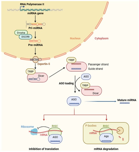
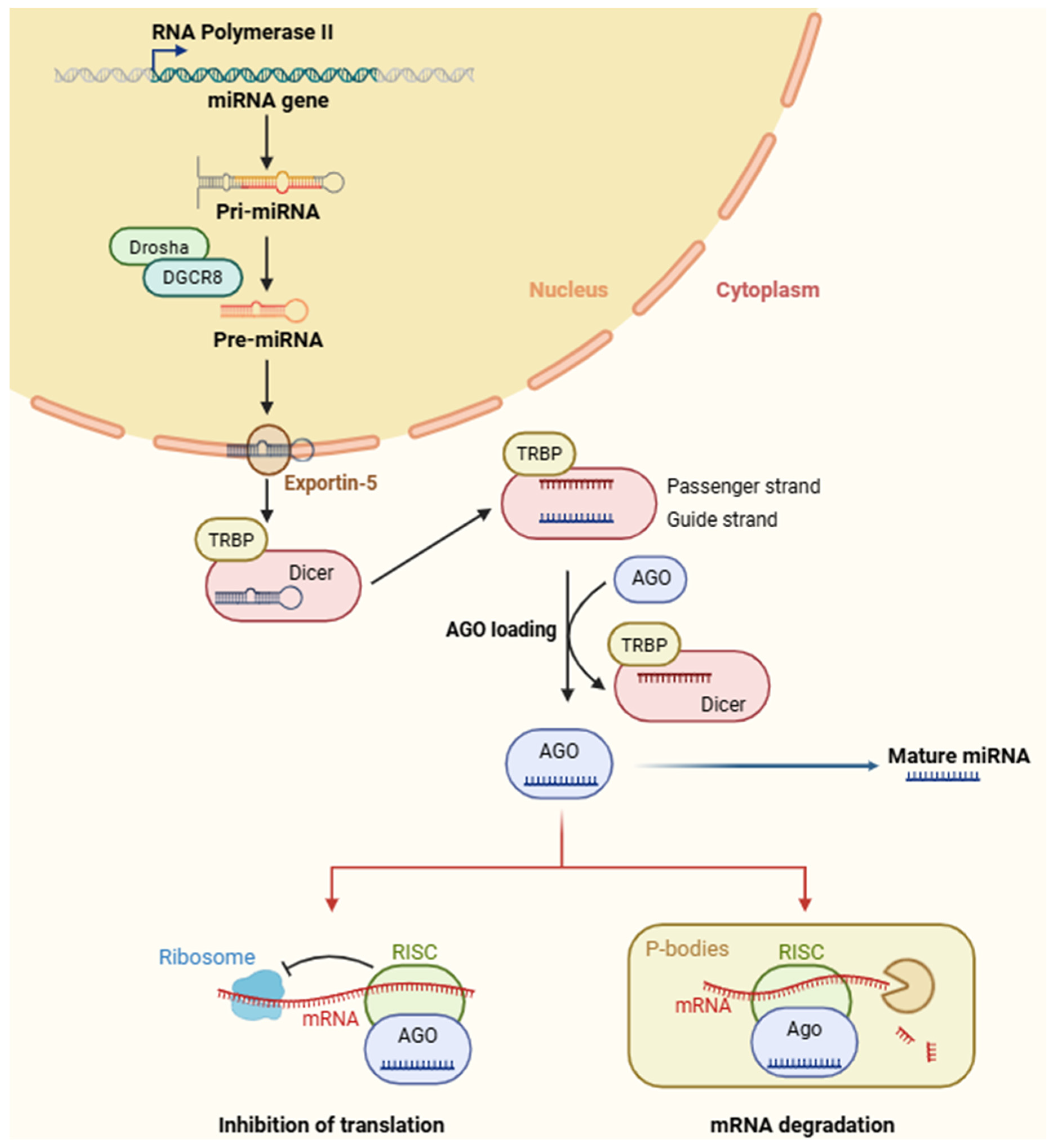

Disclaimer/Publisher’s Note: The statements, opinions and data contained in all publications are solely those of the individual author(s) and contributor(s) and not of MDPI and/or the editor(s). MDPI and/or the editor(s) disclaim responsibility for any injury to people or property resulting from any ideas, methods, instructions or products referred to in the content. |
© 2026 by the author. Licensee MDPI, Basel, Switzerland. This article is an open access article distributed under the terms and conditions of the Creative Commons Attribution (CC BY) license.