


Abstract
Background: Mucormycosis is a rapidly progressive and often fatal invasive fungal infection requiring immediate suspicion-driven diagnosis and treatment, particularly in immunocompromised patients. We report the first documented case of rhino-sino-orbital mucormycosis in a patient with recurrent intrahepatic cholangiocarcinoma and summarize our single-centre experience with eight cases of mucormycosis in oncohematologic patients. Methods: A 50-year-old woman developed rhino-sino-orbital mucormycosis after multimodal treatment for recurrent cholangiocarcinoma. Because of high clinical suspicion, imaging was performed before microbiological confirmation and antifungal therapy was initiated immediately through multidisciplinary management. Results: The patient survived 16 years after the initial cancer diagnosis, seven years after recurrence, and two years following mucormycosis. Our institutional series included eight cases with rhino-sino-orbital, pulmonary, cutaneous, and urinary tract involvement, most diagnosed after 2020, highlighting heterogeneous presentations and breakthrough infection despite prior antifungal exposure. Clinical experience gained from these cases contributed to early recognition and successful management of the present patient. Conclusions: To our knowledge, this is the first reported case of mucormycosis in cholangiocarcinoma. These findings emphasize that mucormycosis should be considered a medical emergency in patients with hematologic malignancies or solid tumors, and antifungal therapy should be initiated based solely on clinical suspicion without waiting for microbiological confirmation, even in atypical presentations.
1. Introduction
Mucormycosis is a rapidly progressive and often fatal invasive fungal infection that primarily affects immunocompromised patients and requires early recognition and prompt treatment [1,2,3]. Cholangiocarcinoma (CCA) is a rare malignancy of the biliary tract characterized by aggressive behavior, frequent late diagnosis, and poor clinical outcomes [4,5]. Although distant metastatic disease may occur, neuro-ophthalmologic and orbital manifestations are exceptionally uncommon [6,7]. Importantly, invasive fungal disease and biliary malignancy may also present with overlapping clinical and radiological features; hepatic mucormycosis involving the biliary tract has been reported to mimic hilar cholangiocarcinoma [8]. In patients with hematologic malignancies, mucormycosis is a particularly severe opportunistic infection, with pulmonary and rhino-sino-orbital/rhinocerebral involvement among the major clinical presentations and with substantial associated mortality [9,10]. The incidence of mucormycosis has increased in recent years, particularly among patients with hematologic malignancies, solid tumors, recipients of hematopoietic stem cell or solid organ transplantation, and in the context of diabetes mellitus, prolonged neutropenia, immunosuppressive therapies, and SARS-CoV-2 infection [11,12,13,14]. Among its clinical forms, rhino-orbito-cerebral mucormycosis is one of the most severe presentations, often associated with rapid progression and high mortality. In this context, orbital involvement represents a diagnostic challenge, as it may mimic a wide range of conditions, including infectious, inflammatory, and neoplastic processes, particularly in patients with underlying malignancies.
Biliary tract cancers are a heterogeneous group of aggressive malignancies associated with high mortality [15]. These tumors comprise distinct anatomical subtypes, including intrahepatic and extrahepatic cholangiocarcinoma, with differing clinical characteristics and outcomes; ampullary carcinoma has been reported to have a more favorable prognosis compared with extrahepatic cholangiocarcinoma [16,17].
Cholangiocarcinoma most commonly involves regional lymph nodes and adjacent organs, whereas distant metastases are uncommon. Brain metastases represent an exceptionally rare event and have been described predominantly in isolated case reports [18,19]. Orbital and neuro-ophthalmologic manifestations of cholangiocarcinoma are exceptionally rare, with only a limited number of orbital, skull-base, and cranial metastases reported in the literature, predominantly as isolated case reports [6,20,21,22].
Once orbital lesions are identified in patients with an underlying malignancy, careful differential diagnosis is essential, including infectious and inflammatory conditions, benign and malignant tumors, and vascular malformations. Among orbital infections, fungal etiologies are uncommon but increasingly recognized, particularly in immunocompromised patients, including those with hematologic malignancies, as well as recipients of hematopoietic stem cell or solid organ transplantation [1,11,12,13].
Therefore, this study aims to describe a rare case of rhino-sino-orbital mucormycosis in a patient with cholangiocarcinoma, together with seven additional cases of mucormycosis in patients with hematologic malignancies, and to highlight the importance of considering mucormycosis in the differential diagnosis of acute orbital and neurological manifestations in oncological patients.
2. Case Presentation
Detailed Case Description
We report the case of a 50-year-old female patient in whom a liver mass in the right hepatic lobe (RHL) was detected during routine ultrasound examination in July 2009 and subsequently confirmed by computed tomography (CT) and magnetic resonance imaging (MRI). CT revealed a polycystic tumor of the right hepatic lobe, measuring 8 cm in diameter, with isodense areas, peripheral hypodense regions, and central isodense enhancement, exerting a mass effect on the vascular structures, without evidence of locoregional or distant metastases (Figure 1).
Figure 1.
Abdominal CT showing a right hepatic lobe polycystic tumor measuring 8 cm in diameter, with isodense enhancement, peripheral hypodense areas, and central isodense enhancement, exerting a mass effect on the vascular structures.
An MRI-guided biopsy was performed. Histopathological examination established the diagnosis of intrahepatic cholangiocarcinoma, with TNM classification: G2, pT1, L0, V0. An extended right hemihepatectomy was performed in Regensburg, with a favorable outcome. Postoperatively, compensatory hypertrophy of the left hepatic lobe and the presence of right-sided hepatorrhaphy material were noted following right lobectomy (Figure 2).
Figure 2.
Compensatory hypertrophy of the left hepatic lobe (LHL) and the presence of hepatorrhaphy material following right lobectomy.
After a 9-year remission period, in September 2018, recurrence of cholangiocarcinoma in the left hepatic lobe was identified, along with high-grade hepatic and retroperitoneal metastases, documented by MRI and histopathological examination following a percutaneous liver biopsy performed at the Department of Gastroenterology in Cluj (Figure 3).
Figure 3.
(a,b) The remaining postoperative left hepatic lobe (LHL) shows a heterogeneous structure with nodular areas suggestive of secondary liver metastases. Paracaval and interaortocaval lymphadenopathy with imaging features similar to those of the hepatic lesions and the lesion at initial diagnosis are also observed.
Six cycles of chemotherapy with gemcitabine and oxaliplatin were administered concurrently with immunogenic cell death (ICD), modulated electrohyperthermia (mEHT), and oncolytic virotherapy with Newcastle disease virus (NDV) in Cologne, Germany. MRI evaluation after 3 and 6 cycles suggested a partial therapeutic response. Molecular investigations and drug sensitivity testing revealed overexpression of the PD-L1 gene, with a certified recommendation for targeted immunotherapy with nivolumab. The patient received nivolumab and gemcitabine concurrently with ICD, mEHT, and oncolytic virotherapy with NDV.
Positron emission tomography–computed tomography (PET–CT) scans performed after 3, 7, and 11 cycles, in June 2019, October 2019, and March 2020, revealed stable hepatic lesions, metabolically inactive residual retroperitoneal lymphadenopathy, and a near-complete metabolic response.
After a 6-month treatment-free interval, in October 2020, CT and PET–CT scans demonstrated progression of hepatic and abdominal lesions. A biopsy of a retroperitoneal lymph node was performed via an ultrasound-guided transabdominal approach, histologically confirming recurrence of cholangiocarcinoma.
Selective intrahepatic radiotherapy (SIRT) was performed in two procedures, two years apart, in November 2018 and 2020, with administration of yttrium-90 microspheres into the left hepatic artery at the Department of Nuclear Medicine, Barmherzige Brüder University Hospital, Regensburg, Germany.
Immunotherapy with the autologous antitumor vaccine IO-VAC was subsequently initiated, with dose 1 administered in August 2022, dose 2 in October 2022, and a booster dose (dose 3) administered six months later, on 10 May 2023, at IOZK, Germany. Treatment with nivolumab at a dose of 140 mg was continued at 3- to 4-week intervals, with the last dose administered on 31 May 2023, two weeks after the IO-VAC booster dose. MEHT, oncolytic virotherapy with NDV, and moderate whole-body hyperthermia (WBH), as well as ozone therapy, Curcumin Direct Spray, and DC vaccine, were administered in combination, with rigorous monitoring of the immune response.
MRI performed in November 2022 showed mild accentuation of internal softening of splenic, hepatic, and lymph node lesions, with no new lesions and no dimensional progression. A PET–CT scan performed in February 2023 revealed retroperitoneal adenopathy with progressive low-avid FDG uptake (Figure 4).
Figure 4.
(a,b) No pathological enhancement is observed in the oculoorbital regions; the liver remains heterogeneous, and several retroperitoneal lymph nodes are present without post-contrast enhancement.
Dental procedures were scheduled for May 2023. Following administration of the autologous antitumor vaccine on 10 May 2023, and nivolumab on 31 May 2023, the patient developed asthenia, abdominal fluid retention, and edema of the lower extremities.
On 24 June 2023, at the age of 64, at 08:00 a.m., the patient presented with marked asthenia, right-sided hemicrania, progressive unilateral right eyelid edema (non-erythematous), conjunctival chemosis, and severe ocular pain on horizontal and vertical eye movements. This was followed by limitation of ocular motility, diplopia, eyelid ptosis with inability to elevate the eyelid, photophobia upon manual elevation of the eyelid, nasolabial swelling, facial asymmetry, and right-sided exophthalmos measuring 19 mm (15 mm on the left).
The symptoms did not respond to the administered analgesics and anti-inflammatory agents or to the ophthalmic drops used. After 13 h, at 21:00, the patient presented to the hospital, where she was evaluated simultaneously by a neurologist and a hematologist. Neurological examination ruled out intracranial pathology, raising the possibility of an orbital cause for the exophthalmos. Prior clinical experience with mucormycosis in patients with hematologic malignancies also raised suspicion of mucormycosis (Figure 5).
Figure 5.
(a–d) Naso-orbital clinical appearance, exophthalmos, ptosis, and gingival lesion.
The differential diagnosis of right-sided exophthalmos (proptosis) included: (1) infections—bacterial (orbital cellulitis, abscess), fungal (Aspergillus, mucormycosis), parasitic (trichinellosis, echinococcosis), and Nocardia, which was considered a possible etiology in this patient; (2) inflammatory conditions—idiopathic orbital inflammatory syndrome (pseudotumor), Langerhans cell histiocytosis, sarcoidosis, and Graves’ ophthalmopathy; (3) tumors—capillary hemangioma, fibrous tumors, fibrosarcoma, optic nerve glioma, granulocytic sarcoma associated with leukemia, lymphomas, plasmacytomas, meningiomas, and metastases, particularly in the context of the patient’s history of cholangiocarcinoma; (4) trauma—orbital fractures and foreign bodies; and (5) other conditions—orbital varices, arteriovenous malformations, mucoceles, and dermoid and epidermoid cysts.
A brain CT scan was performed, revealing right-sided asymmetric exophthalmos caused by an intraorbital retroocular nodular lesion with irregular contours and isodense attenuation, in contact with the superior and inferior rectus muscles as well as the optic nerve, as well as minimal mucosal thickening of the right nasal concha, maxillary sinus, and ethmoid sinus (Figure 6).
Figure 6.
(a–d) Asymmetric right-sided exophthalmos caused by an intraorbital retroocular nodular lesion with irregular margins and heterogeneous contrast enhancement, in contact with the superior and inferior rectus muscles and the optic nerve; minimal mucosal thickening of the right nasal concha, maxillary sinus, and ethmoid sinus.
CT of the thorax, abdomen, and pelvis (CT TAP) revealed a right pleural effusion and a very thin left pleural layer; the left hepatic lobe (LHL) remained heterogeneous, with some hypodense areas not corresponding to the February PET scan; retroperitoneal lymphadenopathy, also evident on the previous PET scan, without radiotracer uptake; a heterogeneous spleen with a nodular lesion, without PET correlation; and moderate ascites (Figure 7).
Figure 7.
(a–d) CT of the thorax, abdomen, and pelvis (TAP CT).
Laboratory findings were as follows: WBC = 6400/mm3; neutrophils = 5080/mm3; Hb = 8 g/dL; platelets = 138,000/mm3; CRP = 7.8 mg/L; TSH = 81.2 mU/L (normal range 0.5–4.7), attributed to nivolumab treatment; CA125 = 127 U/mL (normal range 0–30), in the context of ascites; D-dimer = 2279 ng/mL (normal range 45–500). Evidence of cholestasis and hepatocytolysis was observed: alkaline phosphatase (ALP) = 812 U/L; GGT = 525 U/L; AST (TGO) = 179 U/L; ALT (TGP) = 95 U/L. Multidrug-resistant Klebsiella pneumoniae was isolated from sputum.
Cultures collected from the right nasal cavity and the right conjunctival sac revealed the presence of fungi (Figure 8A–C).
Figure 8.
(A) Mold growth observed on blood agar culture medium. (B) Direct microscopic examination using a lactophenol-stained preparation revealed mycelium composed of unicellular, non-septate, branched hyphae; vertical branches, including sporangiophores bearing sporangia, arising from the mycelium—consistent with organisms of the order Zygomycetales, family Mucoraceae (Mucor species). The hyphae were oriented at approximately 90 degrees, club-shaped, with a bulbous enlargement at one end. The micrographs were captured under 100× magnification. (C) Culture on Sabouraud dextrose agar (SDA), sheep blood agar (SBA), and cystine–lactose–electrolyte-deficient (CLED) agar revealed abundant (++++) filamentous fungal growth within 48 h; the cottony, woolly appearance was suggestive of infection with zygomycetes.
Treatment consisted of prophylactic isolation measures, protection of healthcare personnel with appropriate equipment to prevent transmission to other immunocompromised patients, and frequent ventilation. Amphotericin B was not available at that time; therefore, treatment was initiated with isavuconazole at a dose of 600 mg/day for 2 days, followed by 400 mg/day for 2 weeks, and subsequently 200 mg/day for 6 months.
Dexamethasone was administered for 3 days for its analgesic and anti-inflammatory effects but was subsequently discontinued due to a high suspicion of mucormycosis. Treatment was continued with osmotic and loop diuretics—mannitol, furosemide, and spironolactone—as well as analgesics, albumin, hepatoprotective agents, and propranolol for the associated liver disease. The Klebsiella pneumoniae infection was controlled with third-generation cephalosporins. For established hypothyroidism, attributed to nivolumab therapy, the patient received levothyroxine (Euthyrox) to control thyroid dysfunction. Given the known angioinvasive, microembolic, and necrotic nature of mucormycosis, therapeutic-dose anticoagulation was also administered.
There was also the possibility of orbital metastasis from cholangiocarcinoma concurrent with rhinosinusal mucormycosis. A biopsy was conducted to clarify this was not feasible in the presence of an invasive fungal infection. It was decided to treat the fungal infection first, with biopsy to be performed if the retroorbital tumor persisted.
The clinical course was remarkable: diplopia and pain resolved within 24 h; eyelid edema resolved within 5 days; ocular motility recovered after 5 days; eyelid ptosis was completely corrected within 10 days; exophthalmos resolved within 2 weeks; ascites and peripheral edema resolved within 14 days; and cultures became negative within 10 days.
Imaging findings demonstrated a significant reduction in the orbital mass at 15 days, with near-complete regression at 2 and 3 months after symptom onset. The mucosal thickening of the nasal turbinate, as well as that of the maxillary and ethmoid sinuses, resolved. At that time, the suspicion of concomitant orbital metastasis from cholangiocarcinoma was ruled out (Figure 9).
Figure 9.
Imaging progression: (a) 26 June 2023, at diagnosis; (b) 13 July 2023—2 weeks later; (c) 31 August 2023—2 months later; (d) normalization of the nasal and sinus mucosa. Near-complete remission of the orbital lesion was observed after 3 months.
After 6 months, with the fungal infection in remission (clinically, biologically, microbiologically, and on imaging) and following normalization of liver function, monthly nivolumab infusions were resumed to prevent recurrence or progression of cholangiocarcinoma. The patient’s presentation within 13 h of symptom onset, clinical suspicion, prompt imaging prior to microbiological confirmation, and interdisciplinary collaboration were the cornerstones of therapeutic success in this case, with survival of 16 years from the onset of cholangiocarcinoma, 7 years from recurrence, and 2 year following the mucormycosis infection.
Death occurred 2 years later, on 30 June 2025, due to disease progression with multiple abdominal metastases, subocclusive syndrome secondary to peritoneal carcinomatosis, Escherichia coli sepsis of gastrointestinal origin in the context of intestinal obstruction, gastrointestinal hemorrhage due to esophageal variceal rupture, hypotension, and respiratory failure.
3. Case Series
The experience of the Hematology Clinic in Sibiu includes eight cases of mucormycosis: five cases of rhinosino-orbito-cerebral mucormycosis (2013, 2021, 2023, 2024), one case of pulmonary mucormycosis (2024), two cases of cutaneous mucormycosis (2021, 2024), and one case of urinary mucormycosis. These anatomical categories were not mutually exclusive, as Case 7 initially presented with cutaneous mucormycosis and subsequently progressed to rhino-sino-orbito-cerebral involvement.
- Five cases of rhinosino-orbito-cerebral mucormycosis:
- ⚬
- A single case of ETO-positive acute myeloid leukemia (AML), in remission during consolidation therapy with severe aplasia, associated with insulin-dependent diabetes mellitus and a rural living environment, treated with voriconazole—diagnosed on 19 June 2013, with death due to mucormycosis on 27 June 2013 (case no. 2; Figure 10, Figure 11, Figure 12, Figure 13 and Figure 14).
- ⚬
- A single case of abdominal diffuse large B-cell lymphoma (DLBCL), with colostomy, during consolidation therapy in the aplastic phase, associated with insulin-dependent diabetes mellitus, treated with voriconazole—mucormycosis detected on 22 June 2021, with death due to disease progression on 7 June 2022 (case no. 3; Figure 15 and Figure 16).
- ⚬
- A single case of cholangiocarcinoma undergoing chemoimmunotherapy and dendritic cell vaccination, with recent dental procedures—mucormycosis diagnosed on 20 July 2023; death due to disease progression on 30 June 2025 (case no. 1).
- ⚬
- A single case of B-cell acute lymphoblastic leukemia (ALL) during chemotherapy, with severe aplasia, treated with voriconazole—diagnosed on 21 March 2024, with death due to mucormycosis on 27 March 2024 (case no. 4).
- ⚬
- A single case of high-risk TP53-positive, FLT3-positive acute monoblastic leukemia, with cutaneous onset of mucormycosis on 7 October 2024, progressing to the rhinosino-orbito-cerebral form on 11 November 2024—death due to mucormycosis on 20 December 2024 (case no. 7; Figure 17, Figure 18, Figure 19, Figure 20 and Figure 21)
- One case of pulmonary mucormycosis:
- ⚬
- A patient with T-cell non-Hodgkin lymphoma (NHL) in remission, following peripheral stem cell (PSC) collection, hospitalized for persistent respiratory syncytial virus (RSV) infection and pulmonary bacterial superinfection, treated with prophylactic voriconazole—mucormycosis diagnosed on 11 March 2024, with death due to mucormycosis on 18 March 2024 (case no. 5; Figure 22, Figure 23 and Figure 24).
- Two cases of cutaneous mucormycosis:
- ⚬
- The disease was observed in two patients: one with acute myelomonocytic leukemia following a myelodysplastic syndrome, associated with oropharyngeal and gingival lesions superinfected with mucormycosis on 24 June 2021, medically controlled, with death due to acute myocardial infarction on 1 August 2021 (case no. 6; Figure 25 and Figure 26), and one with high-risk acute monoblastic leukemia, del(17p)-positive and FLT3-positive, with cutaneous onset of mucormycosis progressing to the rhinosino-orbito-cerebral form. The patient had insulin-dependent diabetes mellitus and a rural living environment (case no. 7) both cases were in aplasia and receiving voriconazole prophylaxis.
- One case of urinary mucormycosis:
- ⚬
- A patient with multiple myeloma who had undergone transplantation in 2023. (case no. 8).
Figure 10.
(a,b) Clinical evolution of the rhino-orbito-sinusal lesion in Case No. 2.
Figure 11.
(a,b) Clinical evolution of the rhino-orbito-sinusal lesion in Case No. 2.
Figure 12.
(a–c) Brain CT showing no evidence of intracranial pathology; right-sided exophthalmos with diffuse thickening of the retrobulbar fat, as well as thickening of the right eyelid and cheek, and right maxillary–ethmoid sinusitis.
Figure 13.
(a–g) Brain MRI showing periorbital gadolinium-enhancing infiltration of the right retroorbital fat, maxilloethmoidal sinusitis, and cellulitis involving the right eyelid, cheek, and infratemporal region. The right optic nerve demonstrated diffusion restriction and prechiasmatic gadolinium enhancement. Areas of diffusion restriction were also observed in the right frontobasal and left occipital regions (suggestive of acute ischemia in an embolic context or areas of cerebritis), along with small periventricular demyelinating lesions.
Figure 14.
Mucormycosis: (a) Mold growth on blood agar culture medium; (b) direct microscopic examination using a lactophenol preparation. The micrographs were captured under 100× magnification; (c) culture on Sabouraud dextrose agar.
Figure 15.
(a,b) Clinical appearance of rhinosino-orbital mucormycosis in case no. 3.
Figure 16.
(a–d) Brain MRI in case no. 3.
Figure 17.
(a–o) Evolution of facio-orbital and oral lesions, as well as the solitary cutaneous lesion, in case no. 7.
Figure 18.
(a,b) Orbital CT scan (21 October 2024) showing no acute intracranial or orbital pathology, mild right frontal, maxillary, sphenoid, and ethmoid sinusitis, and right-sided proptosis in case no. 7.
Figure 19.
(a–c) Non-contrast cranial CT scan (3 December 2024) showing no acute intracranial lesions, with mucosal thickening and an isodense collection in the right maxillary sinus, right ethmoid air cells, and sphenoid sinus in case no. 7.
Figure 20.
(a–c) Brain MRI (3 December 2024) demonstrating extension of the inflammatory process from the right sinus cavities, with involvement of the left ethmoid air cells and left frontal sinus, as well as marked progression of intraorbital inflammatory changes in case no. 7.
Figure 21.
(a–c) Non-contrast cranial CT (19 December 2024) showing an intraparenchymal hemorrhagic lesion at the level of the right lentiform nucleus, associated with panventricular hemorrhage, subarachnoid hemorrhage, and diffuse cerebral edema with herniation at the level of the foramen magnum in case no. 7.
Figure 22.
(a,b) CT of the thorax, abdomen, and pelvis (CT TAP) revealing pulmonary consolidation in the mediastinal and pulmonary regions, with air bronchograms and moderate pleural effusion in case no. 5.
Figure 23.
(a,b) Mold growth on an agar-based culture medium; (c,d) filamentous mycelium with a club-shaped structure at one end, observed on direct microscopic examination using a slide and coverslip preparation. The micrographs were captured under 100× magnification.
Figure 24.
(a–e) Hematoxylin–eosin (H&E ×1000 magnification) stain of a peripheral blood smear showing large, hypergranular granulocytes with intracellular vacuoles, toxic green inclusions, and Döhle bodies, as well as schistocytes, consistent with reactive changes associated with severe invasive fungal infection in case no. 5.
Figure 25.
(a–c) Oral lesions involving the gingiva and buccal mucosa, associated with poor dentition, in case no. 6.
Figure 26.
(a–h) Cutaneous mucormycosis lesions at different stages of evolution in case no. 6.
Table 1.
Baseline characteristics and major risk factors of the eight patients with mucormycosis.
Table 2.
Underlying oncologic treatment and relevant exposure prior to mucormycosis.
Table 3.
Clinical presentation, diagnosis, treatment, and outcomes of mucormycosis.
4. Discussion
Mucormycosis, caused by fungi of the order Mucorales, is a rapidly progressive and frequently fatal invasive fungal infection first described in humans by Arnold Paltauf in 1885 [14]. Although uncommon, its burden has become increasingly apparent. Population-based surveillance in the San Francisco Bay Area during 1992–1993 estimated an annual incidence of approximately 1.7 cases per million inhabitants [23,24], while U.S. inpatient data identified 555 mucormycosis-related hospitalizations between 2005 and 2014 and an estimated 1140 hospitalizations in 2019 [24,25]. The disease occurs predominantly in immunocompromised individuals, including hematopoietic stem cell and solid organ transplant recipients, in whom it accounts for approximately 8% and 2% of invasive fungal infections, respectively [24,26]. In oncologic populations, mucormycosis is reported mainly in patients with hematologic malignancies, whereas association with solid tumors is much less frequent [9,12,27,28,29,30]. Infection usually follows inhalation of environmental spores, which may germinate into invasive hyphae and extend from the paranasal sinuses to the orbit or brain by direct spread and angioinvasion. Vascular invasion causes thrombosis, tissue ischemia, necrosis, and infarction. Clinical forms include rhino-orbito-cerebral, pulmonary, cutaneous, gastrointestinal, disseminated, and, rarely, urinary disease. Mortality remains high and varies with site and host factors, reaching 50–85% in rhino-cerebral disease, up to 90% in pulmonary disease, and extremely high levels in gastrointestinal or disseminated infection. Major predisposing factors include neutropenia, diabetes mellitus and metabolic acidosis, transplantation, advanced malignancy, corticosteroid exposure, increased iron availability, and cutaneous trauma. Neutropenia is particularly important in hematologic patients because of the central role of neutrophils in antifungal defense. Voriconazole and echinocandins do not provide adequate activity against Mucorales, and prior voriconazole exposure may contribute to the emergence of mucormycosis in highly immunosuppressed hosts. Its incidence has risen in parallel with the growing number of immunocompromised patients, reaching approximately 1% in some acute leukemia series. Cutaneous disease has been linked to trauma, including injuries associated with natural disasters, such as tsunamis and hurricanes, and more recently with SARS-CoV-2 infection during the COVID-19 pandemic and unusual presentations such as isolated urinary tract involvement have also been described. These observations are relevant to our cohort because they emphasize that mucormycosis cannot be approached as a single-site infection and that host context strongly influences clinical expression. PCR detection of circulating Mucorales DNA may support earlier diagnosis, but it is not routinely available; therefore, diagnosis often begins with clinical suspicion and imaging and is later confirmed microbiologically or histopathologically. Management rests on four principles: early recognition, prompt active antifungal treatment, correction of predisposing factors, and surgical debridement when feasible. In the index case, previous institutional experience and rapid multidisciplinary assessment facilitated recognition, and isavuconazole was started immediately because amphotericin B, considered first-line therapy, was unavailable. Classical risk factors such as neutropenia, hyperglycemia, prior voriconazole exposure, and iron overload were not evident in this patient; instead, combined chemoimmunotherapy, radiotherapy, immunomodulatory treatments, and impaired hepatic function were considered likely contributors to susceptibility. Corticosteroids were discontinued after three days and oncologic treatment was temporarily suspended. Surgical debridement is an important component of therapy, although severe cytopenias or anatomical extension may limit its feasibility; orbital exenteration has been required in advanced rhino-orbital disease. Hyperbaric oxygen has been proposed as an adjunct, but evidence remains limited, and there is no robust evidence for a direct protective or therapeutic effect of ozone therapy against mucormycosis.
Across our eight cases, mucormycosis occurred in one patient with intrahepatic cholangiocarcinoma and seven patients with hematologic neoplasms, including acute myeloid leukemia with t(8;21), acute monoblastic leukemia with del(17p), transformed follicular lymphoma to DLBCL, pre-B acute lymphoblastic leukemia, MEITL, MDS-EB2 evolving to high-risk FLT3-positive acute monoblastic leukemia, and multiple myeloma after autologous transplantation. This biological diversity was accompanied by markedly heterogeneous presentations and outcomes, reflecting the interaction between underlying disease, treatment intensity, and host immune status. Five episodes had rhino-sino-orbito-cerebral involvement, one was pulmonary, two included cutaneous disease, and one involved the urinary tract; these categories were not mutually exclusive because one cutaneous case progressed to rhino-sino-orbito-cerebral disease. Courses ranged from localized infection to ophthalmoplegia, optic nerve involvement, cerebral ischemic or hemorrhagic complications, disseminated intravascular coagulation, multiorgan failure, and death. The pulmonary case was particularly difficult to recognize because prolonged RSV infection, bacterial superinfection, organ dysfunction, and broad-spectrum antimicrobial exposure obscured the fungal process. The urinary case, with nonspecific manifestations, further illustrates that mucormycosis may remain clinically subtle even in profoundly immunocompromised patients. Within the rhino-orbital group, evolution also varied markedly: Case 2 progressed from unilateral eyelid edema to complete ophthalmoplegia, optic nerve involvement, multifocal cerebral ischemic lesions, and death, whereas Case 3 remained comparatively localized, with minimal retro-orbital involvement and rapid resolution under combined antifungal treatment. These contrasts illustrate why the clinical course cannot be inferred from anatomical classification alone. A striking feature was the temporal clustering of diagnoses. The first documented rhino-sino-orbito-cerebral case occurred in 2013, whereas seven of the eight cases were identified after 2020. In later cases, particularly Cases 1 and 3, clinical suspicion was raised within 12–24 h and often before microbiological confirmation; in Case 1, treatment was initiated approximately 13 h after symptom onset. By contrast, the early AML case progressed rapidly and ended in death within eight days despite radiologic suspicion within two days and prompt treatment. This contrast suggests that accumulated institutional experience lowered the threshold for considering mucormycosis and accelerated diagnostic pathways, although disease severity and host factors clearly remained important. In our centre, this experience changed practice: unilateral orbital symptoms, cranial nerve deficits, rapidly progressive facial swelling, and new sinus, pulmonary, or cutaneous lesions during voriconazole exposure now prompt early consideration of mucormycosis. The multidisciplinary pathway used in the cholangiocarcinoma case—integrating hematology, infectious diseases, radiology, ophthalmology, neurology, dentistry, oral and maxillofacial surgery, and intensive care—has subsequently become the institutional model for suspected disease. Earlier cases were more often characterized by advanced disease at recognition and limited therapeutic options, whereas later cases benefited from heightened awareness and faster coordination. A central feature of this series was the combined effect of severe aplasia, metabolic disturbances, and prior antifungal exposure, with profound neutropenia, thrombocytopenia, diabetes/hyperglycemia, and previous voriconazole use frequently accompanying rapidly progressive disease. In contrast, the cholangiocarcinoma patient developed mucormycosis without neutropenia or diabetes, suggesting that prolonged chemoimmunotherapy, radiotherapy, and immunomodulatory treatment may create a distinct therapy-induced immunosuppressive profile and that risk is not confined to classical hematologic settings. Clinical implications and practice changes in our center: mucormycosis is now systematically considered in the differential diagnosis of unilateral orbital symptoms, cranial nerve deficits, or rapidly progressive facial swelling in patients with hematologic malignancies, irrespective of fever or inflammatory markers. In patients receiving voriconazole who develop new sinus, orbital, pulmonary, or cutaneous lesions, breakthrough mucormycosis is actively considered, and early escalation to isavuconazole or amphotericin B is preferred over continuation or intensification of azole therapy.
The host characteristics of our patients are consistent with established risk settings, including hematologic malignancy, transplantation, diabetes mellitus, and corticosteroid exposure [11,14,24,31]. Corticosteroids are a recognized predisposing factor [28,32,33,34], while acute leukemia, prolonged neutropenia, and hematopoietic stem cell transplantation remain major hematologic settings [12,35]. Relapsed or refractory disease, prolonged neutropenia, and corticosteroid exposure also contribute to the cumulative risk of invasive mould infections [31,35,36,37,38,39,40], particularly when several factors coexist [41,42,43]. Although evidence from invasive mould infections overall cannot be fully extrapolated to mucormycosis [24,40,42], our cases similarly demonstrate cumulative immunosuppression related to chemotherapy, corticosteroids, neutropenia, metabolic abnormalities, and other treatment-related factors [35,41,42,43]. At mucormycosis onset, most hematologic patients were in severe aplasia, with neutrophil counts of 0–700/mm3 and frequent profound thrombocytopenia that restricted surgery. Diabetes or hyperglycemia of approximately 180–230 mg/dL was present in several cases. The cholangiocarcinoma patient, however, developed mucormycosis without neutropenia or diabetes, suggesting that prolonged chemoimmunotherapy, radiotherapy, and immunomodulatory treatment can produce a different but clinically relevant immunosuppressive profile. This single observation cannot be generalized, but it supports maintaining diagnostic awareness beyond classical hematologic settings. In several hematologic cases, hyperglycemia coexisted with profound aplasia, potentially amplifying susceptibility and complicating recovery. Prior voriconazole exposure was another recurrent feature; together with corticosteroid exposure, iron overload, and environmental factors, this may have contributed to a clinical context favorable to Mucorales. The important point is not a single isolated risk factor but the cumulative immunologic and metabolic burden carried by these patients. From a clinical perspective, our findings support extending the level of suspicion traditionally applied to hematologic malignancies to other oncologic settings characterized by profound or prolonged immunosuppression. In particular, clinicians managing patients with solid tumors undergoing intensive multimodal treatment, including chemotherapy and immunomodulatory therapies, should maintain awareness of mucormycosis as a potential diagnosis, especially in the presence of additional risk factors such as corticosteroid exposure or neutropenia. Finally, the temporal distribution of cases in our series, with the majority diagnosed after 2020, parallels the increase in mucormycosis incidence reported globally in recent years, including in the context of COVID-19-related immune dysregulation and wide-spread corticosteroid use. While causality cannot be inferred from our data, this observation reinforces the need for heightened clinical vigilance in the current epidemiological context [11,31,33,44,45].
The anatomical distribution in our series also differed from many large hematologic cohorts, in which pulmonary disease generally predominates [12,33], including recent hematologic and intensive-care studies [12,35,36]. Our patients showed substantial extrapulmonary involvement, particularly rhino-sino-orbito-cerebral disease, together with uncommon cutaneous and urinary manifestations [12,28,33,46,47,48]. In the index case, orbital involvement initially raised concern for metastatic disease, illustrating the diagnostic overlap between infectious, inflammatory, and neoplastic orbital lesions [28,49,50]. Urinary involvement, which is rarely reported, likewise demonstrates that mucormycosis may occur outside the thoracic and craniofacial sites most often investigated [28,46,48]. Diagnostic evaluation in high-risk patients should therefore follow the clinical presentation rather than being restricted to pulmonary imaging [47]; absence of pulmonary abnormalities does not exclude invasive fungal infection [28,46], and unusual manifestations warrant a site-oriented approach [33]. This is especially relevant in oncologic patients, in whom orbital, cutaneous, or urinary abnormalities may initially be attributed to tumor progression, bacterial infection, treatment toxicity, or other complications. A flexible anatomical approach is therefore necessary to avoid systematically under-recognizing extrapulmonary disease.
Diagnostic delay is consistently associated with adverse outcome in mucormycosis [31,36,51,52]. Published series have reported delays of up to 26 days, including diagnoses made only post-mortem [51], while ICU cohorts describe a median interval of approximately seven days [36]; delays exceeding 16 days have been independently associated with mortality [52]. In hematologic malignancy-associated zygomycosis, amphotericin B-based therapy initiated ≥6 days after symptom onset was associated with a twofold increase in mortality [53]. Conventional culture and histopathology can further delay confirmation because of limited sensitivity and turnaround times, with culture requiring up to 3–7 days and histopathology approximately 48–72 h [31,35,51,54,55,56]. These limitations support the current emphasis on early clinical suspicion [11,12,31]. In our index case, antifungal therapy was initiated approximately 13 h after symptom onset based on clinical and imaging findings, before microbiological confirmation, consistent with recommendations for prompt treatment when mucormycosis is suspected [31,57]. CT and MRI were central to early site-specific assessment [31,57]. Similar early treatment occurred in selected later cases. Although our small retrospective series cannot establish that a specific 24-h threshold improves survival, it demonstrates the feasibility of a suspicion-driven strategy and is consistent with larger studies linking diagnostic and therapeutic delay to poor outcome [35,52,53]. Thus, in high-risk patients with rapidly progressive or atypical syndromes, treatment decisions may need to rely on host factors, clinical presentation, and imaging rather than waiting for definitive microbiological proof [31,36,51,57]. In selected patients in our series, antifungal therapy was instituted within the first 12–24 h of suspicion, before microbiological confirmation. The favorable evolution observed in some of these cases should not be interpreted as causal evidence from this small series, but it illustrates how a rapid diagnostic pathway can be operationalized in routine practice and is consistent with the broader literature on the prognostic importance of treatment delay.
Breakthrough mucormycosis during antifungal exposure is increasingly recognized in patients with hematologic malignancies [13,58]. It may develop during therapy with agents lacking activity against Mucorales, particularly voriconazole, and less frequently during posaconazole, isavuconazole, or amphotericin B treatment [58]. Prognosis is especially poor when profound immunosuppression and unresolved neutropenia persist [12,58,59]. Voriconazole is particularly relevant because it lacks activity against Mucorales [58,60,61]. Several patients in our series developed mucormycosis despite prior antifungal exposure, and voriconazole was discontinued once mucormycosis was suspected. Persistent, progressive, or atypical lesions during triazole prophylaxis or treatment should therefore prompt diagnostic reassessment rather than continuation of the same regimen without reconsidering the underlying aetiology [52,53]. This approach is consistent with recommendations for infections arising during voriconazole exposure [31,61] and is especially important in hematologic patients, in whom deterioration may initially be attributed to malignancy or concomitant infections [13,62,63]. Breakthrough disease reflects the interaction between host immunosuppression, pathogen biology, and the limitations of antifungal coverage rather than prophylaxis failure alone. Accordingly, ongoing antifungal prophylaxis should not create a false sense of security. In patients with unexplained clinical deterioration or new radiological lesions, prior antifungal exposure should shape the differential diagnosis and therapeutic reassessment but should never reduce vigilance for mucormycosis.
Treatment in our series followed the principle that effective management requires coordinated antifungal therapy, surgical assessment, and correction of modifiable host factors [31,61,64,65]. Liposomal amphotericin B remains a principal first-line agent [31,61], while treatment must be adjusted according to clinical, radiological, and microbiological evolution [61,65]. Earlier cases often received liposomal amphotericin B with posaconazole, whereas isavuconazole was used more extensively in later cases, including as monotherapy when amphotericin B was unavailable. Prolonged therapy, reaching six months in selected patients, was associated with complete clinical and radiological resolution in Cases 1 and 3. Surgical debridement was pursued when feasible [31,36,66], but aplasia, severe thrombocytopenia, coagulation abnormalities, anatomical extension, and clinical instability frequently limited intervention. Correction of underlying factors—including neutrophil recovery, reduction or discontinuation of corticosteroids, glycemic control, removal of infected devices, and management of comorbidities—was integral to care [12,31,59,61]. Rapid coordination among hematology, infectious diseases, radiology, intensive care, and site-specific surgical specialties allowed treatment and surgical decisions to be adapted as disease evolved. This time-sensitive multidisciplinary organization may reduce avoidable delays and operationalizes the core principles emphasized in international recommendations [31,61,65,66]. In practice, management had to remain individualized: critically ill patients sometimes deteriorated too rapidly to sustain planned antifungal combinations or undergo surgery, whereas clinically stable patients could receive prolonged treatment and serial imaging. These differences reinforce that successful care depends not only on the antifungal agent selected but also on the ability to coordinate supportive care, local source control, and recovery of host defenses.
Reported mortality in hematology and HSCT populations ranges from approximately 40% to 80% [35,67], and may approach 70% in critically ill cohorts [36]. Non-recovered neutropenia, corticosteroid exposure, disseminated disease, and delayed appropriate therapy are repeatedly associated with poor outcome [35,53,67], while breakthrough mucormycosis also carries an unfavorable short-term prognosis [58]. Our observations were consistent with these patterns: persistent aplasia, thrombocytopenia, multiple infections, septic shock, disseminated intravascular coagulation, extensive disease, and multiorgan dysfunction characterized unfavorable courses, whereas clinical improvement more often accompanied hematologic recovery, control of modifiable factors, and timely active antifungal treatment [35,53,67]. The comparison between Cases 1 and 2 illustrates that diagnostic timing cannot be considered in isolation. Case 2 deteriorated rapidly despite relatively prompt recognition, in the setting of severe aplasia and metabolic risk factors, whereas Case 1 had no aplasia and achieved complete resolution after suspicion at approximately 13 h and immediate targeted therapy. Thus, prognosis in this series appears to reflect the cumulative interaction of host status, disease extent, and timing of intervention rather than any single variable. Given the small sample, these associations are descriptive and do not establish independent prognostic effects [35,36,52,67]. Nevertheless, across the series the most clinically actionable factors were consistent: maintain early suspicion, initiate active antifungal therapy without unnecessary delay, correct reversible risk factors, and pursue local intervention whenever the hematologic and anatomical situation permits. Poor outcomes occurred particularly when several adverse factors converged, supporting the concept that prognosis is driven by cumulative host- and disease-related severity rather than by a single determinant.
Seven of eight cases were diagnosed after 2020, paralleling increased reporting of mucormycosis during the COVID-19 pandemic, particularly COVID-19-associated mucormycosis [11,32,68]. Proposed contributors include systemic corticosteroid use, immune dysregulation, and worsening or newly recognized diabetes mellitus [32,41,42,43], while intensified immunosuppressive therapies and prolonged hospitalizations may also have influenced the epidemiology of invasive fungal infections. Because our cohort was not restricted to COVID-19-associated disease, this temporal clustering cannot be attributed specifically to SARS-CoV-2. It should instead be interpreted in the context of broader epidemiological changes among highly immunocompromised populations [11,31,33,44,45], reinforcing the need for continued vigilance in the post-pandemic period.
A particularly important contribution of this series is the occurrence of rhino-sino-orbital mucormycosis in a patient with established intrahepatic cholangiocarcinoma after prolonged multimodal oncologic treatment. To our knowledge, mucormycosis complicating an established cholangiocarcinoma has not previously been reported. Conversely, hepatic mucormycosis involving the biliary tract has been described as mimicking hilar cholangiocarcinoma [8], illustrating the reverse diagnostic relationship. In contrast to these reports, our case demonstrates the reverse scenario, in which a known malignancy obscured the recognition of an invasive fungal infection and demonstrates how invasive fungal infection can be obscured by an expected oncologic differential. More broadly, our cohort adds case-level detail to the recognized heterogeneity of mucormycosis. Pulmonary and rhino-orbital/sino-facial disease predominate in larger hematologic series [9,12,47], whereas cutaneous and urinary mucormycosis remain uncommon, and rhino-orbito-cerebral disease requires rapid diagnosis, active antifungal treatment, and surgical evaluation when feasible [31,45,69,70,71,72,73], but severe hematologic abnormalities frequently restricted surgery in our patients. The short diagnostic intervals in selected cases illustrate the feasibility—not proof of efficacy—of a suspicion-driven approach [31,61], particularly given delays reported elsewhere [36,51] and the association between delayed amphotericin B-based treatment and mortality [26,53]. Clinical and imaging findings can therefore support early decisions in high-risk patients [26,74], especially in rapidly progressive rhino-orbito-cerebral disease [75]. Likewise, disease developing despite prior mould-active exposure is consistent with reports of breakthrough mucormycosis [58,76,77] and remains a challenge in hematologic malignancies [58,78,79,80,81,82], requiring reassessment when infection progresses during therapy [58,76]. Taken together, our experience provides a practical framework linking early suspicion, site-directed imaging, rapid antifungal initiation, correction of host factors, and multidisciplinary decision-making across both classical and atypical presentations. In contrast to larger datasets that primarily provide aggregate outcomes, our series documents the sequence of symptom onset, antifungal exposure, imaging evolution, surgical feasibility, and clinical response at case level. This detail helps illustrate the bedside diagnostic traps encountered in patients with complex malignancies, particularly when mucormycosis resembles oncologic progression or develops despite antifungal prophylaxis. The series therefore contributes not by establishing new causal relationships, but by showing how established principles can be applied under real-world constraints.
This study has several strengths, including detailed case-level documentation of eight episodes from symptom onset through clinical suspicion, diagnostic work-up, imaging, antifungal exposure, treatment, surgical decisions, and multidisciplinary management. Inclusion of hematologic malignancies and a solid-tumor case, together with uncommon cutaneous and urinary manifestations, broadens the represented clinical spectrum. However, the retrospective single-centre design and small sample limit generalizability and preclude formal prognostic analysis. Selection bias is possible because only clinically overt, microbiologically or histologically confirmed cases were included, and the observational design does not permit causal inference regarding early suspicion, specific antifungal regimens, multidisciplinary management, or surgery. These findings should therefore be interpreted as descriptive observations consistent with established evidence and current recommendations, and larger multicentre studies are needed to confirm and extend them. Additional integrated, detailed, and in-depth information regarding the detailed clinical, microbiological, laboratory, therapeutic, and outcome data for the eight patients with mucormycosis, as well as the detailed oncologic history, therapeutic lines, and exposure for the eight patients, can be found in Appendix A, Table A1 and Table A2.
5. Conclusions
Mucormycosis remains a rapidly progressive and life-threatening infection in immunocompromised patients, particularly in those with hematologic malignancies and complex oncologic backgrounds. Our series shows that the clinical spectrum of mucormycosis in this population extends beyond classical pulmonary presentations and may include atypical anatomical sites such as rhino-sino-orbital, cutaneous, and urinary involvement, which can complicate and delay diagnosis. In addition, the occurrence of infection despite prior antifungal exposure underscores that prophylaxis does not eliminate risk in highly immunosuppressed patients.
Our experience supports a suspicion-driven approach in high-risk patients, in which early recognition based on clinical and imaging features may allow initiation of antifungal therapy before microbiological confirmation is available. In our series, this approach was combined with rapid multidisciplinary management and correction of underlying risk factors; however, the small retrospective nature of the series does not allow conclusions regarding its efficacy or impact on mortality.
Overall, our findings support maintaining a high index of suspicion for mucormycosis in high-risk patients, including those with atypical presentations or prior antifungal exposure, and are consistent with current recommendations favoring prompt antifungal treatment when mucormycosis is clinically suspected.
Author Contributions
Conceptualization, C.A.C. and M.L.-M.; Data Curation, M.L.-M.; Investigation, B.V., C.C.R., P.M., S.M., D.D., M.E., O.C., O.A., E.V., Ș.L., M.B., P.C. and I.G.; Methodology, M.L.-M. and C.A.C.; Resources, M.L.-M.; Supervision, C.A.C.; Validation, C.A.C.; Visualization, M.L.-M.; Writing—Original Draft, C.A.C.; Writing—Review and Editing, M.L.-M. All authors have read and agreed to the published version of the manuscript.
Funding
This research received no external funding.
Institutional Review Board Statement
This study was conducted in accordance with the Declaration of Helsinki and approved by the Ethics Committee of Emergency Clinical County Hospital of Sibiu Ethics Committee (Code: 29477, approval date: 14 December 2024).
Informed Consent Statement
Informed consent was obtained from all subjects involved in the study. Written informed consent has been obtained from the patient to publish this paper.
Data Availability Statement
The original contributions presented in the case report are included in the article, further inquiries can be directed to the corresponding authors.
Conflicts of Interest
The authors declare no conflicts of interest.
Abbreviations
The following abbreviations are used in this manuscript:
| SARS-CoV-2 | Severe Acute Respiratory Syndrome Coronavirus 2 |
| RHL | Right hepatic lobe |
| CT | Computed tomography |
| MRI | Magnetic resonance imaging |
| TNM | Tumor, Node, Metastasis |
| LHL | Left hepatic lobe |
| ICD | Immunogenic cell death |
| mEHT | Modulated electrohyperthermia |
| NDV | Newcastle disease virus |
| PET–CT | Positron emission tomography–computed tomography |
| SIRT | Selective intrahepatic radiotherapy |
| WBH | Whole body hyperthermia |
| FDG | Fluorodeoxyglucose |
| CT TAP | Computed tomography of the thorax, abdomen, and pelvis |
| WBC | White Blood Cells |
| Hb | Hemoglobin |
| CRP | C-Reactive Protein |
| TSH | Thyroid Stimulating Hormone |
| CA125 | Cancer Antigen 125 |
| ALP | Alkaline phosphatase |
| GGT | Gamma Glutamyl Transferase |
| AST | Aspartate Aminotransferase |
| ALT | Alanine Aminotransferase |
| SDA | Sabouraud dextrose agar |
| SBA | Sheep blood agar |
| CLED | Cystine–lactose–electrolyte deficient |
| AML | Acute myeloid leukemia |
| DLBCL | Diffuse large B cell lymphoma |
| ALL | Acute lymphoblastic leukemia |
| NHL | non-Hodgkin lymphoma |
| PSC | Peripheral stem cell |
| RSV | Respiratory syncytial virus |
| MEITL | Monomorphic epitheliotropic intestinal T-cell lymphoma |
| MDS | Myelodysplastic syndrome |
| DIC | Disseminated intravascular coagulation |
| H&E | Hematoxylin–eosin |
| COVID-19 | Coronavirus Disease 2019 |
| ENT | Ear, Nose, and Throat |
| ICU | Intensive Care Unit |
| HSCT | Hematopoietic Stem Cell Transplantation |
| CAM | COVID-19-associated mucormycosis |
Appendix A
Table A1.
Detailed clinical, microbiological, laboratory, therapeutic, and outcome data for the eight patients with mucormycosis.
Table A2.
Detailed oncologic history, therapeutic lines, and exposure for the eight patients.
References
- Pak, J.; Tucci, V.T.; Vincent, A.L.; Sandin, R.L.; Greene, J.N. Mucormycosis in immunochallenged patients. J. Emerg. Trauma Shock 2008, 1, 106–113. [Google Scholar] [CrossRef] [Scilit]
- Pandilwar, P.K.; Khan, K.; Shah, K.; Sanap, M.; Unnikrishnan, K.S.A.; Nerurkar, S. Mucormycosis: A rare entity with rising clinical presentation in immunocompromised hosts. Int. J. Surg. Case Rep. 2020, 77, 57–61. [Google Scholar] [CrossRef] [Scilit]
- Hernández, J.L.; Buckley, C.J. Mucormycosis. In StatPearls [Internet]; StatPearls Publishing: Treasure Island, FL, USA, 2026. Available online: https://www.ncbi.nlm.nih.gov/books/NBK544364/ (accessed on 10 June 2026).
- Banales, J.M.; Cardinale, V.; Carpino, G.; Marzioni, M.; Andersen, J.B.; Invernizzi, P.; Lind, G.E.; Folseraas, T.; Forbes, S.J.; Fouassier, L.; et al. Cholangiocarcinoma: Current knowledge and future perspectives—Consensus statement from the European Network for the Study of Cholangiocarcinoma (ENS-CCA). Nat. Rev. Gastroenterol. Hepatol. 2016, 13, 261–280. [Google Scholar] [CrossRef] [Scilit]
- Razumilava, N.; Gores, G.J. Cholangiocarcinoma. Lancet 2014, 383, 2168–2179. [Google Scholar] [CrossRef] [Scilit]
- Rico, G.; Smith, S.V.; Siddiqui, Y.; Whyte, A.; Gombos, D.G.; Lee, A.G. Neuro-ophthalmologic manifestations of cholangiocarcinoma: A case series. Eye 2017, 31, 1245–1248. [Google Scholar] [CrossRef] [Scilit]
- Fowler, B.J.; Lam, B.L. Sixth cranial nerve palsy as the presenting sign of metastatic cholangiocarcinoma. Int. Med. Case Rep. J. 2020, 13, 667–671. [Google Scholar] [CrossRef] [Scilit]
- Li, K.W.; Wen, T.F.; Li, G.D. Hepatic mucormycosis mimicking hilar cholangiocarcinoma: A case report and literature review. World J. Gastroenterol. 2010, 16, 1039–1042. [Google Scholar] [CrossRef] [Scilit]
- Pagano, L.; Offidani, M.; Fianchi, L.; Nosari, A.; Candoni, A.; Picardi, M.; Corvatta, L.; D’Antonio, D.; Girmenia, C.; Martino, P.; et al. Mucormycosis in hematologic patients. Haematologica 2004, 89, 207–214. [Google Scholar]
- Lewis, R.E.; Georgiadou, S.P.; Sampsonas, F.; Chamilos, G.; Kontoyiannis, D.P. Risk factors for early mortality in hematological malignancy patients with pulmonary mucormycosis. Mycoses 2014, 57, 10–15. [Google Scholar] [CrossRef] [Scilit]
- Alqarihi, A.; Kontoyiannis, D.P.; Ibrahim, A.S. Mucormycosis in 2023: An update on pathogenesis and management. Front. Cell. Infect. Microbiol. 2023, 13, 1254919. [Google Scholar] [CrossRef] [Scilit]
- Yang, N.; Zhang, L.; Feng, S. Clinical features and treatment progress of invasive mucormycosis in patients with hematological malignancies. J. Fungi 2023, 9, 592. [Google Scholar] [CrossRef] [Scilit]
- Miller, M.A.; Molina, K.C.; Gutman, J.A.; Scherger, S.; Lum, J.M.; Mossad, S.B.; Burgess, M.; Cheng, M.P.; Chuang, S.T.; Jacobs, S.E.; et al. Mucormycosis in hematopoietic cell transplant recipients and in patients with hematological malignancies in the era of new antifungal agents. Open Forum Infect. Dis. 2021, 8, ofaa646. [Google Scholar] [CrossRef] [Scilit]
- Skiada, A.; Pavleas, I.; Drogari-Apiranthitou, M. Epidemiology and diagnosis of mucormycosis: An update. J. Fungi 2020, 6, 265. [Google Scholar] [CrossRef] [Scilit]
- Macarulla, T.; Adeva, J.; Cano Osuna, M.T.; Ruiz Casado, A.; Jiménez Gordo, A.M.; Lamarca, A.; Muñoz, A.M.L.; Cid, R.A.P.; Saurí, T.; Plazas, J.G. SEOM-GEMCAD-TTD clinical guidelines for biliary tract cancer (2025). Clin. Transl. Oncol. 2025, 27, 3293–3306. [Google Scholar] [CrossRef] [Scilit]
- Woo, S.M.; Ryu, J.K.; Lee, S.H.; Yoo, J.W.; Park, J.K.; Kim, Y.T.; Jang, J.Y.; Whe-Kim, S.; Kang, G.H.; Yoon, Y.B. Recurrence and prognostic factors of ampullary carcinoma after radical resection: Comparison with distal extrahepatic cholangiocarcinoma. Ann. Surg. Oncol. 2007, 14, 3195–3201. [Google Scholar] [CrossRef] [Scilit] [PubMed]
- Kim, W.S.; Choi, D.W.; Choi, S.H.; Heo, J.S.; You, D.D.; Lee, H.G. Clinical significance of pathologic subtype in curatively resected ampulla of Vater cancer. J. Surg. Oncol. 2012, 105, 266–272. [Google Scholar] [CrossRef] [Scilit] [PubMed]
- Chindaprasirt, J.; Sookprasert, A.; Sawanyawisuth, K.; Limpawattana, P.; Tiamkao, S. Brain metastases from cholangiocarcinoma: A first case series in Thailand. Asian Pac. J. Cancer Prev. 2012, 13, 1995–1997. [Google Scholar] [CrossRef] [Scilit]
- Cha, S.; Lee, C.H.; Jang, K.Y.; Jeon, S.Y. Unveiling the unprecedented—Very late brain metastasis in cholangiocarcinoma: A case report and comprehensive analysis. Medicine 2024, 103, e39130. [Google Scholar] [CrossRef] [Scilit]
- Fujimoto, K.; Kuroda, J.I.; Makino, K.; Hasegawa, Y.; Kuratsu, J.I. Skull metastasis from intrahepatic cholangiocarcinoma: Report of 3 cases and review of the literature. Neurol. Med. Chir. 2013, 53, 717–721. [Google Scholar] [CrossRef] [Scilit]
- Hanna, J.A.; Mathkour, M.; Gouveia, E.E.; Glynn, R.; Divagaran, A.; Lane, J.B.; Bui, C.J.; Sulaiman, O.A.; Smith, R.D. Cholangiocarcinoma metastasis to the spine and cranium. Ochsner J. 2020, 20, 197–203. [Google Scholar] [CrossRef] [Scilit]
- Wang, D.; He, S.; Chu, L.; Chao, Q.; Zhang, Q.; Shu, H. A metastatic skull tumor from intrahepatic cholangiocarcinoma: A case report and literature review. Medicine 2019, 98, e18291. [Google Scholar] [CrossRef] [Scilit]
- Roden, M.M.; Zaoutis, T.E.; Buchanan, W.L.; Knudsen, T.A.; Sarkisova, T.A.; Schaufele, R.L.; Sein, M.; Sein, T.; Chiou, C.C.; Chu, J.H.; et al. Epidemiology and outcome of zygomycosis: A review of 929 reported cases. Clin. Infect. Dis. 2005, 41, 634–653. [Google Scholar] [CrossRef] [Scilit]
- Centers for Disease Control and Prevention (CDC). Data and Statistics on Mucormycosis; CDC: Atlanta, GA, USA, 2024. Available online: https://www.cdc.gov/mucormycosis/data-research/index.html (accessed on 4 May 2026).
- Kontoyiannis, D.P.; Yang, H.; Song, J.; Kelkar, S.S.; Yang, X.; Azie, N.; Harrington, R.; Fan, A.; Lee, E.; Spalding, J.R. Prevalence, clinical and economic burden of mucormycosis-related hospitalizations in the United States: A retrospective study. BMC Infect. Dis. 2016, 16, 730. [Google Scholar] [CrossRef] [Scilit] [PubMed] [PubMed Central]
- Lanternier, F.; Sun, H.Y.; Ribaud, P.; Singh, N.; Kontoyiannis, D.P.; Lortholary, O. Mucormycosis in organ and stem cell transplant recipients. Clin. Infect. Dis. 2012, 54, 1–8. [Google Scholar] [CrossRef] [Scilit]
- Suo, T.; Xu, M.; Xu, Q. Clinical characteristics and mortality of mucormycosis in hematological malignancies: A retrospective study in Eastern China. Ann. Clin. Microbiol. Antimicrob. 2024, 23, 82. [Google Scholar] [CrossRef] [Scilit] [PubMed] [PubMed Central]
- Jeong, W.; Keighley, C.; Wolfe, R.; Lee, W.L.; Slavin, M.A.; Kong, D.C.M.; Chen, S.C.-A. The epidemiology and clinical manifestations of mucormycosis: A systematic review and meta-analysis of case reports. Clin. Microbiol. Infect. 2019, 25, 26–34. [Google Scholar] [CrossRef] [Scilit]
- Taweesuk, A.; Chongtrakool, P.; Sitthinamsuwan, P.; Phoompoung, P. Hematogenous dissemination of pulmonary mucormycosis manifested as multiple subcutaneous nodules: A case report and review of the literature. BMC Infect. Dis. 2022, 22, 220. [Google Scholar] [CrossRef] [Scilit] [PubMed] [PubMed Central]
- Yamauchi, T.; Misaki, H.; Arai, H.; Iwasaki, H.; Naiki, H.; Ueda, T. An autopsy case of disseminated mucormycosis in a neutropenic patient receiving chemotherapy for the underlying solid malignancy. J. Infect. Chemother. 2002, 8, 103–105. [Google Scholar] [CrossRef] [Scilit] [PubMed]
- Cornely, O.A.; Alastruey-Izquierdo, A.; Arenz, D.; Chen, S.C.A.; Dannaoui, E.; Hochhegger, B.; Hoenigl, M.; Jensen, H.E.; Lagrou, K.; Lewis, R.E.; et al. Global guideline for the diagnosis and management of mucormycosis: An initiative of the European Confederation of Medical Mycology in cooperation with the Mycoses Study Group Education and Research Consortium. Lancet Infect. Dis. 2019, 19, e405–e421. [Google Scholar] [CrossRef] [Scilit]
- Hoenigl, M.; Seidel, D.; Carvalho, A.; Rudramurthy, S.M.; Arastehfar, A.; Gangneux, J.-P.; Nasir, N.; Bonifaz, A.; Araiza, J.; Klimko, N.; et al. The emergence of COVID-19 associated mucormycosis: A review of cases from 18 countries. Lancet Microbe 2022, 3, e543–e552. [Google Scholar] [CrossRef] [Scilit] [PubMed] [PubMed Central]
- Prakash, H.; Chakrabarti, A. Global epidemiology of mucormycosis. J. Fungi 2019, 5, 26. [Google Scholar] [CrossRef] [Scilit] [PubMed] [PubMed Central]
- Prakash, H.; Chakrabarti, A. Epidemiology of mucormycosis in India. Microorganisms 2021, 9, 523. [Google Scholar] [CrossRef] [Scilit] [PubMed] [PubMed Central]
- Cui, Y.; Zhao, R.; Mi, R.; Chen, L.; Wang, L.; Li, D.; Cheng, C.; Li, M.; Wei, X. Hematological diseases-related mucormycosis: A retrospective single center study. PLoS Negl. Trop. Dis. 2025, 19, e0013531. [Google Scholar] [CrossRef] [Scilit]
- Claustre, J.; Larcher, R.; Jouve, T.; Truche, A.-S.; Nseir, S.; Cadiet, J.; Zerbib, Y.; Lautrette, A.; Constantin, J.-M.; Charles, P.-E.; et al. Mucormycosis in intensive care unit: Surgery is a major prognostic factor in patients with hematological malignancy. Ann. Intensive Care 2020, 10, 74. [Google Scholar] [CrossRef] [Scilit]
- Low, C.Y.; Rotstein, C. Emerging fungal infections in immunocompromised patients. F1000 Med. Rep. 2011, 3, 14. [Google Scholar] [CrossRef] [Scilit] [PubMed] [PubMed Central]
- Chu, S.; McCormick, T.S.; Lazarus, H.M.; Leal, L.O.; Ghannoum, M.A. Invasive fungal disease and the immunocompromised host including allogeneic hematopoietic cell transplant recipients: Improved understanding and new strategic approach with sargramostim. Clin. Immunol. 2021, 228, 108731. [Google Scholar] [CrossRef] [Scilit] [PubMed]
- Rodríguez-Goncer, I.; Boán, J.; Carrero-Arribas, R.; Sanchez-Pina, J.M.; Lizasoaín, M.; Fernández-Ruiz, M.; San-Juan, R.; López-Medrano, F.; Pérez-Ayala, A.; Caro-Teller, J.M.; et al. Breakthrough invasive mold infections in hematologic cases: Relevance of the host’s factors. Open Forum Infect. Dis. 2025, 12, ofaf025. [Google Scholar] [CrossRef] [Scilit] [PubMed] [PubMed Central]
- Gras, E.; Azoyan, L.; Monzo-Gallo, P.; Garcia-Vidal, C.; Lanternier, F.; Brissot, E.; Guitard, J.; Lacombe, K.; Dechartres, A.; Surgers, L. Risk factors for invasive mould infections in adult patients with hematological malignancies and/or stem cell transplant: A systematic literature review and meta-analysis. J. Infect. 2025, 91, 106574. [Google Scholar] [CrossRef] [Scilit] [PubMed]
- Bhanuprasad, K.; Manesh, A.; Devasagayam, E.; Varghese, L.; Cherian, L.M.; Kurien, R.; Karthik, R.; Deodhar, D.; Vanjare, H.; Peter, J.; et al. Risk factors associated with the mucormycosis epidemic during the COVID-19 pandemic. Int. J. Infect. Dis. 2021, 111, 267–270. [Google Scholar] [CrossRef] [Scilit] [PubMed] [PubMed Central]
- Sharma, A.; Goel, A. Mucormycosis: Risk factors, diagnosis, treatments, and challenges during COVID-19 pandemic. Folia Microbiol. 2022, 67, 363–387. [Google Scholar] [CrossRef] [Scilit] [PubMed] [PubMed Central]
- Özbek, L.; Topçu, U.; Manay, M.; Esen, B.H.; Bektas, S.N.; Aydın, S.; Özdemir, B.; Khostelidi, S.N.; Klimko, N.; Cornely, O.; et al. COVID-19–associated mucormycosis: A systematic review and meta-analysis of 958 cases. Clin. Microbiol. Infect. 2023, 29, 722–731. [Google Scholar] [CrossRef] [Scilit] [PubMed] [PubMed Central]
- Dalai, S.; Datla, A.V.; Francis, A.A.; Dannana, N.K.; Parappil, H. Endovascular management of mucormycotic aneurysms of the internal carotid artery in post-COVID-19 patients. Cureus 2021, 13, e20812. [Google Scholar] [CrossRef] [Scilit] [PubMed] [PubMed Central]
- Kumar, M.; Kaundal, P.; Sharma, S.; Rana, K. Mucormycosis in the urinary bladder: The devil is in the details. Indian J. Surg. 2023, 85, 975–977. [Google Scholar] [CrossRef] [Scilit]
- Skiada, A.; Pagano, L.; Groll, A.; Zimmerli, S.; Dupont, B.; Lagrou, K.; Lass-Flörl, C.; Bouza, E.; Klimko, N.; Gaustad, P.; et al. Zygomycosis in Europe: Analysis of 230 cases accrued by the registry of the European Confederation of Medical Mycology (ECMM) Working Group on zygomycosis between 2005 and 2007. Clin. Microbiol. Infect. 2011, 17, 1859–1867. [Google Scholar] [CrossRef] [Scilit]
- Nosari, A.; Oreste, P.; Montillo, M.; Carrafiello, G.; Draisci, M.; Muti, G.; Molteni, A.; Morra, E. Mucormycosis in hematologic malignancies: An emerging fungal infection. Haematologica 2000, 85, 1068–1071. [Google Scholar] [PubMed]
- Jindal, J.; Garg, V. Cutaneous mucormycosis: Unveiling rare manifestations. J. Fam. Med. Prim. Care 2025, 14, 2561–2564. [Google Scholar] [CrossRef] [Scilit] [PubMed] [PubMed Central]
- Philip, S.M. Invasive rhino-orbital-cerebral mucormycosis in sinonasal neuroendocrine carcinoma: A case report. Clin. Oncol. Case Rep. 2021, 4. Available online: https://www.scitechnol.com/peer-review/invasive-rhinoorbitalcerebral-mucormycosis-in-sinonasal-neuroendocrine-carcinoma-a-case-report-Ksox.php?article_id=17715 (accessed on 10 June 2026).
- Prabhu, R.M.; Patel, R. Mucormycosis and entomophthoramycosis: A review of the clinical manifestations, diagnosis and treatment. Clin. Microbiol. Infect. 2004, 10, 31–47. [Google Scholar] [CrossRef] [Scilit] [PubMed]
- Leão, B.P.; Abreu, I.; Carvalho, A.C.; Sarmento, A.; Santos, L. Mucormycosis: Literature review and retrospective report of 15 cases from Portugal. Curr. Med. Mycol. 2020, 6, 47–53. [Google Scholar] [CrossRef] [Scilit] [PubMed] [PubMed Central]
- Jeong, S.J.; Lee, J.U.; Song, Y.G.; Lee, K.H.; Lee, M.J. Delaying diagnostic procedure significantly increases mortality in patients with invasive mucormycosis. Mycoses 2015, 58, 746–752. [Google Scholar] [CrossRef] [Scilit] [PubMed]
- Chamilos, G.; Lewis, R.E.; Kontoyiannis, D.P. Delaying amphotericin B–based frontline therapy significantly increases mortality among patients with hematologic malignancy who have zygomycosis. Clin. Infect. Dis. 2008, 47, 503–509. [Google Scholar] [CrossRef] [Scilit]
- Kumar, R.; Singh, M.; Sagar, T.; Bharanidharan Khurana, N.; Kumar, V.; Meher, R.; Malhotra, V.; Goel, R.; Saxena, S.; Kumar, J. Sensitivity of liquid-based cytology in the diagnosis of mucormycosis in COVID-19 treated patients. Cytopathology 2022, 33, 454–462. [Google Scholar] [CrossRef] [Scilit] [PubMed] [PubMed Central]
- Vaezi, A.; Fakhim, H.; Ilkit, M.; Faeli, L.; Fakhar, M.; Alinejad, V.; Wiederhold, N.P.; Badali, H. Rapid and low-cost culture-based method for diagnosis of mucormycosis using a mouse model. Front. Microbiol. 2020, 11, 440. [Google Scholar] [CrossRef] [Scilit]
- Qu, Y.; Xiao, Y.; Liu, L.; Qu, J. Diagnosis of mucormycosis: Current situation, challenges and future prospects. Mycoses 2026, 69, e70175. [Google Scholar] [CrossRef] [Scilit]
- Cornely, O.; Arikan-Akdagli, S.; Dannaoui, E.; Groll, A.H.; Lagrou, K.; Chakrabarti, A.; Lanternier, F.; Pagano, L.I.V.I.O.; Skiada, A.; Akova, M.; et al. ESCMID and ECMM joint clinical guidelines for the diagnosis and management of mucormycosis 2013. Clin. Microbiol. Infect. 2014, 20, 5–26. [Google Scholar] [CrossRef] [Scilit]
- Axell-House, D.B.; Wurster, S.; Jiang, Y.; Kyvernitakis, A.; Lewis, R.E.; Tarrand, J.J.; Raad, I.I.; Kontoyiannis, D.P. Breakthrough mucormycosis developing on Mucorales-active antifungals portrays a poor prognosis in patients with hematologic cancer. J. Fungi 2021, 7, 217. [Google Scholar] [CrossRef] [Scilit] [PubMed] [PubMed Central]
- Jestin, M.; Azoulay, E.; Pène, F.; Bruneel, F.; Mayaux, J.; Murgier, M.; Darmon, M.; Valade, S. Poor outcome associated with mucormycosis in critically ill hematological patients: Results of a multicenter study. Ann. Intensive Care 2021, 11, 31. [Google Scholar] [CrossRef] [Scilit]
- Fung, M.; Schwartz, B.S.; Doernberg, S.B.; Langelier, C.; Lo, M.; Graff, L.; Tan, M.; Logan, A.C.; Chin-Hong, P.; Babik, J.M. Breakthrough invasive fungal infections on isavuconazole prophylaxis and treatment: What is happening in the real-world setting? Clin. Infect. Dis. 2018, 67, 1142–1143. [Google Scholar] [CrossRef] [Scilit] [PubMed] [PubMed Central]
- Tissot, F.; Agrawal, S.; Pagano, L.; Petrikkos, G.; Groll, A.H.; Skiada, A.; Lass-Flörl, C.; Calandra, T.; Viscoli, C.; Herbrecht, R. ECIL-6 guidelines for the treatment of invasive candidiasis, aspergillosis and mucormycosis in leukemia and hematopoietic stem cell transplant patients. Haematologica 2017, 102, 433–444. [Google Scholar] [CrossRef] [Scilit] [PubMed] [PubMed Central]
- Puerta-Alcalde, P.; Monzó-Gallo, P.; Aguilar-Guisado, M.; Ramos, J.C.; Laporte-Amargós, J.; Machado, M.; Martin-Davila, P.; Franch-Sarto, M.; Sánchez-Romero, I.; Badiola, J.; et al. Breakthrough invasive fungal infection among patients with haematologic malignancies: A national, prospective, and multicentre study. J. Infect. 2023, 87, 46–53. [Google Scholar] [CrossRef] [Scilit] [PubMed]
- Mori, G.; Diotallevi, S.; Farina, F.; Lolatto, R.; Galli, L.; Chiurlo, M.; Acerbis, A.; Xue, E.; Clerici, D.; Mastaglio, S.; et al. High-risk neutropenic fever and invasive fungal diseases in patients with hematological malignancies. Microorganisms 2024, 12, 117. [Google Scholar] [CrossRef] [Scilit] [PubMed] [PubMed Central]
- Sipsas, N.V.; Gamaletsou, M.N.; Anastasopoulou, A.; Kontoyiannis, D.P. Therapy of mucormycosis. J. Fungi 2018, 4, 90. [Google Scholar] [CrossRef] [Scilit] [PubMed] [PubMed Central]
- Macedo, P.L.d.G.; Taborda, M.; de Oliveira, V.F.; Magri, A.S.G.K.; Frutuoso, L.L.; de Oliveira, G.M.; Martins, S.T.; Santos, D.W.d.C.L.; Carlesse, F.A.d.M.C.; Cavassin, F.B.; et al. Brazilian task force for the management of mucormycosis. Braz. J. Infect. Dis. 2025, 29, 104579. [Google Scholar] [CrossRef] [Scilit] [PubMed] [PubMed Central]
- Palejwala, S.K.; Zangeneh, T.T.; Goldstein, S.A.; Lemole, G.M. An aggressive multidisciplinary approach reduces mortality in rhinocerebral mucormycosis. Surg. Neurol. Int. 2016, 7, 61. [Google Scholar] [CrossRef] [Scilit] [PubMed] [PubMed Central]
- Shen, M.; Wang, J.; Lei, M.; Wang, Z. The outcome and the risk factors of mucormycosis among patients with hematological diseases: A systematic review and meta-analysis. Front. Cell. Infect. Microbiol. 2024, 10, 1268840. [Google Scholar] [CrossRef] [Scilit]
- Tayabali, K.; Pothiwalla, H.; Narayanan, S. Epidemiology of COVID-19-associated mucormycosis. Curr. Fungal Infect. Rep. 2023, 17, 156–175. [Google Scholar] [CrossRef] [Scilit] [PubMed] [PubMed Central]
- Kontoyiannis, D.P.; Lewis, R.E. How I treat mucormycosis. Blood 2011, 118, 1216–1224. [Google Scholar] [CrossRef] [Scilit] [PubMed] [PubMed Central]
- Safi, M.; Ang, M.J.; Patel, P.; Silkiss, R.Z. Rhino-orbital-cerebral mucormycosis (ROCM) and associated cerebritis treated with adjuvant retrobulbar amphotericin B. Am. J. Ophthalmol. Case Rep. 2020, 19, 100771. [Google Scholar] [CrossRef] [Scilit]
- Uğurlu, Ş.K.; Selim, S.; Kopar, A.; Songu, M. Rhino-orbital mucormycosis: Clinical findings and treatment outcomes of four cases. Turk. J. Ophthalmol. 2015, 45, 169–174. [Google Scholar] [CrossRef] [Scilit]
- Suthananthan, A.E.; Koek, S.A.; Sieunarine, K. Cutaneous mucormycosis in an immunocompromised patient: A case report. J. Surg. Case Rep. 2017, 2017, rjx056. [Google Scholar] [CrossRef] [Scilit][Green Version]
- Muthe, M.M.; Shirsath, S.D.; Shirsath, R.D.; Firke, V.P. A rare case of isolated vesical mucormycosis in a patient with COVID-19 pneumonitis. Indian J. Radiol. Imaging 2022, 32, 408–410. [Google Scholar] [CrossRef] [Scilit]
- Cheruvu, V.P.R.; Khan, M.M. Reconstruction in rhino-orbito-cerebral mucormycosis survivors: A systematic review. Eplasty 2022, 22, e20. [Google Scholar]
- Das, A.K.; Mani, S.K.; Singh, S.K. Surgical management of post-COVID invasive rhino-orbito-cerebral mucormycosis and its outcomes: Role of neurosurgeons in a tertiary care center. Surg. Neurol. Int. 2022, 13, 335. [Google Scholar] [CrossRef] [Scilit]
- Boutin, C.A.; Durocher, F.; Beauchemin, S.; Ziegler, D.; Abou Chakra, C.N.; Dufresne, S.F. Breakthrough invasive fungal infections in patients with high-risk hematological disorders receiving voriconazole and posaconazole prophylaxis: A systematic review. Clin. Infect. Dis. 2024, 79, 151–160. [Google Scholar] [CrossRef] [Scilit]
- Kang, S.H.; Kim, H.S.; Bae, M.N.; Kim, J.; Yoo, J.Y.; Lee, K.Y.; Lee, D.G.; Kim, H.J. Fatal breakthrough mucormycosis in an acute myelogenous leukemia patient while on posaconazole prophylaxis. Infect. Chemother. 2015, 47, 49–54. [Google Scholar] [CrossRef] [Scilit]
- Herrera, F.; Torres, D.; Mendez, G.; Mañez, N.; Jordán, R.; Manzur, A.; Cabral, M.; Alderete, M.; García Allende, N.; Benso, J.; et al. Isavuconazole therapy for patients with hematologic diseases and hematopoietic cell transplantation with and without breakthrough invasive fungal infections. J. Fungi 2025, 11, 648. [Google Scholar] [CrossRef] [Scilit]
- Cătană, A.C.; Vlădoiu, M.G.; Sandu, M.; Olteanu, A.; Mocanu, L.; Mihai, E.; Teodoru, M.; Matei, C.; Zahu, R.; Varady, Z.; et al. A Unique Case of Extramedullary Relapse in Acute Lymphoblastic Leukemia: Testicular to Ocular, Cardiac, and Colonic Involvement and the Role of Sperm Phenotyping in Diagnosis—Case Report and Literature Review. J. Clin. Med. 2025, 14, 405. [Google Scholar] [CrossRef] [Scilit]
- Catana, A.C.; Lazar Benedek, E.; Zaharie, I.; Mocanu, L.; Mera, G.; Popa, C.; Mondoc, L.M. The Efficiency of Allotransplant in a Case of Acute Biphenotypic Myeloid and B-Lymphoid Leukemia (MPAL Myelo/B NOS) That Presented Concurrently with a Mediastinal Granulocytic Sarcoma Co-Expressing Lymphoid Markers Complicated by Cardiac Tamponade. Diagnostics 2026, 16, 953. [Google Scholar] [CrossRef] [Scilit]
- Catana, A.C.; Mondoc, L.M.; Vladoiu, M.G.; Varady, Z.; Dobrea, C.; Sandu, H.M.; Mocanu, L.; Olteanu, A.; Mera, G.; Teodoru, M. Efficacy and Long-Term Remission Following Haploidentical HSCT for Therapy-Related Acute Myelomonocytic Leukemia with Plasmacytoid Dendritic Cells Post-FCR Therapy for CLL: A Case Report. J. Clin. Med. 2026, 15, 1559. [Google Scholar] [CrossRef] [Scilit]
- Apostu, A.P.; Vesa, Ș.C.; Frățilă, S.; Iancu, G.; Bejinariu, N.; Muntean, M.; Șenilă, S.C.; Baba, O.A.; Secășan, C.P.; Ungureanu, L. The effects of the COVID-19 pandemic on the diagnosis and prognosis of melanoma 2 years after the pandemic in two Romanian counties. Front. Med. 2024, 11, 1328488. [Google Scholar] [CrossRef] [Scilit]
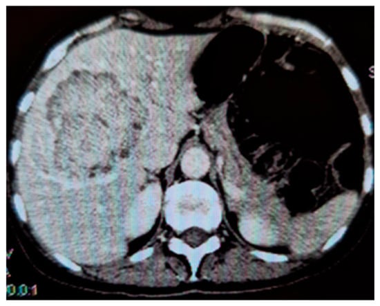
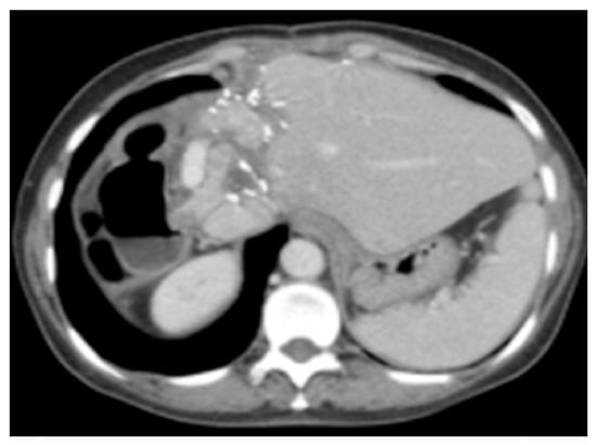
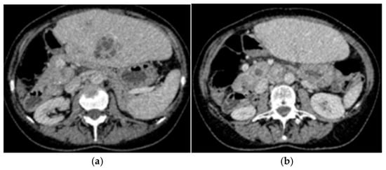
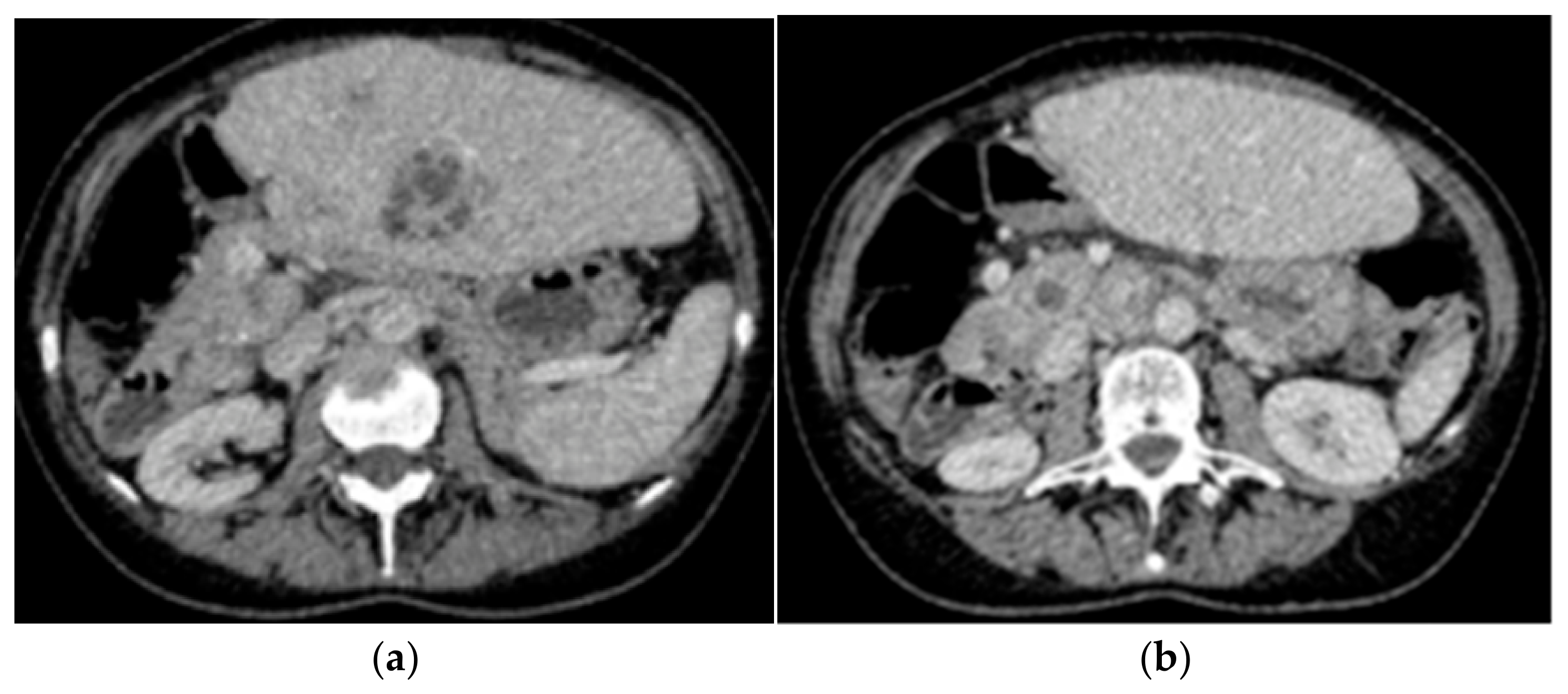
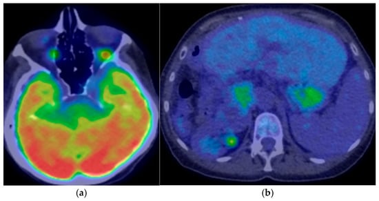
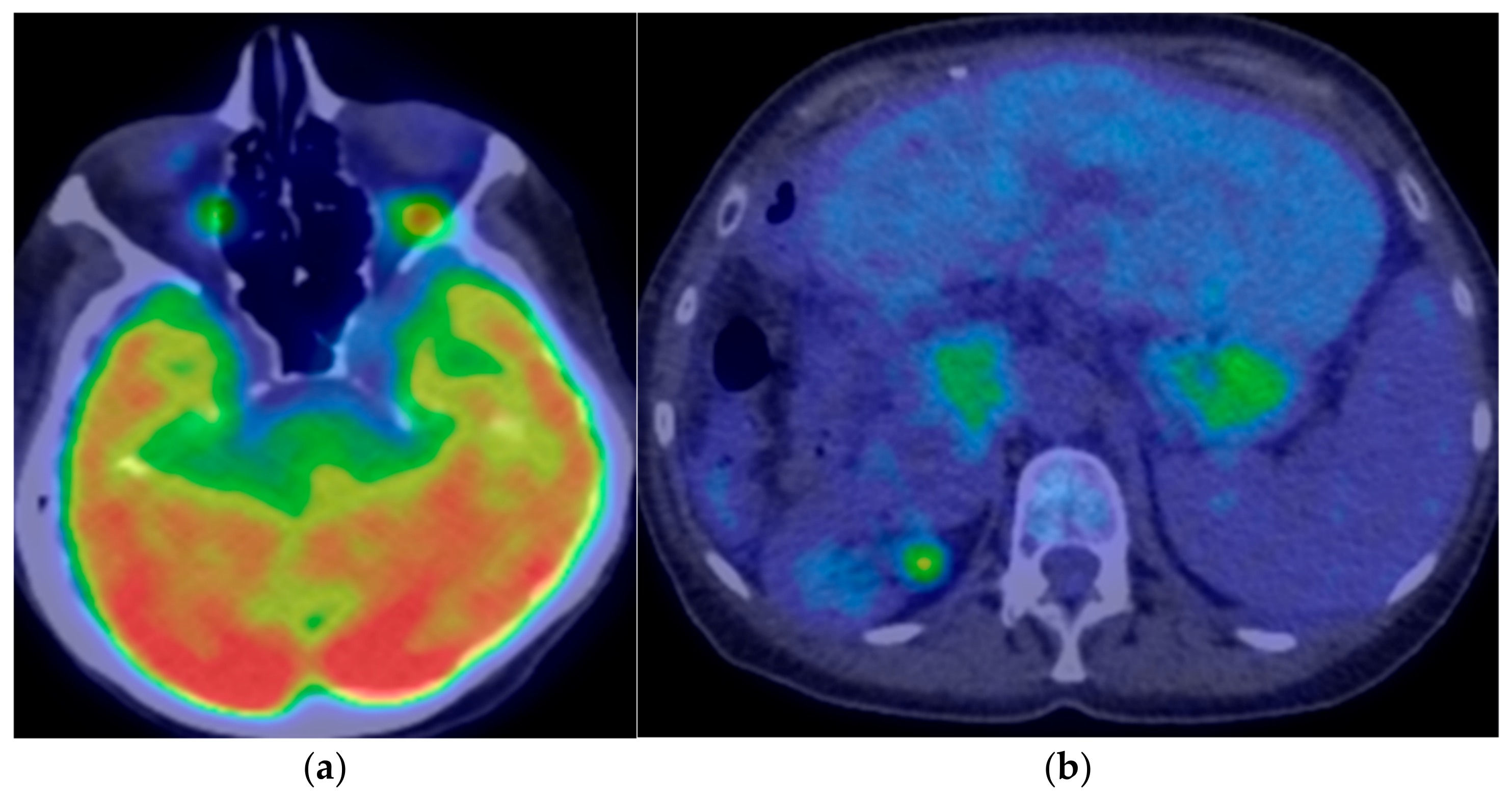
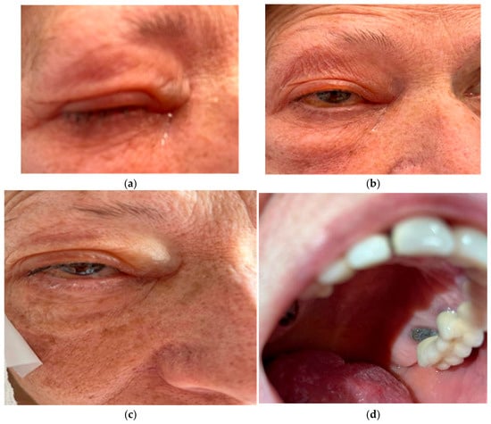
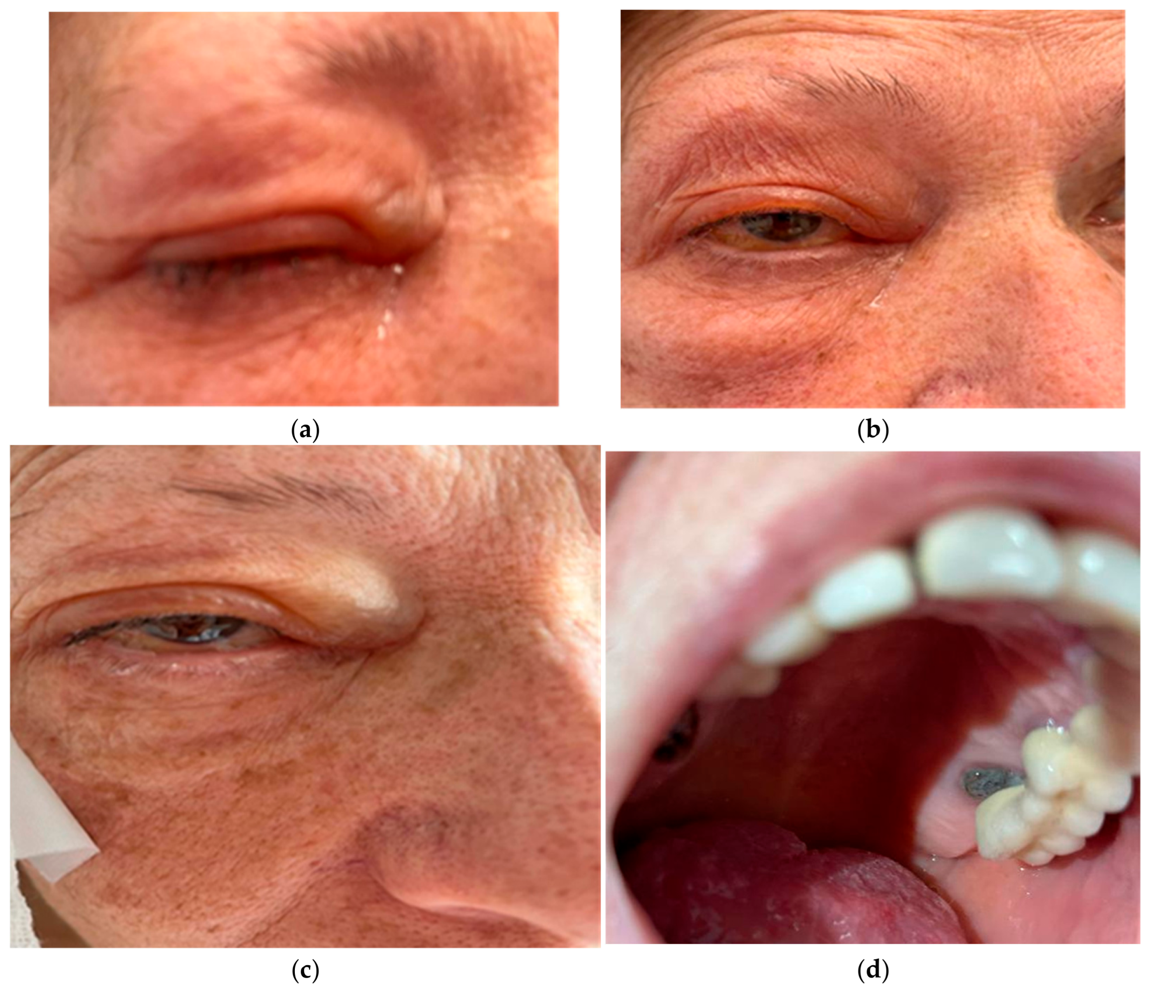
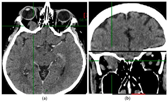
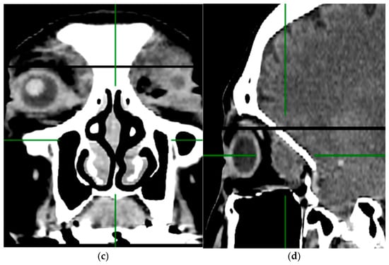
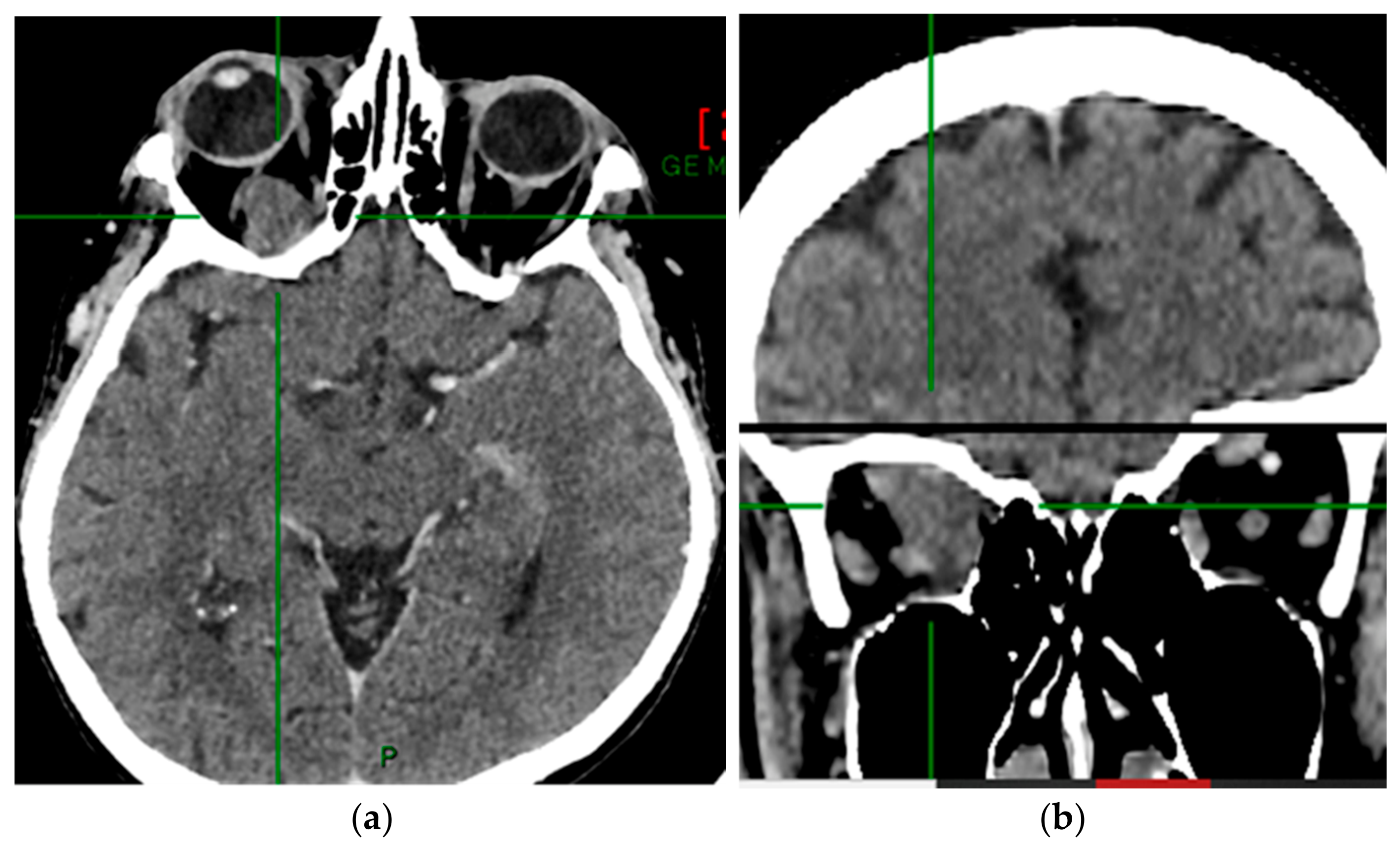
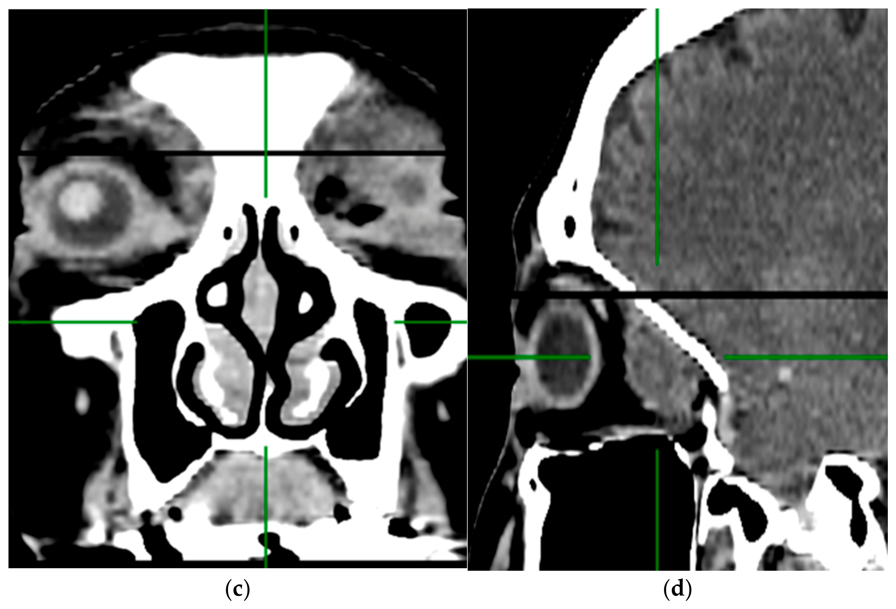
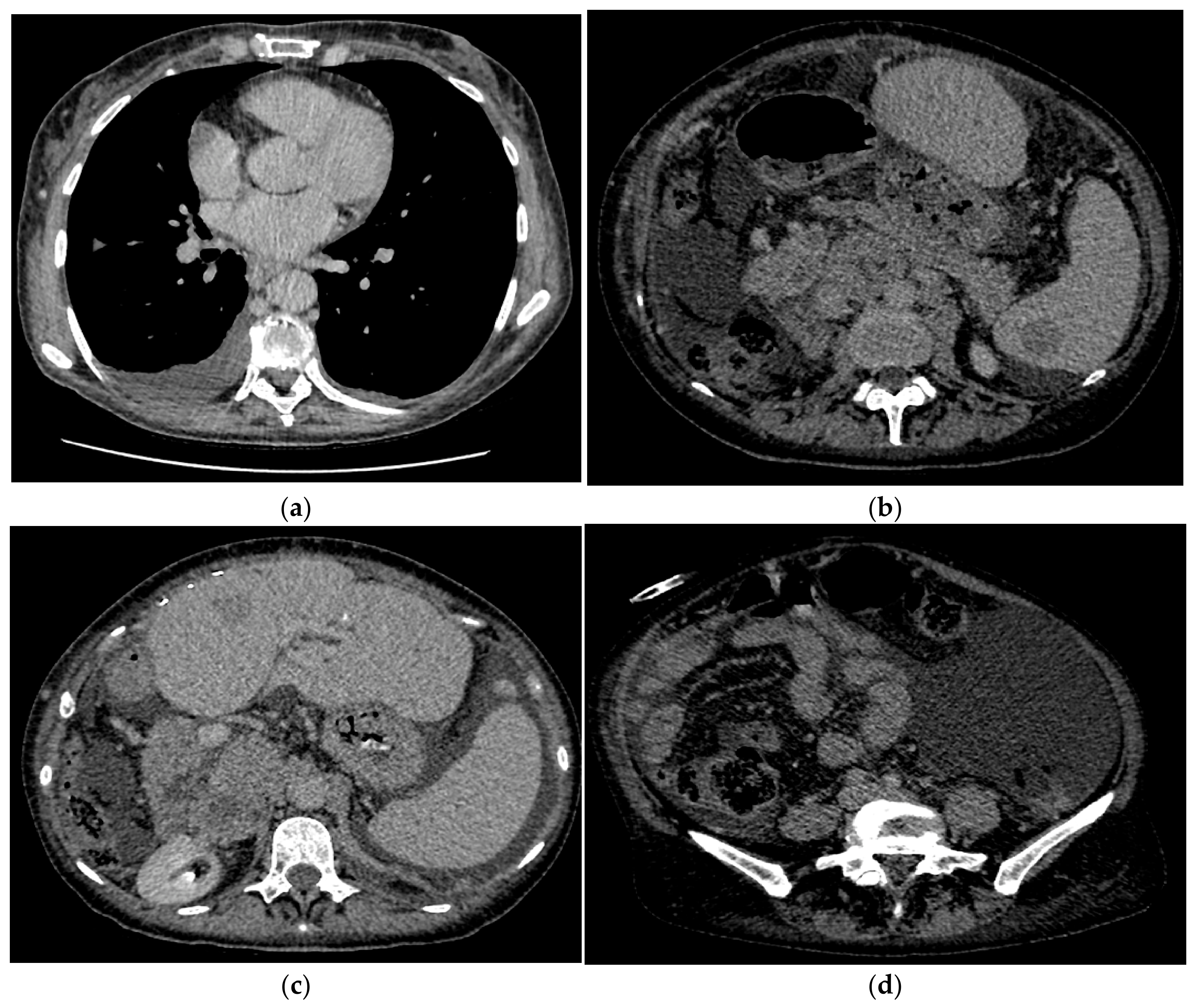
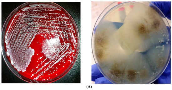
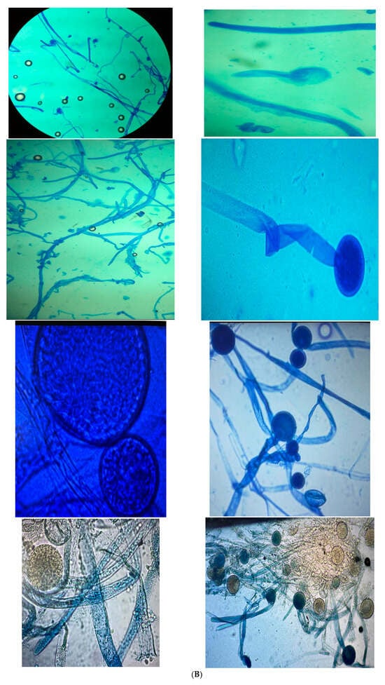
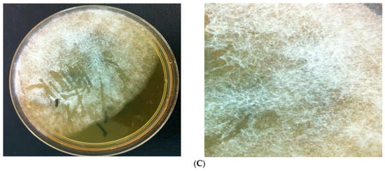
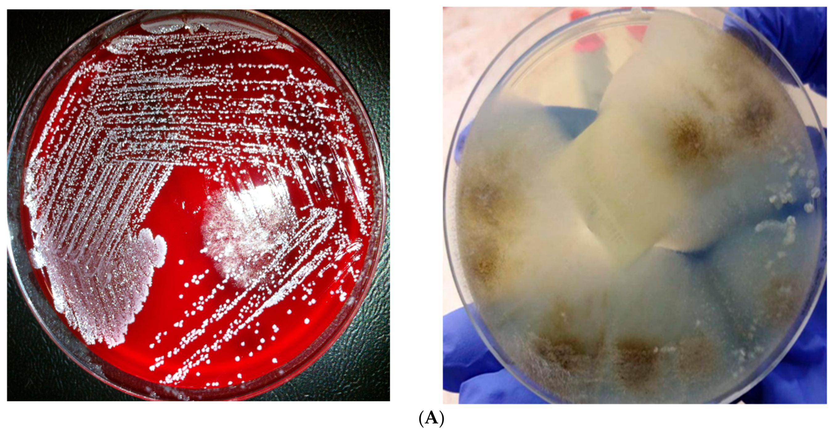
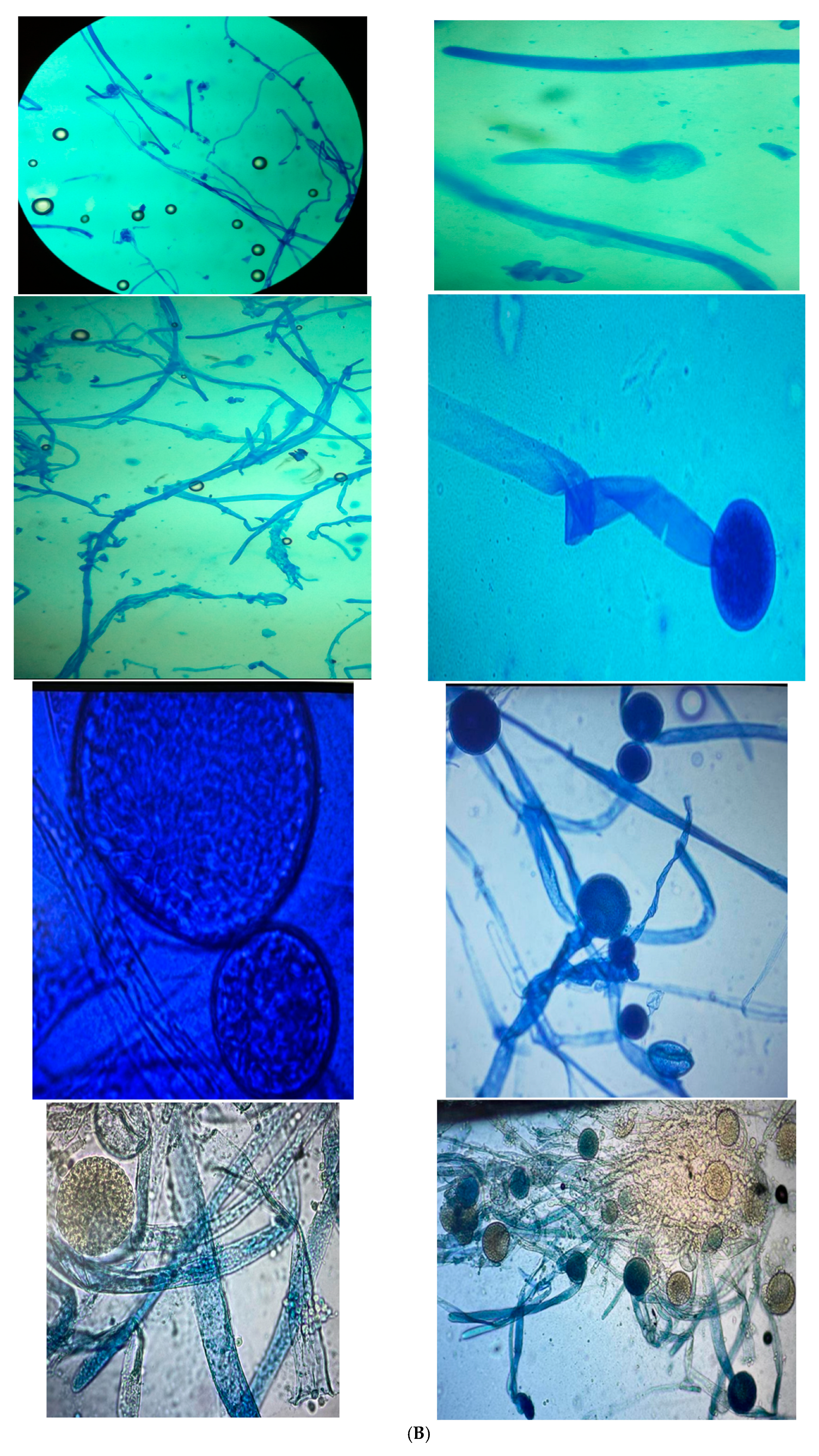
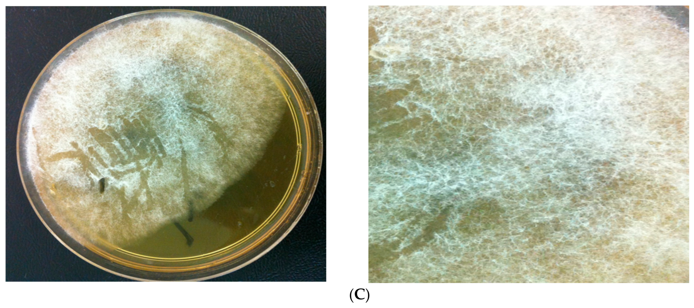
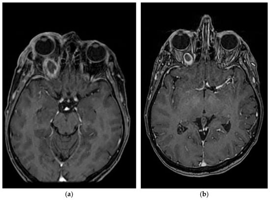
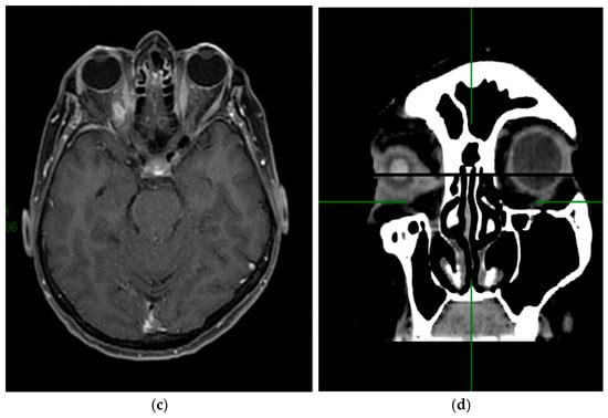
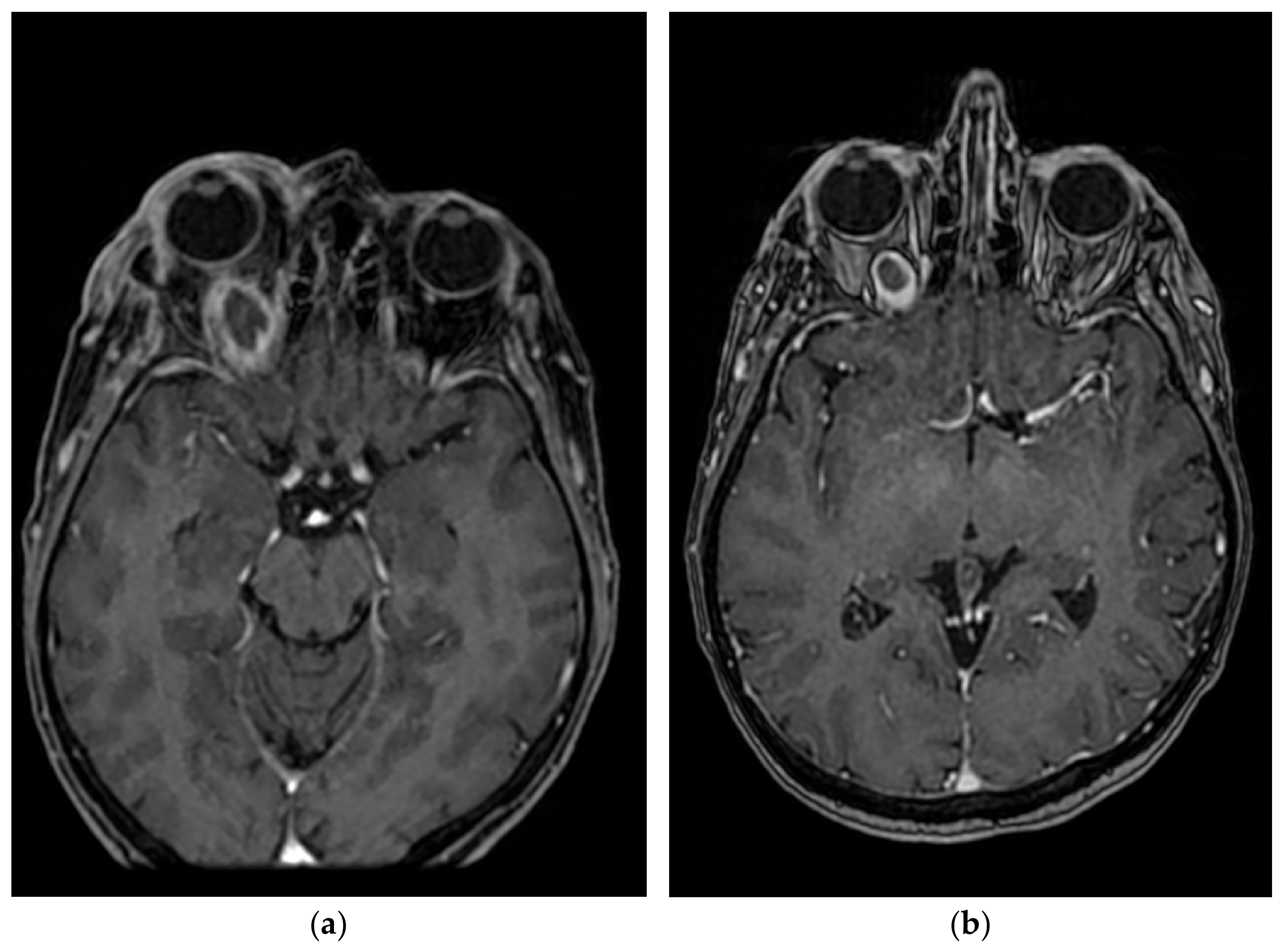
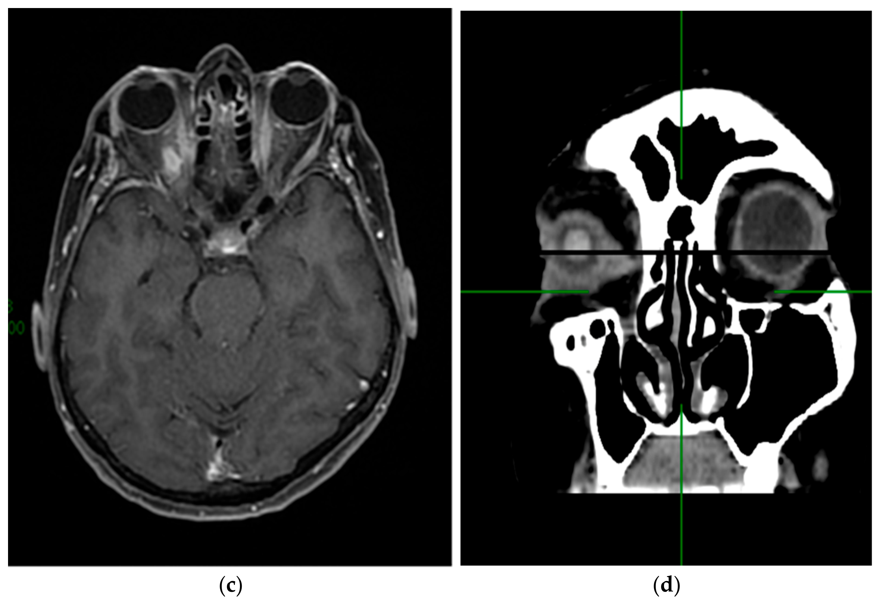
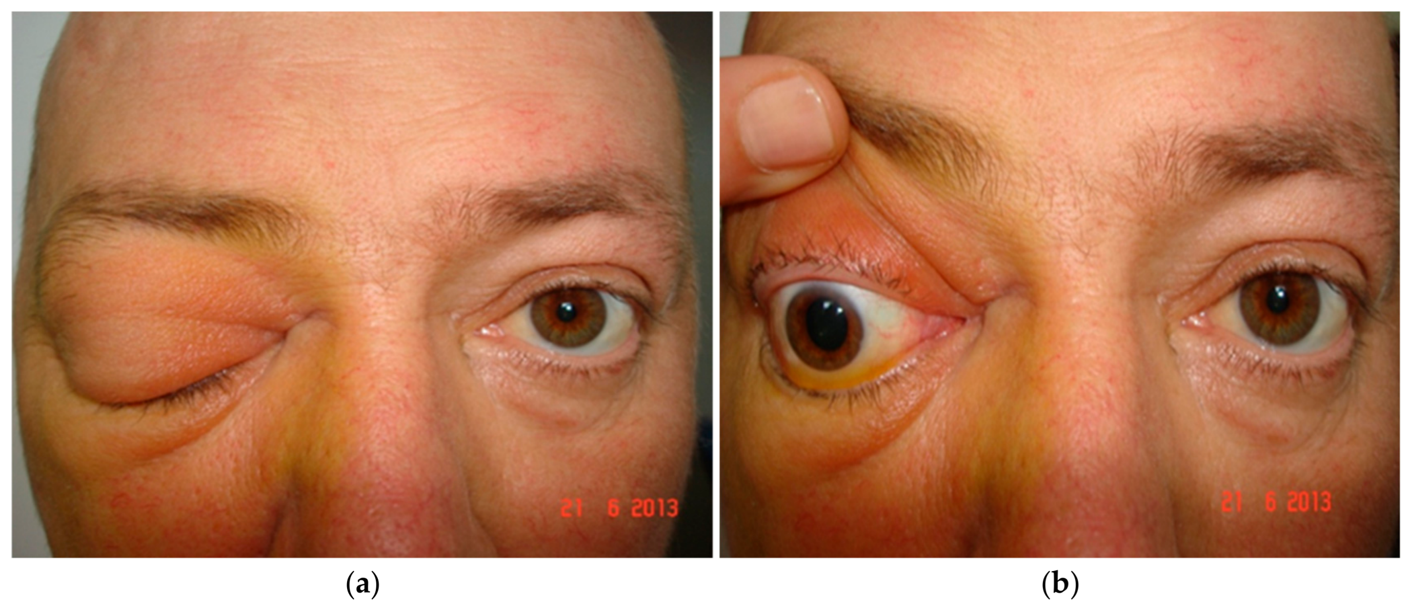
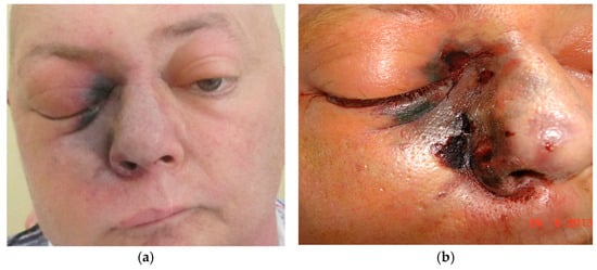
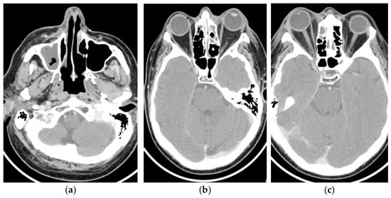
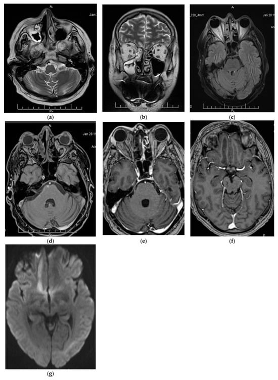
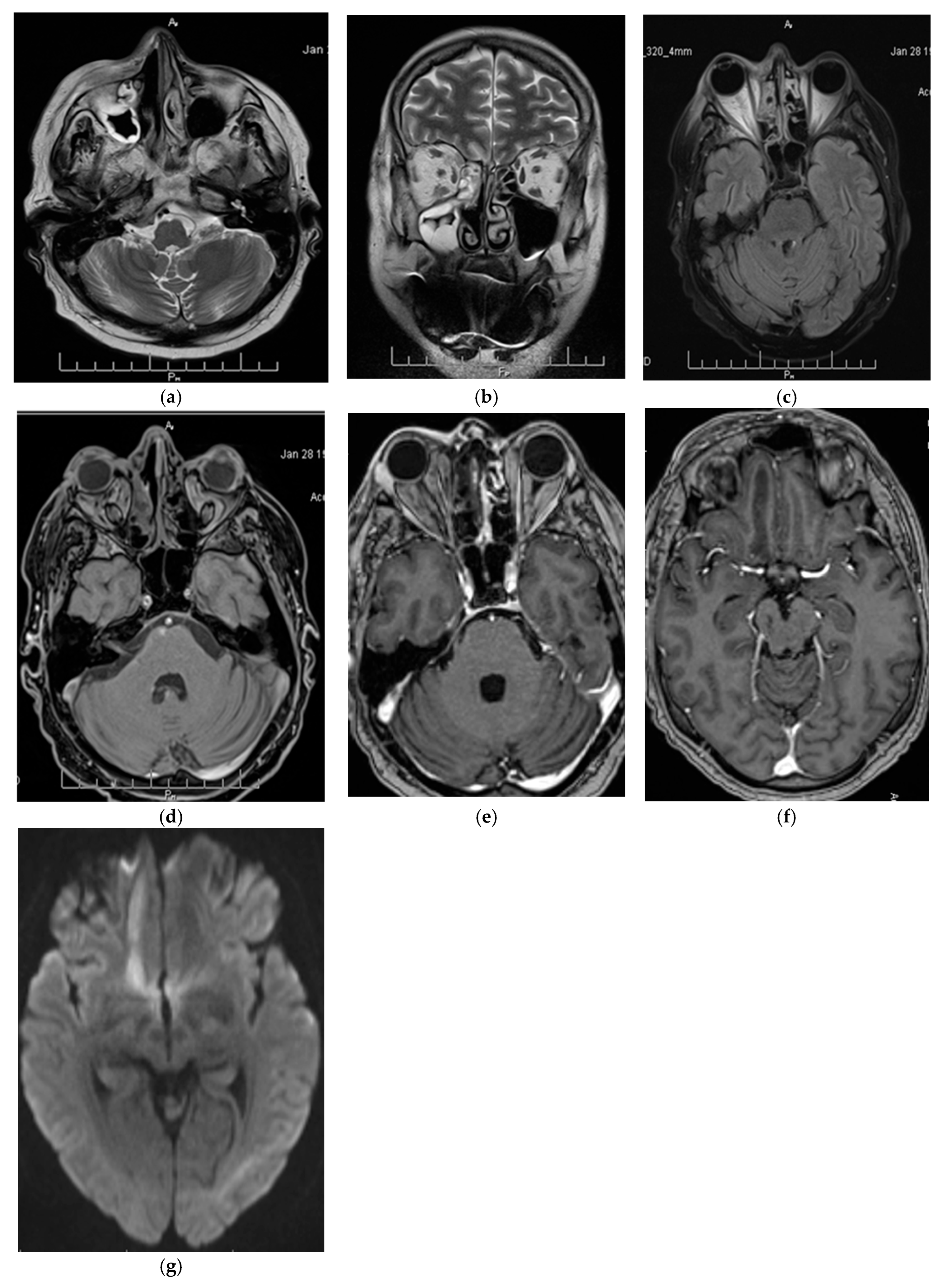
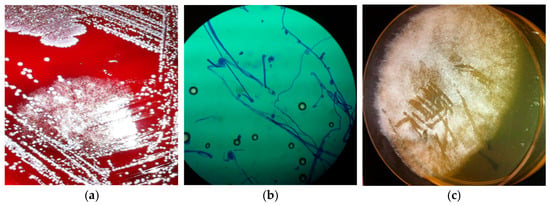
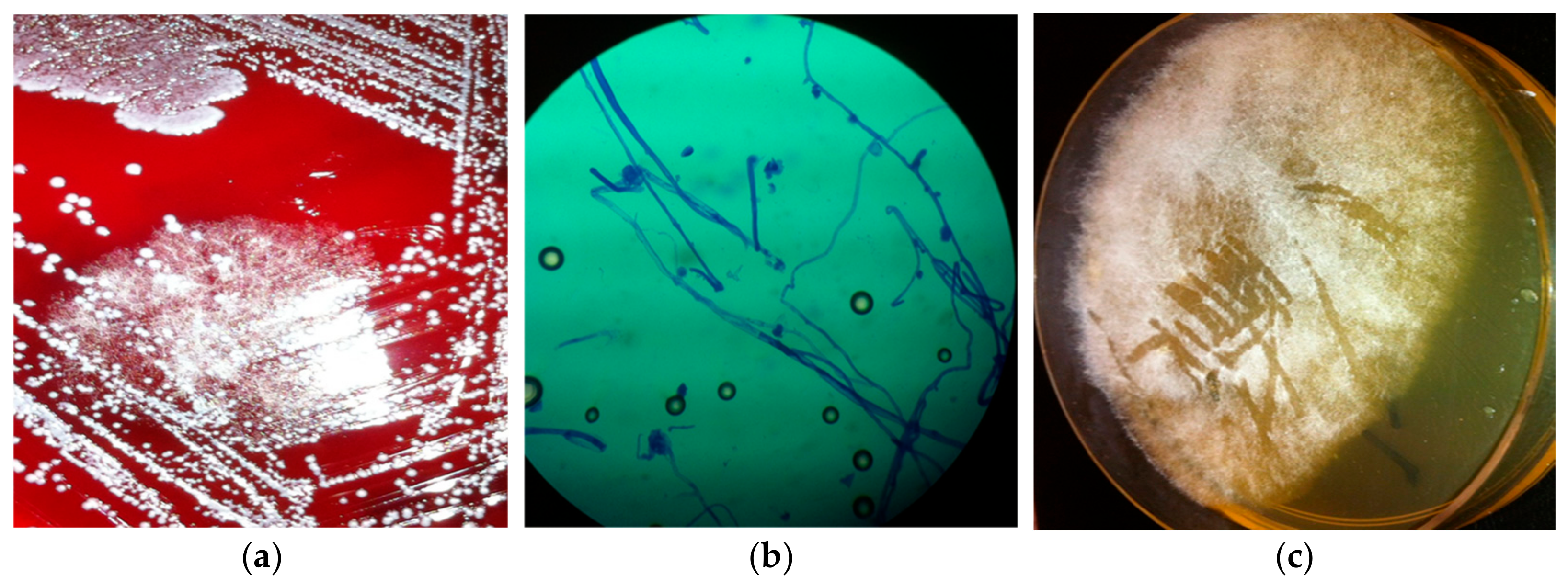
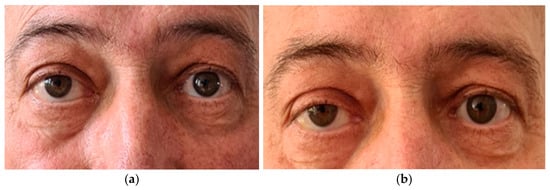
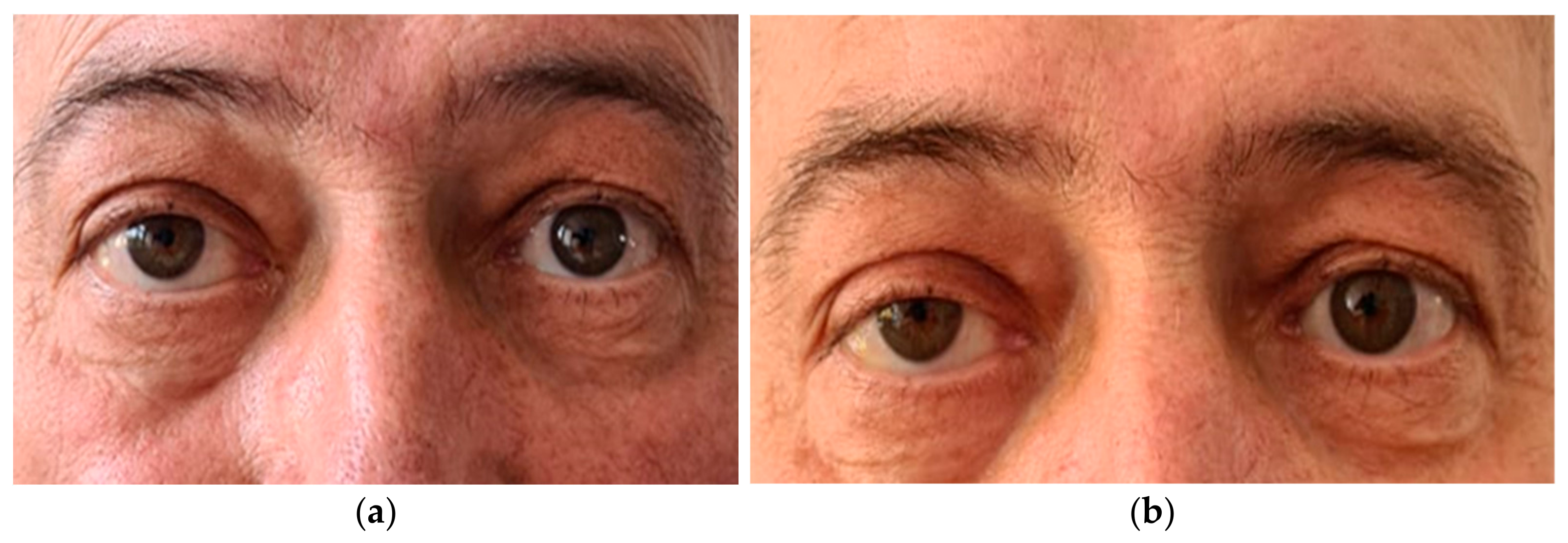
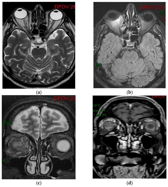
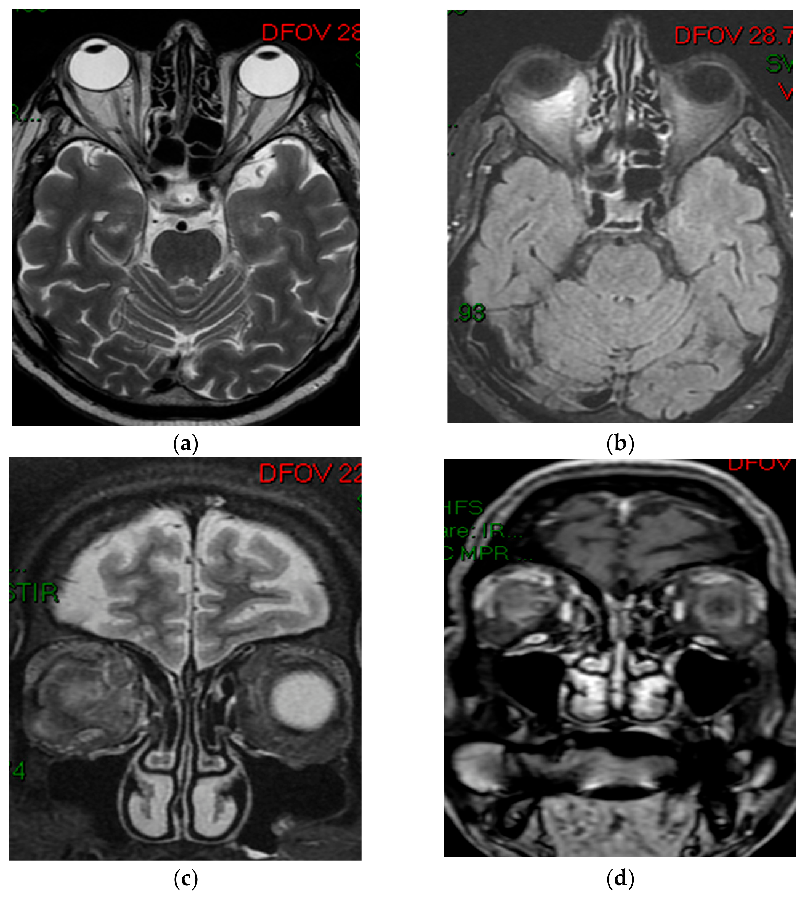
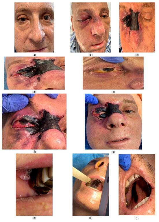
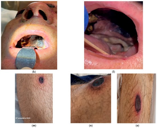
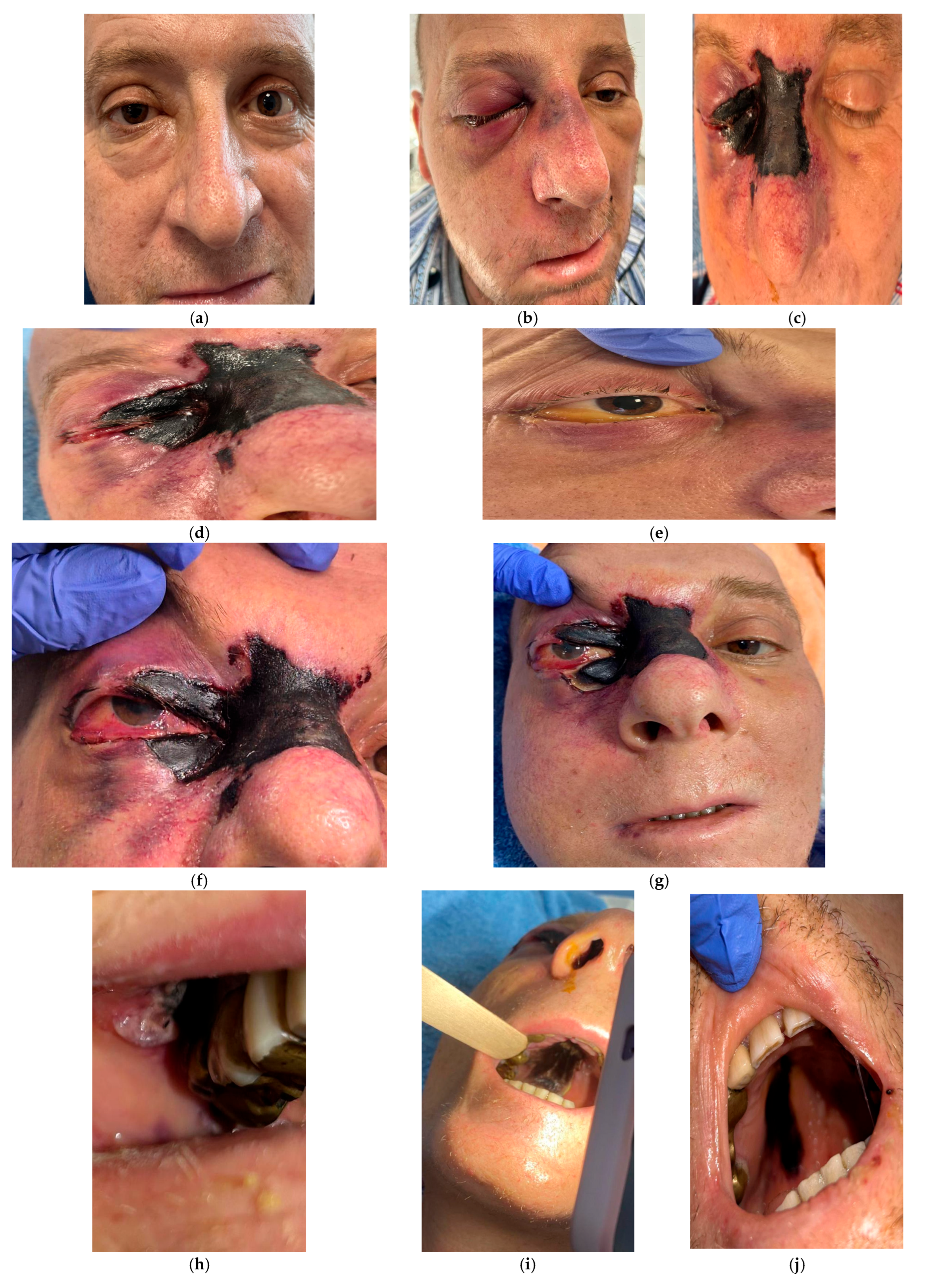
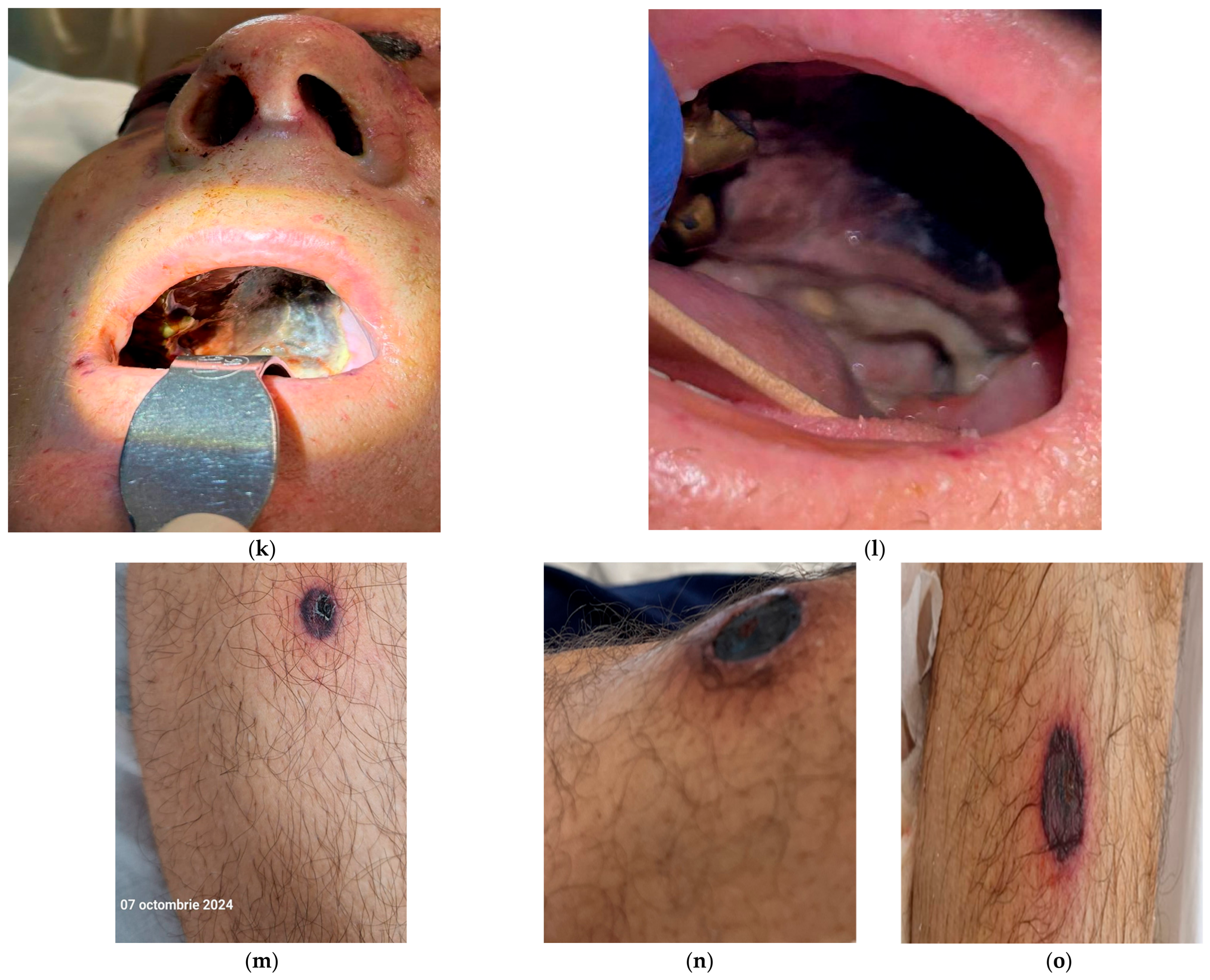
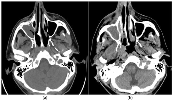
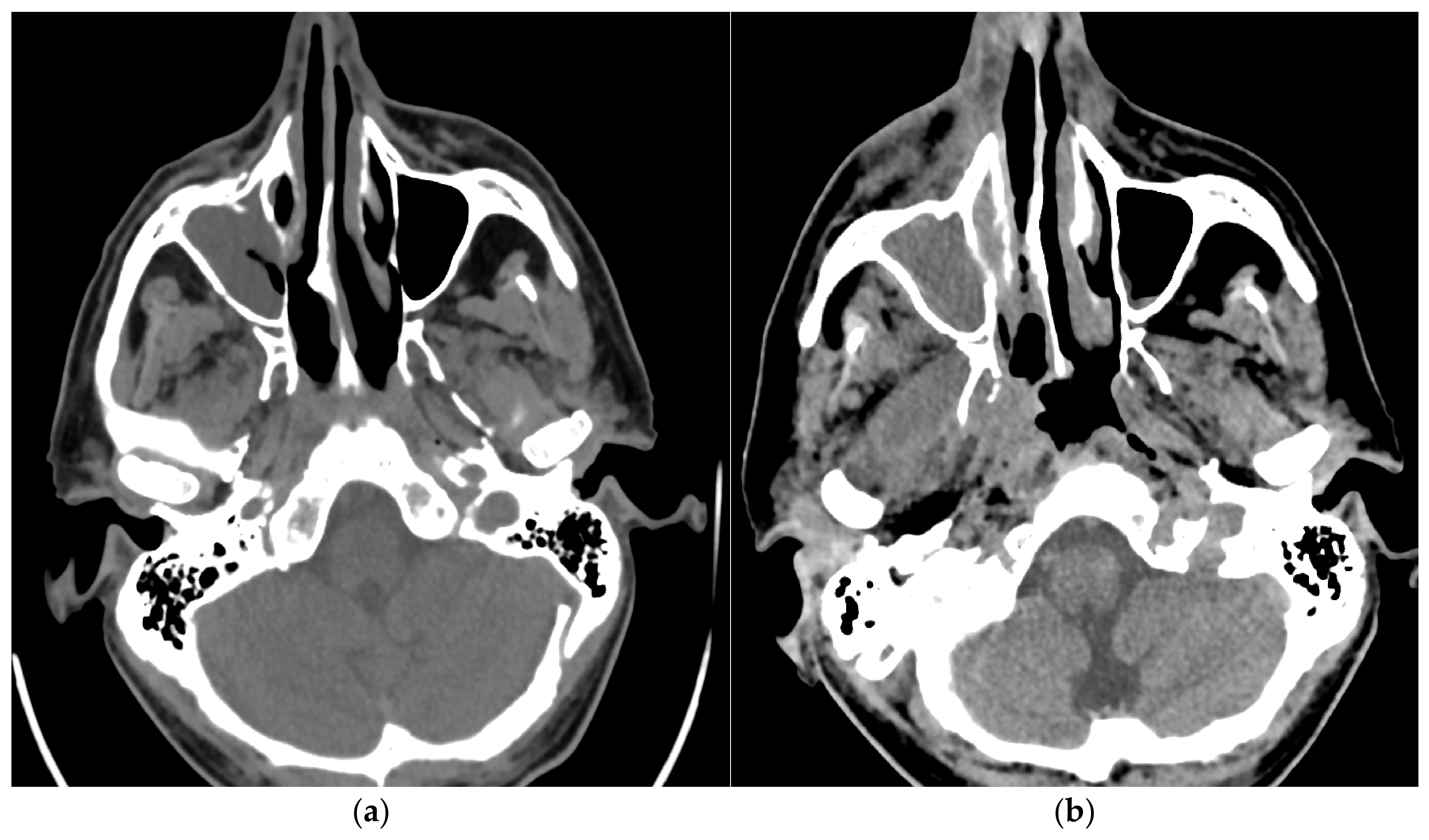
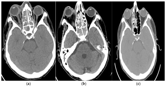
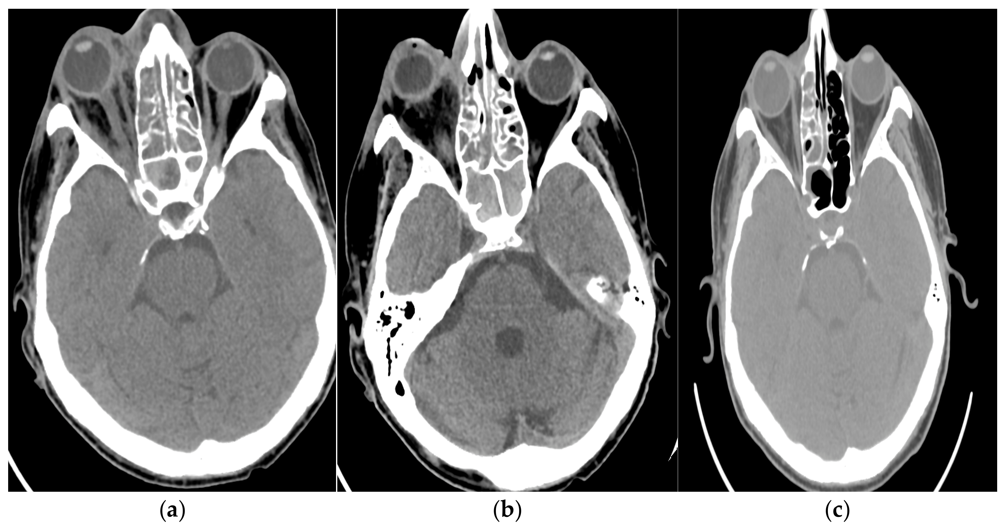
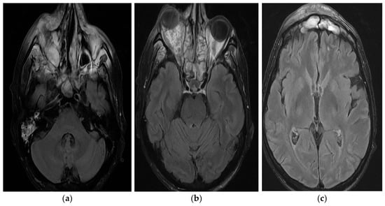
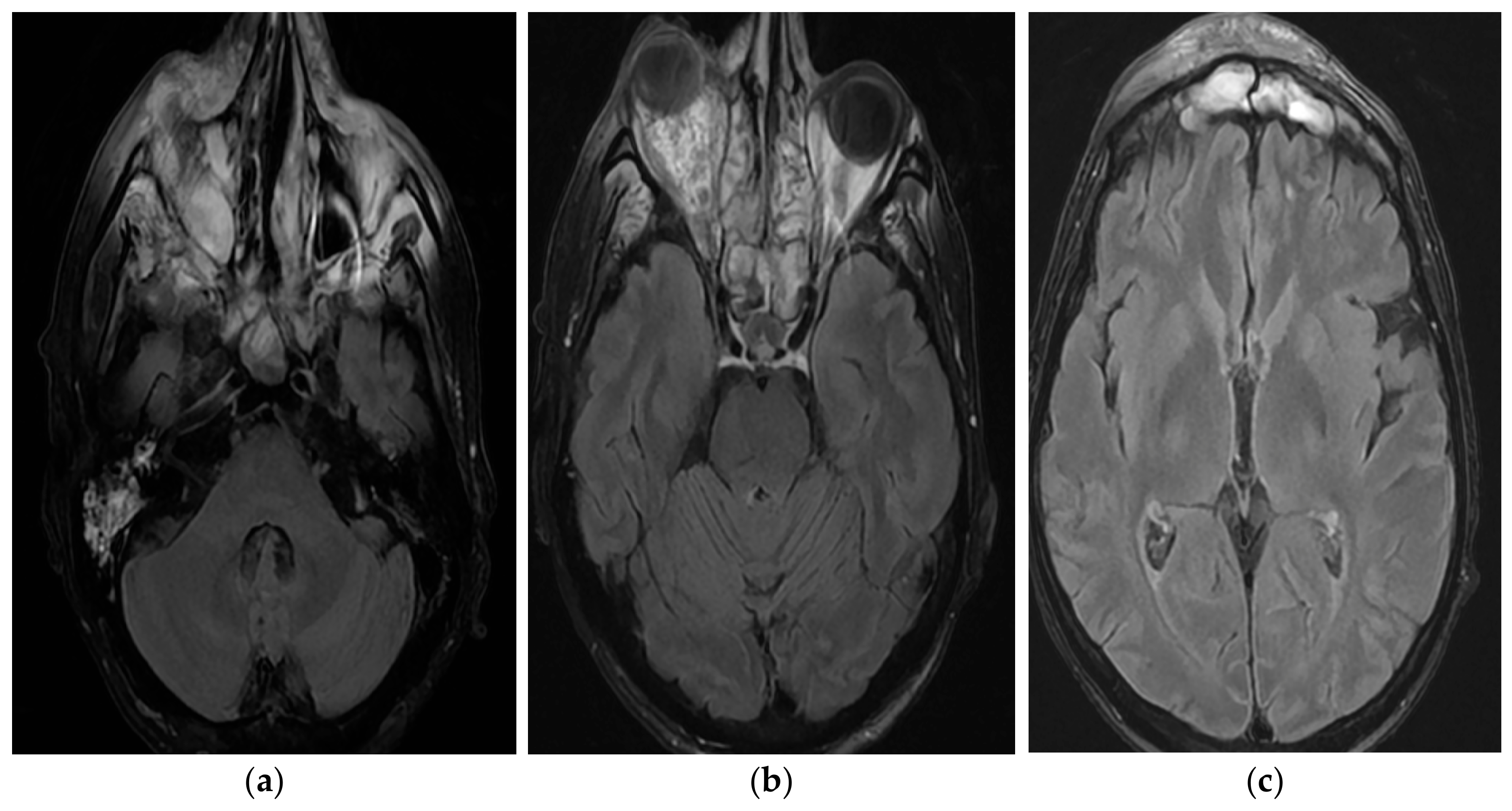
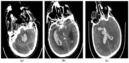
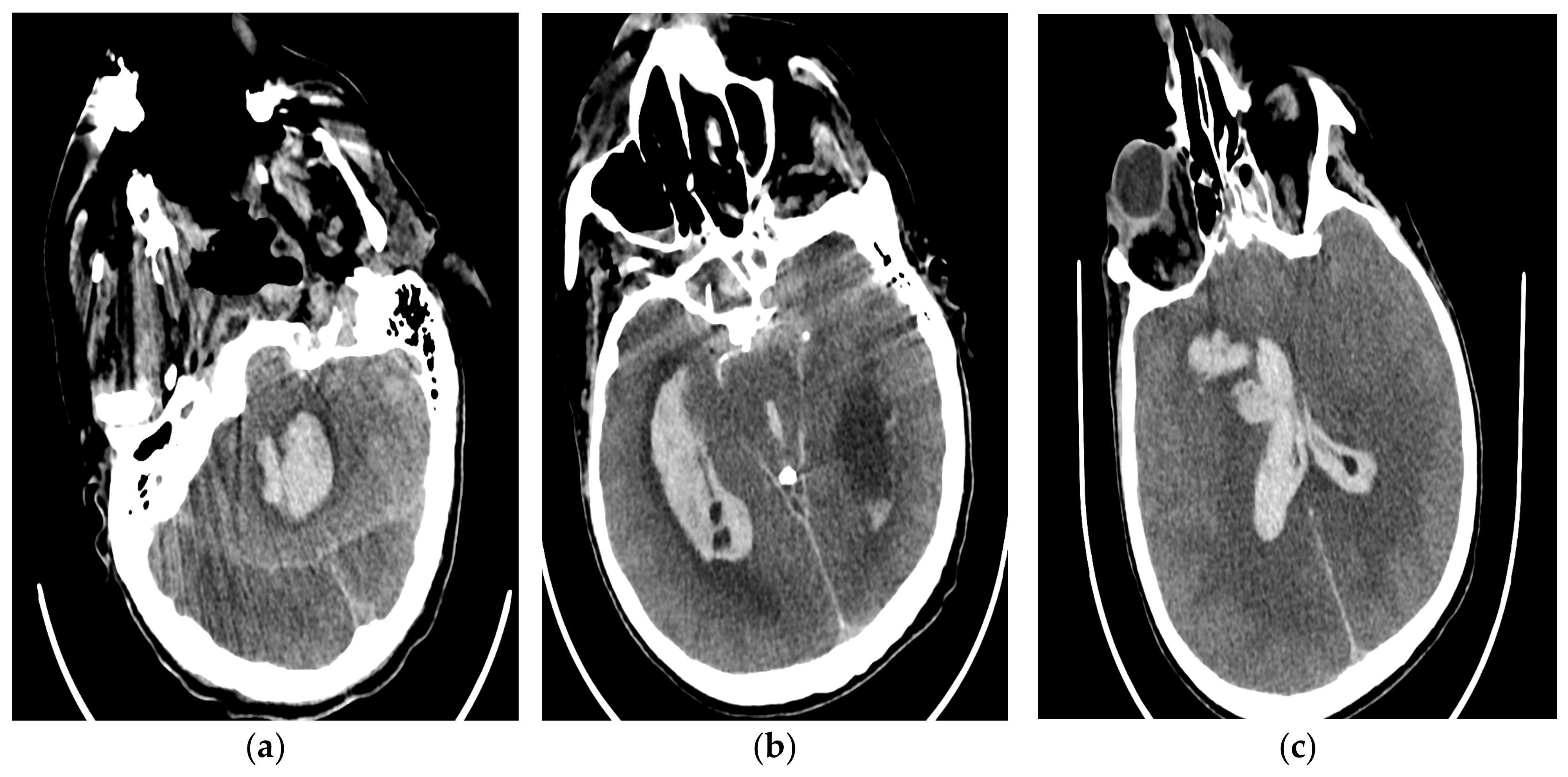
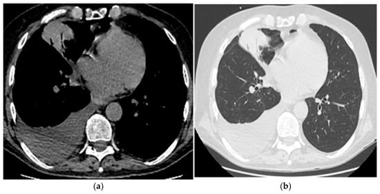
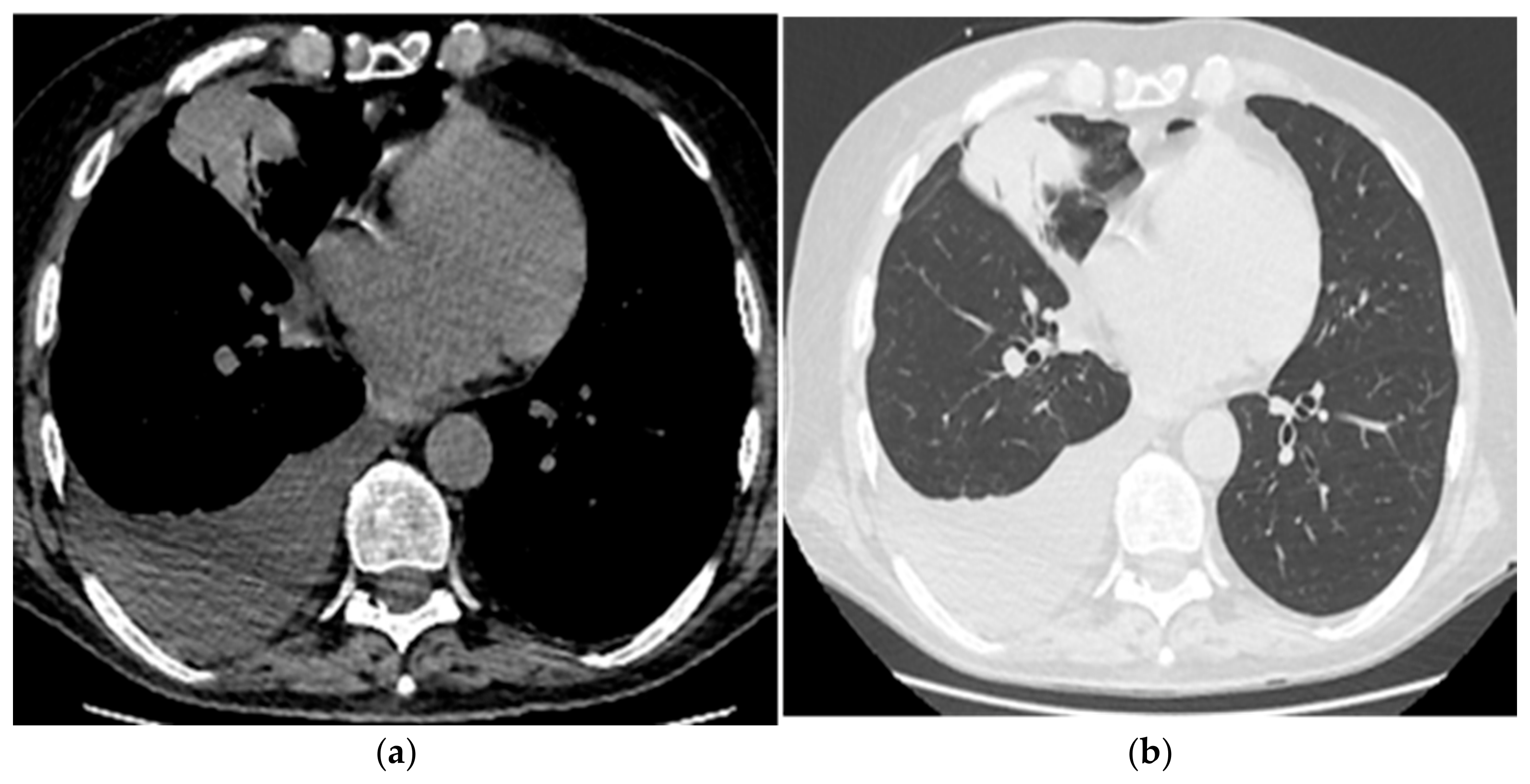
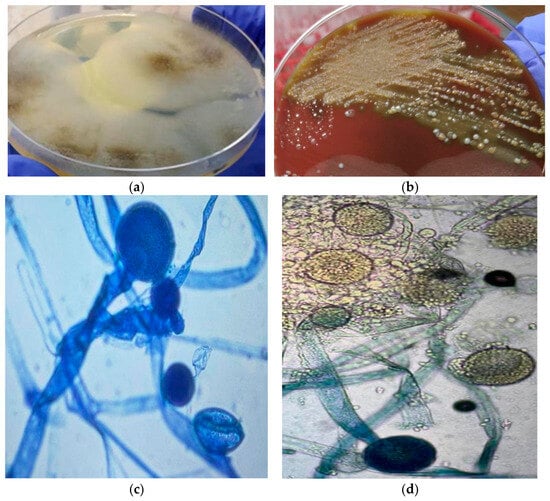
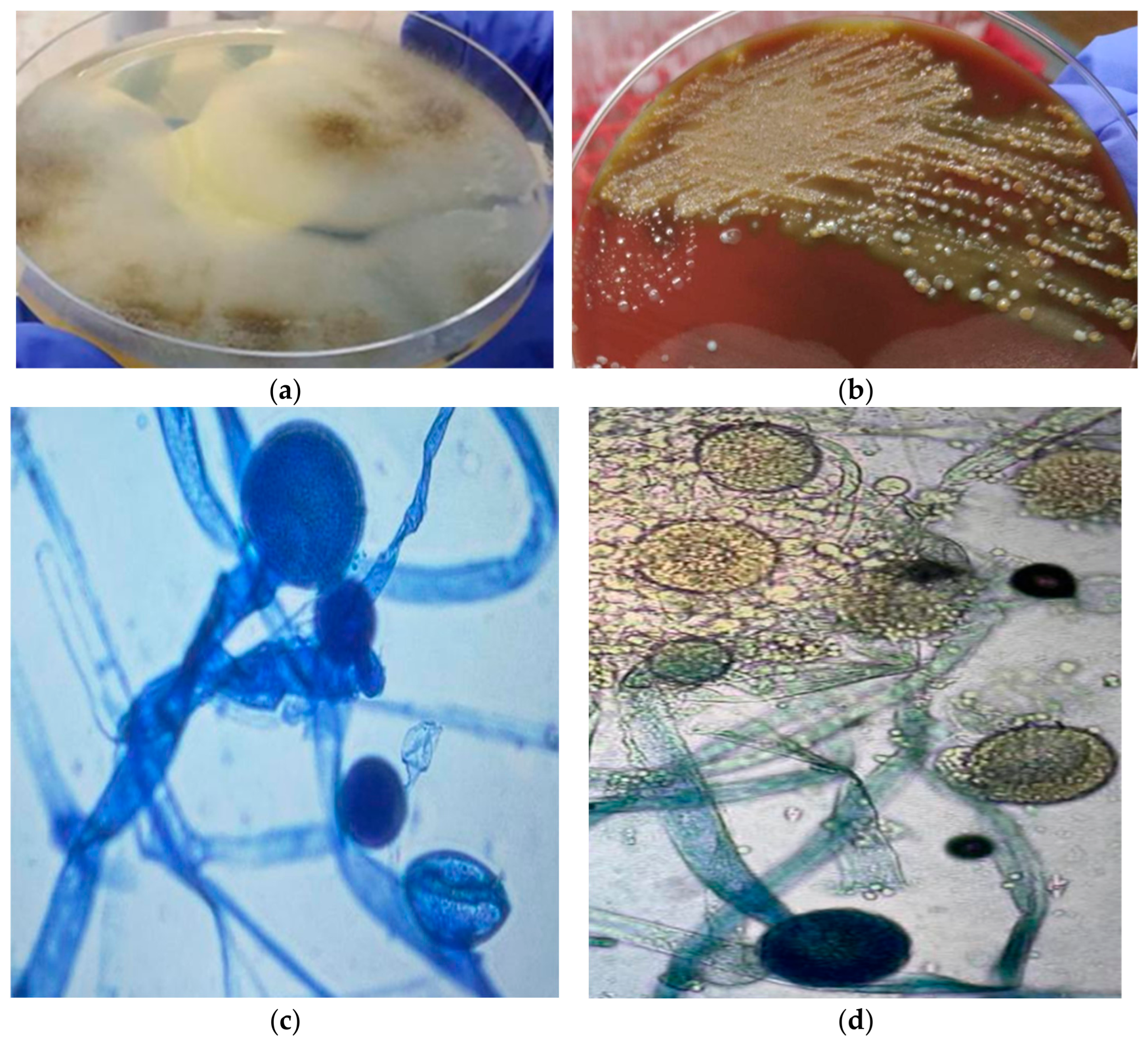
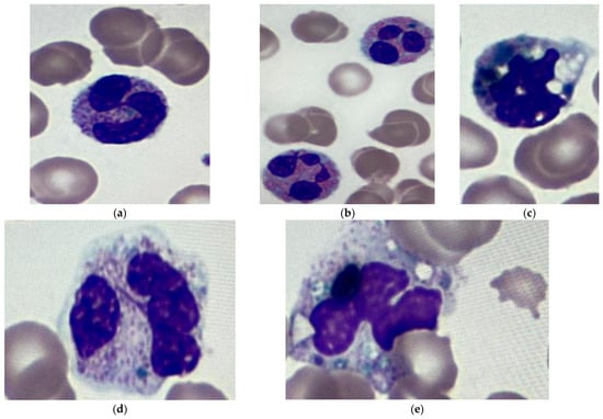
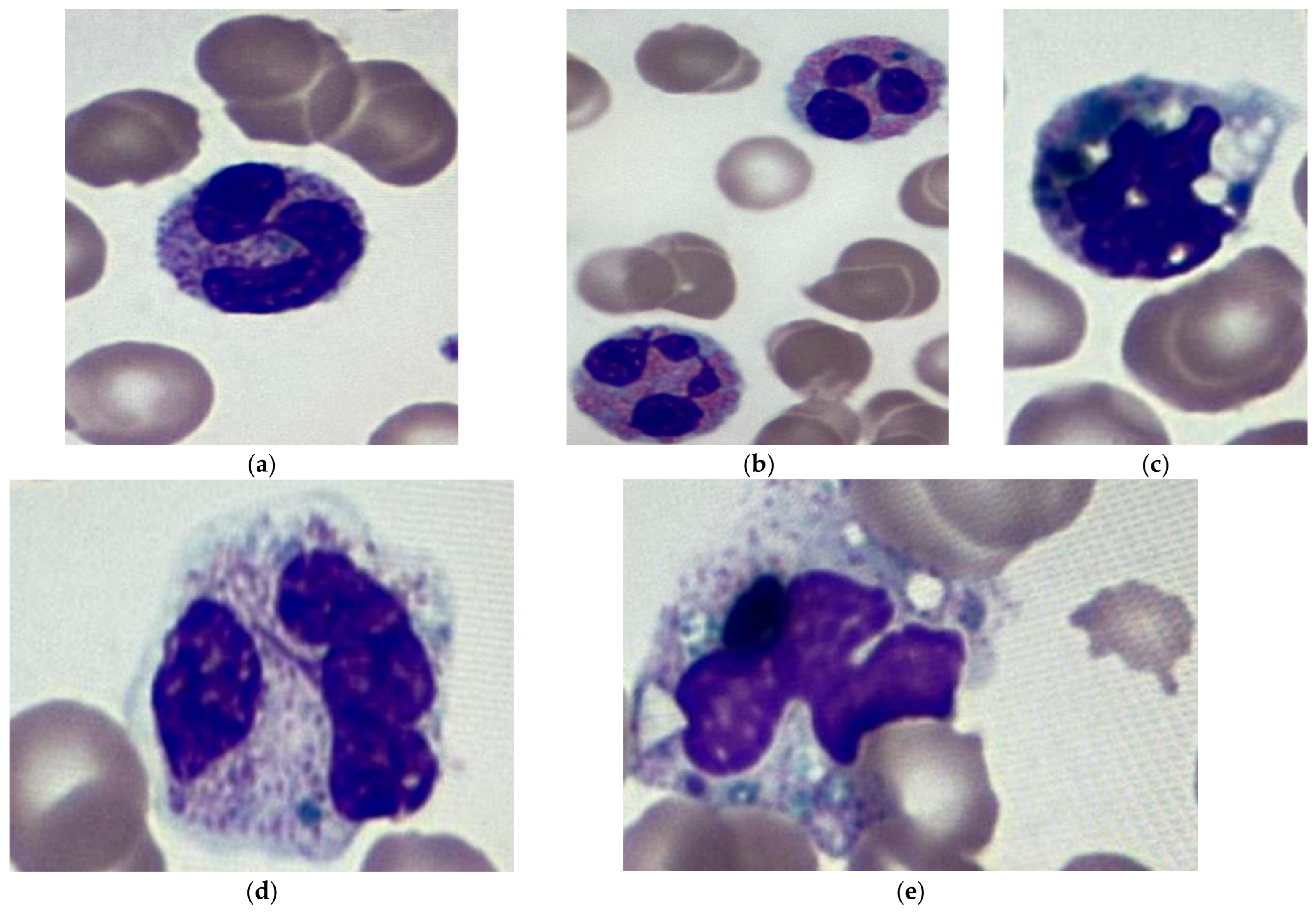
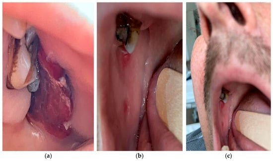
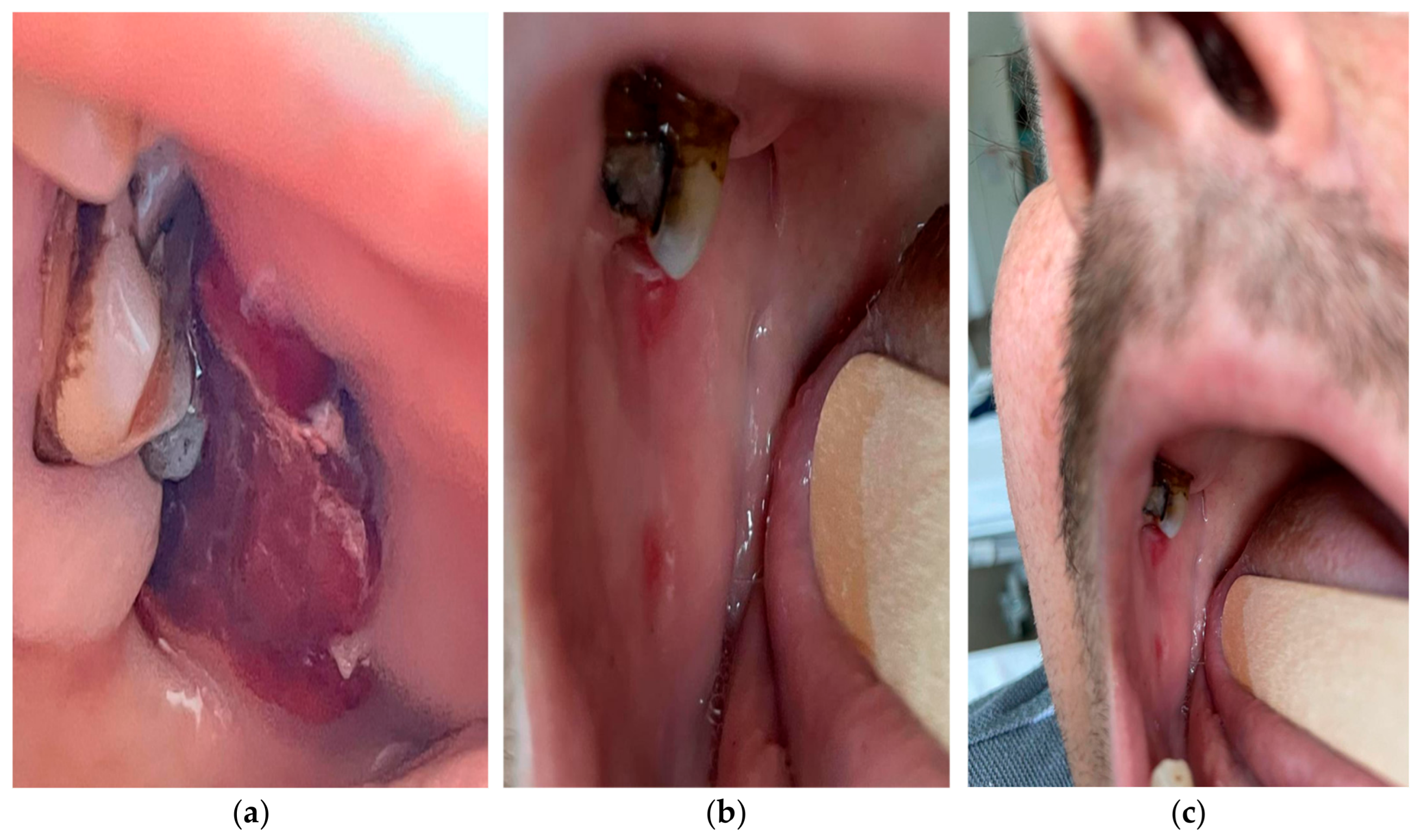
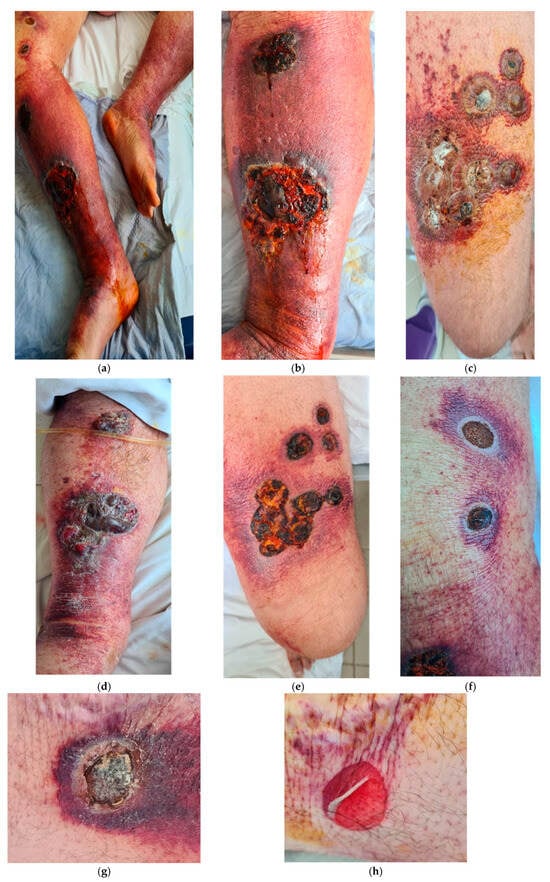
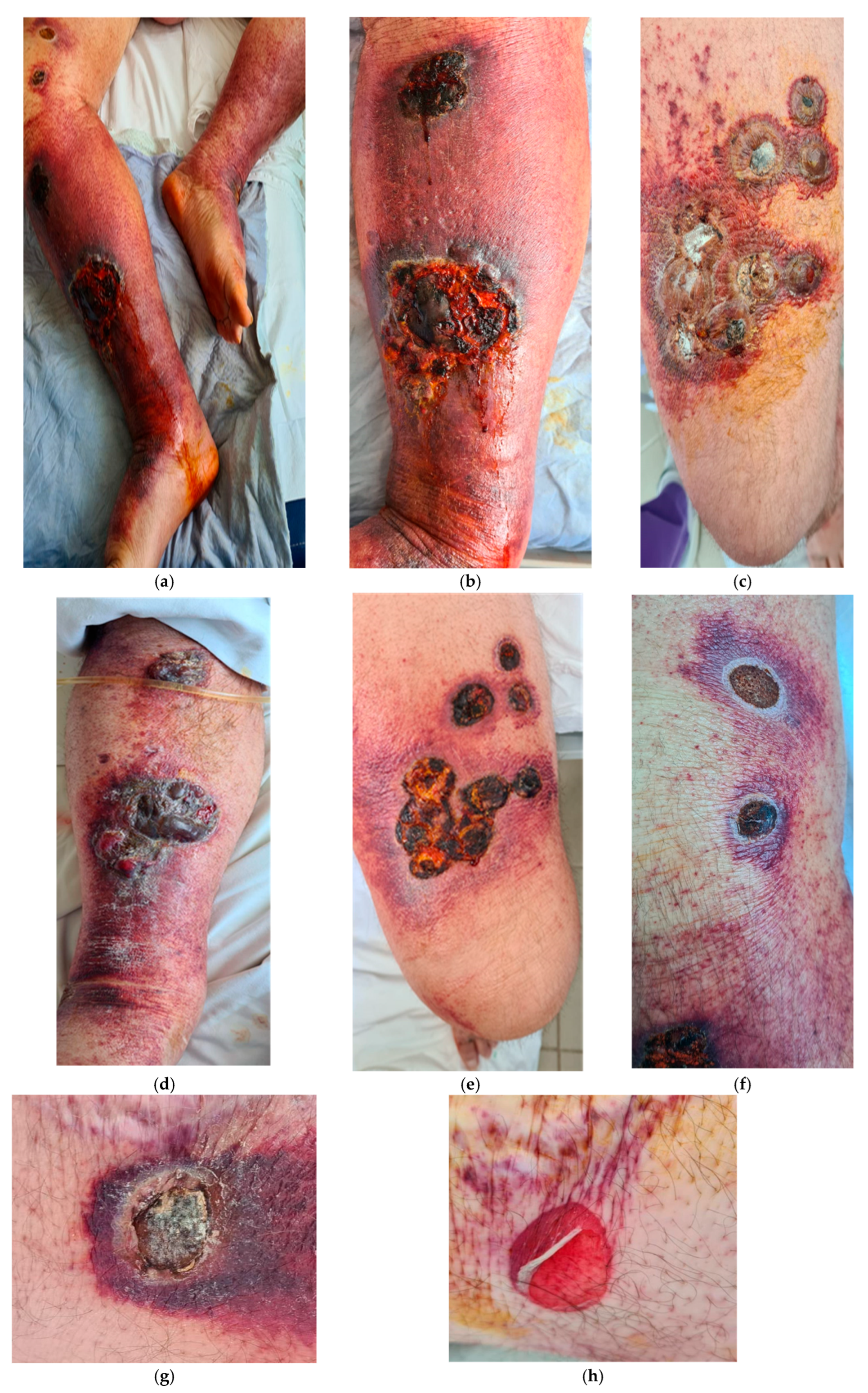

Disclaimer/Publisher’s Note: The statements, opinions and data contained in all publications are solely those of the individual author(s) and contributor(s) and not of MDPI and/or the editor(s). MDPI and/or the editor(s) disclaim responsibility for any injury to people or property resulting from any ideas, methods, instructions or products referred to in the content. |
© 2026 by the authors. Licensee MDPI, Basel, Switzerland. This article is an open access article distributed under the terms and conditions of the Creative Commons Attribution (CC BY) license.