


Abstract
Colletotrichum species are major plant pathogens and emerging opportunistic human pathogens. Due to their vast genetic diversity, existing diagnostic tools often suffer from narrow specificity or labor-intensive workflows. In this study, we developed a rapid, universal, and highly sensitive genus-specific real-time PCR assay utilizing a TaqMan MGB probe targeting the conserved 28S rDNA region. The assay demonstrated exceptional specificity, with no cross-reactivity against closely related fungal taxa or common co-occurring pathogens. The method exhibited high sensitivity, achieving a limit of detection (LOD) of 680 fg of genomic DNA. Furthermore, the assay was successfully validated using simulated environmental samples, where it accurately identified Colletotrichum within complex fungal communities. By providing a robust platform for genus-level screening, this methodology significantly enhances the efficiency of phytosanitary inspections and clinical diagnostics, facilitating timely biosecurity interventions and therapeutic decisions.
1. Introduction
Colletotrichum, the sole genus in the family Glomerellaceae (Glomerellales), is widely recognized as one of the top 10 most important groups of plant pathogenic fungi globally [1]. Exhibiting a cosmopolitan distribution and an extensive host range, species within this genus infect a vast array of agricultural crops, economic trees, and ornamental plants, causing devastating anthracnose diseases. For instance, C. kahawae, the causal agent of Coffee Berry Disease, can result in yield losses of 30–70% or even total crop failure in African Arabica coffee production [2,3]. Consequently, it is listed as a priority quarantine pest in China, the European Union, and various other regions. Similarly, the average anthracnose disease incidence in chili peppers caused by C. scovillei (syn. C. capsici) was reported at 63%, with an average disease intensity of 68% in Indonesia [4]. High-value crops such as strawberries, mangoes, citrus, and avocados are particularly susceptible, facing severe quality degradation and economic downturns due to fruit rot [5]. Additionally, ubiquitous species complexes such as C. gloeosporioides, C. acutatum, and C. boninense are responsible for significant losses in forestry and agriculture worldwide [6,7,8]. Therefore, accurate identification and early warning of this genus are of profound economic and ecological importance.
Beyond their impact on agriculture, Colletotrichum species are emerging as opportunistic human pathogens. They are increasingly implicated in localized infections such as keratitis and phaeohyphomycosis (subcutaneous infections), as well as systemic diseases, particularly in immunocompromised individuals or following traumatic inoculation [9,10]. Clinically relevant species include C. chlorophyti [11], C. coccodes [12], C. dematium [13,14], C. gigasporum [15], C. gloeosporioides [14], C. siamense [16], and C. truncatum [17]. These infections are often linked to direct contact with infected plant material or contaminated soil, posing a growing “One Health” public health risk that necessitates vigilance [18].
As one of the fungal groups most frequently intercepted at ports of entry, Colletotrichum poses a significant challenge to customs and phytosanitary authorities. Rapid screening and interception serve as the first line of defense against biological invasions. However, the taxonomy of Colletotrichum has undergone significant revision, with the genus now organized into approximately 17 species complexes and several singleton species [5,6,7,19]. Currently, routine identification in quarantine and clinical laboratories relies on a traditional workflow involving isolation, morphological observation, and multi-locus (e.g., ITS, cal, gapdh, gs, his3, tub2, and ApMat) phylogenetic analysis [19]. While robust, this approach is labor-intensive, time-consuming, and requires specialized taxonomic expertise, failing to meet the demand for high-throughput, rapid-turnaround testing required for efficient customs clearance and clinical diagnosis. Similarly, while High-Throughput Sequencing (NGS) offers powerful capabilities for analyzing environmental samples, its high cost, long cycle and requirement for complex bioinformatic analysis hinder its routine deployment in frontline inspection.
In terms of molecular diagnostics in Colletotrichum, various methods, including conventional PCR, real-time PCR and Loop-Mediated Isothermal Amplification (LAMP), have been developed. However, the majority of these assays are designed for specific species (e.g., C. acutatum or C. kahawae) [20,21]. Given the high diversity and genetic complexity within the genus, single-species assays are inefficient for initial screening and are prone to false negatives when dealing with unknown or mixed infections. Consequently, existing tools cannot satisfy the practical need for a “genus-level” screen. Therefore, there is an urgent need to develop a rapid, sensitive, and universal molecular detection method capable of covering the vast majority of Colletotrichum species, thereby enhancing the efficiency of quarantine inspection and clinical pathogen screening.
Attempts to develop genus-specific primers have been made, but limitations persist. Mosca et al. [22] designed the degenerate genus-specific primers Coll1F/Coll3Rb based on ITS sequences; however, their specificity validation relied on a limited dataset. By aligning their primer sequences against our comprehensive Colletotrichum ITS dataset [19], we identified a single-nucleotide mismatch in the reverse primer in species belonging to the C. orbiculare species complex (e.g., C. lindemuthianum, C. orbiculare, C. trifolii), suggesting potential detection failures for these taxa. Similarly, Li et al. [23] developed the ITS-based primers ITS-c3/ITS-c4. Sequence alignment against our dataset revealed a single-nucleotide mismatch in the forward primer against C. ocimi and C. pseudoacutatum, as well as multiple mismatches in the reverse primer across several species, indicating that the detection efficiency of this set may be compromised. Furthermore, these methods rely on conventional PCR followed by gel electrophoresis, which is less efficient than real-time quantitative PCR (qPCR) platforms.
Consequently, there is an urgent need to develop a rapid and reliable molecular technique for the universal detection of the genus Colletotrichum. The objective of this study was to establish and validate a novel, genus-specific TaqMan real-time PCR assay. The specificity and robustness of this assay were rigorously evaluated using a comprehensive panel of isolates, which covered the majority of Colletotrichum species complexes (including those reported as human pathogens and serious phytopathogens), closely related fungal families, and other common phytopathogens. Furthermore, the assay’s effectiveness was verified using simulated environmental samples containing complex fungal communities.
2. Materials and Methods
2.1. Fungal Strains and DNA Extraction
A total of 52 Colletotrichum and 28 non-Colletotrichum strains belonging to the orders Botryosphaeriales, Chaetosphaeriales, Diaporthales, Eurotiales, Hypocreales, Mycosphaerellales, and Pleosporales were used in this study (Table 1). Their DNA was extracted using a modified CTAB protocol, and DNA concentration was quantified using NanoDropTM One (Thermo Fisher Scientific, Waltham, MA, USA) [24]. The identities of all Colletotrichum isolates used were confirmed via multi-gene phylogenetic analysis as described in previous studies [8,15,19]. For non-Colletotrichum species, isolates labeled with “LC” (personal culture collection of Prof. Lei Cai, housed in the Institute of Microbiology, Chinese Academy of Sciences) were identified based on ITS sequence similarity, while DNA for isolates labeled with “CBS” was obtained from the Westerdijk Fungal Biodiversity Institute (Utrecht, The Netherlands). Furthermore, to validate the intra-generic conservation of the designed primers, an in silico validation was performed using 97 whole-genome sequences of Colletotrichum available in the NCBI database (Table 1).
2.2. 28S rDNA Sequencing and Design of Primers and Probe
Due to the limited availability of 28S rDNA sequences for Colletotrichum species in GenBank, we sequenced the 28S region of 29 Colletotrichum isolates maintained in our laboratory. PCR amplification was performed using the primers LR0R (5‘-ACCCGCTGAACTTAAGC-3′) and LR5 (5‘-ATCCTGAGGGAAACTTC-3′), with reaction systems and conditions following Liu et al. [19]. The obtained sequences were aligned with those of closely related species and common plant pathogens using MEGA 7.0 software.
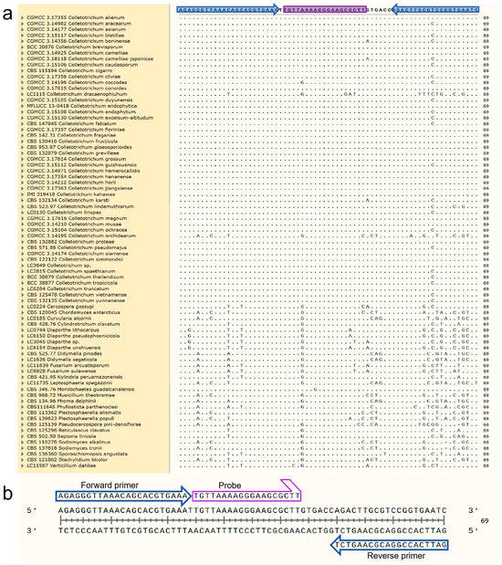
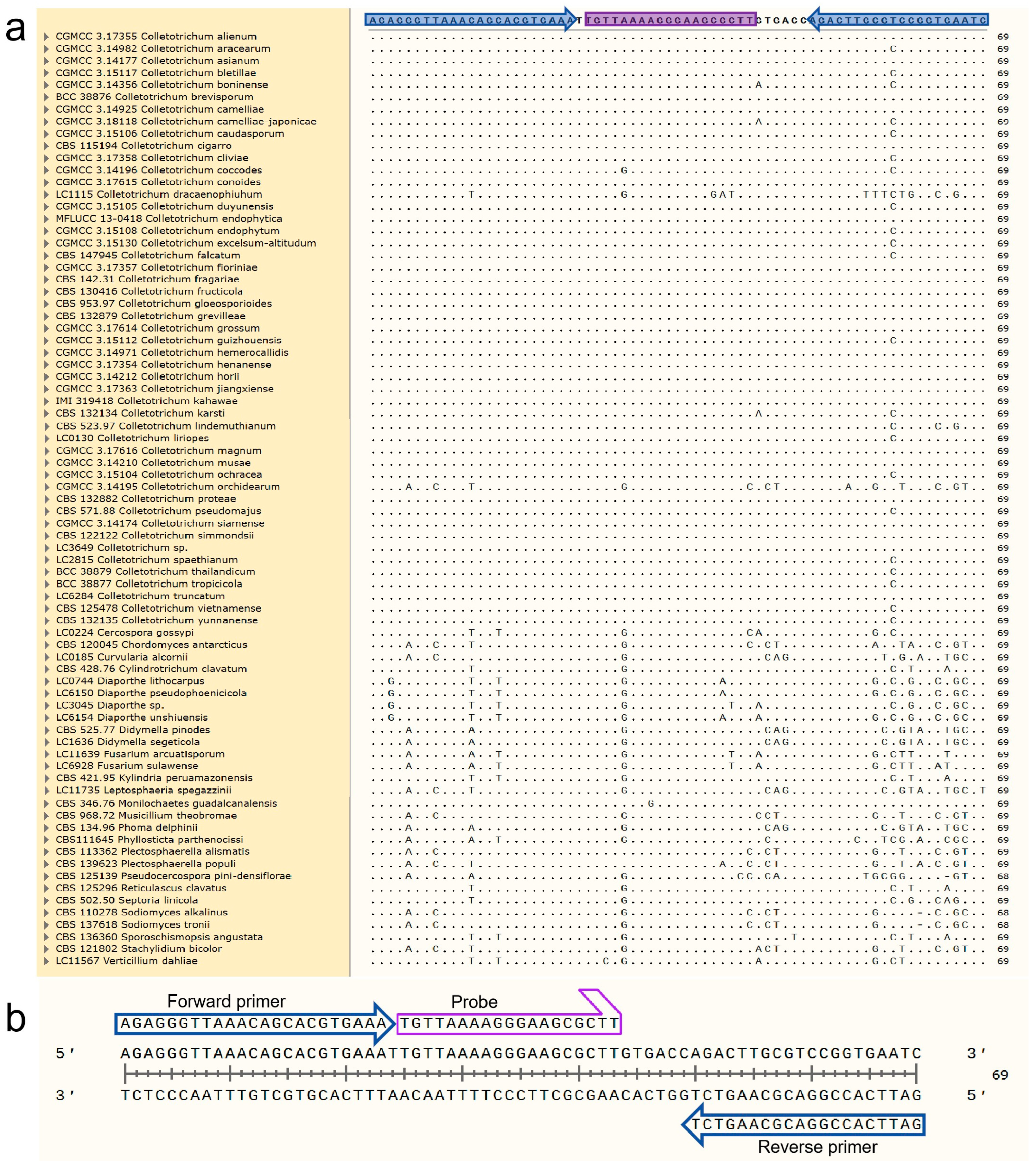
Nucleotide sequence regions exhibiting a consensus specific to Colletotrichum spp. were selected for primer and probe design using Primer Express 3.0 software (Applied Biosystems, Foster City, CA, USA). The resulting assay utilizes the forward primer CLF (5′-AGAGGGTTAAACAGCACGTGAAA-3′), the reverse primer CLR (5′-GATTCACCGGACGCAAGTCT-3′), and the TaqMan probe CLP (5′-FAM-TGTTAAAAGGGAAGCGCTT-MGB-3′), generating a 69 bp amplicon (Figure 1). The Primer Express 3.0.1 software was employed to check for hairpins and potential seco. Specificity was further verified in silico using the BLASTn tool of the National Center for Biotechnology Information (https://blast.ncbi.nlm.nih.gov/Blast.cgi, (accessed on 20 December 2025)). The primers and the probe were synthesized by Invitrogen. The TaqMan® probe was labeled with 6-FAM (fluorescein) at the 5′ end and a non-fluorescent quencher (NFQ) coupled with a Minor Groove Binder (MGB) moiety at the 3′ end.
Figure 1.
Alignment of 28S sequences of Colletotrichum spp. and other genera. (a) Location of forward and reverse primers (blue arrows) and qPCR probe (in purple) designed for the detection of Colletotrichum spp. (b) Detailed schematic of how target gene fragments bind.
Table 1.
Strains or data used in this study.
2.3. Specificity Assessment of the TaqMan qPCR Assay
The specificity of the assay was empirically evaluated using genomic DNA from 78 fungal isolates (indicated as “Primer design & qPCR test” and “qPCR test” in the “Usage” column of Table 1). Sterile distilled water served as a blank control. Real-time PCR was performed using an ABI PRISM 7900HT Fast Real-Time PCR System (Applied Biosystems Company, Carlsbad, CA, USA). The 20 μL reaction mixture consisted of 10 μL of 2× TaqMan® Universal PCR Master Mix, 0.5 μL each of forward primer CLF (10 μmol/L), reverse primer CLR (10 μmol/L), and probe CLP (10 μmol/L), 1 μL of template DNA, and 7.5 μL of sterile water. Thermal cycling conditions were: 95 °C for 10 min, followed by 40 cycles of 95 °C for 15 s and 60 °C for 60 s. Three technical replicates were performed for each sample.
2.4. Optimization of qPCR Conditions
Genomic DNA from C. kahawae (IMI 319418) was used as the template to optimize the qPCR assay. A blank control containing sterile distilled water was included in each run. First, primer concentrations were optimized by testing a gradient of forward and reverse primer concentrations (0.1, 0.2, 0.3, 0.4, 0.5, 0.6, 0.7, 0.8, 0.9, and 1.0 μmol/L) while keeping other component concentrations constant. The optimal primer concentration was selected based on the cycle threshold (Ct) values and the fluorescence intensity of the amplification curves. Subsequently, using the optimized primer concentrations, the probe concentration was optimized by testing a range from 0.1 to 1.0 μmol/L. Three technical replicates were performed for each condition. The thermal cycling conditions were identical to those described in the specificity assessment.
2.5. Sensitivity and Standard Curve Analysis
To assess sensitivity, genomic DNA from C. siamense (CGMCC 3.14193 = CBS 130420) was serially diluted 10-fold using RNase-free ddH2O, ranging from 68 ng/μL to 6.8 fg/μL. The optimized reaction system was used: 20 μL reaction volume containing 10 μL of 2× TaqMan® Universal PCR Master Mix, 0.7 μL of forward primer CLF (10 μmol/L), 0.7 μL of reverse primer CLR (10 μmol/L), 0.5 μL of probe CLP (10 μmol/L), 1 μL of template DNA, and 7.1 μL of sterile water. The cycling conditions were: 95 °C for 10 min, followed by 40 cycles of 95 °C for 15 s and 60 °C for 60 s. Three replicates were performed for each dilution.
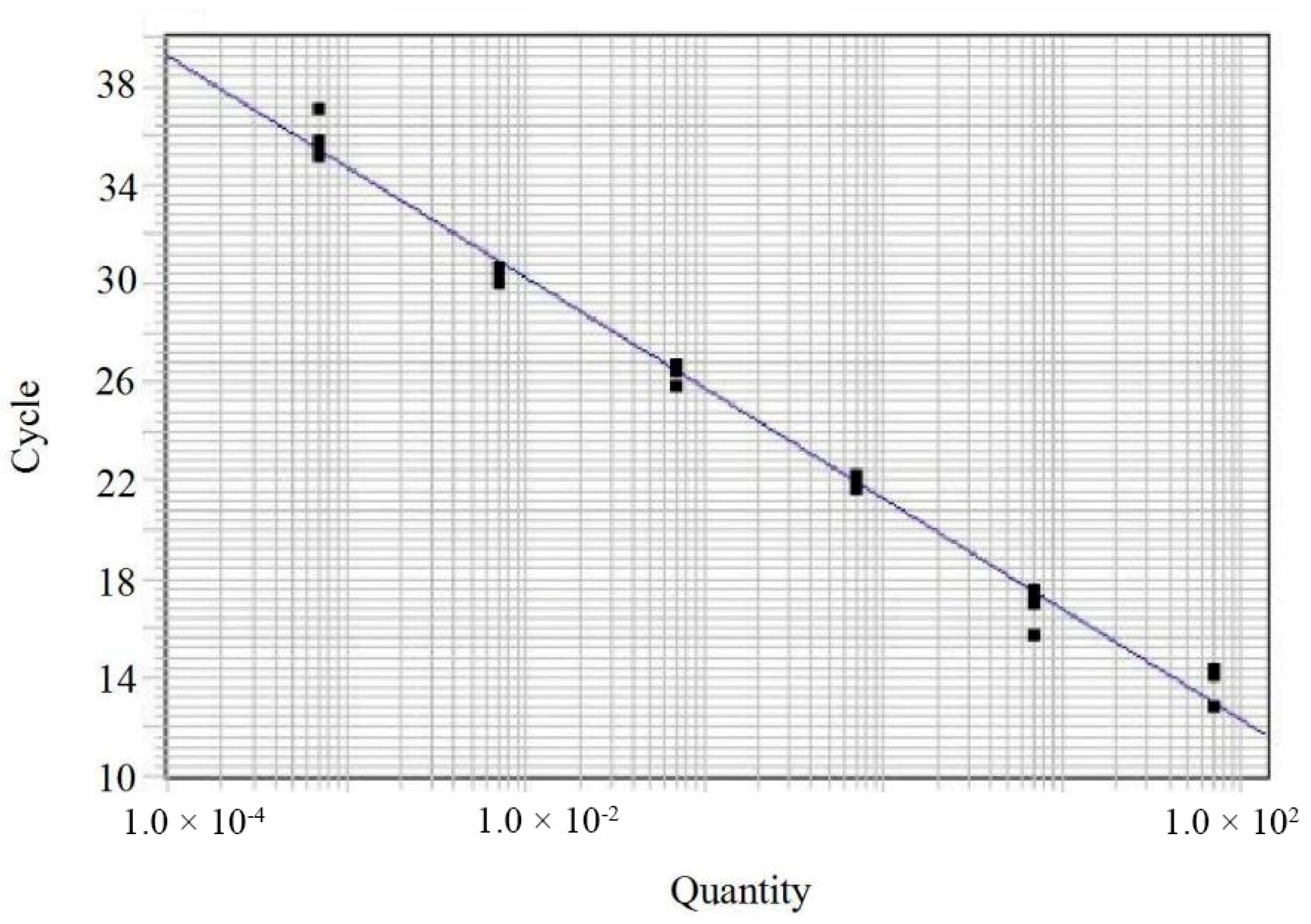
A standard curve was generated using the 10-fold dilution series of C. siamense DNA (dynamic range: 68 ng/μL to 68 fg/μL). The amplification efficiency (E) was calculated using the equation E = (10(−1/slope) − 1) × 100. The slope and linearity (coefficient of determination, R2) were also determined.
2.6. Validation with Mock Environmental Samples
To evaluate the assay’s robustness and exclude potential interference from non-target species in complex matrices, two types of mock environmental samples were prepared using fungal genomic DNA. Mock Sample 1 (Non-target Cocktail): Prepared by pooling 1 μL of DNA from each of the 27 non-Colletotrichum strains (labeled as “qPCR test” or “Primer design & qPCR test” in Table 1) into a single tube. A 1 μL aliquot of this mixture was used as the template. Mock Sample 2 (Target Spiked in Non-target Cocktail): Prepared by transferring 10 μL of Mock Sample 1 to a new tube and adding DNA from 15 randomly selected Colletotrichum spp (1 μL each, as listed in Table 1). A 1 μL aliquot of this final mixture was used as the template.
Genomic DNA of C. kahawae (IMI 319418) served as a positive control, Chordomyces antarcticus (CBS 120045) as a negative control, and sterile distilled water as a blank control. The qPCR was performed using the optimized reaction conditions described above, with three technical replicates for each reaction.
3. Results
3.1. Specificity of the Colletotrichum spp.-Specific qPCR TaqMan Assay
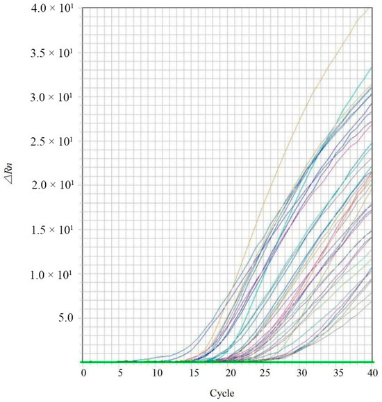
Specificity was evaluated using the 78 fungal isolates listed in Table 1. The results demonstrated that positive amplification signals were observed exclusively in Colletotrichum samples (Figure 2), while no amplification was detected in any non-target species. Variations in the observed cycle threshold (Ct) values were attributed to the lack of DNA concentration normalization across samples during specificity testing. Furthermore, one of the 69 bp target amplicon sequences of Colletotrichum spp. was queried against the NCBI Genome database using BLASTn. The top hits were consistently identified as Colletotrichum species. Subsequent local alignment of at least 200 retrieved sequences from NCBI revealed 100% identity with the target amplicon, confirming the high intrageneric conservation of the designed markers.
Figure 2.
Real-time PCR amplification curves for the specific detection of Colletotrichum spp. The specificity was evaluated by testing the designed primers and probe against genomic DNA from target Colletotrichum isolates and various non-target fungal species. The amplification curves illustrate positive fluorescent signals exclusively for Colletotrichum spp., while no signals (flat lines) were observed for non-target species. The X-axis represents the cycle values, and the Y-axis represents the baseline-corrected normalized fluorescence (ΔRn).
3.2. Optimization of qPCR Conditions
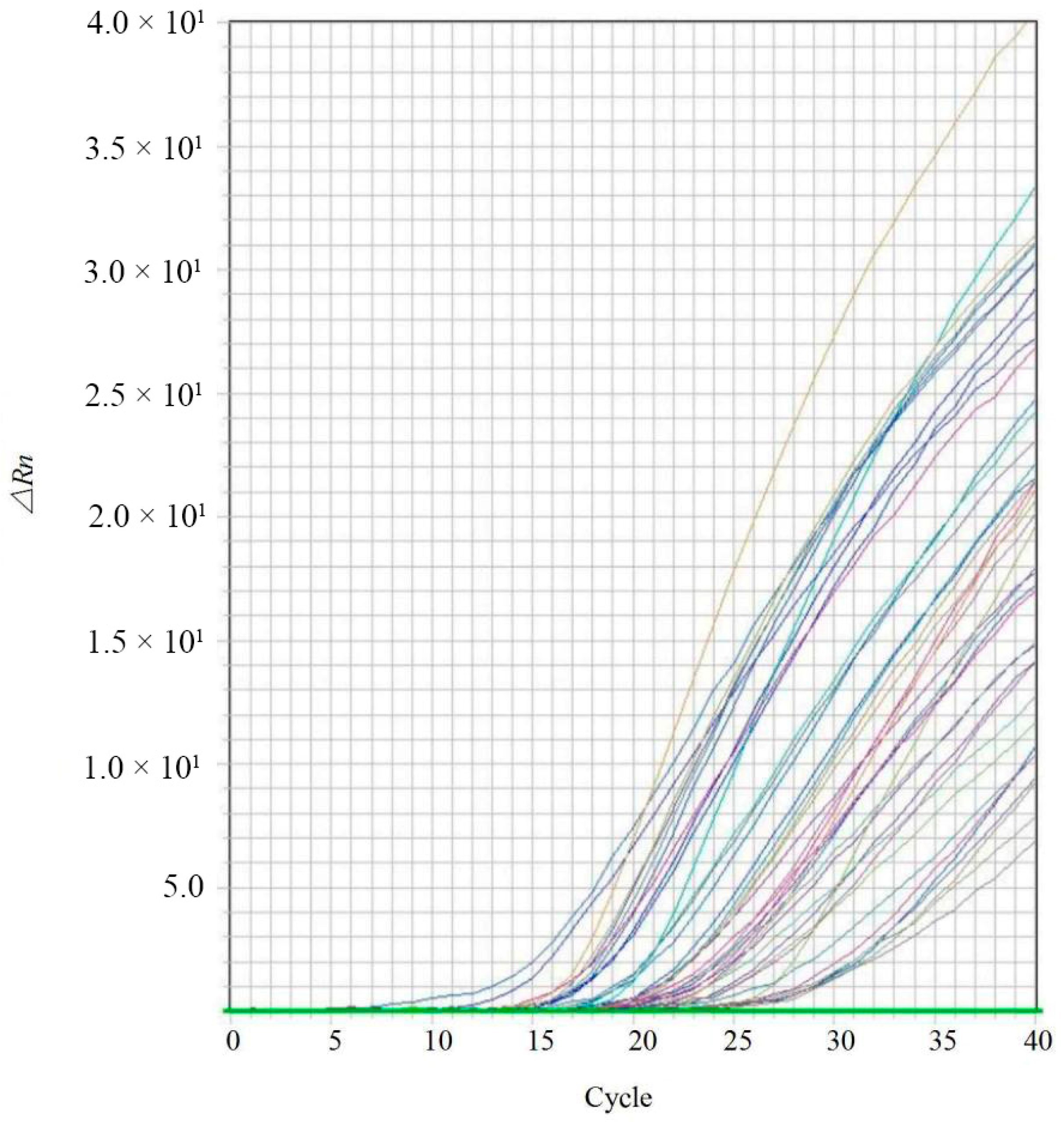
Positive amplification signals were observed at all primer concentrations ranging from 0.1 to 1.0 μmol/L (Figure 3). However, the reaction containing 0.7 μmol/L of primers yielded the highest fluorescence signal (∆Rn) and the lowest Ct values, indicating optimal amplification efficiency. Based on the optimized primer concentration, the probe concentration was subsequently tested. The results indicated that a probe concentration of 0.5 μmol/L produced the maximum ∆Rn and minimum Ct values (Figure 4). Consequently, the optimized qPCR reaction system (20 μL total volume) was established as follows: 10 μL of 2× TaqMan® Universal PCR Master Mix, 0.7 μL of forward primer CLF (10 μmol/L), 0.7 μL of reverse primer CLR (10 μmol/L), 0.5 μL of probe CLP (10 μmol/L), 1 μL of template DNA, and 7.1 μL of sterile water.
3.3. Sensitivity of Real-Time Fluorescent PCR for Detection of Colletotrichum spp.
The sensitivity of the assay was evaluated using a ten-fold serial dilution of genomic DNA from the ex-type strain of C. siamense (CGMCC 3.14193), ranging from 68 ng/μL to 6.8 fg/μL. Positive amplification curves were consistently obtained at concentrations ≥680 fg/μL, establishing the limit of detection (LOD) at 680 fg/μL (Figure 5).
Figure 3.
Optimization of primer concentrations for the Colletotrichum-specific TaqMan qPCR assay. A range of primer concentrations (0.1, 0.2, 0.3, 0.4, 0.5, 0.6, 0.7, 0.8, 0.9, and 1.0 μmol/L) was evaluated while keeping the probe concentration and template DNA constant. Each curve represents the mean amplification plot of a specific primer concentration. The green line represents the threshold line automatically generated by the real-time PCR instrument software.
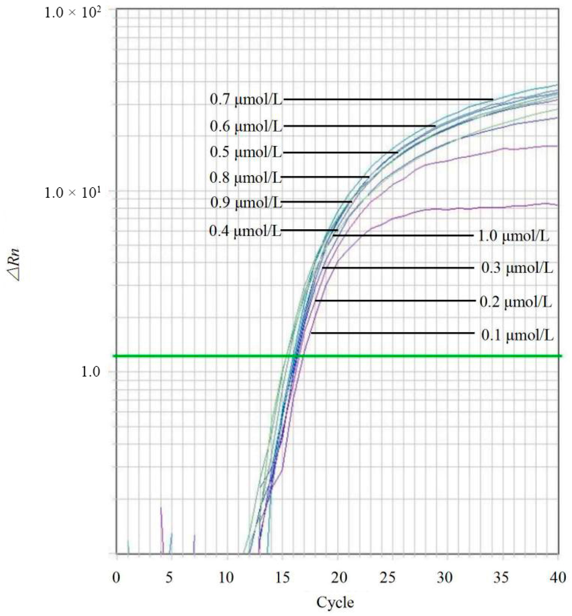
Figure 4.
Optimization of probe concentrations for the Colletotrichum-specific TaqMan qPCR assay. The assay was performed using varying probe concentrations (0.1, 0.2, 0.3, 0.4, 0.5, 0.6, 0.7, 0.8, 0.9, and 1.0 μmol/L) to identify the concentration yielding the highest fluorescence signal and lowest Ct value. Curves correspond to the respective probe concentrations as indicated in the legend. The green line represents the threshold line automatically generated by the real-time PCR instrument software.
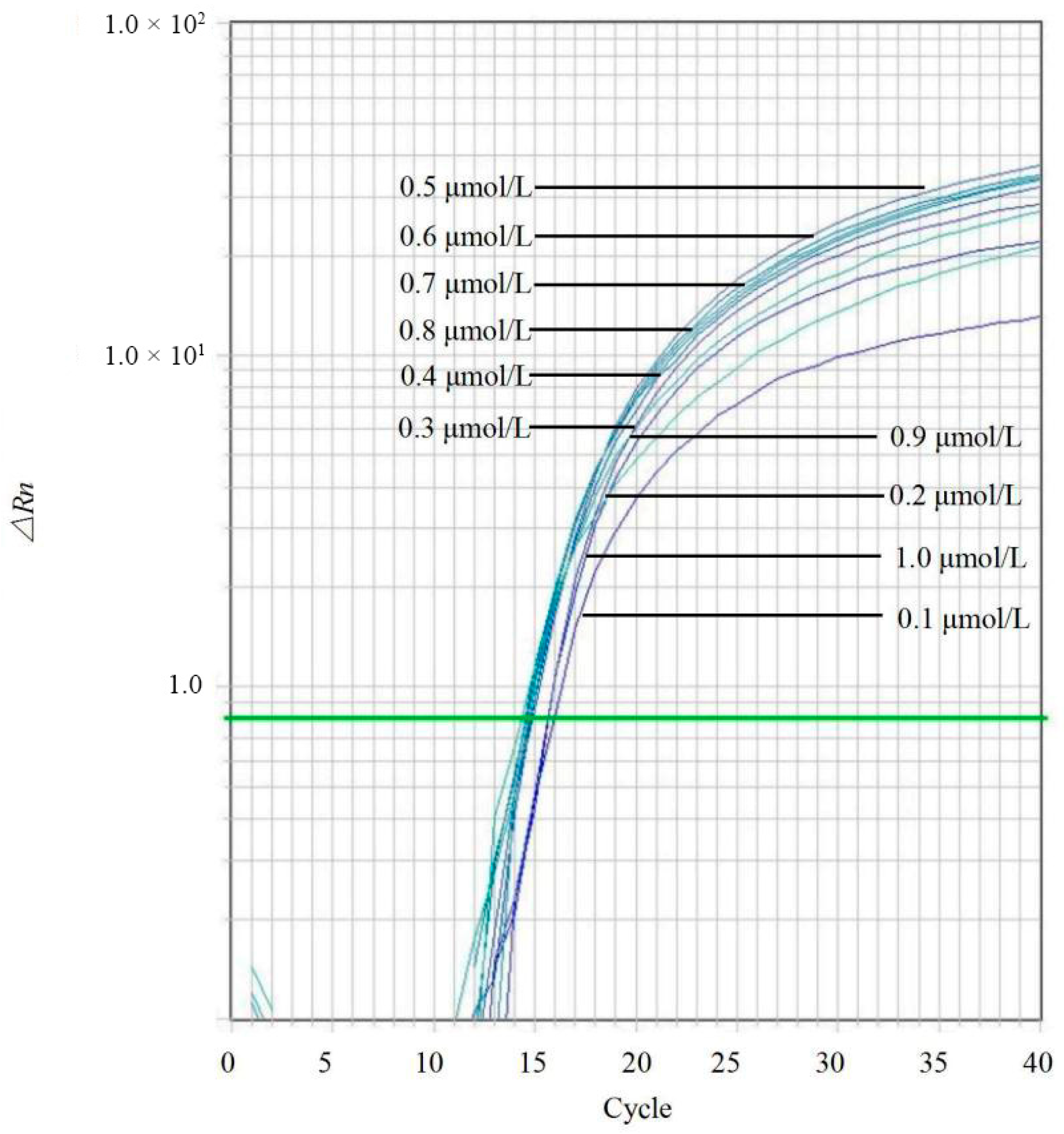
Figure 5.
Sensitivity of the TaqMan qPCR assay using 10-fold serial dilutions of C. siamense genomic DNA as templates. The amplification curves correspond to DNA concentrations ranging from 68 ng/μL to 680 fg/μL per reaction. Each concentration was tested in triplicate to ensure reproducibility. The green line represents the threshold line automatically generated by the real-time PCR instrument software.
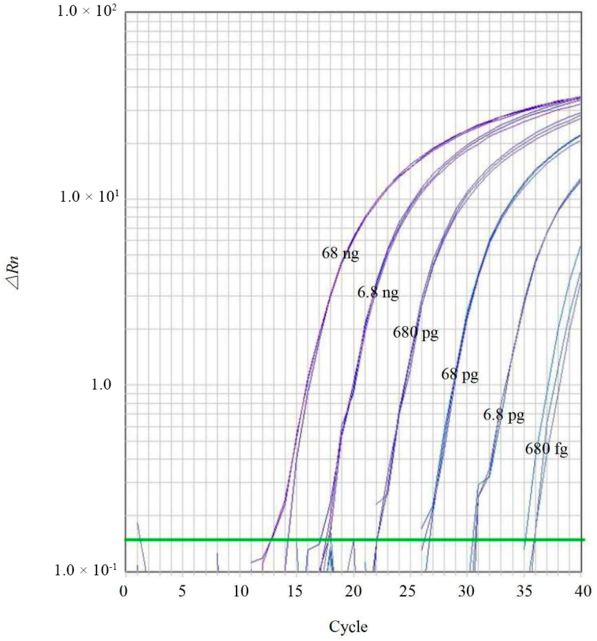
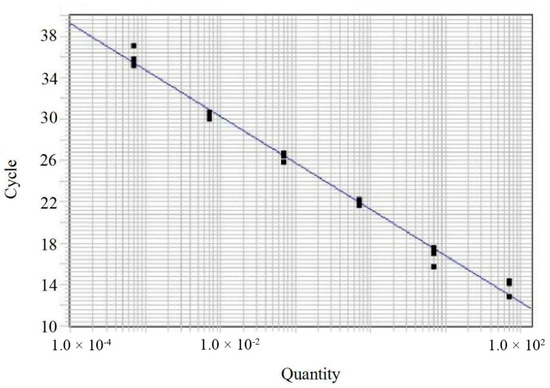
There was a strong inverse linear relationship between the Ct values and the logarithm of the template DNA concentrations. The standard curve generated from the triplicates showed a linear regression equation of y = −4.49x + 21.29, with a coefficient of determination (R2 = 0.9922) (Figure 6). These data demonstrate that the established assay possesses high reproducibility and reliable amplification performance within the tested dynamic range.
Figure 6.
Standard curve of the TaqMan qPCR assay for the quantification of Colletotrichum DNA. The standard curve was generated by plotting the Ct values (cycle, y-axis) against the logarithm of the genomic DNA concentrations (quantity, x-axis) from C. siamense. Each data point represents the mean Ct value derived from three independent replicates.
3.4. Detection of Colletotrichum spp. in Mock Environmental Samples
The specificity and practical applicability of the developed TaqMan qPCR assay were further validated using mock environmental samples. Positive amplification curves were detected in the positive control and Mock Sample 2 (containing Colletotrichum DNA). In contrast, no fluorescence signals were observed in the negative control, the blank control, or Mock Sample 1 (containing only non-Colletotrichum fungal DNA). These results confirm the assay’s ability to accurately detect Colletotrichum species within complex fungal communities without cross-reactivity.
4. Discussion
Specificity is the prerequisite for any reliable molecular diagnostic assay [24]. In this study, the specificity of the designed primer-probe set was rigorously challenged against a comprehensive panel representing the fungal diversity typically encountered in agricultural and clinical ecosystems. By including representatives from 14 Colletotrichum species complexes [19] and, crucially, species from the phylogenetically sister families—Australiascaceae, Plectosphaerellaceae, and Reticulascaceae [25]—we effectively excluded cross-reactivity with proximate taxa. The absence of false-positive signals in these closely related groups, as well as in other common phytopathogens (e.g., members of Botryosphaeriales, Chaetosphaeriales, Diaporthales, Eurotiales, Hypocreales, Mycosphaerellales, and Pleosporales), confirms that the selected 28S rDNA region offers sufficient phylogenetic resolution to discriminate the genus Colletotrichum from other fungal lineages.
In the initial phase of assay development, the ITS region was evaluated as a potential target. However, achieving a balance between intergeneric specificity and intrageneric conservation proved challenging due to the high variability of ITS sequences within the genus and the limitations of ITS as a sole marker for certain Colletotrichum lineages [26]. Consequently, we shifted our focus to the 28S rDNA region, which generally exhibits superior stability at the genus level [27]. A significant hurdle was the paucity of Colletotrichum 28S sequences in public repositories, as this region is not routinely employed in the taxonomy of this genus compared to multi-locus sequence analysis genes like gapdh or tub2 [8]. To bridge this gap, we generated 28S sequences for multiple Colletotrichum strains via in-house sequencing. We then implemented a robust dual-validation strategy, combining empirical qPCR testing with in silico verification. This comprehensive approach confirmed the assay’s efficacy for 14 of the 17 species complexes described by Liu et al. [19].
The remaining three species complexes (C. agaves, C. graminicola, and C. bambusicola) could not be experimentally validated due to the unavailability of reference strains at the time of the study. To evaluate the assay’s potential performance for these groups, sequence alignment analysis was conducted, which identified a single nucleotide polymorphism (SNP), specifically a T-to-C transition, in the central region of the reverse primer binding site. Within the framework of TaqMan chemistry, a single central mismatch generally permits target amplification, although it may delay the threshold cycle by 0.5–2 cycles. While this may slightly influence quantification accuracy, it does not typically compromise qualitative detection [28]. This theoretical resilience was empirically supported in the present study; several species used for validation, such as C. aracearum (CGMCC 3.14982), C. boninense (CGMCC 3.14356), and C. falcatum (CBS 147945), harbor similar mismatches in their reverse primer binding regions (Figure 1). Despite these sequence variations, all such taxa were accurately detected, demonstrating that the assay maintains high diagnostic sensitivity and is capable of providing universal coverage for the genus Colletotrichum.
Previous molecular detection efforts for Colletotrichum have predominantly focused on species-specific assays [20,21,29,30]. While valuable for precise taxonomic identification, these narrow-spectrum assays are often constrained by the genus’s frequent taxonomic reclassifications and the rapid emergence of cryptic pathogenic species [5]. A primer set designed for a specific species today may become taxonomically ambiguous due to future revisions. In contrast, the genus-specific assay established here targets the conserved 28S rDNA region, functioning as a stable and robust “universal” screening tool. This capability is particularly advantageous in phytosanitary inspection, where the primary objective is to rapidly confirm the presence of Colletotrichum infection to trigger immediate control measures, rather than to determine the specific cryptic species immediately.
The high sensitivity and broad specificity of this assay have significant implications for both agriculture and public health. In phytosanitary contexts, Colletotrichum species are notorious for establishing latent infections in asymptomatic plant tissues, rendering visual inspection at customs borders ineffective [31]. With a limit of detection as low as 680 fg, our TaqMan qPCR assay enables the interception of low-titer pathogen DNA in asymptomatic germplasm, serving as a powerful early warning tool.
Furthermore, the significance of this method extends to clinical diagnostics. As Colletotrichum species are increasingly recognized as opportunistic human pathogens causing keratitis and subcutaneous infections [9,14,17], rapid diagnosis is vital. Since first-line antifungal treatments often differ between fungal genera but are consistent within the genus, this assay allows for the rapid confirmation of Colletotrichum etiology, thereby facilitating timely clinical decision-making and the initiation of appropriate antifungal therapy [32].
Ensuring robust performance in complex biological matrices is essential for the practical utility of any diagnostic assay [33]. Although this study utilized simulated samples, they were specifically engineered to mimic the complexity of environmental material by incorporating a diverse background of non-target genomic DNA. This mixture comprised several common phytopathogenic genera, including Cercospora, Curvularia, Diaporthe, Leptosphaeria, Plectosphaerella, Phoma, and Verticillium, as well as the trans-kingdom pathogen Fusarium [34,35]. The lack of cross-reactivity within these complex backgrounds, alongside in silico BLASTn analysis confirming no significant homology between the target sequence and host genomes, highlights the high analytical specificity of the assay. Furthermore, the ability of this qPCR method to directly identify Colletotrichum DNA from environmental samples without the requirement for prior fungal isolation offers a significant advantage for rapid diagnostics. This bypassing of traditional culture-based methods can reduce the diagnostic timeline from several days to about one hour, providing a reliable tool for both agricultural and clinical detection of Colletotrichum infections. Moreover, although the calculated amplification efficiency of this assay was 66.7% (slope = −4.49), falling outside the ideal range for precision absolute quantification, the high correlation coefficient (R2 = 0.9922) ensures sufficient reliability for qualitative screening and semi-quantitative analysis in diagnostic applications.
Author Contributions
Conceptualization, F.L.; methodology, F.L. and W.D.; experiment, F.L. and W.D.; resources, F.L., D.P., L.C. and P.W.C.; writing and editing, F.L., P.W.C. and D.P.; project administration, F.L.; funding acquisition, F.L. and D.P. All authors have read and agreed to the published version of the manuscript.
Funding
This research was financially supported by National Natural Science Foundation of China (32322001), Beijing Nova Program (20230484305), and the Key Research and Development Program of Xizang Autonomous Region in China (XZ202501ZY0019, XZ202301ZY0027G).
Institutional Review Board Statement
Not applicable.
Informed Consent Statement
Not applicable.
Data Availability Statement
The GenBank accession numbers for the public data used in this study are listed in Table 1, further inquiries can be directed to the corresponding author.
Conflicts of Interest
The authors declare no conflicts of interest.
References
- Dean, R.; Van Kan, J.A.; Pretorius, Z.A.; Hammond-Kosack, K.E.; Di Pietro, A.; Spanu, P.D.; Rudd, J.J.; Dickman, M.; Kahmann, R.; Ellis, J.; et al. The Top 10 fungal pathogens in molecular plant pathology. Mol. Plant Pathol. 2012, 13, 414–430. [Google Scholar] [CrossRef] [Scilit]
- Hindorf, H.; Omondi, C.O. A review of three major fungal diseases of Coffea arabica L. in the rainforests of Ethiopia and progress in breeding for resistance in Kenya. J. Adv. Res. 2011, 2, 109–120. [Google Scholar] [CrossRef] [Scilit]
- Ma, Z.; Liu, F.; Tsui, C.K.; Cai, L. Phylogenomics and adaptive evolution of the Colletotrichum gloeosporioides species complex. Commun. Biol. 2025, 8, 593. [Google Scholar] [CrossRef] [Scilit] [PubMed]
- Khalimi, K.; Darmadi, A.A.K.; Suprapta, D.N. First report on the prevalence of Colletotrichum scovillei associated with anthracnose on chili pepper in Bali, Indonesia. Int. J. Agric. Biol. 2019, 22, 363–368. [Google Scholar] [CrossRef] [Scilit]
- Cannon, P.F.; Damm, U.; Johnston, P.R.; Weir, B.S. Colletotrichum: Current status and future directions. Stud. Mycol. 2012, 73, 181–213. [Google Scholar] [CrossRef] [Scilit]
- Damm, U.; Cannon, P.F.; Woudenberg, J.H.C.; Crous, P.W. The Colletotrichum acutatum species complex. Stud. Mycol. 2012, 73, 37–113. [Google Scholar] [CrossRef] [Scilit]
- Damm, U.; Cannon, P.F.; Woudenberg, J.H.C.; Johnston, P.R.; Weir, B.S.; Tan, Y.P.; Shivas, R.G.; Crous, P.W. The Colletotrichum boninense species complex. Stud. Mycol. 2012, 73, 1–36. [Google Scholar] [CrossRef] [Scilit] [PubMed]
- Weir, B.S.; Johnston, P.R.; Damm, U. The Colletotrichum gloeosporioides species complex. Stud. Mycol. 2012, 73, 115–180. [Google Scholar] [CrossRef] [Scilit]
- Guarro, J.; Svidzinski, T.E.; Zaror, L.; Forjaz, M.H.; Gené, J.; Fischman, O. Subcutaneous hyalohyphomycosis caused by Colletotrichum gloeosporioides. J. Clin. Microbiol. 1998, 36, 3060–3065. [Google Scholar] [CrossRef] [Scilit]
- Cano, J.; Guarro, J.; Gené, J. Molecular and morphological identification of Colletotrichum species of clinical interest. J. Clin. Microbiol. 2004, 42, 2450–2454. [Google Scholar] [CrossRef] [Scilit]
- Paniz-Mondolfi, A.E.; Agemy, S.; Cañete-Gibas, C.; Gitman, M.R.; Iacob, C.E.; Necula, I.; Yang, C.Y.; Noguera, L.A.D.; Sanders, C.; Wiederhold, N.P.; et al. First report of human infection caused by Colletotrichum chlorophyti occurring in a post-corneal transplant patient with endophthalmitis. Med. Mycol. Case Rep. 2021, 32, 73–76. [Google Scholar] [CrossRef] [Scilit]
- Kotwal, A.; Biswas, D.; Kakati, B.; Bahadur, H.; Gupta, N. Non traumatic keratitis due to Colletotrichum coccodes: A case report. J. Clin. Diagn. Res. 2015, 9, DD01. [Google Scholar] [CrossRef] [Scilit]
- Buchta, V.; Nekolová, J.; Jirásková, N.; Bolehovská, R.; Wipler, J.; Hubka, V. Fungal keratitis caused by Colletotrichum dematium: Case study and review. Mycopathologia 2019, 184, 441–453. [Google Scholar] [CrossRef] [Scilit] [PubMed]
- Wang, W.; Gong, H.; Yang, X.; Xu, L.; Huang, X.; Su, G.; Tan, Y.; Jing, B.; Zheng, Z.; Chen, Z.; et al. Colletotrichum keratitis: An important fungal infection of nine human eyes. Diagn. Microbiol. Infect. Dis. 2024, 110, 116540. [Google Scholar] [CrossRef] [Scilit]
- Liu, F.; Cai, L.; Crous, P.W.; Damm, U. The Colletotrichum gigasporum species complex. Persoonia 2014, 33, 83–97. [Google Scholar] [CrossRef] [Scilit] [PubMed]
- Werbel, W.A.; Baroncelli, R.; Shoham, S.; Zhang, S.X. Angioinvasive, cutaneous infection due to Colletotrichum siamense in a stem cell transplant recipient: Report and review of prior cases. Transpl. Infect. Dis. 2019, 21, e13153. [Google Scholar] [CrossRef] [Scilit] [PubMed]
- Shivaprakash, M.R.; Appannanavar, S.B.; Dhaliwal, M.; Gupta, A.; Gupta, S.; Gupta, A.; Chakrabarti, A. Colletotrichum truncatum: An unusual pathogen causing mycotic keratitis and endophthalmitis. J. Clin. Microbiol. 2011, 49, 2894–2898. [Google Scholar] [CrossRef] [Scilit]
- Hoog, G.D.; Guarro, J.; Gene, J.F.M.J.; Figueras, M.J. Atlas of Clinical Fungi, 2nd ed.; Central Bureau Voor Schimmelcultures: Utrecht, The Netherlands, 2000; pp. 1–1160. [Google Scholar]
- Liu, F.; Ma, Z.Y.; Hou, L.W.; Diao, Y.Z.; Wu, W.P.; Damm, U.; Song, S.; Cai, L. Updating species diversity of Colletotrichum, with a phylogenomic overview. Stud. Mycol. 2022, 101, 1–56. [Google Scholar] [CrossRef] [Scilit]
- Garrido, C.; Carbú, M.; Fernández-Acero, F.J.; Boonham, N.; Colyer, A.; Cantoral, J.M.; Budge, G. Development of protocols for detection of Colletotrichum acutatum and monitoring of strawberry anthracnose using real-time PCR. Plant Pathol. 2009, 58, 43–51. [Google Scholar] [CrossRef] [Scilit]
- Tao, G.; Hyde, K.D.; Cai, L. Species-specific real-time PCR detection of Colletotrichum kahawae. J. Appl. Microbiol. 2013, 114, 828–835. [Google Scholar] [CrossRef] [Scilit]
- Mosca, S.; Li Destri Nicosia, M.G.; Cacciola, S.O.; Schena, L. Molecular analysis of Colletotrichum species in the carposphere and phyllosphere of olive. PLoS ONE 2014, 9, e114031. [Google Scholar] [CrossRef] [Scilit]
- Li, F.; Zhang, X.L.; Wang, C.; Luo, M. Establishment of a PCR test method for detecting Colletotrichum. Xinjiang Agric. Sci. 2019, 56, 919–926. [Google Scholar]
- Phurbu, D.; Feng, Z.; Cai, L.; Liu, F. Rapid and sensitive diagnosis of plant quarantine fungi Alternaria triticina and Plenodomus libanotidis based on the RPA-CRISPR/Cas12a system. IMA Fungus 2025, 16, e153604. [Google Scholar] [CrossRef] [Scilit]
- Maharachchikumbura, S.S.; Hyde, K.D.; Jones, E.G.; McKenzie, E.H.C.; Bhat, J.D.; Dayarathne, M.C.; Huang, S.-K.; Norphanphoun, C.; Senanayake, I.C.; Perera, R.H.; et al. Families of sordariomycetes. Fungal Divers. 2016, 79, 1–317. [Google Scholar] [CrossRef] [Scilit]
- Crouch, J.A.; Clarke, B.B.; Hillman, B.I. What is the value of ITS sequence data in Colletotrichum systematics and species diagnosis? A case study using the falcate-spored graminicolous Colletotrichum group. Mycologia 2009, 101, 648–656. [Google Scholar] [CrossRef] [Scilit]
- Vu, D.; Groenewald, M.; De Vries, M.; Gehrmann, T.; Stielow, B.; Eberhardt, U.; Al-Hatmi, A.; Groenewald, J.Z.; Cardinali, G.; Houbraken, J.; et al. Large-scale generation and analysis of filamentous fungal DNA barcodes boosts coverage for kingdom fungi and reveals thresholds for fungal species and higher taxon delimitation. Stud. Mycol. 2019, 92, 135–154. [Google Scholar] [CrossRef] [Scilit] [PubMed]
- Bustin, S.; Huggett, J. qPCR primer design revisited. Biomol. Detect. Quantif. 2017, 14, 19–28. [Google Scholar] [CrossRef] [Scilit]
- Karimi, K.; Arzanlou, M.; Pertot, I. Development of novel species-specific primers for the specific identification of Colletotrichum nymphaeae based on conventional PCR and LAMP techniques. Eur. J. Plant Pathol. 2020, 156, 463–475. [Google Scholar] [CrossRef] [Scilit]
- Torres-Calzada, C.; Tapia-Tussell, R.; Quijano-Ramayo, A.; Martin-Mex, R.; Rojas-Herrera, R.; Higuera-Ciapara, I.; Perez-Brito, D. A species-specific polymerase chain reaction assay for rapid and sensitive detection of Colletotrichum capsici. Mol. Biotechnol. 2011, 49, 48–55. [Google Scholar] [CrossRef] [Scilit] [PubMed]
- Prusky, D.; Freeman, S.; Dickman, M.B. Colletotrichum: Host Specificity, Pathology, and Host-Pathogen Interaction; Amer Phytopathological Society: Saint Paul, MN, USA, 2000; pp. 1–393. [Google Scholar]
- O’Quinn, R.P.; Hoffmann, J.L.; Boyd, A.S. Colletotrichum species as emerging opportunistic fungal pathogens: A report of 3 cases of phaeohyphomycosis and review. J. Am. Acad. Dermatol. 2001, 45, 56–61. [Google Scholar] [CrossRef] [Scilit] [PubMed]
- Adil, O.; Shamsi, M.H. Transformative biomedical devices to overcome biomatrix effects. Biosens. Bioelectron. 2025, 279, 117373. [Google Scholar] [CrossRef] [Scilit] [PubMed]
- van Diepeningen, A.D.; de Hoog, G.S. Challenges in Fusarium, a trans-kingdom pathogen. Mycopathologia 2016, 181, 161–163. [Google Scholar] [CrossRef] [Scilit] [PubMed]
- Short, D.P.; O’Donnell, K.; Zhang, N.; Juba, J.H.; Geiser, D.M. Widespread occurrence of diverse human pathogenic types of the fungus Fusarium detected in plumbing drains. J. Clin. Microbiol. 2011, 49, 4264–4272. [Google Scholar] [CrossRef] [Scilit] [PubMed]
Disclaimer/Publisher’s Note: The statements, opinions and data contained in all publications are solely those of the individual author(s) and contributor(s) and not of MDPI and/or the editor(s). MDPI and/or the editor(s) disclaim responsibility for any injury to people or property resulting from any ideas, methods, instructions or products referred to in the content. |
© 2026 by the authors. Licensee MDPI, Basel, Switzerland. This article is an open access article distributed under the terms and conditions of the Creative Commons Attribution (CC BY) license.