


Abstract
Invasive mould diseases (IMDs) are an important component of healthcare-associated infections (HAIs). However, they can be difficult to identify, as the standard definition of HAIs do not always apply and usually have a longer and more variable onset after exposure. Identifying the source of infection is also challenging, as moulds can colonise a wide range of indoor healthcare environments, and multiple sources of exposure are possible. Thus, healthcare-associated IMDs may reflect a broader failure of infection control measures rather than a single isolated event. In addition, IMDs may evolve into outbreaks that are hard to detect and investigate, as they can develop over extended periods before raising concern. Here, we review mould ecology in healthcare settings and describe the patient groups at risk for healthcare-associated IMDs. We also overview the investigative approaches used to identify case clusters—including case finding, environmental sampling and isolate genotyping—summarise major IMD outbreaks and highlight best management practices. Taken together, this knowledge will help identify high-risk situations and guide the implementation of targeted preventive measures and diagnostic strategies that enable the timely diagnosis and effective management of healthcare-associated IMDs.
1. Introduction
Healthcare associated-infections (HAIs) are infections that are acquired directly or indirectly through contact with a healthcare setting [1,2]. They include infections acquired in hospital or in other healthcare environments, as well as those following medical or surgical procedures. As such, HAIs are typically defined as infections occurring ≥48 h after admission or within 30 days after discharge, and they are notable for their substantive morbidity, mortality, and excess healthcare costs [2,3]. HAIs frequently involve antimicrobial-resistant pathogens, including multi-drug-resistant ones. A substantial proportion of HAIs can be avoided through effective infection prevention and control measures.
Invasive mould diseases (IMDs) are an important component of HAIs because they mainly affect immunocompromised or critically ill patients who require prolonged stays in healthcare facilities and have attendant adverse outcomes [4,5]. However, the standard definition of HAIs may not apply to IMDs given the multiple possible sources of exposure and the longer and more variable onset after exposure [6]. In addition, the sources of IMDs are often difficult to pinpoint, with predominantly exogenous exposure being responsible, whereby moulds are ubiquitous and colonise a broad range of indoor healthcare environments or medical devices [7,8,9]. Beyond individual patient cases, IMDs may also evolve into potential clusters and outbreaks that may be difficult to detect and investigate, as they can evolve over extended periods before raising concern [10]. Conversely, moulds can also contaminate sampling devices or laboratory materials, leading to pseudo-outbreaks that must be distinguished from true outbreaks to avoid unnecessary interventions [11].
In this review, we will firstly focus on the ecology of mould in healthcare settings to better understand potential sources of exposure, then describe the patient groups at risk for healthcare-associated IMDs. We also review major IMD outbreaks and describe the case-finding approaches, diagnostic tools used to investigate case clusters, and management strategies.
2. Mould Ecology in Healthcare Settings
Moulds are saprophytic fungi that are found and accumulate in multiple environments, including indoor healthcare settings. A good understanding of their ecology is essential to assess the risk of healthcare-associated IMDs and to adopt prevention strategies.
Moulds can disseminate from outdoor sources into indoor healthcare environments by producing airborne conidia, which can remain suspended in air for hours [12]. Indoor conidia concentrations in non-protected areas generally reflect those of the outdoor environment, where the concentration can exceed 1000 conidia/m3, with the low-pathogenic genera Cladosporium and Alternaria being the most abundant [13]. Of note, the highly pathogenic Aspergillus genus is typically present at lower concentrations (0.2 to 15 conidia/m3), although levels can exceed 100 conidia/m3 under specific conditions [14]. Qualitative and quantitative variations in conidia loads have been documented, with the highest concentrations typically observed in summer and with seasonal shifts in the dominant genera [13,15]. Proximity to construction work or to composting sites can further increase conidia loads.
Moulds can also disseminate in healthcare settings from indoor sources, particularly through ventilation ducts where conidia can accumulate [16]. The regular cleaning of ventilation systems is therefore necessary to prevent contamination of the controlled areas [16]. However, cleaning procedures themselves can transiently increase airborne spore concentrations [16]. Water can also serve as a source of dissemination and contamination in healthcare settings, but it is not known if transmission occurs directly from water or following aerosolization [17,18,19]. In addition, water damage and leaks can favour local mould proliferation. Dusty areas represent another mould hotspot within indoor healthcare environments, as moulds can accumulate in settled dust, necessitating regular cleaning in patient care zones. Storage rooms likewise require careful management to prevent the contamination of materials or medical devices, including incubators, linen, needles or adhesive bandages [7,8,9,20,21].
As moulds are also major phytopathogens, they can be found in potted plants and fresh flowers, as well as on raw fruits and vegetables. Other food products that have been implicated as potential sources of mould infection in immunocompromised patients include outdoor-dried items such as tea, pepper, or other spices and milk-derived products such as cheese or yogurt [8,22,23,24].
Finally, although the patient-to-patient transmission of IMDs is very uncommon, infected patients themselves have been reported as sources of contamination for others: for instance, in the context of mould-infected wounds [25,26], or patients with high pulmonary Aspergillus burdens [27]. Conversely, no transmissions of IMDs through healthcare workers have been reported to date.
3. Definitions and Challenges in Healthcare-Associated IMDs
The defining and detection of healthcare-associated IMDs are challenging, given (i) the heterogeneity of at-risk patients, (ii) the spectrum of pathogens and their differencing ecology, and (iii) the lag between exposure and infection [6]. Hence, the time and place of acquisition is often uncertain unless there is a clear source, e.g., contaminated injectable product. Airborne exposure to the pathogen usually results in a period of colonization prior to infection, yet early clinical and radiological abnormalities are frequently absent, complicating timely diagnosis. In addition, IMDs are usually diagnosed in hospital settings in patients who frequently attend healthcare facilities, making it difficult to distinguish community-acquired secondary IMDs in at-risk individuals from true healthcare-associated IMDs.
Meijer et al. proposed definitions for nosocomial aspergillosis in immunocompromised patients, classifying cases as “possible” when they occur between 7 and 14 days after hospital admission and as “probable” when they manifest more than 14 days after admission or in patients who were discharged within the preceding 14 days [6]. However, these definitions are unlikely to apply uniformly across all patient populations or mould pathogens. Studies have shown that the observed incubation period may range from a couple of days—as short as 48 h in trauma- or burn-related IMD cases—to weeks in haematological malignancy or many months post-organ transplant [28,29].
Grouped cases or outbreaks of healthcare-associated IMDs may be difficult to identify due to their relatively low incidence and the absence of consensus definition. In its simplest form, an outbreak is defined as an increase in the usual incidence of infection [8]; however, this requires knowledge of the baseline incidence. Another proposed definition considers an outbreak to be present when ≥2 cases of a particular IMD are linked in time and place. Finally, outbreaks have also been reported for IMDs involving different mould species or genera (e.g., for mucormycosis).
Grouped cases and outbreaks can reflect an issue in the prevention procedure and/or an unanticipated event. In both situations, they must be investigated to prevent further cases. Atypical IMDs (i.e., IMDs presenting with unusual symptoms, occurring in non-classical populations or involving uncommon pathogens relative to the local epidemiology) should likewise be investigated, as they are more likely to be healthcare-associated. This approach requires robust knowledge of the local epidemiology and therefore systematic surveillance of IMDs diagnosed within a healthcare institution. A conceptual framework for initiating healthcare-associated investigations is presented in Figure 1.
Figure 1.
Conceptual framework for initiating healthcare investigations (IMD: Invasive mould disease). “Classical” and “Atypical” IMD should be classified according to the local epidemiology, including clinical presentation, patient’s risk factor and fungal species).
4. Patients at Risk of Healthcare-Associated IMDs
A broad range of patients are at risk of healthcare-associated IMDs, from those considered high-risk, who require strict preventive measures and usually present with characteristic radio-clinical features, to lower risk patients in whom IMDs are less predictable than high-risk ones and clinical presentations are often atypical.
4.1. High-Risk Patients
4.1.1. Haematology Patients
Patients with haematological malignancy and those undergoing haematopoietic stem-cell transplantation (HSCT) are considered to be at a high risk of IMD, with the greatest vulnerability observed in the acute leukaemia and allogeneic HSCT cohorts [30,31]. Key predisposing factors include prolonged neutropenia (whether in the context of disease or treatment-related factors), relapsed or refractory disease, severe graft versus host disease (GvHD) and long-term corticosteroid use [30]. Traditional assessments of fungal risk have been further complicated by the expanding use of novel disease-related immunosuppressive therapies, with increased IMD susceptibility linked to a broad range of targeted treatments including anti-CD52, TNF-α, Janus kinase (JAK), BTK and PI3K inhibitors [31]. Evidence in chimeric antigen receptor (CAR) T-cell-treated sub-groups is also emerging and, though rare, thought to be greatest in those with previous IMDs, prolonged cytopenia and or cytokine-release syndrome/immune-effector cell-associated neurotoxicity syndrome (CRS/ICANS), particularly where further immunomodulation may be required [32]. Similarly, data on IMDs following bispecific antibody (BsAb) therapy, primarily used in multiple myeloma, remains limited, and surveillance is recommended [33,34].
Clinical presentation varies with the pathogen, with pulmonary, sinus and disseminated disease the most typically reported anatomical sites [35]. Thus, an increased degree of suspicion for IMDs is recommended among high-risk patients presenting with new respiratory symptoms and persistent fever of uncertain aetiology [35]. Beyond antifungal prophylaxis, control mechanisms within the healthcare setting include strict environmental precautions such as the use of high-efficiency particulate air (HEPA) filtration to maintain protected air quality and positive-pressure ventilation (PPV) in high-risk areas [36]. Periodic airborne fungal sampling may also be of benefit in areas where opportunistic fungi are of concern (see later) [36,37,38]. Data on the efficacy of a low microbial diet on reducing IMDs are sparse, though previous studies have supported recommendations to avoid all foods where prior sterilisation or disinfection is not implemented [24,39].
4.1.2. Lung Transplant Recipients
Lung transplant recipients experience the highest burden of IMDs among solid organ transplant (SOT) groups and, from the perspective of environmental mould exposure and outbreak prevention, are appropriately considered alongside other high-risk populations such as patients with acute leukaemia and allogeneic HSCT [40,41,42].
Incidence rates of ~4–9% for IMDs in the year following transplant are frequently described [43,44], but one cohort of 274 patient recently reported an invasive aspergillosis (IA) incidence of 32%, where the median duration to the onset of disease was 6 months post-transplant [45]. Lung transplant recipients are especially susceptible to IMDs through a combination of direct environmental exposure of the allograft to inhaled fungal spores, impaired mucociliary clearance, reduced cough reflex, structural abnormalities, and long-term significant immunosuppression. Additional recognised risk factors include pre- and post-transplant mould colonisation, cytomegalovirus (CMV) infection, airway stenosis, chronic lung allograft dysfunction, renal replacement therapy and episodes of rejection requiring intensified immunosuppression [45,46,47]—with such patients calling for extra vigilance.
4.1.3. Burn Patients
Patients with major burns represent a distinct risk group for IMDs due to the extensive disruption of the skin barrier, prolonged ICU admission, broad-spectrum antibiotic exposure, repeated surgical procedures and burn-associated immune dysfunction [48]. In one large contemporary series, over 20% of burn patients involving >15% of body surface area developed an IMD [48]. Infection frequently results from the direct inoculation of damaged tissue rather than inhalational acquisition. Aspergillus species and Mucorales are the most frequently reported pathogens, but Fusarium and other environmental moulds may be increasing in frequency [49,50]. Infections in burn units have been linked to environmental contamination, contaminated dressings, and construction-associated spore dispersal [50].
Clinical manifestations range from invasive wound infection and soft tissue necrosis to disseminated disease [48,50,51]—these infections are serious, with rapid progression and high mortality. Early diagnosis is challenging because fungal wound infection may be clinically indistinguishable from bacterial infection, colonisation, or non-infectious tissue necrosis. A high index of suspicion combined with early tissue sampling and screening for fungal infection is therefore essential to obtain the best outcome for patients.
4.2. Intermediate Risk Patients
4.2.1. Other Organ Transplant Recipients
Although mould infections are less frequent in other SOT populations, they remain an important cause of morbidity and mortality. Liver transplant recipients experience the next highest burden of IMDs [40,42], with most infections occurring within the first few months after transplantation and often in the context of surgical complications, reoperation, renal replacement therapy, CMV infection or prolonged healthcare exposure [46,52,53]. Heart transplant recipients are similarly vulnerable during the early post-transplant period, particularly those requiring prolonged intensive care support, delayed chest closure, mechanical circulatory support or T-cell depleting therapies [54,55]. Notably, healthcare-associated aspergillosis outbreaks have been described in cardiothoracic and heart transplant units, emphasising the importance of environmental controls in these settings [56]. In contrast, kidney transplant recipients generally experience a lower incidence of IMDs, with most cases occurring beyond the first post-transplant year and in association with intensified immunosuppression or treatment for rejection [40,42,57].
4.2.2. Intensive Care Patients
IMDs are increasingly recognised in non-neutropenic intensive care units (ICUs). The pooled prevalence rate for the most common mould—Aspergillus—among ICU patients was 10% in a recent meta-analysis [58]. Important predisposing factors include prolonged mechanical ventilation, chronic lung disease, corticosteroid exposure, broad-spectrum antimicrobial use, extracorporeal membrane oxygenation (ECMO) and prolonged ICU admission [58]. Particular attention has focused on patients with severe viral pneumonitis, in whom influenza-associated pulmonary aspergillosis (IAPA) and COVID-19-associated pulmonary aspergillosis (CAPA) have emerged as important causes of morbidity and mortality [59]. Airway epithelial damage, immune dysregulation and corticosteroid therapy are thought to act synergistically to increase susceptibility to IMDs in these patients. Healthcare-associated exposures may contribute to additional risk through construction activities, ventilation failures, water damage and contaminated environmental reservoirs, particularly in units housing prolonged mechanically ventilated patients, which therefore necessitate specific prevention measures.
Unlike patients with haematological malignancy, radiological findings are often non-specific, and diagnosis is challenging [59]. A high index of suspicion combined with early bronchoscopy and bronchoalveolar lavage screening for fungal infection is therefore essential to optimise patient outcomes.
4.3. Low or Background Risk
Most patients visiting healthcare settings have a low or background risk of IMDs. Although certain situations, such as the presence of internal or external devices [60], deep wounds [26], or premature neonatal status [20,61] may increase this risk, these conditions alone are not sufficient to develop IMDs. In these populations, the development of IMDs will likely reflect healthcare-associated factors, such as exposure from the environment or devices (e.g., incubators in premature neonates) or direct inoculation through contaminated material or devices, and therefore warrant investigation. Transplant tourism [62] and cosmetic surgery for aesthetic reasons [63] have likewise been associated with healthcare-associated IMDs. In addition, IMDs have also been documented in patients without specific risk factors following the injection of a contaminated product [64,65,66,67].
Although infection is usually localised to the site of inoculation, the clinical presentation in these low-risk patients may be atypical and can involve uncommon or more unusual fungi, for which establishing accountability for causality may be challenging, thereby delaying diagnosis.
5. Investigation of Healthcare-Associated IMDs
The main elements of investigation include case finding, environmental assessment with the consideration of targeted environmental sampling, and comparative mycological and genotypic analyses interpreted in conjunction with temporal, spatial and epidemiological information.
5.1. Diagnosis of IMD and Case Finding
The diagnosis of IMDs involves an assessment of host risk factors, clinical features and mycological findings. It can include targeted or screening strategies regarding whether the patient is symptomatic or not. In either case, mycological findings should be interpreted cautiously, considering the known diagnostic performances and the potential risk of environmental contamination. The main diagnostic elements are summarised in Table 1.
Table 1.
Overview of clinical presentation, diagnostic strategies and interpretation by IMD risk group.
5.1.1. Targeted Diagnosis
In symptomatic patients, investigation includes radiological imaging to assess the site and extent of infection and the biopsy of the involved sites. In identifying an index case, definitive diagnosis relies on the isolation of mould from biopsies, sterile sites, body fluids and blood cultures, as well as direct microscopy and histopathology. Methods and techniques of culture and microscopy-based diagnosis have been well described in consensus definitions and diagnostic guidelines [68,69,70,71,72]. Briefly, a proven case requires the isolation or the amplification and DNA sequencing of a mould from a sterile site or blood culture, or histopathological, cytopathologic or direct microscopic examination of a mould with associated tissue damage [68]. In addition to host factors and clinical features, the mycological evidence of a probable case includes a positive culture from a non-sterile site (such as sputum, bronchial alveolar lavage fluid, or wound biopsy), as well as the detection of circulating fungal antigens and nucleic acids [68]. During the investigation of a potential cluster, the diagnosis of patients with clinical symptoms follows a similar approach to that of the index case.
5.1.2. Screening
In addition to targeted diagnosis in symptomatic patients, screening for IMDs may also be useful to improve early detection in high-risk asymptomatic patients or during investigations of potential clusters. Screening protocols for Aspergillus infections have been established in haematology patients with prolonged neutropenia, and serum biomarker screening may also be considered in burn patients [51,73,74]. Elevated serum galactomannan levels, as well as positive serum Aspergillus PCR results, can precede the first clinical or radiological signs of infection by several days, enabling prospective monitoring [75,76].
Screening approaches for other moulds have been described in high-risk patients, although they have not yet been incorporated into clinical guidelines. Notably, a positive Mucorales PCR in serum can appear 4–9 days before diagnosis and 1–2 days before imaging [77,78,79], and it has been used in both burn and haematology patients to initiate earlier treatment [80,81]. Similarly, serum Fusarium PCR has been shown to detect infection up to one week before confirmation by blood culture or biopsy [82]. Microbial cell-free DNA (mcfDNA) metagenomic testing in serum has been shown to detect pathogen DNA (including Aspergillus and Mucorales) up to three weeks prior to clinical diagnosis and may represent a promising screening tool [83]. Finally, in addition to serum screening, iterative bronchial alveolar lavage in lung transplant patients and serial wound sampling in patients with severe burns may also be used as IMD screening approaches in these high-risk populations [51,84].
5.2. Environmental Investigation and Sampling
The environmental sampling of hospital environments refers to the process of collecting microbiological samples from water, hard surfaces and air for pathogen detection, quantification and monitoring. For hard surface sampling, similar methods of sample collection used for clinical specimens, e.g., the use of swabs, may be performed, whereas for air sampling, either active air capture or the use of settle plates may be carried out [85]. Regardless of methodology, it can be difficult to distinguish between clinically important fungal isolates and contaminants, and the reporting of results requires care [86]. In addition, the interpretation of results is challenging due to the absence of universally accepted fungal burden thresholds to indicate whether the sampled area is “safe” for clinical use.
We first briefly describe common methods of sampling and then discuss the role of performing environmental investigations.
5.2.1. Air Sampling
Active air sampling uses specialised equipment to draw a defined volume of air across an agar surface. Plates are incubated typically at 25–30 °C from 2 to 7 days, with results expressed as colony-forming units per cubic metre (CFU/m3), providing a quantitative and broadly representative assessment of airborne fungi [28,85]. A limitation of this approach is that the sampling process can disrupt airflow, potentially introducing bias into the measurements obtained [86].
Settle plates are a passive air sampling method in which viable fungal spores settle by gravity onto an agar surface over a defined exposure period, followed by incubation. Results are expressed as CFU per unit area per exposure time (CFU/m2 or cm2/time) but are not considered quantitative or qualitative [28,86]. Although simple and cost effective, settle plates are not recommended for fungal air monitoring, as they selectively collect larger particles while single spores can remain suspended in air indefinitely [87].
5.2.2. Surface and Water Sampling
Surface sampling targets areas where fungal contamination is more likely, such as high-touch surfaces and equipment; however, its utility is limited by the lack of standardised methodologies and absence of established threshold values to establish unacceptable fungal burdens [88]. Although simple and inexpensive, the accuracy of surface sampling depends on appropriate sampling and analytical techniques, and it does not directly detect airborne fungi present at the time of sampling [28,85]. Combining surface and air sampling thus provides a more comprehensive assessment of fungi present than either surface or air sampling alone [85].
Water may also represent a potential source of fungi [89], but unlike bacteria, viruses and parasites, there are no standardised methods for assessing fungal contamination in water. The routine testing of hospital water is generally not indicated outside of outbreak investigations, and even then, only after careful consideration. Thresholds for nosocomial fungal contamination of water have not been defined [19,87,90].
5.2.3. Role of Environmental Sampling in Healthcare-Associated IMDs
Environmental sampling may be undertaken routinely or performed in response to an event such as suspected case clusters of infection or a suspected or known compromise in building or environmental infrastructure, e.g., breaches in heating, ventilation and air conditioning (HVAC) systems or visible mould seen on otherwise clean surfaces. Table 2 summarises the authors’ considerations in a reasonable and pragmatic approach to performing environmental sampling in these clinical contexts.
Table 2.
Framework for environmental investigation of suspected healthcare-associated invasive mould disease according to clinical context.
Routine Sampling and Surveillance
Currently, there is no requirement for routine facility-wide environmental fungal surveillance in healthcare facilities [85] because there is lack of robust evidence to support this practice (Table 2) and, importantly, little to predict individual patient risk. However, despite evidence being limited, some authorities regard it as an important adjunct to other environmental precautions in high-risk patient areas, such as HSCT wards and ICUs [19]; these recommendations are largely based on expert opinion and driven by past experiences of the institution with mould outbreaks, and they should be assessed on a case-by-case basis. It is also important to recognize that there are differences between countries in building maintenance requirements that involve the measurement of air quality. If undertaken, this can assist in establishing institutional baselines in high-risk clinical units and detecting deviations from those baselines.
Event-Triggered Sampling
The United States (US) Centre for Disease Control (CDC) and other international guidance documents have suggested that environmental sampling may be positioned as a targeted tool to: (i) support outbreak investigation, (ii) monitor the efficacy of cleaning, and (iii) validate infection control protocols in healthcare facilities, particularly for immunocompromised patients (summarized in Table 2) [6,28,87]. This is also the practice in some Australian Healthcare facilities (authors personal communication). It is also important to acknowledge that, whilst sampling may be performed in the scenarios above, parameters such as the number of sampling locations and the size and physical area of environments to be sampled are most often determined arbitrarily. Further, how often to repeat the sampling will depend on the nature and magnitude of the event and if remediation work has been undertaken.
Role of Air Sampling in Outbreak Investigations
The role of air sampling in outbreak investigations continues to be heavily debated, yet it would be fair to state that, in a potential outbreak, air sampling is often inconclusive and seldom informs clinical decisions. It is sometimes performed in search of environmental sources in conjunction with water and surface sampling (see above) and is seen to be a proactive support approach. Of note, the timing of sampling will influence the result. For instance, when performed close to the onset of an environmental or clinical signal prior to any intervention, sampling may document elevated counts of airborne mould. In contrast, more delayed sampling will likely reflect post-intervention measures and no longer reflect the relevant exposure environment [10,91].
Evidence supporting a correlation between airborne mould burden and IFD is, at best, mixed. In haematology units, many studies including longitudinal surveys have observed IMDs without correlation to measured airborne counts [92,93,94]. Conversely, others have noted temporal links between elevated Aspergillus counts in air and increased rates of IA [10,95]. Similar associations have been observed outside the outbreak scenario [96,97]. Recently, in one US study, the load of Aspergillus species in paediatric haematology/oncology units showed a strong association with the IMD rate (15.9-fold elevation for an increase of 1 CFU/m3 [95% CI 2.8–90.7, p = 0.002]). Here, targeted periodic air monitoring was performed on the premise that it might identify periods during which patients are at an increased risk for IMDs.
Additional limitations of air sampling are that there are no mould burden thresholds to predict the risk of IMDs or that can distinguish a “safe” from an “unsafe” environment. Therefore, the results are unable to direct meaningful clinical action. Indeed, for most patients, a safe lower level of exposure may not exist, as infections have been observed at very low airborne counts in outbreak settings [98]; a continuous trend of dose–response relationship rather than a single threshold measurement is likely to be potentially more useful. Further, differences in host susceptibility limit the generalisability of any threshold that is considered with infection risk reflective of cumulative and dynamic exposure rather than at a single time point. Interpretation is further complicated by methodological differences across institutions, units, and sampling locations which contribute to baseline variability and limit the meaningful contextual interpretation of results in the absence of longitudinal data [96,99]. Hence, any positive air sampling, or indeed positive sampling of the environment, should trigger discussion between the relevant clinical units, infection prevention practitioners and the hospital executive and building representatives in decisions regarding patient placement, cleaning and further surveillance.
Finally, air sampling may underestimate environmental contamination when reservoirs are primarily surface associated. In an A. flavus outbreak in Denmark, there was a substantially greater fungal burden from surface samples than paired air samples [100]. Taken together, air sampling can be considered a tool triggered by an event or incident that has a role in quality assurance and that can support environmental assessment and remediation, but it has limited ability to establish causation or assign patient risk [19].
5.3. Mycological and Genomic Investigations
The first level of the mycological investigation of healthcare-associated IMDs is the accurate identification of the pathogen. The definitive identification of fungal isolates is typically confirmed through matrix-assisted laser desorption/ionisation time-of-flight mass spectrometry (MALDI-TOF MS) or the sequencing of the internal transcribed spacer region (ITS), beta tubulin, calmodulin and/or translation elongation factor genes [101,102,103]. Panfungal PCR assays can be performed directly on clinical specimens through the amplification of the ITS1 and ITS2 regions, followed by melting curve analysis or amplicon sequencing [101,104,105,106]. In histopathology-positive, culture-negative isolates, panfungal PCR is an important adjunct for diagnosis, particularly in identifying Mucorales, which only grows in 15–25% of clinical samples [107,108,109]. Comparing the identification obtained from the patient specimen with that from the environmental sampling or from other patients may be sufficient to support the suspected source of infection or to identify grouped cases, particularly when the involved pathogen is uncommon. For more common pathogens, such as Aspergillus spp., antifungal susceptibility profiles may assist in identifying potential case clusters [110].
The second level of mycological investigation is the genotyping and comparison of genotypes across isolates of the same species. Several genotyping methods have been used, each offering different performance metrics and resolution depending on the objectives [111]. Molecular fingerprinting methods, such as restriction fragment length polymorphism (RFLP), random amplification of polymorphic DNA (RAPD), and amplified fragment length polymorphism (AFLP) analyses, were initially widely used for investigating healthcare-associated IMDs because they are simple to perform, do not require prior knowledge of genetic information [26,112,113,114,115,116], and can be applied to a broad range of pathogens. However, results may be difficult to interpret and often lack sufficient resolution at the strain level. In contrast, tandem repeat-based genotyping methods, such as short tandem repeat (STR), variable number tandem repeat (VNTR), and multi-locus VNTR analysis (MLVA), provide good strain-level resolution but require species-specific protocols, restricting their use to a limited number of moulds. STR genotyping has been widely used for investigating healthcare-associated aspergillosis, supported by a standardized protocol and primer set for A. fumigatus (STRAf) [117]. Multi-locus sequence typing (MLST) provides good intra-laboratory reproducibility but also requires species-specific protocols and dedicated sequence-type (ST) databases. In addition, its resolution at the strain level is generally lower than tandem repeat-based methods. MLST has therefore been used only infrequently for investigating healthcare-associated IMDs, with the exception of the genus Fusarium, for which curated databases are available [60,118,119]. Finally, whole-genome sequencing (WGS) approaches are increasingly used—notably, those based on single-nucleotide polymorphism (SNP) analysis. WGS provides high-resolution analysis; however, SNP number thresholds for strain delineation are not yet fully defined for mould pathogens, vary across species and may lead to an over- or underestimation of diversity in some instances [120]. Core-genome MLST (cgMLST) and whole-genome MLST (wgMLST) represent high-resolution extensions of standard MLST, but their use remains limited by the confined availability of ST databases.
When investigating healthcare-associated IMDs, the choice of the genotyping method depends on the availability of validated STR protocols or curated ST databases for the suspected species as well as resources for WGS analyses. Combining methods with different strain-level resolutions may also be useful to refine the assessment of isolate relatedness [120]. Nevertheless, mycological investigations and genotyping data cannot be used alone to confirm or rule out healthcare-associated IMDs or outbreaks or to establish the mechanism of transmission or the epidemiological causality, and they must be interpreted alongside temporal, spatial, environmental, and clinical epidemiological information.
6. Main Outbreaks of Healthcare-Associated IMDs
6.1. Aspergillus Healthcare-Associated Outbreaks
Several healthcare-associated aspergillosis outbreaks have previously been reported, most often in patients with haematological malignancy and typically occurring in the context of airborne conidia dispersion during construction work [98]. Although A. fumigatus and A. flavus are the most frequently involved species in this setting, mycological and/or genotypic investigations usually have retrieved multiple species or genotypes across patients rather than a single one [10,20,61,112,121]. This pattern reflects shortcomings in environmental control measures, with multiple sources of exposure rather than the expansion of a unique strain from a specific source. This is also highlighted by environmental sampling (see above), which usually reveals a high overall fungal load rather than a dominant single species or genotype [10]. A similar pattern has also been observed in outbreaks related to contaminated or non-functional air-handling units [121]. Therefore, in this context, differences in genotypes between clinical and environmental isolates should not be used to rule out a potential source of contamination.
In some cases, clinical outbreak isolates have been genotypically linked to environmental isolates. This has occurred notably when a specific source of contamination has been identified, such as premature neonates’ incubators, contaminated building materials, or another patient with extensive external lesions [20,26,61,100]. However, even when clinical isolates are linked to an environmental one, there are very few outbreaks in which the same genotype has been found across several patients, making the involvement of a single strain unlikely. Finally, some of these Aspergillus outbreaks have evolved more or less silently over months or even years [10,20,100], further complicating their detection and delaying their management. Table 3 summarises the major Aspergillus healthcare-associated outbreaks where genotyping was performed.
Table 3.
Aspergillus healthcare-associated outbreaks where genotyping was performed.
6.2. Mucorales Healthcare-Associated Outbreaks
Mucorales are the second most frequently reported moulds involved in healthcare-associated outbreaks next to Aspergillus. They have typically occurred in association with contaminated healthcare supplies or other environmental sources and are described in detail by Walther and colleagues [123]. Of 28 healthcare-associated outbreaks, common sources characterized included contact with contaminated medical devices (40.7% of cases), e.g., bandages, wooden tongue depressors, linen, shelving, and air (31.3%). The number of cases in a cluster has ranged from two to 12; diverse patient populations have been affected. The most common pathogen was Rhizopus species, but any Mucorales can cause outbreaks. Contaminated hospital linen, including laundry carts, appears to be a major source of Mucorales infections, presumably caused by suboptimal washing, drying and storage procedures, and they have caused both cutaneous and lung mucormycosis in Hong Kong [124] and the USA, affecting immunocompromised and transplant patients including children [125,126,127].
Where genotyping has been performed, one study demonstrated clonal relatedness between all four haematology patients with lung infection due to Cunninghamella bertholletiae [113], but no environmental isolates were available for study. More recent studies found that, albeit the number of isolates was relatively small, there was very high genetic diversity amongst case isolates (Table 4), with similar high diversity amongst non-outbreak and environmental isolates [114,120,128,129]. All studies showed that the clusters were due to multiple unrelated strains present in the environment, suggesting shortcomings in control measures as for most of the Aspergillus outbreaks.
Table 4.
Outbreaks due to non-Aspergillus filamentous fungi: major healthcare-associated clusters where genotyping was performed (updated from Douglas et al. [130]).
6.3. Other Healthcare-Associated Mould Outbreaks
The knowledge of regional and institutional epidemiological patterns of this heterogeneous group of pathogens is key to diagnosis and management, yet outbreaks often have common themes of likely point sources: inadvertent exposure to contaminated products or devices including post-surgical events, exposure to contaminated environmental surfaces, or through the atmosphere. The numerous reports of healthcare-associated outbreaks caused by various pathogens are beyond the scope of the present review but can be found elsewhere [123,130,137]. We, however, summarise the major outbreaks where genotyping approaches have been used to support epidemiological clustering in Table 4. This was notably the case for Scedosporium and Lomentospora species, in one and two outbreaks, respectively, where genotyping identified identical genotypes across patients [115,116,132]. In addition, in one Lomentospora outbreak, clinical and environmental isolates shared the same genotype, corroborating the source of infection [116].
We also draw particular attention to clusters due to contaminated medicinal products and devices because of the cautionary lessons that must be learned. Historically, such healthcare-associated mould outbreaks focussed on contaminated contact lens solutions and post-cataract surgery contexts caused mainly by Fusarium species (summarised by Douglas et al. [130]). In one of the largest outbreaks, where 156 cases of Fusarium oxysporum species complex (FOSC) endophthalmitis occurred post-cataract surgery, identical genotypes were identified between clinical isolates and isolates cultured from ocular viscoelastic devices, implicating these devices as the source of infection [60]. Other post-cataract surgery outbreaks of Fusarium infection have occurred; in one Turkish study, nine consecutive patients of Fusarium solani species complex (FSSC) endophthalmitis had their procedure in the same operating theatre, but no environmental source was identified [138]. In addition, two concurrent outbreaks of endophthalmitis due to Fusarium incarnatum-equiseti species complex and Bipolaris hawaiiensis (now Curvularia hawaiiensis), respectively, were reported to have arisen from the use of contaminated products post-intraocular installation [133].
In 2012, the first outbreak of fungal meningitis of over 750 patients due to Exserohilum rostratum in the US was reported to have arisen from the administration of contaminated methylprednisolone through epidural, paraspinal, or joint injections, with mortality close to 8% [67], raising the level of public awareness. Genotyping revealed near-identical genomes among 18 isolates, suggesting a common source of infection [135]. Most recently, two outbreaks of FSSC meningitis were reported in Mexico and the US (mortality ≈80%) associated with epidural anesthesia received from May 2022 to February 2023 during surgical procedures in Durango and Matamoros, Mexico [64,65]. Although not all strains underwent genotyping, there was evidence to suggest this point source of infection (Table 4).
6.4. Pseudo-Outbreaks
Apart from true outbreaks, moulds can also be responsible for pseudo-outbreaks, which must be recognized to avoid unnecessary infection control measures and antifungal treatment. Pseudo-outbreaks may arise from laboratory contamination: for example, within incubators or biosafety cabinets. Such situations can be managed and investigated directly in the laboratory by re-analysing the suspected contaminated samples after judicious specimen tracking and appropriate cleaning procedures to exclude a true outbreak. However, sampling devices themselves can suffer from contamination. Under these circumstances, pseudo-outbreaks may be more difficult to detect, as re-analysis of the same sample will still yield a positive culture. Moreover, as the source of contamination is usually unique, isolate genotyping would identify the same genotype across samples and patients. Recently, a pseudo-outbreak affecting 22 patients with suspected Purpureocillium lilacinum cutaneous infection over a 10-month period was reported [11]. All skin samples had been collected by swab scraping in the same clinic. However, investigations revealed that the clinical context and skin lesions were not consistent with P. lilacinum infection. In addition, the refilled saline squeeze bottles used during swab sampling were contaminated with P. lilacinum. WGS SNP genotyping further demonstrated that the same genotype was shared between a clinical sample and the bottles, confirming the device source of contamination. This pseudo-outbreak highlights the importance of case finding and of assessing the accountability of a rare mould as a cause of healthcare-associated IMDs.
7. Management of Healthcare-Associated IMD
A multidisciplinary team approach cannot be over emphasized with thorough communication between the clinical team, infection prevention and control personnel, building infrastructure and engineering services, microbiologists and infectious diseases specialists, the hospital executive and hospital governance. Emphasis should be on (i) best practice antifungal treatments of patients with IFD; (ii) the safe placement of all patients, safe ventilation and water systems, and a safely built environment; and (iii) the review and remediation of cleaning procedures where relevant. In addition, surveillance procedures for IFD should be discussed.
A discussion of preferred, second-line and salvage therapies for individual mould infections and the rationale thereof is beyond the scope of the present review but is clearly detailed in a number of international guidelines for the management of IMDs [72,139,140,141], These guidelines address antifungal use and ancillary measures such as surgery and immunomodulation; as far as possible, they are based on evidence according to the Grading of Recommendations, Assessment, Development and Evaluation (GRADE) approach to assess and classify the supporting body of evidence [142,143]. Where evidence is lacking or is weak to support a recommendation, typically the recommendations are based on expert opinion.
Once a diagnosis is made, antifungal therapy should be instituted immediately or, if an IMD is suspected, instituted as soon as the treating clinician deems appropriate. The timing of antifungal therapy should occur once suspicion for an IMD is strong regardless of environmental studies, since many IMDs are medical emergencies.
7.1. Safe Patient Placement
Safe patient placement involves caring for patients in ward areas that are distant from any identified environmental contamination, including the use of protective environments such as HEPA-filtered rooms for those at high risk of IMDs (see above). If there is suspicion of a case cluster involving a compromised infrastructure, the transfering of the patients outside of this area should be undertaken immediately or as soon as practicable, and antifungal therapy should be carefully considered. Safe placement practice also requires eliminating obvious sources of mould within inpatient settings, such as potted plants and fresh or dried flowers [39].
The safe placement of patients is particularly challenging during periods of hospital construction or renovation, as these activities pose a period of increased risk where there are some reports observing that airborne conidia are dispersed during building work rendering airborne spread [28,98,130]. In this context, however, many experts are of the opinion that air primarily functions as the transmission vehicle rather than the primary source, although the source-transmission mode may be indistinct in situations such as defective HVAC systems [17]. A multidisciplinary approach is recommended to reduce the risk of IMDs during high-risk periods, including minimising dust dispersion and patient exposure and ensuring the regular cleaning and disinfection of surfaces [36].
Specifically, when the question of safe patient placement arises, attention is often drawn to air sampling to assist clinical decisions (see above and Table 2). However, of note, there are currently no international guidelines defining acceptable fungal loads nor recommendations for the frequency of air sampling across different clinical settings [85,86]. Nonetheless, the absence of opportunistic pathogenic moulds, such as Aspergillus species, in HEPA-filtered environments is generally considered desirable [19,85,86].
7.2. Cleaning Procedures
Once an outbreak has been identified, a walk-through of the affected hospital wards should be conducted to identify hazards by visual inspection, including fan settings, open windows, dirty air conditioners, duct contamination and inadequate filtration [144]. Following the identification of possible sites of infection control breaches, remediation is required. This includes enhanced cleaning: higher frequency cleaning using mould active disinfectants and the cleaning and disinfection of air conduits and circulation systems [36].
To prevent the spread of spores during construction, it is recommended that construction workers should not wear external clothing from the work area through clinical spaces [36]. If transit through a construction zone for patients is unavoidable, high-risk patients should be provided with N95/P2 respirator masks. Other infection prevention measures that can be considered during construction include dust trapping with sticky mats, zip wall hoarding, covering waste with plastic sheeting and water misting during demolition; however, there is a lack of evidence to support or refute these measures. The US Environmental Protection Agency does not recommend the routine use of fungicides, and their ineffectiveness has been supported by clinical studies [145,146]. Other cleaning procedures that may require remediation include hospital bed linen and laundry management. Expert opinion, including from the US CDC, suggests keeping healthcare textiles clean through washing processes that include disinfectants, air filtration and keeping surfaces free of lint [21,87].
7.3. Timeline of Surveillance
In a suspected outbreak, prospective active surveillance and retrospective case finding of probable and proven mould infection should occur and be instituted as soon as practicable (Figure 2). This is intuitive good sense, and the interrogation of both clinical records and laboratory information systems is recommended. If not already in place, a registry should be instituted to track cases of invasive mould infection [36]. Data collected should include the collection of post-mortem samples as well as serum galactomannan antigen and nucleic acid testing [6]. Surveillance should be targeted to high-risk patients and include measuring the breakthrough infection rate in high-risk patients on mould prophylaxis, mould-associated mortality and the background incidence of IMDs per bed occupancy day [147]. The surveillance of clinical cases in an outbreak investigation is suggested to occur for 12 months following the last positive case [6]. All of the above recommendations have not been tested against robust supportive evidence but represent pragmatic action steps in suspected IMD case clusters.
Figure 2.
Practical approach to management of a suspected case cluster of invasive mould diseases with suggested timelines (HVAC: heating, ventilation and air conditioning; IMD: Invasive mould disease).
8. Conclusions
In conclusion, a solid understanding of mould ecology in healthcare settings, together with a clear identification of at-risk situations and patient populations, is essential for implementing specific preventive measures and diagnostic strategies that enable the timely diagnosis and management of healthcare-associated IMDs. Notably, atypical infections—whether in terms of pathogens, patient profiles, or unexpected incidence rates—should prompt early investigation, as they are more likely to reflect failures in infection control measures. Such investigations should aim to identify the source of infection to prevent further dissemination and include the search for additional cases as well as targeted environmental sampling. The high-resolution genotyping of isolates from different patients or from environmental sources can also be informative during suspected outbreaks; however, it is not sufficient on its own to rule out a mould outbreak in situations involving multiple introduction methods, such as defective ventilation systems. Finally, beyond targeted antifungal therapy and source control, the effective management of healthcare-associated IMDs requires a multidisciplinary team involving mycologists, infection prevention specialists, and infectious disease physicians.
Author Contributions
Conceptualization, S.I. and S.C.-A.C.; Writing—original draft preparation, S.I. and S.C.-A.C.; Writing—review & editing, S.I., A.U., P.G., J.B., C.L.H. and S.C.-A.C. All authors have read and agreed to the published version of the manuscript.
Funding
This research received no external funding.
Institutional Review Board Statement
Not applicable.
Informed Consent Statement
Not applicable.
Data Availability Statement
No new data were created or analyzed in this study. Data sharing is not applicable to this article.
Conflicts of Interest
The authors declare no conflicts of interest.
References
- New South Wales Health. Healthcare Associated Infection. 2025. Available online: https://www.health.nsw.gov.au/professionals/hai/pages/default.aspx (accessed on 30 July 2026).
- European Centre for Disease Prevention and Control. Healthcare-Associated Infections. 2025. Available online: https://www.ecdc.europa.eu/en/healthcare-associated-infections (accessed on 30 July 2026).
- Lydeamore, M.J.; Mitchell, B.G.; Bucknall, T.; Cheng, A.C.; Russo, P.L.; Stewardson, A.J. Burden of Five Healthcare Associated Infections in Australia. Antimicrob. Resist. Infect. Control 2022, 11, 69. [Google Scholar] [CrossRef] [Scilit] [PubMed]
- Bretagne, S.; Sitbon, K.; Desnos-Ollivier, M.; Garcia-Hermoso, D.; Letscher-Bru, V.; Cassaing, S.; Millon, L.; Morio, F.; Gangneux, J.-P.; Hasseine, L.; et al. Active Surveillance Program to Increase Awareness on Invasive Fungal Diseases: The French RESSIF Network (2012 to 2018). mBio 2022, 13, e00920-22. [Google Scholar] [CrossRef] [Scilit] [PubMed]
- Tio, S.Y.; Chen, S.C.-A.; Hamilton, K.; Heath, C.H.; Pradhan, A.; Morris, A.J.; Korman, T.M.; Morrissey, O.; Halliday, C.L.; Kidd, S.; et al. Invasive Aspergillosis in Adult Patients in Australia and New Zealand: 2017–2020. Lancet Reg. Health-West. Pac. 2023, 40, 100888. [Google Scholar] [CrossRef] [Scilit] [PubMed]
- Meijer, E.F.J.; Marek, A.; Ramage, G.; Chowdhary, A.; Bagrade, L.; Voss, A.; Bal, A.M. A Practical Approach to Investigating Nosocomial Acquisition of Aspergillus. Med. Mycol. 2025, 63, myaf007. [Google Scholar] [CrossRef] [Scilit] [PubMed]
- Rodrigo, N.; Perera, K.N.T.; Ranwala, R.; Jayasinghe, S.; Warnakulasuriya, A.; Hapuarachchi, S. Aspergillus Meningitis Following Spinal Anaesthesia for Caesarean Section in Colombo, Sri Lanka. Int. J. Obstet. Anesth. 2007, 16, 256–260. [Google Scholar] [CrossRef] [Scilit] [PubMed]
- Rammaert, B.; Lanternier, F.; Zahar, J.-R.; Dannaoui, E.; Bougnoux, M.-E.; Lecuit, M.; Lortholary, O. Healthcare-Associated Mucormycosis. Clin. Infect. Dis. 2012, 54, S44–S54. [Google Scholar] [CrossRef] [Scilit] [PubMed]
- Sundermann, A.J.; Clancy, C.J.; Pasculle, A.W.; Liu, G.; Cumbie, R.B.; Driscoll, E.; Ayres, A.; Donahue, L.; Pergam, S.A.; Abbo, L.; et al. How Clean Is the Linen at My Hospital? The Mucorales on Unclean Linen Discovery Study of Large United States Transplant and Cancer Centers. Clin. Infect. Dis. 2019, 68, 850–853. [Google Scholar] [CrossRef] [Scilit] [PubMed]
- Pelaez, T.; Munoz, P.; Guinea, J.; Valerio, M.; Giannella, M.; Klaassen, C.H.W.; Bouza, E. Outbreak of Invasive Aspergillosis After Major Heart Surgery Caused by Spores in the Air of the Intensive Care Unit. Clin. Infect. Dis. 2012, 54, e24–e31. [Google Scholar] [CrossRef] [Scilit] [PubMed]
- Yomogida, K.S.; Laskowski, E.; Hannah, L.; Sajewski, E.; Gable, P.; Moulton-Meissner, H.; Clancy, K.; Howard, M.; Toda, M.; Litvintseva, A.P.; et al. Pseudo-Outbreak of Purpureocillium lilacinum Skin Infections at a Dermatology Clinic—Washington, 2024. MMWR Morb. Mortal. Wkly. Rep. 2026, 75, 64–67. [Google Scholar] [CrossRef] [Scilit] [PubMed]
- Segers, F.J.J.; Dijksterhuis, J.; Giesbers, M.; Debets, A.J.M. Natural Folding of Airborne Fungal Spores: A Mechanism for Dispersal and Long-Term Survival? Fungal Biol. Rev. 2023, 44, 100292. [Google Scholar] [CrossRef] [Scilit]
- Anees-Hill, S.; Douglas, P.; Pashley, C.H.; Hansell, A.; Marczylo, E.L. A Systematic Review of Outdoor Airborne Fungal Spore Seasonality across Europe and the Implications for Health. Sci. Total Environ. 2022, 818, 151716. [Google Scholar] [CrossRef] [Scilit] [PubMed]
- VandenBergh, M.F.Q.; Verweij, P.E.; Voss, A. Epidemiology of Nosocomial Fungal Infections: Invasive Aspergillosis and the Environment. Diagn. Microbiol. Infect. Dis. 1999, 34, 221–227. [Google Scholar] [CrossRef] [Scilit] [PubMed]
- Douglas, P.; Anees-Hill, S.; Macchiarulo, S.; Symon, F.A.; Satchwell, J.; Hansell, A.L.; Marczylo, E.L. Assessing Population Exposure to Airborne Fungi in the UK over One Year Using High-Throughput Sequencing (HTS) Metabarcoding Methods. Environ. Res. 2025, 274, 121227. [Google Scholar] [CrossRef] [Scilit] [PubMed]
- De Waegemaeker, P.; Snoeij, T.; Leroux-Roels, I. Improving Ventilation Hygiene in Wards for Immunocompromised Patients: A Workflow Comparison of Traditional and Innovative Duct Cleaning Methods. Infect. Prev. Pract. 2025, 7, 100472. [Google Scholar] [CrossRef] [Scilit] [PubMed]
- Warris, A.; Klaassen, C.H.W.; Meis, J.F.G.M.; De Ruiter, M.T.; De Valk, H.A.; Abrahamsen, T.G.; Gaustad, P.; Verweij, P.E. Molecular Epidemiology of Aspergillus fumigatus Isolates Recovered from Water, Air, and Patients Shows Two Clusters of Genetically Distinct Strains. J. Clin. Microbiol. 2003, 41, 4101–4106. [Google Scholar] [CrossRef] [Scilit] [PubMed]
- Kauffmann-Lacroix, C.; Bousseau, A.; Dalle, F.; Brenier-Pinchart, M.-P.; Delhaes, L.; Machouart, M.; Gari-Toussaint, M.; Datry, A.; Lacroix, C.; Hennequin, C.; et al. Surveillance mycologique de l’eau pour la prévention des mycoses invasives dans les établissements de santé. La Presse Médicale 2008, 37, 751–759. [Google Scholar] [CrossRef] [Scilit] [PubMed]
- Marek, A.; Meijer, E.F.J.; Tartari, E.; Zakhour, J.; Chowdhary, A.; Voss, A.; Kanj, S.S.; Bal, A.M. Environmental Monitoring for Filamentous Fungal Pathogens in Hematopoietic Cell Transplant Units. Med. Mycol. 2023, 61, myad103. [Google Scholar] [CrossRef] [Scilit] [PubMed]
- Mohammad, N.; Huguenin, A.; Lefebvre, A.; Menvielle, L.; Toubas, D.; Ranque, S.; Villena, I.; Tannier, X.; Normand, A.-C.; Piarroux, R. Nosocomial Transmission of Aspergillus flavus in a Neonatal Intensive Care Unit: Long-Term Persistence in Environment and Interest of MALDI–ToF Mass-Spectrometry Coupled with Convolutional Neural Network for Rapid Clone Recognition. Med. Mycol. 2024, 62, myad136. [Google Scholar] [CrossRef] [Scilit] [PubMed]
- Glowicz, J.; Benowitz, I.; Arduino, M.J.; Li, R.; Wu, K.; Jordan, A.; Toda, M.; Garner, K.; Gold, J.A.W. Keeping Health Care Linens Clean: Underrecognized Hazards and Critical Control Points to Avoid Contamination of Laundered Health Care Textiles. Am. J. Infect. Control 2022, 50, 1178–1181. [Google Scholar] [CrossRef] [Scilit] [PubMed]
- Lee, S.C.; Billmyre, R.B.; Li, A.; Carson, S.; Sykes, S.M.; Huh, E.Y.; Mieczkowski, P.; Ko, D.C.; Cuomo, C.A.; Heitman, J. Analysis of a Food-Borne Fungal Pathogen Outbreak: Virulence and Genome of a Mucor circinelloides Isolate from Yogurt. mBio 2014, 5, e01390-14. [Google Scholar] [CrossRef] [Scilit] [PubMed]
- Brenier-Pinchart, M.P.; Faure, O.; Garban, F.; Fricker-Hidalgo, H.; Mallaret, M.R.; Trens, A.; Lebeau, B.; Pelloux, H.; Grillot, R. Ten-year Surveillance of Fungal Contamination of Food within a Protected Haematological Unit. Mycoses 2006, 49, 421–425. [Google Scholar] [CrossRef] [Scilit] [PubMed]
- Bouakline, A.; Lacroix, C.; Roux, N.; Gangneux, J.P.; Derouin, F. Fungal Contamination of Food in Hematology Units. J. Clin. Microbiol. 2000, 38, 4272–4273. [Google Scholar] [CrossRef] [Scilit] [PubMed]
- Poirier, P.; Nourrisson, C.; Gibold, L.; Chalus, E.; Guelon, D.; Descamp, S.; Traore, O.; Cambon, M.; Aumeran, C. Three Cases of Cutaneous Mucormycosis with Lichtheimia spp. (ex Absidia/Mycocladus) in ICU. Possible Cross-Transmission in an Intensive Care Unit between 2 Cases. J. Mycol. Médicale 2013, 23, 265–269. [Google Scholar] [CrossRef] [Scilit] [PubMed]
- Pegues, D.A.; Lasker, B.A.; McNeil, M.M.; Hamm, P.M.; Lundal, J.L.; Kubak, B.M. Cluster of Cases of Invasive Aspergillosis in a Transplant Intensive Care Unit: Evidence of Person-to-Person Airborne Transmission. Clin. Infect. Dis. 2002, 34, 412–416. [Google Scholar] [CrossRef] [Scilit] [PubMed]
- Lemaire, B.; Normand, A.-C.; Forel, J.-M.; Cassir, N.; Piarroux, R.; Ranque, S. Hospitalized Patient as Source of Aspergillus fumigatus, 2015. Emerg. Infect. Dis. 2018, 24, 1524–1527. [Google Scholar] [CrossRef] [Scilit] [PubMed]
- Bamber, S.; Haiduven, D.; Denning, D.W. Survey of Current National and International Guidance to Reduce Risk of Aspergillosis in Hospitals. J. Hosp. Infect. 2025, 159, 124–139. [Google Scholar] [CrossRef] [Scilit] [PubMed]
- Centers for Disease Control and Prevention. Guidance for Healthcare-Associated Mold Outbreaks. 2024. Available online: https://www.cdc.gov/fungal/php/guidance-investigating-outbreaks/mold-healthcare.html (accessed on 14 July 2026).
- Gras, E.; Azoyan, L.; Monzo-Gallo, P.; Garcia-Vidal, C.; Lanternier, F.; Brissot, E.; Guitard, J.; Lacombe, K.; Dechartres, A.; Surgers, L. Risk Factors for Invasive Mould Infections in Adult Patients with Hematological Malignancies and/or Stem Cell Transplant: A Systematic Literature Review and Meta-Analysis. J. Infect. 2025, 91, 106574. [Google Scholar] [CrossRef] [Scilit] [PubMed]
- Apostolopoulou, A.; Kampouri, E.; Little, J.S. The Changing Epidemiology of Invasive Mould Diseases in Immunosuppressed Patients: What, Why, How? Clin. Microbiol. Infect. 2026, 32, 706–714. [Google Scholar] [CrossRef] [Scilit] [PubMed]
- Little, J.S.; Kampouri, E.; Friedman, D.Z.; McCarty, T.; Thompson, G.R.; Kontoyiannis, D.P.; Vazquez, J.; Baddley, J.W.; Hammond, S.P. The Burden of Invasive Fungal Disease Following Chimeric Antigen Receptor T-Cell Therapy and Strategies for Prevention. Open Forum Infect. Dis. 2024, 11, ofae133. [Google Scholar] [CrossRef] [Scilit] [PubMed]
- Sakalihasan, S.; Lifrange, F.; Czajkowski, M.; Goncette, V.; Duysinx, B.; Lovinfosse, P.; Can, D.; Schils, R.; Hayette, M.-P.; De Voeght, A. Severe Dual Fungal Infection after Bispecific Antibody Therapy: A Case of Invasive Aspergillosis and Mucormycosis in Immunocompromised Patient. IDCases 2025, 39, e02140. [Google Scholar] [CrossRef] [Scilit] [PubMed]
- Reynolds, G.; Cliff, E.R.S.; Mohyuddin, G.R.; Popat, R.; Midha, S.; Liet Hing, M.N.; Harrison, S.J.; Kesselheim, A.S.; Teh, B.W. Infections Following Bispecific Antibodies in Myeloma: A Systematic Review and Meta-Analysis. Blood Adv. 2023, 7, 5898–5903. [Google Scholar] [CrossRef] [Scilit] [PubMed]
- Haidar, G.; Falcione, B.; Nguyen, M. Diagnostic Modalities for Invasive Mould Infections among Hematopoietic Stem Cell Transplant and Solid Organ Recipients: Performance Characteristics and Practical Roles in the Clinic. JoF 2015, 1, 252–276. [Google Scholar] [CrossRef] [Scilit] [PubMed]
- Ruiz-Camps, I.; Aguado, J.M.; Almirante, B.; Bouza, E.; Ferrer-Barbera, C.F.; Len, O.; Lopez-Cerero, L.; Rodríguez-Tudela, J.L.; Ruiz, M.; Solé, A.; et al. Guidelines for the Prevention of Invasive Mould Diseases Caused by Filamentous Fungi by the Spanish Society of Infectious Diseases and Clinical Microbiology (SEIMC). Clin. Microbiol. Infect. 2011, 17, 1–24. [Google Scholar] [CrossRef] [Scilit] [PubMed]
- Pimentel, J.; Dos Santos, G.Q.; Condé, T.O.; Maifrede, S.B.; Scardua, T.O.; Miossi, R.; Dos Reis, T.A.; Ishida, K.; Vicente, C.R.; Gonçalves, S.S. Hospital Indoor Air as a Reservoir of Opportunistic Filamentous Fungi: Species Diversity and Antifungal Susceptibility. Curr. Microbiol. 2026, 83, 228. [Google Scholar] [CrossRef] [Scilit] [PubMed]
- Phillips, B.; Most, Z.M.; Connors, B.; Jackson, P.; Sebert, M.E. Multiyear Environmental Surveillance in a Pediatric Teaching Hospital: Association between Airborne Mold Spores and Invasive Mold Infections. Infect. Control Hosp. Epidemiol. 2025, 46, 1119–1126. [Google Scholar] [CrossRef] [Scilit] [PubMed]
- Partridge-Hinckley, K.; Liddell, G.M.; Almyroudis, N.G.; Segal, B.H. Infection Control Measures to Prevent Invasive Mould Diseases in Hematopoietic Stem Cell Transplant Recipients. Mycopathologia 2009, 168, 329–337. [Google Scholar] [CrossRef] [Scilit] [PubMed]
- Pappas, P.G.; Alexander, B.D.; Andes, D.R.; Hadley, S.; Kauffman, C.A.; Freifeld, A.; Anaissie, E.J.; Brumble, L.M.; Herwaldt, L.; Ito, J.; et al. Invasive Fungal Infections among Organ Transplant Recipients: Results of the Transplant-Associated Infection Surveillance Network (TRANSNET). Clin. Infect. Dis. 2010, 50, 1101–1111. [Google Scholar] [CrossRef] [Scilit] [PubMed]
- Van Delden, C.; Stampf, S.; Hirsch, H.H.; Manuel, O.; Meylan, P.; Cusini, A.; Hirzel, C.; Khanna, N.; Weisser, M.; Garzoni, C.; et al. Burden and Timeline of Infectious Diseases in the First Year After Solid Organ Transplantation in the Swiss Transplant Cohort Study. Clin. Infect. Dis. 2020, 71, e159–e169. [Google Scholar] [CrossRef] [Scilit] [PubMed]
- Hosseini-Moghaddam, S.M.; Ouédraogo, A.; Naylor, K.L.; Bota, S.E.; Husain, S.; Nash, D.M.; Paterson, J.M. Incidence and Outcomes of Invasive Fungal Infection among Solid Organ Transplant Recipients: A Population-based Cohort Study. Transpl. Infect. Dis. 2020, 22, e13250. [Google Scholar] [CrossRef] [Scilit] [PubMed]
- Villalobos, A.P.-C.; Foroutan, F.; Davoudi, S.; Kothari, S.; Martinu, T.; Singer, L.G.; Keshavjee, S.; Husain, S. Statin Use May Be Associated with a Lower Risk of Invasive Aspergillosis in Lung Transplant Recipients. Clin. Infect. Dis. 2023, 76, e1379–e1384. [Google Scholar] [CrossRef] [Scilit] [PubMed]
- Chang, A.; Musk, M.; Lavender, M.; Wrobel, J.; Yaw, M.; Lawrence, S.; Chirayath, S.; Boan, P. Epidemiology of Invasive Fungal Infections in Lung Transplant Recipients in Western Australia. Transpl. Infect. Dis. 2019, 21, e13085. [Google Scholar] [CrossRef] [Scilit] [PubMed]
- Gemert, J.P.V.; Fleurke, G.J.; Akkerman, O.W.; Gan, C.T.; Steenhuis, W.N.; Kerstjens, H.A.M.; Verschuuren, E.A.M.; Postma, D.F. Aspergillus After Lung Transplantation: Prophylaxis, Risk Factors, and the Impact on Chronic Lung Allograft Dysfunction. Transpl. Infect. Dis. 2025, 27, e70020. [Google Scholar] [CrossRef] [Scilit] [PubMed]
- Phoompoung, P.; Villalobos, A.P.C.; Jain, S.; Foroutan, F.; Orchanian-Cheff, A.; Husain, S. Risk Factors of Invasive Fungal Infections in Lung Transplant Recipients: A Systematic Review and Meta-Analysis. J. Heart Lung Transplant. 2022, 41, 255–262. [Google Scholar] [CrossRef] [Scilit] [PubMed]
- Huggins, J.P.; Arthur, D.; Chow, S.-C.; Pease, R.; Stanly, K.; Workman, A.; Reynolds, J.; Alexander, B.D. Risk Factors for Invasive Fungal Infection in Lung Transplant Recipients on Universal Antifungal Prophylaxis. Open Forum Infect. Dis. 2024, 11, ofad640. [Google Scholar] [CrossRef] [Scilit] [PubMed]
- Dudoignon, E.; Chevret, S.; Tsague, S.; Hamane, S.; Chaouat, M.; Plaud, B.; Vicault, E.; Mebazaa, A.; Legrand, M.; Alanio, A.; et al. Risk Factors and Outcome Associated with Fungal Infections in Patients with Severe Burn Injury: 10-Year Retrospective IFI-BURN Study. Clin. Infect. Dis. 2024, 79, 682–689. [Google Scholar] [CrossRef] [Scilit] [PubMed]
- Gur, I.; Zilbert, A.; Toledano, K.; Roimi, M.; Stern, A. Clinical Impact of Fungal Colonization of Burn Wounds in Patients Hospitalized in the Intensive Care Unit: A Retrospective Cohort Study. Trauma Surg. Acute Care Open 2024, 9, e001325. [Google Scholar] [CrossRef] [Scilit] [PubMed]
- González Guerrero, M.C.; Mondragón Eguiluz, J.A.; García Hernández, M.D.L.; Cerón González, G.; Colín Castro, C.A.; Cruz Arenas, E.; Guerrero Almeida, M.D.L.; Vanegas Rodríguez, E.S.; Franco Cendejas, R.; López Jácome, L.E. Fungal Infections in Burn Patients: The Rise of Fusarium as the Most Prevalent in a Burn Center in Mexico City. Med. Mycol. 2025, 63, myaf059. [Google Scholar] [CrossRef] [Scilit] [PubMed]
- Dudoignon, E.; Deniau, B.; Tsague, S.; Hamane, S.; Plaud, B.; Mebazaa, A.; Chaouat, M.; Denis, B.; Dépret, F.; Alanio, A.; et al. Characteristics of Mycological Criteria for the Diagnosis of Invasive Mold Infections in Patients with Severe Burn Injury. J. Clin. Microbiol. 2026, 64, e00950-25. [Google Scholar] [CrossRef] [Scilit] [PubMed]
- Kimura, M.; Rinaldi, M.; Kothari, S.; Giannella, M.; Anjan, S.; Natori, Y.; Phoompoung, P.; Gault, E.; Hand, J.; D’Asaro, M.; et al. Invasive Aspergillosis in Liver Transplant Recipients in the Current Era. Am. J. Transplant. 2024, 24, 2092–2107. [Google Scholar] [CrossRef] [Scilit] [PubMed]
- Husain, S.; Sole, A.; Alexander, B.D.; Aslam, S.; Avery, R.; Benden, C.; Billaud, E.M.; Chambers, D.; Danziger-Isakov, L.; Fedson, S.; et al. The 2015 International Society for Heart and Lung Transplantation Guidelines for the Management of Fungal Infections in Mechanical Circulatory Support and Cardiothoracic Organ Transplant Recipients: Executive Summary. J. Heart Lung Transplant. 2016, 35, 261–282. [Google Scholar] [CrossRef] [Scilit] [PubMed]
- Rabin, A.S.; Givertz, M.M.; Couper, G.S.; Shea, M.M.; Peixoto, D.; Yokoe, D.S.; Baden, L.R.; Marty, F.M.; Koo, S. Risk Factors for Invasive Fungal Disease in Heart Transplant Recipients. J. Heart Lung Transplant. 2015, 34, 227–232. [Google Scholar] [CrossRef] [Scilit] [PubMed]
- Vazquez, R.; Vazquez-Guillamet, M.C.; Suarez, J.; Mooney, J.; Montoya, J.G.; Dhillon, G.S. Invasive Mold Infections in Lung and Heart-lung Transplant Recipients: Stanford University Experience. Transpl. Infect. Dis. 2015, 17, 259–266. [Google Scholar] [CrossRef] [Scilit] [PubMed]
- Kabbani, D.; Goldraich, L.; Ross, H.; Rotstein, C.; Husain, S. Outbreak of Invasive Aspergillosis in Heart Transplant Recipients: The Role of Screening Computed Tomography Scans in Asymptomatic Patients and Universal Antifungal Prophylaxis. Transpl. Infect. Dis. 2018, 20, e12808. [Google Scholar] [CrossRef] [Scilit] [PubMed]
- Leitheiser, S.; Harner, A.; Waller, J.L.; Turrentine, J.; Baer, S.; Kheda, M.; Nahman, N.S.; Colombo, R.E. Risk Factors Associated with Invasive Fungal Infections in Kidney Transplant Patients. Am. J. Med. Sci. 2020, 359, 108–116. [Google Scholar] [CrossRef] [Scilit] [PubMed]
- Trelles, M.; Murillo, J.; Fuenmayor-González, L.; Yu-Liu, Y.; Alexander-León, H.; Acebo, J.; Cuicapuza, D.; Morales, M.; Peña, C.; García-Aguilera, M.F. Prevalence of Invasive Fungal Infection in Critically Ill Patients: A Systematic Review and Meta-Analysis. BMC Infect. Dis. 2025, 25, 896. [Google Scholar] [CrossRef] [Scilit] [PubMed]
- Feys, S.; Carvalho, A.; Clancy, C.J.; Gangneux, J.-P.; Hoenigl, M.; Lagrou, K.; Rijnders, B.J.A.; Seldeslachts, L.; Vanderbeke, L.; Van De Veerdonk, F.L.; et al. Influenza-Associated and COVID-19-Associated Pulmonary Aspergillosis in Critically Ill Patients. Lancet Respir. Med. 2024, 12, 728–742. [Google Scholar] [CrossRef] [Scilit] [PubMed]
- Yoon, S.J.; Kim, S.H.; Bahk, H.J.; Ahn, Y.S.; Lee, J.J.; Kim, H.J.; Lim, H.J.; Choi, M.J.; Shin, J.H.; Lee, Y.-K. Fungal Endophthalmitis Outbreak after Cataract Surgery, South Korea, 2020. Emerg. Infect. Dis. 2022, 28, 2226–2233. [Google Scholar] [CrossRef] [Scilit] [PubMed]
- Etienne, K.A.; Subudhi, C.P.K.; Chadwick, P.R.; Settle, P.; Moise, J.; Magill, S.S.; Chiller, T.; Balajee, S.A. Investigation of a Cluster of Cutaneous Aspergillosis in a Neonatal Intensive Care Unit. J. Hosp. Infect. 2011, 79, 344–348. [Google Scholar] [CrossRef] [Scilit] [PubMed]
- Shoham, S.; Hinestrosa, F.; Moore, J.; O’Donnell, S.; Ruiz, M.; Light, J. Invasive Filamentous Fungal Infections Associated with Renal Transplant Tourism: Fungal Infections in Transplant Tourists. Transpl. Infect. Dis. 2010, 12, 371–374. [Google Scholar] [CrossRef] [Scilit] [PubMed]
- Al-Tarrah, K.; Abdelaty, M.; Behbahani, A.; Mokaddas, E.; Soliman, H.; Albader, A. Cutaneous Mucormycosis Postcosmetic Surgery: A Case Report and Review of the Literature. Medicine 2016, 95, e4185. [Google Scholar] [CrossRef] [Scilit] [PubMed]
- Smith, D.J.; Misas, E.; Gold, J.A.W.; Evert, N.; Dang, T.; Prot, E.; Godwin, S.; Rivas, J.; Pearson, J.; Koenecke, L.; et al. Fungal Meningitis in US Patients Who Received Epidural Anesthesia in Matamoros, Mexico. Clin. Infect. Dis. 2026, 82, e477–e484. [Google Scholar] [CrossRef] [Scilit] [PubMed]
- García-Rodríguez, G.; Duque-Molina, C.; Kondo-Padilla, I.; Zaragoza-Jiménez, C.A.; González-Cortés, V.B.; Flores-Antonio, R.; Villa-Reyes, T.; Vargas-Rubalcava, A.; Ruano-Calderon, L.Á.; Tinoco-Favila, J.C.; et al. Outbreak of Fusarium solani Meningitis in Immunocompetent Persons Associated with Neuraxial Blockade in Durango, Mexico, 2022–2023. Open Forum Infect. Dis. 2024, 11, ofad690. [Google Scholar] [CrossRef] [Scilit] [PubMed]
- Smith, R.M.; Schaefer, M.K.; Kainer, M.A.; Wise, M.; Finks, J.; Duwve, J.; Fontaine, E.; Chu, A.; Carothers, B.; Reilly, A.; et al. Fungal Infections Associated with Contaminated Methylprednisolone Injections. N. Engl. J. Med. 2013, 369, 1598–1609. [Google Scholar] [CrossRef] [Scilit] [PubMed]
- Chiller, T.M.; Roy, M.; Nguyen, D.; Guh, A.; Malani, A.N.; Latham, R.; Peglow, S.; Kerkering, T.; Kaufman, D.; McFadden, J.; et al. Clinical Findings for Fungal Infections Caused by Methylprednisolone Injections. N. Engl. J. Med. 2013, 369, 1610–1619. [Google Scholar] [CrossRef] [Scilit] [PubMed]
- Donnelly, J.P.; Chen, S.C.; Kauffman, C.A.; Steinbach, W.J.; Baddley, J.W.; Verweij, P.E.; Clancy, C.J.; Wingard, J.R.; Lockhart, S.R.; Groll, A.H.; et al. Revision and Update of the Consensus Definitions of Invasive Fungal Disease from the European Organization for Research and Treatment of Cancer and the Mycoses Study Group Education and Research Consortium. Clin. Infect. Dis. 2020, 71, 1367–1376. [Google Scholar] [CrossRef] [Scilit] [PubMed]
- Bassetti, M.; Azoulay, E.; Kullberg, B.-J.; Ruhnke, M.; Shoham, S.; Vazquez, J.; Giacobbe, D.R.; Calandra, T. EORTC/MSGERC Definitions of Invasive Fungal Diseases: Summary of Activities of the Intensive Care Unit Working Group. Clin. Infect. Dis. 2021, 72, S121–S127. [Google Scholar] [CrossRef] [Scilit] [PubMed]
- Bassetti, M.; Giacobbe, D.R.; Agvald-Ohman, C.; Akova, M.; Alastruey-Izquierdo, A.; Arikan-Akdagli, S.; Azoulay, E.; Blot, S.; Cornely, O.A.; Cuenca-Estrella, M.; et al. Invasive Fungal Diseases in Adult Patients in Intensive Care Unit (FUNDICU): 2024 Consensus Definitions from ESGCIP, EFISG, ESICM, ECMM, MSGERC, ISAC, and ISHAM. Intensive Care Med. 2024, 50, 502–515. [Google Scholar] [CrossRef] [Scilit] [PubMed]
- Ullmann, A.J.; Aguado, J.M.; Arikan-Akdagli, S.; Denning, D.W.; Groll, A.H.; Lagrou, K.; Lass-Flörl, C.; Lewis, R.E.; Munoz, P.; Verweij, P.E.; et al. Diagnosis and Management of Aspergillus Diseases: Executive Summary of the 2017 ESCMID-ECMM-ERS Guideline. Clin. Microbiol. Infect. 2018, 24, e1–e38. [Google Scholar] [CrossRef] [Scilit] [PubMed]
- Hoenigl, M.; Salmanton-García, J.; Walsh, T.J.; Nucci, M.; Neoh, C.F.; Jenks, J.D.; Lackner, M.; Sprute, R.; Al-Hatmi, A.M.S.; Bassetti, M.; et al. Global Guideline for the Diagnosis and Management of Rare Mould Infections: An Initiative of the European Confederation of Medical Mycology in Cooperation with the International Society for Human and Animal Mycology and the American Society for Microbiology. Lancet Infect. Dis. 2021, 21, e246–e257. [Google Scholar] [CrossRef] [Scilit] [PubMed]
- Lamoth, F.; Calandra, T. Early Diagnosis of Invasive Mould Infections and Disease. J. Antimicrob. Chemother. 2017, 72, i19–i28. [Google Scholar] [CrossRef] [Scilit] [PubMed]
- Maertens, J.A.; Klont, R.; Masson, C.; Theunissen, K.; Meersseman, W.; Lagrou, K.; Heinen, C.; Crepin, B.; Eldere, J.V.; Tabouret, M.; et al. Optimization of the Cutoff Value for the Aspergillus Double-Sandwich Enzyme Immunoassay. Clin. Infect. Dis. 2007, 44, 1329–1336. [Google Scholar] [CrossRef] [Scilit] [PubMed]
- Maertens, J.; Van Eldere, J.; Verhaegen, J.; Verbeken, E.; Verschakelen, J.; Boogaerts, M. Use of Circulating Galactomannan Screening for Early Diagnosis of Invasive Aspergillosis in Allogeneic Stem Cell Transplant Recipients. J. Infect. Dis. 2002, 186, 1297–1306. [Google Scholar] [CrossRef] [Scilit] [PubMed]
- Cuenca-Estrella, M.; Meije, Y.; Diaz-Pedroche, C.; Gomez-Lopez, A.; Buitrago, M.J.; Bernal-Martinez, L.; Grande, C.; Juan, R.S.; Lizasoain, M.; Rodriguez-Tudela, J.L.; et al. Value of Serial Quantification of Fungal DNA by a Real-Time PCR-Based Technique for Early Diagnosis of Invasive Aspergillosis in Patients with Febrile Neutropenia. J. Clin. Microbiol. 2009, 47, 379–384. [Google Scholar] [CrossRef] [Scilit] [PubMed]
- Millon, L.; Caillot, D.; Berceanu, A.; Bretagne, S.; Lanternier, F.; Morio, F.; Letscher-Bru, V.; Dalle, F.; Denis, B.; Alanio, A.; et al. Evaluation of Serum Mucorales Polymerase Chain Reaction (PCR) for the Diagnosis of Mucormycoses: The MODIMUCOR Prospective Trial. Clin. Infect. Dis. 2022, 75, 777–785. [Google Scholar] [CrossRef] [Scilit] [PubMed]
- Millon, L.; Herbrecht, R.; Grenouillet, F.; Morio, F.; Alanio, A.; Letscher-Bru, V.; Cassaing, S.; Chouaki, T.; Kauffmann-Lacroix, C.; Poirier, P.; et al. Early Diagnosis and Monitoring of Mucormycosis by Detection of Circulating DNA in Serum: Retrospective Analysis of 44 Cases Collected through the French Surveillance Network of Invasive Fungal Infections (RESSIF). Clin. Microbiol. Infect. 2016, 22, 810.e1–810.e8. [Google Scholar] [CrossRef] [Scilit] [PubMed]
- Brown, L.; Tschiderer, L.; Alanio, A.; Barnes, R.A.; Chen, S.C.-A.; Cogliati, M.; Cruciani, M.; Donnelly, J.P.; Hagen, F.; Halliday, C.; et al. The Diagnosis of Mucormycosis by PCR in Patients at Risk: A Systematic Review and Meta-Analysis. eClinicalMedicine 2025, 81, 103115. [Google Scholar] [CrossRef] [Scilit] [PubMed]
- Legrand, M.; Gits-Muselli, M.; Boutin, L.; Garcia-Hermoso, D.; Maurel, V.; Soussi, S.; Benyamina, M.; Ferry, A.; Chaussard, M.; Hamane, S.; et al. Detection of Circulating Mucorales DNA in Critically Ill Burn Patients: Preliminary Report of a Screening Strategy for Early Diagnosis and Treatment. Clin. Infect. Dis. 2016, 63, 1312–1317. [Google Scholar] [CrossRef] [Scilit] [PubMed]
- Bellanger, A.-P.; Gbaguidi-Haore, H.; Berceanu, A.; Gouzien, L.; El Machhour, C.; Bichard, D.; Lanternier, F.; Scherer, E.; Millon, L.; French Mycoses Study Group; et al. Use of the Mucorales qPCR on Blood to Screen High-Risk Hematology Patients Is Associated with Better Survival. Med. Mycol. 2024, 62, myae030. [Google Scholar] [CrossRef] [Scilit] [PubMed]
- Dellière, S.; Guitard, J.; Sabou, M.; Angebault, C.; Moniot, M.; Cornu, M.; Hamane, S.; Bougnoux, M.-E.; Imbert, S.; Pasquier, G.; et al. Detection of Circulating DNA for the Diagnosis of Invasive Fusariosis: Retrospective Analysis of 15 Proven Cases. Med. Mycol. 2022, 60, myac049. [Google Scholar] [CrossRef] [Scilit] [PubMed]
- Heldman, M.R.; Ahmed, A.A.; Liu, W.; Vo, A.; Keane-Candib, J.; Stevens-Ayers, T.; Boeckh, M.; Blauwkamp, T.A.; Fisher, C.E.; Hill, J.A. Serial Quantitation of Plasma Microbial Cell-Free DNA Before and After Diagnosis of Pulmonary Invasive Mold Infections After Hematopoietic Cell Transplant. J. Infect. Dis. 2024, 229, 576–587. [Google Scholar] [CrossRef] [Scilit] [PubMed]
- Husain, S.; Bhaskaran, A.; Rotstein, C.; Li, Y.; Bhimji, A.; Pavan, R.; Kumar, D.; Humar, A.; Keshavjee, S.; Singer, L.G. A Strategy for Prevention of Fungal Infections in Lung Transplantation: Role of Bronchoalveolar Lavage Fluid Galactomannan and Fungal Culture. J. Heart Lung Transplant. 2018, 37, 886–894. [Google Scholar] [CrossRef] [Scilit] [PubMed]
- Surwill, D.L.; Cruz, P.; Buttner, M.P.; Pharr, J.R.; Lough, N.; Roehr, T.T. Airborne Fungal Monitoring in Healthcare Environments: A Systematic Review. JoF 2026, 12, 336. [Google Scholar] [CrossRef] [Scilit] [PubMed]
- Tomazin, R.; Matos, T. Mycological Methods for Routine Air Sampling and Interpretation of Results in Operating Theaters. Diagnostics 2024, 14, 288. [Google Scholar] [CrossRef] [Scilit] [PubMed]
- Centers for Disease Control and Prevention. Guidelines for Environmental Infection Control in Health-Care Facilities: Recommendations of CDC and the Healthcare Infection Control Practices Advisory Committee (HICPAC) 2024. Available online: https://www.cdc.gov/infection-control/hcp/environmental-control/environmental-sampling.html (accessed on 24 May 2026).
- Warren, B.G.; Graves, A.; Barrett, A.; Nelson, A.; Stiegel, M.; Smith, B.; Schwartz, I.; Anderson, D.J. Comparative Analysis of Sampling and Detection Methods for Fungal Contamination on Common Healthcare Environment Surface Materials. Infect. Control Hosp. Epidemiol. 2024, 45, 1253–1256. [Google Scholar] [CrossRef] [Scilit] [PubMed]
- Anaissie, E.J.; Kuchar, R.T.; Rex, J.H.; Francesconi, A.; Kasai, M.; Müller, F.C.; Lozano-Chiu, M.; Summerbell, R.C.; Dignani, M.C.; Chanock, S.J.; et al. Fusariosis Associated with Pathogenic Fusarium Species Colonization of a Hospital Water System: A New Paradigm for the Epidemiology of Opportunistic Mold Infections. Clin. Infect. Dis. 2001, 33, 1871–1878. [Google Scholar] [CrossRef] [Scilit] [PubMed]
- Olawale, K.S.; Oladele, R.O.; Peters, R.F.; Ekeng, B.E.; Ogunsola, F.T. Fungal Contamination of the Water Distribution System of a Tertiary Hospital Water Supply System in a Resource-Limited Setting. Ther. Adv. Infect. 2024, 11, 20499361241265953. [Google Scholar] [CrossRef] [Scilit] [PubMed]
- Park, J.H.; Ryu, S.H.; Lee, J.Y.; Kim, H.J.; Kwak, S.H.; Jung, J.; Lee, J.; Sung, H.; Kim, S.-H. Airborne Fungal Spores and Invasive Aspergillosis in Hematologic Units in a Tertiary Hospital during Construction: A Prospective Cohort Study. Antimicrob. Resist. Infect. Control 2019, 8, 88. [Google Scholar] [CrossRef] [Scilit] [PubMed]
- Lai, K. A Cluster of Invasive Aspergillosis in a Bone Marrow Transplant Unit Related to Construction and the Utility of Air Sampling. Am. J. Infect. Control 2001, 29, 333–337. [Google Scholar] [CrossRef] [Scilit] [PubMed]
- Thio, C.L.; Smith, D.; Merz, W.G.; Streifel, A.J.; Bova, G.; Gay, L.; Miller, C.B.; Perl, T.M. Refinements of Environmental Assessment During an Outbreak Investigation of Invasive Aspergillosis in a Leukemia and Bone Marrow Transplant Unit. Infect. Control Hosp. Epidemiol. 2000, 21, 18–23. [Google Scholar] [CrossRef] [Scilit] [PubMed]
- Reboux, G.; Gbaguidi-Haore, H.; Bellanger, A.P.; Demonmerot, F.; Houdrouge, K.; Deconinck, E.; Bertrand, X.; Millon, L. A 10-Year Survey of Fungal Aerocontamination in Hospital Corridors: A Reliable Sentinel to Predict Fungal Exposure Risk? J. Hosp. Infect. 2014, 87, 34–40. [Google Scholar] [CrossRef] [Scilit] [PubMed]
- Arnow, P.M.; Sadigh, M.; Costas, C.; Weil, D.; Chudy, R. Endemic and Epidemic Aspergillosis Associated with In-Hospital Replication of Aspergillus Organisms. J. Infect. Dis. 1991, 164, 998–1002. [Google Scholar] [CrossRef] [Scilit] [PubMed]
- Alberti, C.; Bouakline, A.; Ribaud, P.; Lacroix, C.; Rousselot, P.; Leblanc, T.; Derouin, F. Relationship between Environmental Fungal Contamination and the Incidence of Invasive Aspergillosis in Haematology Patients. J. Hosp. Infect. 2001, 48, 198–206. [Google Scholar] [CrossRef] [Scilit] [PubMed]
- Gheith, S.; Saghrouni, F.; Normand, A.-C.; Bannour, W.; Khelif, A.; Piarroux, R.; Ben Said, M.; Njah, M.; Ranque, S. Microsatellite Typing of Aspergillus flavus Strains in a Tunisian Onco-Hematology Unit. Mycopathologia 2016, 181, 175–184. [Google Scholar] [CrossRef] [Scilit] [PubMed]
- Vonberg, R.-P.; Gastmeier, P. Nosocomial Aspergillosis in Outbreak Settings. J. Hosp. Infect. 2006, 63, 246–254. [Google Scholar] [CrossRef] [Scilit] [PubMed]
- García-Gutiérrez, L.; Mellado, E.; Martin-Sanchez, P.M. Contribution of DNA Metabarcoding to the Environmental Fungal Assessments in Hospitals. Microb. Ecol. 2025, 89, 3. [Google Scholar] [CrossRef] [Scilit] [PubMed]
- Gewecke, A.; Sieber, R.N.; Hallstrøm, S.; Stegger, M.; Kolarik, B.; Knudsen, J.D.; Andersen, B.; Hall, A.; Vissing, N.H.; Arendrup, M.C. Environmental and Phylogenetic Investigations of Aspergillus flavus Outbreak Linked to Contaminated Building Materials, Denmark, 2025. Emerg. Infect. Dis. 2026, 32, 376–387. [Google Scholar] [CrossRef] [Scilit] [PubMed]
- Lau, A.; Chen, S.; Sorrell, T.; Carter, D.; Malik, R.; Martin, P.; Halliday, C. Development and Clinical Application of a Panfungal PCR Assay To Detect and Identify Fungal DNA in Tissue Specimens. J. Clin. Microbiol. 2007, 45, 380–385. [Google Scholar] [CrossRef] [Scilit] [PubMed]
- Imbert, S.; Normand, A.C.; Gabriel, F.; Cassaing, S.; Bonnal, C.; Costa, D.; Lachaud, L.; Hasseine, L.; Kristensen, L.; Schuttler, C.; et al. Multi-Centric Evaluation of the Online MSI Platform for the Identification of Cryptic and Rare Species of Aspergillus by MALDI-TOF. Med. Mycol. 2019, 57, 962–968. [Google Scholar] [CrossRef] [Scilit] [PubMed]
- Normand, A.-C.; Blaize, M.; Imbert, S.; Packeu, A.; Becker, P.; Fekkar, A.; Stubbe, D.; Piarroux, R. Identification of Molds with Matrix-Assisted Laser Desorption Ionization–Time of Flight Mass Spectrometry: Performance of the Newly Developed MSI-2 Application in Comparison with the Bruker Filamentous Fungi Database and MSI-1. J. Clin. Microbiol. 2021, 59, e01299-21. [Google Scholar] [CrossRef] [Scilit] [PubMed]
- Valero, C.; De La Cruz-Villar, L.; Zaragoza, Ó.; Buitrago, M.J. New Panfungal Real-Time PCR Assay for Diagnosis of Invasive Fungal Infections. J. Clin. Microbiol. 2016, 54, 2910–2918. [Google Scholar] [CrossRef] [Scilit] [PubMed]
- Landlinger, C.; Preuner, S.; Bašková, L.; Van Grotel, M.; Hartwig, N.G.; Dworzak, M.; Mann, G.; Attarbaschi, A.; Kager, L.; Peters, C.; et al. Diagnosis of Invasive Fungal Infections by a Real-Time Panfungal PCR Assay in Immunocompromised Pediatric Patients. Leukemia 2010, 24, 2032–2038. [Google Scholar] [CrossRef] [Scilit] [PubMed]
- Imbert, S.; Portejoie, L.; Pfister, E.; Tauzin, B.; Revers, M.; Uthurriague, J.; Hernandez-Grande, M.; Lafon, M.-E.; Jubert, C.; Issa, N.; et al. A Multiplex PCR and DNA-Sequencing Workflow on Serum for the Diagnosis and Species Identification for Invasive Aspergillosis and Mucormycosis. J. Clin. Microbiol. 2023, 61, e01409-22. [Google Scholar] [CrossRef] [Scilit] [PubMed]
- Lass-Flörl, C. Zygomycosis: Conventional Laboratory Diagnosis. Clin. Microbiol. Infect. 2009, 15, 60–65. [Google Scholar] [CrossRef] [Scilit] [PubMed]
- Morrissey, C.O. Diagnosis and Management of Invasive Fungal Infections Due to Non- Aspergillus Moulds. J. Antimicrob. Chemother. 2025, 80, i17–i39. [Google Scholar] [CrossRef] [Scilit] [PubMed]
- Pfister, E.; Brousse, X.; Blanchard, E.; Issa, N.; Gabriel, F.; Jubert, C.; Kaminski, H.; Forcade, E.; Dumas, P.-Y.; Delhaes, L.; et al. Utility of Mucorales Polymerase Chain Reaction to Diagnose Rhizomucor Infections in Neutropenic Patients. Med. Mycol. 2025, 63, myaf013. [Google Scholar] [CrossRef] [Scilit] [PubMed]
- Joste, V.; Delouis, M.; Mouhajir, A.; Gera Denis-Petit, S.; Moënne-Locoz, P.; Kernéis, S.; Houzé, S.; Bonnal, C.; Coppée, R. Genomic Investigation of an Antifungal-Resistant Aspergillus fumigatus Outbreak in a French Hospital. Med. Mycol. 2025, 63, myaf012. [Google Scholar] [CrossRef] [Scilit] [PubMed]
- Ramadan, A.A. Bacterial Typing Methods from Past to Present: A Comprehensive Overview. Gene Rep. 2022, 29, 101675. [Google Scholar] [CrossRef] [Scilit]
- Lutz, B.D.; Jin, J.; Rinaldi, M.G.; Wickes, B.L.; Huycke, M.M. Outbreak of Invasive Aspergillus Infection in Surgical Patients, Associated with a Contaminated Air-Handling System. Clin. Infect. Dis. 2003, 37, 786–793. [Google Scholar] [CrossRef] [Scilit] [PubMed]
- Verhasselt, H.L.; Radke, J.; Schmidt, D.; Killengray, D.; Scharmann, U.; Rickerts, V.; Hansen, W.; Seidel, D.; Falces-Romero, I.; Buer, J.; et al. Comparison of Genotyping Methods for Cunninghamella bertholletiae. Mycoses 2019, 62, 519–525. [Google Scholar] [CrossRef] [Scilit] [PubMed]
- Gamarra, S.; Chaves, M.S.; Cabeza, M.S.; Macedo, D.; Leonardelli, F.; Franco, D.; Boleas, M.; Garcia-Effron, G. Mucormycosis Outbreak Due to Rhizopus Microsporus after Arthroscopic Anterior Cruciate Ligament Reconstruction Surgery Evaluated by RAPD and MALDI-TOF Mass Spectrometry. J. Mycol. Médicale 2018, 28, 617–622. [Google Scholar] [CrossRef] [Scilit] [PubMed]
- Ruiz-Díez, B.; Martín-Díez, F.; Rodríguez-Tudela, J.L.; Alvárez, M.; Martínez-Suárez, J.V. Use of Random Amplification of Polymorphic DNA (RAPD) and PCR-Fingerprinting for Genotyping a Scedosporium Prolificans (Inflatum) Outbreak in Four Leukemic Patients. Curr. Microbiol. 1997, 35, 186–190. [Google Scholar] [CrossRef] [Scilit] [PubMed]
- Guerrero, A.; Torres, P.; Duran, M.T.; Ruiz-Díez, B.; Rosales, M.; Rodriguez-Tudela, J.L. Airborne Outbreak of Nosocomial Scedosporium Prolificans Infection. Lancet 2001, 357, 1267–1268. [Google Scholar] [CrossRef] [Scilit] [PubMed]
- De Valk, H.A.; Meis, J.F.G.M.; Curfs, I.M.; Muehlethaler, K.; Mouton, J.W.; Klaassen, C.H.W. Use of a Novel Panel of Nine Short Tandem Repeats for Exact and High-Resolution Fingerprinting of Aspergillus fumigatus Isolates. J. Clin. Microbiol. 2005, 43, 4112–4120. [Google Scholar] [CrossRef] [Scilit] [PubMed]
- Moretti, M.L.; Busso-Lopes, A.F.; Tararam, C.A.; Moraes, R.; Muraosa, Y.; Mikami, Y.; Gonoi, T.; Taguchi, H.; Lyra, L.; Reichert-Lima, F.; et al. Airborne Transmission of Invasive Fusariosis in Patients with Hematologic Malignancies. PLoS ONE 2018, 13, e0196426. [Google Scholar] [CrossRef] [Scilit] [PubMed]
- O’Donnell, K.; Gueidan, C.; Sink, S.; Johnston, P.R.; Crous, P.W.; Glenn, A.; Riley, R.; Zitomer, N.C.; Colyer, P.; Waalwijk, C.; et al. A Two-Locus DNA Sequence Database for Typing Plant and Human Pathogens within the Fusarium Oxysporum Species Complex. Fungal Genet. Biol. 2009, 46, 936–948. [Google Scholar] [CrossRef] [Scilit] [PubMed]
- Nguyen, M.H.; Kaul, D.; Muto, C.; Cheng, S.J.; Richter, R.A.; Bruno, V.M.; Liu, G.; Beyhan, S.; Sundermann, A.J.; Mounaud, S.; et al. Genetic Diversity of Clinical and Environmental Mucorales Isolates Obtained from an Investigation of Mucormycosis Cases among Solid Organ Transplant Recipients. Microb. Genom. 2020, 6, e000473. [Google Scholar] [CrossRef] [Scilit] [PubMed]
- Hiel, S.J.P.; Hendriks, A.C.A.; Eijkenboom, J.J.A.; Bosch, T.; Coolen, J.P.M.; Melchers, W.J.G.; Anröchte, P.; Camps, S.M.T.; Verweij, P.E.; Zhang, J.; et al. Aspergillus Outbreak in an Intensive Care Unit: Source Analysis with Whole Genome Sequencing and Short Tandem Repeats. JoF 2024, 10, 51. [Google Scholar] [CrossRef] [Scilit] [PubMed]
- Guinea, J.; García De Viedma, D.; Peláez, T.; Escribano, P.; Muñoz, P.; Meis, J.F.; Klaassen, C.H.W.; Bouza, E. Molecular Epidemiology of Aspergillus fumigatus: An In-Depth Genotypic Analysis of Isolates Involved in an Outbreak of Invasive Aspergillosis. J. Clin. Microbiol. 2011, 49, 3498–3503. [Google Scholar] [CrossRef] [Scilit] [PubMed]
- Walther, G.; Wagner, L.; Kurzai, O. Outbreaks of Mucorales and the Species Involved. Mycopathologia 2019, 185, 765–781. [Google Scholar] [CrossRef] [Scilit] [PubMed]
- Cheng, V.C.C.; Chen, J.H.K.; Wong, S.C.Y.; Leung, S.S.M.; So, S.Y.C.; Lung, D.C.; Lee, W.-M.; Trendell-Smith, N.J.; Chan, W.-M.; Ng, D.; et al. Hospital Outbreak of Pulmonary and Cutaneous Zygomycosis Due to Contaminated Linen Items from Substandard Laundry. Clin. Infect. Dis. 2016, 62, 714–721. [Google Scholar] [CrossRef] [Scilit] [PubMed]
- Teal, L.J.; Schultz, K.M.; Weber, D.J.; Gergen, M.F.; Miller, M.B.; DiBiase, L.M.; Sickbert-Bennett, E.E.; Rutala, W.A. Invasive Cutaneous Rhizopus Infections in an Immunocompromised Patient Population Associated with Hospital Laundry Carts. Infect. Control Hosp. Epidemiol. 2016, 37, 1251–1253. [Google Scholar] [CrossRef] [Scilit] [PubMed]
- Duffy, J.; Harris, J.; Gade, L.; Sehulster, L.; Newhouse, E.; O’Connell, H.; Noble-Wang, J.; Rao, C.; Balajee, S.A.; Chiller, T. Mucormycosis Outbreak Associated with Hospital Linens. Pediatr. Infect. Dis. J. 2014, 33, 472–476. [Google Scholar] [CrossRef] [Scilit] [PubMed]
- Sundermann, A.J.; Clancy, C.J.; Pasculle, A.W.; Liu, G.; Cheng, S.; Cumbie, R.B.; Driscoll, E.; Ayres, A.; Donahue, L.; Buck, M.; et al. Remediation of Mucorales-Contaminated Healthcare Linens at a Laundry Facility Following an Investigation of a Case Cluster of Hospital-Acquired Mucormycosis. Clin. Infect. Dis. 2022, 74, 1401–1407. [Google Scholar] [CrossRef] [Scilit] [PubMed]
- Garcia-Hermoso, D.; Criscuolo, A.; Lee, S.C.; Legrand, M.; Chaouat, M.; Denis, B.; Lafaurie, M.; Rouveau, M.; Soler, C.; Schaal, J.-V.; et al. Outbreak of Invasive Wound Mucormycosis in a Burn Unit Due to Multiple Strains of Mucor circinelloides f. Circinelloides Resolved by Whole-Genome Sequencing. mBio 2018, 9, e00573-18. [Google Scholar] [CrossRef] [Scilit] [PubMed]
- Marek, C.; Croxen, M.A.; Dingle, T.C.; Bharat, A.; Schwartz, I.S.; Wiens, R.; Smith, S. The Use of Genome Sequencing to Investigate an Outbreak of Hospital-acquired Mucormycosis in Transplant Patients. Transpl. Infect. Dis. 2019, 21, e13163. [Google Scholar] [CrossRef] [Scilit] [PubMed]
- Douglas, A.P.; Stewart, A.G.; Halliday, C.L.; Chen, S.C.-A. Outbreaks of Fungal Infections in Hospitals: Epidemiology, Detection, and Management. JoF 2023, 9, 1059. [Google Scholar] [CrossRef] [Scilit] [PubMed]
- Rickerts, V.; Böhme, A.; Viertel, A.; Behrendt, G.; Jacobi, V.; Tintelnot, K.; Just-Nübling, G. Cluster of Pulmonary Infections Caused by Cunninghamella bertholletiae in Immunocompromised Patients. Clin. Infect. Dis. 2000, 31, 910–913. [Google Scholar] [CrossRef] [Scilit] [PubMed]
- Bernhardt, A.; Seibold, M.; Rickerts, V.; Tintelnot, K. Cluster Analysis of Scedosporium Boydii Infections in a Single Hospital. Int. J. Med. Microbiol. 2015, 305, 724–728. [Google Scholar] [CrossRef] [Scilit] [PubMed]
- Mikosz, C.A.; Smith, R.M.; Kim, M.; Tyson, C.; Lee, E.H.; Adams, E.; Straif-Bourgeois, S.; Sowadsky, R.; Arroyo, S.; Grant-Greene, Y.; et al. Fungal Endophthalmitis Associated with Compounded Products. Emerg. Infect. Dis. 2014, 20, 248–256. [Google Scholar] [CrossRef] [Scilit] [PubMed]
- Carlesse, F.; Amaral, A.-P.C.; Gonçalves, S.S.; Xafranski, H.; Lee, M.-L.M.; Zecchin, V.; Petrilli, A.S.; Al-Hatmi, A.M.; Hagen, F.; Meis, J.F.; et al. Outbreak of Fusarium Oxysporum Infections in Children with Cancer: An Experience with 7 Episodes of Catheter-Related Fungemia. Antimicrob. Resist. Infect. Control. 2017, 6, 93. [Google Scholar] [CrossRef] [Scilit] [PubMed]
- Litvintseva, A.P.; Hurst, S.; Gade, L.; Frace, M.A.; Hilsabeck, R.; Schupp, J.M.; Gillece, J.D.; Roe, C.; Smith, D.; Keim, P.; et al. Whole-Genome Analysis of Exserohilum Rostratum from an Outbreak of Fungal Meningitis and Other Infections. J. Clin. Microbiol. 2014, 52, 3216–3222. [Google Scholar] [CrossRef] [Scilit] [PubMed]
- Etienne, K.A.; Roe, C.C.; Smith, R.M.; Vallabhaneni, S.; Duarte, C.; Escandón, P.; Castañeda, E.; Gómez, B.L.; De Bedout, C.; López, L.F.; et al. Whole-Genome Sequencing to Determine Origin of Multinational Outbreak of Sarocladium kiliense Bloodstream Infections. Emerg. Infect. Dis. 2016, 22, 476–481. [Google Scholar] [CrossRef] [Scilit] [PubMed]
- Litvintseva, A.P.; Brandt, M.E.; Mody, R.K.; Lockhart, S.R. Investigating Fungal Outbreaks in the 21st Century. PLoS Pathog. 2015, 11, e1004804. [Google Scholar] [CrossRef] [Scilit] [PubMed]
- Güngel, H.; Eren, M.H.; Pınarcı, E.Y.; Altan, Ç.; Baylançiçek, D.O.; Kara, N.; Gürsel, T.; Yegenoğlu, Y.; Susever, S. An Outbreak of Fusarium solani Endophthalmitis after Cataract Surgery in an Eye Training and Research Hospital in Istanbul: Fusarium solani Endophthalmitis after Cataract Surgery. Mycoses 2011, 54, e767–e774. [Google Scholar] [CrossRef] [Scilit] [PubMed]
- Douglas, A.P.; Smibert, O.C.; Bajel, A.; Halliday, C.L.; Lavee, O.; McMullan, B.; Yong, M.K.; Van Hal, S.J.; Chen, S.C.-A.; the Australasian Antifungal Guidelines Steering Committee. Consensus Guidelines for the Diagnosis and Management of Invasive Aspergillosis, 2021. Intern. Med. J. 2021, 51, 143–176. [Google Scholar] [CrossRef] [Scilit] [PubMed]
- Bupha-Intr, O.; Butters, C.; Reynolds, G.; Kennedy, K.; Meyer, W.; Patil, S.; Bryant, P.; Morrissey, C.O.; Australasian Antifungal Guidelines Steering Committee. Consensus Guidelines for the Diagnosis and Management of Invasive Fungal Disease Due to Moulds Other than Aspergillus in the Haematology/Oncology Setting, 2021. Intern. Med. J. 2021, 51, 177–219. [Google Scholar] [CrossRef] [Scilit] [PubMed]
- Cornely, O.A.; Alastruey-Izquierdo, A.; Arenz, D.; Chen, S.C.A.; Dannaoui, E.; Hochhegger, B.; Hoenigl, M.; Jensen, H.E.; Lagrou, K.; Lewis, R.E.; et al. Global Guideline for the Diagnosis and Management of Mucormycosis: An Initiative of the European Confederation of Medical Mycology in Cooperation with the Mycoses Study Group Education and Research Consortium. Lancet Infect. Dis. 2019, 19, e405–e421. [Google Scholar] [CrossRef] [Scilit] [PubMed]
- Guyatt, G.H.; Oxman, A.D.; Vist, G.E.; Kunz, R.; Falck-Ytter, Y.; Alonso-Coello, P.; Schünemann, H.J. GRADE: An Emerging Consensus on Rating Quality of Evidence and Strength of Recommendations. BMJ 2008, 336, 924–926. [Google Scholar] [CrossRef] [Scilit] [PubMed]
- Guyatt, G.; Oxman, A.D.; Akl, E.A.; Kunz, R.; Vist, G.; Brozek, J.; Norris, S.; Falck-Ytter, Y.; Glasziou, P.; deBeer, H. GRADE Guidelines: 1. Introduction—GRADE Evidence Profiles and Summary of Findings Tables. J. Clin. Epidemiol. 2011, 64, 383–394. [Google Scholar] [CrossRef] [Scilit] [PubMed]
- Sehulster, L.; Chinn, R.Y.W.; CDC; HICPAC. Guidelines for Environmental Infection Control in Health-Care Facilities. Recommendations of CDC and the Healthcare Infection Control Practices Advisory Committee (HICPAC). MMWR Recomm. Rep. 2003, 52, 1–42. [Google Scholar] [PubMed]
- Meda, M.; Gentry, V.; Preece, E.; Nagy, C.; Kumari, P.; Wilson, P.; Hoffman, P. Assessment of Mould Remediation in a Healthcare Setting Following Extensive Flooding. J. Hosp. Infect. 2024, 146, 1–9. [Google Scholar] [CrossRef] [Scilit] [PubMed]
- Environmental Protection Agency Mold Remediation in Schools and Commercial Buildings . 2008. Available online: https://www.epa.gov/mold/printable-version-mold-remediation-schools-and-commercial-buildings (accessed on 24 May 2026).
- Khanina, A.; Tio, S.Y.; Ananda-Rajah, M.R.; Kidd, S.E.; Williams, E.; Chee, L.; Urbancic, K.; Thursky, K.A.; Australasian Antifungal Guidelines Steering Committee. Consensus Guidelines for Antifungal Stewardship, Surveillance and Infection Prevention, 2021. Intern. Med. J. 2021, 51, 18–36. [Google Scholar] [CrossRef] [Scilit] [PubMed]
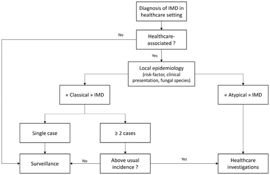
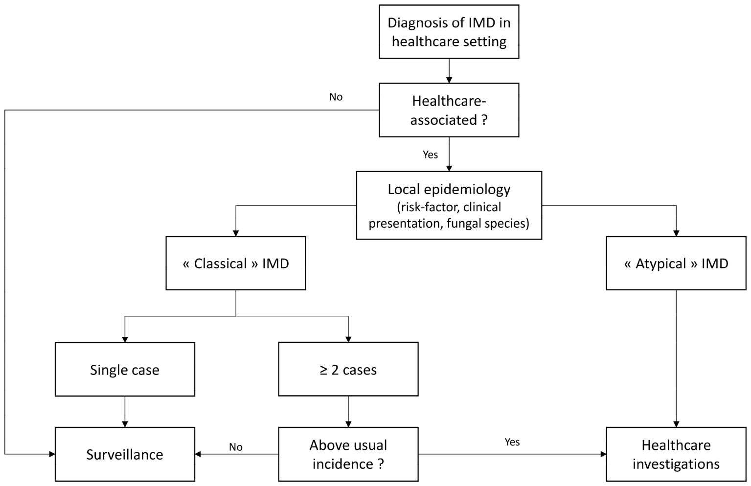
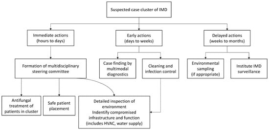
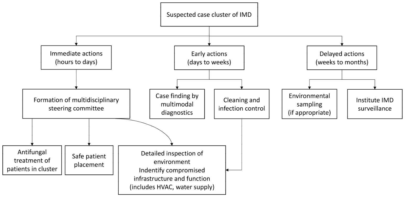

Disclaimer/Publisher’s Note: The statements, opinions and data contained in all publications are solely those of the individual author(s) and contributor(s) and not of MDPI and/or the editor(s). MDPI and/or the editor(s) disclaim responsibility for any injury to people or property resulting from any ideas, methods, instructions or products referred to in the content. |
© 2026 by the authors. Licensee MDPI, Basel, Switzerland. This article is an open access article distributed under the terms and conditions of the Creative Commons Attribution (CC BY) license.