


Abstract
Minimal access cardiac surgery (MACS) can mitigate the increasing risk profile of cardiac surgery patients and is associated with improved postoperative outcomes. One of the ways to manage the steep learning curve of MACS is the use of surgical simulation training. We conducted a narrative review to identify the relevant literature discussing MACS simulation training. We identified 20 studies using our search strategy. Various platforms were represented: high-fidelity (n = 8), low-fidelity (n = 6), and animal studies (n = 6). Virtual reality (VR) appeared in two wet-lab studies as an adjunct. The surgical approach was video-assisted thoracoscopic surgery (VATS) in 11 and robotic-assisted thoracoscopic surgery (RATS) in nine. The most simulated procedure was minimal access mitral valve (MV) repair (n = 16). Most studies (n = 16) evaluated the impact of simulation training on the surgical skill of participants with varying baseline MACS experience. A small proportion of included studies (n = 4) carried out only fidelity testing. While some standardised assessment tools were used, there was considerable variation in how surgical skill and fidelity were assessed. There are an increasing number of publications on MACS simulation training, with equal focus on bench and animal models. MV procedures were the most simulated, suggesting a drive towards increasing the scope of minimal access MV training.
1. Introduction
Despite the increasing risk profile of patients, outcomes following cardiac surgery continue to improve worldwide. This is related to improvements in preoperative optimisation, surgical practice, and perioperative management. As has been seen for all surgical specialties, there has been a move towards more minimal access approaches to undertaking cardiac surgery—with both video-assisted (VATS) and robotic (RATS) techniques being increasingly adopted as an alternative to the traditional median sternotomy. The reported advantages of these techniques include reduced surgical trauma and postoperative pain, lower infection risk, superior cosmesis, and possibly, reduced length of stay [1]. These factors are contributing to increasing patient and referring physician demand for minimal access cardiac surgery (MACS) [2].
As has been seen in other surgical specialties transitioning to minimal access techniques, there is a steep learning curve, and new skills must be learnt by surgeons used to operating open. There are different skills required by surgeons performing VATS procedures. This is due to having a restricted two-dimensional field of view, reduced tactile sensation and feedback, and requiring familiarity with long-shafted, straight instruments. For RATS, there are again additional skills required.
For various reasons, including reduced resident doctor working hours, simulation has become an integral part of surgical training, allowing surgeons and surgical trainees to develop surgical skills outside of the operating theatre. There are many advantages of this approach to surgical training, including the opportunity to acquire surgical dexterity in safe environments, minimising potential risk to patients. In addition, there is opportunity to make objective assessment and gain individualised feedback of simulated surgical skills.
Over recent years simulators of various fidelity have been developed in open cardiac surgery, from simple coronary anastomosis simulators using rubber tubes, through to live operating on porcine models [3,4]. Fidelity here refers to the degree to which the simulator and surrounding environment resemble operating room conditions, and it can be broadly split into high-fidelity simulation (HFS) and low-fidelity simulation (LFS). HFS may be perceived by some to be superior to LFS due to its closer resemblance to reality in terms of anatomical accuracy, as well as how real it feels to use, i.e., psychological fidelity.
With the increasing trend towards minimal access techniques, a demand has developed for MACS simulators. In this review, we aim to describe the current scope of simulation training in robotic-assisted and video-assisted MACS through a discussion of the range of simulation models and assessment tools reported in the literature, also considering trainee perspectives.
2. Materials and Methods
A narrative review was conducted. We searched PubMed, Embase, and Web of Science using MESH terms and keywords. Full search criteria is provided in Supplementary Tables S1–S3. Studied discussing VATS or RATS simulation in adult cardiac surgery for training purposes were included. Full exclusion criteria are provided in the adapted PRISMA diagram in Supplementary Figure S1. Articles were limited to those in the English language and published between 1995 and 2026. We identified 20 studies using the search strategy. Table 1 and Table 2 summarise the findings of studies simulating VATS and RATS procedures, respectively. The details of included models, including materials used, assessment tools, and simulation cost, is provided in this section to supplement the methodology.
Table 1.
The literature on simulation of video-assisted cardiac surgery.
Table 2.
The literature on simulation of robot-assisted cardiac surgery.
2.1. Simulation Materials
Many HFSs are 3D-printed from CT-derived anonymised patient data. The use of 3D models within MACS allows a tactile perception of complex pathologies which is not possible through computerised 3D reconstructions, with higher anatomical accuracy than is permitted by LFS [25]. The use of CT-derived data also permits simulation of patient-specific anatomy, which can be valuable in the preoperative planning of complex procedures. While the one-off cost of printers can be quite high, printing materials can be relatively cheap. Polyvinyl alcohol (PVA) is often used with injection moulding due to being water-resistant and readily available. Unlike silicone, it does not require specialist printing equipment. However, silicone can offer more realistic tensile strength and elasticity than PVA where cardiac structures are concerned [26]. In the included studies, the 3D printing approach was used primarily for simulating mitral valve (MV) procedures [7,9,10,21,22,24].
In contrast, the LFS models evaluated in Table 1 and Table 2 used commonly found household items such as baby bottles and associated feeding teats [13], as well as Lego® (Billund, Denmark) classic blocks and Lego® Technic™ parts (The LEGO Group), and kitchen sponges [13]. Verberkmoes and Verberkmoes-Broeders [5] evaluated their baby bottle model for video-assisted MV and tricuspid valve surgery, which cost $9 per simulator. The baby bottle acted as a holder for the MV structures, while the feeding teat represented the annulus and subvalvular apparatuses; a dental dam (commonly used in endodontic surgery) was used to simulate the leaflets of the MV. The simulator’s fidelity was validated by comparing the suture tension and tactile response to those of the human MV, which were found to be adequately similar.
2.2. Assessment Tools
Simulation in MACS needs to be paired with fidelity testing and standardised tools to assess surgical performance. Table 3 summarises commonly used tools within the included literature to assess technical skill and perceived competence, as well as the structural fidelity of simulators.
Table 3.
Prevalent assessment tools for minimal access cardiac surgery simulation.
The GEARS scale was created by Goh et al. [27], who adapted the Global Assessment of Laparoscopic Skills (GOALS) scale [28] for assessment of elements of robotic surgery. GEARS has six elements, including depth perception, bimanual skill, efficiency, force control, autonomy, and robot control. All of these elements, except depth perception, are reported to differentiate between individuals based on their level of expertise (e.g., consultants and trainees) [29]. The 3D vision afforded by modern robotic systems may compensate for the less experienced depth perception of trainees, therefore leading to similar scores between trainees and consultants. This led to the adaptation of GEARS into the modified GEARS (mGEARS), which excludes depth perception as a parameter. The included studies used both GEARS and mGEARS in conjunction with animal and virtual reality (VR) models [16,17,18], suggesting the adaptability of both scales to various simulation platforms.
2.3. Simulation Cost
The included studies highlight a lack of economic transparency in MACS simulation literature. While the approximate cost per-cadaver during wet-lab sessions is a well-known premium, the same transparency is not afforded to bench models or VR interfaces. The cost of well-known minimal access cardiac surgery platforms such as the Simurghy trainer, for example, is not available to the public. From the included literature, seven explicitly stated the cost of their simulation platforms (Table 4).
Table 4.
Studies with inclusion of simulator costs.
3. Results and Synthesis
3.1. Results Summary
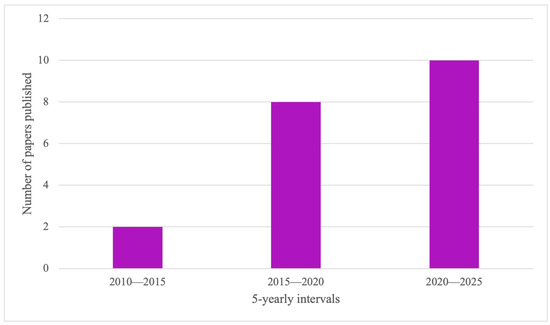
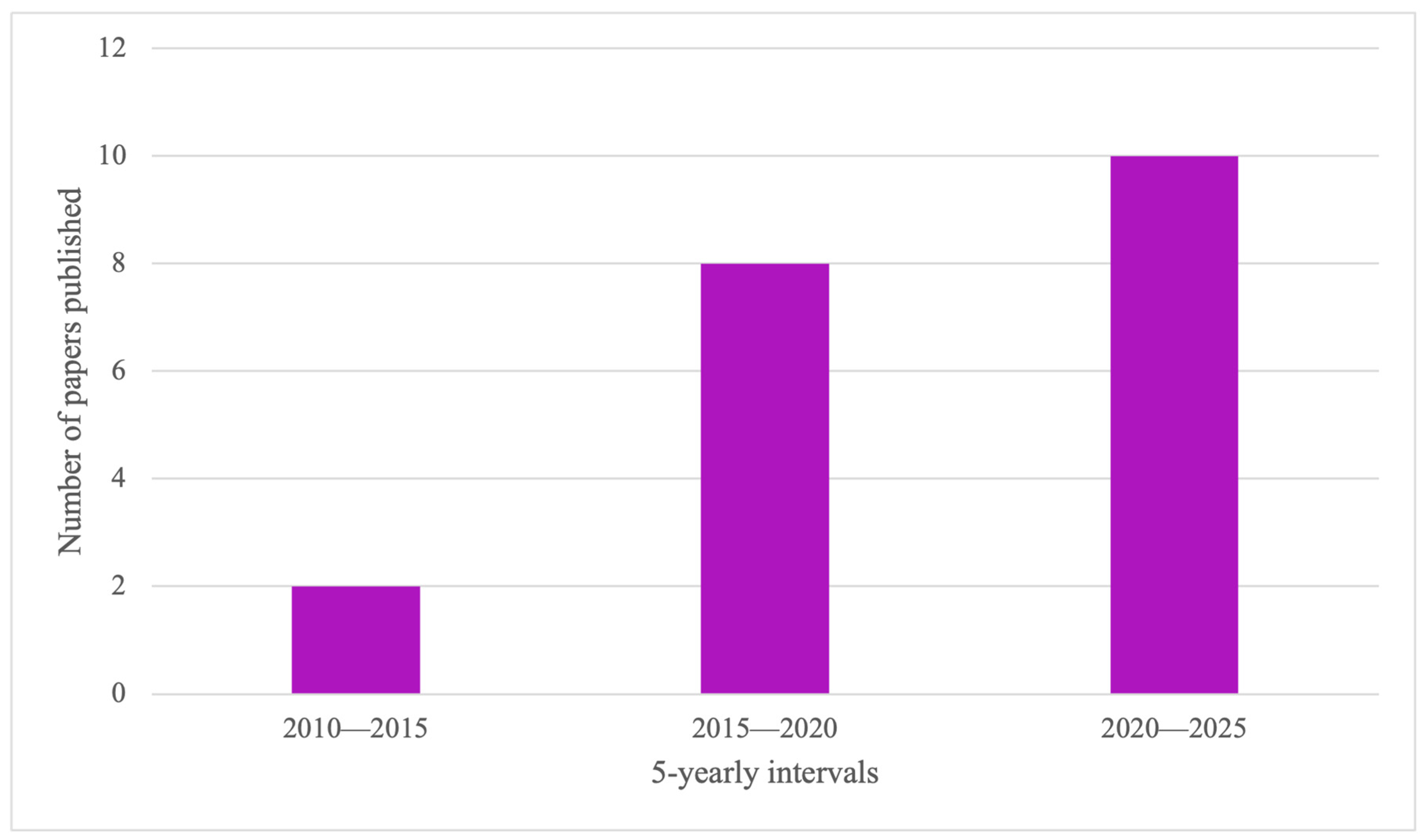
We identified 20 studies using the search strategy. Table 1 and Table 2 summarise the findings of studies simulating VATS and RATS procedures, respectively. Various simulation platforms were represented. HFS was used in eight studies, while LFS and animal models (porcine and calf) both appeared in six studies each. VR was used as an adjunct to wet labs in two studies. There were eleven studies simulating VATS procedures, and nine simulating RATS. Valvular procedures were the most simulated, with 16 studies simulating some aspect of MV replacement/repair. The number of papers published on VATS and RATS simulation training in adult cardiac surgery appears to be rising in recent years as per Figure 1.
Figure 1.
Graph depicting the number of studies published in minimal access simulation training (VATS and RATS) in adult cardiac surgery in the recent years.
3.2. Surgical Procedure Frequency
The representation of MV procedures in the literature may arise from a combination of procedural difficulty (MV procedures may often involve intricate repair rather than total replacement [30]) and the higher volume of minimally invasive MV surgery (MIMVS) performed compared to other valves including aortic.
The technicalities of minimal access MV repair include, but are not limited to, maintaining a mini-thoracotomy incision of less than 5 cm, as well as the technique of aortic occlusion within limited space. The success of surgical outcomes of minimal access MV procedures is reported to directly correlate with hospital volume of minimal access MV cases [31]. Cases with complex anatomical pathologies are often deemed high risk and referred to experienced centres.
This creates a vacuum of opportunities for trainees at lower-experience centres to increase their exposure to the true breadth of minimal access MV surgery. The frequency of simulated minimal access MV operations in the literature may be reflective of the high demand for out-of-theatre training opportunities.
The British and Irish Society for Minimally Invasive Cardiac Surgery (BISMICS) consensus statement on minimal access MV training in the UK states that compared to sternotomy, patients undergoing minimal access MV surgery experience lower wound sepsis and postoperative bleeding. This is paired with higher patient satisfaction related to a quicker return to activities of daily living due to a shorter postoperative recovery period [32]. BISMICS recommend team-based simulation and wet-lab sessions to increase trainee familiarity with long-shafted instruments.
This can increase suture accuracy and, therefore, decrease the time taken for procedures such as ring annuloplasty and leaflet plasty [7]. Several studies in the literature showed that both time and accuracy in performing MV tasks could be significantly improved with both LFS and HFS [7,8,18,20].
Robotic internal mammary artery (IMA) harvest was also commonly represented (17–20). The prevalence of robotic IMA harvest in the literature may reflect increased training in minimal access techniques for long-established procedures such as CABG.
3.3. Levels of Simulators
Simulation can be categorised based on fidelity and biological format/platform [33]. The fidelity of surgical simulation (low or high) is often correlated with its closeness to reality. LFSs may be classed as those allowing simulation of only simple skills, while HFSs allow a more realistic simulation of an operation from start to finish. This approach to fidelity considers only physical fidelity, however. Fidelity, as a whole, also concerns subjective operator-dependant measures such as how realistic a simulator feels to use: psychological fidelity [34].
While none of the included studies directly compared HFS and LFS, it is generally agreed that an increasing level of fidelity is associated with increasing cost but not necessarily with superior training value [35,36]. The ideal simulator should strike the perfect balance between keeping costs low and training value high [34].
Simulation may utilise biological or non-biological platforms. Biological platforms include animal models and human cadavers, while non-biological formats include bench models and VR simulators. While human cadaveric training is considered the gold standard simulation technique for surgical training [37], facility hire and cost per cadaver limit its availability. These barriers, along with ethical considerations, make it extremely rare for this training to be at the disposal of cardiac surgery trainees worldwide. This may, in the near future, encourage a shift away from cadaveric training as the gold standard and towards integrated surgical training programs which more readily utilise various types of simulation.
Of the animal models appearing in the literature, models of porcine origin were the most common [16,17,18,19,23], while one study utilised a foetal bovine calf model [20]. The wider literature does not explicitly compare bovine and porcine models for simulation. Most comparisons made between bovine and porcine hearts relate to their performance as bioprosthetic valves [38,39] which is outside the remit of this review.
Animal models significantly reduced the sequential operating times of robotic IMA harvest in a calf model in a study carried out by Ashraf et al. [20]. When comparing animal models to other simulation platforms, in one randomised controlled trial (RCT) comparing VR and porcine models for IMA robotic harvesting and MV annuloplasty, trainees were randomised to one of the following simulation environments: wet lab (using a porcine model), dry lab, or VR [17].
The wet-lab group showed the greatest improvement in time-based scoring (TBS) assessment. Although the VR group did meet the levels of proficiency set by the study experts for TBS and GEARS, this was not as high as the wet-lab group. In another RCT by the same group, candidates assigned to practice with VR before assessment of IMA harvest and MV annuloplasty on a porcine model performed significantly better than the control group who did not undergo VR training beforehand [16].
VR as an adjunctive platform may ensure that trainees can make the most of expendable wet-lab materials by familiarising themselves with the procedure beforehand. VR allows for repeated practice of focused skills without using up consumables (as with simulators) or altering the surgical anatomy of the simulation field (as in wet labs), allowing all participating trainees to have an equal learning experience.
In a multi-centre evaluation of U.K.-based minimal access cardiac surgery courses by Moorjani et al. [40], the courses with access to the live operating simulation models rated highest in scoring for content and materials as well as for organisation and facilities.
High-fidelity bench models were often used in the literature for minimal access MV surgery procedures [6,8,9,10,11]. As discussed in the previous section, the prevalence of such models for minimal access MV surgery may arise from the lack of trainee access to MV cases [41].
HFS improved the time taken and accuracy of annular sutures placed with long-shafted instruments in two proof-of-concept studies [8,10], and the speed of knot tying in another [15]. In a study by Nia et al. [8], consultants and residents improved their time and anatomic accuracy in placing anterior and posterior annular sutures using long-shafted instruments in a model thoracic torso for MIMVS. In a study using a 3D-printed telesimulator including a printed valvular apparatus and left ventricle, Cheheili Sobbi et al. [10], demonstrated that 100% of their MACS experts and trainees could place an anatomically correct suture post-simulation training compared to only 48% before, with a significant improvement in the time taken to place sutures as well.
In another study by Sobbi et al. [9], HFS improved the self-reported confidence levels of both trainees and consultants in suture placement, valve implantation, and suture knotting using a knot pusher. A group of 25 medical students in Wang et al.’s [11] study also experienced significant improvement in neochorda implantation and annuloplasty sutures and demonstrated shorter duration of knot tying after HFS training, suggesting the educational value of simulation across all training levels.
3.4. Trainee Perception
In a country-wide survey of surgeons and trainees at 12 institutions in Canada, a questionnaire was aimed at unveiling trainee perceptions and confidence levels when it came to simulation training [42]. The survey saw that the majority of participants indicated a desire for expanded simulation training at their centres [42]. There was a prevalent belief that being able to practice the required skills before operating would improve theatre confidence in residents. Similarly, seniors indicated they would feel more comfortable with allowing residents to operate on patients if they knew residents had undergone simulation training beforehand. This suggests that simulation training may act as a gateway towards theatre opportunities for residents, not only through improvements in their own confidence but also through that of their seniors’ in them. Using an LFS for MV and aortic valve (AV) replacement in a study by En Yean et al. [12] and a calf model for IMA harvest in a study by Ashraf et al. [20], participants experienced significantly increased scores on SSES and PCS scales following simulator training.
4. Discussion
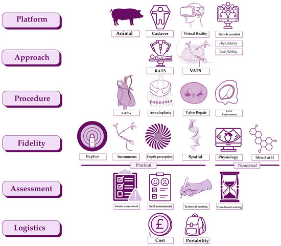
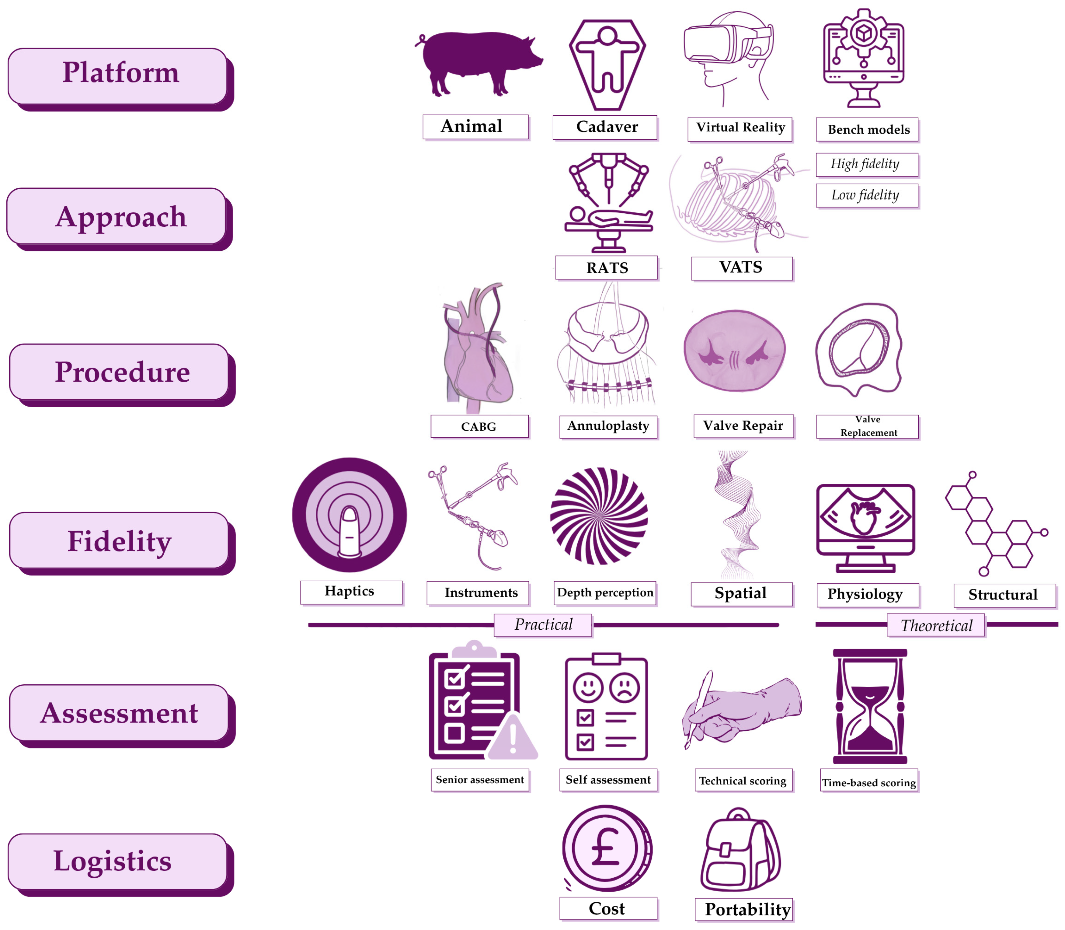
Simulation training in MACS provides budding surgeons with a safe environment to practice requisite skills. The ideal simulation platform mirrors the real-life operating environment while allowing repeated practice of both focused skills and whole procedures under expert guidance. The included literature evaluates VATS and RATS simulation platforms for valvular and coronary procedures [43,44]. The methods used include assessment of surgical performance and isolated fidelity testing. The various components of simulation training in VATS and RATS adult cardiac surgery are summarised in Figure 2.
Figure 2.
Concept map for simulation training in minimal access adult cardiac surgery. Our review finds simulation training in minimal access cardiac surgery training to be defined by six major components. These are: simulation platform i.e., animal or cadaveric; surgical approach i.e., robotic or video-assisted; type of procedure simulated; fidelity of simulation; assessment tools used to evaluate simulation users; as well as the logistics of both local and wide-spread implementation of the simulation in question as a training tool. The fidelity of the platform is a multifactorial property which is defined by haptics, the usability of long-shafted instruments within the simulation field, depth perception, the realism of spatial limitations to those in theatre, as well as the structural and physiological resemblance of the simulator to real-life. Figure created with Canva Visual Suite 2.0 and Procreate for iPad version 5.4.8 (Savage Interactive Pty Ltd., Tasmania, Auatralia). VATS, video-assisted thoracoscopic surgery; RATS, robotic-assisted thoracoscopic surgery; CABG, coronary artery bypass grafting.
The main limitation of the included papers is the heterogeneity of assessment tools. used. For example, both GEARS and mGEARS do not include any time-based parameters. Cross-clamp and bypass times are important in contributing to cardiac surgery outcomes, and therefore, time does matter in cardiac surgery. Atroshchenko et al. [18] carried out a multicentre trial using porcine simulation where both trainees and experienced participants carried out left atrial (LA) closure, IMA harvest, and MV annular suturing. They found that mGEARS and TBS assessments could both discriminate between different levels of competence in LA closure and MV annular suturing but not IMA harvest.
This raises an important point: technical scoring systems may not bear the same utility in evaluating all surgical tasks. On two separate occasions, Valdis et al. [16,17] used GEARS to evaluate the impact of simulation raining on IMA dissection skill in trainees and found a significant improvement in the GEARS scores of participants who underwent adjunctive VR and wet-lab training prior to assessment.
In contrast, Atroshchenko et al. [18] suggest that there was insufficient validity evidence to support the assessment of IMA harvest using GEARS in their study. This cannot be attributed solely to GEARS, and the simulation platform used may have a part to play in the contrasting results of Valdis et al. [16,17] and Atroshchenko et al. [18].
One solution could be the pre-validation of scoring systems for the specific task being assessed. This could be achieved by comparing the baseline scores of consultants and trainees to validate the efficacy of the score in discriminating competence levels in that task.
Some studies paired technical scoring scales, such as the Objective Structured Assessment of Technical Skills (OSAT), with psychological scales such as the perceived competence scale (PCS) or surgical self-efficacy scale (SEES) [12]. While self-evaluation may not be as reliable as senior evaluation, such hybrid assessment tools allow for the consideration of trainee perspectives. This may shed light on the perceived educational value of these platforms by those who are most likely to utilise and benefit from them.
In studies with structural data as their primary outcome, there was less consistency in the type of scoring tools used, particularly where efficacy testing of bench models was concerned [5,21,22]. As many of these bench models are newly designed, authors often used customised scales to report on components which were relevant to the model being studied.
The studies with structural data as their primary outcome all concerned the simulation of mitral or other valvular procedures. While there was no one standardised scoring system, commonly assessed structural properties were rupture strength (relating to the chorda tendinae) or elastic modulus (e.g., relating to the leaflets) [21,22] as relating to the real value of these measures in the MV in vivo. Karl et al., 2024 [24] also investigated the ability of their dynamic high-fidelity simulator to mimic real-world resolution of regurgitant fractions and volumes following MV repair.
5. Future Directions
The scope of the discussed literature indicates several avenues for future work. It is unclear if confidence levels and self- or senior-evaluated technical scores correlate with the perceived realism of simulators (namely bench models). Such analyses would provide more insight into apt directions for achieving the perfect balance between confidence-inspiring realism and cost, which is often cited as the main barrier to HFS [29,34].
Trainee perception of HFS as superior to other learning strategies due to its novelty may lead to the false deduction that they gain more from HFS than other types of simulation, e.g., LFS, leading them to overestimate their abilities [45]. Therefore, it is essential that levels of perceived realism, confidence level, and skill are interpreted in relation to each other to achieve an ideal simulation which is equally economical and educational [43]. Addressing the barrier of economic transparency is an important step in this.
The reported scales for perceived confidence and skill related to simulation training in minimal access cardiac surgery need to be standardised. One group who may bring this to realisation are the expert workforces of MACS societies, both national and international. Published expert consensus on the appropriate scales for simulation research could have considerable utility in standardising the research in this field for analysis. While simulation platforms are not yet the norm in medical and surgical education, increased interest in simulation training is evidenced by the increase in publications in recent years. Their widespread potential is especially topical given the increasing restrictions and ethical concerns surrounding the use of animal tissue in scientific research [46,47].
6. Conclusions
Our review finds a steady increase in publications relating to minimal access cardiac surgery in the past decade, with increasing awareness of novel simulation platforms, including VR and HFS bench models. These models appear to predominantly pursue the simulation of minimal access MV surgery, which may reflect the increasing number of MIMVS cases being performed and a push towards increasing training scope in MIMVS by recent national consensus statements. Technologically innovative platforms are not yet equivalent to wet labs or cadaveric simulation both with regards to perceived utility and conceptual realism. However, using technology as an adjunct may optimise both the time and cost related to delivering gold-standard cadaveric training. With time, increased user familiarity with and economic tailoring of these more novel platforms to surgical need may transform the status quo of how we deliver MICS training: to maximise trainee educational benefit and patient safety.
Supplementary Materials
The following supporting information can be downloaded at: https://www.mdpi.com/article/10.3390/jcdd13050180/s1, Figure S1: Adapted PRISMA diagram showing inclusion and exclusion criteria for article selection during screening process; Table S1: PubMed search strategy <1995 to 1 February 2026>; Table S2: Embase search strategy <1995–2026>; Table S3: Web of Science search strategy <1995–2026>.
Author Contributions
Conceptualization, J.M.A.; methodology, F.H.N. and J.M.A.; validation, F.H.N. and J.M.A.; formal analysis, F.H.N. and J.M.A.; investigation, F.H.N. and J.M.A.; resources, F.H.N. and J.M.A.; data curation, F.H.N. and J.M.A.; writing—original draft preparation, F.H.N. and J.M.A.; writing—review and editing, F.H.N. and J.M.A.; visualization, F.H.N. and J.M.A.; supervision, J.M.A.; project administration, F.H.N. and J.M.A. All authors have read and agreed to the published version of the manuscript.
Funding
This research received no external funding.
Institutional Review Board Statement
Ethical review and approval were waived for this study due to this being a review article.
Informed Consent Statement
Not applicable.
Data Availability Statement
No new data were created or analysed in this study. Data sharing is not applicable to this article.
Acknowledgments
No further acknowledgements.
Conflicts of Interest
The authors declare no conflicts of interest.
Abbreviations
The following abbreviations are used in this manuscript:
| AV | Aortic valve |
| BISMICS | British and Irish Society for Minimally Invasive Cardiac Surgery |
| CABG | Coronary artery bypass grafting |
| CO | Cardiac output |
| GEARS | Global Evaluative Assessment of Robotic Skill |
| HFS | High-fidelity simulation |
| IMA | Internal mammary artery |
| LFS | Low-fidelity simulation |
| LVAD | Left-ventricular assist device |
| MACS | Minimal access cardiac surgery |
| mGEARS | Modified Global Evaluative Assessment of Robotic Skill |
| MICA | Minimally invasive coronary anastomosis |
| MICS | Minimally invasive cardiac surgery |
| MIDCAB | Minimally invasive direct coronary artery bypass |
| MIMVS | Minimally invasive mitral valve repair |
| MV | Mitral valve |
| MVR | Mitral valve repair |
| OPCAB | Off-pump coronary artery bypass |
| OSAT | Objective Structured Assessment of Technical Skills |
| PCS | Perceived competency scale |
| PVA | Polyvinyl alcohol |
| RATS | Robotic-assisted thoracoscopic surgery |
| RF | Regurgitant fraction |
| RVOL | Regurgitant volume |
| RCT | Randomised controlled trial |
| SSES | Surgical self-efficacy scale |
| SVP | Stroke volume set by pump |
| TBS | Time-based score |
| TECAB | Totally endoscopic coronary artery bypass |
| TV | Tricuspid valve |
| VATS | Video-assisted thoracoscopic surgery |
| VR | Virtual reality |
References
- Ilcheva, L.; Risteski, P.; Tudorache, I.; Häussler, A.; Papadopoulos, N.; Odavic, D.; Biefer, H.R.C.; Dzemali, O. Beyond Conventional Operations: Embracing the Era of Contemporary Minimally Invasive Cardiac Surgery. J. Clin. Med. 2023, 12, 7210. [Google Scholar] [CrossRef] [Scilit] [PubMed]
- Kirmani, B.; Knowles, A.; Saravanan, P.; Zacharias, J. Establishing minimally invasive cardiac surgery in a low-volume mitral surgery centre. Ind. Mark. Manag. 2021, 103, 444–451. [Google Scholar] [CrossRef] [Scilit]
- Ribeiro, I.B.; Ngu, J.M.; Lam, B.-K.; Edwards, R.A. Simulation-Based Skill Training for Trainees in Cardiac Surgery: A Systematic Review. Ann. Thorac. Surg. 2018, 105, 972–982. [Google Scholar] [CrossRef] [Scilit]
- Rad, A.A.; Hajzamani, D.; Nia, P.S. Simulation-based training in cardiac surgery: A systematic review. Interdiscip. Cardiovasc. Thorac. Surg. 2023, 37, ivad079. [Google Scholar] [CrossRef] [Scilit]
- Verberkmoes, N.J.; Verberkmoes-Broeders, E.M. A novel low-fidelity simulator for both mitral valve and tricuspid valve surgery: The surgical skills trainer for classic open and minimally invasive techniques†. Interact. Cardiovasc. Thorac. Surg. 2013, 16, 97–101. [Google Scholar] [CrossRef] [Scilit]
- Nia, P.S.; Daemen, J.H.; Maessen, J.G. Development of a high-fidelity minimally invasive mitral valve surgery simulator. J. Thorac. Cardiovasc. Surg. 2019, 157, 1567–1574. [Google Scholar] [CrossRef] [Scilit]
- Jebran, A.-F.; Saha, S.; Waezi, N.; Al-Ahmad, A.; Niehaus, H.; Danner, B.C.; Baraki, H.; Kutschka, I. Design and training effects of a physical reality simulator for minimally invasive mitral valve surgery. In Proceedings of the 48th Annual Meeting of the German-Society-of-Thoracic-and-Cardiovascular-Surgery, Wiesbaden, Germany, 16–19 February 2019; pp. 409–415. [Google Scholar]
- Nia, P.S.; Heuts, S.; Daemen, J.H.T.; Olsthoorn, J.R.; Chitwood, W.R.; Maessen, J.G. The EACTS simulation-based training course for endoscopic mitral valve repair: An air-pilot training concept in action. Interact. Cardiovasc. Thorac. Surg. 2020, 30, 691–698. [Google Scholar] [CrossRef] [Scilit]
- Sobbi, S.C.; Jung, Y.; Fillet, M.; Bakhtiary, F.; Maessen, J.G.; Nia, P.S. Simulation-based training for endoscopic mitral valve repair: The impact on basic surgical skills for placement of sutures at mitral valve annulus during 2-h training workshop. Interdiscip. Cardiovasc. Thorac. Surg. 2024, 38, ivae003. [Google Scholar] [CrossRef] [Scilit]
- Sobbi, S.C.; Hamid, U.I.; Rad, A.A.; Fillet, M.; Maesen, J.; Nia, P.S. Telesimulation Training for Endoscopic Mitral Valve Surgery: An Air-Pilot Training Concept for Distance Training. Innov. Technol. Tech. Cardiothorac. Vasc. Surg. 2024, 19, 169–174. [Google Scholar] [CrossRef] [Scilit] [PubMed]
- Wang, C.; Karl, R.; Sharan, L.; Grizelj, A.; Fischer, S.; Karck, M.; De Simone, R.; Romano, G.; Engelhardt, S. Surgical training of minimally invasive mitral valve repair on a patient-specific simulator improves surgical skills. Eur. J. Cardio-Thoracic Surg. 2024, 65, ezad387. [Google Scholar] [CrossRef] [Scilit] [PubMed]
- Yean, L.E.; bin Hashim, S.A. Role of surgical simulation on self-reported confidence level on cardiothoracic surgical trainees. J. Cardiothorac. Surg. 2024, 19, 293. [Google Scholar] [CrossRef] [Scilit] [PubMed]
- Azmi, M.I.; Nair, A.K.; Hashim, S.A. Self-Practice Program for Beating-Heart Minimally Invasive Coronary Anastomosis Using a Homemade Low-Fidelity Simulator: A Proof of Concept. Innov. Technol. Tech. Cardiothorac. Vasc. Surg. 2024, 19, 175–183. [Google Scholar] [CrossRef] [Scilit] [PubMed]
- Ujihira, K.; Yamada, A. Novel Dry-Lab Training Method for Totally Endoscopic Coronary Anastomosis. Innov. Technol. Tech. Cardiothorac. Vasc. Surg. 2017, 12, 363–369. [Google Scholar] [CrossRef] [Scilit]
- Engelhardt, S.; Sauerzapf, S.; Brčić, A.; Karck, M.; Wolf, I.; De Simone, R. Replicated mitral valve models from real patients offer training opportunities for minimally invasive mitral valve repair. In Proceedings of the 32nd Annual Meeting of the European-Association-for-Cardio-Thoracic-Surgery, Milan, Italy, 18–20 October 2018; pp. 43–50. [Google Scholar]
- Valdis, M.; Chu, M.W.A.; Schlachta, C.M.; Kiaii, B. Validation of a Novel Virtual Reality Training Curriculum for Robotic Cardiac Surgery a Randomized Trial. Innov. Technol. Tech. Cardiothorac. Vasc. Surg. 2015, 10, 383–388. [Google Scholar] [CrossRef] [Scilit]
- Valdis, M.; Chu, M.W.; Schlachta, C.; Kiaii, B. Evaluation of robotic cardiac surgery simulation training: A randomized controlled trial. J. Thorac. Cardiovasc. Surg. 2016, 151, 1498–1505.e2. [Google Scholar] [CrossRef] [Scilit]
- Atroshchenko, G.V.; Navarra, E.; Valdis, M.; Sandoval, E.; Hashemi, N.; Cerny, S.; Pereda, D.; Palmen, M.; Bjerrum, F.; Bruun, N.H.; et al. Simulation-based assessment of robotic cardiac surgery skills: An international multicenter, cross-specialty trial. JTCVS Open 2023, 16, 619–627. [Google Scholar] [CrossRef] [Scilit] [PubMed]
- Atroshchenko, G.V.; Navarra, E.; Valdis, M.; Sandoval, E.; Hashemi, N.; Cerny, S.; Pereda, D.; Palmen, M.; Bjerrum, F.; Bruun, N.H.; et al. Examining the learning curves in robotic cardiac surgery wet lab simulation training. Interdiscip. Cardiovasc. Thorac. Surg. 2024, 40, ivae227. [Google Scholar] [CrossRef] [Scilit] [PubMed]
- Ashraf, S.F.; Seese, L.; Hasan, I.S.; Babu, A.N.; Balkhy, H.H.; Kiaii, B.B.; Guy, T.S.; Kaczorowski, D.J.; Bonatti, J. Development and Validation of a Low-Cost, High-Fidelity Simulation Model for Robotic Internal Mammary Artery Harvest Using the da Vinci Xi Robot. Innov. Technol. Tech. Cardiothorac. Vasc. Surg. 2024, 19, 633–639. [Google Scholar] [CrossRef] [Scilit]
- Yamada, T.; Osaka, M.; Uchimuro, T.; Yoon, R.; Morikawa, T.; Sugimoto, M.; Suda, H.; Shimizu, H. Three-Dimensional Printing of Life-Like Models for Simulation and Training of Minimally Invasive Cardiac Surgery. Innov. Technol. Tech. Cardiothorac. Vasc. Surg. 2017, 12, 459–465. [Google Scholar] [CrossRef] [Scilit]
- Premyodhin, N.; Mandair, D.; Ferng, A.S.; Leach, T.S.; Palsma, R.P.; Albanna, M.Z.; I Khalpey, Z. 3D printed mitral valve models: Affordable simulation for robotic mitral valve repair. Interact. Cardiovasc. Thorac. Surg. 2017, 26, 71–76. [Google Scholar] [CrossRef] [Scilit]
- AlJamal, Y.N.; Burgin, R.; Kitahara, H.; Gonzalez, G.; Balkhy, H.H. Inexpensive and Easy to Set Up Robotic Cardiac Simulator Offers “Unlimited” Endoscopic Coronary Artery Bypass Grafting Experience: Proof of Concept. Innov. Technol. Tech. Cardiothorac. Vasc. Surg. 2023, 18, 419–423. [Google Scholar] [CrossRef] [Scilit]
- Karl, R.; Romano, G.; Marx, J.; Eden, M.; Schlegel, P.; Stroh, L.; Fischer, S.; Hehl, M.; Kühle, R.; Mohl, L.; et al. An ex-vivo and in-vitro dynamic simulator for surgical and transcatheter mitral valve interventions. Int. J. Comput. Assist. Radiol. Surg. 2024, 19, 411–421. [Google Scholar] [CrossRef] [Scilit]
- Yoo, S.J.; Hussein, N.; Peel, B.; Coles, J.; van Arsdell, G.S.; Honjo, O.; Haller, C.; Lam, C.Z.; Seed, M.; Barron, D. 3D Modeling and Printing in Congenital Heart Surgery: Entering the Stage of Maturation. Front. Pediatr. 2021, 9, 621672. [Google Scholar] [CrossRef] [Scilit]
- Shabbak, A.; Masoumkhani, F.; Fallah, A.; Amani-Beni, R.; Mohammadpour, H.; Shahbazi, T.; Bakhshi, A. 3D Printing for Cardiovascular Surgery and Intervention: A Review Article. Curr. Probl. Cardiol. 2024, 49, 102086. [Google Scholar] [CrossRef] [Scilit]
- Goh, A.C.; Goldfarb, D.W.; Sander, J.C.; Miles, B.J.; Dunkin, B.J. Global Evaluative Assessment of Robotic Skills: Validation of a Clinical Assessment Tool to Measure Robotic Surgical Skills. J. Urol. 2012, 187, 247–252. [Google Scholar] [CrossRef] [Scilit]
- Sánchez, R.; Rodríguez, O.; Rosciano, J.; Vegas, L.; Bond, V.; Rojas, A.; Sanchez-Ismayel, A. Robotic surgery training: Construct validity of Global Evaluative Assessment of Robotic Skills (GEARS). J. Robot. Surg. 2016, 10, 227–231. [Google Scholar] [CrossRef] [Scilit] [PubMed]
- Howard, T.; Iyengar, K.P.; Vaishya, R.; Ahluwalia, R. High-fidelity virtual reality simulation training in enhancing competency assessment in orthopaedic training. Br. J. Hosp. Med. 2023, 84, 1–8. [Google Scholar] [CrossRef] [Scilit]
- Pope, N.H.; Ailawadi, G. Minimally Invasive Valve Surgery. J. Cardiovasc. Transl. Res. 2014, 7, 387–394. [Google Scholar] [CrossRef] [Scilit] [PubMed]
- Akmaz, B.; van Kuijk, S.M.J.; Nia, P.S. Association between individual surgeon volume and outcome in mitral valve surgery: A systematic review. J. Thorac. Dis. 2021, 13, 4500–4510. [Google Scholar] [CrossRef] [Scilit] [PubMed]
- A Vohra, H.; Salmasi, M.Y.; Chien, L.; Caputo, M.; Baghai, M.; Deshpande, R.; Akowuah, E.; Ahmed, I.; Tolan, M.; Bahrami, T.; et al. BISMICS consensus statement: Implementing a safe minimally invasive mitral programme in the UK healthcare setting. Open Heart 2020, 7, e001259. [Google Scholar] [CrossRef] [Scilit]
- Badash, I.; Burtt, K.; Solorzano, C.A.; Carey, J.N. Innovations in surgery simulation: A review of past, current and future techniques. Ann. Transl. Med. 2016, 4, 453. [Google Scholar] [CrossRef] [Scilit]
- Johnson, G.G.; Brindley, P.G.; Gillman, L.M. Fidelity in surgical simulation: Further lessons from the S.T.A.R.T.T. course. Can. J. Surg. 2020, 63, E161–E166. [Google Scholar] [CrossRef] [Scilit]
- Massoth, C.; Röder, H.; Ohlenburg, H.; Hessler, M.; Zarbock, A.; Pöpping, D.M.; Wenk, M. High-fidelity is not superior to low-fidelity simulation but leads to overconfidence in medical students. BMC Med. Educ. 2019, 19, 29. [Google Scholar] [CrossRef] [Scilit] [PubMed]
- Lefor, A.K.; Harada, K.; Kawahira, H.; Mitsuishi, M. The effect of simulator fidelity on procedure skill training: A literature review. Int. J. Med. Educ. 2020, 11, 97–106. [Google Scholar] [CrossRef] [Scilit] [PubMed]
- Hughes, J.; Katsogridakis, E.; Junaid-Siddiqi, A.; Pollard, J.; Bedford, J.; Coe, P.; Jones, D. The development of cadaveric simulation for core surgical trainees. Bull. R. Coll. Surg. Engl. 2019, 101, 38–43. [Google Scholar] [CrossRef] [Scilit]
- Persson, M.; Glaser, N.; Franco-Cereceda, A.; Nilsson, J.; Holzmann, M.J.; Sartipy, U. Porcine vs Bovine Bioprosthetic Aortic Valves: Long-Term Clinical Results. Ann. Thorac. Surg. 2021, 111, 529–535. [Google Scholar] [CrossRef] [Scilit]
- Fu, B.; Liu, X.; Wei, R.; Chen, Q.; Guo, Z.; Jiang, N. Bovine pericardial versus porcine stented replacement mitral valves: Early hemodynamic performance and clinical results of a randomized comparison of the Perimount and the Mosaic valves. J. Thorac. Dis. 2021, 13, 262–269. [Google Scholar] [CrossRef] [Scilit]
- Moorjani, N.; Lewis, M.; Shah, R.; Barnard, S.; Graham, T.; Rathinam, S. Implementation of a novel portfolio of structured, curriculum-aligned, simulation-based, cardiothoracic surgery training courses: Evolving the delivery of surgical education. J. Thorac. Cardiovasc. Surg. 2017, 154, 2009–2016. [Google Scholar] [CrossRef] [Scilit]
- Aerts, L.; Nia, P.S. Mastering the learning curve of endoscopic mitral valve surgery. Front. Cardiovasc. Med. 2023, 10, 1162330. [Google Scholar] [CrossRef] [Scilit]
- White, A.; Singh, G.; Moon, M.C.; Zheng, B.; Turner, S.R. Current Status of Simulation Use in Canadian Cardiac Surgery Training Programs. Can. J. Cardiol. 2021, 37, 1639–1643. [Google Scholar] [CrossRef] [Scilit] [PubMed]
- Sadideen, H.; Hamaoui, K.; Saadeddin, M.; Kneebone, R. Simulators and the simulation environment: Getting the balance right in simulation-based surgical education. Int. J. Surg. 2012, 10, 458–462. [Google Scholar] [CrossRef] [Scilit] [PubMed]
- Hassanabad, A.F.; Hamid, U.I.; Nia, P.S. An international survey-based assessment of minimally invasive mitral valve surgery. Interdiscip. Cardiovasc. Thorac. Surg. 2023, 37, ivad154. [Google Scholar] [CrossRef] [Scilit] [PubMed]
- Carey, J.M.; Rossler, K. The How When Why of High Fidelity Simulation. In StatPearls; StatPearls Publishing: Treasure Island, FL, USA, 2025. Available online: http://www.ncbi.nlm.nih.gov/books/NBK559313 (accessed on 9 March 2026).
- Kiani, A.K.; Pheby, D.; Henehan, G.; Brown, R.; Sieving, P.; Sykora, P.; Marks, R.; Falsini, B.; Capodicasa, N.; Miertus, S.; et al. Ethical considerations regarding animal experimentation. J. Prev. Med. Hyg. 2022, 63, E255–E266. [Google Scholar] [CrossRef] [Scilit] [PubMed]
- Germani, F.; März, J.W.; Luciani, A.; Devuyst, O.; Biller-Andorno, N. Ethical and legal considerations of digital animal models: Pioneering reduction and replacement. Trends Biotechnol. 2025, 44, 593–595. [Google Scholar] [CrossRef] [Scilit]

Disclaimer/Publisher’s Note: The statements, opinions and data contained in all publications are solely those of the individual author(s) and contributor(s) and not of MDPI and/or the editor(s). MDPI and/or the editor(s) disclaim responsibility for any injury to people or property resulting from any ideas, methods, instructions or products referred to in the content. |
© 2026 by the authors. Licensee MDPI, Basel, Switzerland. This article is an open access article distributed under the terms and conditions of the Creative Commons Attribution (CC BY) license.