


Abstract
Cardiac rehabilitation (CR) is one of the most evidence-based interventions in secondary cardiovascular prevention, yet it operates under a finite-duration model that assumes patients will independently maintain exercise for life. This assumption breaks down precisely in Phase III, the unsupervised lifelong maintenance stage, where adherence collapses and the gains of supervised training erode—which is why this review focuses on it. This narrative review examines whether the molecular mechanisms of exercise-induced muscle memory, established in healthy skeletal muscle across myonuclear, epigenetic, non-coding RNA, proteomic, and immune substrates and shown to outlast the functional adaptations they support, apply to the skeletal muscle of patients with ischemic heart disease, heart failure, pulmonary arterial hypertension, and peripheral arterial disease, which presents a convergently disrupted molecular baseline compounded by ageing, inflammation, sarcopenia, and routine pharmacotherapy. The key finding is that, whereas this molecular record is well established in healthy muscle, whether it is preserved, attenuated, or abolished in cardiovascular disease remains entirely uninvestigated. We therefore propose the periodic, strategic administration of exercise, an “exercise vaccine”, as a mechanistically grounded hypothesis that could lower the biological cost of re-adaptation and redefine Phase III CR, and we outline the training–detraining–retraining studies with serial muscle biopsy needed to test it.
1. Introduction
Cardiovascular disease (CVD) remains the leading cause of mortality and disability-adjusted life years (DALYs) globally. According to Global Burden of Disease (GBD) 2023 estimates, CVD accounted for 19.2 million deaths and 437 million DALYs, a 1.4-fold rise in DALYs since 1990, driven by population growth, ageing, and metabolic risk factors [1]. This burden is unevenly distributed: age-standardized DALY rates are highest in low- and low-middle Socio-demographic Index regions [1], whereas in Europe CVD caused more than 3 million deaths and 68 million DALYs in 2023, the leading cause of death and disability in the region [2]. Socioeconomic disparities persist even within Europe [3], and the resulting economic burden represents approximately 11% of total European Union (EU) healthcare expenditure [4].
Cardiac rehabilitation (CR) is a structured, multidisciplinary intervention combining exercise training, risk-factor control, psychosocial counselling, and patient education [5], and is among the most effective strategies in secondary cardiovascular prevention, with benefits across clinical outcomes, health-related quality of life (HRQoL), and healthcare utilization [6]. In heart failure (HF), exercise-based CR reduces all-cause hospital admissions and improves HRQoL, without a clear effect on all-cause mortality [7], with benefits sustained over long-term follow-up and tied to the cumulative dose of sessions attended [8]. Supervised home-based and telehealth-delivered CR yields outcomes equivalent to center-based programmes [9,10] and can sustain physical activity (PA) while limiting detraining [11], yet this equivalence holds mainly for patients with adequate digital access and health literacy and may exclude the older and socioeconomically disadvantaged groups most affected by the access barriers noted above. Yet despite this evidence base, CR remains inaccessible to most eligible patients, and even those who complete a programme are left without a structured framework to maintain their gains, a critical gap that current cardiovascular care fails to address.
The consequences of this second gap are well documented. In patients with coronary artery disease (CAD) undergoing combined strength and aerobic training, a substantial part of the adaptations gained is lost within three months of detraining [11]; lifelong exercise is therefore required to sustain cardiovascular function and muscular strength, yet real-world adherence is poor [12]. In European cohorts only 40% of patients with CAD meet recommended PA targets [12], and adherence to structured post-rehabilitation programmes falls to approximately 54%, dropping to as low as 9% among working-age patients vs. 60% among retirees [13]. This lifelong maintenance stage, the so-called Phase III of CR, is precisely where the assumption of self-sustained exercise is made and where it breaks down: it remains comparatively understudied relative to the supervised Phases I and II, lacks a structured re-engagement framework when training lapses, and is therefore the stage at which a molecularly justified model of periodic reinforcement would be most needed.
In response to this dual failure of access and continuity, we propose the concept of periodic, structured and supervised exercise interventions, herein termed exercise vaccines (used as an operational analogy to the primary-dose-and-booster logic of vaccination, not a claim of literal immunization): time-limited interventions delivered at defined intervals after Phase II CR to periodically reactivate the molecular and physiological adaptations acquired during formal rehabilitation. The framework reconceptualizes skeletal muscle as an endocrine organ [14] and structured exercise as a systemic, pleiotropic stimulus whose effects are molecular, measurable, and reversible in its absence. It does not propose extending conventional supervised rehabilitation, but a fundamentally different delivery paradigm: periodic interventions triggered by clinical decision-making within existing cardiology follow-up, reactivating patients before they fall below the physiological threshold at which cardiovascular risk escalates (Figure 1).
Figure 1.
The current model of cardiac rehabilitation (CR) vs. the proposed “exercise vaccine” model. Left: the conventional pathway Phase I (in-hospital), Phase II (structured, supervised outpatient CR) and Phase III (autonomous maintenance), in which functional capacity (VO2peak) rises during supervised Phase II but subsequently declines below the functional risk threshold during Phase III as exercise adherence wanes. Right: the proposed model, in which periodic supervised “booster” doses of exercise during Phase III exploit the molecular memory encoded by prior training (myonuclear, epigenetic, proteomic, non-coding RNA and trained-immunity substrates) to reduce the biological cost of re-adaptation and sustain functional capacity above the risk threshold over time. Arrows indicate an increase (↑) or a decrease (↓).
Despite this established molecular basis in healthy skeletal muscle, its relevance to cardiovascular disease has not been tested. This review is accordingly framed around two linked questions. Given that the molecular substrates of exercise-induced muscle memory are established in healthy skeletal muscle, do they also operate in the pathologically altered skeletal muscle of cardiovascular patients? And, if so, could periodic and strategically timed reinforcement interventions preserve or restore these adaptations more efficiently than continuous self-directed exercise?
The aim of this review is to propose the conceptual framework of exercise vaccines as a periodic, structured exercise intervention model for cardiovascular patients beyond Phase II CR, grounded in existing clinical and molecular evidence, and to outline its potential as a basis for future research and clinical implementation.
2. Relevant Sections
Search strategy. This is a narrative, conceptually driven review and did not follow a systematic, pre-registered protocol; full PRISMA compliance was therefore not pursued. The relevant literature was identified by searching PubMed, Scopus, Web of Science, Google Scholar, and MEDLINE, with no date or language restriction (from database inception to the most recent search). The search combined MeSH and free-text terms grouped into five concept blocks: (i) muscle memory and its molecular substrates (e.g., muscle memory, myonuclear, satellite cells, DNA methylation, epigenetics, proteomics, microRNA/long non-coding RNA, trained immunity); (ii) exercise and training, including detraining and retraining (exercise, resistance training, endurance/aerobic training, high-intensity interval training, detraining, retraining); (iii) skeletal muscle; (iv) cardiovascular conditions (ischemic heart disease, myocardial infarction, coronary artery disease, heart failure, pulmonary arterial hypertension, peripheral arterial disease); and (v) cardiac rehabilitation and secondary prevention. Blocks were combined with the Boolean operators AND/OR to address the three strands of the review: the molecular evidence for muscle memory in healthy and trained muscle (blocks i–iii), skeletal-muscle alterations in cardiovascular disease (blocks iii–iv), and the cardiac-rehabilitation context (blocks ii, iv–v). Many of the included studies were retrieved through PubMed, complemented by the remaining databases and by backward and forward citation tracking of key articles. Studies were selected on the basis of relevance to the conceptual framework rather than against pre-specified quantitative eligibility criteria, prioritizing primary mechanistic studies and, where available, direct human evidence; background references (definitions, epidemiology, historical context) were included where needed to support the argument, whereas sources without relevance to skeletal muscle, to the memory substrates considered, or to the cardiovascular conditions reviewed were not included. Consistent with the aim of the review, a deliberate distinction was maintained throughout between evidence obtained in healthy or trained individuals and the absence of direct evidence in patients with cardiovascular disease.
2.1. Limitations of Current Cardiac Rehabilitation
The limitations of current CR, and of its Phase III maintenance stage, operate at three interacting levels: factors intrinsic to the patient, factors inherent to the programme model, and factors rooted in the wider health-system and socioeconomic context.
2.1.1. Participant-Level Factors
Several barriers are intrinsic to the patient. Sex is a major axis of inequity: although CVD causes more deaths in women than in men across European Society of Cardiology (ESC) member countries (1.6 vs. 1.5 million annually; 39% vs. 34% of all deaths) [2], women are 36% less likely than men to enroll in CR [15], reflecting interpersonal, logistical, programme-level and health-system barriers that converge differentially on them [16]. Age adds a further layer: advanced age and frailty interact with a pathophysiological triad of senescence, sarcopenia and cardiovascular disease, in which the age-related loss of muscle mass, worsened by myocardial ischemia, drives a catabolic state of chronic inflammation and insulin resistance that compromises functional capacity [17]; older adults are consequently referred and retained less often and may be better served by intensive inpatient formats than by extensive outpatient ones [18].
At the individual level, engagement is further shaped by time-discounting, whereby the value of a delayed reward such as future health is discounted relative to immediate costs [19]. The result is poor long-term participation: physical activity declines as early as two months after discharge, particularly in less educated or younger patients, in women, and in those with percutaneous coronary intervention, chronic heart failure or diabetes [20]; adherence to Phase III then erodes sharply, with roughly two-thirds of eligible patients in a Scottish cohort failing to initiate or sustain the programme [21], exercise showing the highest discontinuation of all CR components in a Danish cohort (13% in-hospital and a further 22% during community maintenance) [22], and about 49% of patients in the Italian ICAROS registry discontinuing prescribed exercise one year after formal CR despite high medication adherence [23].
Importantly, sustaining exercise is not synonymous with sustaining its benefits. The prevailing clinical assumption that a patient who remains active is thereby adequately treated conflates physical activity with therapeutic dose. It is the legacy of a universal, closed-number paradigm, epitomized by fixed population targets such as 150 min of weekly activity, that prescribes volume while specifying neither an intensity individualized to the patient’s current capacity, nor the progression required to keep the stimulus supra-threshold, nor the periodic reassessment needed to detect when it has ceased to be. Yet a specific threshold dose is required for clinical benefit [24], and because this dose and the patient’s response are rarely re-evaluated throughout Phase III, its trajectory remains effectively unknown [25]. Continued activity at an unmonitored, sub-threshold level can therefore coexist with progressive loss of adaptation: three months of detraining after eight months of combined training produced a 10% fall in peak oxygen uptake (VO2peak), with losses in exercise time and strength [11], a decline that is clinically meaningful given the established cardiovascular benefit of higher VO2peak [26]. This mismatch carries a hidden opportunity cost: because the patient appears active and the fixed target appears met, the model is judged to be working and the incentive to reassess or change it disappears, so that a patient counted as a therapeutic success may in fact be silently losing the very adaptations the programme was meant to preserve.
2.1.2. Programme-Level Factors
Other barriers are inherent to the programme model. Although international guidelines uniformly recommend CR for secondary prevention, only a small proportion of eligible patients use it and many of those who do fail to adhere to or complete it [27]. A central reason is structural rigidity: the traditional model, designed more than 60 years ago, is still followed by 70–80% of programmes worldwide and remains a one-size-fits-all, service-oriented scheme rather than a flexible, patient-centered one [28]. This rigidity is most evident in Phase III, which consists of regular group-based outpatient aftercare intended to maintain physical activity and reduce long-term risk [13], yet Phase III remains markedly understudied relative to Phase II, with few analyses of physical-activity patterns or adherence predictors and findings that are difficult to compare across heterogeneous programmes [25].
2.1.3. Contextual and Health-System Factors
A third set of barriers is rooted in the wider health-system and socioeconomic context. The epidemiological backdrop is an ageing European population with a high and worsening risk-factor burden, projected to drive as much as a 90% rise in CVD prevalence between 2025 and 2050, within health systems designed for acute episodic care rather than chronic lifelong management [29]. Against this backdrop, CR is structurally under-provided: it is available in only half of countries globally [30] and, even where offered, timely access is limited, with a median 42-day wait from referral to intake and each additional day independently reducing enrolment by 1% [31]. The EUROASPIRE V registry quantifies the resulting gap: fewer than half of coronary patients were advised to attend CR and only 32% ultimately enrolled, while most failed to reach blood-pressure, low-density lipoprotein cholesterol and glucose targets [32]. These deficits are reinforced by health-system economics that systematically undervalue prevention, as behavioural change and its benefits accrue slowly and seem distant to short budget horizons [33]. The neglect is economically paradoxical: a systematic review of its cost-effectiveness reported incremental cost-effectiveness ratios for exercise-based CR as low as approximately US $1065 per quality-adjusted life-year (QALY), with several analyses finding it dominant, cheaper and more effective than usual care, so that systems systematically underfund an intervention that is frequently cost-saving rather than merely cost-effective [34]. This is compounded by a systemic rule of rescue that prioritizes visible acute interventions over long-term management, pushing preventive investment into the background [35].
Taken together, these patient-, programme- and system-level barriers converge on a Phase III care model that fails patients simultaneously through inequitable access, a rigid one-size-fits-all design, and the erosion of hard-won physiological gains once supervised follow-up ends.
2.2. Cellular Mechanisms
A periodic reinforcement strategy, however, requires a durable substrate to act upon, a molecular record left by prior training that a booster could reactivate rather than rebuild, so the following sections examine the cellular basis of this muscle memory in healthy skeletal muscle before asking whether it survives in cardiovascular disease.
2.2.1. Historical Origins: From Mechanical Muscle to the Endocrine Muscle
Skeletal muscle contraction was first described as the sliding of myosin filaments over actin filaments [36,37]. However, chronic exercise induces a 2.0-fold increase in mitochondrial protein content and respiratory enzyme activity in skeletal muscle [38]. This adaptive capacity extends to fibre-type transformation, capillarisation, and oxidative-glycolytic enzyme adaptation, with adaptation magnitude depending on training volume and modality [39].
The term “myokine” was first introduced by Bengt Saltin in 2003 [40], in a study identifying interleukin-6 (IL-6) as a candidate exercise factor released by contracting skeletal muscle [41]. Skeletal muscle is now recognized as a secretory organ, releasing myokines that mediate crosstalk with adipose tissue, liver, pancreas, bone, and brain [42]. This myokine-signalling framework extends to additional organs and systems, including skin, the vascular bed, the gut, pancreatic β-cells, the immune system, and cancer [43]. Indeed, secretome analysis of human myocyte culture medium has revealed over 600 myokines mediating this crosstalk, the majority of which remain poorly understood [40]. Specific myokines, including myostatin, irisin, brain-derived neurotrophic factor (BDNF), and apelin, are implicated in the development and progression of cardiovascular disease itself, with several showing dynamic changes across stages of myocardial infarction, heart failure, atherosclerosis, hypertension, and diabetes mellitus (DM) [44].
In addition to this secretory function, a coactivator inducing mitochondrial biogenesis in response to a thermogenic stimulus was isolated in 1998 [45]. This same coactivator, peroxisome proliferator-activated receptor-γ coactivator-1α (PGC-1α), was subsequently shown to increase approximately twofold in skeletal muscle within 18 h of a single bout of exercise [46]. While PGC-1α governs oxidative adaptation, a separate pathway governs hypertrophic adaptation: the protein kinase B/mechanistic target of rapamycin (Akt/mTOR) pathway is a crucial regulator of skeletal muscle hypertrophy, capable of preventing muscle atrophy in vivo [47].
These pathways do not operate as a simple, bidirectional switch. Endurance-type stimulation activates AMP-activated protein kinase (AMPK) and reduces phosphorylation of the tuberous sclerosis complex 2 (TSC2), whereas resistance-type stimulation activates protein kinase B (PKB) and increases phosphorylation of TSC2 and mechanistic target of mTOR at PKB-sensitive sites, a pattern described as an AMPK-PKB switch [48]. The Hippo pathway also contributes to skeletal muscle mass regulation: its effector, Yes-associated protein (YAP), positively regulates basal skeletal muscle mass and protein synthesis through interaction with transcriptional enhancer factor domain 1–4 (TEAD 1–4), with YAP abundance increasing following motor nerve injury as a mechanism to mitigate neurogenic muscle atrophy [49].
On the other hand, sustained elevation of myoplasmic calcium during prolonged contraction activates calcium/calmodulin-dependent protein kinase IV (CaMKIV), which induces expression of PGC-1α and activates its gene promoter in cultured myotubes [50]. Vascular endothelial growth factor (VEGF), generated within skeletal myofibers, is required for exercise-induced capillary angiogenesis, with its targeted deletion abolishing the angiogenic response to training [51]. Exercise-induced heat and oxidative load additionally upregulate the 70-kilodalton heat shock protein (HSP70), whose expression declines with inactivity and ageing and whose loss has been implicated in muscle atrophy and impaired regenerative capacity [52]. A muscle-derived factor cleaved from fibronectin type III domain-containing protein 5 (FNDC5) under PGC-1α control acts at a distance, driving brown-fat-like remodelling of white adipose tissue [53], although whether this response occurs comparably in exercising humans remains contested [54].
Even within pathways long considered settled, the underlying biology has proven more intricate than first assumed. Sirtuin 1 (SIRT1), once proposed as the master regulator linking cellular energy status to PGC-1α activation, is dispensable for exercise-induced mitochondrial biogenesis in skeletal muscle, with regulation of the competing acetyltransferase general control non-derepressible 5 (GCN5) identified as a more plausible mechanism [55]. PGC-1α itself is not mandatory for adaptive gene responses to exercise and training in knockout mice [56]. PGC-1α and mTOR are accordingly best understood as two well-defined nodes within a considerably larger network of multiple integrated and often redundant responses that contract skeletal muscle deploys to meet the metabolic and mechanical demands of exercise [57].
Within this molecular network, skeletal muscle memory has been defined as “the capacity of skeletal muscle to respond differently to environmental stimuli in an adaptive (positive) or maladaptive (negative) manner if the stimuli have been encountered previously” [58]. A retraining advantage in previously trained individuals was first identified physiologically in the early 1990s: six weeks of retraining following 30–32 weeks of detraining produced significant increases in fibre cross-sectional area compared with detraining values in previously resistance-trained women [59]. A comparable retraining advantage was documented in men in the late 1990s, before the myokine framework and the molecular study of exercise-induced muscle memory had become established: in eleven elderly men (65–77 years), the strength acquired over 24 weeks of resistance training was largely retained after 12 weeks of detraining and fully regained after only 8 weeks of retraining [60]. In keeping with the functional emphasis of that era, fibre cross-sectional area reverted during detraining, and the retained strength was attributed largely to neural adaptation, a distinct and goal-dependent component of the training response rather than a lesser one. Its cellular and epigenetic basis was elucidated only later, distinguishing two underlying mechanisms: a cellular memory, attributed to the retention of myonuclei acquired during a prior period of muscle growth, and an epigenetic memory, attributed to the persistence of DNA hypomethylation, the removal of methyl groups from specific gene regions, which generally increases their transcriptional accessibility, alongside other chromatin modifications such as histone acetylation, established during that same period [58].
In summary, skeletal muscle has been redefined for over six decades from a purely mechanical effector into a biochemically adaptable tissue, a node within a dense molecular signalling network, and a secretory organ with cardiometabolic reach. Within this molecular complexity, muscle memory refers to the persistence of components of this adaptive response beyond the training period, conferring an advantage upon subsequent re-exposure to the same stimulus.
2.2.2. Molecular Mechanisms: Myonuclear Persistence, Epigenetic Reprogramming, Proteomic Memory, Non-Coding RNA Regulation, and Trained Immunity
Historical Origins and Definition of the Concept of Muscle Memory
A mononucleated cell, wedged between the muscle fibre plasma membrane and the surrounding basement membrane, was described by electron microscopy in frog skeletal muscle in February 1961 [61]. In parallel, an equivalent cell was reported in the same month in frog muscle spindle fibres [62]; both authors used the term satellite cell and proposed it as a latent progenitor capable of contributing to muscle growth and repair. In adult skeletal muscle, satellite cell nuclei represent 3–6% of all muscle nuclei, a proportion that declines from 30–35% at birth, and are maintained in a state of quiescence between stimuli, a state actively regulated rather than passively assumed [63]. Once activated, satellite cells proliferate, differentiate into myogenic progenitor cells, and fuse to existing myofibers, providing additional nuclei to the growing myofiber, or return to quiescence to replenish the satellite cell pool [64]. This fusion is executed by two muscle-specific transmembrane proteins, Myomaker [65] and Myomerger [66], which remodel the plasma membranes of the differentiating progenitor cell and the existing myofiber to merge them into a single cytoplasm. Each myonucleus is considered to oversee a finite volume of surrounding cytoplasm, a relationship termed the myonuclear domain [64].
Therefore, satellite cells constitute a dedicated myogenic progenitor population capable of expanding the myonuclear content of adult skeletal muscle fibres in response to appropriate stimuli. Beyond myonuclear dynamics, skeletal muscle adaptation to exercise is also proposed to involve a second, distinct mechanism: retained modifications to the DNA methylome that persist through detraining, discussed later in this section. These satellite-cell-derived myonuclei constitute the cellular substrate proposed to underlie the “cellular” component of muscle memory defined above [58].
The Foundational Hypothesis of Myonuclear Permanence
The prevailing paradigm held that myonuclei were added by satellite cell fusion during hypertrophy and removed by apoptosis during atrophy, such that recovery from atrophy would require replenishment of myonuclei from muscle stem cells [67]. This paradigm was directly challenged by evidence from three distinct hypertrophic stimuli: pharmacological, surgical, and physiological.
Myonuclei acquired following a 14-day exposure to anabolic steroids in mice remained elevated for at least three months, despite fibre cross-sectional area returning to baseline within three weeks of steroid withdrawal [68]. Upon subsequent overload, fibre cross-sectional area increased by 31% in the steroid-exposed group, compared with a non-significant 6% increase in controls [68]. This retention was described as exceeding 10% of the mouse lifespan, a proportion that cannot be translated mathematically to human lifespan given the non-linearity of ageing between species [69].
In vivo time-lapse microscopy of individually labelled myonuclei in mice showed no loss of myonuclei in fast- or slow-twitch fibres over 28 days, despite a greater than 50% reduction in fibre cross-sectional area [70]. Using a model of synergist ablation overload followed by denervation, new myonuclei were added before any major increase in fibre size, with both pre-existing and newly acquired nuclei retained for at least three months during subsequent severe atrophy [71]. No corresponding loss of myonuclei was observed in a separate model of hindlimb suspension, despite a strong increase in apoptotic activity among other nuclear populations within the muscle tissue [72].
In a physiological resistance-type exercise model based on repeated stretch-shortening contractions, muscle mass remained elevated by 17% three months after training cessation, accompanied by greater total RNA content, heightened ribosomal gene expression, and increased myonuclear circularity [73].
Juvenile exercise also establishes lasting myonuclear memory: rats trained by climbing at 4–9 weeks of age retained elevated myonuclear numbers after 10 weeks of detraining and showed a 19% greater fibre cross-sectional area increase upon adult retraining compared with naïve controls [74].
In endurance-trained mice, myonuclear accretion in larger muscle fibres was dependent on PGC-1α, and this accretion was blunted by muscle-specific PGC-1α knockout [75]. In a non-hypertrophic model of eccentric downhill running in rats, myonuclear accretion in the soleus and vastus intermedius rose from a baseline of 0.9–1.9% to 3.1–3.4% after an initial bout followed by normal cage activity, and to 4.2–5.3% with continued daily exercise [76].
Taken together, these animal model studies position myonuclear accretion as a broader cellular response to muscular loading, not confined to hypertrophic stimulation: pharmacological, surgical, resistance-type, endurance, and eccentric non-hypertrophic models all converge on new myonuclei being incorporated, and in several of these models, pharmacological, surgical, resistance-type, and juvenile exercise, myonuclear retention has been demonstrated beyond the intervention itself.
Animal Evidence of Myonuclear Loss in Pathological Atrophy
Despite this evidence for myonuclear retention, other experimental models of pathological atrophy demonstrate significant myonuclear loss: in a hindlimb suspension animal model of disuse atrophy, apoptotic deoxyribonucleic acid (DNA) fragmentation was significantly increased after 14 days, with some terminal deoxynucleotidyl transferase (TDT)-positive nuclei within the fibre lamina identified as most likely myonuclei [77]. TDT-based staining cannot, however, unequivocally confirm that apoptotic nuclei are myonuclei rather than other nuclear populations present within the muscle tissue [70]. In a model of prolonged denervation, the pro-apoptotic protein B-cell lymphoma 2-associated X protein (Bax) was elevated by 115% and the anti-apoptotic protein B-cell lymphoma 2 (Bcl-2) decreased by 89% after 42 days, producing a 16-fold increase in the Bax-to-Bcl-2 ratio [78]. Myonuclear loss is further duration-dependent: denervated rat extensor digitorum longus (EDL) muscle contained 20 to 60% fewer myonuclei than controls after 10 weeks [79].
Apoptotic signalling also differs between fibre types in ageing: in aged Fischer 344/Brown Norway rats, the expression of Bax, Bcl-2, caspase-3 and caspase-9 differed between the fast-twitch EDL and the slow-twitch soleus in a pattern not consistent with mitochondria-mediated apoptosis alone [80]. In the progressive weighted wheel running (PoWeR) model, myonuclear density returned to pre-training levels after 12 weeks of detraining specifically in the plantaris, a predominantly fast-twitch muscle [81]. The same model revealed that detraining reduces micro-ribonucleic acid (miRNA) miR-1 levels and produces significant myonuclear elongation, while the soleus remained resistant to detraining-induced myonuclear loss [82]. The experimental models described above share a common feature: atrophy is induced by a locally acting stimulus, mechanical unloading, denervation, or cessation of exercise, confined to the affected muscle [83]. In ageing animal models more broadly, the severity of skeletal muscle apoptosis increases progressively and correlates with the degree of muscle mass and strength decline through three main pathways: mitochondria-mediated, tumor necrosis factor alpha (TNF-α)-mediated, and caspase-independent [84].
In an experimental model of chronic heart failure induced by monocrotaline injection in rats, terminal deoxynucleotidyl transferase dUTP nick end labelling (TUNEL)-positive myonuclei increased from 0.0025 ± 0.005 to 0.031 ± 0.012 per fibre cross-section in the tibialis anterior [85], with progressive myonuclear apoptosis also documented in the slow-twitch soleus, accompanied by reduced Bcl-2 and elevated caspase-3, increasing from day 17 to day 30 of heart failure development [86]. Both studies were conducted using TUNEL-based staining, which does not unequivocally distinguish myonuclei from other nuclear populations; no study employing more recent nuclear identification methodologies has replicated these findings in cardiac disease animal models. In a rat model of congestive heart failure induced by aortic banding, skeletal muscle showed a slow-to-fast fibre type shift and a 35% and 45% reduction in mitochondrial oxidative capacity in the soleus and white gastrocnemius, respectively, accompanied by impaired regulation of respiration and phosphotransferase systems [87]. To the best of our knowledge, no study has specifically quantified myonuclear content in an animal model of ischemic heart disease.
In animal models of cancer cachexia, TUNEL-positive nuclei have been detected both outside and inside muscle fibres, suggesting myonuclear death; however, most studies do not unequivocally localize apoptotic nuclei to the myonuclear compartment [88]. Concomitantly, mitochondrial uncoupling has been documented in skeletal muscle of Lewis lung carcinoma-bearing mice, with adenosine triphosphate (ATP) synthesis rate and tricarboxylic acid (TCA) cycle flux reduced by 49% and 22%, respectively [89]. In a rat model of cigarette smoke-induced chronic obstructive pulmonary disease (COPD), skeletal muscle exhibited a shift from oxidative to glycolytic fibre types, reduced citrate synthase activity, and elevated TNF-α, with activation of the ubiquitin–proteasome pathway; specific myonuclear loss has not, to the best of our knowledge, been assessed in this model [90].
In a model of pure sedentary behaviour, confinement of adult Wistar rats to reduced-size cages for 7 days produced a 17.2% reduction in the soleus-to-body weight ratio, with significantly reduced antioxidant enzyme activities, superoxide dismutase (SOD), catalase (CAT) and glutathione peroxidase (GPX), and elevated oxidative stress biomarkers, advanced protein oxidation products (AOPPs) and 4-hydroxynonenal (4-HNE) [91]; myonuclear content was not, to the best of our knowledge, assessed in this model.
Collectively, atrophy in animal models produces changes at multiple levels: loss and morphological remodelling of myonuclei, molecular alterations including persistent changes in miRNA levels, and progressive apoptotic signalling whose nature and magnitude differ according to the atrophic stimulus, whether mechanical, systemic, age-related, or disease-driven.
Human Evidence: Myonuclear Controversy and Metabolic Reversibility
The first direct human evidence of myonuclear dynamics with resistance training and detraining was provided by sequential vastus lateralis biopsies in 15 young men across 30 and 90 days of training and 3, 10, 30, 60 and 90 days of detraining; no significant increase in myonuclear number was detected during training, precluding a rigorous test of the muscle memory hypothesis [92]. Whether myonuclear permanence operates in humans remains largely ambiguous, a debate that extends across training modalities [93].
In a unilateral training model combining resistance exercise and blood flow restriction followed by 20 weeks of detraining and 5 weeks of bilateral retraining, no significant myonuclear accretion was detected, and no retraining advantage was observed in the previously trained leg [94]. A secondary analysis of these data suggested that newly acquired myonuclei were lost during detraining in most subjects who had shown an increase [95]; the original authors responded that the small and variable training effects precluded any conclusion in either direction [96]. In older men studied with pericentriolar material 1 (PCM1) based myonuclear identification, resistance training increased type II fibre cross-sectional area (CSA) and myonuclear number; detraining produced a trend towards myonuclear loss (−12%), and 12 weeks of retraining produced type II fibre hypertrophy with significant myonuclear accretion [97]. The most recent human evidence confirmed myonuclear accretion during unilateral arm training and retention of the elevated myonuclear number during 16 weeks of detraining, 33% more myonuclei in type II fibres of the trained arm than in the contralateral control, constituting the first direct confirmation of myonuclear permanence in humans, although the incremental gain in fibre CSA after retraining did not differ between arms, and the transcriptional response was more pronounced in the untrained control (1338 vs. 822 differentially expressed genes); the authors concluded that the physiological benefit of myonuclear retention remains to be determined [98].
Whether this pattern extends to aerobic resistance type training remains comparatively underexplored. Fourteen weeks of endurance cycling in men aged 70 to 80 years significantly increased satellite cell number, VO2peak, citrate synthase activity, and type IIA fibre area, but myonuclear number was not significantly affected [99]. Twelve weeks of combined lower-body endurance and upper-body resistance training in elderly subjects produced no significant change in myonuclear content or myonuclear domain size [100]. Six months of continuous endurance-type cycling in obese men with type 2 diabetes (T2D) produced no change in satellite cell content or leg lean mass [101]. In contrast, a full competitive season in junior cyclists produced a 2.25-fold increase in myonuclei per unit fibre length and a 2.38-fold decrease in fibre area per nucleus, accompanied by a 3.1-fold decrease in satellite cell content, consistent with satellite cell fusion during the competitive season following proliferation in the preceding pre-competitive season [102]. The divergence appears driven mainly by training exposure rather than subject phenotype: only a full competitive season of very high volume produced accretion [102], whereas shorter, lower-volume interventions did not. Because that study also involved young athletes, volume and phenotype remain confounded, but the data point to cumulative training volume as the primary driver, with age and metabolic impairment as likely attenuating factors.
Beyond myonuclear dynamics, detraining in humans produces rapid reductions in oxidative enzyme activities; citrate synthase declined from 10.0 to 7.7 mol·kg−1 within 3 weeks, with succinate dehydrogenase activity estimated to decline with a half-life of 12 days, alongside reduced capillary density and diminished mitochondrial ATP production [103]. These muscular metabolic changes are completely reversed in recently trained individuals but remain above sedentary values in long-term athletes [104].
Overall, human myonuclear evidence with resistance training has moved from ambiguity towards partial confirmation of accretion and retention, whereas aerobic evidence remains scarce and largely null except under sustained, high-volume competitive exposure. Both modalities converge on a broader principle: skeletal muscle adaptation is time-limited and substantially reversible once training ceases.
Critically, the human studies were conducted almost exclusively in healthy young or older men, using unilateral, blood-flow-restriction, or high-volume resistance and endurance protocols; none reproduces the Phase III cardiac rehabilitation setting of older, frequently female and multimorbid patients, on cardioactive medication, exercising at individualized moderate intensities under supervision. Whether these memory phenomena operate in cardiovascular disease, and to what degree, remains unknown—and that is the empirical gap this framework sets out to define.
Modulatory Variables in Humans
Sex exerts limited influence on the myonuclear and metabolic response to resistance training in young adults: training-induced increases in satellite cell content in type II fibres and myonuclear content in type I and II fibres were similar between males and females and related to the magnitude of fibre hypertrophy [105]. The first study to jointly assess estrogen receptor alpha (ERα) signalling and satellite cell dynamics found no consistent link between ERα markers and hypertrophy, particularly in women, despite men showing higher baseline ERα content; an acute reduction in cytoplasmic ERα was negatively correlated with hypertrophy in men but not in women [106]. In postmenopausal monozygotic twin pairs discordant for hormone replacement therapy (HRT), the HRT co-twin showed improved single-fibre specific force in type I and type IIa fibres alongside altered myonuclear organization in type I fibres, without a change in fibre CSA [107]. In lifelong endurance exercisers, myofiber nuclei, myonuclear domain, and satellite cell content did not differ between men and women [108]. In sedentary adults with overweight or obesity, resting sex differences in muscle fibre-type protein composition and substrate-handling proteins were present at baseline but equalized after 8 weeks of endurance training, with similar upregulation of mitochondrial proteins involved in the TCA cycle, oxidative phosphorylation, and β-oxidation in both sexes [109].
The menstrual cycle and hormonal contraception exert modest and inconsistently demonstrated effects on resistance training outcomes. An umbrella review found highly variable results across meta-analyses, with insufficient evidence to conclude that cycle phase meaningfully affects strength, performance, or hypertrophy [110]. A meta-analysis of 226 women found greater delayed-onset muscle soreness (DOMS) and strength loss in the early follicular phase than the mid-luteal phase, with no phase difference in creatine kinase [111]. A multilevel meta-analysis of 325 women found no significant effect of oral contraceptive (OC) use on hypertrophy, power, or strength [112], despite conflicting individual trials reporting impaired [113] or unchanged [114] lean mass gains. Creatine kinase was higher in the OC withdrawal phase than the active phase after eccentric exercise [115].
Age operates as a dose-dependent gradient: a meta-regression of 35 resistance training studies (ages 59–88.5 years) found that age was negatively associated with type I and type II myosin heavy chain (MHC) fibre hypertrophy [116]. In men aged approximately 69 years, type I fibre hypertrophy was comparable to that of young men but delayed to the sixth rather than the third week; neither myonuclear accretion nor myonuclear domain ceiling accounted for this response [117]. Lifelong endurance runners preserved satellite cell density relative to fibre area vs. age-matched untrained controls [118]. In men and women aged 83–94 years, 12 weeks of heavy resistance training produced no fibre hypertrophy, myonuclear addition, or satellite cell pool expansion [119]. Percentage gains in skeletal muscle mitochondrial content with exercise training modalities were not influenced by age, sex, menopause, or disease [120].
Genetic background, nutritional status, chronic medication, body composition, comorbidity, and circadian timing each independently shape adaptation to both resistance and aerobic exercise. In humans, genetic variability and heterogeneous training responses, partly explained by polymorphisms in alpha-actinin-3 (ACTN3) R577X [121], angiotensin-converting enzyme (ACE) insertion/deletion [122,123], and myostatin (MSTN) K153R [124], contrast with the genetically homogeneous rodent models that produce more uniform myonuclear responses [58].
Protein supplementation augments fibre hypertrophy without changing myonuclear or satellite cell content in frail elderly adults, indicating myonuclear domain expansion [125]. A review of nutritional regulation of muscle stem cells suggests that protein supplementation augments the early myogenic response to a single bout of aerobic exercise without enhancing the muscle stem cell response to aerobic training itself [126]. Creatine supplementation amplifies training-induced satellite cell and myonuclear accretion compared with placebo [127]. Omega-3 supplementation increases type II fibre hypertrophy without additional myonuclear accretion [128]. Sodium bicarbonate ingestion before high-intensity interval training (HIIT) augments the increase in PGC-1α mRNA during recovery [129].
Pharmacological modulators include ACE inhibitors, which attenuate myonuclear addition in overloaded slow-twitch muscle [130] and reduce myonuclear accretion while increasing fibre number after myotrauma [131] and angiotensin II receptor blockers, which have no additive hypertrophic effect in elderly men [132]. Chronic statin use reduces the maximal oxygen consumption (VO2max) response to aerobic training by approximately 30% without affecting fat oxidation adaptation [133]. Metformin blunts lean mass and thigh muscle area gains after 14 weeks of resistance training in older adults [134]. Of particular relevance to cardiovascular patients, statin therapy carries, beyond this attenuated aerobic adaptation, a recognized risk of myotoxicity spanning myalgia to overt myopathy [135]; β-adrenergic blockade blunts the post-exercise rise in skeletal muscle mitochondrial protein synthesis [136]; and sodium-glucose cotransporter-2 (SGLT2) inhibitors have raised concern for adverse skeletal muscle effects, including myopathy and the promotion of sarcopenia [137]. Because these agents are prescribed routinely, and frequently in combination, in secondary cardiovascular prevention, they constitute systematic confounders of any attempt to characterize exercise-induced muscle memory in these populations.
Obesity and cellular senescence reduce the satellite cell response to exercise [138,139]. Low-grade systemic inflammation, reflected by elevated C-reactive protein, is associated with 21% fewer paired box protein 7 (Pax7)-positive satellite cells per fibre and impaired anabolic signalling after resistance exercise and protein feeding [140]. Vitamin D supplementation during resistance training produces no additional hypertrophy or strength benefit but increases the shift toward type IIa fibres and reduces myostatin mRNA expression in young men [141]. Obesity and metabolic syndrome are the strongest negative predictors of gains in lean mass and lean-to-fat mass ratio in older adults undergoing resistance training and protein supplementation [142].
Smoking reduces baseline muscle volume in military recruits without affecting the proportional training response; the attenuated anabolic environment in smokers is reflected by reduced fractional muscle protein synthesis and elevated myostatin and muscle atrophy F-box (MAFbx) expression [143,144].
In patients with non-dialysis chronic kidney disease (CKD), skeletal muscle mitochondrial mass and expression of PGC-1α, nuclear respiratory factor 1 (NRF-1), nuclear factor erythroid 2-related factor 2 (Nrf2), mitochondrial transcription factor A (TFAM), mitofusin 2 (MFN2), and superoxide dismutase 1/2 (SOD1/2) are reduced; 12 weeks of aerobic or combined training increases PGC-1α without restoring mitochondrial mass or other markers [145]. In CKD patients, resistance training resolves the acute inflammatory response to unaccustomed exercise but does not alter myogenin or myoblast determination protein 1 (MyoD) mRNA expression [146].
Circadian timing does not produce additional hypertrophy benefits at the aggregate level [147], though evening training may confer greater gains in muscle CSA in the latter phases of prolonged interventions [148], and morning exercise produces greater reductions in cardiometabolic risk markers in adults with metabolic syndrome [149].
Overall, sex exerts modest, outcome-specific, and unevenly studied influence on both myonuclear and metabolic outcomes; age operates as a dose-dependent gradient that is asymmetric between training modalities; and genetic background, nutritional and ergogenic status, chronic medication, body composition and associated comorbidity, lifestyle factors, renal function, and circadian timing each independently shape the magnitude and cellular basis of adaptation to both resistance and aerobic exercise. These modifiers were established almost entirely in non-cardiovascular populations (young adults, postmenopausal women, athletes, overweight sedentary individuals) and are often modality-specific; their effects in Phase III patients on cardiovascular pharmacotherapy remain undefined.
Epigenetic Reprogramming: The Foundational Study and Its Validation
Prior to human evidence, C2C12 murine myoblasts exposed to TNF-α during early proliferative lifespan showed retained hypermethylation of MyoD cytosine-phosphate-guanine (CpG) islands 30 population doublings later, impairing differentiation capacity upon a later TNF-α challenge, establishing that skeletal muscle cells possess an epigenetic memory of catabolic stimuli [150].
In adult human skeletal muscle, genes hypomethylated after 7 weeks of resistance training remained hypomethylated throughout 7 weeks of detraining despite return of muscle mass to baseline, with epigenetic sensitivity detectable within 30 min of a single acute exercise bout and retained 22 weeks later [151].
A bioinformatic cross-validation combining pooled transcriptome data (110 acute and 181 chronic resistance exercise samples) with the methylome dataset above confirmed that the epigenetically regulated genes are not an artefact of the original small cohort, identifying predominant roles in extracellular matrix (ECM) remodelling, mechanotransduction, and transforming growth factor beta (TGF-β) signalling [152].
Collectively, these studies identify DNA methylation as a molecular substrate for skeletal muscle memory, retained across both catabolic and anabolic stimuli, and independent of the myonuclear mechanisms described above.
Additional Epigenetic Marks Induced by Exercise
The epigenetic response of human skeletal muscle to exercise extends well beyond the DNA methylation paradigm established for resistance training. A single bout of aerobic exercise induced dose-dependent hypomethylation of PGC-1α, pyruvate dehydrogenase kinase 4 (PDK4), and peroxisome proliferator-activated receptor delta (PPARδ) promoters in human vastus lateralis, with concurrent increases in mRNA expression [153]. This responsiveness is not confined to aerobic stimuli: high-intensity resistance exercise reduced p38 mitogen-activated protein kinase (p38 MAPK) phosphorylation and H3K4me3 in human muscle biopsies, linking lactate accumulation to histone methylation remodelling [154]. When sustained over weeks, DNA methylation changes acquire the character of memory: two months of HIIT produced thousands of hypomethylated CpG positions that were retained throughout 3 months of detraining and into retraining, with enrichment in genes governing calcium signalling and lactate transport [155]. At the population scale, a meta-analysis of 3176 human skeletal muscle samples confirmed that higher aerobic fitness associates with younger epigenetic and transcriptomic profiles, exercise training shifts these profiles toward a younger state, and disuse ages the transcriptome, effects concentrated in genes related to metabolism and mitochondrial function [156].
Beyond DNA methylation, exercise remodels the histone modification landscape of human skeletal muscle through complementary mechanisms. A single bout of aerobic exercise increased H3K36 acetylation, associated with transcriptional elongation, alongside phosphorylation-dependent nuclear export of histone deacetylase (HDAC) 4 and 5 via AMPK and calcium/calmodulin-dependent protein kinase II (CaMKII), delineating a contraction-to-chromatin signalling axis [157]. Resistance exercise extended this histone remodelling to acetylated H3 (+235% acutely, +40% chronically), H3K4me1 (+290%), and H3K27me3 (+849%) [158]. HIIT produced the first comprehensive acetylomic map of human skeletal muscle, 1263 lysine acetyl-sites on 464 proteins, with increased acetylation of mitochondrial complex V proteins and upregulation of E1A binding protein p300 (EP300) [159].
Epigenetic remodelling by exercise is not confined to the nuclear genome. Resistance training in older men hypomethylated 63% of mitochondrial DNA (mtDNA) CpG sites, with enrichment in the D-loop region that controls mtDNA replication and transcription, and increased complex III and IV protein expression [160]. In Olympic champions, mtDNA D-loop methylation was approximately 36% lower than in non-champions, with sex as a significant modulator; the absence of correlation between mtDNA and nuclear DNA methylation indicates that both genomic compartments are regulated independently [161].
One candidate modification remains unresolved. Lysine lactylation (Kla), identified in human and mouse cells, with 28 Kla sites on core histones and lactate as the obligate metabolic precursor [162], has been detected in human skeletal muscle but has not, to the best of our knowledge, been shown to increase in response to acute or chronic resistance training, acute endurance cycling, or physiologically plausible lactate concentrations in vitro [163,164,165]. Given the high lactate concentrations generated during high-intensity exercise, this constitutes an active gap directly relevant to the exercise vaccine hypothesis.
Taken together, exercise induces a multi-layered epigenetic response in human skeletal muscle, encompassing nuclear and mitochondrial DNA methylation, histone acetylation, and histone methylation, that extends across aerobic, resistance, and high-intensity modalities, shifts the epigenome toward a younger state, and persists beyond the exercise stimulus. Mechanistically, this remodelling is coupled to mitochondrial metabolism, which supplies the substrates of chromatin-modifying enzymes (acetyl-CoA for acetylation, NAD+ for deacetylation), positioning mitochondria as a metabolic processor that links bioenergetic state to the epigenome [166]. Whether lysine lactylation constitutes an additional exercise-responsive epigenetic layer in human skeletal muscle remains an open question. Clinically, each bout aerobic, resistance, or high intensity, leaves transient epigenetic marks that, on repeated exposure, plausibly consolidate into the retained signature seen after sustained training, giving a molecular rationale for exercise delivered as periodic boosters rather than continuous volume alone. These marks were, however, characterized almost entirely in young, healthy, or athletic muscle, and their value in older cardiovascular patients remains untested.
Mechanistic Validation and Extension to Other Populations
Exercise-induced epigenetic marks are not inert chemical signatures; they carry functional consequences that propagate across the organellar hierarchy of the skeletal muscle cell. TCA cycle-derived citrate translocates to the nucleus as acetyl-CoA for histone acetylation, while α-ketoglutarate, succinate, and fumarate regulate DNA and lysine demethylase activity [167].
Acute resistance exercise in 30 human participants induced hypomethylation of ribosomal DNA (rDNA) enhancer regions and MYC-associated loci in vastus lateralis biopsies, increasing rDNA transcriptional capacity and ribosomal biogenesis, a limiting step in skeletal muscle hypertrophy [168].
Six weeks of endurance training remodeled enhancer activity genome-wide in human skeletal muscle, with exercise-remodeled enhancers enriched for single-nucleotide polymorphisms (SNPs) from genome-wide association studies (GWAS) of chronic disease risk [169].
In a murine model, the gene expression signature of late-life exercise overlapped with that of partial molecular reprogramming induced by OCT4, KLF4, SOX2, and MYC (OKSM) Yamanaka factors, with MYC as the most exercise-responsive reprogramming factor; a corresponding, more limited signature was detectable in human exercise transcriptomes [170].
The universality of this epigenetic response has been tested across populations with fundamentally different biological starting points. Recurrent resistance training rejuvenated and amplified methylome and transcriptome responses in both young (22 ± 2 years) and older (65 ± 5 years) men [171]. In breast cancer survivors 10–14 years after diagnosis, skeletal muscle retained hypermethylated signatures relative to age-matched healthy controls; five months of aerobic training reconfigured the muscle methylome toward healthy reference patterns [172]. In sedentary individuals spanning a wide range of insulin sensitivities, 8 weeks of supervised exercise altered vastus lateralis DNA methylation with concurrent improvements in aerobic capacity and insulin-stimulated glucose disposal [173]. In virally suppressed people with human immunodeficiency virus (HIV), 24 weeks of combined exercise modified skeletal muscle DNA methylation profiles, identifying 237 differentially methylated positions after training; HIV serostatus exerted a stronger influence on the muscle methylome than exercise itself [174].
The epigenetic response also varies by modality and dose. Change of direction running produced a distinct methylome and transcriptome signature compared with straight-line running in human skeletal muscle [175], and higher resistance exercise loads (80% of one-repetition maximum, 1RM) produced differential methylation and mRNA responses compared with lower loads (30% 1RM) [176].
These mechanistic and population findings converge on a single principle: exercise leaves a functional molecular imprint, epigenetic, translational, and transcriptomic, that primes the organism for faster and deeper adaptation upon re-exposure. That imprint is dose- and modality-dependent (Table 1).
Table 1.
Population, modality, training intensity, and principal epigenetic outcome of the human exercise studies discussed in Section Mechanistic Validation and Extension to Other Populations. Studies are listed in order of appearance.
Proteomic Memory
Whether skeletal muscle retains a molecular memory at the protein level, distinct from myonuclear or epigenetic mechanisms, was tested for the first time in 30 untrained men and women across 10 weeks of resistance training, 10 weeks of detraining, and 10 weeks of retraining, with vastus lateralis biopsies analyzed by high-resolution mass spectrometry. Most training-induced proteins, particularly those involved in aerobic energy metabolism, reversed toward baseline during detraining and re-increased with retraining, whereas a smaller subset involved in calcium binding, muscle contraction, and the cytoskeleton, including calpain-2 (CAPN2), carbonyl reductase 1 (CBR1), and histone H1.5, remained elevated throughout detraining, indicating a retained proteomic signature independent of ongoing training [177].
An active literature search identified no other study applying quantitative proteomics across this training–detraining–retraining design in human skeletal muscle. The training stimulus tested was exclusively resistance exercise; to the best of our knowledge, whether an equivalent proteomic memory exists after aerobic or high-intensity interval training has not been examined. The observation that reversible, non-retained proteins were concentrated in aerobic energy metabolism pathways offers an indirect signal, not a tested conclusion, that proteomic memory may differ between resistance and aerobic stimuli.
Non-Coding RNA Regulation of Exercise Adaptation
Beyond epigenetic marks acting directly on DNA and histones, exercise regulates skeletal muscle adaptation through non-coding RNA species that operate post-transcriptionally. Micro-ribonucleic acids (miR-1 and miR-133a) decrease during skeletal muscle hypertrophy in mice, establishing that specific myomiRs are exercise-responsive and inversely related to growth signalling [178]. In humans, ageing differentially affects skeletal muscle miRNA expression both at rest and following an anabolic stimulus of resistance exercise combined with essential amino acids, indicating that the miRNA response to exercise is itself age-modulated, consistent with the broader age-dependent pattern already established for myonuclear and epigenetic outcomes [179]. Reduced myomiR expression sustained over the lifespan does not, however, adversely affect skeletal muscle morphology in mice, indicating that myomiR abundance is not strictly required for normal muscle structure and tempering a direct causal interpretation of myomiRs as necessary memory substrates [180].
Long non-coding RNAs (lncRNAs) constitute a second layer of post-transcriptional regulation. Taurine-upregulated gene 1 (TUG1) is upregulated in human skeletal muscle following a single bout of exercise and positively correlates with the induction of PGC-1α; knockdown of Tug1 in differentiating myotubes impairs mitochondrial respiration and blunts the transcriptional response to an in vitro model of exercise, indicating a direct regulatory role upstream of the PGC-1α network already established in this section [181].
To the best of our knowledge, no study has tracked microRNA or long non-coding RNA expression across a complete human training, detraining, and retraining design analogous to the methylome studies described above. The closest available evidence remains the murine PoWeR model, in which detraining reduced miR-1 levels and produced myonuclear elongation [82], but whether an equivalent, retained non-coding RNA signature exists in human skeletal muscle following detraining is unexamined.
Non-coding RNAs therefore constitute a further, mechanistically distinct layer of exercise-responsive regulation, upstream of and interacting with the transcriptional networks already described whose contribution to the persistence of skeletal muscle memory remains incompletely characterised.
Trained Immunity in Exercise-Conditioned Macrophages
A further candidate mechanism connects the exercise vaccine hypothesis directly to its own immunological analogy. Trained immunity, the metabolic and epigenetic reprogramming of innate immune cells that produces an altered response upon secondary stimulation, has recently been demonstrated because of exercise itself. Chronic moderate-intensity training in mice produces persistent metabolic rewiring and durable changes in chromatin accessibility in bone marrow-derived macrophages, tempering their inflammatory response to a subsequent lipopolysaccharide (LPS) challenge and shifting their phenotype toward an anti-inflammatory, M2-like profile [182].
This reprogramming is not restricted to circulating or bone marrow-derived populations; skeletal muscle fibres themselves actively signal to macrophages within the exercised tissue. PGC-1α, the same coactivator central to the mitochondrial and hypertrophic adaptations described earlier in this section, controls the muscle-derived secretion of brain natriuretic peptide (BNP), establishing a direct paracrine signalling axis from contracting myofibers to resident and infiltrating macrophages during exercise [183].
To the best of our knowledge, whether the trained-immunity phenotype induced by exercise in macrophages persists through a period of detraining, analogous to the myonuclear, epigenetic, and proteomic memory already documented in the myocyte itself, has not been examined [184].
Trained immunity in exercise-conditioned macrophages therefore represents a biologically literal, rather than purely metaphorical, correlate of the exercise vaccine concept: a cellular population reprogrammed by an initial exposure to respond differently upon a subsequent one. Clinically, cardiovascular disease is sustained by chronic low-grade inflammation; a durable, exercise-induced shift in macrophages toward an anti-inflammatory, M2-like phenotype would therefore be a testable, non-pharmacological route to attenuate it, trackable in patients before training, after it, and across detraining. This reframes the exercise vaccine booster as a falsifiable mechanistic target rather than an analogy.
Synthesis and Formal Controversy
The permanence of training-acquired myonuclei remains the most actively contested question in the field. A meta-analysis of 147 studies found myonuclear accretion retained in rodents but not in humans following atrophy, concluding that permanence does not support skeletal muscle memory in humans and that epigenetic mechanisms may be more relevant [64]. A subsequent human study directly contradicted this: myonuclei acquired during 10 weeks of unilateral strength training were retained through 16 weeks of detraining, 33% higher in previously trained than control muscle. Yet the incremental gain in fibre size after retraining did not differ between arms, and the transcriptional response was more pronounced in the untrained control (1338 vs. 822 differentially expressed genes); the physiological benefit of this retained structure remains undetermined [98]. Structural permanence and functional advantage are dissociable.
This contradiction may be more apparent than real, reflecting methodological heterogeneity: healthy muscle after the mild detraining of the confirmatory study [98] and the severely disused, denervated, or aged muscle pooled in the meta-analysis [64] are fundamentally different starting states, not comparable baselines from which to judge myonuclear loss.
This controversy does not need resolving for the scientific case to hold. Retained DNA methylation, remodeled histone marks, non-coding RNA regulation, retained proteomic signatures, and trained immunity in exercise-conditioned macrophages are each documented in humans or in directly relevant animal models, independently of myonuclear number. Their strongest signal is convergence: calpain-2 emerged independently as a hypomethylated gene by methylome analysis [152] and as a retained protein after detraining by proteomics [177], two unrelated technologies, different cohorts, the same molecular substrate. Skeletal muscle memory is better understood as reprogramming distributed across the epigenome, the non-coding transcriptome, the proteome, and the immune microenvironment of the muscle, rather than as a property of any single mechanism. A recent critical evaluation of muscle-memory theory reaches the same conclusion from the opposite direction: while questioning the permanence of myonuclei, it locates durable memory in persistent methylome, proteome, and transcriptome adaptations rather than in myonuclear number [185].
Additional candidate mechanisms, including myokine-carrying extracellular vesicles [186] and extracellular matrix remodelling [187], have been proposed to contribute to exercise-induced skeletal muscle plasticity but were not examined in sufficient depth to warrant independent treatment here.
None of the studies reviewed here were conducted in a cardiovascular disease population. Whether this molecular record survives, is blunted, or is abolished in the chronic inflammatory and hypoperfused environment of cardiovascular disease is addressed directly in the sections that follow.
2.3. Temporal Limits of Muscle Memory
2.3.1. Animal Models: Evidence Across Different Time Frames
In animal models, exercise leaves a molecular record in skeletal muscle through multiple independent mechanisms whose temporal durability varies by mechanism, fibre type, and duration of detraining.
At the myonuclear level, myonuclei acquired during synergist ablation overload were retained through severe atrophy caused by subsequent denervation lasting a considerable period of the animal’s lifespan [71]. Myonuclei elevated by 14 days of testosterone propionate, producing a 66% increase in myonuclear number and a 77% increase in fibre CSA, persisted for at least 3 months after drug withdrawal despite return of fibre size to baseline; a subsequent 14-day overload stimulus produced 44% hypertrophy in the pre-exposed muscle vs. 17% in controls, confirming a functionally consequential myonuclear memory [68]. Over longer detraining periods, this permanence is not sustained: after 12 weeks of detraining following 8 weeks of PoWeR, myonuclear density in the plantaris, predominantly fast twitch, returned toward pre-training levels concomitant with loss of fibre size [81,185]. Extending this model to 6 months of detraining, myonuclei in the gastrocnemius and plantaris returned to untrained levels, whereas the soleus, a predominantly slow-twitch muscle that showed no functional detraining, retained elevated myonuclear number throughout; in the gastrocnemius, the muscle-enriched microRNA miR-1 remained lower with training and stayed lower after detraining [82].
At the mitochondrial level, resistance training in rats increased myonuclear number, and this elevated content was retained during a long-term detraining period; the retained myonuclei were associated with greater mitochondrial biogenesis and higher expression of both mitochondrial and nuclear genes encoding mitochondrial proteins during subsequent retraining, with in vitro confirmation that myotubes with higher myonuclear number display greater mitochondrial oxidative phosphorylation (OxPhos) function [188].
At the epigenetic level, nucleus type-specific methylome analysis in mice after 8 weeks of PoWeR and 12 weeks of detraining demonstrated retained CpG hypo- and hyper-methylation in myonuclear promoter regions relative to untrained controls, providing the first nucleus-specific evidence of epigenetic memory in skeletal muscle and establishing that this molecular record persists at least 12 weeks after training cessation [189].
At the proteomic level, to the best of our knowledge, no animal study has examined retained protein signatures across a training–detraining–retraining design. Across these substrates, myonuclear permanence holds over days to weeks but erodes progressively over longer intervals in fast-twitch muscle; the mitochondrial and epigenetic records share a similar temporal window of weeks to months, but their durability beyond that window remains mechanistically unresolved.
2.3.2. Human Models: Evidence Across Different Time Frames
In humans, direct evidence for the temporal limits of muscle memory is more constrained than in animal models, both in the number of studies and in the range of detraining intervals examined; yet the available data converge on a principle with direct clinical relevance: molecular memory consistently outlasts functional memory across multiple substrates.
At the epigenetic level, in 8 untrained men after 7 weeks of resistance training (4 × 10 repetitions, progressive load), training-induced DNA hypomethylation was retained throughout 7 weeks of detraining despite return of lean mass to pre-training levels and remained detectable 22 weeks later at reloading [151]. This retention was confirmed in younger and older men after a 12-week programme (8 exercises, 65–80% 1RM, 3×/week), where age-specific hypomethylation of vinculin (VCL) persisted across 12 weeks of detraining into retraining [171]. In high-intensity interval training, thousands of differentially methylated positions (DMPs) retained a hypomethylated state throughout 3 months of detraining and into retraining, without a corresponding retention of VO2max gains [155].
At the proteomic level, in untrained adults after 10 weeks of resistance training (8–10RM, twice weekly), a subset of training-induced proteins, including CBR1 and CAPN2, remained elevated throughout 10 weeks of detraining [177]. Whether equivalent proteomic memory exists after aerobic or high-intensity interval training, or persists beyond 10 weeks, remains uninvestigated to the best of our knowledge.
At the myonuclear level, in 12 adults untrained in the elbow flexors, myonuclei acquired over 10 weeks of unilateral resistance training (6–12RM, 2–3×/week) were retained through 16 weeks of detraining, remaining 33% higher in previously trained than in control muscle [98]. Indirect evidence from older adults after 24 weeks of resistance training (4 × 8 repetitions, 80% 1RM) suggests that functional gains were only partly preserved one year after training cessation [190], indicating that functional memory decays well within that window even when molecular substrates may persist.
At the transcriptomic and metabolic levels, the available evidence is predominantly negative or absent. In the human study to examine transcriptional memory of aerobic exercise with an explicit long-term detraining interval, no transcriptome differences were detected between previously trained and untrained legs after 9 months of detraining [191].
Across substrates, molecular memory consistently outlasts functional memory: epigenetic memory persisting for at least 3 months, proteomic for at least 10 weeks, and myonuclear for at least 16 weeks. Whether equivalent retention exists after aerobic exercise, or for metabolic signatures, remains uninvestigated to the best of our knowledge. This gap between molecular retention and functional decay defines the interval within which a booster stimulus could reactivate a pre-existing molecular programme rather than initiating adaptation from baseline.
2.3.3. Modulatory Factors Affecting Temporal Limits
The temporal durability of muscle memory is not fixed. Age is the best-documented modulator of how long the molecular record of prior exercise persists and therefore of the optimal interval between exercise doses in any preventive or clinical programme.
In young adults undergoing repeated lower-limb immobilization, the second atrophic episode elicited attenuated transcriptional perturbations in oxidative and mitochondrial pathways compared with the first episode, a protective molecular memory characterised by transcriptional resilience, despite similar atrophy to initial disuse [192]. In aged muscle exposed to the same repeated disuse paradigm, the pattern reversed: exaggerated suppression of aerobic metabolism genes despite recovery of expression after initial atrophy, depletion of nicotinamide adenine dinucleotide (NAD+) and mtDNA, and activation of proteasomal and DNA-damage pathways, a detrimental molecular memory in which prior disuse rendered muscle more susceptible to subsequent wasting rather than more resilient [192]. Epigenetic memory of resistance training differs by age in human skeletal muscle: VCL hypomethylation persisted into retraining in older but not younger adults, indicating that the epigenetic mechanisms of memory differ qualitatively, not only quantitatively, between age groups [171].
The duration of the detraining period also modulates whether myonuclear erosion occurs at all. Brief unloading periods, days to weeks in rodents and weeks to a few months in humans, are typically not associated with a lower number of myonuclear domains; loss becomes detectable only with more prolonged detraining intervals, and the myonuclear domain itself may be more flexible during atrophy than previously assumed [185].
Several modulatory factors remain without direct human evidence to the best of our knowledge: the influence of sex and nutritional strategies on the durability of muscle memory has not been examined in any training–detraining–retraining design. Although age has been shown to modulate myonuclear and epigenetic memory, its effect on the temporal limits of metabolic or proteomic memory has not been examined in designs of this type to the best of our knowledge.
2.3.4. Synthesis: The Temporal Boundaries of Muscle Memory
Evidence from both animal models and human studies demonstrates that the molecular record of prior exercise is retained after functional capacity has returned to baseline [98,151,155,177] (Table 2). In humans, epigenetic memory persists for at least 3 months of complete exercise cessation [155], proteomic memory for at least 10 weeks [177], and myonuclear memory for at least 16 weeks [98]. This temporal hierarchy is not uniform: age modulates both the direction and durability of molecular memory [171,192], and the modulatory factors governing metabolic and proteomic memory remain uninvestigated.
Table 2.
Landmark studies of the molecular substrates of exercise-induced muscle memory. Studies are grouped by memory substrate, in the order presented in Section 2.2.2 and Section 2.3.
The temporal durability of non-coding RNA regulation and of trained immunity in exercise-conditioned macrophages, both introduced above as candidate memory substrates, has not been examined in any detraining design. Whether molecular memory acquired through one exercise modality transfers to or interacts with memory of a different modality within the same muscle is similarly unexamined, to the best of our knowledge [155].
How this molecular record behaves in the pathological muscle environment of cardiovascular disease remains to be established.
2.4. Skeletal Muscle Dysfunction in Cardiovascular Diseases
2.4.1. Ischaemic Heart Disease
Preclinical evidence from animal models demonstrates that myocardial infarction induces rapid and pleiotropic disruption of skeletal muscle; whether an equivalent molecular response occurs in humans remains, to the best of our knowledge, uncharacterized in the published literature.
The most detailed transcriptomic evidence comes from genome-wide RNA sequencing of skeletal muscle, heart, liver and adipose tissue in mice 24 h after left anterior descending (LAD) coronary artery ligation, which identified 641 differentially expressed genes (DEGs) in skeletal muscle, with more than 97% changing in the same direction as in the infarcted myocardium [193]. Pathway analysis demonstrated downregulation of oxidative phosphorylation and mitochondrial function alongside upregulation of reactive oxygen species (ROS) production and immune response pathways, a transcriptional signature closely paralleling the cardiac response [193]. A second murine study demonstrated that post-infarction skeletal muscle atrophy is mechanistically driven by suppression of the insulin-like growth factor 1 (IGF-1)/phosphatidylinositol 3-kinase (PI3K)/Akt signalling axis, with concomitant upregulation of the E3 ubiquitin ligases muscle RING finger protein 1 (MuRF-1) and MAFbx, reduced expression of Pax7 and myogenic regulatory factors (MRFs), increased ROS production, and accelerated myocyte apoptosis in the gastrocnemius muscle [194].
In humans, published evidence is restricted to functional measures. In patients studied 21 ± 8 days after a first myocardial infarction, 12 weeks of aerobic training increased peak oxygen uptake and enhanced skeletal muscle deoxygenation during exercise as measured by near-infrared spectroscopy at the vastus lateralis, with the enhancement in deoxygenation correlated with the improvement in aerobic capacity, documenting a peripheral muscular contribution to exercise intolerance without molecular characterization of the underlying tissue [195]. In a larger population with coronary artery disease recruited 4 to 12 weeks after myocardial infarction, percutaneous coronary intervention or coronary artery bypass grafting, aerobic interval training and continuous training equally improved peak oxygen uptake over 12 weeks, again without biopsy-based assessment of skeletal muscle [196]. To the best of our knowledge, no human study has characterised the molecular environment of skeletal muscle, including fibre type distribution, mitochondrial function, inflammatory infiltration, myonuclear content, or epigenetic remodelling, specifically in ischemic heart disease or the post-infarction period.
Therefore, the molecular environment of post-infarction skeletal muscle, documented in preclinical models by mitochondrial suppression, proteolytic activation, satellite cell dysfunction, and systemic inflammatory signalling, constitutes a fundamentally different baseline from that of healthy or moderately trained muscle. In the absence of molecular characterization of this tissue in humans, no training–detraining–retraining design has been applied in this population to determine whether post-infarction skeletal muscle retains the capacity to form and sustain muscle memory.
2.4.2. Heart Failure
Heart failure, whether with reduced (HFrEF) or preserved ejection fraction (HFpEF), is the cardiovascular condition in which skeletal muscle dysfunction is most extensively documented by direct biopsy-based evidence in humans, with both phenotypes sharing a common pattern of skeletal myopathy but differing in severity and molecular profile.
At the structural level, fibre-type distribution and capillarisation are altered in both phenotypes. In 25 patients with HFrEF compared with 25 healthy controls, vastus lateralis biopsy demonstrated a greater proportion of type II fibres and, in less impaired patients, greater type I fibre capillarisation that partially compensated for reduced blood flow [197]. In 22 older patients with HFpEF vs. 43 age-matched healthy controls, vastus lateralis biopsy demonstrated reduced type I oxidative fibre proportion and decreased capillary-to-fibre ratio, both positively correlated with VO2peak [198]. A systematic review and meta-analysis of 35 biopsy studies across both phenotypes confirmed lower relative type I fibre area (mean difference −8.3%, 95% CI −12.3 to −4.4), higher type II and IIx areas, and reduced capillaries per fibre particularly in type IIa and IIx fibres, alongside lower IGF-1 expression and elevated myostatin [199].
Beyond structural remodelling, mitochondrial dysfunction is a further hallmark of HFpEF skeletal myopathy. Mitochondrial content, citrate synthase activity, and MFN2 expression were significantly reduced in HFpEF vastus lateralis and directly related to VO2peak and six-minute walk distance [200]. High-resolution respirometry of fresh muscle fibre bundles from HFpEF patients confirmed markedly reduced maximal mitochondrial bioenergetic capacity, including both complex I-linked and complex II-linked respiration, associated with peak oxygen uptake, six-minute walk distance, and leg strength [201].
At the cellular level, regenerative capacity is impaired and proteolytic signalling is activated in heart failure. Primary myoblasts cultured from vastus lateralis biopsies of patients with HFrEF showed attenuated proliferation, reduced tumor necrosis factor receptor 2 (TNFR2) expression, and diminished IL-6 secretion compared with healthy donors, a profile consistent with impaired regenerative and anti-inflammatory capacity [202]. Although both wasting and weakness are partly rescued by aerobic and resistance exercise training in heart failure, the effects of satellite cell dynamics remain poorly explored [203]. Proteolytic activation via the ubiquitin–proteasome system was directly quantified in percutaneous skeletal muscle biopsies from both HFpEF and HFrEF patients, demonstrating increased MuRF-1 protein expression, ubiquitinated protein content, and proteasome activity compared with sedentary controls, with proteasome activity significantly elevated in both phenotypes and correlated with IL-6 [204].
On the other hand, sex-specific differences in skeletal muscle pathology have also been reported in HFrEF. RNA sequencing of pectoralis major biopsies from 22 men and 16 women with New York Heart Association (NYHA) class I–III HFrEF identified 5629 differentially expressed genes between sexes, with upregulation of catabolic pathways and downregulation of mitochondrial terms predominantly in men, and higher IGF-1 expression in women [205]. Structurally, women with HFrEF showed a pro-oxidative phenotype with more type I fibres, higher muscle capillarity, and lower expression of catabolic genes related to the ubiquitin–proteasome system compared with men; the authors concluded that these preliminary data question whether muscle pathology is a primary mechanism contributing to greater symptoms in women with HFrEF, given that women with worse symptoms paradoxically present with less severe muscle abnormalities than men [205].
To the best of our knowledge, an equivalent high-resolution functional characterization of mitochondrial dysfunction has not been conducted in HFrEF; sex differences have not been examined in HFpEF; myonuclear content per fibre and myonuclear domain have not been directly quantified in skeletal muscle biopsies of patients with either phenotype; and no epigenetic, proteomic, and metabolic memory mechanisms have been examined in heart failure skeletal muscle. Both phenotypes therefore present a profoundly altered molecular baseline in which no training–detraining–retraining design has been applied to determine whether muscle memory is preserved.
2.4.3. Pulmonary Arterial Hypertension
Skeletal muscle dysfunction in pulmonary arterial hypertension (PAH) is well documented by direct biopsy and correlates with exercise capacity. Patients with idiopathic PAH displayed a lower proportion of type I muscle fibres, lower maximal voluntary contraction, and an increased muscular phosphofructokinase/3-hydroxyacyl-CoA-dehydrogenase ratio; maximal oxygen uptake correlated with quadriceps strength [206]. Vastus lateralis biopsy in a separate PAH cohort demonstrated a lower proportion of type I and IIa fibres than a healthy reference population, with predominance of type IIa and IIx fibres [207].
Beyond fibre-type composition, mitochondrial function in PAH skeletal muscle is contested. In nine patients with idiopathic PAH, muscle biopsy showed normal function and subunit protein abundance across the oxidative phosphorylation complexes, with abnormal 31P-magnetic resonance spectroscopy bioenergetics attributed to impaired oxygen delivery rather than an intrinsic mitochondrial defect [208]. In contrast, proteomic analysis of quadriceps biopsies from eight idiopathic PAH patients vs. eight matched controls found decreased pyruvate dehydrogenase (PDH), a key oxidative enzyme, increased lactate dehydrogenase (LDH), and most downregulated proteins related to mitochondrial structure and function [209].
Molecular signalling for muscle growth and breakdown is altered in PAH. Quadriceps biopsies demonstrated reduced phosphorylated Akt and phosphorylated p70 ribosomal S6 kinase (p70S6K), consistent with suppressed anabolic signalling, alongside increased atrogin-1 and MuRF-1, both markers of ubiquitin–proteasome-mediated proteolysis [210]. Contractile dysfunction has also been demonstrated at the single quadriceps fibre level in patients with PAH [211].
Reduced capillary density contributes independently to exercise intolerance. In ten patients with PAH vs. ten matched controls, quadriceps biopsy assessing fibre type and capillarity, combined with near-infrared spectroscopy during exercise, showed that reduced capillary density influences exercise tolerance and quadriceps strength at least partly through impaired muscle oxygen supply [212].
Therefore, PAH skeletal muscle presents a molecularly disrupted baseline, structural, vascular, bioenergetic, and proteolytic, against which myonuclear content, satellite cell number, and epigenetic, proteomic, and metabolic memory mechanisms remain entirely uncharacterized, and no training–detraining–retraining design has been applied to determine whether this profoundly altered muscle retains the capacity to form and sustain muscle memory. This characterization must be qualified: PAH (WHO Group 1) is etiologically heterogeneous, idiopathic and heritable forms, and forms associated with connective-tissue disease, congenital heart disease or HIV, several carrying systemic disease that affects muscle independently of the pulmonary vasculature. Drawn largely from idiopathic PAH, these findings should not be generalized as a single “PAH skeletal muscle” phenotype across subgroups.
2.4.4. Peripheral Arterial Disease
Skeletal muscle myopathy in peripheral arterial disease (PAD) is among the most extensively characterised of any cardiovascular condition, driven by repeated cycles of exercise-induced ischemia and resting reperfusion. Mitochondrial dysfunction is the foundational hallmark: calf muscle biopsies from PAD patients demonstrate reduced oxidative capacity and structural mitochondrial abnormalities, establishing defective mitochondria as central regulators of the reduced strength and function characteristic of this myopathy [213]. Building on this, subsequent histomorphological and biochemical characterization identified oxidative stress secondary to ischemia–reperfusion, alongside a shift in muscle fibre-type composition, as core additional mechanisms of PAD myopathy [214].
The most detailed recent characterization comes from gastrocnemius biopsies in 113 individuals with PAD, which revealed myofibers entirely devoid of intermyofibrillar mitochondria in 76.1% of samples, predominantly within type I fibres, alongside neural cell adhesion molecule (NCAM)-positive fibres, a marker of denervation, in 80% of samples; both abnormalities correlated with disease severity, measured by ankle-brachial index, and provided the first direct visualization of an ongoing type II-to-type I fibre-type transition in this disease [215].
To the best of our knowledge, no myonuclear, epigenetic, proteomic, or metabolic memory mechanisms have been examined in PAD skeletal muscle, and no training–detraining–retraining design has been applied in this population despite exercise being a first-line, guideline-recommended therapy for PAD.
2.4.5. Common Molecular Pathways Across Cardiovascular Disease Phenotypes
In conditions such as HF, PAH, and PAD, skeletal muscles share molecular disturbances with direct human evidence; to the best of our knowledge, equivalent evidence in ischemic heart disease is absent in humans, restricted instead to preclinical models.
Ubiquitin–proteasome-mediated proteolysis is the most consistently activated pathway across conditions with human evidence. Percutaneous biopsies from patients with HFpEF and HFrEF showed increased MuRF-1 protein expression, ubiquitinated protein content, and proteasome activity compared with sedentary controls, correlated with IL-6 [204]. In PAH, quadriceps biopsies show elevated atrogin-1 and MuRF-1 [210]. Suppressed anabolic signalling accompanies this proteolytic activation, though the specific markers measured differ between conditions: reduced IGF-1 expression has been reported in HF [199], and reduced Akt and p70S6 kinase phosphorylation in PAH [210], the same growth-suppressive axis, probed at different points along its signalling cascade. To the best of our knowledge, equivalent proteolytic or anabolic signalling markers have not been examined in PAD skeletal muscle, and human data on this axis in ischemic heart disease remain entirely absent, restricted to the murine evidence described above [194].
Mitochondrial impairment is a shared structural endpoint across all four conditions, though its origin is disputed within each individually. HFpEF shows reduced mitochondrial content, citrate synthase activity, and respiratory capacity by direct biopsy [200,201], alongside disturbed energy-metabolism enzymes in both HF phenotypes [204]; PAH shows either normal mitochondrial protein function attributed to impaired oxygen delivery, or reduced oxidative enzyme expression by proteomics, depending on the study [208,209]; and PAD shows the most direct structural evidence of the four conditions, with mitochondrial dysfunction established as a foundational hallmark by early biopsy work [213] and myofibers entirely devoid of intermyofibrillar mitochondria confirmed in the majority of recent biopsy samples [215]. In ischemic heart disease, mitochondrial suppression has been documented only transcriptionally in murine myocardial infarction models [193]; to the best of our knowledge, no human biopsy-based assessment of mitochondrial structure or function exists for this condition. Whether mitochondrial impairment reflects an intrinsic cellular defect or a downstream consequence of impaired oxygen or substrate delivery remains unresolved across all conditions with available evidence.
A shift toward a more glycolytic, fatigable fibre-type profile has been directly confirmed by biopsy in HF [197,198,199], PAH [206,207,212], and PAD [214,215], the most consistent structural finding across the four conditions; reduced capillary density accompanies this shift in HF and PAH [197,198,199,206,207,212], but, to the best of our knowledge, has not been directly quantified in PAD or characterised in ischemic heart disease, where evidence remains restricted to functional and transcriptomic-in-animal data.
Regenerative capacity is impaired in HF, where myoblast proliferation is attenuated [202] and satellite cell dynamics during exercise remain poorly explored [203]; the equivalent has not been examined in PAH or PAD, and only animal evidence exists for ischemic disease [194].
To the best of our knowledge, myonuclear, epigenetic, proteomic, and metabolic memory mechanisms remain entirely unstudied in this altered muscle, and no training–detraining–retraining design has been applied across any of the four conditions.
Skeletal muscle in cardiovascular disease is therefore not a passive bystander but an actively remodeled tissue, converging on shared proteolytic, mitochondrial, and structural signatures across HF, PAH, and PAD (Figure 2 and Figure 3). Ischemic heart disease, despite being the most prevalent of the four conditions, is the one for which this molecular characterization is least developed in humans, a gap that stands in direct contrast to its clinical weight and represents a priority for future biopsy-based research. Whether this profoundly altered tissue retains the capacity for molecular memory demonstrated in healthy and moderately trained muscle remains, to the best of our knowledge, entirely unknown.
Figure 2.
Shared and distinct mechanisms of skeletal muscle dysfunction across ischemic heart disease (IHD), heart failure (HF), pulmonary arterial hypertension (PAH), and peripheral arterial disease (PAD). Each cell indicates the strongest level of available evidence: direct human (biopsy) evidence, disputed or conflicting human evidence, preclinical (animal) evidence only, or not examined/uncharacterized, as detailed in Section 2.4. Supporting references: ubiquitin–proteasome proteolysis, IHD [194] (animal), HF [204], PAH [210]; suppressed anabolic signalling, IHD [194] (animal), HF (↓ IGF-1) [199], PAH (↓ Akt–p70S6K) [210]; mitochondrial impairment, IHD [193] (animal, transcriptomic), HF [200,201], PAH (disputed) [208,209], PAD [213,215]; glycolytic fibre-type shift, HF [197,198,199], PAH [206,207,212], PAD [214,215]; reduced capillary density, HF [197,198,199], PAH [206,207,212]; impaired regenerative capacity, IHD [194] (animal), HF [202,203]. Shared proteolytic, mitochondrial and glycolytic fibre-type signatures contrast with the near-absence of direct human evidence in ischemic heart disease, while muscle-memory mechanisms (myonuclear, epigenetic, proteomic, metabolic) remain uncharacterized across all four conditions. Arrows indicate an increase (↑) or a decrease (↓).
Figure 3.
Skeletal muscle in cardiovascular disease and its implications for the “exercise vaccine” hypothesis. Left: shared and recurrent pathological features of skeletal muscle across cardiovascular conditions, proteolytic activation (↑ ubiquitin–proteasome activity, ↑ MuRF-1 and atrogin-1, ↓ IGF-1–Akt anabolic signalling), mitochondrial impairment, a glycolytic fibre-type shift (↓ type I, ↑ type II/IIx fibres), reduced capillarisation with impaired oxygen delivery, and impaired regenerative capacity, compounded by additional modulators (ageing, chronic low-grade inflammation, sarcopenia and frailty, pharmacotherapy, and sex/comorbidity), and a proposed mitochondrial–epigenetic constraint in which dysregulated metabolites and cofactors (acetyl-CoA, NAD+, α-ketoglutarate) alter epigenetic regulation and may restrict epigenetic plasticity. Right: the central open question, whether exercise-induced muscle memory is preserved (Scenario A), attenuated (Scenario B) or abolished (Scenario C) in cardiovascular disease, which, with potential inter-individual variability in optimal booster timing, dose and modality, defines the need for training–detraining–retraining studies with serial skeletal muscle biopsies in cardiovascular populations. Arrows indicate an increase (↑) or a decrease (↓).
3. Discussion
This review synthesizes the evidence on the molecular mechanisms of exercise-induced muscle memory and examines whether this knowledge is transferable to the skeletal muscle of patients with cardiovascular disease, a question for which, to the best of our knowledge, no direct experimental precedent exists and whose answer could redefine the approach to exercise training in CR and in the long-term management of these populations.
The cellular and molecular mechanisms by which exercise leave a durable record in skeletal muscles are now established across multiple levels in healthy and moderately trained individuals. Non-coding RNA regulation and trained immunity in exercise-conditioned macrophages constitute two further layers of this molecular record, though, to the best of our knowledge, their temporal durability remains uninvestigated.
The molecular changes established by prior exercise accelerate re-adaptation upon retraining, making periodic booster doses more efficient than training initiated without prior experience [58,151]. These temporal windows, however, were established in that population; whether they apply to the skeletal muscle that is chronically and metabolically altered in cardiovascular patients, characterised by persistent proteolytic activation and ongoing fibre atrophy [204,210], remains entirely unknown.
Exercise could be dosed as a periodic intervention in which a time-limited stimulus leaves a persistent molecular effect and facilitates re-adaptation upon future exposure to the same stimulus. This pattern, a biological benefit that extends beyond the intervention itself and that, applied periodically, could sustain the capacity for muscular re-adaptation over time, is what we would term an exercise vaccine, an analogy that is operational throughout most of this record but, in the specific case of trained immunity in exercise-conditioned macrophages, corresponds to a literal immunological mechanism rather than a purely operational one.
It is important to note at this point that the evidence reviewed does not support the interpretation that molecular memory confers functional protection during detraining: VO2max gains are not retained despite epigenetic retention [155], fibre cross-sectional area does not differ between previously trained and untrained muscle after retraining despite myonuclear retention [98], and the transcriptional response to retraining is more pronounced in previously untrained muscle [98]. What evidence does support is that a prior molecular record accelerates and reduces the cost of re-adaptation upon retraining, the benefit is one of efficiency, not immunity. This distinction is fundamental to a rigorous formulation of the exercise vaccine concept.
The molecular environment documented across cardiovascular conditions renders this extrapolation unjustifiable without direct evidence. Ischaemic heart disease, paradoxically the most prevalent of the four conditions reviewed, remains the least molecularly characterised in humans, with evidence restricted to preclinical models [193,194].
Furthermore, the skeletal muscle of cardiovascular patients does not operate in isolation from other biological determinants. Advanced age, chronic low-grade systemic inflammation, and sarcopenia independently attenuate anabolic signalling and regenerative responses to exercise in skeletal muscle [216,217], compounding the molecular deterioration attributable to the cardiovascular condition itself.
The molecular baseline from which muscle memory would have to form in these populations is therefore shaped by the interaction of cardiovascular pathophysiology and the three biological determinants cited above [216,217], a combination that has not been studied in any training–detraining–retraining design.
Beyond the influence of these determinants on muscular plasticity, the mitochondrial impairment documented in skeletal muscle of cardiovascular patients does not solely alter cellular bioenergetics. Mitochondria have been reconceptualized as biological information processors, the mitochondrial information processing system (MIPS), whose metabolic products directly remodel the nuclear epigenome [166].
If this framework holds, the mitochondrial dysfunction documented in skeletal muscle of cardiovascular patients may not only impair energy production but impose an active epigenetic constraint: in heart failure, dysregulation of acetyl-CoA, NAD+, and α-ketoglutarate, substrates and cofactors for histone acetyltransferases, sirtuins, and demethylases respectively, has been shown to generate aberrant histone hypoacetylation and DNA hypermethylation [218].
Whether this capacity for molecular memory is preserved or compromised in the skeletal muscle of cardiovascular patients is, to the best of our knowledge, an unanswered question.
This question acquires critical relevance when considered against the magnitude of the underlying public health burden: cardiovascular disease is the leading cause of mortality and disability globally, with 626 million prevalent cases in 2023, more than double the 311 million recorded in 1990 [1], and healthcare costs attributable to cardiovascular conditions are projected to rise nearly fourfold between 2020 and 2050 [219].
The available evidence does not extend to decades of follow-up [28], and the clinical recommendation that patients maintain exercise for life, the so-called Phase III of CR, rests on a wish rather than direct long-term trial evidence.
For this reason, this intervention carries a critical methodological vulnerability: dropout rates across CR programmes range widely, with figures between 12% and 56% reported depending on the phase and follow-up modality [220]. If molecular memory were preserved in the pathologically altered skeletal muscle of these patients, periodic supervised exercise sessions could replace the current Phase III structure. If, however, memory is attenuated or abolished, the exercise vaccine concept requires modification, but that question has not been asked, despite the fact that one in four survivors of an acute coronary event experience at least one emergency hospital admission for cardiovascular disease within two years [28], a figure that reflects the persistent gaps between guideline recommendations and sustained clinical practice in secondary prevention [221].
Without a molecular rationale for periodic reinforcement, the transition from supervised CR to autonomous maintenance is a clinical handover without scientific foundation. It is within this gap that the exercise vaccine concept as applied to cardiovascular disease is situated, proposed here, to the best of our knowledge, for the first time in this form. It is not a validated intervention but a mechanistically informed hypothesis, one grounded in evidence from healthy muscle that cannot yet be extrapolated to the pathologically altered skeletal muscle of patients with ischemic heart disease, heart failure, pulmonary arterial hypertension, or peripheral arterial disease.
Validating the exercise vaccine concept will require research designed on a timescale determined by the biology under study, not by funding cycles, what has been called slow science [222]. Furthermore, historical analysis of public health policy supports this perspective: historical research can produce findings that compel a reconsideration of the validity of current approaches and a reassessment of the feasibility of radical alternatives [223].
The clinical urgency of preventing functional decline after CR is further substantiated by recent longitudinal evidence. In a UK Biobank cohort of 11,530 adults with two cardiorespiratory fitness assessments separated by a mean of 4.3 years, even modest annual improvements in fitness were independently associated with lower incidence of cardiovascular disease and cardiovascular mortality, whilst declining fitness showed a progressive, dose-dependent increase in both outcomes [224]. This dose–response relationship between fitness trajectory and cardiovascular outcome provides direct empirical support for the principle underlying the exercise vaccine hypothesis: that the periodic reactivation of cardiorespiratory capacity, rather than its one-time acquisition during Phase II CR, constitutes a clinically consequential target. The mechanistic gap identified in this review is directly reflected in the most recent clinical guidance in the field. The 2026 ESC Guidelines on Cardiac Rehabilitation, the first ESC document dedicated exclusively to CR across all cardiac conditions, explicitly identify the determinants of long-term dropout and non-adherence after CR as poorly understood, and designate the evaluation of maintenance strategies, including booster sessions, peer support, and digital follow-up, as a research priority [225]. Crucially, the evidence gaps tabulated in those guidelines do not include any reference to the molecular basis of periodic exercise reinforcement, confirming that the mechanistic rationale examined in this review represents an entirely unexplored dimension of the CR maintenance problem and that the exercise vaccine hypothesis addresses a gap recognized at the highest level of clinical guideline development. Nevertheless, several limitations must be acknowledged. First, it remains unknown how biological ageing, chronic low-grade inflammation, sarcopenia, and other conditions could modify the capacity for muscle memory formation in the pathologically altered skeletal muscle of cardiovascular patients, and, to an even greater extent, the relative contribution of each factor when they coexist in the same individual remains entirely uncharacterized. Second, human biopsy studies of skeletal muscle in cardiovascular disease are predominantly cross-sectional and small, which severely restricts causal inference and generalization. Third, the molecular characterization of skeletal muscle in ischemic heart disease lacks direct human evidence, limiting comparison across the conditions reviewed. Fourth, studies of muscle memory have been conducted exclusively in healthy or athletic populations; sex, training dose, nutritional status, and the optimal temporal interval for re-administration of the booster stimulus in clinical context remain uninvestigated. Fifth, patients with cardiovascular disease routinely receive statins [135], β-blockers [136], and SGLT2 inhibitors [137], agents with documented effects on skeletal muscle that constitute confounding variables requiring control in future studies of muscle memory in these populations.
Answering these questions will require specific experimental designs, including training–detraining–retraining studies with serial muscle biopsy in cardiovascular populations.
4. Conclusions
The molecular mechanisms of exercise-induced muscle memory are established in healthy human skeletal muscle, spanning myonuclear, epigenetic, non-coding RNA, proteomic, and immune substrates, and consistently outlast the functional benefits of a single training period. Whether this molecular record persists, is attenuated, or is abolished in the metabolically and pathophysiologically altered skeletal muscle of patients with ischemic heart disease, heart failure, pulmonary arterial hypertension, or peripheral arterial disease remains, to the best of our knowledge, unknown.
The exercise vaccine concept, periodic reinforcement exploiting a persistent molecular substrate rather than initiating adaptation anew, is accordingly not a validated clinical strategy but an untested, mechanistically grounded hypothesis, whose confirmation or refutation would directly determine whether the current, time-limited structure of Phase III CR can be replaced by a molecularly justified model of periodic supervised reinforcement.
In summary, three findings frame the hypothesis: in healthy skeletal muscle, exercise leaves a multi-substrate molecular record that converges across independent methods and outlasts the functional adaptations it supports; the skeletal muscle of cardiovascular disease presents a convergently disrupted baseline, potentially compounded by a mitochondrial constraint on the epigenetic machinery; and no training–detraining–retraining study has tested whether this record forms or persists there. The hypothesis is thus directly testable: the priority is the first such study in cardiovascular skeletal muscle, and a positive result would reframe Phase III from open-ended self-maintenance into scheduled, molecularly timed reinforcement.
5. Future Directions
Two complementary lines of investigation are required to move the exercise vaccine concept from hypothesis to evidence. The first is mechanistic: determining whether the molecular substrates of muscle memory, epigenetic, proteomic, myonuclear, and mitochondrial form, persist, and facilitate re-adaptation in the skeletal muscle of cardiovascular patients as they do in healthy trained individuals, while disentangling the contribution of cardiovascular pathophysiology from that of standard pharmacotherapy. The second is clinical: evaluating whether periodic supervised reinforcement interventions produce measurable benefits in cardiovascular outcomes independently of mechanistic confirmation. These two questions are separable and can be pursued in parallel.
Author Contributions
Conceptualization: M.G.-B.; writing & original draft preparation: M.G.-B.; writing, review & editing: M.G.-B., A.B.-S., A.G.-F., A.C.-G., I.S.M.-C., J.B.-R., J.R.G.-L., J.F.O.-F.; editing: A.C.-G.; supervision: M.G.-B. All authors have read and agreed to the published version of the manuscript.
Funding
This research received no external funding.
Institutional Review Board Statement
Not applicable.
Informed Consent Statement
Not applicable.
Data Availability Statement
No new data were created or analyzed in this study. Data sharing is not applicable to this article.
Conflicts of Interest
The authors declare no conflicts of interest.
Abbreviations
The following abbreviations are used in this manuscript:
| 1RM | One repetition maximum |
| 4-HNE | 4-hydroxynonenal |
| ACE | Angiotensin-converting enzyme |
| ACTN3 | Alpha-actinin-3 |
| Akt/PKB | Protein kinase B |
| AMPK | AMP-activated protein kinase |
| AOPP | Advanced oxidation protein products |
| ATP | Adenosine triphosphate |
| Bax | B-cell lymphoma 2-associated X protein |
| Bcl-2 | B-cell lymphoma 2 |
| BDNF | Brain-derived neurotrophic factor |
| BNP | Brain natriuretic peptide |
| CAD | Coronary artery disease |
| CaMKII | Calcium/calmodulin-dependent protein kinase II |
| CaMKIV | Calcium/calmodulin-dependent protein kinase IV |
| CAPN2 | Calpain-2 |
| CAT | Catalase |
| CBR1 | Carbonyl reductase 1 |
| CKD | Chronic kidney disease |
| COPD | Chronic obstructive pulmonary disease |
| CpG | Cytosine-phosphate-guanine |
| CR | Cardiac rehabilitation |
| CSA | Cross-sectional area |
| CVD | Cardiovascular disease |
| DALYs | Disability-adjusted life years |
| DEGs | Differentially expressed genes |
| DM | Diabetes mellitus |
| DMP | Differentially methylated position |
| DNA | Deoxyribonucleic acid |
| DOMS | Delayed-onset muscle soreness |
| ECM | Extracellular matrix |
| EDL | Extensor digitorum longus |
| EP300 | E1A binding protein p300 |
| ERα | Estrogen receptor alpha |
| ESC | European Society of Cardiology |
| EU | European Union |
| FDNC5 | Fibronectin type III domain-containing protein 5 |
| GBD | Global Burden of Disease |
| GCN5 | General control non-derepressible 5 |
| GPX | Glutathione peroxidase |
| GWAS | Genome-wide association studies |
| HDAC | Histone deacetylase |
| HF | Heart failure |
| HFpEF | Heart failure with preserved ejection fraction |
| HFrEF | Heart failure with reduced ejection fraction |
| HIIT | High-intensity interval training |
| HIV | Human immunodeficiency virus |
| HRQoL | Health-related quality of life |
| HRT | Hormone replacement therapy |
| HSP70 | 70-kilodalton heat shock protein |
| IGF-1 | Insulin-like growth factor 1 |
| IL-6 | Interleukin-6 |
| LAD | Left anterior descending artery |
| LDH | Lactate dehydrogenase |
| lncRNA | Long non-coding RNA |
| LPS | Lipopolysaccharide |
| MAFbx | Muscle atrophy F-box |
| MAPK | Mitogen-activated protein kinase |
| MFN2 | Mitofusin 2 |
| MHC | Myosin heavy chain |
| MIPS | Mitochondrial information processing system |
| miRNA | Micro ribonucleic acid |
| MRFs | Myogenic regulatory factors |
| MSTN | Myostatin |
| mtDNA | Mitochondrial DNA |
| mTOR | Mechanistic target of rapamycin |
| MuRF-1 | Muscle RING finger protein 1 |
| MyoD | Myoblast determination protein 1 |
| NAD+ | Nicotinamide adenine dinucleotide |
| NCAM | Neural cell adhesion molecule |
| NRF-1 | Nuclear respiratory factor 1 |
| Nrf2 | Nuclear factor erythroid 2-related factor 2 |
| NYHA | New York Heart Association |
| OC | Oral contraceptive |
| OKSM | OCT4, KLF4, SOX2, and MYC |
| OxPhos | Oxidative phosphorylation |
| p70S6K | p70 ribosomal S6 kinase |
| PA | Physical activity |
| PAD | Peripheral arterial disease |
| PAH | Pulmonary arterial hypertension |
| Pax7 | Paired box protein 7 |
| PCM1 | Pericentriolar material 1 |
| PDH | Pyruvate dehydrogenase |
| PDK4 | Pyruvate dehydrogenase kinase 4 |
| PGC-1α | Peroxisome proliferator-activated receptor-γ coactivator-1α |
| PI3K | Phosphatidylinositol 3-kinase |
| PoWeR | Progressive weighted wheel running |
| PPARδ | Peroxisome proliferator-activated receptor delta |
| rDNA | Ribosomal DNA |
| RNA | Ribonucleic acid |
| ROS | Reactive oxygen species |
| SGLT2 | Sodium-glucose cotransporter-2 |
| SIRT1 | Sirtuin 1 |
| SNP | Single-nucleotide polymorphism |
| SOD | Superoxide dismutase |
| T2D | Type 2 diabetes |
| TCA | Tricarboxylic acid |
| TDT | Terminal deoxynucleotidyl transferase |
| TEAD | Transcriptional enhancer factor domain |
| TFAM | Mitochondrial transcription factor A |
| TGF-β | Transforming growth factor beta |
| TNF-α | Tumor necrosis factor alpha |
| TNFR2 | Tumor necrosis factor receptor 2 |
| TSC2 | Tuberous sclerosis complex 2 |
| TUG1 | Taurine-upregulated gene 1 |
| TUNEL | Terminal deoxynucleotidyl transferase dUTP nick end labelling |
| VCL | Vinculin |
| VEGF | Vascular endothelial growth factor |
| VO2max | Maximal oxygen uptake |
| VO2peak | Peak oxygen uptake |
| YAP | Yes-associated protein |
References
- GBD 2023 Cardiovascular Burden Collaborators. Global, regional, and national burden of cardiovascular diseases and risk factors in 204 countries and territories, 1990–2023. J. Am. Coll. Cardiol. 2025, 86, 22. [Google Scholar] [CrossRef] [Scilit] [PubMed]
- Timmis, A.; Petersen, S.E.; Van Belle, E.; Townsend, N.; Conrad, N.; Kaplon-Cieslicka, A.; Kazakiewicz, D.; Kavousi, M.; Cvijic, M.; Ignatiuk, B.; et al. European Society of Cardiology: Cardiovascular disease statistics 2025. Eur. Heart J. 2026, 47, 3499–3543. [Google Scholar] [CrossRef] [Scilit] [PubMed]
- Luengo-Fernandez, R.; Walli-Attaei, M.; Gray, A.; Torbica, A.; Maggioni, A.P.; Huculeci, R.; Bairami, F.; Aboyans, V.; Timmis, A.D.; Vardas, P.; et al. Economic burden of cardiovascular diseases in the European Union: A population-based cost study. Eur. Heart J. 2023, 44, 4752–4767. [Google Scholar] [CrossRef] [Scilit] [PubMed]
- Abreu, A.; Frederix, I.; Dendale, P.; Janssen, A.; Doherty, P.; Piepoli, M.F.; Völler, H.; Davos, C.H. Standardization and quality improvement of secondary prevention through cardiovascular rehabilitation programmes in Europe: The avenue towards EAPC accreditation programme. Eur. J. Prev. Cardiol. 2021, 28, 496–509. [Google Scholar] [CrossRef] [Scilit] [PubMed]
- Mueller, A.S.; Kim, S.M. Cardiac rehabilitation in the modern era: Evidence, equity, and evolving delivery models across the cardiovascular spectrum. J. Clin. Med. 2025, 14, 5573. [Google Scholar] [CrossRef] [Scilit] [PubMed]
- Dibben, G.O.; Faulkner, J.; Oldridge, N.; Rees, K.; Thompson, D.R.; Zwisler, A.-D.; Taylor, R.S. Exercise-based cardiac rehabilitation for coronary heart disease: A meta-analysis. Eur. Heart J. 2023, 44, 452–469. [Google Scholar] [CrossRef] [Scilit] [PubMed]
- Molloy, C.; Long, L.; Mordi, I.R.; Bridges, C.; Sagar, V.A.; Davies, E.J.; Coats, A.J.S.; Dalal, H.; Rees, K.; Singh, S.J.; et al. Exercise-based cardiac rehabilitation for adults with heart failure. Cochrane Database Syst. Rev. 2024, 3, CD003331. [Google Scholar] [CrossRef] [Scilit] [PubMed]
- Medina-Inojosa, J.R.; Grace, S.L.; Supervia, M.; Stokin, G.; Bonikowske, A.R.; Thomas, R.; Lopez-Jimenez, F. Dose of cardiac rehabilitation to reduce mortality and morbidity: A population-based study. J. Am. Heart Assoc. 2021, 10, e021356. [Google Scholar] [CrossRef] [Scilit] [PubMed]
- McDonagh, S.T.J.; Dalal, H.; Moore, S.; Clark, C.E.; Dean, S.G.; Jolly, K.; Cowie, A.; Afzal, J.; Taylor, R.S. Home-based versus centre-based cardiac rehabilitation. Cochrane Database Syst. Rev. 2023, 10, CD007130. [Google Scholar] [CrossRef] [Scilit] [PubMed]
- Taylor, R.S.; Dalal, H.M.; McDonagh, S.T.J. The role of cardiac rehabilitation in improving cardiovascular outcomes. Nat. Rev. Cardiol. 2022, 19, 180–194. [Google Scholar] [CrossRef] [Scilit] [PubMed]
- Volaklis, K.A.; Douda, H.T.; Kokkinos, P.F.; Tokmakidis, S.P. Physiological alterations to detraining following prolonged combined strength and aerobic training in cardiac patients. Eur. J. Cardiovasc. Prev. Rehabil. 2006, 13, 375–380. [Google Scholar] [CrossRef] [Scilit]
- De Bacquer, D.; Astin, F.; Kotseva, K.; Pogosova, N.; De Smedt, D.; De Backer, G.; Rydén, L.; Wood, D.; Jennings, C. Poor adherence to lifestyle recommendations in patients with coronary heart disease: Results from the EUROASPIRE surveys. Eur. J. Prev. Cardiol. 2022, 29, 383–395. [Google Scholar] [CrossRef] [Scilit] [PubMed]
- Tilgner, N.; Nehls, D.; Lichtmess, C.; Kober, A.; Küsel, C.; Radloff, L.; Gabrys, L. Adherence to exercise and fitness following exercise-based outpatient cardiac rehabilitation: A cross-sectional survey for Germany. BMC Sports Sci. Med. Rehabil. 2022, 14, 191. [Google Scholar] [CrossRef] [Scilit] [PubMed]
- Pedersen, B.K.; Febbraio, M.A. Muscle as an endocrine organ: Focus on muscle-derived interleukin-6. Physiol. Rev. 2008, 88, 1379–1406. [Google Scholar] [CrossRef] [Scilit] [PubMed]
- Samayoa, L.; Grace, S.L.; Gravely, S.; Scott, L.B.; Marzolini, S.; Colella, T.J.F. Sex differences in cardiac rehabilitation enrollment: A meta-analysis. Can. J. Cardiol. 2014, 30, 793–800. [Google Scholar] [CrossRef] [Scilit] [PubMed]
- Resurrección, D.M.; Motrico, E.; Rigabert, A.; Rubio-Valera, M.; Conejo-Cerón, S.; Pastor, L.; Moreno-Peral, P. Barriers for nonparticipation and dropout of women in cardiac rehabilitation programmes: A systematic review. J. Women’s Health 2017, 26, 849–859. [Google Scholar] [CrossRef] [Scilit] [PubMed]
- Springer, J.; Springer, J.I.; Anker, S.D. Muscle wasting and sarcopenia in heart failure and cardiovascular diseases. J. Cachexia Sarcopenia Muscle 2017, 86, 873–884. [Google Scholar]
- González-Salvado, V.; Peña-Gil, C.; Lado-Baleato, O.; Cadarso-Suárez, C.; Prada-Ramallal, G.; Prescott, E.; Wilhelm, M.; Eser, P.; Iliou, M.-C.; Zeymer, U.; et al. Offering, participation and adherence to cardiac rehabilitation programmes in the elderly: A European comparison based on the EU-CaRE multicentre observational study. Eur. J. Prev. Cardiol. 2021, 28, 558–568. [Google Scholar] [CrossRef] [Scilit] [PubMed]
- Barlow, P.; McKee, M.; Reeves, A.; Galea, G.; Stuckler, D. Time-discounting and tobacco smoking: A systematic review and network analysis. Int. J. Epidemiol. 2017, 46, 860–869. [Google Scholar] [CrossRef] [Scilit] [PubMed]
- Reid, R.D.; Morrin, L.I.; Pipe, A.L.; Dafoe, W.A.; Higginson, L.A.J.; Wielgosz, A.T.; McDonald, P.W.; Plotnikoff, R.C.; Courneya, K.S.; Oldridge, N.B.; et al. Determinants of physical activity after hospitalization for coronary artery disease: The TEACH study. Eur. J. Cardiovasc. Prev. Rehabil. 2006, 13, 529–537. [Google Scholar] [CrossRef] [Scilit] [PubMed]
- Sharp, J.; Freeman, C. Patterns and predictors of uptake and adherence to cardiac rehabilitation. J. Cardiopulm. Rehabil. Prev. 2009, 29, 241–247. [Google Scholar] [CrossRef] [Scilit] [PubMed]
- Mikkelsen, T.; Thomsen, K.K.; Tchijevitch, O. Non-attendance and drop-out in cardiac rehabilitation among patients with ischaemic heart disease. Dan. Med. J. 2014, 61, A4919. [Google Scholar] [PubMed]
- Griffo, R.; Temporelli, P.L.; Fattirolli, F.; Ambrosetti, M.; Tramarin, R.; Vestri, A.R.; De Feo, S.; Tavazzi, L. ICAROS (Italian survey on CardiAc RehabilitatiOn and Secondary prevention after cardiac revascularization): Temporary report of the first prospective, longitudinal registry of the cardiac rehabilitation network GICR/IACPR. Monaldi Arch. Chest Dis. 2012, 78, 73–78. [Google Scholar] [CrossRef] [Scilit] [PubMed]
- del Pozo-Cruz, B.; Carrick-Ranson, G.; Reading, S.; Nolan, P.; Dalleck, L.C. The relationship between exercise dose and health-related quality of life with a phase III cardiac rehabilitation programme. Qual. Life Res. 2018, 27, 993–998. [Google Scholar] [CrossRef] [Scilit] [PubMed]
- Hannan, A.L.; Harders, M.P.; Hing, W.; Climstein, M.; Coombes, J.S.; Furness, J. Impact of wearable physical activity monitoring devices with exercise prescription or advice in the maintenance phase of cardiac rehabilitation: Systematic review and meta-analysis. BMC Sports Sci. Med. Rehabil. 2019, 11, 14. [Google Scholar] [CrossRef] [Scilit] [PubMed]
- Letnes, J.M.; Dalen, H.; Vesterbekkmo, E.K.; Wisløff, U.; Nes, B.M. Peak oxygen uptake and incident coronary heart disease in a healthy population: The HUNT Fitness Study. Eur. Heart J. 2019, 40, 1633–1639. [Google Scholar] [CrossRef] [Scilit] [PubMed]
- Santiago de Araújo Pio, C.; Chaves, G.S.S.; Davies, P.; Taylor, R.S.; Grace, S.L. Interventions to promote patient utilization of cardiac rehabilitation. Cochrane Database Syst. Rev. 2019, 2, CD007131. [Google Scholar] [CrossRef] [Scilit] [PubMed]
- Redfern, J.; Gallagher, R.; Maiorana, A.; Candelaria, D.; Hollings, M.; Gauci, S.; O’nEil, A.; Chaseling, G.K.; Zhang, L.; Thomas, E.E.; et al. Cardiac rehabilitation and secondary prevention of CVD: Time to think about cardiovascular health rather than rehabilitation. npj Cardiovasc. Health 2024, 1, 22. [Google Scholar] [CrossRef] [Scilit] [PubMed]
- OECD. The State of Cardiovascular Health in the European Union; OECD Publishing: Paris, France, 2025. [Google Scholar]
- Turk-Adawi, K.; Supervia, M.; Lopez-Jimenez, F.; Pesah, E.; Ding, R.; Britto, R.R.; Bjarnason-Wehrens, B.; Derman, W.; Abreu, A.; Babu, A.S.; et al. Cardiac Rehabilitation Availability and Density around the Globe. eClinicalMedicine 2019, 13, 31–45. [Google Scholar] [CrossRef] [Scilit] [PubMed]
- Russell, K.L.; Holloway, T.M.; Brum, M.; Caruso, V.; Chessex, C.; Grace, S.L. Cardiac rehabilitation wait times: Effect on enrolment. J. Cardiopulm. Rehabil. Prev. 2011, 31, 373–377. [Google Scholar] [PubMed]
- Kotseva, K.; De Backer, G.; De Bacquer, D.; Rydén, L.; Hoes, A.; Grobbee, D.; Maggioni, A.; Marques-Vidal, P.; Jennings, C.; Abreu, A.; et al. Lifestyle and impact on cardiovascular risk factor control in coronary patients across 27 countries: Results from the ESC-EORP EUROASPIRE V registry. Eur. J. Prev. Cardiol. 2019, 26, 824–835. [Google Scholar] [CrossRef] [Scilit] [PubMed]
- Woolf, S.H. A closer look at the economic argument for disease prevention. JAMA 2009, 301, 536–538. [Google Scholar] [CrossRef] [Scilit] [PubMed]
- Shields, G.E.; Wells, A.; Doherty, P.; Heagerty, A.; Buck, D.; Davies, L.M. Cost-effectiveness of cardiac rehabilitation: A systematic review. Heart 2018, 104, 1403–1410. [Google Scholar] [CrossRef] [Scilit] [PubMed]
- McKie, J.; Richardson, J. The rule of rescue. Soc. Sci. Med. 2003, 56, 2407–2419. [Google Scholar] [CrossRef] [Scilit] [PubMed]
- Huxley, A.F.; Niedergerke, R. Structural changes in muscle during contraction: Interference microscopy of living muscle fibres. Nature 1954, 173, 971–973. [Google Scholar] [CrossRef] [Scilit] [PubMed]
- Huxley, H.; Hanson, J. Changes in the cross-striations of muscle during contraction and stretch and their structural interpretation. Nature 1954, 173, 973–976. [Google Scholar] [CrossRef] [Scilit] [PubMed]
- Holloszy, J.O. Biochemical adaptations in muscle. Effects of exercise on mitochondrial oxygen uptake and respiratory enzyme activity in skeletal muscle. J. Biol. Chem. 1967, 242, 2278–2282. [Google Scholar] [PubMed]
- Saltin, B.; Gollnick, P.D. Skeletal muscle adaptability: Significance for metabolism and performance. In Handbook of Physiology, Section 10: Skeletal Muscle; American Physiological Society: Rockville, MA, USA, 1983; pp. 555–631. [Google Scholar]
- Lee, J.H.; Jun, H.-S. Role of myokines in regulating skeletal muscle mass and function. Front. Physiol. 2019, 10, 42. [Google Scholar] [CrossRef] [Scilit] [PubMed]
- Pedersen, B.K.; Steensberg, A.; Fischer, C.; Keller, C.; Keller, P.; Plomgaard, P.; Febbraio, M.; Saltin, B. Searching for the exercise factor: Is IL-6 a candidate? J. Muscle Res. Cell Motil. 2003, 24, 113–119. [Google Scholar] [CrossRef] [Scilit] [PubMed]
- Pedersen, B.K.; Febbraio, M.A. Muscles, exercise and obesity: Skeletal muscle as a secretory organ. Nat. Rev. Endocrinol. 2012, 8, 457–465. [Google Scholar] [CrossRef] [Scilit] [PubMed]
- Severinsen, M.C.K.; Pedersen, B.K. Muscle–organ crosstalk: The emerging roles of myokines. Endocr. Rev. 2020, 41, 594–609. [Google Scholar] [CrossRef] [Scilit] [PubMed]
- Lyu, J.-X.; Guo, D.-D.; Song, Y.-C.; Zhang, M.-R.; Ge, F.-Q.; Zhao, J.; Zhu, H.; Hang, P.-Z. Circulating myokines as novel biomarkers for cardiovascular diseases. Rev. Cardiovasc. Med. 2024, 25, 56. [Google Scholar] [CrossRef] [Scilit] [PubMed]
- Puigserver, P.; Wu, Z.; Park, C.W.; Graves, R.; Wright, M.; Spiegelman, B.M. A cold-inducible coactivator of nuclear receptors linked to adaptive thermogenesis. Cell 1998, 92, 829–839. [Google Scholar] [CrossRef] [Scilit] [PubMed]
- Baar, K.; Wende, A.R.; Jones, T.E.; Marison, M.; Nolte, L.A.; Chen, M.; Kelly, D.P.; Holloszy, J.O. Adaptations of skeletal muscle to exercise: Rapid increase in the transcriptional coactivator PGC-1. FASEB J. 2002, 16, 1879–1886. [Google Scholar] [CrossRef] [Scilit] [PubMed]
- Bodine, S.C.; Stitt, T.N.; Gonzalez, M.; Kline, W.O.; Stover, G.L.; Bauerlein, R.; Zlotchenko, E.; Scrimgeour, A.; Lawrence, J.C.; Glass, D.J.; et al. Akt/mTOR pathway is a crucial regulator of skeletal muscle hypertrophy and can prevent muscle atrophy in vivo. Nat. Cell Biol. 2001, 3, 1014–1019. [Google Scholar] [CrossRef] [Scilit] [PubMed]
- Atherton, P.J.; Babraj, J.; Smith, K.; Singh, J.; Rennie, M.J.; Wackerhage, H. Selective activation of AMPK-PGC-1α or PKB-TSC2-mTOR signaling can explain specific adaptive responses to endurance or resistance training-like electrical muscle stimulation. FASEB J. 2005, 19, 786–788. [Google Scholar] [CrossRef] [Scilit] [PubMed]
- Watt, K.I.; Turner, B.J.; Hagg, A.; Zhang, X.; Davey, J.R.; Qian, H.; Beyer, C.; Winbanks, C.E.; Harvey, K.F.; Gregorevic, P. The Hippo pathway effector YAP is a critical regulator of skeletal muscle fibre size. Nat. Commun. 2015, 6, 6048. [Google Scholar] [CrossRef] [Scilit] [PubMed]
- Wu, H.; Kanatous, S.B.; Thurmond, F.A.; Gallardo, T.; Isotani, E.; Bassel-Duby, R.; Williams, R.S. Regulation of mitochondrial biogenesis in skeletal muscle by CaMK. Science 2002, 296, 349–352. [Google Scholar] [CrossRef] [Scilit] [PubMed]
- Olfert, I.M.; Howlett, R.A.; Wagner, P.D.; Breen, E.C. Myocyte vascular endothelial growth factor is required for exercise-induced skeletal muscle angiogenesis. Am. J. Physiol.-Regul. Integr. Comp. Physiol. 2010, 299, R1059–R1067. [Google Scholar] [CrossRef] [Scilit] [PubMed]
- Senf, S.M. Skeletal muscle heat shock protein 70: Diverse functions and therapeutic potential for wasting disorders. Front. Physiol. 2013, 4, 330. [Google Scholar] [CrossRef] [Scilit] [PubMed]
- Boström, P.; Wu, J.; Jedrychowski, M.P.; Korde, A.; Ye, L.; Lo, J.C.; Rasbach, K.A.; Boström, E.A.; Choi, J.H.; Long, J.Z.; et al. A PGC1-α-dependent myokine that drives brown-fat-like development of white fat and thermogenesis. Nature 2012, 481, 463–468. [Google Scholar] [CrossRef] [Scilit] [PubMed]
- Timmons, J.A.; Baar, K.; Davidsen, P.K.; Atherton, P.J. Is irisin a human exercise gene? Nature 2012, 488, E9–E10. [Google Scholar] [CrossRef] [Scilit] [PubMed]
- Philp, A.; Chen, A.; Lan, D.; Meyer, G.A.; Murphy, A.N.; Knapp, A.E.; Olfert, I.M.; McCurdy, C.E.; Marcotte, G.R.; Hogan, M.C.; et al. Sirtuin 1 (SIRT1) deacetylase activity is not required for mitochondrial biogenesis or peroxisome proliferator-activated receptor-γ coactivator-1α (PGC-1α) deacetylation following endurance exercise. J. Biol. Chem. 2011, 286, 30561–30570. [Google Scholar] [CrossRef] [Scilit] [PubMed]
- Leick, L.; Wojtaszewski, J.F.P.; Johansen, S.T.; Kiilerich, K.; Comes, G.; Hellsten, Y.; Hidalgo, J.; Pilegaard, H. PGC-1α is not mandatory for exercise- and training-induced adaptive gene responses in mouse skeletal muscle. Am. J. Physiol.-Endocrinol. Metab. 2008, 294, E463–E474. [Google Scholar] [CrossRef] [Scilit] [PubMed]
- Hawley, J.A.; Hargreaves, M.; Joyner, M.J.; Zierath, J.R. Integrative biology of exercise. Cell 2014, 159, 738–749. [Google Scholar] [CrossRef] [Scilit] [PubMed]
- Sharples, A.P.; Turner, D.C. Skeletal muscle memory. Am. J. Physiol.-Cell Physiol. 2023, 324, C1274–C1294. [Google Scholar] [CrossRef] [Scilit] [PubMed]
- Staron, R.S.; Leonardi, M.J.; Karapondo, D.L.; Malicky, E.S.; Falkel, J.E.; Hagerman, F.C.; Hikida, R.S. Strength and skeletal muscle adaptations in heavy-resistance-trained women after detraining and retraining. J. Appl. Physiol. 1991, 70, 631–640. [Google Scholar] [CrossRef] [Scilit] [PubMed]
- Taaffe, D.R.; Marcus, R. Dynamic muscle strength alterations to detraining and retraining in elderly men. Clin. Physiol. Funct. Imaging 1997, 17, 311–324. [Google Scholar] [CrossRef] [Scilit] [PubMed]
- Mauro, A. Satellite cell of skeletal muscle fibres. J. Biophys. Biochem. Cytol. 1961, 9, 493–495. [Google Scholar] [CrossRef] [Scilit] [PubMed]
- Katz, B. The termination of the afferent nerve fibre in the muscle spindle of the frog. Philos. Trans. R. Soc. B 1961, 243, 221–240. [Google Scholar] [CrossRef] [Scilit]
- Relaix, F.; Zammit, P.S. Satellite cells are essential for skeletal muscle regeneration: The cell on the edge returns center stage. Development 2012, 139, 2845–2856. [Google Scholar] [CrossRef] [Scilit] [PubMed]
- Rahmati, M.; McCarthy, J.J.; Malakoutinia, F. Myonuclear permanence in skeletal muscle memory: A systematic review and meta-analysis of human and animal studies. J. Cachexia Sarcopenia Muscle 2022, 13, 2276–2297. [Google Scholar] [CrossRef] [Scilit] [PubMed]
- Millay, D.P.; O’rourke, J.R.; Sutherland, L.B.; Bezprozvannaya, S.; Shelton, J.M.; Bassel-Duby, R.; Olson, E.N. Myomaker is a membrane activator of myoblast fusion and muscle formation. Nature 2013, 499, 301–305. [Google Scholar] [CrossRef] [Scilit] [PubMed]
- Quinn, M.E.; Goh, Q.; Kurosaka, M.; Gamage, D.G.; Petrany, M.J.; Prasad, V.; Millay, D.P. Myomerger induces fusion of non-fusogenic cells and is required for skeletal muscle development. Nat. Commun. 2017, 8, 15665. [Google Scholar] [CrossRef] [Scilit] [PubMed]
- Gundersen, K.; Bruusgaard, J.C. Nuclear domains during muscle atrophy: Nuclei lost or paradigm lost? J. Physiol. 2008, 586, 2675–2681. [Google Scholar] [CrossRef] [Scilit] [PubMed]
- Egner, I.M.; Bruusgaard, J.C.; Eftestøl, E.; Gundersen, K. A cellular memory mechanism aids overload hypertrophy in muscle long after an episodic exposure to anabolic steroids. J. Physiol. 2013, 591, 6221–6230. [Google Scholar] [CrossRef] [Scilit] [PubMed]
- Dutta, S.; Sengupta, P. Men and mice: Relating their ages. Life Sci. 2016, 152, 244–248. [Google Scholar] [CrossRef] [Scilit] [PubMed]
- Bruusgaard, J.C.; Gundersen, K. In vivo time-lapse microscopy reveals no loss of murine myonuclei during weeks of muscle atrophy. J. Clin. Investig. 2008, 118, 1450–1457. [Google Scholar] [CrossRef] [Scilit] [PubMed]
- Bruusgaard, J.C.; Johansen, I.B.; Egner, I.M.; Rana, Z.A.; Gundersen, K. Myonuclei acquired by overload exercise preceded hypertrophy and are not lost on detraining. Proc. Natl. Acad. Sci. USA 2010, 107, 15111–15116. [Google Scholar] [CrossRef] [Scilit] [PubMed]
- Bruusgaard, J.C.; Egner, I.M.; Larsen, T.K.; Dupre-Aucouturier, S.; Desplanches, D.; Gundersen, K. No change in myonuclear number during muscle unloading and reloading. J. Appl. Physiol. 2012, 113, 290–296. [Google Scholar] [CrossRef] [Scilit] [PubMed]
- Rader, E.P.; Baker, B.A. Elevated muscle mass accompanied by transcriptional and nuclear alterations several months following cessation of resistance-type training in rats. Physiol. Rep. 2022, 10, e15476. [Google Scholar] [CrossRef] [Scilit] [PubMed]
- Eftestøl, E.; Ochi, E.; Juvkam, I.S.; Hansson, K.A.; Gundersen, K. A juvenile climbing exercise establishes a muscle memory boosting the effects of exercise in adult rats. Acta Physiol. 2022, 236, e13879. [Google Scholar] [CrossRef] [Scilit] [PubMed]
- Battey, E.; Furrer, R.; Ross, J.; Handschin, C.; Ochala, J.; Stroud, M.J. PGC-1α regulates myonuclear accretion after moderate endurance training. J. Cell. Physiol. 2022, 237, 696–705. [Google Scholar] [CrossRef] [Scilit] [PubMed]
- Smith, H.K.; Maxwell, L.; Rodgers, C.D.; McKee, N.H.; Plyley, M.J. Exercise-enhanced satellite cell proliferation and new myonuclear accretion in rat skeletal muscle. J. Appl. Physiol. 2001, 90, 1407–1414. [Google Scholar] [CrossRef] [Scilit] [PubMed]
- Allen, D.L.; Linderman, J.K.; Roy, R.R.; Bigbee, A.J.; Grindeland, R.E.; Mukku, V.; Edgerton, V.R. Apoptosis: A mechanism contributing to remodelling of skeletal muscle in response to hindlimb unweighting. Am. J. Physiol. Cell Physiol. 1997, 273, C579–C587. [Google Scholar] [CrossRef] [Scilit] [PubMed]
- Adhihetty, P.J.; O’Leary, M.F.N.; Chabi, B.; Wicks, K.L.; Hood, D.A. Effect of denervation on mitochondrially mediated apoptosis in skeletal muscle. J. Appl. Physiol. 2007, 102, 1143–1151. [Google Scholar] [CrossRef] [Scilit] [PubMed]
- Schmalbruch, H.; Lewis, D. Dynamics of nuclei of muscle fibres and connective tissue cells in normal and denervated rat muscles. Muscle Nerve 2000, 23, 617–626. [Google Scholar] [CrossRef] [Scilit]
- Rice, K.M.; Blough, E.R. Sarcopenia-related apoptosis is regulated differently in fast- and slow-twitch muscles of the ageing F344/N×BN rat model. Mech. Ageing Dev. 2006, 127, 670–679. [Google Scholar] [CrossRef] [Scilit] [PubMed]
- Dungan, C.M.; Murach, K.A.; Frick, K.K.; Jones, S.R.; Crow, S.E.; Englund, D.A.; Vechetti, I.J., Jr.; Figueiredo, V.C.; Levitan, B.M.; Satin, J.; et al. Elevated myonuclear density during skeletal muscle hypertrophy in response to training is reversed during detraining. Am. J. Physiol.-Cell Physiol. 2019, 316, C649–C654. [Google Scholar] [CrossRef] [Scilit] [PubMed]
- Murach, K.A.; Mobley, C.B.; Zdunek, C.J.; Frick, K.K.; Jones, S.R.; McCarthy, J.J.; Peterson, C.A.; Dungan, C.M. Muscle memory: Myonuclear accretion, maintenance, morphology, and miRNA levels with training and detraining in adult mice. J. Cachexia Sarcopenia Muscle 2020, 11, 1705–1722. [Google Scholar] [CrossRef] [Scilit] [PubMed]
- Brooks, N.E.; Myburgh, K.H. Skeletal muscle wasting with disuse atrophy is multi-dimensional: The response and interaction of myonuclei, satellite cells and signalling pathways. Front. Physiol. 2014, 5, 99. [Google Scholar] [CrossRef] [Scilit] [PubMed]
- Marzetti, E.; Privitera, G.; Simili, V.; Wohlgemuth, S.E.; Aulisa, L.; Pahor, M.; Leeuwenburgh, C. Multiple pathways to the same end: Mechanisms of myonuclear apoptosis in sarcopenia of ageing. Sci. World J. 2010, 10, 340–349. [Google Scholar] [CrossRef] [Scilit] [PubMed]
- Vescovo, G.; Zennaro, R.; Sandri, M.; Carraro, U.; Leprotti, C.; Ceconi, C.; Ambrosio, G.B.; Libera, L.D. Apoptosis of skeletal muscle myofibers and interstitial cells in experimental heart failure. J. Mol. Cell. Cardiol. 1998, 30, 2449–2459. [Google Scholar] [CrossRef] [Scilit] [PubMed]
- Libera, L.D.; Zennaro, R.; Sandri, M.; Ambrosio, G.B.; Vescovo, G. Apoptosis and atrophy in rat slow skeletal muscles in chronic heart failure. Am. J. Physiol.-Cell Physiol. 1999, 277, C982–C986. [Google Scholar] [CrossRef] [Scilit] [PubMed]
- De Sousa, E.; Veksler, V.; Bigard, X.; Mateo, P.; Ventura-Clapier, R. Heart failure affects mitochondrial but not myofibrillar intrinsic properties of skeletal muscle. Circulation 2000, 102, 1847–1853. [Google Scholar] [CrossRef] [Scilit] [PubMed]
- Martin, A.; Freyssenet, D. Phenotypic features of cancer cachexia-related loss of skeletal muscle mass and function: Lessons from human and animal studies. J. Cachexia Sarcopenia Muscle 2021, 12, 252–273. [Google Scholar] [CrossRef] [Scilit] [PubMed]
- Tzika, A.A.; Fontes-Oliveira, C.C.; Shestov, A.A.; Constantinou, C.; Psychogios, N.; Righi, V.; Mintzopoulos, D.; Busquets, S.; Lopez-Soriano, F.J.; Milot, S.; et al. Skeletal muscle mitochondrial uncoupling in a murine cancer cachexia model. Int. J. Oncol. 2013, 43, 886–894. [Google Scholar] [CrossRef] [Scilit] [PubMed]
- Gosker, H.R.; Langen, R.C.; Bracke, K.R.; Joos, G.F.; Brusselle, G.G.; Steele, C.; Ward, K.A.; Wouters, E.F.M.; Schols, A.M.W.J. Extrapulmonary manifestations of chronic obstructive pulmonary disease in a mouse model of chronic cigarette smoke exposure. Am. J. Respir. Cell Mol. Biol. 2009, 40, 710–716. [Google Scholar] [CrossRef] [Scilit] [PubMed]
- Gungor-Orhan, I.; Akin, S.; Powers, S.K.; Olgaz-Bingol, S.; Demirel, H.A. Sedentary lifestyle induces oxidative stress and atrophy in rat skeletal muscle. Exp. Physiol. 2025, 110, 857–865. [Google Scholar] [CrossRef] [Scilit] [PubMed]
- Kadi, F.; Schjerling, P.; Andersen, L.L.; Charifi, N.; Madsen, J.L.; Christensen, L.R.; Andersen, J.L. The effects of heavy resistance training and detraining on satellite cells in human skeletal muscles. J. Physiol. 2004, 558, 1005–1012. [Google Scholar] [CrossRef] [Scilit] [PubMed]
- Snijders, T.; Aussieker, T.; Holwerda, A.; Parise, G.; van Loon, L.J.C.; Verdijk, L.B. The concept of skeletal muscle memory: Evidence from animal and human studies. Acta Physiol. 2020, 229, e13465. [Google Scholar] [CrossRef] [Scilit] [PubMed]
- Psilander, N.; Eftestøl, E.; Cumming, K.T.; Juvkam, I.; Ekblom, M.M.; Sunding, K.; Wernbom, M.; Holmberg, H.-C.; Ekblom, B.; Bruusgaard, J.C.; et al. Effects of training, detraining, and retraining on strength, hypertrophy, and myonuclear number in human skeletal muscle. J. Appl. Physiol. 2019, 126, 1636–1645. [Google Scholar] [CrossRef] [Scilit] [PubMed]
- Murach, K.A.; Dungan, C.M.; Dupont-Versteegden, E.E.; McCarthy, J.J.; Peterson, C.A. “Muscle memory” not mediated by myonuclear number? Secondary analysis of human detraining data. J. Appl. Physiol. 2019, 127, 1814–1816. [Google Scholar] [CrossRef] [Scilit] [PubMed]
- Eftestøl, E.; Psilander, N.; Cumming, K.T.; Juvkam, I.; Ekblom, M.M.; Sunding, K.; Wernbom, M.; Holmberg, H.-C.; Ekblom, B.; Bruusgaard, J.C.; et al. Muscle memory: Are myonuclei ever lost? J. Appl. Physiol. 2020, 128, 456–457. [Google Scholar] [CrossRef] [Scilit] [PubMed]
- Blocquiaux, S.; Gorski, T.; Van Roie, E.; Ramaekers, M.; Van Thienen, R.; Nielens, H.; Delecluse, C.; De Bock, K.; Thomis, M. The effect of resistance training, detraining and retraining on muscle strength and power, myofibre size, satellite cells and myonuclei in older men. Exp. Gerontol. 2020, 133, 110860. [Google Scholar] [CrossRef] [Scilit] [PubMed]
- Cumming, K.T.; Reitzner, S.M.; Hanslien, M.; Skilnand, K.; Seynnes, O.R.; Horwath, O.; Psilander, N.; Sundberg, C.J.; Raastad, T. Muscle memory in humans: Evidence for myonuclear permanence and long-term transcriptional regulation after strength training. J. Physiol. 2024, 602, 4171–4193. [Google Scholar] [CrossRef] [Scilit] [PubMed]
- Charifi, N.; Kadi, F.; Féasson, L.; Denis, C. Effects of endurance training on satellite cell frequency in skeletal muscle of old men. Muscle Nerve 2003, 28, 87–92. [Google Scholar] [CrossRef] [Scilit] [PubMed]
- Verney, J.; Kadi, F.; Charifi, N.; Féasson, L.; Saafi, M.A.; Castells, J.; Piehl-Aulin, K.; Denis, C. Effects of combined lower body endurance and upper body resistance training on the satellite cell pool in elderly subjects. Muscle Nerve 2008, 38, 1147–1154. [Google Scholar] [CrossRef] [Scilit] [PubMed]
- Snijders, T.; Verdijk, L.B.; Hansen, D.; Dendale, P.; van Loon, L.J. Continuous endurance-type exercise training does not modulate satellite cell content in obese type 2 diabetes patients. Muscle Nerve 2010, 43, 393–401. [Google Scholar] [CrossRef] [Scilit] [PubMed]
- Frese, S.; Reuber, M.; Suhr, F.; Konou, T.M.; Tappe, K.A.; Toigo, M.; Jung, H.H.; Henke, C.; Steigleder, R.; Strissel, P.L.; et al. Long-term endurance exercise in humans stimulates cell fusion of myoblasts along with fusogenic endogenous retroviral genes in vivo. PLoS ONE 2015, 10, e0132099. [Google Scholar] [CrossRef] [Scilit] [PubMed]
- Coyle, E.F.; Martin, W.H.; Sinacore, D.R.; Joyner, M.J.; Hagberg, J.M.; Holloszy, J.O. Time course of loss of adaptations after stopping prolonged intense endurance training. J. Appl. Physiol. 1984, 57, 1857–1864. [Google Scholar] [CrossRef] [Scilit] [PubMed]
- Mujika, I.; Padilla, S. Muscular characteristics of detraining in humans. Med. Sci. Sports Exerc. 2001, 33, 1297–1303. [Google Scholar] [CrossRef] [Scilit] [PubMed]
- Sawan, S.A.; Hodson, N.; Babits, P.; Malowany, J.M.; Kumbhare, D.; Moore, D.R. Satellite cell and myonuclear accretion is related to training-induced skeletal muscle fibre hypertrophy in young males and females. J. Appl. Physiol. 2021, 131, 871–880. [Google Scholar] [CrossRef] [Scilit] [PubMed]
- Bergamasco, J.G.A.; Scarpelli, M.C.; Godwin, J.S.; Mesquita, P.H.C.; Chaves, T.S.; Silva, D.G.; Bittencourt, D.; Dias, N.F.; Medalha, R.A.; Angleri, V.; et al. Estrogen receptor signaling markers are poor predictors of muscle hypertrophy outcomes in young women and men. J. Appl. Physiol. 2025, 140, 339–350. [Google Scholar] [CrossRef] [Scilit] [PubMed]
- Qaisar, R.; Renaud, G.; Hedstrom, Y.; Pöllänen, E.; Ronkainen, P.; Kaprio, J.; Alen, M.; Sipilä, S.; Artemenko, K.; Bergquist, J.; et al. Hormone replacement therapy improves contractile function and myonuclear organization of single muscle fibres from postmenopausal monozygotic female twin pairs. J. Physiol. 2013, 591, 2333–2344. [Google Scholar] [CrossRef] [Scilit] [PubMed]
- Montenegro, C.F.; Skiles, C.; Kuszmaul, D.J.; Gouw, A.; Minchev, K.; Chambers, T.L.; Raue, U.; Trappe, T.A.; Trappe, S. Fast and slow myofiber nuclei, satellite cells, and size distribution with lifelong endurance exercise in men and women. Physiol. Rep. 2024, 12, e16052. [Google Scholar] [CrossRef] [Scilit] [PubMed]
- Dreher, S.I.; Goj, T.; von Toerne, C.; Hoene, M.; Irmler, M.; Ouni, M.; Jähnert, M.; Beckers, J.; de Angelis, M.H.; Peter, A.; et al. Sex differences in resting skeletal muscle and the acute and long-term response to endurance exercise in individuals with overweight and obesity. Mol. Metab. 2025, 98, 102185. [Google Scholar] [CrossRef] [Scilit] [PubMed]
- Colenso-Semple, L.M.; D’Souza, A.C.; Elliott-Sale, K.J.; Phillips, S.M. Current evidence shows no influence of women’s menstrual cycle phase on acute strength performance or adaptations to resistance exercise training. Front. Sports Act. Living 2023, 5, 1054542. [Google Scholar] [CrossRef] [Scilit] [PubMed]
- Romero-Parra, N.; Cupeiro, R.; Alfaro-Magallanes, V.M.; Rael, B.; Rubio-Arias, J.Á.; Peinado, A.B.; Benito, P.J.; IronFEMME Study Group. Exercise-induced muscle damage during the menstrual cycle: A systematic review and meta-analysis. J. Strength Cond. Res. 2020, 35, 549–561. [Google Scholar]
- Nolan, D.; McNulty, K.L.; Manninen, M.; Egan, B. The effect of hormonal contraceptive use on skeletal muscle hypertrophy, power and strength adaptations to resistance exercise training: A systematic review and multilevel meta-analysis. Sports Med. 2024, 54, 105–125. [Google Scholar] [CrossRef] [Scilit] [PubMed]
- Riechman, S.E.; Lee, C.W. Oral contraceptive use impairs muscle gains in young women. J. Strength Cond. Res. 2022, 36, 3074–3080. [Google Scholar] [CrossRef] [Scilit] [PubMed]
- Sung, E.-S.; Han, A.; Hinrichs, T.; Vorgerd, M.; Platen, P. Effects of oral contraceptive use on muscle strength, muscle thickness, and fibre size and composition in young women undergoing 12 weeks of strength training: A cohort study. BMC Women’s Health 2022, 22, 150. [Google Scholar] [CrossRef] [Scilit] [PubMed]
- Romero-Parra, N.; Rael, B.; Alfaro-Magallanes, V.M.; Janse de Jonge, X.; Cupeiro, R.; Peinado, A.B.; IronFEMME Study Group. The effect of the oral contraceptive cycle phase on exercise-induced muscle damage after eccentric exercise in resistance-trained women. J. Strength Cond. Res. 2021, 35, 353–359. [Google Scholar] [CrossRef] [Scilit] [PubMed]
- Straight, C.R.; Fedewa, M.V.; Toth, M.J.; Miller, M.S. Improvements in skeletal muscle fibre size with resistance training are age-dependent in older adults: A systematic review and meta-analysis. J. Appl. Physiol. 2020, 129, 392–403. [Google Scholar] [CrossRef] [Scilit] [PubMed]
- Brook, M.S.; Wilkinson, D.J.; Tarum, J.; Mitchell, K.W.; Lund, J.L.; Phillips, B.E.; Szewczyk, N.J.; Kadi, F.; Greenhaff, P.L.; Smith, K.; et al. Neither myonuclear accretion nor a myonuclear domain size ceiling is a feature of the attenuated hypertrophic potential of aged human skeletal muscle. GeroScience 2023, 45, 451–462. [Google Scholar] [CrossRef] [Scilit] [PubMed]
- Mackey, A.L.; Karlsen, A.; Couppé, C.; Mikkelsen, U.R.; Nielsen, R.H.; Magnusson, S.P.; Kjaer, M. Differential satellite cell density of type I and II fibres with lifelong endurance running in old men. Acta Physiol. 2014, 210, 612–627. [Google Scholar] [CrossRef] [Scilit] [PubMed]
- Karlsen, A.; Bechshøft, R.L.; Malmgaard-Clausen, N.M.; Andersen, J.L.; Schjerling, P.; Kjaer, M.; Mackey, A.L. Lack of muscle fibre hypertrophy, myonuclear addition, and satellite cell pool expansion with resistance training in 83-94-year-old men and women. Acta Physiol. 2019, 227, e13271. [Google Scholar] [CrossRef] [Scilit] [PubMed]
- Mølmen, K.S.; Almquist, N.W.; Skattebo, Ø. Effects of exercise training on mitochondrial and capillary growth in human skeletal muscle: A systematic review and meta-regression. Sports Med. 2025, 55, 115–144. [Google Scholar] [CrossRef] [Scilit] [PubMed]
- Clarkson, P.M.; Devaney, J.M.; Gordish-Dressman, H.; Thompson, P.D.; Hubal, M.J.; Urso, M.; Price, T.B.; Angelopoulos, T.J.; Gordon, P.M.; Moyna, N.M.; et al. ACTN3 genotype is associated with increases in muscle strength in response to resistance training in women. J. Appl. Physiol. 2005, 99, 154–163. [Google Scholar] [CrossRef] [Scilit] [PubMed]
- Pescatello, L.S.; Kostek, M.A.; Gordish-Dressman, H.; Thompson, P.D.; Seip, R.L.; Price, T.B.; Angelopoulos, T.J.; Clarkson, P.M.; Gordon, P.M.; Moyna, N.M.; et al. ACE ID genotype and the muscle strength and size response to unilateral resistance training. Med. Sci. Sports Exerc. 2006, 38, 1074–1081. [Google Scholar] [CrossRef] [Scilit] [PubMed]
- Vaughan, D.; Huber-Abel, F.A.; Graber, F.; Hoppeler, H.; Flück, M. The angiotensin converting enzyme insertion/deletion polymorphism alters the response of muscle energy supply lines to exercise. Eur. J. Appl. Physiol. 2013, 113, 1719–1729. [Google Scholar] [CrossRef] [Scilit] [PubMed]
- Kruszewski, M.; Aksenov, M.O. Association of myostatin gene polymorphisms with strength and muscle mass in athletes: A systematic review and meta-analysis of the MSTN rs1805086 mutation. Genes 2022, 13, 2055. [Google Scholar] [CrossRef] [Scilit] [PubMed]
- Dirks, M.L.; Tieland, M.; Verdijk, L.B.; Losen, M.; Nilwik, R.; Mensink, M.; de Groot, L.C.; van Loon, L.J. Protein supplementation augments muscle fibre hypertrophy but does not modulate satellite cell content during prolonged resistance-type exercise training in frail elderly. J. Am. Med. Dir. Assoc. 2017, 18, 608–615. [Google Scholar] [CrossRef] [Scilit] [PubMed]
- Beaudry, K.M.; Binet, E.R.; Collao, N.; De Lisio, M. Nutritional regulation of muscle stem cells in exercise and disease: The role of protein and amino acid dietary supplementation. Front. Physiol. 2022, 13, 915390. [Google Scholar] [CrossRef] [Scilit] [PubMed]
- Olsen, S.; Aagaard, P.; Kadi, F.; Tufekovic, G.; Verney, J.; Olesen, J.L.; Suetta, C.; Kjær, M. Creatine supplementation augments the increase in satellite cell and myonuclei number in human skeletal muscle induced by strength training. J. Physiol. 2006, 573, 525–534. [Google Scholar] [CrossRef] [Scilit] [PubMed]
- Brook, M.; Din, U.; Tarum, J.; Selby, A.; Quinlan, J.; Bass, J.; Gharahdaghi, N.; Boereboom, C.; Abdulla, H.; Franchi, M.; et al. Omega-3 supplementation during unilateral resistance exercise training in older women: A within subject and double-blind placebo-controlled trial. Clin. Nutr. ESPEN 2021, 46, 394–404. [Google Scholar] [CrossRef] [Scilit] [PubMed]
- Percival, M.E.; Martin, B.J.; Gillen, J.B.; Skelly, L.E.; MacInnis, M.J.; Green, A.E.; Tarnopolsky, M.A.; Gibala, M.J. Sodium bicarbonate ingestion augments the increase in PGC-1α mRNA expression during recovery from intense interval exercise in human skeletal muscle. J. Appl. Physiol. 2015, 119, 1303–1312. [Google Scholar] [CrossRef] [Scilit] [PubMed]
- Westerkamp, C.M.; Gordon, S.E. Angiotensin-converting enzyme inhibition attenuates myonuclear addition in overloaded slow-twitch skeletal muscle. Am. J. Physiol.-Regul. Integr. Comp. Physiol. 2005, 289, R1223–R1231. [Google Scholar] [CrossRef] [Scilit] [PubMed]
- Johnston, A.P.W.; Bellamy, L.M.; De Lisio, M.; Parise, G. Captopril treatment induces hyperplasia but inhibits myonuclear accretion following severe myotrauma in murine skeletal muscle. Am. J. Physiol.-Regul. Integr. Comp. Physiol. 2011, 301, R363–R369. [Google Scholar] [CrossRef] [Scilit] [PubMed]
- Heisterberg, M.F.; Andersen, J.L.; Schjerling, P.; Lund, A.; Dalskov, S.; Jønsson, A.O.; Warming, N.; Fogelstrøm, M.; Kjaer, M.; Mackey, A.L. Losartan has no additive effect on the response to heavy-resistance exercise in human elderly skeletal muscle. J. Appl. Physiol. 2018, 125, 1536–1554. [Google Scholar] [CrossRef] [Scilit] [PubMed]
- Alvarez-Jimenez, L.; Morales-Palomo, F.; Moreno-Cabañas, A.; Ortega, J.F.; Mora-Rodriguez, R. Effects of statins on fat oxidation improvements after aerobic exercise training. J. Clin. Endocrinol. Metab. 2023, 108, e139–e147. [Google Scholar] [CrossRef] [Scilit] [PubMed]
- Walton, R.G.; Dungan, C.M.; Long, D.E.; Tuggle, S.C.; Kosmac, K.; Peck, B.D.; Bush, H.M.; Tezanos, A.G.V.; McGwin, G.; Windham, S.T.; et al. Metformin blunts muscle hypertrophy in response to progressive resistance exercise training in older adults: A randomized, double-blind, placebo-controlled, multicenter trial: The MASTERS trial. Aging Cell 2019, 18, e13039. [Google Scholar] [CrossRef] [Scilit] [PubMed]
- Turner, R.M.; Pirmohamed, M. Statin-related myotoxicity: A comprehensive review of pharmacokinetic, pharmacogenomic and muscle components. J. Clin. Med. 2019, 9, 22. [Google Scholar] [CrossRef] [Scilit] [PubMed]
- Robinson, M.M.; Bell, C.; Peelor, F.F.; Miller, B.F. β-Adrenergic receptor blockade blunts postexercise skeletal muscle mitochondrial protein synthesis rates in humans. Am. J. Physiol.-Regul. Integr. Comp. Physiol. 2011, 301, R327–R334. [Google Scholar] [CrossRef] [Scilit] [PubMed]
- Stöllberger, C.; Finsterer, J.; Schneider, B. Effect of sodium-glucose cotransporter-2 inhibitors on skeletal muscle. Eur. J. Intern. Med. 2025, 141, 106420. [Google Scholar] [CrossRef] [Scilit] [PubMed]
- Podraza-Farhanieh, A.; Spinelli, R.; Zatterale, F.; Nerstedt, A.; Gogg, S.; Blüher, M.; Smith, U. Physical training reduces cell senescence and associated insulin resistance in skeletal muscle. Mol. Metab. 2025, 95, 102130. [Google Scholar] [CrossRef] [Scilit] [PubMed]
- Pugh, J.K.; Faulkner, S.H.; Turner, M.C.; Nimmo, M.A. Satellite cell response to concurrent resistance exercise and high-intensity interval training in sedentary, overweight/obese, middle-aged individuals. Eur. J. Appl. Physiol. 2018, 118, 225–238. [Google Scholar] [CrossRef] [Scilit] [PubMed]
- Draganidis, D.; Jamurtas, A.Z.; Chondrogianni, N.; Mastorakos, G.; Jung, T.; Grune, T.; Papadopoulos, C.; Papanikolaou, K.; Papassotiriou, I.; Papaevgeniou, N.; et al. Low-grade systemic inflammation interferes with anabolic and catabolic characteristics of the aged human skeletal muscle. Oxidative Med. Cell. Longev. 2021, 2021, 8376915. [Google Scholar] [CrossRef] [Scilit] [PubMed]
- Agergaard, J.; Trøstrup, J.; Uth, J.; Iversen, J.V.; Boesen, A.; Andersen, J.L.; Schjerling, P.; Langberg, H. Does vitamin-D intake during resistance training improve the skeletal muscle hypertrophic and strength response in young and elderly men?—A randomized controlled trial. Nutr. Metab. 2015, 12, 32. [Google Scholar] [CrossRef] [Scilit] [PubMed]
- Nilsson, M.I.; Xhuti, D.; de Maat, N.M.; Hettinga, B.P.; Tarnopolsky, M.A. Obesity and metabolic disease impair the anabolic response to protein supplementation and resistance exercise: A retrospective analysis of a randomized clinical trial with implications for aging, sarcopenic obesity, and weight management. Nutrients 2024, 16, 4407. [Google Scholar] [CrossRef] [Scilit] [PubMed]
- Puthucheary, Z.; Kordi, M.; Rawal, J.; Eleftheriou, K.I.; Payne, J.; Montgomery, H.E. The relationship between lower limb bone and muscle in military recruits, response to physical training and influence of smoking status. Sci. Rep. 2015, 5, 9323. [Google Scholar] [CrossRef] [Scilit] [PubMed]
- Petersen, A.M.W.; Magkos, F.; Atherton, P.; Selby, A.; Smith, K.; Rennie, M.J.; Pedersen, B.K.; Mittendorfer, B. Smoking impairs muscle protein synthesis and increases the expression of myostatin and MAFbx in muscle. Am. J. Physiol.-Endocrinol. Metab. 2007, 293, E843–E848. [Google Scholar] [CrossRef] [Scilit] [PubMed]
- Watson, E.L.; Baker, L.A.; Wilkinson, T.J.; Gould, D.W.; Graham-Brown, M.P.; Major, R.W.; Ashford, R.U.; Philp, A.; Smith, A.C. Reductions in skeletal muscle mitochondrial mass are not restored following exercise training in patients with chronic kidney disease. FASEB J. 2020, 34, 1755–1767. [Google Scholar] [CrossRef] [Scilit] [PubMed]
- Watson, E.L.; Viana, J.L.; Wimbury, D.; Martin, N.; Greening, N.J.; Barratt, J.; Smith, A.C. The effect of resistance exercise on inflammatory and myogenic markers in patients with chronic kidney disease. Front. Physiol. 2017, 8, 541. [Google Scholar] [CrossRef] [Scilit] [PubMed]
- Grgic, J.; Lazinica, B.; Garofolini, A.; Schoenfeld, B.J.; Saner, N.J.; Mikulic, P. The effects of time of day-specific resistance training on adaptations in skeletal muscle hypertrophy and muscle strength: A systematic review and meta-analysis. Chronobiol. Int. 2019, 36, 449–460. [Google Scholar] [CrossRef] [Scilit] [PubMed]
- Küüsmaa, M.; Schumann, M.; Sedliak, M.; Kraemer, W.J.; Newton, R.U.; Malinen, J.-P.; Nyman, K.; Häkkinen, A.; Häkkinen, K. Effects of morning versus evening combined strength and endurance training on physical performance, muscle hypertrophy, and serum hormone concentrations. Appl. Physiol. Nutr. Metab. 2016, 41, 1285–1294. [Google Scholar] [CrossRef] [Scilit] [PubMed]
- Morales-Palomo, F.; Moreno-Cabañas, A.; Alvarez-Jimenez, L.; Mora-Gonzalez, D.; Ortega, J.F.; Mora-Rodriguez, R. Efficacy of morning versus afternoon aerobic exercise training on reducing metabolic syndrome components: A randomized controlled trial. J. Physiol. 2024, 602, 6463–6477. [Google Scholar] [CrossRef] [Scilit] [PubMed]
- Sharples, A.P.; Polydorou, I.; Hughes, D.C.; Owens, D.J.; Hughes, T.M.; Stewart, C.E. Skeletal muscle cells possess a ‘memory’ of acute early life TNF-α exposure: Role of epigenetic adaptation. Biogerontology 2016, 17, 603–617. [Google Scholar] [CrossRef] [Scilit] [PubMed]
- Seaborne, R.A.; Strauss, J.; Cocks, M.; Shepherd, S.; O’Brien, T.D.; Van Someren, K.A.; Bell, P.G.; Murgatroyd, C.; Morton, J.P.; Stewart, C.E.; et al. Human skeletal muscle possesses an epigenetic memory of hypertrophy. Sci. Rep. 2018, 8, 1898. [Google Scholar] [CrossRef] [Scilit] [PubMed]
- Turner, D.C.; Seaborne, R.A.; Sharples, A.P. Comparative transcriptome and methylome analysis in human skeletal muscle anabolism, hypertrophy and epigenetic memory. Sci. Rep. 2019, 9, 4251. [Google Scholar] [CrossRef] [Scilit] [PubMed]
- Barres, R.; Yan, J.; Egan, B.; Treebak, J.T.; Rasmussen, M.; Fritz, T.; Caidahl, K.; Krook, A.; O’Gorman, D.J.; Zierath, J.R. Acute exercise remodels promoter methylation in human skeletal muscle. Cell Metab. 2012, 15, 405–411. [Google Scholar] [CrossRef] [Scilit] [PubMed]
- Willkomm, L.; Gehlert, S.; Jacko, D.; Schiffer, T.; Bloch, W. p38 MAPK activation and H3K4 trimethylation is decreased by lactate in vitro and high intensity resistance training in human skeletal muscle. PLoS ONE 2017, 12, e0176609. [Google Scholar] [CrossRef] [Scilit] [PubMed]
- Pilotto, A.M.; Turner, D.C.; Mazzolari, R.; Crea, E.; Brocca, L.; Pellegrino, M.A.; Miotti, D.; Bottinelli, R.; Sharples, A.P.; Porcelli, S. Human skeletal muscle possesses an epigenetic memory of high-intensity interval training. Am. J. Physiol.-Cell Physiol. 2025, 328, C258–C272. [Google Scholar] [CrossRef] [Scilit] [PubMed]
- Voisin, S.; Seale, K.; Jacques, M.; Landen, S.; Harvey, N.R.; Haupt, L.M.; Griffiths, L.R.; Ashton, K.J.; Coffey, V.G.; Thompson, J.M.; et al. Exercise is associated with younger methylome and transcriptome profiles in human skeletal muscle. Aging Cell 2024, 23, e13859. [Google Scholar] [CrossRef] [Scilit] [PubMed]
- McGee, S.L.; Fairlie, E.; Garnham, A.P.; Hargreaves, M. Exercise-induced histone modifications in human skeletal muscle. J. Physiol. 2009, 587, 5951–5958. [Google Scholar] [CrossRef] [Scilit] [PubMed]
- Lim, C.; Shimizu, J.; Kawano, F.; Kim, H.J.; Kim, C.K. Adaptive responses of histone modifications to resistance exercise in human skeletal muscle. PLoS ONE 2020, 15, e0231321. [Google Scholar] [CrossRef] [Scilit] [PubMed]
- Hostrup, M.; Lemminger, A.K.; Stocks, B.; Gonzalez-Franquesa, A.; Larsen, J.K.; Quesada, J.P.; Thomassen, M.; Weinert, B.T.; Bangsbo, J.; Deshmukh, A.S. High-intensity interval training remodels the proteome and acetylome of human skeletal muscle. eLife 2022, 11, e69802. [Google Scholar] [CrossRef] [Scilit] [PubMed]
- Ruple, B.A.; Godwin, J.S.; Mesquita, P.H.C.; Osburn, S.C.; Vann, C.G.; Lamb, D.A.; Sexton, C.L.; Candow, D.G.; Forbes, S.C.; Frugé, A.D.; et al. Resistance training rejuvenates the mitochondrial methylome in aged human skeletal muscle. FASEB J. 2021, 35, e21864. [Google Scholar] [CrossRef] [Scilit] [PubMed]
- Teglas, T.; Torma, F.; Bori, Z.; Aczel, D.; Babszky, G.; Kawamura, T.; Higuchi, M.; Yaodong, G.; Lee, M.; Horvath, S.; et al. Epigenetic insights of Olympic champions: Nuclear and mitochondrial DNA methylation and regulators of aging. GeroScience 2026, 48, 7923–7938. [Google Scholar] [CrossRef] [Scilit] [PubMed]
- Zhang, D.; Tang, Z.; Huang, H.; Zhou, G.; Cui, C.; Weng, Y.; Liu, W.; Kim, S.; Lee, S.; Perez-Neut, M.; et al. Metabolic regulation of gene expression by histone lactylation. Nature 2019, 574, 575–580. [Google Scholar] [CrossRef] [Scilit] [PubMed]
- Mattingly, M.L.; Ruple, B.A.; Sexton, C.L.; Godwin, J.S.; McIntosh, M.C.; Smith, M.A.; Plotkin, D.L.; Michel, J.M.; Anglin, D.A.; Kontos, N.J.; et al. Resistance training in humans and mechanical overload in rodents do not elevate muscle protein lactylation. Front. Physiol. 2023, 14, 1281702. [Google Scholar] [CrossRef] [Scilit] [PubMed]
- Mattingly, M.L.; Anglin, D.A.; Ruple, B.A.; Scarpelli, M.C.; Bergamasco, J.G.; Godwin, J.S.; Mobley, C.B.; Frugé, A.D.; Libardi, C.A.; Roberts, M.D. Acute and Chronic Resistance Training, Acute Endurance Exercise, nor Physiologically Plausible Lactate In Vitro Affect Skeletal Muscle Lactylation. Int. J. Mol. Sci. 2024, 25, 12216. [Google Scholar] [CrossRef] [Scilit] [PubMed]
- Kong, H.; Han, J.; Guo, L.; Zhang, X.A. New insights into lactate in exercise adaptations: Does protein lactylation play a role? Am. J. Physiol.-Endocrinol. Metab. 2025, 329, E405–E419. [Google Scholar] [CrossRef] [Scilit] [PubMed]
- Picard, M.; Shirihai, O.S. Mitochondrial signal transduction. Cell Metab. 2022, 34, 1620–1653. [Google Scholar] [CrossRef] [Scilit] [PubMed]
- Seaborne, R.A.; Sharples, A.P. The interplay between exercise metabolism, epigenetics, and skeletal muscle remodeling. Exerc. Sport Sci. Rev. 2020, 48, 188–200. [Google Scholar] [CrossRef] [Scilit] [PubMed]
- Figueiredo, V.C.; Wen, Y.; Alkner, B.; Fernandez-Gonzalo, R.; Norrbom, J.; Vechetti, I.J.; Valentino, T.; Mobley, C.B.; Zentner, G.E.; Peterson, C.A.; et al. Genetic and epigenetic regulation of skeletal muscle ribosome biogenesis with exercise. J. Physiol. 2021, 599, 3363–3384. [Google Scholar] [CrossRef] [Scilit] [PubMed]
- Williams, K.; Carrasquilla, G.D.; Ingerslev, L.R.; Hochreuter, M.Y.; Hansson, S.; Pillon, N.J.; Donkin, I.; Versteyhe, S.; Zierath, J.R.; Kilpeläinen, T.O.; et al. Epigenetic rewiring of skeletal muscle enhancers after exercise training supports a role in whole-body function and human health. Mol. Metab. 2021, 53, 101290. [Google Scholar] [CrossRef] [Scilit] [PubMed]
- Jones, R.G.; Dimet-Wiley, A.; Haghani, A.; da Silva, F.M.; Brightwell, C.R.; Lim, S.; Khadgi, S.; Wen, Y.; Dungan, C.M.; Brooke, R.T.; et al. A molecular signature defining exercise adaptation with ageing and in vivo partial reprogramming in skeletal muscle. J. Physiol. 2023, 601, 763–782. [Google Scholar] [CrossRef] [Scilit] [PubMed]
- Blocquiaux, S.; Gorski, T.; Van Roie, E.; Ramaekers, M.; Van Thienen, R.; Nielens, H.; Delecluse, C.; De Bock, K.; Thomis, M. Recurrent training rejuvenates and enhances transcriptome and methylome responses in young and older human muscle. JCSM Rapid Commun. 2022, 5, 10–32. [Google Scholar] [CrossRef] [Scilit]
- Gorski, P.P.; Raastad, T.; Ullrich, M.; Turner, D.C.; Hallén, J.; Savari, S.I.; Nilsen, T.S.; Sharples, A.P. Aerobic exercise training resets the human skeletal muscle methylome 10 years after breast cancer treatment and survival. FASEB J. 2023, 37, e22720. [Google Scholar] [CrossRef] [Scilit] [PubMed]
- Garcia, L.A.; Zapata-Bustos, R.; Day, S.E.; Campos, B.; Hamzaoui, Y.; Wu, L.; Leon, A.D.; Krentzel, J.; Coletta, R.L.; De Filippis, E.; et al. Can exercise training alter human skeletal muscle DNA methylation? Metabolites 2022, 12, 222. [Google Scholar] [CrossRef] [Scilit] [PubMed]
- Jankowski, C.M.; Konigsberg, I.R.; Wilson, M.P.; Sun, J.; Brown, T.T.; Julian, C.G.; Erlandson, K.M. Skeletal muscle DNA methylation: Effects of exercise and HIV. Aging Cell 2024, 23, e14025. [Google Scholar] [CrossRef] [Scilit] [PubMed]
- Maasar, M.-F.; Turner, D.C.; Gorski, P.P.; Seaborne, R.A.; Strauss, J.A.; Shepherd, S.O.; Cocks, M.; Pillon, N.J.; Zierath, J.R.; Hulton, A.T.; et al. The comparative methylome and transcriptome after change of direction compared to straight line running exercise in human skeletal muscle. Front. Physiol. 2021, 12, 619447. [Google Scholar] [CrossRef] [Scilit] [PubMed]
- Sexton, C.L.; Godwin, J.S.; McIntosh, M.C.; Ruple, B.A.; Osburn, S.C.; Hollingsworth, B.R.; Kontos, N.J.; Agostinelli, P.J.; Kavazis, A.N.; Ziegenfuss, T.N.; et al. Skeletal muscle DNA methylation and mRNA responses to a bout of higher versus lower load resistance exercise in previously trained men. Cells 2023, 12, 263. [Google Scholar] [CrossRef] [Scilit] [PubMed]
- Hulmi, J.J.; Halonen, E.J.; Sharples, A.P.; O’Connell, T.M.; Kuikka, L.; Lappi, V.; Salokas, K.; Keskitalo, S.; Varjosalo, M.; Ahtiainen, J.P. Human skeletal muscle possesses both reversible proteomic signatures and a retained proteomic memory after repeated resistance training. J. Physiol. 2025, 603, 2655–2673. [Google Scholar] [CrossRef] [Scilit] [PubMed]
- McCarthy, J.J.; Esser, K.A. MicroRNA-1 and microRNA-133a expression are decreased during skeletal muscle hypertrophy. J. Appl. Physiol. 2007, 102, 306–313. [Google Scholar] [CrossRef] [Scilit] [PubMed]
- Drummond, M.J.; McCarthy, J.J.; Fry, C.S.; Esser, K.A.; Rasmussen, B.B. Aging differentially affects human skeletal muscle microRNA expression at rest and after an anabolic stimulus of resistance exercise and essential amino acids. Am. J. Physiol.-Endocrinol. Metab. 2008, 295, E1333–E1340. [Google Scholar] [CrossRef] [Scilit] [PubMed]
- Vechetti, I.J., Jr.; Wen, Y.; Chaillou, T.; Murach, K.A.; Alimov, A.P.; Figueiredo, V.C.; Dal-Pai-Silva, M.; McCarthy, J.J. Life-long reduction in myomiR expression does not adversely affect skeletal muscle morphology. Sci. Rep. 2019, 9, 5483. [Google Scholar] [CrossRef] [Scilit] [PubMed]
- Trewin, A.J.; Silver, J.; Dillon, H.T.; Della Gatta, P.A.; Parker, L.; Hiam, D.S.; Lee, Y.P.; Richardson, M.; Wadley, G.D.; Lamon, S. Long non-coding RNA Tug1 modulates mitochondrial and myogenic responses to exercise in skeletal muscle. BMC Biol. 2022, 20, 164. [Google Scholar] [CrossRef] [Scilit] [PubMed]
- Murugathasan, M.; Jafari, A.; Amandeep, A.; Hassan, S.A.; Chihata, M.; Abdul-Sater, A.A. Moderate exercise induces trained immunity in macrophages. Am. J. Physiol.-Cell Physiol. 2023, 325, C429–C442. [Google Scholar] [CrossRef] [Scilit] [PubMed]
- Furrer, R.; Eisele, P.S.; Schmidt, A.; Beer, M.; Handschin, C. Paracrine cross-talk between skeletal muscle and macrophages in exercise by PGC-1α-controlled BNP. Sci. Rep. 2017, 7, 40789. [Google Scholar] [CrossRef] [Scilit] [PubMed]
- Voskoboynik, Y.; McCulloch, A.D.; Sahoo, D. Macrophages on the run: Exercise balances macrophage polarization for improved health. Mol. Metab. 2024, 90, 102058. [Google Scholar] [CrossRef] [Scilit] [PubMed]
- Serrano, N.; Dupont-Versteegden, E.E.; Murach, K.A. Muscle memory theory: A critical evaluation. J. Physiol. 2025, 603, 4705–4711. [Google Scholar] [CrossRef] [Scilit] [PubMed]
- Whitham, M.; Parker, B.L.; Friedrichsen, M.; Hingst, J.R.; Hjørne, A.P.; Hughes, W.E.; Egan, C.L.; Hein, L.K.; Huang, C.; Hinck, K.; et al. Skeletal muscle releases extracellular vesicles with distinct protein and microRNA signatures that function in the muscle microenvironment. PNAS Nexus 2022, 1, pgac173. [Google Scholar] [CrossRef] [Scilit] [PubMed]
- Hjorth, M.; Norheim, F.; Meen, A.J.; Pourteymour, S.; Lee, S.; Holen, T.; Jensen, J.; Birkeland, K.I.; Martinov, V.N.; Langleite, T.M.; et al. The effect of acute and long-term physical activity on extracellular matrix and serglycin in human skeletal muscle. Physiol. Rep. 2015, 3, e12473. [Google Scholar] [CrossRef] [Scilit] [PubMed]
- Lee, H.; Kim, K.; Kim, B.; Shin, J.; Rajan, S.; Wu, J.; Chen, X.; Brown, M.D.; Lee, S.; Park, J. A cellular mechanism of muscle memory facilitates mitochondrial remodelling following resistance training. J. Physiol. 2018, 596, 4413–4426. [Google Scholar] [CrossRef] [Scilit] [PubMed]
- Wen, Y.; Dungan, C.M.; Mobley, C.B.; Valentino, T.; von Walden, F.; A Murach, K. Nucleus type-specific DNA methylomics reveals epigenetic “memory” of prior adaptation in skeletal muscle. Function 2021, 2, zqab038. [Google Scholar] [CrossRef] [Scilit] [PubMed]
- Snijders, T.; Leenders, M.; de Groot, L.; van Loon, L.; Verdijk, L. Muscle mass and strength gains following 6 months of resistance type exercise training are only partly preserved within one year with autonomous exercise continuation in older adults. Exp. Gerontol. 2019, 121, 71–78. [Google Scholar] [CrossRef] [Scilit] [PubMed]
- E Lindholm, M.; Giacomello, S.; Solnestam, B.W.; Fischer, H.; Huss, M.; Kjellqvist, S.; Sundberg, C.J. The impact of endurance training on human skeletal muscle memory, global isoform expression and novel transcripts. PLoS Genet. 2016, 12, e1006294. [Google Scholar] [CrossRef] [Scilit] [PubMed]
- Turner, D.C.; Raastad, T.; Ullrich, M.; Christiansen, S.F.; Sutherland, H.; Boot, J.; Wozniak, E.; Mein, C.; Dalbram, E.; Treebak, J.T.; et al. Repeated disuse atrophy imprints a molecular memory in skeletal muscle: Transcriptional resilience in young adults and susceptibility in aged muscle. Adv. Sci. 2026, 13, e22726. [Google Scholar] [CrossRef] [Scilit] [PubMed]
- Arif, M.; Klevstig, M.; Benfeitas, R.; Doran, S.; Turkez, H.; Uhlén, M.; Clausen, M.; Wikström, J.; Etal, D.; Zhang, C.; et al. Integrative transcriptomic analysis of tissue-specific metabolic crosstalk after myocardial infarction. eLife 2021, 10, e66921. [Google Scholar] [CrossRef] [Scilit] [PubMed]
- Feng, L.; Li, B.; Xi, Y.; Cai, M.; Tian, Z. Aerobic exercise and resistance exercise alleviate skeletal muscle atrophy through IGF-1/IGF-1R-PI3K/Akt pathway in mice with myocardial infarction. Am. J. Physiol.-Cell Physiol. 2022, 322, C164–C176. [Google Scholar] [CrossRef] [Scilit] [PubMed]
- Takagi, S.; Murase, N.; Kime, R.; Niwayama, M.; Osada, T.; Katsumura, T. Aerobic training enhances muscle deoxygenation in early post-myocardial infarction. Eur. J. Appl. Physiol. 2016, 116, 673–685. [Google Scholar] [CrossRef] [Scilit] [PubMed]
- Conraads, V.M.; Pattyn, N.; De Maeyer, C.; Beckers, P.J.; Coeckelberghs, E.; Cornelissen, V.A.; Denollet, J.; Frederix, G.; Goetschalckx, K.; Hoymans, V.Y.; et al. Aerobic interval training and continuous training equally improve aerobic exercise capacity in patients with coronary artery disease. Am. Heart J. 2015, 169, 408–415. [Google Scholar]
- Niemeijer, V.M.; Snijders, T.; Verdijk, L.B.; van Kranenburg, J.; Groen, B.B.L.; Holwerda, A.M.; Spee, R.F.; Wijn, P.F.F.; van Loon, L.J.C.; Kemps, H.M.C. Skeletal muscle fiber characteristics in patients with chronic heart failure: Impact of disease severity and relation with muscle oxygenation during exercise. J. Appl. Physiol. 2018, 125, 1266–1276. [Google Scholar] [CrossRef] [Scilit] [PubMed]
- Kitzman, D.W.; Nicklas, B.; Kraus, W.E.; Lyles, M.F.; Eggebeen, J.; Morgan, T.M.; Haykowsky, M. Skeletal muscle abnormalities and exercise intolerance in older patients with heart failure and preserved ejection fraction. Am. J. Physiol.-Heart Circ. Physiol. 2014, 306, H1364–H1370. [Google Scholar] [CrossRef] [Scilit] [PubMed]
- Prokopidis, K.; McCluskey, M.; Schmid, V.; Irlik, K.; Florek, S.; Phillips, B.E. Skeletal muscle morphology and metabolism in patients with heart failure compared to healthy controls: A systematic review and meta-analysis. J. Appl. Physiol. 2026, 140, 862–871. [Google Scholar] [CrossRef] [Scilit] [PubMed]
- Molina, A.J.; Bharadwaj, M.S.; Van Horn, C.; Nicklas, B.J.; Lyles, M.F.; Eggebeen, J.; Haykowsky, M.J.; Brubaker, P.H.; Kitzman, D.W. Skeletal muscle mitochondrial content, oxidative capacity, and Mfn2 expression are reduced in older patients with heart failure and preserved ejection fraction and are related to exercise intolerance. JACC Heart Fail. 2016, 4, 636–645. [Google Scholar] [CrossRef] [Scilit] [PubMed]
- Scandalis, L.; Kitzman, D.W.; Nicklas, B.J.; Lyles, M.; Brubaker, P.; Nelson, M.B.; Gordon, M.; Stone, J.; Bergstrom, J.; Neufer, P.D.; et al. Skeletal muscle mitochondrial respiration and exercise intolerance in patients with heart failure with preserved ejection fraction. JAMA Cardiol. 2023, 8, 575–584. [Google Scholar] [CrossRef] [Scilit] [PubMed]
- Sente, T.; Van Berendoncks, A.M.; Jonckheere, A.I.; Rodenburg, R.J.; Lauwers, P.; Van Hoof, V.; Wouters, A.; Lardon, F.; Hoymans, V.Y.; Vrints, C.J. Primary skeletal muscle myoblasts from chronic heart failure patients exhibit loss of anti-inflammatory and proliferative activity. BMC Cardiovasc. Disord. 2016, 16, 107. [Google Scholar] [CrossRef] [Scilit] [PubMed]
- Gallagher, H.; Hendrickse, P.W.; Pereira, M.G.; Bowen, T.S. Skeletal muscle atrophy, regeneration, and dysfunction in heart failure: Impact of exercise training. J. Sport Health Sci. 2023, 12, 557–567. [Google Scholar] [CrossRef] [Scilit] [PubMed]
- Adams, V.; Wunderlich, S.; Mangner, N.; Hommel, J.; Esefeld, K.; Gielen, S.; Halle, M.; Ellingsen, Ø.; Van Craenenbroeck, E.M.; Wisløff, U.; et al. Ubiquitin-proteasome-system and enzymes of energy metabolism in skeletal muscle of patients with HFpEF and HFrEF. ESC Heart Fail. 2021, 8, 2556–2568. [Google Scholar] [CrossRef] [Scilit] [PubMed]
- Wood, N.; Critchlow, A.; Cheng, C.W.; Straw, S.; Hendrickse, P.W.; Pereira, M.G.; Wheatcroft, S.B.; Egginton, S.; Witte, K.K.; Roberts, L.D.; et al. Sex differences in skeletal muscle pathology in patients with heart failure and reduced ejection fraction. Circ. Heart Fail. 2024, 17, e011471. [Google Scholar] [CrossRef] [Scilit] [PubMed]
- Mainguy, V.; Maltais, F.; Saey, D.; Gagnon, P.; Martel, S.; Simon, M.; Provencher, S. Peripheral muscle dysfunction in idiopathic pulmonary arterial hypertension. Thorax 2010, 65, 113–117. [Google Scholar] [CrossRef] [Scilit] [PubMed]
- Breda, A.P.; Pereira de Albuquerque, A.L.; Jardim, C.; Morinaga, L.K.; Suesada, M.M.; Fernandes, C.J.C.; Dias, B.; Lourenço, R.B.; Salge, J.M.; Souza, R. Skeletal muscle abnormalities in pulmonary arterial hypertension. PLoS ONE 2014, 9, e114101. [Google Scholar] [CrossRef] [Scilit] [PubMed]
- Sithamparanathan, S.; Rocha, M.C.; Parikh, J.D.; Rygiel, K.A.; Falkous, G.; Grady, J.P.; Hollingsworth, K.G.; Trenell, M.I.; Taylor, R.W.; Turnbull, D.M.; et al. Skeletal muscle mitochondrial oxidative phosphorylation function in idiopathic pulmonary arterial hypertension: In vivo and in vitro study. Pulm. Circ. 2017, 7, 154–160. [Google Scholar] [CrossRef] [Scilit] [PubMed]
- Malenfant, S.; Potus, F.; Fournier, F.; Breuils-Bonnet, S.; Pflieger, A.; Bourassa, S.; Tremblay, È.; Nehmé, B.; Droit, A.; Bonnet, S.; et al. Skeletal muscle proteomic signature and metabolic impairment in pulmonary hypertension. J. Mol. Med. 2015, 93, 573–584. [Google Scholar] [CrossRef] [Scilit] [PubMed]
- Batt, J.; Ahmed, S.S.; Correa, J.; Bain, A.; Granton, J. Skeletal muscle dysfunction in idiopathic pulmonary arterial hypertension. Am. J. Respir. Cell Mol. Biol. 2014, 50, 74–86. [Google Scholar] [CrossRef] [Scilit] [PubMed]
- Manders, E.; Ruiter, G.; Bogaard, H.-J.; Stienen, G.J.; Vonk-Noordegraaf, A.; de Man, F.S.; Ottenheijm, C.A. Quadriceps muscle fibre dysfunction in patients with pulmonary arterial hypertension. Eur. Respir. J. 2015, 45, 1737–1740. [Google Scholar] [CrossRef] [Scilit] [PubMed]
- Malenfant, S.; Potus, F.; Mainguy, V.; Leblanc, E.; Malenfant, M.; Ribeiro, F.; Saey, D.; Maltais, F.; Bonnet, S.; Provencher, S. Impaired skeletal muscle oxygenation and exercise tolerance in pulmonary hypertension. Med. Sci. Sports Exerc. 2015, 47, 2273–2282. [Google Scholar] [CrossRef] [Scilit] [PubMed]
- Pipinos, I.I.; Judge, A.R.; Selsby, J.T.; Zhu, Z.; Swanson, S.A.; Nella, A.A.; Dodd, S.L. The myopathy of peripheral arterial occlusive disease: Part 1. Functional and histomorphological changes and evidence for mitochondrial dysfunction. Vasc. Endovasc. Surg. 2007, 41, 481–489. [Google Scholar] [CrossRef] [Scilit] [PubMed]
- Pipinos, I.I.; Judge, A.R.; Selsby, J.T.; Zhu, Z.; Swanson, S.A.; Nella, A.A.; Dodd, S.L. The myopathy of peripheral arterial occlusive disease: Part 2. Oxidative stress, neuropathy, and shift in muscle fibre type. Vasc. Endovasc. Surg. 2008, 42, 101–112. [Google Scholar] [CrossRef] [Scilit] [PubMed]
- Kosmac, K.; Wang, R.D.; Stewart, J.; Kaur, P.; Ismaeel, A.; Ahsan, H.; Hartnell, L.; E Dupont-Versteegden, E.; McDermott, M.M.; Sufit, R.L.; et al. Gastrocnemius myofibre type and mitochondrial alterations associated with peripheral artery disease severity. Function 2025, 6, zqaf047. [Google Scholar] [CrossRef] [Scilit] [PubMed]
- Kunz, H.E.; Lanza, I.R. Age-associated inflammation and implications for skeletal muscle responses to exercise. Exp. Gerontol. 2023, 177, 112177. [Google Scholar] [CrossRef] [Scilit] [PubMed]
- Anagnostou, D.; Theodorakis, N.; Hitas, C.; Kreouzi, M.; Pantos, I.; Vamvakou, G.; Nikolaou, M. Sarcopenia and cardiogeriatrics: The links between skeletal muscle decline and cardiovascular ageing. Nutrients 2025, 17, 282. [Google Scholar] [CrossRef] [Scilit] [PubMed]
- Huang, Z.; Song, S.; Zhang, X.; Zeng, L.; Sun, A.; Ge, J. Metabolic substrates, histone modifications, and heart failure. Biochim. Biophys. Acta-Gene Regul. Mech. 2023, 1866, 194898. [Google Scholar] [CrossRef] [Scilit] [PubMed]
- Kazi, D.S.; Elkind, M.S.; Deutsch, A.; Dowd, W.N.; Heidenreich, P.; Khavjou, O.; Mark, D.; Mussolino, M.E.; Ovbiagele, B.; Patel, S.S.; et al. Forecasting the economic burden of cardiovascular disease and stroke in the united states through 2050: A presidential advisory from the American Heart Association. Circulation 2024, 150, e89–e101. [Google Scholar] [CrossRef] [Scilit] [PubMed]
- Aharon, K.B.; Gershfeld-Litvin, A.; Amir, O.; Nabutovsky, I.; Klempfner, R. Improving cardiac rehabilitation patient adherence via personalized interventions. PLoS ONE 2022, 17, e0273815. [Google Scholar] [CrossRef] [Scilit] [PubMed]
- Laranjo, L.; Lanas, F.; Sun, M.C.; Chen, D.A.; Hynes, L.; Imran, T.F.; Kazi, D.S.; Kengne, A.P.; Komiyama, M.; Kuwabara, M.; et al. World Heart Federation Roadmap for secondary prevention of cardiovascular disease: 2023 update. Glob. Heart 2024, 19, 8. [Google Scholar] [CrossRef] [Scilit] [PubMed]
- Owens, B. Long-term research: Slow science. Nature 2013, 495, 300–303. [Google Scholar] [CrossRef] [Scilit] [PubMed]
- Szreter, S. The capacity to surprise: On the importance of history for public health policy. Am. J. Public Health 2020, 110, 337–338. [Google Scholar] [CrossRef] [Scilit] [PubMed]
- López-Bueno, R.; Andersen, L.L.; Polo-Lopez, A.; Joensuu, L.; Suso-Marti, L.; Nunez-Cortes, R.; Calatayud, J. Longitudinal changes of cardiorespiratory fitness are associated with cardiovascular disease and mortality: Evidence from the UK Biobank. Eur. J. Prev. Cardiol. 2026, zwag383. [Google Scholar] [CrossRef] [Scilit] [PubMed]
- Bäck, M.; Wilhelm, M.; Marcin, T.; Machado, F.V.C.; Sandberg, A.; Buccheri, S.; Conde, A.C.; Czapla, M.; De Rosa, S.; Gibson, I.; et al. 2026 ESC Guidelines on cardiac rehabilitation. Eur. Heart J. 2026, 47, 5631–5729. [Google Scholar] [CrossRef] [Scilit] [PubMed]
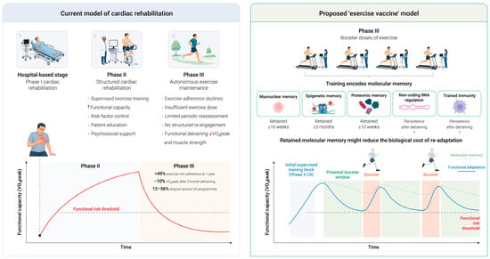
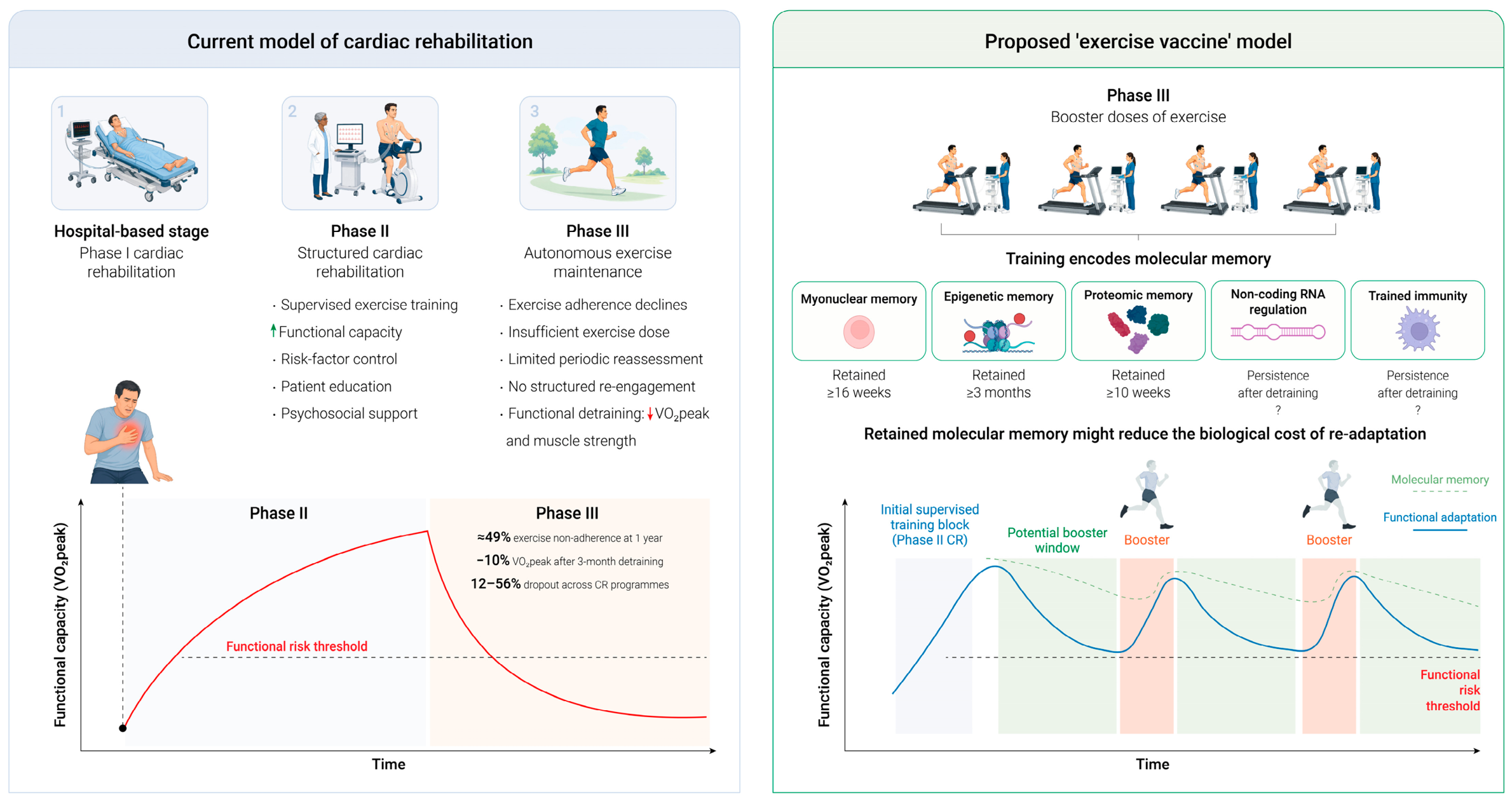
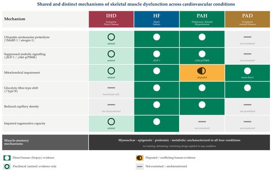
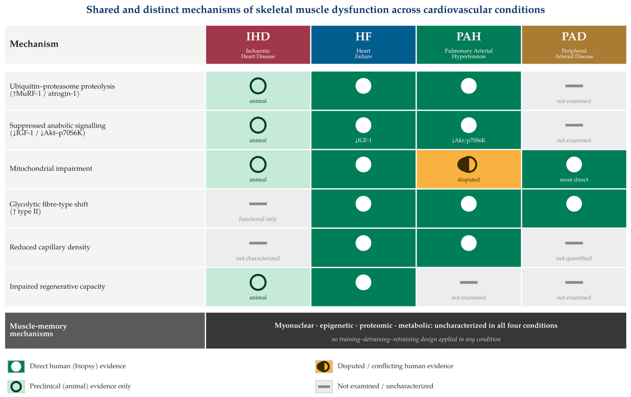
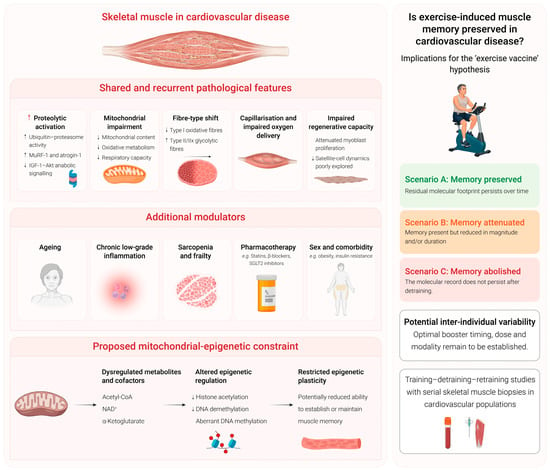
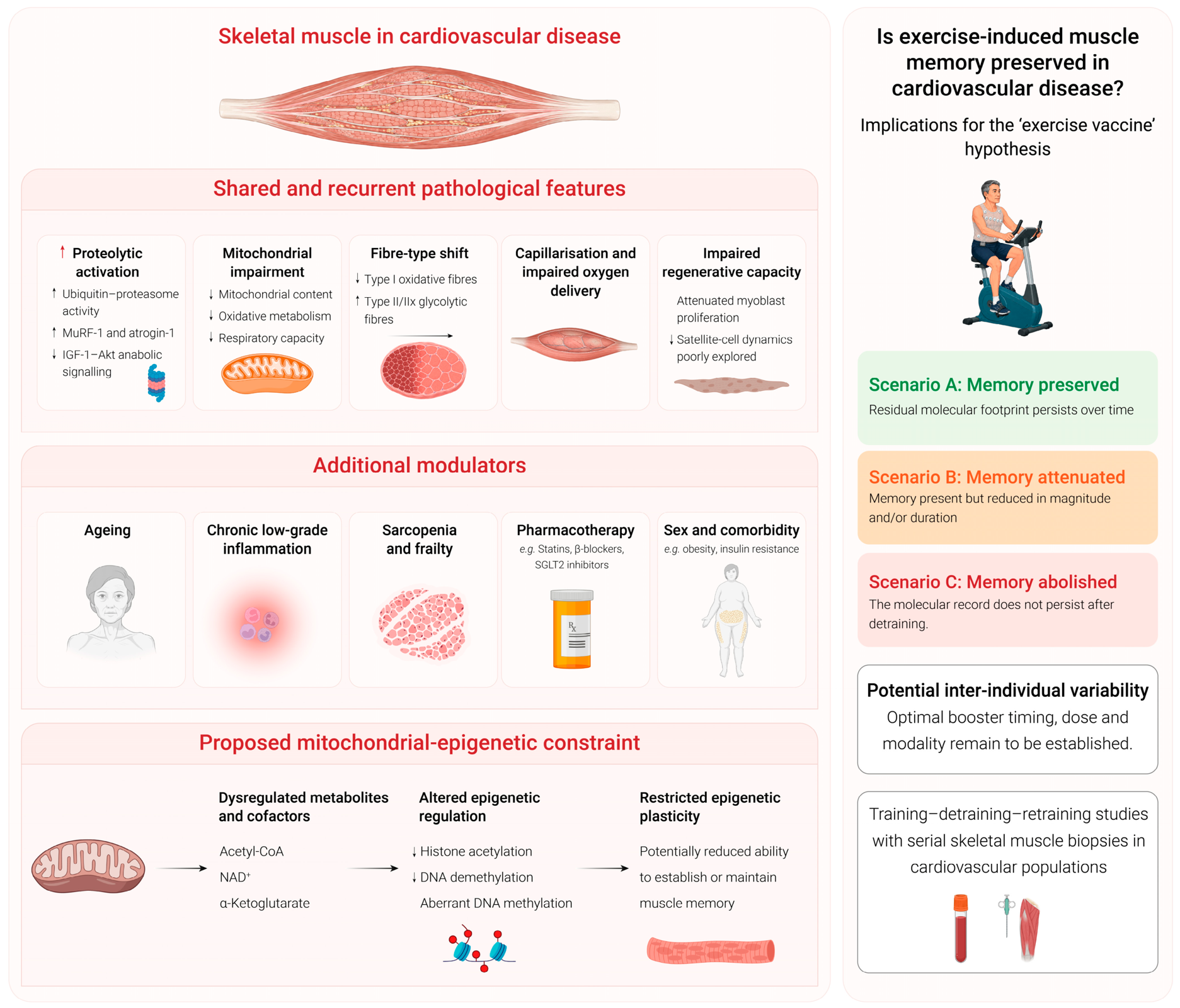

Disclaimer/Publisher’s Note: The statements, opinions and data contained in all publications are solely those of the individual author(s) and contributor(s) and not of MDPI and/or the editor(s). MDPI and/or the editor(s) disclaim responsibility for any injury to people or property resulting from any ideas, methods, instructions or products referred to in the content. |
© 2026 by the authors. Licensee MDPI, Basel, Switzerland. This article is an open access article distributed under the terms and conditions of the Creative Commons Attribution (CC BY) license.