


Abstract
Advances in surgical and medical management have improved survival for individuals with Fontan physiology (FP), increasing attention to long-term neurocognitive impairments. We propose the Cardiopulmonary–Brain Axis Hypothesis, identifying respiratory muscle performance (RMP) as a potentially modifiable contributor to executive function in FP. Drawing on evidence from cardiovascular and respiratory physiology, autonomic neuroscience, cerebrovascular biology, and neurocognitive research, this framework explores potential relationships among RMP, FP, respiratory muscle metaboreflex activation, autonomic regulation, cerebral perfusion, and executive function. Fontan-specific studies demonstrate the influence of respiration on venous return and pulmonary blood flow, while selected inspiratory muscle training (IMT) studies report improvements in respiratory function. Building on these findings, we hypothesize that improvements in RMP following IMT may enhance respiratory pump efficiency, favorably influence Fontan hemodynamics, attenuate respiratory muscle metaboreflex activation, and support autonomic regulation and cerebral perfusion, with potential downstream improvements in executive function. However, evidence supporting these relationships varies, with several mechanisms derived primarily from non-Fontan populations. The pathway remains untested, and improvements in executive function following IMT have not been established in FP. The Cardiopulmonary–Brain Axis Hypothesis provides a biologically plausible, testable framework to guide future longitudinal and interventional investigations examining the potential relationships between RMP, cardiopulmonary function, and brain health.
1. Introduction
Advances in surgical techniques, perioperative management, and multidisciplinary care have transformed the Fontan procedure from a life-saving palliative operation into a chronic model of cardiovascular care, allowing most individuals born with single-ventricle congenital heart disease to survive well into adulthood [1,2,3]. As survival has improved, the clinical focus has shifted from reducing mortality to optimizing long-term health, quality of life, and the prevention and management of Fontan-associated multisystem complications [1,2,3]. Although cardiovascular, pulmonary, hepatic, and lymphatic sequelae remain major contributors to morbidity, neurodevelopmental impairment has emerged as one of the most important determinants of lifelong functional outcomes [4,5,6]. Among these impairments, executive function, including attention, working memory, inhibitory control, cognitive flexibility, processing speed, planning, and organization, is particularly important because it influences academic achievement, treatment adherence, self-management, psychosocial functioning, and overall quality of life [4,5,6,7,8,9].
Current paradigms primarily attribute neurocognitive dysfunction in Fontan physiology (FP) to developmental and perioperative neurologic injury. Altered fetal cerebral oxygen delivery, delayed brain maturation, chronic hypoxemia, perioperative ischemic injury, and white matter abnormalities are well-established contributors to adverse neurodevelopmental outcomes and are supported by both neuroimaging and longitudinal clinical studies [4,5,6,7,8,9,10,11,12]. These mechanisms provide an essential foundation for understanding brain injury in congenital heart disease; however, they primarily describe developmental or historical contributors that are not readily modifiable after Fontan completion. Furthermore, they do not fully explain the considerable variability in executive functioning observed among individuals with similar cardiac anatomy, surgical histories, or clinical characteristics, suggesting that ongoing physiological factors may continue to influence neurocognitive performance throughout childhood and adulthood [4,5,6,7,8,9].
The Fontan circulation possesses several unique physiological characteristics that support this variation. In the absence of a subpulmonary ventricle, pulmonary blood flow depends on passive venous return generated by the pressure gradient between the systemic and pulmonary circulations and is substantially influenced by respiratory-generated intrathoracic pressure changes [13,14,15,16,17]. Real-time phase-contrast cardiovascular magnetic resonance studies have demonstrated that respiration significantly modulates systemic venous return, ventricular filling, stroke volume, and pulmonary blood flow in individuals with FP, highlighting the central role of the respiratory pump in maintaining circulatory efficiency [13,14,15]. Respiratory muscle weakness, restrictive pulmonary mechanics, impaired ventilatory efficiency, diminished exercise capacity, and autonomic dysfunction are also frequently observed following Fontan palliation, yet these impairments have largely been investigated as independent clinical manifestations rather than as interconnected components of a shared physiological system [16,17,18,19,20,21,22,23].
Growing evidence from both FP and broader cardiovascular research suggests that respiratory muscle performance (RMP) may influence physiological processes extending well beyond ventilation. Fontan-specific training studies have included pediatric to adult populations. Notably, a home-based exercise program incorporating inspiratory muscle training (IMT) permitted participation beginning at 6 years of age, while targeted IMT trials have directly studied children as young as approximately 8 years, supporting the potential applicability of respiratory muscle interventions beginning in childhood [23]. Improvements in RMP have been associated with enhanced respiratory-driven flow dynamics and cardiopulmonary interactions, attenuation of the respiratory muscle metaboreflex, improved autonomic regulation, favorable cerebrovascular responses, and enhanced exercise performance [20,21,22,23,24,25,26,27,28,29,30,31,32]. More broadly, emerging evidence describing the heart-brain axis demonstrates that cardiovascular function and cerebral physiology are closely interconnected, with alterations in cardiac performance influencing cerebral blood flow, neurovascular regulation, and cognitive health [29,30,31,32]. These observations raise the possibility that RMP may represent an important physiological link between Fontan hemodynamics and brain function.
Accordingly, we propose the Cardiopulmonary–Brain Axis Hypothesis, a conceptual framework identifying RMP as a potentially modifiable contributor to cardiopulmonary physiology that may indirectly influence executive function. The novelty lies in proposing IMT as a means to investigate whether improvements in RMP are associated with subsequent changes in Fontan hemodynamics, autonomic regulation, cerebral perfusion, and executive function. These proposed links vary considerably in evidentiary support and have not been validated as a continuous causal pathway in FP. Respiratory muscle training (RMT), particularly IMT, is therefore considered an experimental means of testing the hypothesis, not an established neurorehabilitation treatment. The purposes of this article are to present (1) the biological and physiological rationale supporting the Cardiopulmonary–Brain Axis Hypothesis, (2) distinguish direct Fontan findings from indirect evidence, and (3) identify approaches to test the proposed hypotheses.
Approach to Literature Identification and Evidence Synthesis
A narrative literature review was conducted from May through August 2026, including PubMed/MEDLINE and Google Scholar. The literature was identified and reviewed collaboratively by physical therapists with board certification in cardiovascular and pulmonary and pediatric physical therapy, together with physical therapy students. Representative search concepts included “Fontan circulation” or “single ventricle” combined with “respiration,” “respiratory muscle performance,” “inspiratory muscle training,” “venous return,” “cardiac output,” “autonomic regulation,” “respiratory muscle metaboreflex,” “cerebral blood flow,” “neurodevelopment,” and “executive function.” Publications were considered for their relevance to the proposed physiological links of the Cardiopulmonary–Brain Axis Hypothesis, with emphasis on Fontan-specific investigations, human clinical or mechanistic studies, and pertinent reviews. Evidence from other populations was included when direct Fontan evidence was unavailable and was identified as indirect support. This narrative, hypothesis-generating synthesis did not employ a systematic screening protocol or formal risk-of-bias assessment. Final review of this manuscript was completed by a cardiologist board-certified in pediatrics, pediatric cardiology, and adult congenital heart disease.
2. The Cardiopulmonary–Brain Axis Hypothesis
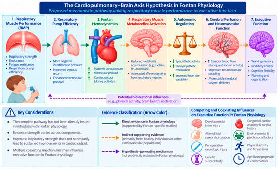
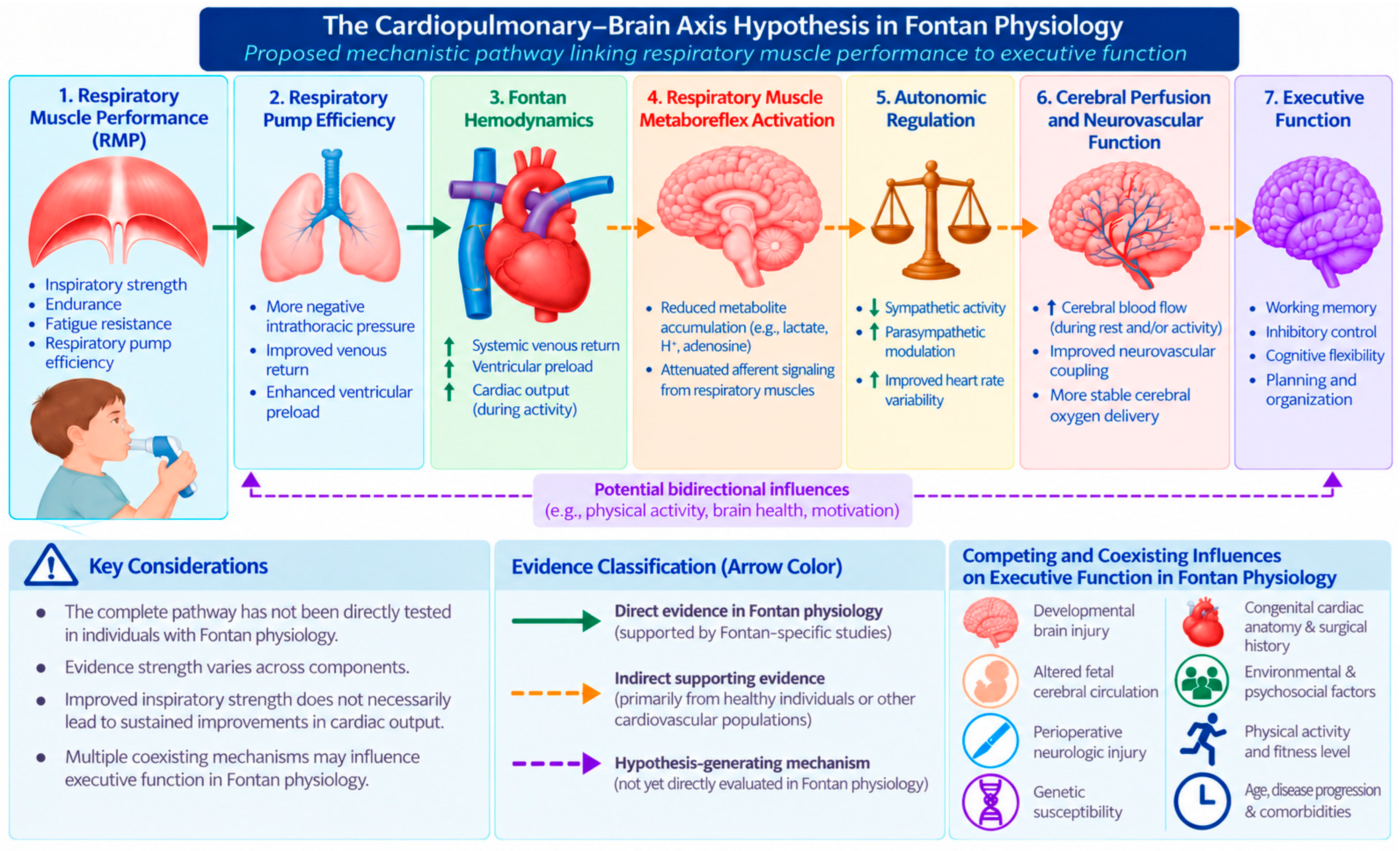
The Cardiopulmonary–Brain Axis Hypothesis proposes a set of potentially connected, testable relationships between RMP, respiratory mechanics, Fontan hemodynamics, autonomic activity, cerebral physiology, and executive function (Figure 1). These relationships should not be interpreted as a demonstrated linear causal sequence. IMT offers one possible intervention to examine if changes in inspiratory muscle capacity are accompanied by temporally ordered changes in proposed mediators and outcomes.
Figure 1.
The Cardiopulmonary–Brain Axis Hypothesis in Fontan physiology. Proposed mechanistic pathway linking RMP to executive function through respiratory pump efficiency, Fontan hemodynamics, respiratory muscle metaboreflex activation, autonomic regulation, and cerebral perfusion. IMT is proposed as a potential intervention targeting RMP. Solid green arrows indicate direct Fontan-specific evidence, dashed orange arrows indicate indirect supporting evidence, and dotted purple arrows represent hypothesized relationships or potential bidirectional influences. Competing factors influencing executive function are also highlighted. The complete pathway remains untested, and potential benefits of IMT on cerebral perfusion and executive function require further investigation.
For the purposes of this hypothesis, RMP refers to measurable properties of respiratory muscle capacity, including maximal inspiratory and expiratory pressure generation, inspiratory endurance, and fatigue resistance. Respiratory mechanics describe the behavior of the lungs, chest wall, and airways, whereas respiratory pump efficiency reflects the resulting interaction of muscular effort, mechanics, breathing pattern, and cardiopulmonary loading conditions; neither is synonymous with RMP. IMT primarily targets inspiratory muscle strength, endurance, and fatigue resistance. The effects of IMT on expiratory performance, respiratory pump efficiency, Fontan blood flow, or cerebral outcomes cannot be assumed from an increase in maximal inspiratory pressure alone.
The central premise of the Cardiopulmonary–Brain Axis Hypothesis is that RMP may be a modifiable contributor to respiratory pressure generation and subsequent cardiopulmonary responses in FP. Respiratory-generated intrathoracic pressure changes influence systemic venous return, pulmonary blood flow, ventricular preload, and cardiac output within the preload-dependent Fontan circulation [13,14,15,16,17,18,19]. However, these interactions are influenced by breathing pattern, pressure magnitude, lung volume, airway resistance, and imposed respiratory loads, such that greater inspiratory pressure generation does not necessarily translate into improved hemodynamics. Within this context, reduced RMP may compromise respiratory pump efficiency and increase respiratory muscle loading and susceptibility to fatigue, potentially contributing to respiratory muscle metaboreflex activation and altered autonomic regulation [19,24,25,26,27,28]. These responses could, in turn, influence cerebral perfusion and neurovascular function, with potential implications for executive function [21,22,23,24,25,26,27,28,29,30,31,32]. Conversely, improvements in RMP following IMT may favorably influence respiratory pump efficiency and selected hemodynamic responses, potentially modifying downstream autonomic and cerebrovascular processes. Nevertheless, these proposed relationships remain largely untested in individuals with FP, and improvements in inspiratory muscle strength cannot be assumed to produce sustained hemodynamic or neurocognitive benefits.
This hypothesis does not propose that RMP is the sole determinant of executive function. Executive function remains inherently multifactorial and is influenced by developmental brain injury, congenital cardiac anatomy, genetic susceptibility, surgical history, environmental factors, and ongoing cardiovascular and environmental influences across the lifespan [4,5,6,7,8,9,10,11,12]. Rather, the Cardiopulmonary–Brain Axis Hypothesis identifies RMP as a previously underrecognized, clinically modifiable physiological contributor that may interact with these established determinants to influence cognitive performance across the lifespan.
Four testable hypotheses associated with this framework include: (1) targeted IMT can modify prespecified inspiratory muscle outcomes; (2) some participants may exhibit associated changes in respiratory-flow dynamics and Fontan hemodynamics, although direction and magnitude may vary; (3) changes in autonomic indices or cerebral blood flow and oxygen delivery may follow, but these outcomes are distinct and their causal links are unproven; and (4) changes in prespecified executive function outcomes may occur only if the proposed downstream pathway is operative. Controlled trials with repeated mediator assessments are needed to distinguish these possibilities from confounding, reverse causation, and nonspecific training effects. The following sections examine the existing evidence linked to each component of this proposed pathway and identify the key mechanistic studies needed to validate or refute the hypothesis.
3. Biological Rationale Supporting the Cardiopulmonary–Brain Axis Hypothesis
The proposed Cardiopulmonary–Brain Axis integrates evidence from direct and indirect research. Respiratory modulation of venous and pulmonary blood flow has been demonstrated in FP [13,14,15,16,17], and selected Fontan intervention studies have evaluated IMT and cardiopulmonary outcomes [20,21,22,23]. In contrast, the respiratory muscle metaboreflex and its putative downstream effects on cerebral physiology and executive function have not been demonstrated as an integrated causal sequence in Fontan populations. Evidence from healthy adults and other cardiovascular conditions is therefore treated as indirect mechanistic support, not confirmation of this hypothesis.
The following sections examine each component of the proposed pathway sequentially. First, we review the dependence of Fontan hemodynamics on respiratory-generated intrathoracic pressure and RMP. Next, we examine the role of respiratory muscle fatigue and the respiratory muscle metaboreflex in autonomic regulation. We then review evidence linking cardiovascular physiology with cerebral perfusion and neurovascular function with executive function as the primary predicted clinical manifestation of the proposed pathway. Finally, we consider the broader physiological implications of RMP beyond neurocognitive outcomes. Together, these sections provide the mechanistic foundation for the Cardiopulmonary–Brain Axis Hypothesis and identify key areas requiring future investigation.
3.1. Respiratory Muscle Performance and Fontan Hemodynamics
The Fontan circulation is uniquely dependent on respiratory mechanics to maintain cardiovascular performance. Unlike normal biventricular circulation, pulmonary blood flow occurs in the absence of a subpulmonary ventricle and is therefore driven by the pressure gradient between the systemic and pulmonary circulations rather than active ventricular propulsion [1]. Consequently, respiratory-generated intrathoracic pressure changes become a primary determinant of systemic venous return, ventricular preload, and cardiac output, placing the respiratory pump at the center of circulatory regulation [1,2,3].
Recent advances in real-time phase-contrast cardiovascular magnetic resonance (CMR) imaging have substantially improved our understanding of the interaction between respiration and Fontan hemodynamics. In contrast to conventional breath-held imaging, real-time CMR captures beat-to-beat physiological changes throughout the respiratory cycle and has demonstrated that inspiration significantly augments inferior vena cava flow, ventricular filling, stroke volume, and pulmonary blood flow, whereas expiration produces the opposite effect [13,14,15]. These respiratory-dependent fluctuations are not trivial but represent a fundamental characteristic of FP, emphasizing that respiratory-generated negative intrathoracic pressure functions as an essential driver of pulmonary blood flow rather than simply facilitating ventilation. Studies have demonstrated that approximately 30% of Fontan flow and 55% of hepatic venous flow are influenced by respiratory mechanics, underscoring the substantial contribution of breathing to Fontan hemodynamics, with normal and forced inspiration significantly increasing blood flow pulsatility in patients with FP [16,17]. Recent evidence demonstrates that collateral blood flow does not compensate for inadequate ventricular preload or diastolic dysfunction, reinforcing the importance of preserving efficient respiratory pump function for maintaining circulatory performance [15].
The dependence of Fontan hemodynamics on respiration provides a strong physiological rationale for investigating RMP as a potentially modifiable contributor to cardiovascular function. RMP encompasses the ability of the inspiratory and expiratory musculature to generate and sustain respiratory pressures, while respiratory pump efficiency reflects the interaction between muscle performance, respiratory mechanics, and cardiovascular loading conditions. In FP, respiratory-generated intrathoracic pressure changes facilitate systemic venous return, pulmonary blood flow, and ventricular filling, suggesting that improvements in RMP may enhance respiratory pump efficiency and support hemodynamic function [13,14,15,16,17,18,19]. Conversely, reduced RMP may limit effective respiratory pressure generation and increase respiratory muscle loading, potentially placing additional demands on an already preload-dependent circulation [16,17,18,19]. However, the magnitude and direction of these effects depend on breathing pattern, lung volume, airway resistance, and imposed respiratory loads.
Consistent with this physiological model, respiratory dysfunction is common following Fontan palliation. Individuals with FP frequently demonstrate restrictive pulmonary mechanics, inspiratory muscle weakness, impaired ventilatory efficiency, diminished respiratory endurance, and reduced exercise capacity compared with healthy peers [16,17,18]. These impairments are likely multifactorial, reflecting the combined effects of altered thoracic mechanics, restrictive pulmonary physiology, chronic venous congestion, physical deconditioning, and the increased work of breathing associated with the Fontan circulation [16,17,18]. Collectively, these abnormalities may reduce respiratory pump efficiency and further limit cardiovascular performance, particularly during exercise when circulatory demands increase.
Importantly, RMP represents a potentially modifiable target for rehabilitation. Studies investigating IMT in individuals with FP have reported improvements in inspiratory muscle strength and selected measures of ventilatory efficiency and exercise capacity, with improvements in resting cardiac output also observed in a limited study [20,21,22,23]. Although the mechanisms underlying these responses remain incompletely understood, these findings suggest that enhancing inspiratory muscle performance may improve respiratory pump efficiency and favorably influence Fontan hemodynamics. However, the magnitude and durability of these effects, particularly across different ages and Fontan phenotypes, require further investigation. Collectively, these observations provide a physiological foundation for the Cardiopulmonary–Brain Axis Hypothesis and support investigating whether training-related improvements in RMP are accompanied by changes in autonomic regulation, cerebral perfusion, and executive function.
3.2. Respiratory Muscle Fatigue, the Respiratory Metaboreflex, and Autonomic Regulation
Beyond its role in intrathoracic pressure generation, RMP may influence cardiovascular regulation through neural reflex pathways, including the respiratory muscle metaboreflex. RMP influences cardiovascular function not only through its effects on intrathoracic pressure generation but also through neural reflex pathways that regulate autonomic cardiovascular control. One of the most important mechanisms is the respiratory muscle metaboreflex, a protective reflex activated when the respiratory muscles experience increased work or fatigue. During periods of sustained inspiratory muscle demand, metabolite accumulation within the diaphragm and accessory respiratory muscles stimulates metabolically sensitive group III and IV afferent fibers, increasing sympathetic nervous system activity and redistributing blood flow away from locomotor muscles to preserve respiratory muscle function [24,25,26,27]. Although this response is physiologically beneficial during acute exercise, exaggerated activation has been associated with increased sympathetic vasoconstrictor activity, reduced locomotor muscle perfusion, greater peripheral muscle fatigue, and impaired exercise tolerance [24,25,26,27]. Evidence supporting these mechanisms has been derived predominantly from studies of healthy adults and adults with cardiovascular disease, with limited direct investigation in pediatric cardiovascular populations. Therefore, while FP may plausibly increase inspiratory muscle loading through restrictive mechanics, reduced reserve, and exercise-related ventilatory demands [16,17,18,19], direct demonstration of a respiratory muscle metaboreflex in Fontan patients is lacking. Accordingly, any contribution of this reflex to sympathetic activation or exercise limitation in Fontan physiology remains a hypothesis extrapolated primarily from non-Fontan studies [24,25,26,27].
Autonomic dysfunction is a well-recognized feature of FP and is characterized by impaired chronotropic response, reduced parasympathetic modulation, and diminished heart rate variability (HRV), a noninvasive marker of autonomic nervous system regulation. Early physiological investigations demonstrated markedly impaired cardiac autonomic nervous activity following the Fontan operation [19], while more recent studies have shown that reduced HRV is associated with impaired exercise capacity, chronotropic incompetence, and Fontan-associated organ complications [28]. These findings suggest that autonomic dysfunction reflects not only abnormal cardiovascular regulation but also overall disease severity within the Fontan circulation. Although autonomic dysfunction is likely multifactorial, the respiratory muscle metaboreflex provides a biologically plausible mechanism through which greater RMP may influence less sympathetic activation and greater autonomic balance.
Evidence from other cardiovascular populations provides additional support for the proposed pathway. Studies suggest that IMT may reduce respiratory muscle fatigue, attenuate respiratory muscle metaboreflex activation, improve skeletal muscle oxygenation, and favorably influence autonomic regulation [24,25,26,27]. Although these responses have not been directly established in individuals with FP, they suggest that improving RMP may influence sympathetic activation and promote more favorable autonomic regulation. Given the role of autonomic mechanisms in cerebrovascular regulation, these adaptations could potentially influence cerebral perfusion and neurovascular function [29,30,31,32]. Within the Cardiopulmonary–Brain Axis Hypothesis, attenuation of respiratory muscle metaboreflex activation therefore represents a proposed mechanism connecting improvements in RMP and Fontan hemodynamics with downstream cerebrovascular and executive function outcomes. However, direct evaluation of these relationships in Fontan populations is needed, as measures such as heart rate variability provide indirect estimates of autonomic modulation and may be influenced by breathing pattern, medications, exercise intensity, and disease severity.
3.3. Cerebral Perfusion and Neurovascular Function
Cerebral perfusion is critically dependent on adequate cardiovascular function and dynamic neurovascular regulation. Although cerebral autoregulation maintains relatively stable cerebral blood flow across a range of systemic blood pressures, cerebral perfusion ultimately depends on sufficient cardiac output, appropriate cerebrovascular tone, and intact neurovascular coupling [29,30,31,32]. Emerging evidence describing the heart-brain axis recognizes that cardiovascular dysfunction can directly influence cerebral blood flow, oxygen delivery, and long-term brain health through interconnected hemodynamic, autonomic, and vascular mechanisms [29,30,31,32]. Within this framework, alterations in RMP that influence Fontan hemodynamics and autonomic regulation may also affect cerebral perfusion.
The Fontan circulation presents several physiological challenges that may compromise cerebral blood flow and oxygen delivery. Reduced ventricular preload, chronically elevated central venous pressure, diminished cardiac output, impaired exercise capacity, and autonomic dysfunction collectively create conditions that may adversely influence cerebral physiology [1,2,3,19,33]. Importantly, alterations in cerebral physiology and brain maturation are evident well before Fontan completion, during critical periods of central nervous system development. Studies of fetuses and infants with congenital heart disease, including single-ventricle physiology, have demonstrated altered cerebral oxygen delivery and metabolism, impaired brain growth, and abnormalities in brain maturation [9,10,11,12]. Fogel and colleagues further demonstrated abnormalities in cerebral blood flow across staged single-ventricle palliation, including during infancy before the bidirectional Glenn, before Fontan completion, and following Fontan surgery [33]. These abnormalities may persist later in development; subsequent work demonstrated altered cerebral blood flow after Fontan completion and associations with aortopulmonary collateral flow [34]. More recently, Romanowicz and colleagues quantified regional cerebral blood flow and oxygen delivery in adolescents with Fontan circulation and demonstrated reduced cerebral oxygen delivery despite relatively preserved global cerebral blood flow, suggesting that abnormalities in oxygen transport may persist even when total cerebral perfusion appears maintained [35]. Collectively, these findings suggest that altered cerebral physiology begins early in the single-ventricle pathway and may persist across childhood and adolescence following Fontan completion. Whether interventions targeting modifiable cardiopulmonary factors, including RMP, can influence cerebral physiology or neurocognitive outcomes remains unknown and represents an important area for future investigation.
Autonomic regulation provides an additional mechanistic link between Fontan hemodynamics and cerebral perfusion. Sympathetic activation influences cerebrovascular resistance, cerebral autoregulation, and neurovascular coupling, while parasympathetic activity contributes to vascular homeostasis and favorable endothelial function [34,35,36,37]. Consequently, autonomic dysfunction may impair the brain’s ability to appropriately regulate cerebral blood flow during physiological stress. Consistent with this concept, broader cardiovascular literature has increasingly recognized the bidirectional relationship between cardiac function and brain health, emphasizing that impaired cardiovascular performance contributes to cognitive dysfunction through reductions in cerebral perfusion, altered vascular regulation, endothelial dysfunction, and chronic cerebral hypoperfusion [34,35,36,37]. These observations support the biological plausibility that autonomic improvements following RMT could extend beyond cardiovascular regulation to influence cerebrovascular physiology.
Although direct evidence linking RMT with cerebral perfusion in FP is not yet available, emerging evidence from other populations provides proof of concept for this proposed mechanism. IMT has been shown to improve cerebrovascular reactivity in healthy older adults, suggesting that RMP may directly influence cerebrovascular regulation independent of changes in traditional cardiovascular risk factors [36]. Furthermore, recent work has identified associations between cardiorenal dysfunction and cerebrovascular risk in congenital heart disease, reinforcing the concept that multisystem cardiovascular physiology influences long-term brain health [37]. Collectively, these findings support the hypothesis that RMP may influence cerebral perfusion through coordinated effects on Fontan hemodynamics, autonomic regulation, and vascular function.
Within the Cardiopulmonary–Brain Axis Hypothesis, cerebral perfusion and neurovascular function represent proposed physiological mediators linking cardiopulmonary performance with executive function. Improvements in RMP may enhance respiratory pump efficiency, favorably influence Fontan hemodynamics, and support autonomic regulation, potentially creating more favorable conditions for cerebral blood flow, oxygen delivery, and neurovascular function. These outcomes are closely related but physiologically distinct, as cerebral oxygen delivery depends on both blood flow and arterial oxygen content, while cerebrovascular autoregulation and reactivity reflect the capacity to regulate blood flow in response to physiological changes. In individuals with FP, elevated central venous pressure, oxygen saturation, collateral flow, ventricular function, and cerebrovascular regulation may independently influence cerebral physiology [33,34,35,36,37]. Although these relationships provide a physiological rationale for investigating the proposed pathway, whether IMT-induced improvements in RMP translate into measurable cerebrovascular benefits remains unknown. Cerebral blood flow, oxygen delivery, and neurovascular function are therefore considered candidate mediators requiring direct investigation rather than established consequences of IMT. The following section examines executive function as the primary clinical outcome of interest within this proposed framework.
3.4. Executive Function as the Proposed Clinical Outcome
Executive function represents the primary clinical outcome predicted by the Cardiopulmonary–Brain Axis Hypothesis because it depends on distributed neural networks that are particularly sensitive to alterations in cerebral perfusion, oxygen delivery, and neurovascular regulation. Executive dysfunction is among the most consistently reported neurodevelopmental sequelae in individuals with FP. Across childhood, adolescence, and adulthood, impairments have been described in attention, working memory, processing speed, cognitive flexibility, behavioral regulation, and other executive domains compared with healthy peers [4,5,6,7,8,9]. These deficits are associated with poorer educational achievement, psychosocial functioning, health-related quality of life, and the ability to independently manage increasingly complex medical care [4,5,6,7,8,9]. Current evidence suggests that executive dysfunction is multifactorial, reflecting the combined influences of altered fetal brain development, congenital cardiac anatomy, perioperative neurological injury, chronic cardiovascular adaptations, and lifelong physiological stress [4,5,6,7,8,9,10,11,12].
Although executive dysfunction in individuals with FP is multifactorial, the Cardiopulmonary–Brain Axis Hypothesis proposes RMP as one potentially modifiable physiological contributor to neurocognitive health. Altered brain development, perioperative neurological injury, underlying cardiac anatomy and function, oxygen saturation, physical activity, and socioeconomic factors may independently influence neurocognitive outcomes [4,5,6,7,8,9,10,11,12]. Additionally, neurocognitive or functional limitations may reduce participation in physical activity, potentially contributing to reduced conditioning and RMP. These competing influences emphasize the importance of investigating RMP within a broader physiological and developmental framework without assuming a direct causal relationship.
Emerging evidence linking physical activity, cardiorespiratory fitness, and exercise capacity with executive function and broader neurodevelopmental outcomes in individuals with congenital heart disease and FP provides additional support for investigating modifiable cardiopulmonary contributors to brain health [38,39,40]. Although IMT differs mechanistically from aerobic exercise, both may influence overlapping physiological pathways relevant to neurocognitive function. Whether improvements in RMP produce similar downstream benefits remains unknown, but these associations support further investigation of respiratory muscle rehabilitation as a potentially novel approach to addressing neurocognitive health in FP.
Accordingly, executive function represents the primary clinical outcome of interest for evaluating the Cardiopulmonary–Brain Axis Hypothesis, given the documented executive function difficulties in individuals with FP [4,5,6,7,8,9]. Future studies should prespecify age-appropriate executive function domains and distinguish performance-based neuropsychological measures from assessments of everyday executive behaviors, such as the Behavior Rating Inventory of Executive Function, Second Edition (BRIEF-2). Improvements in executive function following IMT would provide preliminary evidence of potential neurocognitive benefit, while concurrent assessment of cardiopulmonary, autonomic, and cerebrovascular responses would be necessary to investigate the proposed mechanisms [13,14,15,16,17,18,19,24,25,26,27,28,29,30,31,32,33,34,35,36,37]. This framework supports future research examining whether targeted respiratory muscle rehabilitation can contribute to improved neurocognitive outcomes in individuals with FP.
3.5. Broader Physiological Implications of the Cardiopulmonary–Brain Axis Hypothesis
Although executive function represents the primary clinical outcome of the Cardiopulmonary–Brain Axis Hypothesis, the proposed physiological mechanisms may have broader implications for the multisystem consequences of FP. Chronically elevated central venous pressure, reduced cardiac output, and impaired end-organ perfusion contribute to lymphatic, hepatic, renal, and gastrointestinal complications [1,2,3,41,42,43,44,45]. Because respiration influences both systemic venous return and lymphatic transport, improvements in RMP and respiratory pump efficiency may theoretically support more favorable pressure gradients for these processes. This relationship is particularly relevant in FP, where elevated venous pressure impairs lymphatic drainage and contributes to complications such as protein-losing enteropathy, plastic bronchitis, and pleural effusions [41,42,43,44,45].
Beyond lymphatic function, improvements in respiratory pump efficiency may potentially influence other organ systems affected by chronic venous congestion and reduced cardiac output. However, whether IMT or other respiratory muscle interventions meaningfully improve lymphatic function, end-organ perfusion, or associated clinical complications remains unknown. These possibilities represent exploratory extensions of the proposed physiological framework rather than established therapeutic effects or necessary components of the Cardiopulmonary–Brain Axis Hypothesis. Future studies should prioritize direct evaluation of the proposed cardiopulmonary, autonomic, cerebrovascular, and executive function relationships while considering multisystem outcomes as secondary areas for investigation.
4. Clinical and Research Implications
The Cardiopulmonary–Brain Axis Hypothesis provides a novel conceptual framework for investigating RMP as a therapeutic target in individuals with FP. Rather than viewing IMT training solely as an intervention to improve inspiratory muscle strength or exercise capacity, this hypothesis proposes that RMP may influence interconnected cardiovascular, autonomic, and cerebrovascular processes relevant to executive function. If supported, this framework has the potential to expand the goals of cardiopulmonary rehabilitation beyond traditional measures of functional capacity to include neurocognitive health.
Among respiratory muscle training approaches, IMT represents a practical intervention for testing the proposed hypothesis because it specifically targets inspiratory muscle performance and is noninvasive, relatively inexpensive, portable, and readily integrated into both home- and clinic-based rehabilitation programs. Previous studies have investigated IMT in Fontan populations, with home-based programs enrolling children as early as 6 years of age and actual interventional trials including children as young as approximately 8 years [20,21,22,23]. These studies suggest that IMT is feasible and may improve RMP, ventilatory efficiency, exercise capacity, and selected cardiovascular outcomes in individuals with FP [20,21,22,23]. However, findings have varied across study designs, training protocols, and participant characteristics, with some controlled trials demonstrating limited or no improvements in exercise capacity [22,46]. These findings support the feasibility and potential clinical utility of IMT while emphasizing the need for further investigation of its physiological mechanisms and long-term effects.
The proposed hypothesis generates several testable predictions. First, improvements in RMP may favorably influence respiratory-driven Fontan hemodynamics, including systemic venous return, ventricular filling, and cardiac output. Second, these adaptations may attenuate respiratory muscle metaboreflex activation and influence autonomic regulation. Third, improvements in cardiopulmonary physiology may contribute to more favorable cerebral blood flow, oxygen delivery, and neurovascular function, potentially supporting executive function. These relationships remain hypothetical and may vary across individuals and developmental stages. Accordingly, future studies should evaluate each proposed mechanism using prespecified physiological and neurocognitive outcomes, appropriate comparison groups, and longitudinal assessments capable of examining the temporal relationships among these variables. Table 1 presents a proposed experimental framework for investigating these pathways across the lifespan in individuals with FP.
Table 1.
Proposed experimental framework for evaluating the Cardiopulmonary–Brain Axis Hypothesis across the lifespan in individuals with Fontan physiology.
Testing these predictions will require interdisciplinary collaboration among cardiology, rehabilitation, neuropsychology, exercise physiology, and neuroimaging specialists. A staged research approach incorporating feasibility studies, controlled interventions, and longitudinal assessment of proposed physiological mediators would help determine whether improvements in RMP precede measurable changes in Fontan hemodynamics, autonomic regulation, cerebral physiology, and executive function. Study designs should account for developmental stage, cardiac anatomy and function, oxygen saturation, pulmonary mechanics, medication use, and baseline neurodevelopmental status, while incorporating appropriate safety monitoring and comparison conditions. Such investigations would help distinguish direct training effects from associated physiological changes and identify which components of the proposed pathway may be clinically meaningful.
5. Evidence Supporting the Proposed Pathway
The evidence informing the Cardiopulmonary–Brain Axis Hypothesis varies in strength and applicability, ranging from direct Fontan-specific physiological observations and intervention findings to indirect evidence from other populations. Table 2 distinguishes these evidence levels and identifies the areas requiring further investigation.
5.1. Interpretation of Fontan-Specific Inspiratory Muscle Training Evidence
Fontan-specific IMT studies have reported promising but heterogeneous outcomes. In an uncontrolled adult pilot, Wu et al. [20] observed improved peak work rate without significant changes in peak oxygen uptake or maximal inspiratory pressure. Laohachai et al. [21] reported increased inspiratory muscle strength and resting cardiac output in an imaging subgroup following six weeks of IMT, although the uncontrolled design and smaller subgroup limit causal interpretation. In a pediatric randomized trial, Neidenbach et al. [22] observed improved resting oxygen saturation without significant improvement in exercise capacity. Walzer et al. [23] reported changes in respiratory pressures and oxygen pulse following combined IMT and endurance training, making it difficult to isolate IMT-specific effects.
Additional randomized findings highlight the importance of distinguishing respiratory adaptations from broader clinical benefits. Turquetto et al. [46] randomized 42 participants aged 12–30 years to aerobic training, IMT, or a non-exercise control group. Peak oxygen uptake improved in both training groups, with a larger reported increase after aerobic training (23%) than IMT (9%); IMT improved inspiratory pressure and spirometric outcomes, whereas significant autonomic changes were observed with aerobic training. In contrast, Fritz et al. [48] randomized 42 adults with Fontan circulation and found no significant improvement in peak oxygen uptake or lung-function measures following six months of IMT compared with controls, although resting oxygen saturation improved. Together, these studies support further investigation of IMT as a feasible, potentially useful rehabilitation approach, but do not establish consistent hemodynamic benefits or effects on cerebral physiology or executive function.
Table 2.
Current evidence supporting and informing the Cardiopulmonary–Brain Axis Hypothesis in Fontan physiology.
5.2. Limitations of the Proposed Framework
The Cardiopulmonary–Brain Axis Hypothesis is derived from a narrative synthesis rather than a systematic review, and the evidence supporting its proposed relationships differs in strength and directness. Fontan-specific studies establish respiratory modulation of venous and pulmonary blood flow and document selected IMT responses, but no longitudinal investigation has tested the complete sequence linking RMP, Fontan hemodynamics, autonomic regulation, cerebral physiology, and executive function. Evidence concerning respiratory muscle metaboreflex responses and cerebrovascular adaptations to IMT is drawn predominantly from non-Fontan populations. Consequently, associations among individual components cannot establish mediation or causation.
Interpretation must also account for heterogeneity in age, developmental stage, sex, ventricular anatomy and function, surgical pathway, fenestration and collateral status, oxygen saturation, pulmonary mechanics, medication exposure, and physical activity. Previous studies have identified respiratory and exercise-related impairments in individuals with Fontan circulation compared with healthy peers [16,17,18]. Additionally, Jackson et al. [50] identified executive-function deficits across multiple domains in individuals with congenital heart disease compared with healthy controls, while Meyer et al. [51] reported sex-related differences in cardiac function, functional capacity, and clinical outcomes among individuals with Fontan circulation. These findings highlight the importance of accounting for developmental and biological variability when investigating the proposed relationships. Neurocognitive outcomes are further influenced by prenatal and perioperative neurological injury, baseline neurodevelopment, and environmental and socioeconomic factors. Reverse causation is possible if neurocognitive or functional limitations reduce physical activity and conditioning. Moreover, respiratory pressure effects depend on breathing pattern, lung volume, imposed loads, and prevailing hemodynamics; increased inspiratory strength should not be assumed to improve cardiac output in every individual with Fontan circulation.
Future studies should account for age, developmental stage, and sex through developmentally appropriate and age-normed assessments, prespecified age stratification where feasible, and statistical adjustment. Potential sex-related differences should be explored rather than assumed, particularly in sufficiently powered studies. Given the relatively small and heterogeneous Fontan population, subgroup and mediation analyses should remain exploratory unless adequately powered. Future controlled and longitudinal investigations should distinguish respiratory muscle capacity from respiratory mechanics and circulatory responses, assess cerebral blood flow and oxygen delivery as separate constructs, and recognize the measurement limitations of heart rate variability and cerebrovascular imaging modalities. Appropriate comparators, prespecified outcomes, repeated physiological measurements, blinded assessment where feasible, and systematic monitoring of adverse events will be essential to determine which proposed relationships are supported. Until then, respiratory muscle training as a strategy to improve executive function in Fontan circulation remains investigational.
6. Conclusions
The Cardiopulmonary–Brain Axis Hypothesis proposes RMP as a potentially modifiable physiological contributor linking respiratory-dependent Fontan hemodynamics with autonomic regulation, cerebral physiology, and executive function. By integrating established principles of Fontan circulation with emerging evidence from respiratory physiology, autonomic regulation, cerebrovascular function, and neurodevelopment, this hypothesis provides a biologically plausible framework for investigating respiratory muscle training as a potential neurorehabilitation strategy in individuals with FP. Although evidence supports individual components of the proposed pathway, their integration into a causal sequence has not been demonstrated, and improvements in RMP cannot currently be assumed to translate into enhanced cerebral perfusion or executive function. Rather than identifying RMP as the sole determinant of neurocognitive outcomes, this framework positions it as one potentially modifiable factor within the complex physiological and developmental influences associated with FP.
Future mechanistic and interventional studies should evaluate whether improvements in RMP are accompanied by measurable changes in Fontan hemodynamics, autonomic regulation, cerebral perfusion, and executive function, while accounting for competing physiological and developmental influences. Establishing these relationships could expand the role of cardiopulmonary rehabilitation beyond respiratory function and exercise capacity, identifying new opportunities to support integrated cardiovascular and neurocognitive health. Although its application as a neurorehabilitation strategy remains investigational, the Cardiopulmonary–Brain Axis Hypothesis offers a testable foundation for advancing more targeted, physiology-informed rehabilitation approaches across the lifespan in individuals with FP.
Author Contributions
Conceptualization, A.C. and L.P.C.; methodology, A.C. and L.P.C.; software, A.C.; investigation, A.C., H.H., K.H., V.M., D.C., G.C., J.D., J.S. and L.P.C.; resources, A.C., H.H., K.H., V.M., D.C., G.C., J.D., J.S. and L.P.C.; data curation, A.C. and L.P.C.; writing—original draft preparation, A.C., H.H., K.H., V.M., D.C., G.C., J.D., J.S. and L.P.C.; writing—review and editing, A.C., J.G.M., L.P.C. and D.K.; and supervision, D.K.; funding acquisition, A.C. and L.P.C. All authors have read and agreed to the published version of the manuscript.
Funding
This research was funded in part by a Promotion of Doctoral Studies Scholarship from the Foundation for Physical Therapy Research (1462814) via the Scot C. Irwin Award, which is generously supported by the APTA Academy of Cardiovascular and Pulmonary Physical Therapy Human Performance Endowment Fund.
Institutional Review Board Statement
Not applicable.
Informed Consent Statement
Not applicable.
Data Availability Statement
The original contributions presented in this study are included in the article. Further inquiries can be directed to the corresponding author.
Acknowledgments
During the preparation of this manuscript, the authors used ChatGPT powered by GPT-6 (OpenAI) for the purposes of assisting with the visual development of the conceptual figure and refinement of organization. The authors have reviewed and edited the output and take full responsibility for the content of this publication.
Conflicts of Interest
The authors declare no conflict of interest.
References
- Rychik, J.; Atz, A.M.; Celermajer, D.S.; Deal, B.J.; Gatzoulis, M.A.; Gewillig, M.H.; Hsia, T.-Y.; Hsu, D.T.; Kovacs, A.H.; McCrindle, B.W.; et al. Evaluation and management of the child and adult with Fontan circulation: A scientific statement from the American Heart Association. Circulation 2019, 140, e234–e284. [Google Scholar] [CrossRef] [Scilit] [PubMed]
- Perrin, N.; Dore, A.; van de Bruaene, A.; Mongeon, F.-P.; Mondésert, B.; Poirier, N.; Miró, J.; Khairy, P.; Ibrahim, R.; Chaix, M.-A. The Fontan circulation: From ideal to failing hemodynamics and drug therapies for optimization. Can. J. Cardiol. 2022, 38, 1059–1071. [Google Scholar] [CrossRef] [Scilit] [PubMed]
- Hassan, A.; Chegondi, M.; Porayette, P. Five decades of Fontan palliation: What have we learned? What should we expect? J. Int. Med. Res. 2023, 51, 3000605231209156. [Google Scholar] [CrossRef] [Scilit] [PubMed]
- Calderon, J.; Newburger, J.W.; Rollins, C.K. Neurodevelopmental and mental health outcomes in patients with Fontan circulation: A state-of-the-art review. Front. Pediatr. 2022, 10, 826349. [Google Scholar] [CrossRef] [Scilit] [PubMed]
- Verrall, C.E.; Yang, J.Y.; Chen, J.; Schembri, A.; D’UDekem, Y.; Zannino, D.; Kasparian, N.A.; du Plessis, K.; Grieve, S.M.; Welton, T.; et al. Neurocognitive dysfunction and smaller brain volumes in adolescents and adults with a Fontan circulation. Circulation 2021, 143, 878–891. [Google Scholar] [CrossRef] [Scilit] [PubMed]
- Feldmann, M.; Bataillard, C.; Ehrler, M.; Ullrich, C.; Knirsch, W.; Gosteli-Peter, M.A.; Held, U.; Latal, B. Cognitive and executive function in congenital heart disease: A meta-analysis. Pediatrics 2021, 148, e2021050875. [Google Scholar] [CrossRef] [Scilit] [PubMed]
- Howell, H.B.; Zaccario, M.; Kazmi, S.H.; Desai, P.; Sklamberg, F.E.; Mally, P. Neurodevelopmental outcomes of children with congenital heart disease: A review. Curr. Probl. Pediatr. Adolesc. Health Care 2019, 49, 100685. [Google Scholar] [CrossRef] [Scilit] [PubMed]
- Derridj, N.; Guedj, R.; Calderon, J.; Houyel, L.; Lelong, N.; Bertille, N.; Goffinet, F.; Bonnet, D.; Khoshnood, B. Long-term neurodevelopmental outcomes of children with congenital heart defects. J. Pediatr. 2021, 237, 109–114.e5. [Google Scholar] [CrossRef] [Scilit] [PubMed]
- Selvanathan, T.; Smith, J.M.; Miller, S.P.; Field, T.S. Neurodevelopment and cognition across the lifespan in patients with single-ventricle physiology: Abnormal brain maturation and accumulation of brain injuries. Can. J. Cardiol. 2022, 38, 977–987. [Google Scholar] [CrossRef] [Scilit] [PubMed]
- Volpe, J.J. Fetal origin of brain dysmaturation in congenital heart disease-Challenges and opportunities for interventions. J. Neonatal. Perinat. Med. 2022, 15, 489–494. [Google Scholar] [CrossRef] [Scilit] [PubMed]
- Sun, L.; Macgowan, C.K.; Sled, J.G.; Yoo, S.-J.; Manlhiot, C.; Porayette, P.; Grosse-Wortmann, L.; Jaeggi, E.; McCrindle, B.W.; Kingdom, J.; et al. Reduced fetal cerebral oxygen consumption is associated with smaller brain size in fetuses with congenital heart disease. Circulation 2015, 131, 1313–1323. [Google Scholar] [CrossRef] [Scilit] [PubMed]
- Morton, P.D.; Ishibashi, N.; Jonas, R.A. Neurodevelopmental abnormalities and congenital heart disease: Insights into altered brain maturation: Insights into altered brain maturation. Circ. Res. 2017, 120, 960–977. [Google Scholar] [CrossRef] [Scilit] [PubMed]
- van der Woude, S.F.S.; Rijnberg, F.M.; Hazekamp, M.G.; Jongbloed, M.R.M.; Kenjeres, S.; Lamb, H.J.; Westenberg, J.J.M.; Roest, A.A.W.; Wentzel, J.J. The influence of respiration on blood flow in the Fontan circulation: Insights for imaging-based clinical evaluation of the total cavopulmonary connection. Front. Cardiovasc. Med. 2021, 8, 683849. [Google Scholar] [CrossRef] [Scilit] [PubMed]
- Körperich, H.; Barth, P.; Gieseke, J.; Müller, K.; Burchert, W.; Esdorn, H.; Kececioglu, D.; Beerbaum, P.; Laser, K.T. Impact of respiration on stroke volumes in paediatric controls and in patients after Fontan procedure assessed by MR real-time phase-velocity mapping. Eur. Heart J. Cardiovasc. Imaging 2015, 16, 198–209. [Google Scholar] [CrossRef] [Scilit] [PubMed]
- Blessing, M.C.M.; Körperich, H.; Barth, P.M.; Michel, M.; Dubowy, O.; Forreiter, S.C.M.; Laser, K.T. Influence of respiration on collateral flow in the Fontan population using real-time phase-contrast cardiovascular magnetic resonance: Collateral flow does not protect the ventricle from volume deficiency and diastolic dysfunction. J. Thorac. Imaging 2023, 38, W64–W74. [Google Scholar] [CrossRef] [Scilit] [PubMed]
- Laohachai, K.; Ayer, J. Impairments in pulmonary function in Fontan patients: Their causes and consequences. Front. Pediatr. 2022, 10, 825841. [Google Scholar] [CrossRef] [Scilit] [PubMed]
- Abdulkarim, A.; Shaji, S.; Elfituri, M.; Gunsaulus, M.; Zafar, M.A.; Zaidi, A.N.; Pass, R.H.; Feingold, B.; Kurland, G.; Kreutzer, J.; et al. Pulmonary complications in patients with Fontan circulation: JACC review topic of the week. J. Am. Coll. Cardiol. 2023, 81, 2434–2444. [Google Scholar] [CrossRef] [Scilit] [PubMed]
- Ladrón-Abia, R.; Ramos, P.C.; Pérez, I.V.; De Liger, R.C.F.; Wals-Rodríguez, A.-J.; Puras, M.J.R.; García, B.M.; Gallego, P. Respiratory muscle function and exercise performance in adult patients with Fontan circulation. J. Clin. Med. 2023, 12, 4593. [Google Scholar] [CrossRef] [Scilit] [PubMed]
- Ohuchi, H.; Hasegawa, S.; Yasuda, K.; Yamada, O.; Ono, Y.; Echigo, S. Severely impaired cardiac autonomic nervous activity after the Fontan operation. Circulation 2001, 104, 1513–1518. [Google Scholar] [CrossRef] [Scilit] [PubMed]
- Wu, F.M.; Opotowsky, A.R.; Denhoff, E.R.; Gongwer, R.; Gurvitz, M.Z.; Landzberg, M.J.; Shafer, K.M.; Valente, A.M.; Uluer, A.Z.; Rhodes, J. A pilot study of inspiratory muscle training to improve exercise capacity in patients with Fontan physiology. Semin. Thorac. Cardiovasc. Surg. 2018, 30, 462–469. [Google Scholar] [CrossRef] [Scilit] [PubMed]
- Laohachai, K.; Winlaw, D.; Selvadurai, H.; Gnanappa, G.K.; D’Udekem, Y.; Celermajer, D.; Ayer, J. Inspiratory muscle training is associated with improved inspiratory muscle strength, resting cardiac output, and the ventilatory efficiency of exercise in patients with a Fontan circulation. J. Am. Heart Assoc. 2017, 6, e005750. [Google Scholar] [CrossRef] [Scilit] [PubMed]
- Neidenbach, R.; Freilinger, S.; Stöcker, F.; Ewert, P.; Nagdyman, N.; Oberhoffer-Fritz, R.; Pieper, L.; Kaemmerer, H.; Hager, A. Clinical aspects and targeted inspiratory muscle training in children and adolescents with Fontan circulation: A randomized controlled trial. Cardiovasc. Diagn. Ther. 2023, 13, 11–24. [Google Scholar] [CrossRef] [Scilit] [PubMed]
- Walzer, L.; Sallmon, H.; Kelm, M.; Dirks, S.; Meyer, M.; Kramer, P.; Wolfarth, B.; Thouet, T.; Ovroutski, S.; Berger, F.; et al. Home-based long-term physical endurance and inspiratory muscle training in children and adults with Fontan circulation. Front. Cardiovasc. Med. 2024, 11, 1411758. [Google Scholar] [CrossRef] [Scilit] [PubMed]
- e Lima, T.B.W.; Silva, B.B.d.S.; de Souza, A.P.S.; Aires, D.M.F.; Fagundes, M.G. The effect of inspiratory muscle training on the inspiratory muscle metaboreflex: A systematic review. Can. J. Respir. Ther. 2025, 61, 51–59. [Google Scholar] [CrossRef] [Scilit] [PubMed]
- Moreno, A.M.; Toledo-Arruda, A.C.; Lima, J.S.; Duarte, C.S.; Villacorta, H.; Nóbrega, A.C. Inspiratory muscle training improves intercostal and forearm muscle oxygenation in patients with chronic heart failure: Evidence of the origin of the respiratory metaboreflex. J. Card. Fail. 2017, 23, 672–679. [Google Scholar] [CrossRef] [Scilit] [PubMed]
- Dempsey, J.A.; Romer, L.; Rodman, J.; Miller, J.; Smith, C. Consequences of exercise-induced respiratory muscle work. Respir. Physiol. Neurobiol. 2006, 151, 242–250. [Google Scholar] [CrossRef] [Scilit] [PubMed]
- Sheel, A.W.; Boushel, R.C.; Dempsey, J.A. Competition for blood flow distribution between respiratory and locomotor muscles: Implications for muscle fatigue. J. Appl. Physiol. 2018, 125, 820–831. [Google Scholar] [CrossRef] [Scilit] [PubMed]
- Okólska, M.; Łach, J.; Matusik, P.T.; Pająk, J.; Mroczek, T.; Podolec, P.; Tomkiewicz-Pająk, L. Heart rate variability and its associations with organ complications in adults after Fontan operation. J. Clin. Med. 2021, 10, 4492. [Google Scholar] [CrossRef] [Scilit] [PubMed]
- Haque, S.; Dutta, P. The heart-brain axis in the context of cardiovascular disease. Physiology 2026, 41, 147–158. [Google Scholar] [CrossRef] [Scilit] [PubMed]
- Testai, F.D.; Gorelick, P.B.; Chuang, P.-Y.; Dai, X.; Furie, K.L.; Gottesman, R.F.; Iturrizaga, J.C.; Lazar, R.M.; Russo, A.M.; Seshadri, S.; et al. Cardiac contributions to brain health: A scientific statement from the American heart association. Stroke 2024, 55, e425–e438. [Google Scholar] [CrossRef] [Scilit] [PubMed]
- Meng, L.; Hou, W.; Chui, J.; Han, R.; Gelb, A.W. Cardiac 0utput and cerebral blood Flow: The integrated regulation of brain perfusion in adult humans. Anesthesiology 2015, 123, 1198–1208. [Google Scholar] [CrossRef] [Scilit] [PubMed]
- Castle-Kirszbaum, M.M.; Parkin, W.G.M.; Goldschlager, T.F.; Lewis, P.M. Cardiac output and cerebral blood flow: A systematic review of cardio-cerebral coupling: A systematic review of cardio-cerebral coupling. J. Neurosurg. Anesthesiol. 2022, 34, 352–363. [Google Scholar] [CrossRef] [Scilit] [PubMed]
- Fogel, M.A.; Li, C.; Elci, O.U.; Pawlowski, T.; Schwab, P.J.; Wilson, F.; Nicolson, S.C.; Montenegro, L.M.; Diaz, L.; Spray, T.L.; et al. Neurological injury and cerebral blood flow in single ventricles throughout staged surgical reconstruction. Circulation 2017, 135, 671–682. [Google Scholar] [CrossRef] [Scilit] [PubMed]
- Fogel, M.A.; Donnelly, E.; Crandell, I.; Hanlon, A.; Whitehead, K.K.; Harris, M.; Partington, S.; Biko, D.; Flynn, T.; Nicolson, S.; et al. Cerebral blood flow, brain injury, and aortic-pulmonary collateral flow after the Fontan operation. Am. J. Cardiol. 2023, 208, 164–170. [Google Scholar] [CrossRef] [Scilit] [PubMed]
- Romanowicz, J.; Park, S.; Bunn, J.; Jacobsen, R.M.; Fonseca, B.; Zablah, J.E.; Englund, E.K.; Barker, A.J.; Davidson, J.A. Magnetic resonance quantification of regional blood flow and oxygen delivery to the brain, gut, kidneys, and lower extremities in adolescents with a Fontan circulation compared to biventricular controls. J. Cardiovasc. Magn. Reson. 2025, 27, 101907. [Google Scholar] [CrossRef] [Scilit] [PubMed]
- Freeberg, K.A.; Craighead, D.H.; Heinbockel, T.C.; Rossman, M.J.; Jackman, R.A.; Jankowski, L.R.; Ludwig, K.R.; Chonchol, M.; Bailey, E.F.; Seals, D.R. Time-efficient, high-resistance inspiratory muscle strength training increases cerebrovascular reactivity in midlife and older adults. Am. J. Physiol. Heart Circ. Physiol. 2023, 325, H1059–H1068. [Google Scholar] [CrossRef] [Scilit] [PubMed]
- Martínez-Quintana, E.; Rodríguez-González, F. Cardiorenal biomarkers and cerebrovascular risk in patients with congenital heart disease. J. Clin. Med. 2026, 15, 2440. [Google Scholar] [CrossRef] [Scilit] [PubMed]
- Clifton, A.; Kirk-Sanchez, N.; Cipriano, G.; Moore, J.G.; Cahalin, L.P. Physical activity and executive functioning in children and adolescents with congenital heart defects: A scoping review. J. Cardiovasc. Dev. Dis. 2024, 11, 309. [Google Scholar] [CrossRef] [Scilit] [PubMed]
- Verrall, C.E.; Tran, D.L.; Yang, J.Y.-M.; Lubans, D.R.; Winlaw, D.S.; Ayer, J.; Celermajer, D.; Cordina, R. Exercise as therapy for neurodevelopmental and cognitive dysfunction in people with a Fontan circulation: A narrative review. Front. Pediatr. 2023, 11, 1111785. [Google Scholar] [CrossRef] [Scilit] [PubMed]
- Cooney, S.J.; Campbell, K.; Wolfe, K.; DiMaria, M.V.; Rausch, C.M. Is neurodevelopment related to exercise capacity in single ventricle patients who have undergone Fontan palliation? Pediatr. Cardiol. 2021, 42, 408–416. [Google Scholar] [CrossRef] [Scilit] [PubMed]
- Gordon-Walker, T.T.; Bove, K.; Veldtman, G. Fontan-associated liver disease: A review. J. Cardiol. 2019, 74, 223–232. [Google Scholar] [CrossRef] [Scilit] [PubMed]
- Itkin, M.; Rockson, S.G.; Burkhoff, D. Pathophysiology of the lymphatic system in patients with heart failure: JACC state-of-the-art review. J. Am. Coll. Cardiol. 2021, 78, 278–290. [Google Scholar] [CrossRef] [Scilit] [PubMed]
- Daniels, C.J.; Bradley, E.A.; Landzberg, M.J.; Aboulhosn, J.; Beekman, R.H.; Book, W.; Gurvitz, M.; John, A.; John, B.; Marelli, A.; et al. Fontan-associated liver disease: Proceedings from the American College of cardiology stakeholders meeting, October 1 to 2, 2015, Washington DC. J. Am. Coll. Cardiol. 2017, 70, 3173–3194. [Google Scholar] [CrossRef] [Scilit] [PubMed]
- Kreutzer, C.; Kreutzer, G. The lymphatic system: The Achilles heel of the Fontan-Kreutzer circulation. World. J. Pediatr. Congenit. Heart Surg. 2017, 8, 613–623. [Google Scholar] [CrossRef] [Scilit] [PubMed]
- Rochérodríguez, M.; DiNardo, J.A. The lymphatic system in the Fontan patient-pathophysiology, imaging, and interventions: What the anesthesiologist should know. J. Cardiothorac. Vasc. Anesth. 2022, 36, 2669–2678. [Google Scholar] [CrossRef] [Scilit] [PubMed]
- Turquetto, A.L.R.; dos Santos, M.R.; Agostinho, D.R.; Sayegh, A.L.C.; de Souza, F.R.; Amato, L.P.; Barnabe, M.S.R.; de Oliveira, P.A.; Liberato, G.; Binotto, M.A.; et al. Aerobic exercise and inspiratory muscle training increase functional capacity in patients with univentricular physiology after Fontan operation: A randomized controlled trial. Int. J. Cardiol. 2021, 330, 50–58. [Google Scholar] [CrossRef] [Scilit] [PubMed]
- Khetani, M.A.; Cliff, A.B.; Schelly, C.; Daunhauer, L.; Anaby, D. Decisional support algorithm for collaborative care planning using the Participation and Environment Measure for children and Youth (PEM-CY): A mixed methods study. Phys. Occup. Ther. Pediatr. 2014, 35, 231–252. [Google Scholar] [CrossRef] [Scilit] [PubMed]
- Fritz, C.; Müller, J.; Oberhoffer, R.; Ewert, P.; Hager, A. Inspiratory muscle training did not improve exercise capacity and lung function in adult patients with Fontan circulation: A randomized controlled trial. Int. J. Cardiol. 2020, 305, 50–55. [Google Scholar] [CrossRef] [Scilit] [PubMed]
- Rocco, P.G.L.; Clifton, A.; Kirk-Sanchez, N.; Cahalin, L.P. Impact of respiratory muscle training on cognitive functioning with a focus on executive function across the lifespan: A scoping review. Cardiopulm. Phys. Ther. J. 2026, ahead of print. [Google Scholar] [CrossRef] [Scilit]
- Jackson, W.M.; Davis, N.; Calderon, J.; Lee, J.J.; Feirsen, N.; Bellinger, D.C.; Sun, L.S. Executive functions in children with heart disease: A systematic review and meta-analysis. Cardiol. Young 2021, 31, 1914–1922. [Google Scholar] [CrossRef] [Scilit] [PubMed]
- Meyer, S.L.; Wolff, D.; Ridderbos, F.-J.S.; Hagdorn, Q.A.; Eshuis, G.; van Melle, J.P.; Ebels, T.; Willems, T.P.; Berger, R.M. Sex differences in cardiac function and clinical outcome in patients with a Fontan circulation. Int. J. Cardiol. Congenit. Heart Dis. 2021, 5, 100197. [Google Scholar] [CrossRef] [Scilit]

Disclaimer/Publisher’s Note: The statements, opinions and data contained in all publications are solely those of the individual author(s) and contributor(s) and not of MDPI and/or the editor(s). MDPI and/or the editor(s) disclaim responsibility for any injury to people or property resulting from any ideas, methods, instructions or products referred to in the content. |
© 2026 by the authors. Licensee MDPI, Basel, Switzerland. This article is an open access article distributed under the terms and conditions of the Creative Commons Attribution (CC BY) license.