


Simple Summary
The equine tarsal (hock) joint may show anatomical variations, including those involving os tarsale primum. In this study, twelve equine tarsal joints were examined using macroscopic and radiographic methods. In most cases, six tarsal bones were identified, whereas in one specimen an additional bone was observed, resulting in a configuration of seven bones. These findings contribute to the understanding of anatomical variations and their interpretation in diagnostic imaging.
Abstract
Anatomical variation in the equine tarsus may involve either fusion of the os tarsale primum with the os tarsale secundum or the presence of a separate os tarsale primum. The distal row of tarsal bones is therefore of particular anatomical interest, as an independent first tarsal bone has been only sporadically documented in previous studies. In this study, twelve equine tarsal joints were examined using macroscopic and radiographic methods. In eleven specimens, six tarsal bones were identified, whereas in one specimen (1/12, 8.3%), seven tarsal bones were observed due to the presence of an independent os tarsale primum with an irregular cuboid shape. Radiographic examination confirmed and complemented the macroscopic findings. These results document the morphological characteristics of the distal tarsal region in the examined specimens and may support further anatomical and diagnostic studies of the equine tarsal joint.
1. Introduction
The equine tarsal joint has a complex anatomical structure and intricate arrangement of its osseous components, which creates challenges for both anatomical description and diagnostic imaging. Detailed knowledge of tarsal osteoanatomy is essential for accurate diagnosis and appropriate clinical decision-making [1,2]. This is particularly relevant for the distal row of the tarsal bones, where variations in fusion or independent presentation of individual elements have been reported.
Embryological studies indicate that ossification of the tarsal bones begins during the fetal period and may influence the final morphological configuration of the tarsus [3]. These developmental processes suggest that variations in the degree of fusion between os tarsale I and os tarsale II may be due to anatomical variants rather than pathological conditions.
Classically, os tarsale I (t I) and os tarsale II (t II) are described as a single fused structure, referred to as os cuneiforme mediale or os cuneiforme mediointermedium [4,5,6,7,8,9]. This synostosis reduces the number of tarsal bones to six, with three bones forming the distal row. The os cuneiforme mediale is typically characterized by a medioplantar position, a flattened proximodistal shape, and articular facets for os tarsi centrale and os metatarsale II [7]. However, several studies have reported variability in this fusion pattern, ranging from complete synostosis to partial or independent presentation of t I and t II [10,11,12,13].
Advanced imaging modalities complement classical anatomical descriptions and allow more precise evaluation of skeletal structures. Computed tomography has demonstrated a high correlation with cross-sectional anatomy, supporting the identification of anatomical variations [10]. In clinical practice, radiography remains the primary imaging modality for evaluation of the tarsal region, mainly for detection of fractures and traumatic lesions [14]. In this context, recognition of an independent os tarsale I is important, as it may be misinterpreted as a fracture line or bone fragmentation.
Furthermore, specific configurations of the tarsal bones, such as wedging of the central and third tarsal bones, have been reported as a potential predisposing factor for degenerative joint disease [15,16].
The aim of the present study was to describe the morphological characteristics of a separately developed os tarsale primum in the equine distal tarsal row and to evaluate its diagnostic and clinical relevance based on anatomical and radiographic findings.
2. Materials and Methods
Twelve anatomical specimens, each representing an individual equine tarsal joint, were available in the teaching anatomical collection of the Department of Veterinary Anatomy, Histology and Embryology, Faculty of Veterinary Medicine, Trakia University. The specimens were obtained from animals used for educational purposes and were prepared using standard veterinary anatomical dissection techniques. Preparation included systematic removal of soft tissues, mechanical cleaning of osseous structures, and subsequent preservation for instructional use. No chemical fixation or additional experimental processing was applied.
All twelve specimens were subjected to macroscopic anatomical examination. The specimens were selected from the collection without prior case selection; no information regarding age, breed, or sex was available. The sample included six left and six right tarsal joints.
During routine examination of the teaching collection, one specimen was identified as exhibiting a separate os tarsale primum. This specimen was subsequently subjected to detailed macroscopic and radiographic evaluation and is presented in this report. Radiographic examination was performed with an X-ray system (Philips Healthcare, Hamburg, Germany), and the images were processed with direct radiography (Philips Bucky Diagnost C 94, Hamburg, Germany). The exposure parameters were 55 kV and 10 mAs. The focal distance was 85 cm, and the specimens were imaged in mediolateral and dorsoplantar projections. The specimens were manually positioned and stabilized in the desired anatomical orientation during radiographic acquisition.
The radiographic examination was performed at the Radiology unit of the University Veterinary Hospital, Faculty of Veterinary Medicine, Trakia University. To ensure consistency, all radiographic images were acquired and interpreted by the same experienced veterinary radiologist.
3. Results
3.1. Tarsal Bones (Macroscopic Findings)
The tarsal bones of the examined anatomical specimens were short and arranged in three rows: proximal (crural), intermediate (central), and distal (metatarsal). The proximal row was represented by the talus and calcaneus, and the intermediate row by the os tarsi centrale.
In eleven specimens (11/12, 91.7%), the distal row consisted of six tarsal bones due to fusion of os tarsale primum and os tarsale secundum, consistent with the typical anatomical configuration of the equine tarsus.
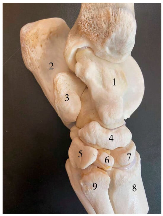
In one specimen (1/12, 8.3%), a well-defined os tarsale primum of irregular cuboid shape, larger than the os tarsale secundum, was identified in the distal row. In this specimen, a total of seven tarsal bones were included, and no fusion between os tarsale primum and os tarsale secundum was observed (Figure 1).
Figure 1.
Medial view of a skeletal anatomical preparation of the left equine tarsal region. (1) talus; (2) calcaneus; (3) sustentaculum tali; (4) os tarsi centrale; (5) os tarsale primum; (6) os tarsale secundum; (7) os tarsale tertium; (8) os metatarsale tertium; (9) os metatarsale secundum.
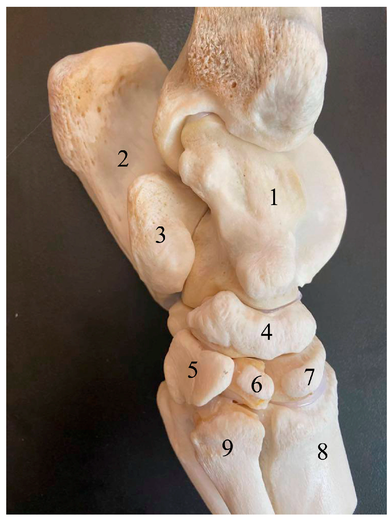
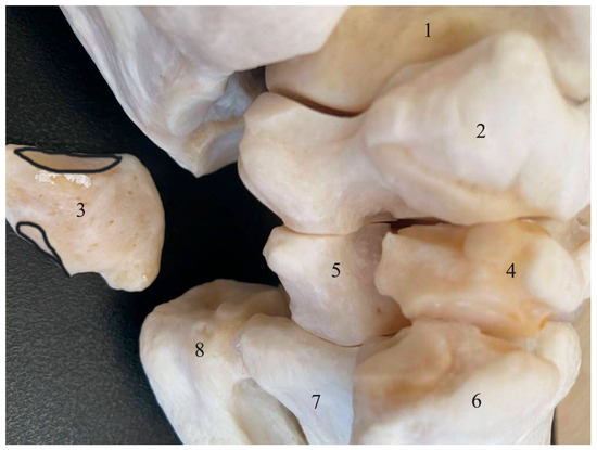
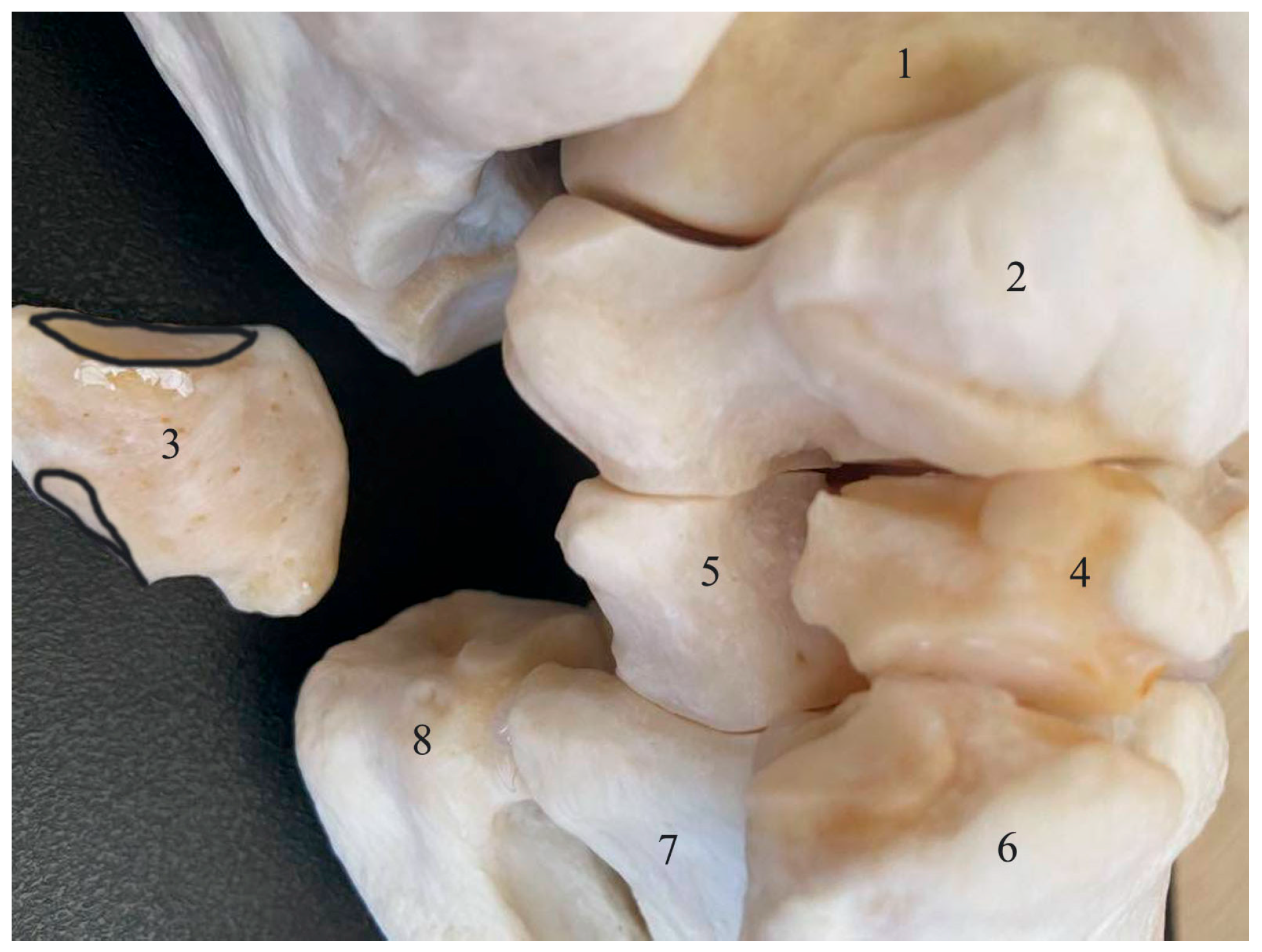
After separation of os tarsale primum, the first tarsal bone in this specimen, two well-defined articular facets were observed proximally and distally, corresponding to articulation with the os tarsi centrale and the second and third metatarsal bones (Figure 2).
Figure 2.
Medioplantar view of a skeletal anatomical preparation of the left equine tarsal region (os tarsale primum is dissected and clearly separated, and its articular facets are highlighted). (1) Talus; (2) os tarsi centrale; (3) os tarsale primum; (4) os tarsale secundum; (5) os tarsale tertium; (6) os metatarsale secundum; (7) os metatarsale tertium; (8) os metatarsale quartum.
3.2. Radiographic Findings
Radiographic examination of the specimen showing the anatomical variation confirmed and complemented the macroscopic findings.
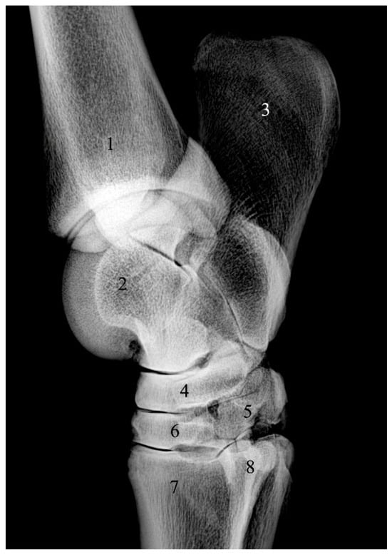
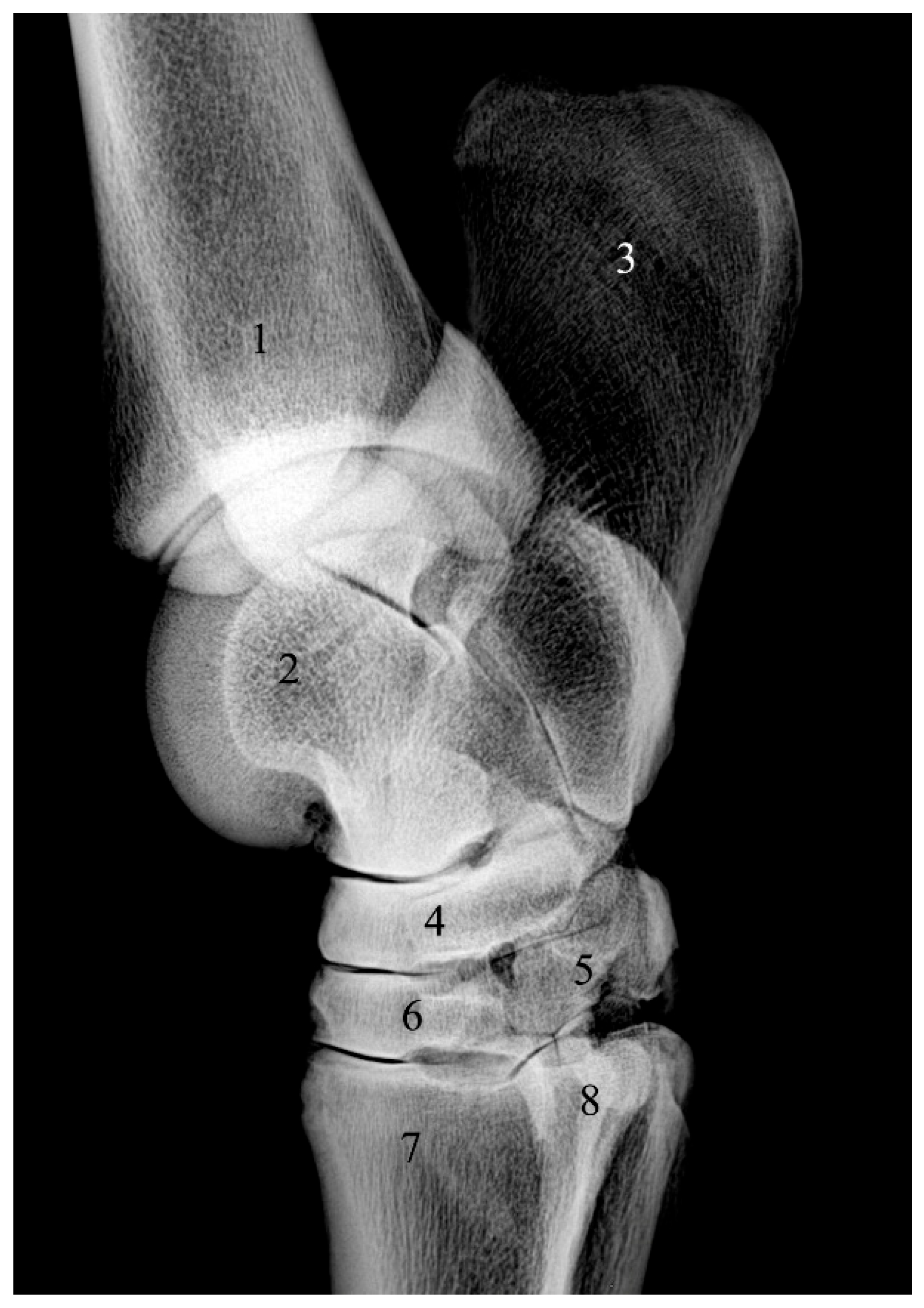
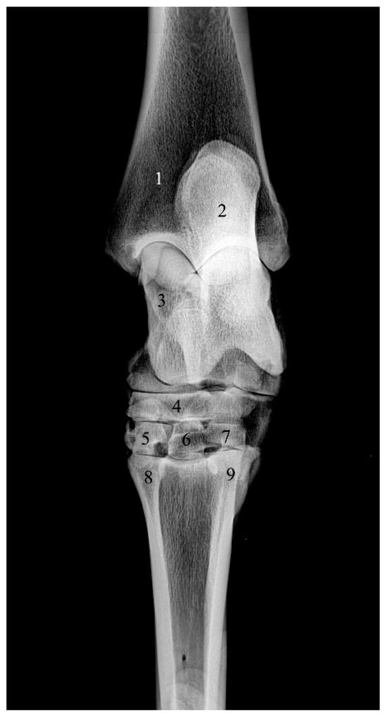
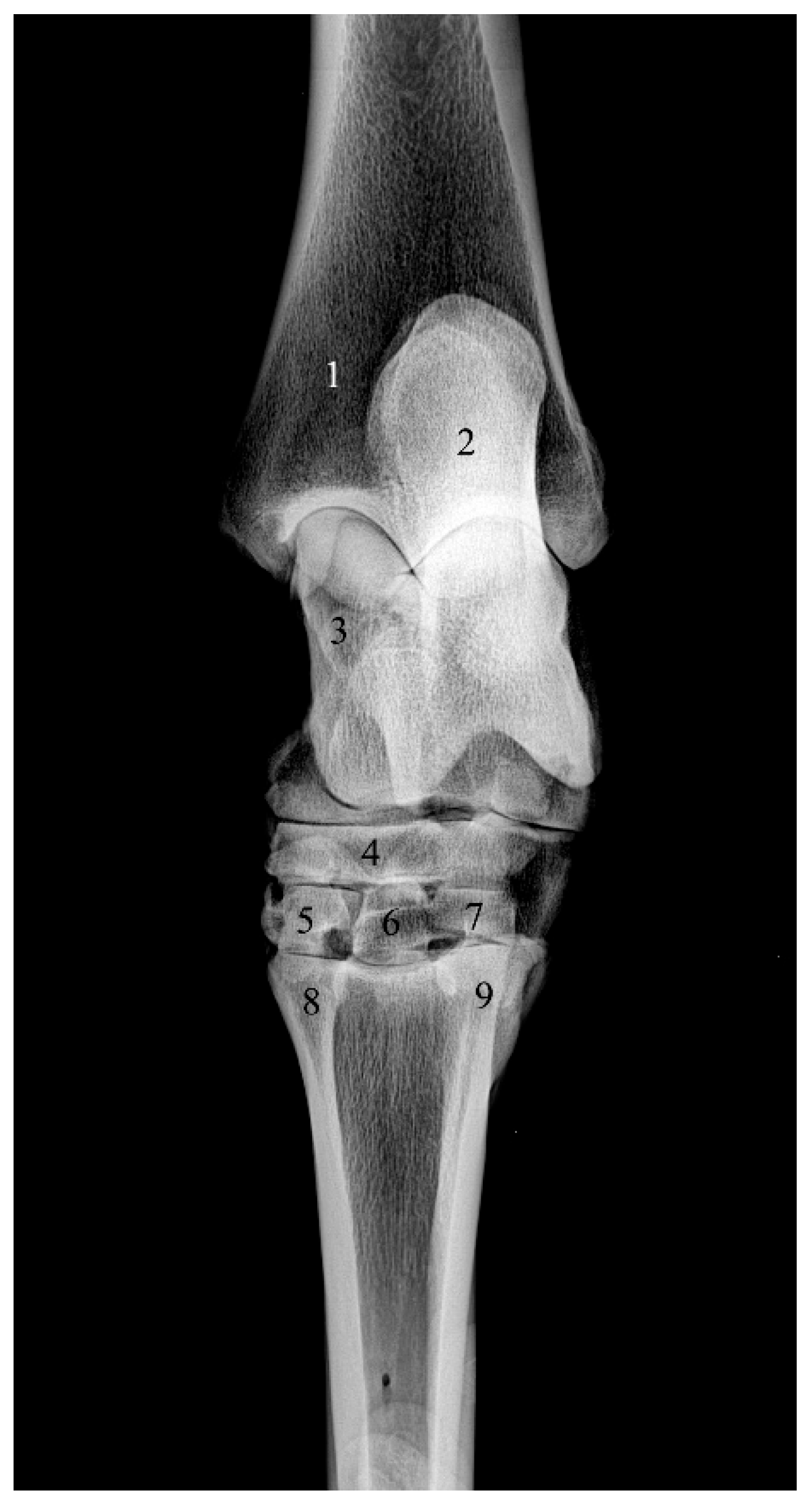
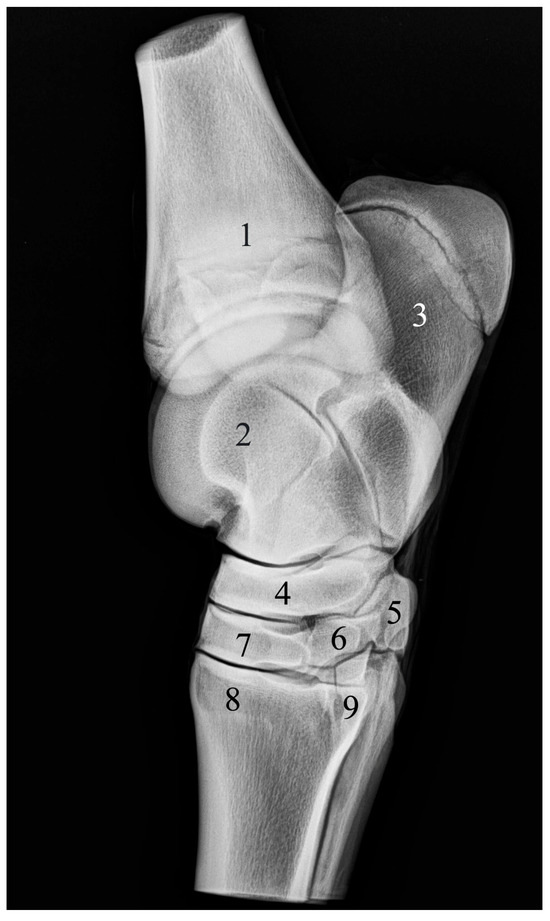
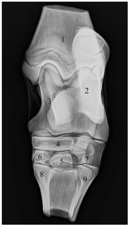
In eleven specimens, radiographs demonstrated the typical configuration of six tarsal bones, with fusion of os tarsale I and os tarsale II (Figure 3 and Figure 4). In these specimens, the fused os cuneiforme mediale articulated proximally with the os tarsi centrale and distally with the second and third metatarsal bones.
Figure 3.
Radiographic image of the right equine tarsal region. Mediolateral projection. (1) Tibia; (2) talus; (3) calcaneus; (4) os tarsi centrale; (5) os tarsale primum et secundum; (6) os tarsale tertium; (7) os metatarsale tertium; (8) os metatarsale secundum.
Figure 4.
Radiographic image of the right equine tarsal region. Dorsoplantar projection. (1) Tibia; (2) calcaneus; (3) talus; (4) os tarsi centrale; (5) os tarsale primum et secundum; (6) os tarsale tertium; (7) os tarsale quartum; (8) os metatarsale secundum; (9) os metatarsale quartum.
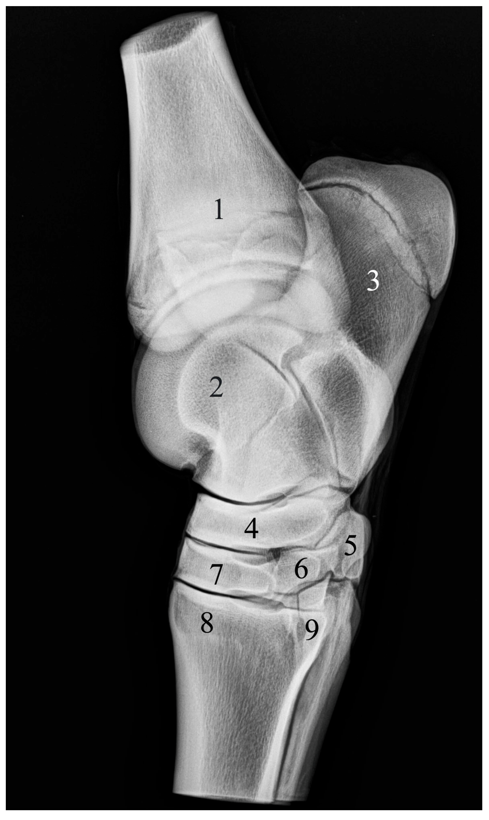
In the single specimen (1/12), mediolateral projection revealed a well-defined radiopaque structure corresponding to the os tarsale primum, in contact with the os tarsi centrale, os tarsale tertium, and the second and third metatarsal bones. The structure was clearly separated from the os tarsale secundum, with no evidence of synostosis or fusion (Figure 5).
Figure 5.
Radiographic image of the right equine tarsal region. Mediolateral projection. (1) Tibia; (2) talus; (3) calcaneus; (4) os tarsi centrale; (5) os tarsale primum; (6) os tarsale secundum; (7) os tarsale tertium; (8) os metatarsale tertium; (9) os metatarsale secundum.
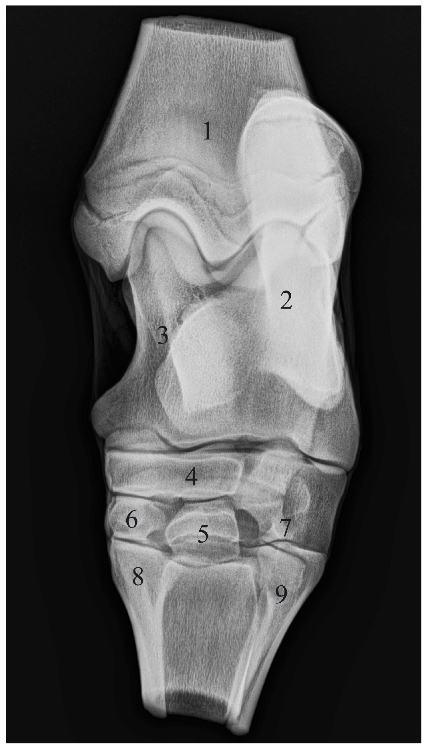
In the dorsoplantar projection of the same specimen, the three-row arrangement of the tarsal bones was confirmed. The proximal row consisted of the talus and calcaneus, and the intermediate row was represented by the os tarsi centrale. The os tarsale primum was identified in the distal row as a distinct radiopaque structure with a well-defined cortical margin and a radiolucent trabecular bone, in contact with the os tarsi centrale, os tarsale secundum, os tarsale tertium, and the second and third metatarsal bones (Figure 6).
Figure 6.
Radiographic image of the right equine tarsal region. Dorsoplantar projection. (1) Tibia; (2) calcaneus; (3) talus; (4) os tarsi centrale; (5) os tarsale primum superimpossed over os tarsale tertium; (6) os tarsale secundum; (7) os tarsale quartum; (8) os metatarsale secundum; (9) os metatarsale quartum.
4. Discussion
4.1. Key Findings
The present findings describe an unusual anatomical configuration of the distal tarsal bones in the horse, characterized by the presence of a distinct os tarsale primum and an overall arrangement of seven tarsal bones. This configuration was consistently identified through combined macroscopic and radiographic evaluation, providing both complementary anatomical and imaging evidence of this variation.
4.2. Anatomical Context and Variability
The observed configuration differs from the classical description of a fused os cuneiforme mediale (os cuneiforme mediointermedium) as reported in standard anatomical literature [4,5,6,7,8,9]. However, previous studies have documented variability in the degree of fusion and morphological presentation of the distal tarsal bones [10,11,12,13], suggesting that different patterns of ossification and fusion may occur within this anatomical region. The present observation provides additional evidence supporting the existence of such anatomical variability described in earlier anatomical and developmental studies.
4.3. Clinical and Diagnostic Implications
The identification of an independent os tarsale primum may be relevant for the interpretation of equine tarsal radiographs as such a structure could potentially be misinterpreted as a fracture line or bone fragment during routine diagnostic evaluation [14]. The absence of synostosis between os tarsale primum and os tarsale secundum in the examined specimen indicates a morphological variation rather than a pathological condition [3]. Recognition of this anatomical variation, which represents a single individual anatomical finding, is important for improving diagnostic accuracy and supporting appropriate differential diagnosis in the tarsal region [1,2,3]. In addition, awareness of such variability may contribute to more precise interpretation of degenerative or traumatic changes in the equine hock region [15,16].
4.4. Limitations
This study is limited by the examination of a single specimen derived from a collection of teaching materials. As only one out of twelve examined tarsal specimens exhibited this configuration, this represents an isolated case within the examined sample, and no population-level conclusions can be drawn regarding its frequency or distribution. Furthermore, the lack of complete metadata (including age, breed, and sex) for the examined specimens limits broader comparison with previously reported anatomical datasets.
4.5. Future Directions
Further investigations including a larger number of specimens, well-documented population data, and additional imaging modalities (e.g., computed tomography and oblique radiographic projections) are required to better characterize the frequency, developmental basis, and potential clinical relevance of this anatomical variation.
5. Conclusions
The present study documents an anatomical variation observed in a single equine tarsal specimen, representing an isolated case within the examined collection, expressed in a well-shaped and well-developed os tarsale primum, which was in anatomical contact with os tarsi centrale, os tarsale secundum, os tarsale tertium and mt II, and mt III. These features may be relevant for the anatomical understanding and the interpretation of diagnostic imaging of the equine tarsal region, particularly in cases where there is potential for misinterpretation in suspected trauma.
Author Contributions
Conceptualization, D.V., D.K. and K.S.-Y.; methodology, D.V., H.H., N.G. and K.S.-Y.; investigation, K.S.-Y., D.V., T.D. and A.L.-D. All authors have read and agreed to the published version of the manuscript.
Funding
This research received no external funding.
Institutional Review Board Statement
Not applicable.
Informed Consent Statement
Not applicable.
Data Availability Statement
The original contributions presented in this study are included in the article. Further inquiries can be directed to the corresponding authors.
Conflicts of Interest
The authors declare no conflict of interest.
Abbreviations
| t I | os tarsale I |
| t II | os tarsale II |
| mt II | os metatarsale secundum |
| mt III | os metatarsale tertium |
References
- Vanderperren, K.; Raes, E.; Hoegaerts, M.; Saunders, J.H. Diagnostic imaging of the equine tarsal region using radiography and ultrasonography. Part 1: The soft tissues. Vet. J. 2009, 179, 179–187. [Google Scholar] [PubMed]
- Schumacher, J.; Wilhite, R. How to Inject the Tarsal Joints? In Proceedings of the AAEP Proceedings, Anaheim, CA, USA, 5 December 2012. [Google Scholar]
- Soana, S.; Gnudi, G.; Bertoni, G.; Botti, P. Anatomo-radiographic study on the osteogenesis of carpal and tarsal bones in horse fetus. Anat. Histol. Embryol. 1998, 27, 301–305. [Google Scholar] [CrossRef] [Scilit] [PubMed]
- Gigov, T.S.; Gadev, H. Skeleton of the pes. In Anatomy of Domestic Animals, 2nd ed.; Gigov, T.S., Gadev, H., Eds.; Zemizdat: Sofia, Bulgaria, 1982; Volume 1, pp. 53–54. [Google Scholar]
- Liebich, H.G.; König, H.E.; Maerl, J. Hindlimb or pelvic limb (Membra pelvina). In Veterinary Anatomy of Domestic Mammals, 1st ed.; König, H.E., Liebich, H.G., Eds.; Schattauer GmbH: Stuttgart, Germany, 2004; pp. 197–262. [Google Scholar]
- Kovachev, G.; Georgiev, G.; Vodenicharov, A. Bones of the pelvic limb—Ossa membri pelvini. In Anatomy of Domestic Animals, 4th ed.; Vodenicharov, A., Ed.; KOTA Publishing House: Stara Zagora, Bulgaria, 2016; pp. 149–166. [Google Scholar]
- Barone, R. Squelette du pied. In Anatomie Comparée des Mammifères Domestiques, cinquième ed.; Barone, R., Ed.; Association Centrale d’Entraide Vétérinaire: Paris, France, 2020; pp. 701–737. [Google Scholar]
- Budras, K.D.; Sack, W.O.; Rock, S. Chapter 3: Pelvic Limb. In Anatomy of the Horse, 5th ed.; Budras, K.D., Sack, W.O., Rock, S., Eds.; Schlutersche Verlagsgesellschaft mbH & Co. KG: Hannover, Germany, 2009; pp. 16–31. [Google Scholar]
- Salomon, F.V.; Geyer, H.; Gille, U. Skeleton pedis: Ossa tarsi, Ossa metatrsalia. In Anatomie für die Tiermedizin; Enke Verlag: Stuttgart, Germany, 2005; pp. 78–80. [Google Scholar]
- Raes, E.V.; Bergman, E.H.; van der Veen, H.; Vanderperren, K.; Van der Vekens, E.; Saunders, J.H. Comparison of cross-sectional anatomy and computed tomography of the tarsus in horses. Am. J. Vet. Res. 2011, 72, 1209–1221. [Google Scholar] [CrossRef] [Scilit] [PubMed]
- Taylor, S.G. Some Studies on Equine Hock Joint Disease Whith Particular Reference to Tarsal Osteoarthritis. MVM Dissertation, Department of Veterinary Surgery, University of Glasgow, Glasgow, UK, December 1977. [Google Scholar]
- Gadjev, S.T. Skeleton of the pes. In Anatomy of Domestic Animals, 1st ed.; Gadjev, S.T., Ed.; Jusator: Sofia, Bulgaria, 2000; pp. 170–178. [Google Scholar]
- Ley, C.J.; Ekman, S.; Hansson, K.; Björnsdóttir, S.; Boyde, A. Osteochondral lesions in distal tarsal joints of icelandic horses reveal strong associations between hyaline and calcified cartilage abnormalities. Eur. Cells Mater. 2014, 27, 213–236. [Google Scholar] [CrossRef] [Scilit] [PubMed]
- Knuchell, J.A.; Spriet, M.; Galuppo, L.D.; Katzman, S.A. Fracture of the central tarsal bone in nonracehorses: Four cases. Vet. Radiol. Ultrasound 2016, 57, 403–409. [Google Scholar] [CrossRef] [Scilit] [PubMed]
- Sprackman, L.; Dakin, S.G.; May, S.A.; Weller, R. Relationship between the shape of the central and third tarsal bones and the presence of tarsal osteoarthritis. Vet. J. 2015, 204, 94–98. [Google Scholar] [CrossRef] [Scilit] [PubMed]
- Skelly-Smith, E.; Ireland, J.; Dyson, S. The centrodistal joint interosseous ligament region in the tarsus of the horse: Normal appearance, abnormalities and possible association with other tarsal lesions, including osteoarthritis. Equine Vet. J. 2016, 48, 457–465. [Google Scholar] [CrossRef] [Scilit] [PubMed]
Disclaimer/Publisher’s Note: The statements, opinions and data contained in all publications are solely those of the individual author(s) and contributor(s) and not of MDPI and/or the editor(s). MDPI and/or the editor(s) disclaim responsibility for any injury to people or property resulting from any ideas, methods, instructions or products referred to in the content. |
© 2026 by the authors. Licensee MDPI, Basel, Switzerland. This article is an open access article distributed under the terms and conditions of the Creative Commons Attribution (CC BY) license.