


Simple Summary
Gallbladder rupture in dogs is a serious condition, but rare complications can be difficult to detect. In this case, an abnormal connection formed between the gallbladder and the liver. Conventional ultrasound showed unclear findings, making diagnosis challenging. A contrast-enhanced ultrasonography clearly revealed the connection and helped identify the liver lesion as inflammation rather than a tumor. Surgery confirmed gallbladder rupture with adhesion to the liver. This case shows that contrast-enhanced ultrasound can improve diagnosis and support better treatment decisions in dogs with gallbladder rupture.
Abstract
Cholecystohepatic communication is a rare complication of gallbladder rupture in dogs, and its imaging features are not well described. A 10-year-old Pomeranian showed a gallbladder wall defect, a tortuous tubular structure extending into an adjacent ill-defined hepatic lesion. Contrast-enhanced ultrasonography revealed marked enhancement of the tubular structure and weaker heterogeneous enhancement of the hepatic lesion. Laparotomy confirmed gallbladder rupture with severe adhesion at the suspected communication site. Contrast-enhanced ultrasonography enabled delineation of the enhancing tubular structure and improved characterization of the adjacent hepatic inflammation, increasing diagnostic confidence in cholecystohepatic communication secondary to gallbladder rupture.
1. Introduction
Gallbladder rupture is an uncommon but clinically important condition in dogs. It can be consequential to mucocele, cholelithiasis, cholecystitis, bacterial infections, and gallbladder neoplasia in dogs [1,2,3]. Gallbladder rupture typically results in bile leakage into the peritoneal cavity, leading to bile peritonitis [4]. Extension of the perforation into the adjacent tissue is considered rare. Only a few cases of cholecystocutaneous fistula secondary to gallbladder rupture have been reported [5,6,7,8]. Inflammatory adhesion between the ruptured gallbladder and liver surface may progress to focal dissection of bile into the hepatic tissue or formation of a cholecystohepatic communication [9,10,11,12,13,14,15]. However, to the authors’ knowledge, fistulous communication between a ruptured gallbladder and an internal visceral organ has not been reported in veterinary medicine.
Ultrasonography is the primary diagnostic imaging modality for evaluating gallbladder integrity. However, distinguishing inflammatory adhesion, focal parenchymal involvement, or potential communication tracts from surrounding soft tissue can be challenging on conventional ultrasonography [16]. In contrast, contrast-enhanced ultrasonography (CEUS) uses an intravascular microbubble-based contrast agent to allow real-time evaluation of tissue microvascular perfusion based on the echogenicity of the contrast agent [17]. In hepatobiliary disease, CEUS can improve lesion conspicuity and may help differentiate vascularized inflammatory tissue from non-enhancing bile, debris, or necrotic material. CEUS may also help overcome the limitations of conventional ultrasonography by improving lesion conspicuity and better delineating abnormal structures associated with gallbladder rupture [18].
This report describes a dog with gallbladder rupture and severe adhesion to the adjacent liver parenchyma, in which CEUS was used to assess the actual communication between the ruptured gallbladder and the adjacent hepatic lesion and to improve visualization of a tubular structure suspected to represent cholecystohepatic communication. To the authors’ knowledge, this is the first report describing CEUS findings of suspected cholecystohepatic communication secondary to gallbladder rupture in dogs.
2. Case Description
A 10-year-old neutered male Pomeranian presented with partial anorexia and vomiting for 5 days. Physical examination revealed cranial abdominal pain. Laboratory examination showed mild anemia (hematocrit, 27.9%; reference range, 37.3–61.7%) and markedly increased alkaline phosphatase (1590 U/L; reference range, 23–212 U/L) and C-reactive protein (101 mg/L; reference range, 0–10 mg/L) levels. Prothrombin time (10.0 s; reference range, 7.4–8.8 s) and activated partial thromboplastin time (64 s; reference range, 12–28 s) were elevated.
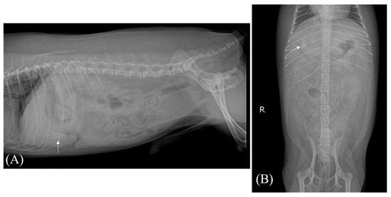
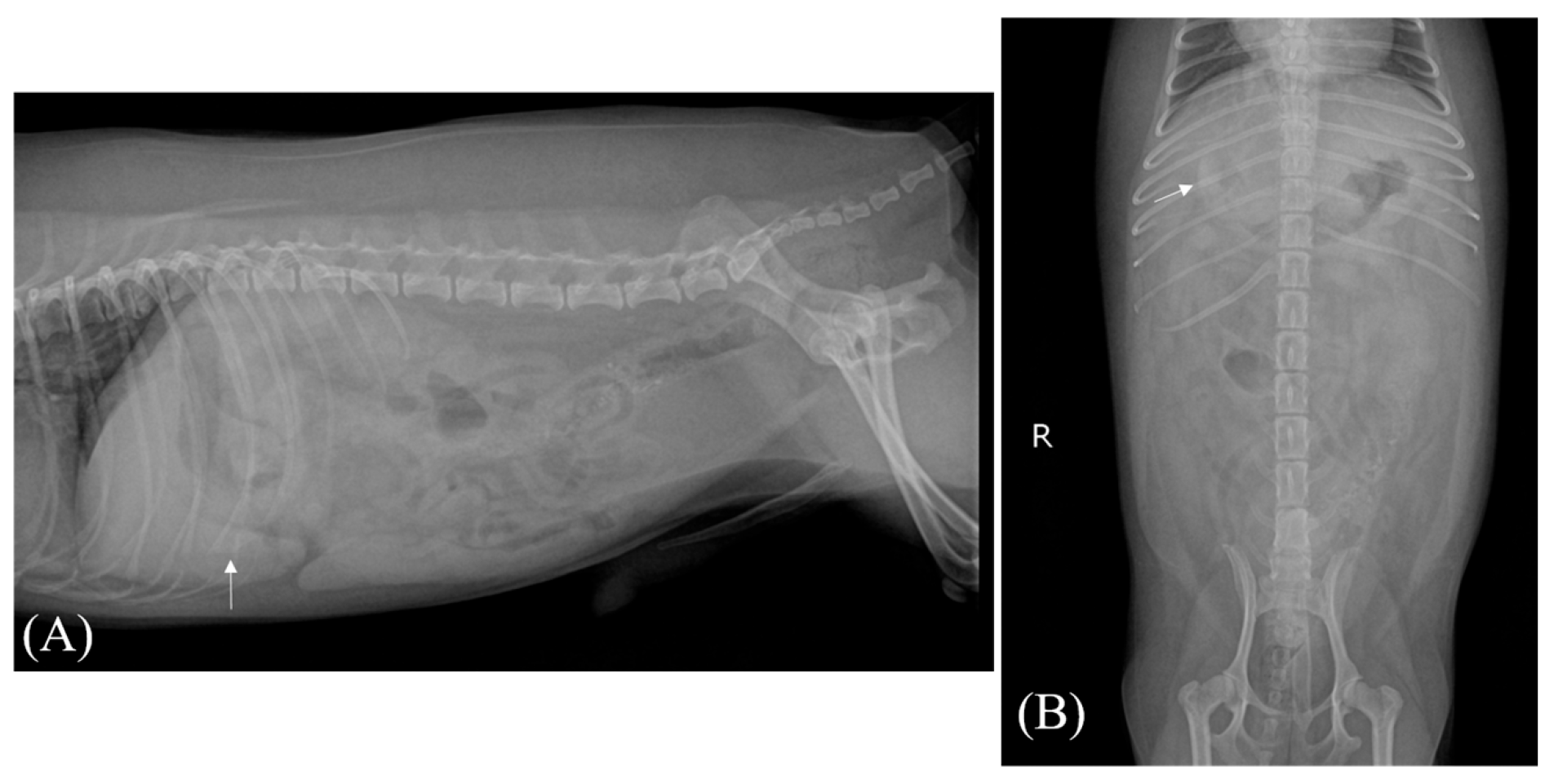
Right lateral and ventrodorsal abdominal radiographs were acquired with a digital radiography unit (EVA-HF525, Gemss-Medical) equipped with a table-integrated cesium iodide flat-panel detector (FDX4343R, Gemss-Medical). The system allowed a maximum tube voltage of 125 kV and a maximum tube current of 500 mA. Abdominal radiography revealed mild hepatomegaly, decreased serosal details in the cranial abdomen, and radiopaque cholelithiasis (Figure 1). Decreased serosal detail suggested the presence of a small amount of peritoneal effusion. In addition, a radiopaque structure identified in the anatomical region of the gallbladder was considered most consistent with cholelithiasis.
Figure 1.
Abdominal radiographic images of the patient. On the lateral (A) and ventrodorsal (B) views, radiopaque cholelithiasis is observed in the right cranial portion of the liver, with mild hepatomegaly and decreased serosal detail.
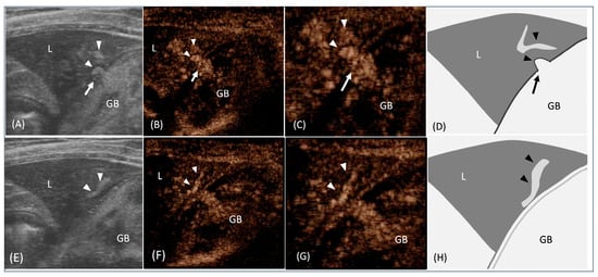
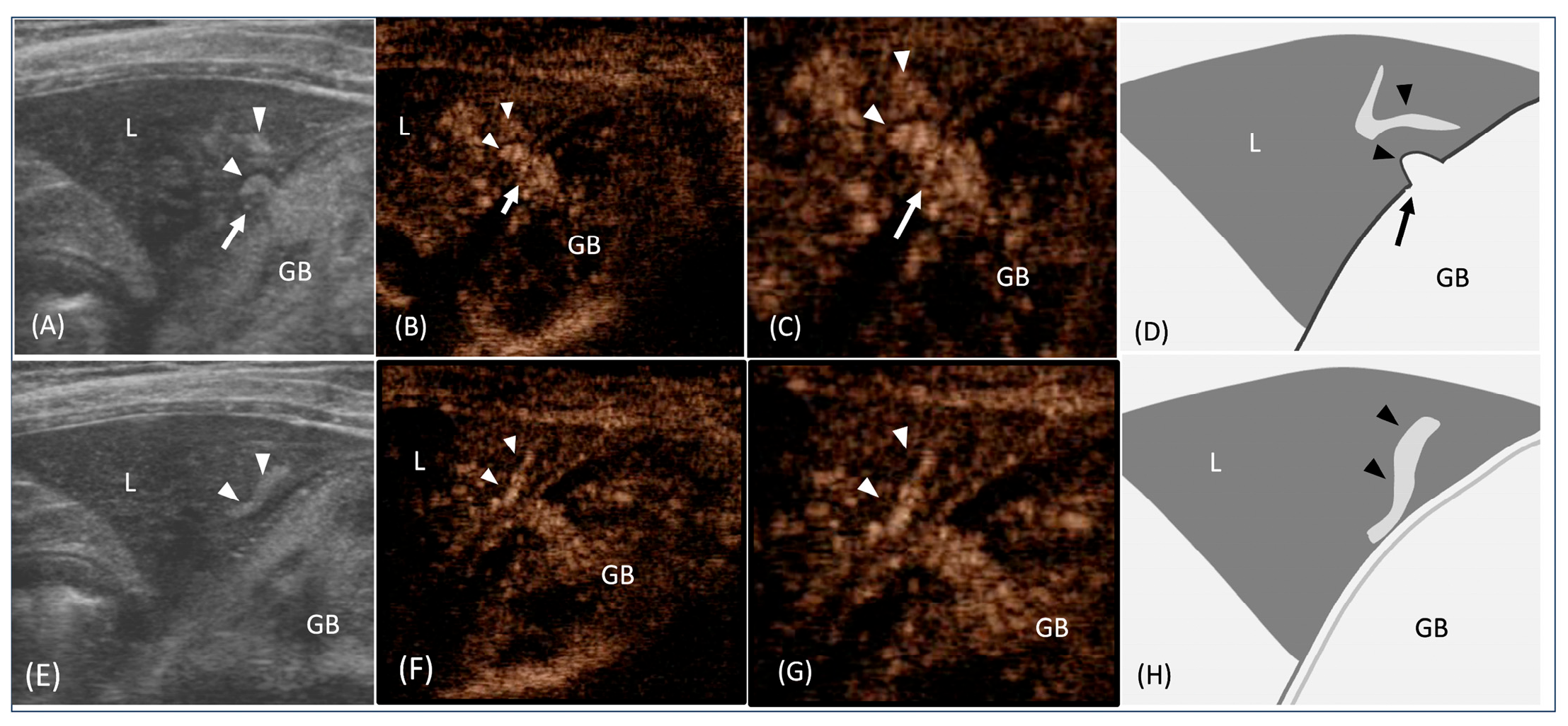
Ultrasonography was performed on the patient in dorsal and lateral recumbency without sedation or anesthesia, using manual restraint. Conventional ultrasonography and CEUS were performed using the same ultrasound unit (Prosound Alpha 7, Hitachi-Aloka, Tokyo, Japan) with a 10 MHz linear transducer. Conventional ultrasonography showed a distended gallbladder containing hyperechoic bile sludge and choleliths with acoustic shadowing. The gallbladder wall was thickened and showed a double-rim sign. A 2.3 mm focal wall defect was identified in the right cranial aspect of the gallbladder fundus (Figure 2).
Figure 2.
Conventional ultrasonography and contrast-enhanced ultrasonography (CEUS) of the cholecystohepatic communication secondary to gallbladder rupture. (A) Conventional ultrasonography shows a focal gallbladder (GB) wall defect (thin arrow) and adjacent echogenic structure (arrowheads) extending from a focal gallbladder defect (thin arrow) toward the liver (L). (B) CEUS image of the same plane and (C) magnified CEUS image demonstrate enhancement of the structure (arrowheads) and its continuity with the gallbladder (GB) lumen through the defect (thin arrow). (D) Schematic illustration of the tubular tract (black arrowheads) passing through the gallbladder wall defect (black arrow), consistent with cholecystohepatic communication. (E) When the echogenic structure is traced cranially, it appears as a tortuous tubular structure (arrowheads) coursing within the right medial liver lobe (L). (F) CEUS of the same plane and (G) magnified CEUS image clearly delineate the course of the enhancing tract (arrowheads) within the hepatic parenchyma. (H) Schematic illustration showing the tubular tract extending through the right medial liver lobe.
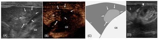
Reactive hyperechoic fat change was observed throughout the cranial abdominal cavity, with a small amount of echogenic peritoneal fluid surrounding the gallbladder and within the abdominal cavity. A tortuous echogenic tubular structure extended from the gallbladder lumen through the defect into the right medial liver lobe. Adjacent to the tubular structure, an ill-defined hypoechoic hepatic mass measuring 11 × 7 mm was found (Figure 3).
Figure 3.
Conventional ultrasonography and contrast-enhanced ultrasonography (CEUS) of the hepatic mass located at the terminal portion of the tubular tract and the overall appearance of the gallbladder. (A) When the tortuous hyperechoic tubular structure identified in Figure 1 is followed to its cranial end, an ill-defined hypoechoic mass (arrows) is observed within the hepatic parenchyma. (B) On CEUS, the indistinct mass (arrows) becomes clearly delineated as a hypoechoic lesion due to contrast enhancement of the surrounding hepatic parenchyma. (C) Schematic illustration depicting the hypoechoic mass located at the terminal end of the tract within the liver. (D) Conventional ultrasonography of the gallbladder reveals abundant sludge and choleliths within the lumen, a double-rim sign along the gallbladder wall (arrow), and surrounding hyperechoic edematous change. L, liver; GB, gallbladder.
CEUS was performed in extended pure harmonic detection mode, with the focal zone positioned near the bottom of the image for homogeneous acoustic energy distribution. The transmitted acoustic energy was reduced to 10%, and the mechanical index was 0.22. Perfluorobutane contrast agent (Sonazoid, GE Healthcare, Oslo, Norway) was agitated before administration and injected at a dose of 0.015 mL/kg as a rapid intravenous bolus through a 24-gauge catheter placed in a cephalic vein, followed immediately by a 5 mL bolus of 0.9% sodium chloride solution. During CEUS, enhancement was evaluated qualitatively according to the temporal enhancement pattern of the surrounding hepatic parenchyma, including the wash-in, peak enhancement, and subsequent wash-out phases. Contrast enhancement was monitored continuously for 10 min after contrast administration. The tortuous tubular structure demonstrated clear hyperenhancement relative to the hepatic parenchyma, allowing its course from the gallbladder defect into the liver to be easily visualized (Figure 1). The adjacent hypoechoic hepatic mass showed weaker, heterogeneous enhancement compared with the surrounding liver during the contrast phase, making it distinguishable from normal parenchyma (Figure 3).
Based on these imaging features, including gallbladder wall discontinuity, a contrast-enhancing tubular structure connecting the gallbladder to the liver, and a heterogeneously enhancing hepatic lesion, cholecystohepatic communication secondary to gallbladder rupture with a suspected hepatic abscess was diagnosed.
Exploratory laparotomy was performed as an emergency procedure under general anesthesia. The ventral abdomen was clipped and aseptically prepared, and a ventral midline celiotomy was performed. Exploratory laparotomy confirmed gallbladder rupture with marked adhesion between the gallbladder and the adjacent hepatic parenchyma. The adhered region of the liver corresponded to the ultrasonographic site of suspected communication and was excised along with the gallbladder. Histopathologic examination was not performed. The patient recovered uneventfully, and the dog was managed with intravenous fluid therapy, analgesics, and antimicrobial treatment. Appetite and vomiting improved, and no immediate postoperative complications were observed. The dog was discharged in stable condition; however, the dog was subsequently lost to follow-up.
A generative artificial intelligence tool (ChatGPT-5, OpenAI, San Francisco, CA, USA) was used only for language editing and improvement of clarity during the preparation of this manuscript. It was not used for data analysis, image interpretation, or generation of scientific conclusions.
3. Discussion
In this case, ultrasonography identified a focal defect in the gallbladder wall and an echogenic tubular structure extending directly into the right medial liver lobe. These findings raised suspicion of cholecystohepatic communication, a complication that has been rarely reported in human patients [15,16]. In previously reported human patients with cholecystohepatic communication, gallbladders were found to be partial or complete intrahepatic [9,10,11,12,13,14,15]. This intrahepatic gallbladder is associated with a higher incidence of cholelithiasis, and rupture of the intrahepatic gallbladder is generally considered a very rare evolution of cholelithiasis [14,16]. The mechanism of intrahepatic gallbladder rupture involves a cascade of events triggered by obstruction of the cystic duct. Obstruction of the cystic duct leads to biliary stasis and gallbladder distension with a consequential increase in intraluminal pressure, compromise of the venous and lymphatic drainage, and vascular damage that results in gallbladder rupture intrahepatically rather than intraperitoneally [9,19,20].
In dogs, the most common underlying conditions associated with gallbladder rupture include gallbladder mucocele, necrotizing cholecystitis, cholelithiasis, and biliary obstruction. Conventional ultrasonography can help differentiate these underlying biliary diseases based on gallbladder contents, wall characteristics, and pericholecystic changes. Gallbladder mucocele typically appears as immobile, organized echogenic bile with stellate or finely striated patterns, whereas cholecystitis is more commonly associated with gallbladder wall thickening, a double-rim sign, biliary sludge, and pericholecystic inflammatory change. Cholelithiasis is identified as hyperechoic intraluminal material with distal acoustic shadowing, and biliary obstruction may be suggested by gallbladder distension and extrahepatic or intrahepatic bile duct dilation. In the present case, typical ultrasonographic features of gallbladder mucocele were not observed, and marked adhesion between the gallbladder and the adjacent liver was confirmed during surgery. Therefore, severe inflammation and subsequent necrosis of the gallbladder wall were considered the most likely cause of rupture.
In addition to the findings of gallbladder rupture, this case showed a tortuous echogenic tubular structure extending from the gallbladder wall defect into the adjacent hepatic parenchyma and an ill-defined hepatic lesion near the terminal portion of the structure. These findings were unusual for uncomplicated gallbladder rupture, cholecystitis, or cholelithiasis. Moreover, conventional ultrasonography could not clearly determine whether it represented passive leakage of biliary material or a communication with the adhered hepatic parenchyma.
Gallbladder rupture with cholecystohepatic communication has been rarely reported in human medicine [15,16]. Despite chronic processes with communication formation, its clinical progress is often acute [16]. Ultrasonography can make an imaging diagnosis of this disease by showing a distended gallbladder, a thickened gallbladder, and a “hole sign” between the gallbladder and the liver [12]. In addition, hepatic abscess with continuity into the gallbladder or the presence of echogenic stones in the hepatic abscess increases confidence in the diagnosis of cholecystohepatic communication [12]. However, to the authors’ knowledge, this complication has not been previously described in dogs.
Although conventional ultrasonography is the primary imaging modality for evaluating gallbladder disease and gallbladder rupture in dogs, complex sequelae such as inflammatory adhesion, focal bile leakage, and communication with adjacent hepatic parenchyma may be difficult to distinguish. This diagnostic difficulty has also been reported in human medicine because intrahepatic gallbladder perforation, hepatic abscess, and neoplasia may have overlapping imaging appearances [16]. CEUS provides real-time evaluation of tissue perfusion and lesion enhancement and has been used in human medicine to improve characterization of hepatobiliary lesions and inflammatory complications. In veterinary medicine, CEUS has been applied to characterize hepatic lesions based on contrast-enhancement patterns, to detect gallbladder wall defects, and to differentiate gallbladder sludge from inflammatory polyps [21,22,23,24,25]. While these conventional applications have predominantly focused on evaluating either the liver or the gallbladder as an isolated structure, their integrated application to evaluate both organs simultaneously—specifically in suspected cholecystohepatic communication secondary to gallbladder rupture—has not yet been reported. In this case, CEUS demonstrated marked enhancement of the tubular structure and allowed its entire course from the gallbladder defect to the adjacent liver to be clearly traced. The enhancement pattern suggested that the tubular structure was vascularized through an inflammatory process during the development of communication rather than only static bile or debris [19].
Conventional ultrasonography revealed an ill-defined mass in the right medial lobe of the liver. Considering the echogenicity, shape, size, and location of the mass, differentials that were considered included abscess, nodular hyperplasia, hematoma, or neoplasia [22]. CEUS enabled differentiation of the adjacent ill-defined liver mass in the right medial lobe from normal parenchyma by revealing weaker, heterogeneous enhancement during the distribution phase. This finding was more suggestive of an inflammatory process than neoplasia by demonstrating a lack of vascularity within it [26,27]. These CEUS characteristics increased confidence that the gallbladder rupture had progressed to form a communication with adhered hepatic parenchyma, and a hepatic abscess was the most likely diagnosis.
The imaging findings were confirmed by the surgical observations, which revealed gallbladder rupture and pronounced adhesion between the gallbladder and the liver at the site corresponding to the suspected communication. Recent studies have highlighted the utility of computed tomography in evaluating gallbladder rupture; however, CT was not performed in the present case because the lesion appeared relatively localized [28,29]. In addition, histopathologic examination of the hepatic lesion was not performed, which represents a limitation of this report. Although histopathologic confirmation was not obtained, the agreement between CEUS findings, conventional ultrasonography, and surgical exploration strengthened the interpretation that a cholecystohepatic communication had developed. Similar to human cases, chronic inflammation, gallbladder distension, and local adhesions may have contributed to focal extension of biliary contents into the hepatic parenchyma [15,16].
4. Conclusions
In conclusion, this report presented the diagnostic value of CEUS for characterizing complex sequelae of gallbladder rupture. By improving visualization of abnormal structures and enhancing contrast between affected and normal tissue, CEUS can help in identifying rare complications such as cholecystohepatic communication and in distinguishing inflammatory hepatic lesions from other differentials. Despite the rarity of this condition, cholecystohepatic communication should be considered in dogs with gallbladder ruptures and adjacent hepatic lesions.
Author Contributions
Conceptualization, H.C. and J.C.; data curation, S.-K.L., S.P. and J.C.; formal analysis, S.-K.L., S.P. and J.C.; funding acquisition, J.C.; investigation, J.C.; methodology, S.-K.L., S.H., S.P., S.K. and J.C.; project administration, J.C.; supervision, J.C.; validation, J.C.; writing—original draft, H.C., S.-K.L. and J.C.; writing—review and editing, H.C., S.-K.L., S.H., S.P., S.K. and J.C. All authors have read and agreed to the published version of the manuscript.
Funding
This research was supported by the Basic Science Research Program through the National Research Foundation of Korea (NRF) funded by the Ministry of Science, ICT and Future Planning (NRF-2021R1A2C200573011).
Institutional Review Board Statement
Ethical review and approval were not required for this case report in accordance with local legislation and institutional requirements. Informed consent was obtained from the owner of the animal.
Informed Consent Statement
The owner provided informed consent for the use of their animal’s information and medical records for academic and research purposes.
Data Availability Statement
The original contributions presented in this study are included in the article. Further inquiries can be directed to the corresponding author.
Acknowledgments
During the preparation of this manuscript, the authors used ChatGPT, a generative artificial intelligence tool developed by OpenAI, for language editing and improvement of clarity. The authors reviewed and edited the content as needed and take full responsibility for the final version of the manuscript.
Conflicts of Interest
The authors declare no conflicts of interest.
Abbreviations
The following abbreviations are used in this manuscript:
| CEUS | Contrast-enhanced ultrasonography |
References
- Crews, L.J.; Feeney, D.A.; Jessen, C.R.; Rose, N.D.; Matise, I. Clinical, ultrasonographic, and laboratory findings associated with gallbladder disease and rupture in dogs: 45 cases (1997–2007). J. Am. Vet. Med. Assoc. 2009, 234, 359–366. [Google Scholar] [CrossRef] [Scilit]
- Besso, J.; Wrigley, R.; Gliatto, J.; Webster, C.R.L. Ultrasonographic appearance and clinical findings in 14 dogs with gallbladder mucocele. Vet. Radiol. Ultrasound 2000, 41, 261–271. [Google Scholar] [CrossRef] [Scilit]
- Choi, J.; Kim, A.; Keh, S.; Oh, J.; Kim, H.; Yoon, J. Comparison between ultrasonographic and clinical findings in 43 dogs with gallbladder mucoceles. Vet. Radiol. Ultrasound 2014, 55, 202–207. [Google Scholar] [CrossRef] [Scilit]
- Center, S.A. Diseases of the gallbladder and biliary tree. Vet. Clin. N. Am. Small Anim. Pract. 2009, 39, 543–598. [Google Scholar] [CrossRef] [Scilit]
- Fabbi, M.; Volta, A.; Quintavalla, F.; Zubin, E.; Manfredi, S.; Martini, F.M.; Mantovani, L.; Tribaudino, M.; Gnudi, G. Cholecystocutaneous fistula containing multiple gallstones in a dog. Can. Vet. J. 2014, 55, 1163–1166. [Google Scholar] [PubMed] [PubMed Central]
- Marquardt, S.A.; Rochat, M.C.; Johnson-Neitman, J.L. Spontaneous cholecystocutaneous fistula in a dog. J. Am. Anim. Hosp. Assoc. 2012, 48, 43–49. [Google Scholar] [CrossRef] [Scilit] [PubMed]
- Kligman, K.C.; Jehn, C.T.; Davis, K.M.; Taylor, D.P.; Conway, J.A. Spontaneous external biliary fistula in a dog. J. Am. Vet. Med. Assoc. 2007, 231, 919–923. [Google Scholar] [CrossRef] [Scilit]
- Testault, I.; Sarran, D.; Kolb, H. Spontaneous cholecystocutaneous fistula in a dog. Rev. Vet. Clin. 2022, 57, 75–79. [Google Scholar] [CrossRef] [Scilit]
- Hussain, T.; Adams, M.; Ahmed, M.; Arshad, N.; Solkar, M. Intrahepatic perforation of the gallbladder causing liver abscesses: Case studies and literature review of a rare complication. Ann. R. Coll. Surg. Engl. 2016, 98, 88–91. [Google Scholar] [CrossRef] [Scilit]
- Jayasinghe, G.; Adam, J.; Abdul-Aal, Y. Unusual presentation of gallbladder perforation. Int. J. Surg. Case Rep. 2016, 18, 42–44. [Google Scholar] [CrossRef] [Scilit] [PubMed]
- Jo, S.; Song, S. Intrahepatic Abscess due to Gallbladder Perforation. Korean J. Gastroenterol. 2019, 74, 119–122. [Google Scholar] [CrossRef] [Scilit]
- Kochar, K.; Vallance, K.; Mathew, G.; Jadhav, V. Intrahepatic perforation of the gall bladder presenting as liver abscess: Case report, review of literature and Niemeier’s classification. Eur. J. Gastroenterol. Hepatol. 2008, 20, 240–244. [Google Scholar] [CrossRef] [Scilit]
- Lin, H.C.; Chen, T.S. Gallbladder perforation in cholecystitis with liver abscess formation and septic thrombophlebitis of portal vein mimicking presentation of liver malignancy. Adv. Dig. Med. 2014, 1, 95–99. [Google Scholar] [CrossRef] [Scilit]
- Uryu, N.H.; Godoy, G.d.F.; Alves Junior, W.F.; Amim, I.B.; Fontes, E.V.L.C.; Branco, A.C.; Farah, J.F.d.M. Rare Association between Gallbladder Perforation, Distant Hepatic Abscess Formation and Choledocolithiasis: Case Report and Literature Review. Ann. Clin. Case Rep. 2022, 7, 2192. [Google Scholar] [CrossRef]
- Singh, K.; Singh, A.; Vidyarthi, S.H.; Jindal, S.; Thounaojam, C.K. Spontaneous intrahepatic type II gallbladder perforation: A rare cause of liver abscess–case report. J. Clin. Diagn. Res. 2013, 7, 2012–2014. [Google Scholar] [CrossRef] [Scilit] [PubMed]
- Donati, M.; Biondi, A.; Basile, F.; Gruttadauria, S. An atypical presentation of intrahepatic perforated cholecystitis: A modern indication to open cholecystectomy. Report of a case. BMC Surg. 2014, 14, 6. [Google Scholar] [CrossRef] [Scilit] [PubMed]
- Wilson, S.R.; Burns, P.N. Microbubble-enhanced US in body imaging: What role? Radiology 2010, 257, 24–39. [Google Scholar] [CrossRef] [Scilit]
- Bargellini, P.; Orlandi, R.; Paloni, C.; Rubini, G.; Fonti, P.; Peterson, M.E.; Rishniw, M.; Boiti, C. Evaluation of contrast-enhanced ultrasonography as a method for detecting gallbladder necrosis or rupture in dogs. Vet. Radiol. Ultrasound 2016, 57, 611–620. [Google Scholar] [CrossRef] [Scilit]
- Shah, M.; Mori, W. A clinic-pathological study of spontaneous internal biliary fistula. Pathol. Int. 1973, 23, 349–358. [Google Scholar] [CrossRef] [Scilit]
- Hollanda, E.S.; Torres, U.S.; Gual, F.; Oliveira, E.P.; Cardoso, L.V.; Criado, D.A.B. Spontaneous perforation of gallbladder with intrahepatic biloma formation: Sonographic signs and correlation with computed tomography. Radiol. Bras. 2013, 46, 320–322. [Google Scholar] [CrossRef] [Scilit]
- Jaffey, J.A.; Graham, A.; VenEerde, E.; Hostnik, E.; Alvarez, W.; Arango, J.; Jacobs, C.; DeClue, A.E. Gallbladder mucocele: Vari-ables associated with outcome and the utility of ultrasonography to identify gallbladder rupture in 219 dogs (2007–2016). J. Vet. Intern. Med. 2018, 32, 195–200. [Google Scholar] [CrossRef] [Scilit]
- Nakamura, K.; Sasaki, N.; Murakami, M.; Kmara, W.R.B.; Ohta, H.; Yamasaki, M.; Takagi, S.; Osaki, T.; Takiguchi, M. Contrast-enhanced characterization of canine focal liver lesions. J. Vet. Intern. Med. 2010, 24, 1290–1297. [Google Scholar] [CrossRef] [Scilit] [PubMed]
- Toaldo, M.B.; Diana, A.; Bettini, G.; Donato, P.D.; Linta, N.; Cipone, M. Imaging diagnosis-cholangiocellular adenoma: Contrast-enhanced ultrasonographic findings of a benign tumor mimicking malignant neoplasia in a dog. Vet. Radiol. Ultrasound 2013, 54, 71–74. [Google Scholar] [CrossRef] [Scilit] [PubMed]
- Burti, S.; Zotti, A.; Rubini, G.; Orlandi, R.; Bargellini, P.; Bonsembiante, F.; Banzato, T. Contrast-enhanced ultrasound features of malignant focal liver masses in dogs. Sci. Rep. 2020, 10, 6076. [Google Scholar] [CrossRef] [Scilit]
- Lee, J.; Kang, J.; Heo, S.; Lee, K.; Yoon, H. Case Report: Imaging Features of Gallbladder Sessile Polyp Confirmed by Contrast-Enhanced Ultrasonography and Dynamic Computed Tomography in a Dog With Asymptomatic Chronic Cholecystitis. Front. Vet. Sci. 2022, 9, 836414. [Google Scholar] [CrossRef] [Scilit] [PubMed]
- Li, R.; Guo, Y.; Hua, X.; He, Y.; Ding, J.; Guo, A.; Luo, M. Characterization of focal liver lesions: Comparison of pulse-inversion harmonic contrast-enhanced sonography with contrast-enhanced CT. J. Clin. Ultrasound 2007, 35, 109–117. [Google Scholar] [CrossRef] [Scilit]
- Kanemoto, H.; Ohno, K.; Nakashima, K.; Takahashi, M.; Fujino, Y.; Nishimura, R.; Tsumjimoto, H. Characterization of canine focal liver lesions with contrast-enhanced ultrasound using a novel contrast agent—Sonazoid. Vet. Radiol. Ultrasound 2009, 50, 188–194. [Google Scholar] [CrossRef] [Scilit]
- Gordon, C.R.; Fernandez, N.; Schwarz, T. CT findings of gall bladder rupture in two dogs with gall bladder mucocele. Vet. Rec. Case Rep. 2017, 5, e000481. [Google Scholar] [CrossRef] [Scilit]
- Cordella, A.; Gianesini, G.; Zoia, A.; Ventura, L.; Bertolini, G. Multi-phase MULTIDETECTOR-row computed tomographic features and laboratory findings in dogs with gallbladder rupture. Res. Vet. Sci. 2022, 31, 137–143. [Google Scholar] [CrossRef] [Scilit]
Disclaimer/Publisher’s Note: The statements, opinions and data contained in all publications are solely those of the individual author(s) and contributor(s) and not of MDPI and/or the editor(s). MDPI and/or the editor(s) disclaim responsibility for any injury to people or property resulting from any ideas, methods, instructions or products referred to in the content. |
© 2026 by the authors. Licensee MDPI, Basel, Switzerland. This article is an open access article distributed under the terms and conditions of the Creative Commons Attribution (CC BY) license.