


Simple Summary
This report describes systemic granulomatous disease in an orbiculate batfish from an aquarium in South Korea. Gross and histopathological examinations revealed severe multifocal granulomas containing acid-fast bacteria across multiple organs, including the gills, spleen, and kidney. Subsequent molecular analysis showed that the detected bacterium was interpreted as being most closely related to Mycobacterium marinum. These findings highlight the need for continued monitoring of bacterial infections in aquarium fish and support awareness of potential zoonotic risks to handlers and aquarists.
Abstract
In this report, we describe systemic granulomatous mycobacteriosis in an orbiculate batfish from an aquarium in South Korea. Gross examination of the deceased fish showed multifocal nodular lesions in multiple internal organs including the gills, spleen, and kidney. Histopathological analysis demonstrated severe chronic systemic granulomatous inflammation, and Ziehl–Neelsen staining highlighted abundant intralesional acid-fast bacilli. Molecular analysis based on partial sequencing of the 16S ribosomal RNA (rRNA) and heat shock protein 65 (hsp65) genes showed that the detected organism was most closely related to Mycobacterium marinum. Because the molecular analysis was performed using partial sequences obtained from formalin-fixed, paraffin-embedded tissues, definitive species-level identification was not possible. This case represents systemic granulomatous mycobacteriosis associated with a Mycobacterium marinum-like organism in orbiculate batfish in an aquarium in South Korea and emphasizes the need for continuous disease surveillance and improved diagnostic awareness of non-tuberculous mycobacterial infections in ornamental and public aquarium fish.
1. Introduction
Mycobacterium marinum is a slow-growing, non-tuberculous mycobacterium [1]. It belongs to the Mycobacterium marinum complex, which comprises closely related species such as M. shottsii, M. pseudoshottsii, and M. ulcerans [2]. It is an opportunistic pathogen found in both freshwater and marine environments, particularly in tropical and subtropical regions, and is capable of infecting a wide range of organisms as well as humans [3]. In aquatic environments, M. marinum has a broad host range that includes amphibians, reptiles, and various fish such as goldfish, zebrafish, salmon, and bass [4,5]. The pathological spectrum in fish ranges from localized lesions to systemic granulomatous diseases affecting internal organs, including the liver, spleen, kidney, and heart [6,7]. Histologically, granulomatous lesions in fish consist primarily of aggregates of epithelioid macrophages and peripheral lymphocytes, with variable central necrosis [8]. Infected fish may exhibit lethargy, anorexia, emaciation, and ulcerative skin lesions, although subclinical infections are also common [3,9]. Diagnosis is established using a combination of clinical findings, histopathology, bacterial culture, and molecular identification based on sequence analysis of the 16S ribosomal RNA (rRNA) and heat shock protein 65 (hsp65) genes [10].
In addition to its veterinary relevance, M. marinum is recognized as a zoonotic pathogen [11]. In humans, it is associated with fish tank granuloma and typically presents as localized cutaneous or subcutaneous lesions following exposure to infected fish or contaminated water [3]. Immunosuppressed individuals are at a greater risk for disseminated infection [3]. Therefore, M. marinum and related species pose a potential occupational hazard for aquaculture workers and fish handlers. Despite its significance, documented reports of systemic mycobacterial infections in public aquarium species remain limited. Orbiculate batfish are tropical marine fish commonly displayed because of their distinctive appearance and schooling behavior. To the best of our knowledge, systemic granulomatous mycobacteriosis associated with a Mycobacterium marinum-like organism has not previously been reported in orbiculate batfish in an aquarium in South Korea. This study describes such a case and highlights the importance of routine disease surveillance and biosecurity in managed aquatic systems.
2. Case Description
2.1. Gross Findings
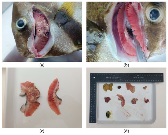
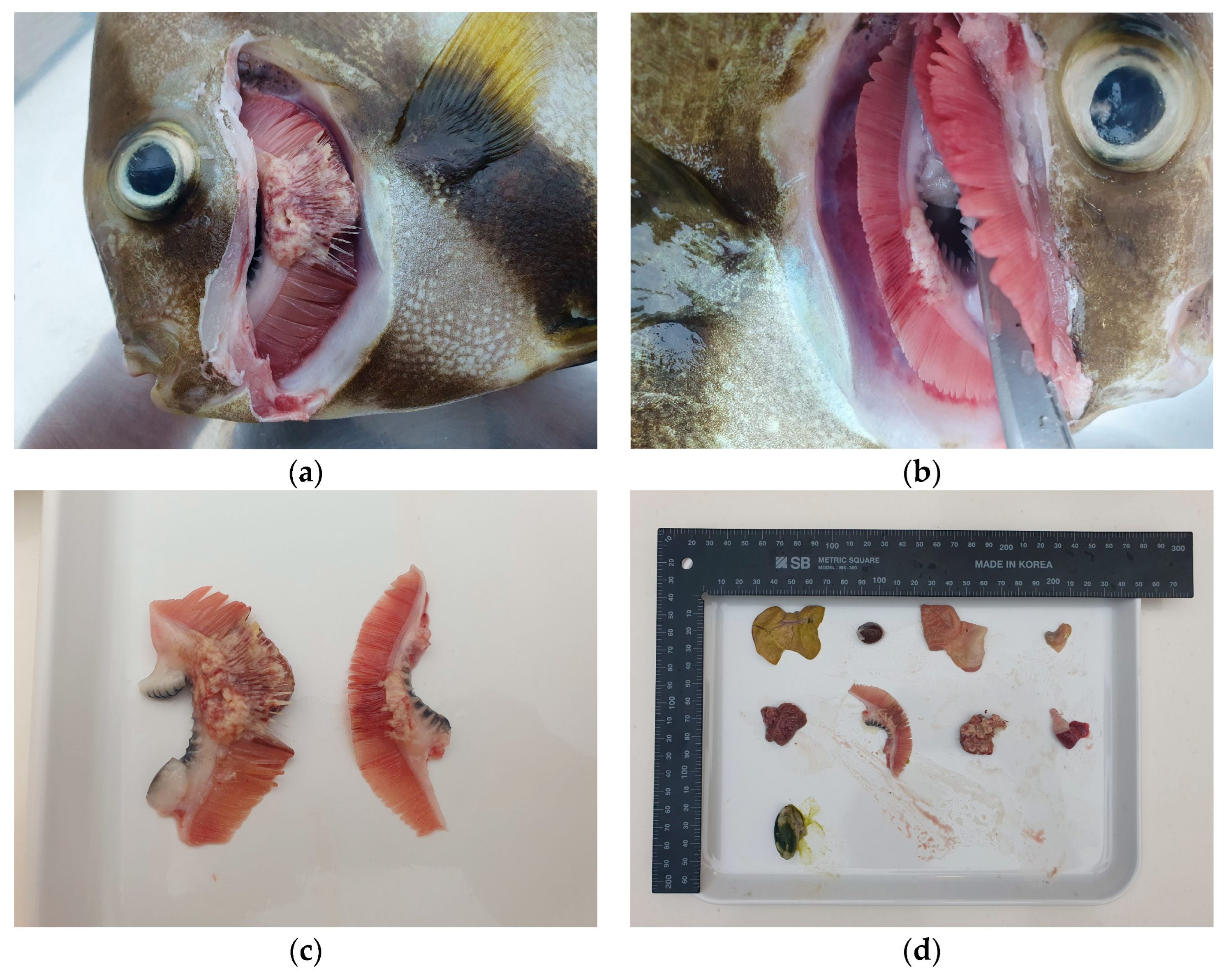
A 33-cm-long, 1.3 kg orbiculate batfish (Platax orbicularis) was found dead in an aquarium in South Korea and was subsequently submitted for necropsy. On gross examination, the gill filaments were diffusely swollen and distorted by multifocal to coalescing pale to whitish nodular lesions, as shown in Figure 1. These nodules were irregular in shape and variable in size, with some areas exhibiting partial coalescence, multifocal hemorrhage, and loss of normal filament architecture (Figure 1a,c). After opening the right operculum, similar nodular lesions were also observed along the gill (Figure 1b). Representative sections of gills and internal organs were collected, fixed in 10% neutral buffered formalin, and processed into formalin-fixed, paraffin-embedded (FFPE) tissues for histopathological evaluation (Figure 1d). Detailed husbandry and environmental records, including water quality, stocking density, filtration, and information on cohabiting species, were not available for retrospective review in this case.
Figure 1.
Gross lesions in an orbiculate batfish (Platax orbicularis). (a) Multifocal nodular lesions were observed on the left gill filaments after removal of the left operculum. (b) Right gill filaments, separated after a right opercular incision, revealing nodular lesions between the filaments. (c) Gross image of the left and right gill showing multifocal hemorrhage and pale nodular lesions. (d) Representative internal organs collected at necropsy for subsequent histopathological examination.
2.2. Histopathological Findings
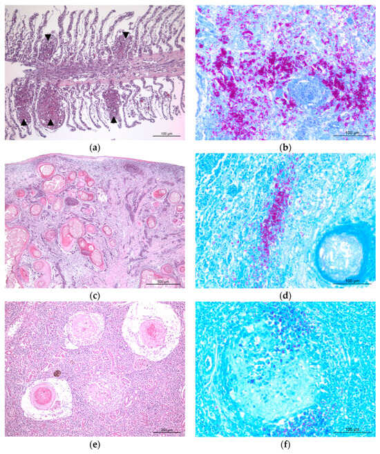
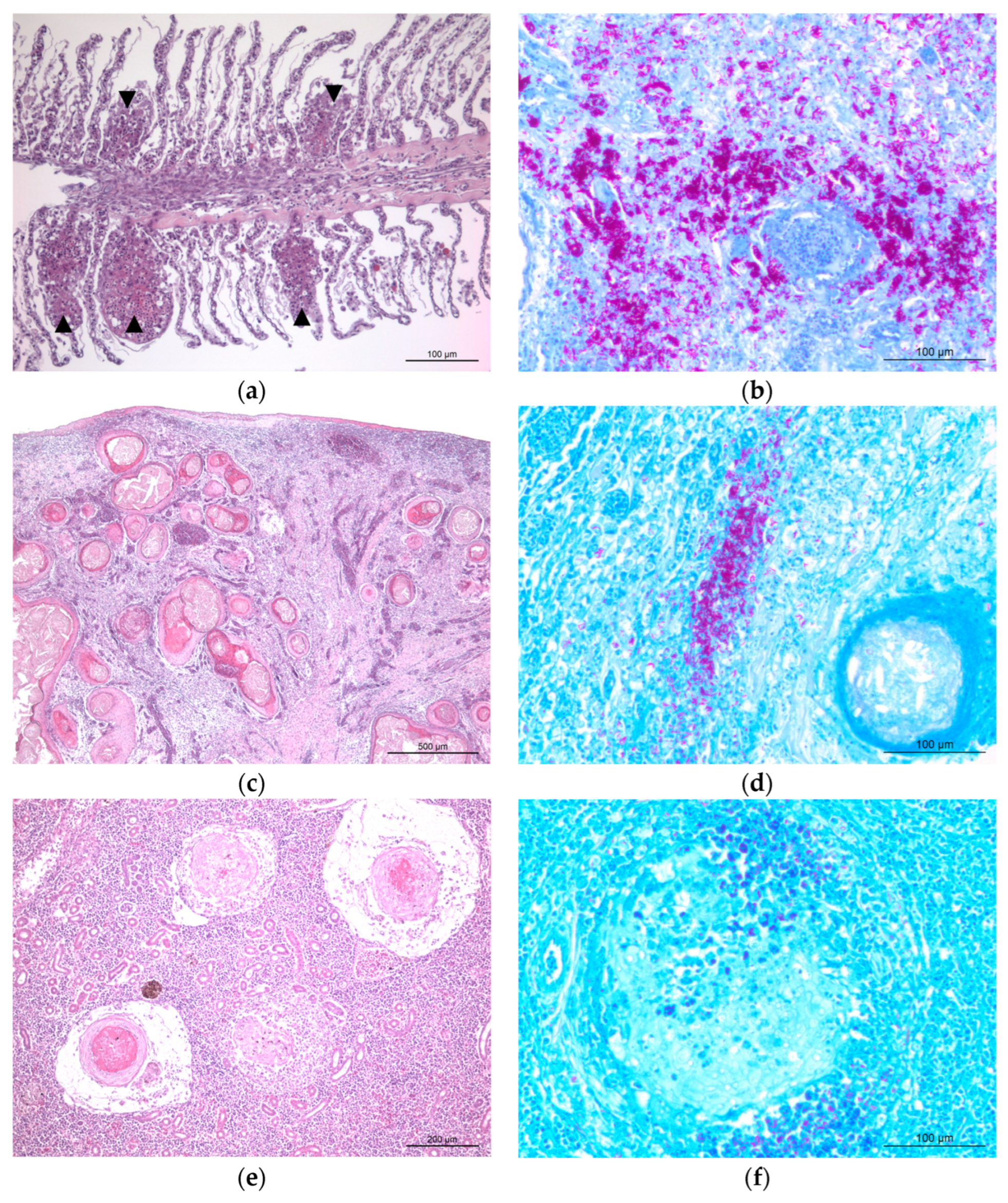
For histopathological evaluation, FFPE tissue sections were stained with either hematoxylin and eosin (H&E) for routine morphological assessment and with Ziehl–Neelsen stain for the detection of acid-fast bacilli. In the gill, multifocal granulomatous lesions were observed, resulting in marked disruption of the normal filament architecture and branchial arch structure (Figure 2a). Ziehl–Neelsen-stained sections of the gill demonstrated abundant rod-shaped acid-fast bacilli within the granulomatous lesions (Figure 2b). The most severe lesions were identified in the parenchymal organs, particularly the spleen and kidney. In the spleen, the normal hematopoietic architecture was extensively replaced by multifocal to coalescing granulomas characterized by prominent central necrotic cores composed of eosinophilic cellular debris and surrounded by epithelioid macrophages (Figure 2c). Ziehl–Neelsen staining of the spleen highlighted abundant acid-fast bacilli within the necrotic cores and adjacent inflammatory cells (Figure 2d). Similar granulomatous lesions with central necrosis were observed in the kidney (Figure 2e), and Ziehl–Neelsen staining demonstrated numerous acid-fast bacilli within these lesions (Figure 2f). Overall, these findings were consistent with chronic systemic granulomatous inflammation associated with acid-fast bacilli.
Figure 2.
Representative histopathological lesions of systemic granulomatous mycobacteriosis in an orbiculate batfish. Scale bars as indicated. (a) H&E-stained section of the gill showing multifocal granulomatous inflammation (arrowheads). (b) Ziehl–Neelsen-stained section of the gill demonstrating numerous acid-fast bacilli within granulomatous lesions. (c) H&E-stained section of the spleen showing multifocal to coalescing granulomas with central necrotic cores. (d) Ziehl–Neelsen-stained section of the spleen highlighting abundant acid-fast bacilli within the necrotic core and surrounding inflammatory cells. (e) H&E-stained section of the kidney showing multifocal granulomatous lesions with central necrosis. (f) Ziehl–Neelsen-stained section of the kidney demonstrating acid-fast bacilli within granulomatous lesions.
2.3. Molecular Analysis
To further characterize the acid-fast bacteria observed in the histological sections, molecular analysis was performed. Although fresh tissue samples are generally preferred for optimal pathogen identification, only FFPE specimens were available in the present case. Therefore, the FFPE tissues originally prepared for histological examination were used for molecular identification. Despite the technical challenges of detecting mycobacterial DNA from FFPE specimens, primarily because of nucleic acid degradation and cross-linking induced by formalin fixation, DNA extraction and subsequent PCR amplifications were successfully achieved [12]. In brief, DNA was extracted from the FFPE tissue sections (10 µm thick) using the QIAamp DNA FFPE Advanced Kit (QIAGEN, Hilden Germany) according to the manufacturer’s instructions, and the final product was eluted in the elution buffer. To improve analytical sensitivity, a nested PCR strategy was used to target both 16S rRNA and hsp65 genes. For the amplification of the hsp65 gene, two primer sets were adopted from a previous study [13]. For the 16S rRNA, specific primer sets were designed to optimize amplification from the FFPE-derived templates. In the first round of PCR, the extracted DNA served as the initial template, and resulting primary PCR products were then used as templates for the second-round (nested) PCR. The sequences of all forward and reverse primers used for these reactions are listed in Table 1. A no-template negative control was included in each PCR run, and pre-PCR setup, amplification, and post-PCR handling were performed in physically separated work areas using aerosol-resistant filter tips to minimize contamination. Both rounds of PCR amplifications were performed under the following conditions: initial denaturation at 94 °C for 2 min, followed by 30 cycles of denaturation at 94 °C for 30 s, annealing at 50 °C for 30 s, and extension at 68 °C for 30 s, with a final extension at 72 °C for 5 min. Amplicons were confirmed by agarose gel electrophoresis and subjected to direct sequencing. The expected amplicon size of the second-round 16S rRNA product was 200 bp, whereas those of the first- and second-round PCR products were 463 bp and 439 bp, respectively.
Table 1.
Primer sets used for the nested PCR amplification of mycobacterial genes and expected amplicon sizes.
Basic Local Alignment Search Tool (BLAST, https://blast.ncbi.nlm.nih.gov/Blast.cgi, accessed on 21 April 2026) analysis of partial 16S rRNA and hsp65 gene sequences demonstrated that the detected organisms were most closely related to M. marinum, as summarized in Table 2. Specifically, the 16S rRNA sequences obtained from the gill and spleen exhibited 97.3 and 95.7% identity, respectively, to reference sequences of M. marinum. Furthermore, the hsp65 sequences derived from the gill and kidney showed higher similarities, 99.5 and 100.0%, respectively.
Table 2.
Results of BLAST analysis of partial 16S rRNA and hsp65 gene sequences.
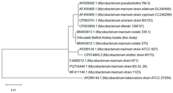
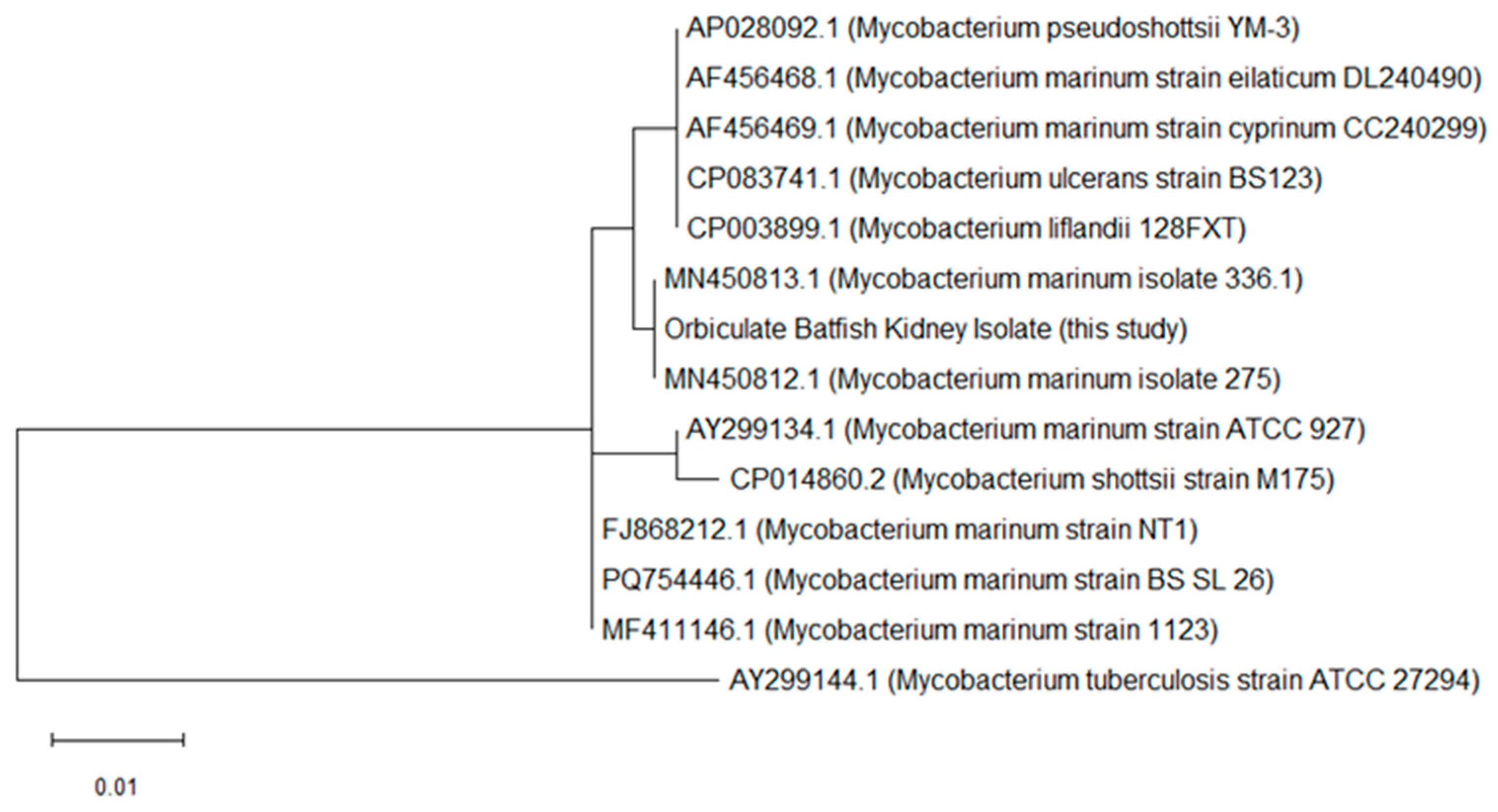
Phylogenetic analysis based on the partial hsp65 gene sequences was performed using the Maximum Likelihood method and the Tamura-Nei model in MEGA version 12. Twelve reference sequences representing closely related taxa within the M. marinum complex and related fish-associated non-tuberculous mycobacteria were included, together with the highest-quality sequence obtained from the kidney tissue of the orbiculate batfish in this study. The sequences were aligned using MUSCLE, and the resulting phylogenetic tree demonstrated that the kidney-derived sequence clustered closely with reference strains of M. marinum (Figure 3). Specifically, it formed a tight clade with M. marinum isolates, 275 (MN450812.1) and 336.1 (MN450813.1), while remaining separated from other closely related mycobacterial species, including M. pseudoshottsii, M. ulcerans, and M. shottsii. Because the other sequences obtained in this study were shorter and of lower comparative value for tree construction, they were used for BLAST-based comparison rather than inclusion in the phylogenetic tree. These molecular findings, together with the histopathological evidence of acid-fast bacilli, support the presence of a systemic granulomatous disease associated with a pathogen closely related to M. marinum.
Figure 3.
Phylogenetic tree based on partial hsp65 gene sequence obtained from the kidney tissue of the orbiculate batfish in this study (GenBank accession number: PZ290492) and closely related Mycobacterium strains. A reference sequence of M. tuberculosis was additionally included to provide phylogenetic context for the tree interpretation. Sequence alignment and phylogenetic tree construction were performed using the Maximum Likelihood method with the Tamura-Nei model in MEGA version 12. Bootstrap analysis was performed with 1000 replicates. Positions containing gaps and missing data were eliminated using the complete deletion option. GenBank accession numbers and strain names are shown for all sequences included in the analysis. The scale bar indicates the number of nucleotide substitutions per site.
3. Discussion
This study describes a case of systemic granulomatous disease in an orbiculate batfish from an aquarium in South Korea, characterized by multifocal granulomas in multiple organs and abundant intralesional acid-fast bacilli. Histopathological examination revealed granulomatous lesions in the gills, spleen, and kidney, with the most severe changes observed in the parenchymal organs. These findings are consistent with previous reports of piscine mycobacteriosis, in which granulomas composed of epithelioid macrophages and associated inflammatory cells are commonly observed. The distribution of lesions across multiple organs supports the diagnosis of a systemic mycobacterial infection. Differential diagnoses for multifocal granulomatous lesions in fish include infections caused by other non-tuberculous mycobacteria, Nocardia spp., certain fungal agents, and chronic parasitic or foreign-body-associated granulomatous inflammation; however, the presence of abundant intralesional acid-fast bacilli strongly supported a mycobacterial etiology in the present case. Similar systemic granulomatous lesions associated with Mycobacterium spp. have been described in other ornamental and cultured fish species, particularly involving the spleen, kidney, and gills [14,15]. The lesion distribution and abundant intralesional acid-fast bacilli observed in the present case were broadly consistent with those previously reported in piscine mycobacteriosis [14,15]. However, unlike culture-confirmed cases, the present report relied on FFPE-based molecular analysis, which limited definitive species-level identification [12].
Molecular characterization based on the partial sequencing of the 16S rRNA and hsp65 genes suggested that the detected organism was most closely related to M. marinum. However, definitive species-level identification remained challenging because the analysis was performed using DNA extracted from FFPE tissues, which can compromise DNA length and quality. In addition, the phylogenetic analysis was based on a single highest-quality hsp65 sequence, and branch-support values were not displayed in the current tree presentation. Bacterial culture and isolation were also not available in this case, representing an important diagnostic limitation. Accordingly, the etiological agent is most appropriately and cautiously designated as an M. marinum-like organism rather than definitively identified as M. marinum. Because only FFPE specimens had been preserved for retrospective analysis, fresh tissues suitable for mycobacterial culture were not available in this case; therefore, culture-based confirmation could not be performed.
In aquatic environments, M. marinum and related species are widely distributed and are recognized as opportunistic pathogens infecting a broad range of marine and fresh-water fish. In the present case, the severe systemic granulomatous lesions observed in the orbiculate batfish were consistent with findings reported in previous studies across susceptible species. Because detailed tank history, water-quality data, stocking density, filtration records, quarantine information, and information on tank mates were unavailable in this case, environmental factors are discussed here only as general risk factors reported in aquarium mycobacteriosis.
In addition to its veterinary significance, M. marinum is a well-recognized zoonotic pathogen responsible for fish tank granuloma in humans. Accordingly, the presence of a M. marinum-like organism in aquarium fish warrants general awareness of potential occupational exposure among aquarists, fish handlers, and other personnel frequently exposed to aquatic environments. However, because no viable bacterial isolate, human exposure assessment, or human infection data were available in this case, the zoonotic implications should be interpreted cautiously. These findings highlight the importance of implementing suitable biosecurity measures and maintaining awareness of potential zoonotic risks in aquatic systems.
4. Conclusions
This report describes a case of systemic granulomatous disease in an orbiculate batfish from a public aquarium in South Korea. Gross and histopathological findings, together with molecular analysis based on partial 16S rRNA and hsp65 gene sequences, indicated that the detected organism was most closely related to Mycobacterium marinum. Because definitive species-level identification was limited using partial FFPE-derived sequences and the lack of bacterial isolation, the etiologic agent is best interpreted as a M. marinum-like organism. This case highlights the importance of routine disease surveillance and improved diagnostic awareness of non-tuberculous mycobacterial infections in aquarium fish, as well as the need for appropriate biosecurity awareness regarding potential zoonotic risk.
Author Contributions
Conceptualization, S.-H.D., S.O. and C.Y.K.; methodology, C.Y.K. and S.-H.D.; software, C.Y.K.; validation, C.Y.K., Y.-H.G. and S.-H.D.; investigation, S.O. and C.Y.K.; resources, S.O. and S.-H.D.; writing—original draft preparation, C.Y.K. and S.-H.D.; writing—review and editing, C.Y.K. and S.-H.D.; visualization, C.Y.K. and S.-H.D.; supervision, C.Y.K. and S.-H.D.; project administration, S.-H.D.; funding acquisition, S.-H.D. All authors have read and agreed to the published version of the manuscript.
Funding
This work was supported by Konkuk University in 2024 (2024-A019-0063).
Institutional Review Board Statement
Not applicable.
Informed Consent Statement
Not applicable.
Data Availability Statement
The data presented in this study are openly available. The nucleotide sequences obtained in this study were submitted to GenBank (accession numbers: PZ274886, PZ274891, PZ290491, and PZ290492).
Conflicts of Interest
The authors declare no conflicts of interest.
References
- Akram, S.M.; Aboobacker, S. Mycobacterium marinum Infection. In StatPearls; StatPearls Publishing: Treasure Island, FL, USA, 2023. [Google Scholar]
- Tsai, M.-A.; Wang, P.-C.; Yoshida, S.; Aono, A.; Mitarai, S.; Wada, T.; Chen, S.-C. Establishment of Loop-Mediated Isothermal Amplification for Rapid and Convenient Detection of Mycobacterium marinum Complex. J. Microbiol. Methods 2019, 164, 105671. [Google Scholar] [CrossRef] [Scilit] [PubMed]
- Hashish, E.; Merwad, A.; Elgaml, S.; Amer, A.; Kamal, H.; Elsadek, A.; Marei, A.; Sitohy, M. Mycobacterium marinum Infection in Fish and Man: Epidemiology, Pathophysiology and Management; a Review. Vet. Q. 2018, 38, 35–46. [Google Scholar] [CrossRef] [Scilit] [PubMed]
- Broussard, G.W.; Ennis, D.G. Mycobacterium marinum Produces Long-Term Chronic Infections in Medaka: A New Animal Model for Studying Human Tuberculosis. Comp. Biochem. Physiol. Part C Toxicol. Pharmacol. 2007, 145, 45–54. [Google Scholar] [CrossRef] [Scilit] [PubMed]
- Kumar, P.; Cameron, J.; Saviola, B.; Venketaraman, V. Mycobacterium marinum Immune Evasion in Zebrafish. Pathogens 2025, 14, 908. [Google Scholar] [CrossRef] [Scilit] [PubMed]
- Avsever, M.L.; Çavuşoğlu, C.; Günen, M.Z.; Yazıcıoğlu, Ö.; Eskiizmirliler, S.; Didinen, B.I.; Tunalıgil, S.; Erdal, G.; Özden, M. The First Report of Mycobacterium marinum Isolated from Cultured Meagre, Argyrosomus Regius. Bull. Eur. Fish Pathol. 2014, 34, 124–129. [Google Scholar]
- Aubry, A.; Mougari, F.; Reibel, F.; Cambau, E. Mycobacterium marinum. Microbiol. Spectr. 2017, 5, 735–752. [Google Scholar] [CrossRef] [Scilit] [PubMed]
- Bayou, K. Overview on Fish Mycobacteriosis with Special Reference to Pathology, Diagnosis and Human Health. World J. Fish Mar. Sci. 2017, 9, 9–16. [Google Scholar]
- Parikka, M.; Hammarén, M.M.; Harjula, S.-K.E.; Halfpenny, N.J.A.; Oksanen, K.E.; Lahtinen, M.J.; Pajula, E.T.; Iivanainen, A.; Pesu, M.; Rämet, M. Mycobacterium marinum Causes a Latent Infection That Can Be Reactivated by Gamma Irradiation in Adult Zebrafish. PLoS Pathog. 2012, 8, e1002944. [Google Scholar] [CrossRef] [Scilit] [PubMed]
- Ucko, M.; Colorni, A. Mycobacterium marinum Infections in Fish and Humans in Israel. J. Clin. Microbiol. 2005, 43, 892–895. [Google Scholar] [CrossRef] [Scilit] [PubMed]
- Das, S.; Pettersson, B.M.F.; Behra, P.R.K.; Mallick, A.; Cheramie, M.; Ramesh, M.; Shirreff, L.; DuCote, T.; Dasgupta, S.; Ennis, D.G.; et al. Extensive Genomic Diversity among Mycobacterium marinum Strains Revealed by Whole Genome Sequencing. Sci. Rep. 2018, 8, 12040. [Google Scholar] [CrossRef] [Scilit] [PubMed]
- Pourahmad, F.; Thompson, K.D.; Adams, A.; Richards, R.H. Detection and identification of aquatic mycobacteria in formalin-fixed, paraffin-embedded fish tissues. J. Fish Dis. 2009, 32, 409–419. [Google Scholar] [CrossRef] [Scilit] [PubMed]
- Fata, A.; Bojdy, A.; Maleki, M.; Hosseini Farash, B.R.; Ghazvini, K.; Tajzadeh, P.; Vakili, V.; Moghaddas, E.; Mastroeni, P.; Rahmani, S. Fish Tank Granuloma: An Emerging Skin Disease in Iran Mimicking Cutaneous Leishmaniasis. PLoS ONE 2019, 14, e0221367. [Google Scholar] [CrossRef] [Scilit] [PubMed]
- Gauthier, D.T.; Rhodes, M.W. Mycobacteriosis in fishes: A review. Vet. J. 2009, 180, 33–47. [Google Scholar] [CrossRef] [Scilit] [PubMed]
- Jacobs, J.M.; Stine, C.B.; Baya, A.M.; Kent, M.L. A review of mycobacteriosis in marine fish. J. Fish Dis. 2009, 32, 119–130. [Google Scholar] [CrossRef] [Scilit] [PubMed]

Disclaimer/Publisher’s Note: The statements, opinions and data contained in all publications are solely those of the individual author(s) and contributor(s) and not of MDPI and/or the editor(s). MDPI and/or the editor(s) disclaim responsibility for any injury to people or property resulting from any ideas, methods, instructions or products referred to in the content. |
© 2026 by the authors. Licensee MDPI, Basel, Switzerland. This article is an open access article distributed under the terms and conditions of the Creative Commons Attribution (CC BY) license.