


Simple Summary
Canine adenovirus type 2 (CAV-2) causes respiratory disease in dogs and is also being developed as a viral vector for delivering genes into cells. Antibodies that recognise the virus are valuable laboratory tools, but most existing antibodies target two of the three main proteins that build the virus shell, and reagents against the third one, the penton base, are scarce. In this study we produced a mouse monoclonal antibody, named 3D5, using a portion of the CAV-2 penton base protein made in bacteria. Antibody 3D5 recognises both the purified protein and the virus growing in cultured dog kidney cells, and it binds to a short stretch of eight amino acids (RYKGATEG, residues 29–36) near the beginning of the penton base. This stretch is almost identical in the CAV-2 and CAV-1 viruses examined, suggesting that 3D5 could help detect both. Because the antibody does not block virus infection, its main use would be as a laboratory detection reagent rather than as a treatment.
Abstract
Canine adenovirus type 2 (CAV-2) is an important respiratory pathogen of dogs and has also emerged as a promising non-human adenoviral vector for gene delivery. However, antibody reagents targeting the CAV-2 penton base remain poorly characterized compared with those directed against the hexon and fiber proteins. In this study, we generated and characterized a mouse monoclonal antibody (mAb), designated 3D5, against the CAV-2 penton base. Recombinant penton base protein expressed in Escherichia coli was used for mouse immunization and hybridoma screening. mAb 3D5 was identified as an IgG2b antibody with a kappa light chain and specifically recognized both recombinant penton base protein and viral antigen in CAV-2-infected MDCK cells by Western blotting and immunofluorescence assays. Sequential truncation mapping localized the antibody-binding region to the N terminus of the penton base, and a recombinant fragment spanning residues 29–36 (RYKGATEG) retained 3D5 reactivity under denaturing conditions. This sequence was identical in 31/33 CAV-2 and 4/4 CAV-1 accession records, and mAb 3D5 reacted with a recombinant CAV-1 preparation. MAb 3D5 showed no neutralizing activity. These results identify a N-terminal linear reactive region recognized by mAb 3D5 and provide a renewable monoclonal antibody reagent for CAV-2 antigen detection and further investigation of penton base antigenicity.
1. Introduction
Canine adenovirus type 2 (CAV-2) is a non-enveloped, icosahedral virus with a linear double-stranded DNA genome of approximately 31 kb and belongs to the genus Mastadenovirus within the family Adenoviridae [1]. Canine adenoviruses are classified into two types: CAV-1, the etiological agent of infectious canine hepatitis, and CAV-2, one of the viral pathogens associated with canine infectious respiratory disease (CIRD) [2]. CAV-2 continues to circulate in domestic and wild canid populations worldwide [3]. In China, a molecular survey of 121 dogs with respiratory disease in parts of Sichuan and Chongqing detected CAV-2 DNA in 29.8% of the examined nasopharyngeal swab samples [4]. Sporadic CAV-2 infections associated with enteric disease or neurological signs have also been reported [5,6,7]. Beyond its veterinary significance, CAV-2 has attracted interest as a non-human adenoviral vector for gene therapy because of its neuronal tropism, efficient retrograde axonal transport, and the absence of pre-existing CAV-2-specific immunity in humans [8,9,10,11]. These veterinary and biomedical applications highlight the need for well-defined antibodies for CAV-2 antigen detection and biological investigation.
The CAV-2 capsid contains three principal structural proteins: hexon, fiber, and penton base [1,9]. The fiber mediates attachment to host cells by binding the D1 domain of the coxsackievirus and adenovirus receptor, whereas the penton base anchors the fiber at each capsid vertex and contributes to adenovirus internalization and endosomal escape [12,13]. Unlike the penton bases of human adenovirus types 2 and 5, the CAV-2 penton base lacks the canonical Arg-Gly-Asp integrin-binding motif; nevertheless, alpha-v integrins may contribute to CAV-2 attachment or entry in a cell-dependent manner [8]. Structural analysis has described the CAV-2 capsid organization [14], but antibody accessibility of the reactive fragment identified here on intact virions remains unresolved.
Monoclonal antibodies against canine adenoviruses have previously been generated by whole-virus immunization, and both type-specific and cross-reactive antibodies against CAV-1 and CAV-2 have been described [15]. Previous studies have used monoclonal antibodies for immunochromatographic detection of CAV [16], characterization of neutralizing epitopes within hexon [17], and ELISA-based detection of CAV-1 antigen [18].
In comparison, the penton base remains insufficiently characterized as an antibody target. Although a penton base-reactive mouse monoclonal antibody was recently used to identify a CAV-2 isolate by Western blotting [19].
In this study, we generated a mouse monoclonal antibody, designated 3D5, against the CAV-2 penton base using a prokaryotically expressed recombinant penton base protein as the immunogen. The hybridoma-derived antibody was evaluated for reactivity with recombinant penton base protein and CAV-2-infected MDCK cells by Western blotting and indirect immunofluorescence assays. A N-terminal linear reactive region was mapped to residues 29–36. This study provides a renewable antibody reagent for CAV-2 antigen detection and further investigation of penton base antigenicity.
2. Materials and Methods
2.1. Cells, Viruses, and Reagents
Madin-Darby canine kidney (MDCK, ATCC CCL-34) cells were maintained in Dulbecco’s modified Eagle’s medium (DMEM) supplemented with 10% fetal bovine serum (FBS), 100 U/mL penicillin, and 100 μg/mL streptomycin at 37 °C in a 5% CO2 incubator. The CAV-2 strain preserved in our laboratory [20] was propagated in MDCK cells and stored at −80 °C. E. coli DH5α and BL21(DE3) competent cells were used for plasmid propagation and protein expression, respectively. Mouse myeloma SP2/0 (ATCC CRL-1581) cells were maintained for hybridoma production. Restriction enzymes (EcoRI and KpnI) and PrimeSTAR DNA polymerase were purchased from Takara Biotechnology (Dalian, China). The recombinant protein was purified from the soluble fraction using Ni-NTA agarose (TEASEN Cat# 20502ES50). Complete and incomplete Freund’s adjuvants, polyethylene glycol (PEG) 1500, and HAT/HT media were obtained from Sigma-Aldrich (St. Louis, MO, USA).
Six- to eight-week-old female BALB/c mice were purchased from the Liaoning Changsheng Biotechnology Co., Ltd. (Benxi, Liaoning, China). All animal procedures were approved by the Animal Ethics Committee of Harbin Veterinary Research Institute (HVRI), Chinese Academy of Agricultural Sciences (CAAS; Approval No. 251211-05-GR, No. 251211-05-GR).
2.2. Expression and Purification of Recombinant CAV-2 Penton Protein
The CAV-2 penton gene was amplified from viral DNA using primers CAV2-Pen-F 5′-GGCATATGGAGCTCGGTACCatggagttttcgtcgtctcc-3′ (Kpn I site, underlined) and CAV2-Pen-R 5′-GACTGCAGGTCGACAAGCTTGAATTCctagaaggttttactgga-3′ (EcoR I site, underlined) and cloned into the pCold TF vector (Takara) to generate pCold TF-penton. The recombinant plasmid was transformed into E. coli BL21(DE3). Protein expression was induced with 0.5 mM IPTG at 16 °C for 18 h. Bacterial pellets were resuspended in PBS (pH 7.4) and lysed by sonication. The His-tagged recombinant protein was purified from the supernatant by Ni-NTA affinity chromatography and analyzed by SDS-PAGE (12% gel). Protein concentration was determined using the BCA assay ((Thermo Fisher Scientific, Rockford, IL, USA; Cat. No. 23227).).
2.3. Generation of Monoclonal Antibodies
A total of ten BALB/c mice were immunized subcutaneously with 50 μg of purified recombinant penton protein emulsified 1:1 (v/v) with complete Freund’s adjuvant for the primary immunization, followed by two booster injections at 2-week intervals with incomplete Freund’s adjuvant. One week after the second booster, serum antibody titers were determined by IFA using CAV-2-infected MDCK cells. The mouse with the highest titer received a final intraperitoneal booster of 50 μg antigen without adjuvant. Three days later, splenocytes were fused with SP2/0 myeloma cells at a 5:1 ratio using 50% PEG 1500. Hybridomas were selected in HAT medium and screened by IFA and Western blot. Positive clones were subcloned three times by limiting dilution to obtain stable cell lines. Antibody isotypes were determined using a Mouse Monoclonal Antibody Isotyping Kit (Biodragon, Suzhou, China; Cat. No. BF06001). For ascites production, the stable hybridoma cells were injected intraperitoneally into pristane-primed BALB/c mice, and ascitic fluid was collected 7–14 days post-injection.
2.4. Immunofluorescence Assay (IFA)129
MDCK cells grown on coverslips were infected with CAV-2 at an MOI of 1.0 for 36 h, fixed with cold methanol at −20 °C for 30 min, and blocked with 5% BSA in PBS for 1 h at 37 °C. Cells were incubated with hybridoma supernatant (1:100 dilution) overnight at 4 °C, followed by FITC-conjugated goat anti-mouse IgG (1:200) for 1 h at 37 °C. Fluorescence signals were observed under a fluorescence microscope (Olympus, Japan). Uninfected cells and SP2/0 supernatant served as negative controls.
2.5. Western Blot Analysis
Purified recombinant penton protein (~1 μg/lane) was resolved by SDS-PAGE (12% gel) and transferred onto Amersham™ Protran™ 0.2 μm nitrocellulose membrane (Cytiva, Germany; Cat. No. 10600001). using a semi-dry transfer apparatus eBlot II rapid wet transfer system (GenScript, Nanjing, China).) at 15 V for 40 min. For detection of the viral antigen, MDCK cells grown in 6-well plates were either mock-infected or infected with CAV-2 at an MOI of 1.0. At 36 h post-infection, cells were harvested, washed twice with ice-cold PBS, and lysed in RIPA lysis buffer (Beyotime, Shanghai, China; Cat. No. P0013B)supplemented with 1× protease inhibitor cocktail (Roche, Cat# 11836170001) on ice for 30 min. Lysates were clarified by centrifugation at 12,000× g for 10 min at 4 °C, and protein concentrations were determined using the BCA assay (Thermo Fisher Scientific, Cat# 23225). Equal amounts of total protein (20 μg/lane) from infected and mock-infected cell lysates, along with purified recombinant penton protein (~1 μg/lane), were resolved by SDS-PAGE (12% gel) and transferred onto PVDF membranes. Membranes were blocked with 5% skimmed milk in TBST (Tris-buffered saline with 0.05% Tween-20) for 2 h at room temperature, incubated with anti-penton mAb (1:500) or anti-His tag antibody (1:2000; Proteintech, Cat# 66005-1-Ig) overnight at 4 °C, and then with IRDye® 800CW Goat anti-Mouse IgG (H+L) secondary antibody (LI-COR, Lincoln, NE, USA Cat. No. 926-32210, diluted 1:8000) for 1 h at room temperature. The membrane was scanned and analyzed with an Odyssey infrared scanner (Li-Cor Biosciences, USA).
2.6. Epitope Mapping of mAb 3D5
A sequential truncation strategy was used to map the linear epitope recognized by mAb 3D5. Overlapping fragments covering the CAV-2 penton base were initially constructed, and the reactive region was progressively subdivided into smaller fragments based on the results of each round of Western blot screening. The corresponding DNA fragments were amplified using specific primers (Table 1) or commercially synthesized by RuiBiotech Co., Ltd. (Beijing, China) and cloned into the pCold TF vector. Recombinant proteins were expressed in E. coli BL21(DE3) and their reactivity with mAb 3D5 was evaluated by Western blotting, with anti-His detection used as an expression control.
Table 1.
Truncated expression PCR primers of the CAV-2 penton gene.
2.7. Computational Structural and Bioinformatic Analysis
An existing five-subunit AlphaFold3 model [21] of the CAV-2 penton base was used to map the mAb 3D5-reactive fragment spanning residues 29–36. Solvent-accessible surface areas (SASA) were calculated in the complete pentamer using the Shrake–Rupley implementation in Biopython 1.88, with a 1.4 Å probe and 2048 sampling points per atom. Relative solvent accessibility was expressed as a percentage of the maximum ASA values reported by Tien et al. [22]. Mean SASA per residue was calculated across all five copies of the fragment.
Structural visualization and secondary-structure assignment were performed using the PyMOL Molecular Graphics System (version 3.1.0) [23]. Residues 29–36 were highlighted in each subunit. Their position and local conformation were examined in the complete pentamer and a magnified view of chain A, with a stick representation showing the local residue arrangement.
Penton amino acid sequences from CAV-2 and CAV-1 strains were retrieved from GenBank and aligned. Accession numbers are listed in Supplementary Table S1. Sequence conservation of the mAb 3D5-reactive region was assessed based on this alignment.
3. Results
3.1. Expression and Purification of Recombinant CAV-2 Penton Base in Escherichia coli
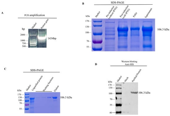
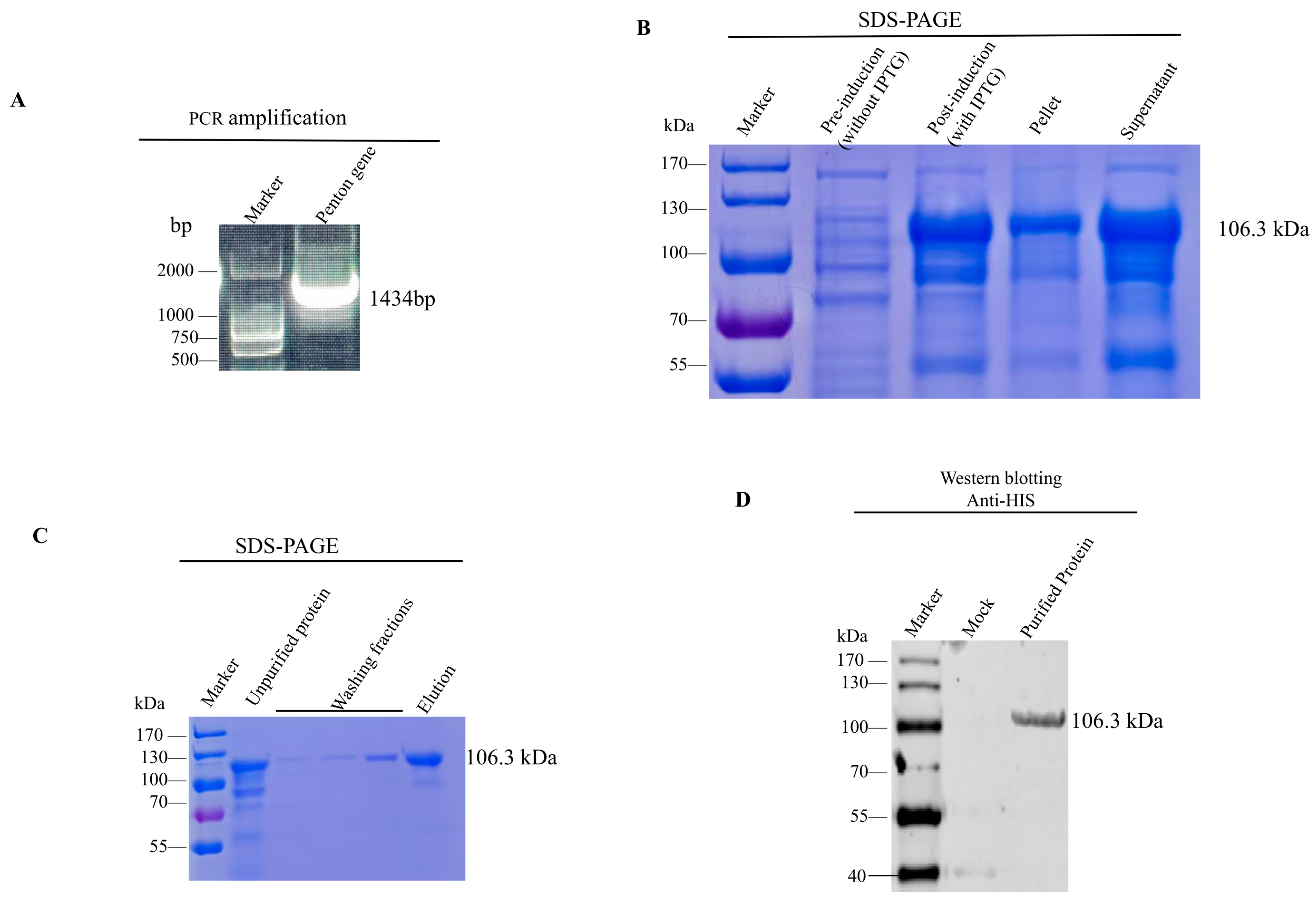
The penton base gene of CAV-2 was amplified by PCR and cloned into the prokaryotic expression vector pCold TF (Figure 1A), in which penton base is expressed as an N-terminal His–trigger factor (TF) fusion. The recombinant plasmid pCold TF-penton was verified by restriction enzyme digestion and DNA sequencing. The recombinant plasmid was then transformed into E. coli BL21(DE3) cells and expression was induced with IPTG. SDS-PAGE analysis confirmed soluble expression of the recombinant penton base protein (Figure 1B). This mobility is consistent with the calculated mass of 106.3 kDa for the His–TF–penton base fusion, comprising TF (~48 kDa) and penton base (478 aa, ~53.6 kDa). The fusion protein was purified from the soluble fraction by Ni–NTA affinity chromatography under native conditions and eluted with 250 mM imidazole. SDS-PAGE of the eluate showed a predominant band at ~100 kDa (Figure 1C). Western blotting with an anti-His antibody detected a single band at the same position (Figure 1D).
Figure 1.
Expression and purification of the recombinant canine adenovirus type 2 (CAV-2) penton base protein. (A) The amplification of the target gene by PCR. (B) SDS-PAGE analysis of total cell lysates from E. coli BL21(DE3) following IPTG-induced expression of the recombinant penton base fusion protein. (C) SDS-PAGE analysis of the purified recombinant protein eluted from Ni-NTA affinity chromatography. (D) Western blot analysis of the purified protein probed with an anti-His-tag monoclonal antibody, confirming the identity of the fusion protein. Molecular weight markers (in kDa) are indicated on the left.
3.2. Generation and Characterization of Monoclonal mAb 3D5
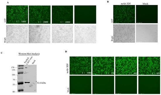
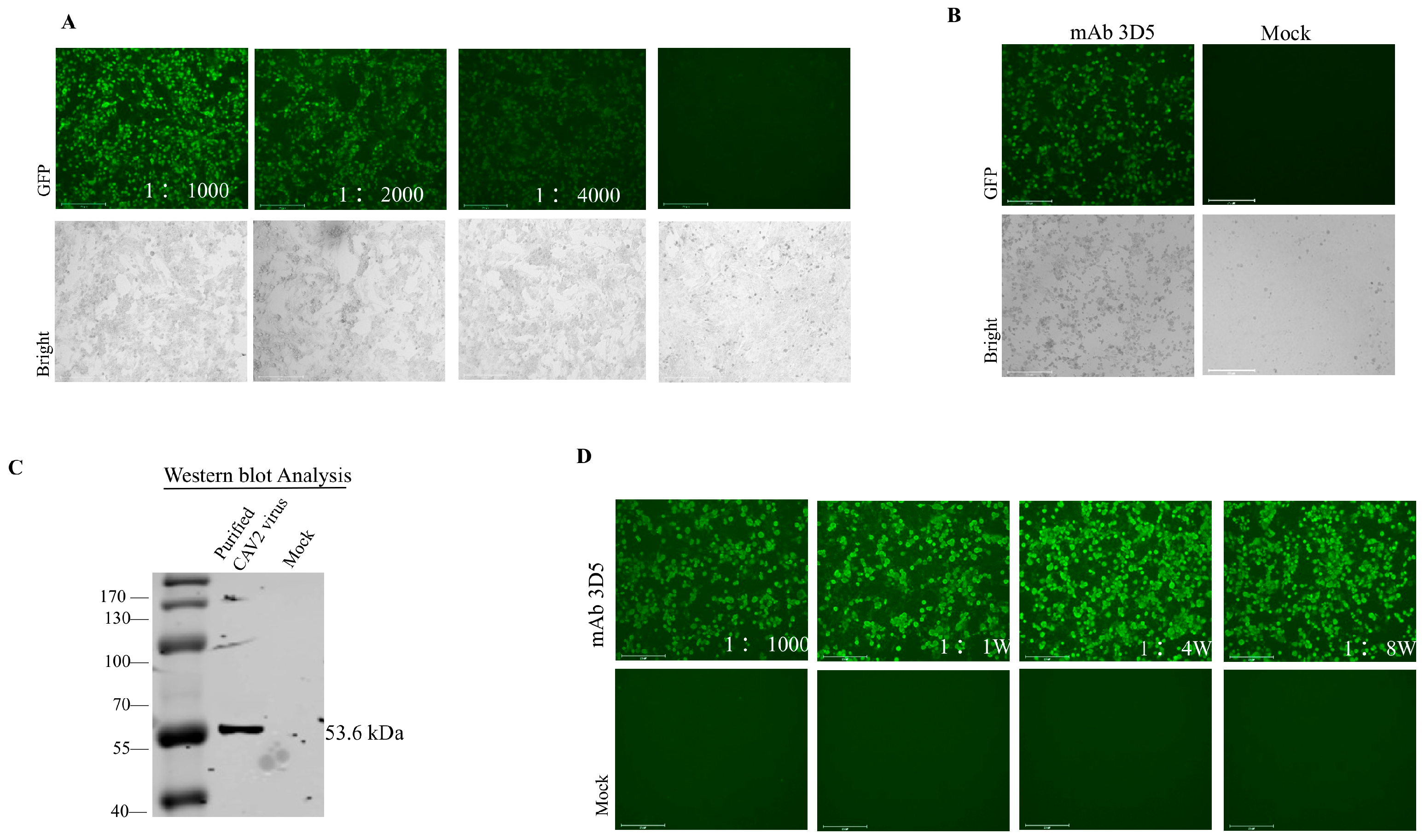
BALB/c mice immunized with the purified His–TF–penton base fusion protein (Figure 1C) developed serum antibodies that stained CAV-2-infected MDCK cells by indirect immunofluorescence assay (IFA) at titers of ≥1:4000, with no staining of uninfected MDCK cells (Figure 2A). Spleen cells were fused with SP2/0 myeloma cells, and screened by IFA. One clone-secreted antibody reactive with penton base by IFA, designated 3D5, was subjected to three rounds of limiting dilution to clonality, expanded in culture, and inoculated into pristane-primed BALB/c mice for ascites production. Isotyping identified mAb 3D5 as IgG2b with kappa light chains.
Figure 2.
Generation and characterization of a novel monoclonal antibody (mAb 3D5) targeting the CAV-2 penton base. (A) Serum antibody titers from immunized mice were determined by indirect immunofluorescence assay (IFA). (B) IFA validation of mAb 3D5 specificity. (C) Western blot analysis demonstrating that mAb 3D5 specifically recognizes the native penton base protein in CAV-2-infected MDCK cell lysates. (D) IFA test the ascitic fluid titer of mAb 3D5.
In IFA, mAb 3D5 stained CAV-2-infected MDCK cells with a fluorescence pattern, whereas uninfected cells remained negative (Figure 2B). In Western blotting, mAb 3D5 recognized the ~100 kDa His–TF–penton base fusion and, in lysates of CAV-2-infected MDCK cells, a band of approximately 55 kDa consistent with the penton base monomer (calculated 53.6 kDa); no band was detected at this position in uninfected cell lysates (Figure 2C).
The ascites titer of mAb 3D5 was 1:80,000 by IFA (Figure 2D). Because neither TF nor the His tag is present in MDCK cells, recognition of the 55 kDa species in infected but not uninfected lysates establishes that mAb 3D5 is directed against CAV-2 penton base rather than against the expression tags. The neutralizing activity of mAb 3D5 was evaluated by a virus neutralization assay. No neutralization was observed at the highest antibody concentration tested (1:4–1:512 during virus preincubation; Table S1, Supplementary S2).
3.3. Identification of the mAb 3D5-Reactive Region
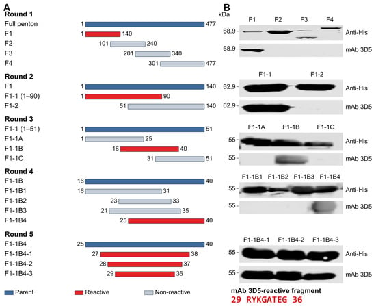
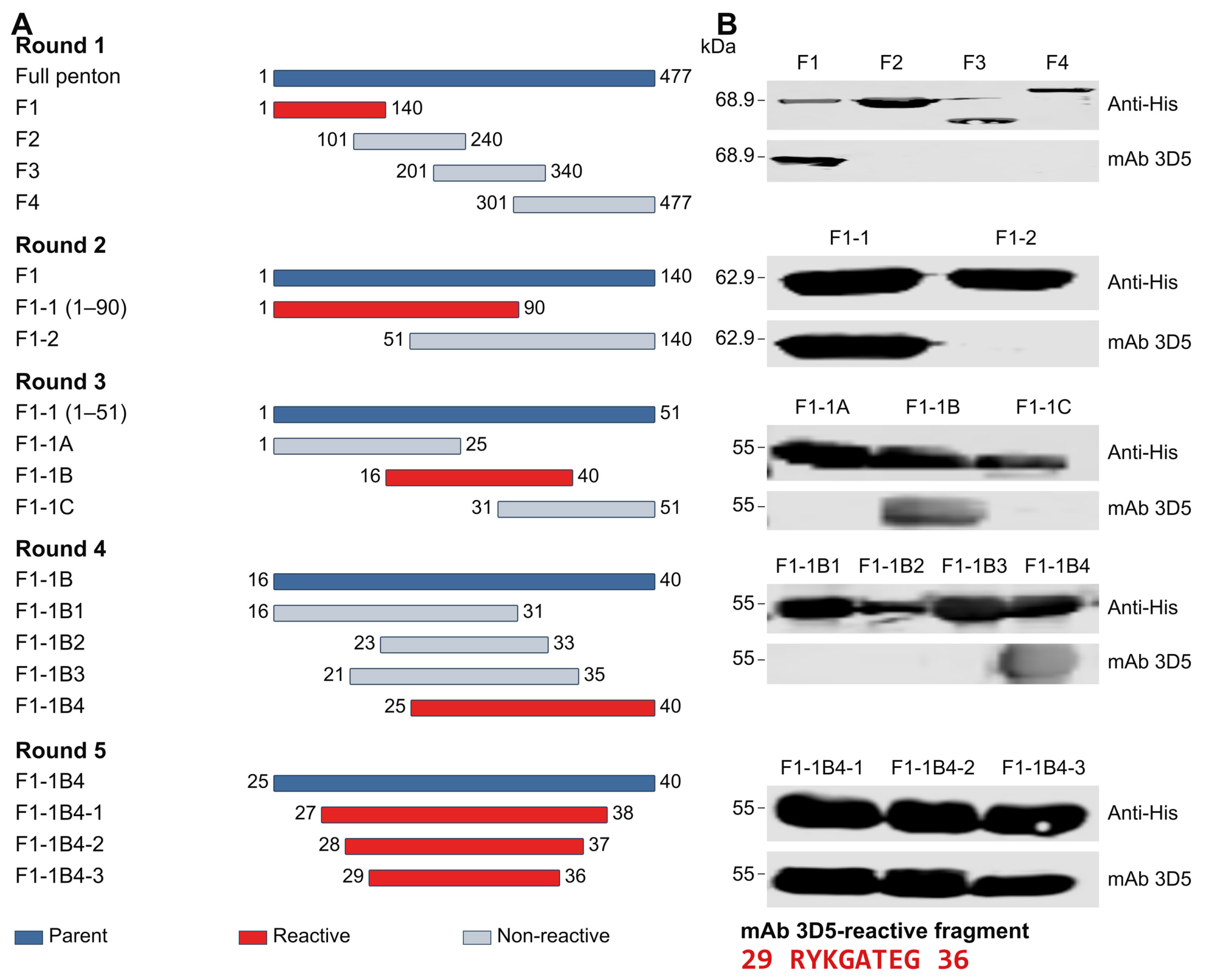
To define the linear epitope recognized by mAb 3D5, a series of penton base truncation fragments were expressed as recombinant proteins and analyzed by Western blotting (Figure 3A). Four overlapping fragments covering the entire penton base protein were generated: F1 (aa 1–140), F2 (aa 101–240), F3 (aa 201–340), and F4 (aa 301–477). mAb 3D5 reacted with F1 but not with F2–F4, placing the reactive region within the N-terminal 140 residues. Narrowing F1 to F1-1 (aa 1–90) and F1-2 (aa 51–140), reactivity was retained only with F1-1. Further division of the F1-1 construct spanning aa 1–51 into F1-1A (aa 1–25), F1-1B (aa 16–40), and F1-1C (aa 31–51) revealed that only F1-1B was recognized by mAb 3D5. Further truncation identified reactive F1-1B4 (aa 25–40), whereas F1-1B1 (aa 16–31), F1-1B2 (aa 23–33), and F1-1B3 (aa 21–35) were non-reactive. Additional constructs F1-1B4-1 (aa 27–38), F1-1B4-2 (aa 28–37), and F1-1B4-3 (aa 29–36) retained mAb 3D5 reactivity. The directly expressed 25–31 and 26–30 constructs were non-reactive. Thus, residues 29–36 (RYKGATEG) were identified as an mAb 3D5-reactive fragment in the recombinant-fragment Western blot assay (Figure 3B). Further residue-level mapping was not performed, and the precise epitope boundaries remain undefined.
Figure 3.
Western blot analysis identifying the minimal linear reactive region recognized by mAb 3D5. (A) Schematic representation of the truncated penton base constructs used for epitope delineation. (B) Western blot analysis of the truncation mutants, identifying the region recognized by mAb 3D5.
3.4. Conservation of the mAb 3D5-Reactive Region and Predicted Structural Accessibility
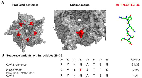
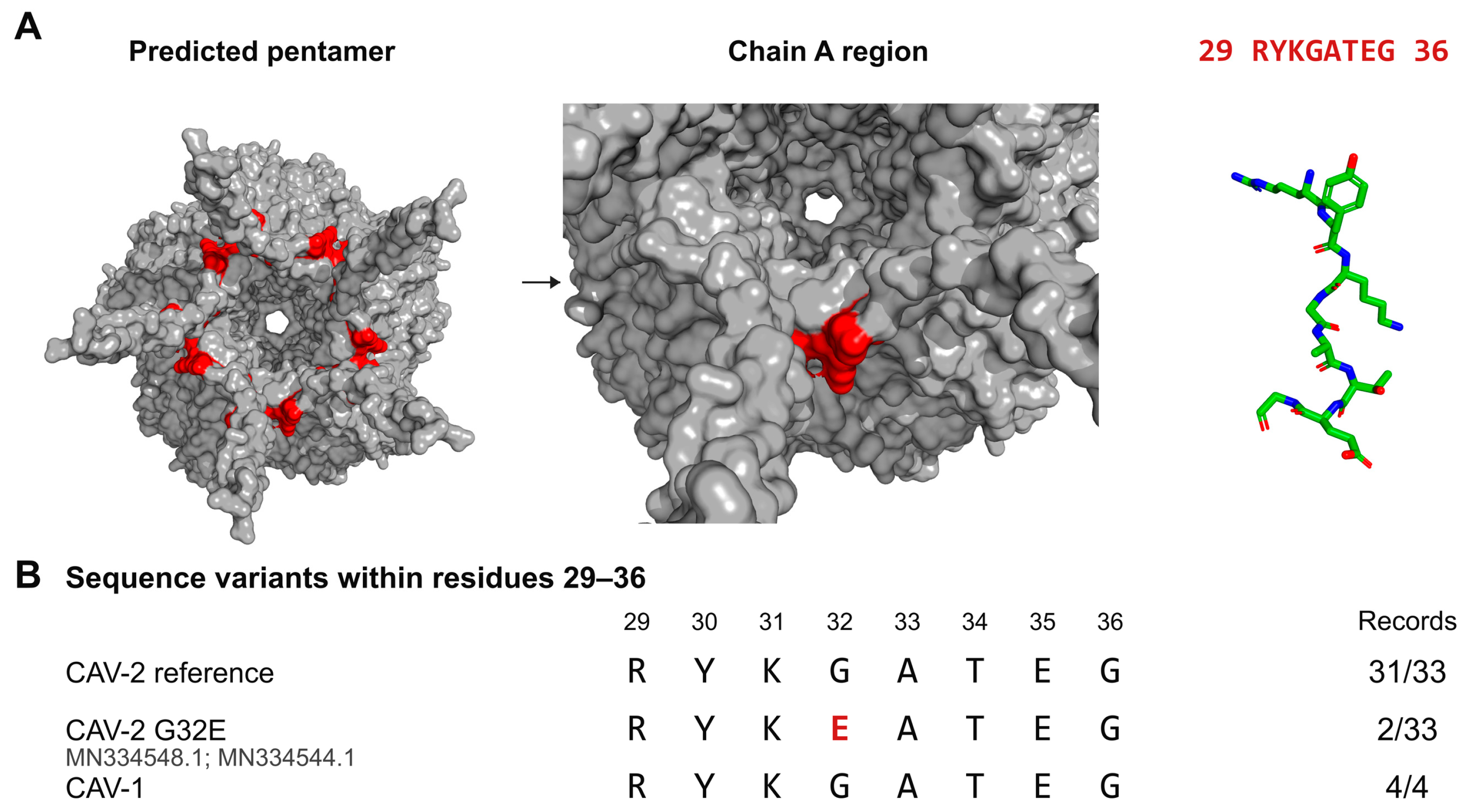
The mAb 3D5-reactive fragment 29RYKGATEG36 was mapped onto the predicted CAV-2 penton-base pentamer using PyMOL, with the corresponding segment highlighted in all five subunits (Figure 4A). Residues 29–36 were modelled as an extended coil in the N-terminal segment, downstream of a short α-helix spanning residues 11–17. The overall pentamer view showed the fragment’s positions across the five subunits, while the magnified A-chain view and stick representation illustrated its local arrangement. In the complete predicted pentamer, residues 29–36 showed heterogeneous relative solvent accessibility (14–69%), with a mean SASA of 87.5 Å2 per residue. These model-based estimates do not establish antibody accessibility on intact virions.
Figure 4.
Structural localization and sequence conservation of the mAb 3D5-reactive fragment in the CAV-2 penton base. (A) Localization of the 29RYKGATEG36 fragment (red) mapped onto the predicted CAV-2 penton base pentamer. The fragment lies in the N-terminal segment upstream of the ordered core and is modelled as an extended coil immediately C-terminal to a short α-helix (residues 11–17). (B) Sequence comparison of residues 29–36 across CAV-2 and CAV-1 penton sequences, showing the RYKGATEG sequence in 31/33 CAV-2 and 4/4 CAV-1 records and G32E in two CAV-2 records. The predicted model does not establish accessibility on intact virions.
Penton amino acid sequences from 33 CAV-2 and 4 CAV-1 strains were retrieved from GenBank and aligned, with accession numbers listed in Supplementary Table S1. The 29RYKGATEG36 sequence was present in 31/33 CAV-2 and all four CAV-1 accession records examined (Figure 4B; Supplementary Table S1). The two CAV-2 variants encoded G32E (MN334548.1 and MN334544.1). Western blotting also detected mAb 3D5 reactivity with unpurified recombinant CAV-1 penton-base expression preparations (Figure S4).
Thus, conservation of the reactive fragment in most CAV-2 sequences examined, together with the observed antibody reactivity, supports further evaluation of mAb 3D5 as a reagent for CAV-2 antigen detection.
4. Discussion
In this study, we identified an N-terminal reactive fragment, 29RYKGATEG36, of the CAV-2 penton base recognized by mAb 3D5 in recombinant-fragment Western blotting. The fragment was largely conserved in the sequence records examined, and antibody reactivity was also detected with a recombinant CAV-1 expression preparation. These findings localize mAb 3D5 recognition to a short N-terminal region and provide a basis for further evaluation of its antigen-detection applications. However, recognition of the eight-residue fragment does not establish precise epitope boundaries or the contributions of individual residues.
The penton base forms part of the adenovirus capsid vertex and contributes to viral entry and assembly. In human adenovirus 2, its RGD motif mediates interactions with cellular integrins involved in internalization [13]. CAV-2 lacks a canonical RGD motif, and RGD-independent entry has been reported [8]. In the present study, structural mapping placed the reactive fragment in an N-terminal coil of the predicted pentamer. Calculating solvent accessibility in the complete pentamer incorporated occlusion by neighbouring subunits and showed heterogeneous accessibility across the fragment. This provides structural context for its localization, but the predicted coil conformation does not demonstrate flexibility or assembly-dependent exposure. Moreover, accessibility to a small solvent probe does not establish accessibility to an antibody on an intact virion. The model therefore supports positional mapping without confirming the fragment’s native conformation or exposure.
The RYKGATEG sequence was present in 31 of 33 CAV-2 accession records and all four CAV-1 records examined. The two CAV-2 variants encoded G32E. Reactivity with an unpurified recombinant CAV-1 penton-base expression preparation provides experimental evidence of recognition under Western blot conditions. However, the G32E variants were not tested, and recognition of CAV-1-infected cells or intact CAV-1 virions was not established. Sequence conservation therefore supports further investigation of binding breadth but cannot demonstrate equivalent antigenicity or antibody affinity among variants.
Capsid-directed monoclonal antibodies have been investigated for adenovirus detection, including group-specific hexon antibodies [24]. For CAV, an immunochromatographic strip developed using hexon-specific monoclonal antibodies detected both CAV-1 and CAV-2 [16]. The penton-base region described here offers an additional target for investigating CAV antigen detection alongside existing hexon-based approaches. The present data support mAb 3D5 recognition in Western blotting and fixed-cell immunofluorescence, whereas performance in antigen-capture assays and clinical specimens requires separate validation. No detectable neutralizing activity was observed against the tested CAV-2 strain under the assay conditions used. Accordingly, mAb 3D5 is characterized primarily as an antigen-recognition reagent under the tested conditions. This result defines the observed functional profile of mAb 3D5 without establishing a universal absence of neutralizing activity against other strains or CAV-1.
Several limitations should be acknowledged. Structural interpretation relied on a predicted model, and antibody accessibility in the complete capsid remains unresolved. The sequence dataset was limited in size and geographic coverage, and accession records do not necessarily represent independent isolates. CAV-1 reactivity was assessed using an unpurified recombinant expression preparation, while quantitative binding affinity and the effects of sequence variation were not determined. These limitations restrict the conclusions to antigen recognition under the tested conditions and support further evaluation before broader diagnostic applications are proposed.
5. Conclusions
In conclusion, mAb 3D5 recognized the N-terminal fragment 29RYKGATEG36 of the CAV-2 penton base in Western blotting. This sequence was conserved in most CAV-2 and all four CAV-1 accession records examined, and mAb 3D5 also reacted with the tested recombinant CAV-1 preparation. Structural mapping indicated heterogeneous predicted solvent accessibility of the fragment in the pentamer. These findings support further evaluation of mAb 3D5 for CAV antigen detection, while reactivity with sequence variants and binding to intact virions remain to be established.
Supplementary Materials
The following supporting information can be downloaded at: https://www.mdpi.com/article/10.3390/vetsci13111251/s1: Table S1: Accession numbers of the CAV-2 and CAV-1 penton base sequences used for sequence alignment.
Author Contributions
Writing—review and editing, Q.J. and H.J.; writing—original draft preparation, Q.J.; methodology, Q.J., S.M., W.D., M.L. and J.H.; investigation, Q.J.; data curation, J.H.; visualization, H.K.; supervision, H.J. and J.L.; funding acquisition, Q.J. and H.J. All authors have read and agreed to the published version of the manuscript.
Funding
This study was financially supported by the National Key Research and Development Program of China (Grant No. 2023YFE0126200, awarded to H.J.) and the R&D and Application of Veterinary Biological Products for the Belt and Road Initiative (Grant No. JD 25E003). The funders had no role in the study design, data collection and analysis, decision to publish, or preparation of the manuscript. We declare that we have no competing interests.
Institutional Review Board Statement
All animal experiments received approval from the Institutional Animal Care and Use Committee (IACUC) of the Harbin Veterinary Research Institute (HVRI), Chinese Academy of Agricultural Sciences (CAAS) (Approval No. 251211-05-GR, 6 May 2025).
Informed Consent Statement
Not applicable.
Data Availability Statement
The original contributions presented in this study are included in the article/Supplementary Material. Further inquiries can be directed to the corresponding author(s).
Conflicts of Interest
The authors declare no conflicts of interest.
Abbreviations
The following abbreviations are used in this manuscript:
| aa | Amino acid |
| BSA | Bovine serum albumin |
| CAV-1 | Canine adenovirus type 1 |
| CAV-2 | Canine adenovirus type 2 |
| DMEM | Dulbecco’s modified Eagle’s medium |
| FBS | Fetal bovine serum |
| IFA | Immunofluorescence assay |
| IgG | Immunoglobulin G |
| IPTG | Isopropyl β-d-1-thiogalactopyranoside |
| mAb | Monoclonal antibody |
| MDCK | Madin-Darby canine kidney |
| MOI | Multiplicity of infection |
| Ni-NTA | Nickel-nitrilotriacetic acid |
| PBS | Phosphate-buffered saline |
| PCR | Polymerase chain reaction |
| RMSD | Root-mean-square deviation |
| SASA | Solvent-accessible surface area |
| SDS-PAGE | Sodium dodecyl sulfate–polyacrylamide gel electrophoresis |
| TBST | Tris-buffered saline with Tween-20 |
| TF | Trigger factor |
References
- Davison, A.J.; Benkő, M.; Harrach, B. Genetic content and evolution of adenoviruses. J. Gen. Virol. 2003, 84, 2895–2908. [Google Scholar] [CrossRef] [Scilit] [PubMed]
- Zhu, Y.; Xu, J.; Lian, S.; Zhang, R.; Hou, J.; Wang, M.; Yan, X. Difference Analysis Between Canine Adenovirus Types 1 And 2. Front. Cell. Infect. Microbiol. 2022, 12, 854876. [Google Scholar] [CrossRef] [Scilit] [PubMed]
- Balboni, A.; Verin, R.; Morandi, F.; Poli, A.; Prosperi, S.; Battilani, M. Molecular epidemiology of canine adenovirus type 1 and type 2 in free-ranging red foxes (Vulpes vulpes) in Italy. Vet. Microbiol. 2013, 162, 551–557. [Google Scholar] [CrossRef] [Scilit] [PubMed]
- Wang, J.; Zhou, N.; Chen, X.; Yue, H.; Tang, C. Isolation, Identification and Pathogenicity of Canine Adenovirus Type 2 Strain with Natural Deletion in E3 Gene. Acta Vet. Zootech. Sin. 2023, 54, 2982–2990. [Google Scholar] [CrossRef]
- Macartney, L.; Cavanagh, H.M.A.; Spibey, N. Isolation of canine adenovirus-2 from the faeces of dogs with enteric disease and its unambiguous typing by restriction endonuclease mapping. Res. Vet. Sci. 1988, 44, 9–14. [Google Scholar] [CrossRef] [Scilit]
- Benetka, V.; Weissenböck, H.; Kudielka, I.; Pallan, C.; Rothmüller, G.; Möstl, K. Canine adenovirus type 2 infection in four puppies with neurological signs. Vet. Rec. 2006, 158, 91–94. [Google Scholar] [CrossRef] [Scilit] [PubMed]
- Chander, V.; Sharma, G.K.; Bhatt, M.; Nandi, S.; Mahajan, S.; Singh, M.; Mahendran, K.; Karikalan, M.; Pawde, A.M.; Gupta, V.; et al. Isolation and genetic characterization of canine adenovirus type 2 from a domestic dog showing neurological symptoms. Braz. J. Microbiol. 2021, 52, 2521–2528. [Google Scholar] [CrossRef] [Scilit] [PubMed]
- Soudais, C.; Boutin, S.; Hong, S.S.; Chillon, M.; Danos, O.; Bergelson, J.M.; Boulanger, P.; Kremer, E.J. Canine Adenovirus Type 2 Attachment and Internalization: Coxsackievirus-Adenovirus Receptor, Alternative Receptors, and an RGD-Independent Pathway. J. Virol. 2000, 74, 10639–10649. [Google Scholar] [CrossRef] [Scilit] [PubMed]
- Bru, T.; Salinas, S.; Kremer, E.J. An Update on Canine Adenovirus Type 2 and Its Vectors. Viruses 2010, 2, 2134–2153. [Google Scholar] [CrossRef] [Scilit] [PubMed]
- Soudais, C.; Laplace-Builhe, C.; Kissa, K.; Kremer, E.J. Preferential transduction of neurons by canine adenovirus vectors and their efficient retrograde transport in vivo. FASEB J. 2001, 15, 2283–2285. [Google Scholar] [CrossRef] [Scilit] [PubMed]
- Kremer, E.J. CAR chasing: Canine adenovirus vectors—All bite and no bark? J. Gene Med. 2004, 6, S139–S151. [Google Scholar] [CrossRef] [Scilit] [PubMed]
- Seiradake, E.; Lortat-Jacob, H.; Billet, O.; Kremer, E.J.; Cusack, S. Structural and Mutational Analysis of Human Ad37 and Canine Adenovirus 2 Fiber Heads in Complex with the D1 Domain of Coxsackie and Adenovirus Receptor. J. Biol. Chem. 2006, 281, 33704–33716. [Google Scholar] [CrossRef] [Scilit] [PubMed]
- Zubieta, C.; Schoehn, G.; Chroboczek, J.; Cusack, S. The Structure of the Human Adenovirus 2 Penton. Mol. Cell 2005, 17, 121–135. [Google Scholar] [CrossRef] [Scilit] [PubMed]
- Schoehn, G.; El Bakkouri, M.; Fabry, C.M.S.; Billet, O.; Estrozi, L.F.; Le, L.; Curiel, D.T.; Kajava, A.V.; Ruigrok, R.W.H.; Kremer, E.J. Three-Dimensional Structure of Canine Adenovirus Serotype 2 Capsid. J. Virol. 2008, 82, 3192–3203. [Google Scholar] [CrossRef] [Scilit] [PubMed]
- Whetstone, C.A. Monoclonal antibodies to canine adenoviruses 1 and 2 that are type-specific by virus neutralization and immunofluorescence. Vet. Microbiol. 1988, 16, 1–8. [Google Scholar] [CrossRef] [Scilit] [PubMed]
- Wang, S.; Wen, Y.; An, T.; Duan, G.; Sun, M.; Ge, J.; Li, X.; Yang, K.; Cai, X. Development of an Immunochromatographic Strip for Rapid Detection of Canine Adenovirus. Front. Microbiol. 2019, 10, 2882. [Google Scholar] [CrossRef] [Scilit] [PubMed]
- Wang, S.; Wang, C.; Ren, X.; Xue, W.; He, H.; Zhu, Y.; Wang, H.; Wang, G.; Cai, X. Identification of Two Novel Linear Neutralizing Epitopes within the Hexon Protein of Canine Adenovirus Using Monoclonal Antibodies. Vaccines 2021, 9, 135. [Google Scholar] [CrossRef] [Scilit] [PubMed]
- Wang, B.; Xu, J.; Zhang, H.; Lian, S.; Duan, Y.; Zhang, H.; Hou, W.; Yin, B.; Zhu, Y. Establishment of ELISA method for canine adenovirus type 1. Front. Vet. Sci. 2024, 11, 1440124. [Google Scholar] [CrossRef] [Scilit] [PubMed]
- Wang, C.-R.; Wang, D.-M.; Yin, G.-Y.; Wang, Y.-H.; Lu, M.; Zhang, Z.-W.; Wen, Y.; Gao, D.-Z.; Hong, J.; Fu, P.-F. Isolation and characterization of canine adenovirus type 2 (CAV-HN45) and its selective infection of human cervical cancer cells with preliminary oncolytic potential. Front. Vet. Sci. 2025, 12, 1692395. [Google Scholar] [CrossRef] [Scilit] [PubMed]
- Yu, Z.; Jiang, Q.; Liu, J.; Guo, D.; Quan, C.; Li, B.; Qu, L. A simplified system for generating recombinant E3-deleted canine adenovirus-2. Plasmid 2015, 77, 1–6. [Google Scholar] [CrossRef] [Scilit] [PubMed]
- Abramson, J.; Adler, J.; Dunger, J.; Evans, R.; Green, T.; Pritzel, A.; Ronneberger, O.; Willmore, L.; Ballard, A.J.; Bambrick, J.; et al. Accurate structure prediction of biomolecular interactions with AlphaFold 3. Nature 2024, 630, 493–500. [Google Scholar] [CrossRef] [Scilit] [PubMed]
- Tien, M.Z.; Meyer, A.G.; Sydykova, D.K.; Spielman, S.J.; Wilke, C.O. Maximum Allowed Solvent Accessibilites of Residues in Proteins. PLoS ONE 2013, 8, e80635. [Google Scholar] [CrossRef] [Scilit] [PubMed]
- Schrödinger, L.L.C. The PyMOL Molecular Graphics System, Version 3.1.0. Available online: https://www.pymol.org/ (accessed on 10 November 2026).
- Cepko, C.L.; Whetstone, C.A.; Sharp, P.A. Adenovirus hexon monoclonal antibody that is group specific and potentially useful as a diagnostic reagent. J. Clin. Microbiol. 1983, 17, 360–364. [Google Scholar] [CrossRef] [Scilit] [PubMed]

Disclaimer/Publisher’s Note: The statements, opinions and data contained in all publications are solely those of the individual author(s) and contributor(s) and not of MDPI and/or the editor(s). MDPI and/or the editor(s) disclaim responsibility for any injury to people or property resulting from any ideas, methods, instructions or products referred to in the content. |
© 2026 by the authors. Licensee MDPI, Basel, Switzerland. This article is an open access article distributed under the terms and conditions of the Creative Commons Attribution (CC BY) license.