


Simple Summary
Porcine reproductive and respiratory syndrome causes substantial economic losses in pig production worldwide. Laboratory tests that detect antibodies against the virus can support disease monitoring, but their results must be interpreted in light of vaccination history. In this study, we produced a viral protein called NSP7 and generated monoclonal antibodies that recognize it. We then used one of these antibodies to develop a test for detecting NSP7-specific antibodies in pig serum. The test showed 95.74% agreement with a commercial test when evaluated using 94 clinical samples. It detected antibody responses in experimentally infected pigs and pigs given the tested live attenuated vaccine, whereas samples from pigs given the tested inactivated vaccine were negative. The test may support PRRSV antibody monitoring, although it cannot distinguish natural infection from live attenuated vaccination. Further testing is needed to assess its performance against genetically diverse PRRSV strains.
Abstract
Porcine reproductive and respiratory syndrome (PRRS) remains an important infectious disease of pigs, and serological assays are widely used to monitor antibody responses to the PRRS virus (PRRSV). This study aimed to develop and evaluate a blocking enzyme-linked immunosorbent assay (bELISA) targeting PRRSV nonstructural protein 7 (NSP7). Recombinant NSP7 was expressed in Escherichia coli, purified, and used to generate monoclonal antibodies. Monoclonal antibody 5E2 was selected to establish the bELISA. A cutoff of 40% inhibition was determined via receiver operating characteristic analysis of 198 serum samples classified using a commercial ELISA. No cross-reactivity was detected with the tested sera positive for pseudorabies virus (PRV), African swine fever virus (ASFV), classical swine fever virus (CSFV), or porcine circovirus type 2 (PCV2). Intra-assay and interassay coefficients of variation were below 10%, and agreement with the commercial ELISA was 95.74% among 94 clinical serum samples. The assay detected antibody responses in experimentally infected pigs. Samples from pigs given the tested inactivated vaccine were negative, whereas those from pigs given the tested live attenuated vaccine were positive. These findings support the potential of the NSP7-based bELISA as a complementary tool for PRRSV serology. The assay does not distinguish natural infection from live attenuated vaccination, and its performance across PRRSV genotypes requires further validation.
1. Background
Porcine reproductive and respiratory syndrome (PRRS), caused by the PRRS virus (PRRSV), is a major infectious disease that poses a major threat to the global swine industry. The disease was first reported in the United States in 1987 [1,2]. Recombination contributes to viral diversity, and pathogenicity varies among strains. Some highly virulent strains cause mortality approaching 100% under specific experimental conditions, whereas other recombinant strains have low pathogenicity and may cause no clinical disease. This disease is characterized by reproductive failure in pregnant sows and respiratory distress in growing pigs [3]. Its widespread occurrence and effects on herd health cause substantial economic losses.
Vaccination remains the primary and most critical strategy for the prevention and control of PRRS. However, currently available commercial vaccines generally fail to provide satisfactory protective efficacy [4], and viral genetic diversity and continued evolution complicate vaccine-based control [5]. Given the lack of highly effective and stable vaccines against PRRSV, the development of accurate and rapid diagnostic methods has become increasingly important for disease surveillance and control [6]. Enzyme–linked immunosorbent assay (ELISA) are widely used for PRRSV serology because they are relatively simple to perform and suitable for high-throughput testing [7,8]. Commercial assays commonly target the immunogenic nucleocapsid (N) protein. Although these assays can detect antibodies against diverse strains, they generally cannot distinguish vaccine-induced antibodies from those induced by field-virus infection [9].
PRRSV has a positive-sense, single-stranded positive-sense RNA virus with a genome of approximately 15 kb containing at least 10 open reading frames (ORFs) [10]. Among these, ORF1a and ORF1b mainly encode nonstructural proteins that are essential for viral replication and transcription [11]. NSP7 has been implicated in replication and modulation of host immune responses [12,13]. Its sequence is relatively conserved within PRRSV types, with reported identity values of 96.7–97.4% within PRRSV-1 and 84.9–100% within PRRSV-2, compared with approximately 45% between the two types [14]. Additionally, NSP7 is also immunogenic and can induce detectable antibody responses in infected pigs [15,16]. Because NSP7 is produced during viral replication, antibodies against it may help distinguish exposure to replicating virus from immunization with an inactivated vaccine. This distinction does not extend to live attenuated vaccination.
Here, we successfully expressed recombinant NSP7 protein in E. coli, generated NSP7-specific monoclonal antibodies, and developed a blocking ELISA (bELISA). We evaluated its analytical performance, agreement with a commercial assay, and detection of antibody responses in experimentally infected and vaccinated pigs. The aim was to assess its potential as a complementary tool for PRRSV serological surveillance.
2. Materials and Methods
2.1. Cells, Viruses, Sera, and Strains
MARC-145 and SP2/0 cells were purchased from Shanghai Jinyuan Biotechnology Co., Ltd. (Shanghai, China). MARC-145 cells were cultured in Dulbecco’s modified Eagle’s medium (DMEM; Gibco, Waltham, MA, USA) supplemented with 10% fetal bovine serum (FBS; ExCell Bio, Suzhou, China), 100 U/mL penicillin, and 100 μg/mL streptomycin (Gibco, USA) at 37 °C in 5% CO2. SP2/0 murine myeloma cells were cultured in RPMI 1640 medium (Gibco, USA) supplemented with 20% FBS. E. coli DH5α and BL21 (DE3) strain were maintained in our laboratory. Clinical serum samples were obtained from the Detection Laboratory of the Animal Disease Diagnostic Center at Huazhong Agricultural University. Their PRRSV antibody status was determined using a commercial ELISA (BioChek, Reeuwijk, The Netherlands).
2.2. Construction of the NSP7 Expression Plasmid
The PRRSV NSP7 gene sequence (777 bp) was obtained from a PRRSV strain isolated in our laboratory. The gene was amplified by PCR using the forward primer 5′-CGCGGATCCTCGCTGACTGGTGCCCTCG-3′ and 5′-CCGAAGCTTTTCCCACTGAGCTCTTCTATTCTCG-3′ and cloned into pET-28a to express full-length, His-tagged NSP7. Protein expression was assessed by SDS–PAGE and Western blotting. The recombinant protein was purified by affinity chromatography, and its concentration was measured spectrophotometrically.
2.3. Expression and Purification of Recombinant NSP7 Protein
The recombinant prokaryotic expression plasmid pET-28a-NSP7 was transformed into competent E. coli DH5α cells. A single colony was selected and cultured, and plasmid DNA was extracted using a plasmid miniprep kit (Omgga, Norcross, GA, USA). The construct was verified by restriction enzyme digestion, and the bacterial culture was preserved. The plasmid was subsequently transformed into competent E. coli BL21(DE3) cells and verified by sequencing. A stored bacterial culture was inoculated into fresh culture medium at 1:1000 for recovery and expansion, followed by transfer into fresh medium at 1:1000. The resulting culture was then inoculated into fresh medium at a ratio of 1:100. After 2 h of incubation, isopropyl β-D-1-thiogalactopyranoside (IPTG) was added to a final concentration of 0.8 mM, and protein expression was induced at 37 °C with shaking at 180 rpm for 4 h. The recombinant NSP7 protein was purified from the soluble fraction by nickel-nitrilotriacetic acid (Ni-NTA) affinity chromatography. The purified protein was dialyzed to remove salts and subsequently concentrated using a 10-kDa molecular-weight-cutoff ultrafiltration device. Sucrose was then used for further concentration of the protein preparation. Protein concentration was determined using a bicinchoninic acid (BCA) protein assay kit (Beyotime Biotechnology, Shanghai, China), according to the manufacturer’s instructions.
2.4. Generation of NSP7-Specific Monoclonal Antibodies
BALB/c mice aged 4–6 weeks were immunized intramuscularly with 15 μg of purified NSP7 per mouse mixed with QuickAntibody-Mouse3W (Biodragon Biotech, Suzhou, China). Two injections were administered over three weeks. Antibody titers were measured three weeks after the last injection. Mice were euthanized 10 days after the final immunization, and the splenocytes were collected and fused with SP2/0 myeloma cells. The fused cells were cultured for 7 days in semisolid hypoxanthine–aminopterin–thymidine (HAT)-selective medium at 37 °C in a humidified incubator with 5% CO2. Hybridomas were screened via ELISA after reaching confluence. Positive clones underwent three rounds of limiting dilution methods to obtain monoclonal cell lines.
2.5. Production and Characterization of Monoclonal Antibodies
Mice were inoculated intraperitoneally with hybridoma cells, and ascitic fluid was collected ten days later. Antibodies were purified using affinity chromatography and characterized via ELISA, indirect immunofluorescence assay (IFA), and Western blotting (WB).
For Western blotting, purified NSP7 was separated by SDS–PAGE and transferred to polyvinylidene difluoride (PVDF) membranes (Merck Millipore, Billerica, MA, USA). Membranes were blocked with 5% skim milk at room temperature and incubated with horseradish peroxidase (HRP)-conjugated NSP7-specific monoclonal antibodies. Bands were visualized using an enhanced chemiluminescence (ECL) chromogenic detection kit (Thermo Fisher Scientific, Waltham, MA, USA). Original uncropped blots are shown in Supplementary Figure S1
For immunohistochemistry (IHC), lung tissues from PRRSV-infected pigs were fixed in 4% paraformaldehyde, embedded in paraffin, and sectioned. The tissue sections were incubated with a laboratory-generated HRP-conjugated monoclonal antibody against the PRRSV NSP7 protein. Immunoreactive signals were visualized by chromogenic detection to evaluate the reactivity and specificity of the monoclonal antibody.
2.6. Development of the NSP7-Based Blocking ELISA
Checkerboard titration was used to optimize the antigen-coating concentration and working concentration of monoclonal antibody (mAb) 5E2 using two reference swine sera S1002 and S0. Blocking solutions and incubation times then compared, followed by serum and antibody incubation times. Plates with 96 wells (Corning Inc., Kennebunk, ME, USA) were coated with 100 μL/well NSP7 at concentrations of 0.25–8 μg/mL prepared by twofold serial dilution and incubated for 12 h at 4 °C. After three washes with PBST, plates were blocked with 5% skim milk in PBST for 2 h at 37 °C and washed three more times. Undiluted positive and negative sera (100 μL per well) were separately added to alternate columns, and plates were then incubated for 1 h at 37 °C.
After washing, HRP-conjugated mAb 5E2 (1:100,000; 100 μL per well) was added, and plates were incubated for 1 h at 37 °C. Plates were washed again, and 100 μL of 3,3′,5,5′-tetramethylbenzidine (TMB) substrate was added to each well. Color was developed for 10 min at room temperature in the dark. The reaction was stopped with 50 μL of 2 M H2SO4 per well. Optical density at 450 nm (OD450) was measured, and data were transferred to an Excel worksheet. Percent inhibition (PI) was calculated as follows: PI (%) = [(OD450 of the negative control − OD450 of the sample)/OD450 of the negative control] × 100.
2.7. Assessment of Assay Repeatability
Three PRRSV antibody-positive and three antibody-negative serum samples were used to assess repeatability. For intra-assay testing, each sample was analyzed in triplicate using one batch of precoated ELISA plates. For interassay testing, the same samples were analyzed using three plate batches. Mean PI values and coefficients of variation (CVs) were calculated from the three replicate measurements for each sample.
2.8. Statistical Analysis
Receiver Operating Characteristic (ROC) curve analysis was performed using IBM SPSS Statistics version 21 to determine the cutoff value for the NSP7 blocking ELISA. A total of 198 serum samples, including 123 PRRSV antibody-negative samples and 75 PRRSV antibody-positive samples classified using a commercial ELISA kit (BioChek, Reeuwijk, The Netherlands), were analyzed. The optimal cutoff value was determined based on the ROC curve analysis using the maximum Youden index method (Youden index = sensitivity + specificity − 1), and the cutoff yielding the highest Youden index was selected as the optimal cutoff.
3. Results
3.1. Expression, Purification, and Identification of Recombinant PRRSV NSP7 Protein
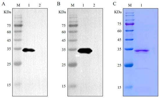
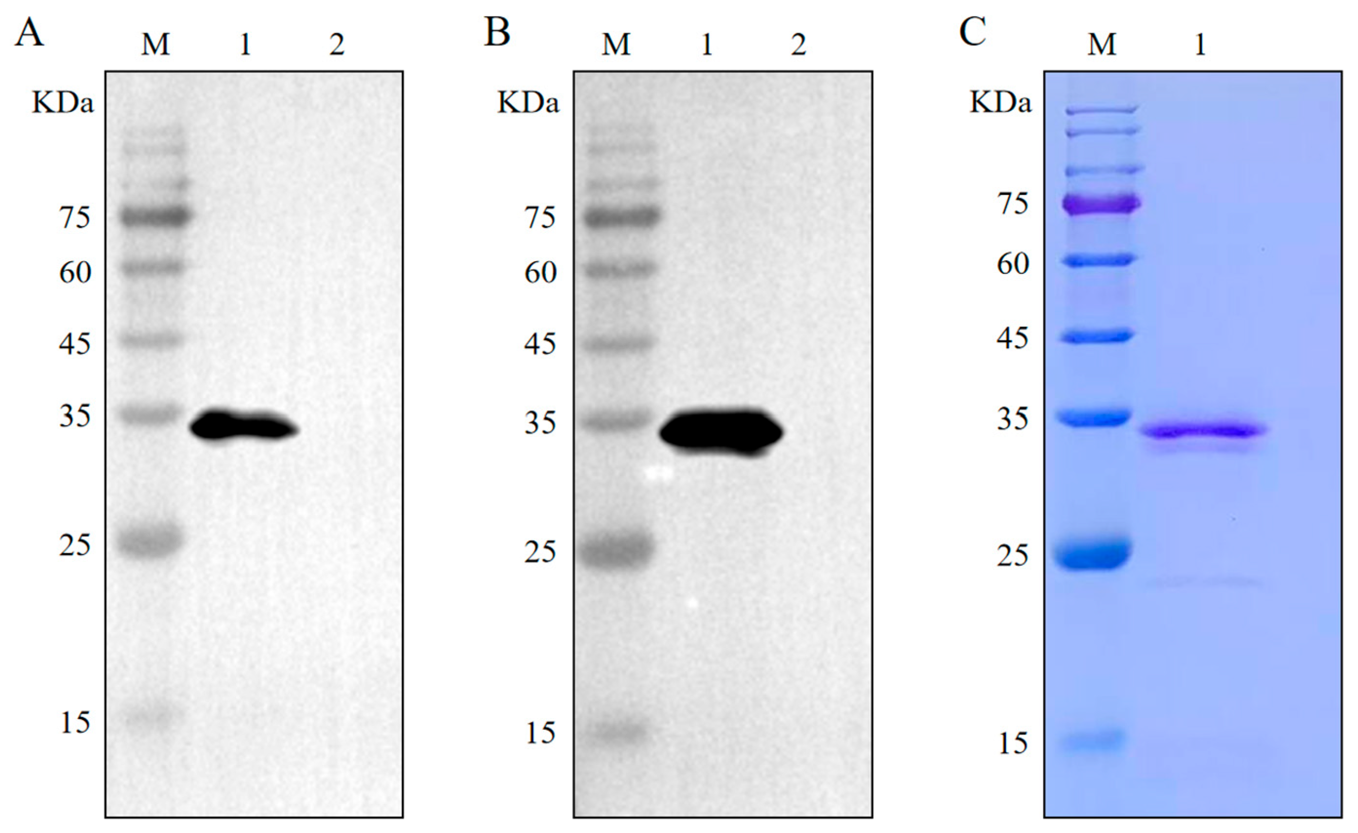
The recombinant plasmid pET28-NSP7, containing a His tag, was transformed into E. coli. The expression of the PRRSV NSP7 protein (35 kDa) was analyzed via Western blot analysis (Figure 1A). Owing to the faint appearance of two bands in the experimental results, PRRSV-positive serum was employed as the primary antibody for further confirmation of successful NSP7 protein expression. The results revealed a distinct band at 35 kDa, confirming successful expression (Figure 1B). Additionally, the presence of two bands suggested specificity, possibly related to the placement of the purified tag at the C-terminus of the vector. The PRRSV NSP7 protein was purified using Ni-NTA agarose (Qiagen, Hilden, Germany) under native conditions. Purified NSP7 protein was obtained through Ni-NTA agarose chromatography (Figure 1C).
Figure 1.
Purification and identification of the NSP7 protein. (A) The NSP7 protein can react with HRP-conjugated mouse anti His-Tag MAb. Lane M: protein marker; lane 1: nsp7 protein; lane 2: negative control. (B) The NSP7 protein can react with PRRSV-positive pig serum. Lane M: protein marker, lane 1: nsp7 protein; lane 2: negative control. (C) The purification of the recombinant protein was assessed by SDS–PAGE. M: protein marker; 1: purified protein.
3.2. Characterization of NSP7-Specific Monoclonal Antibodies
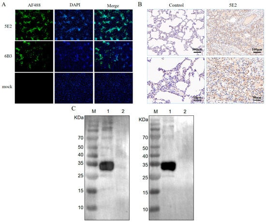
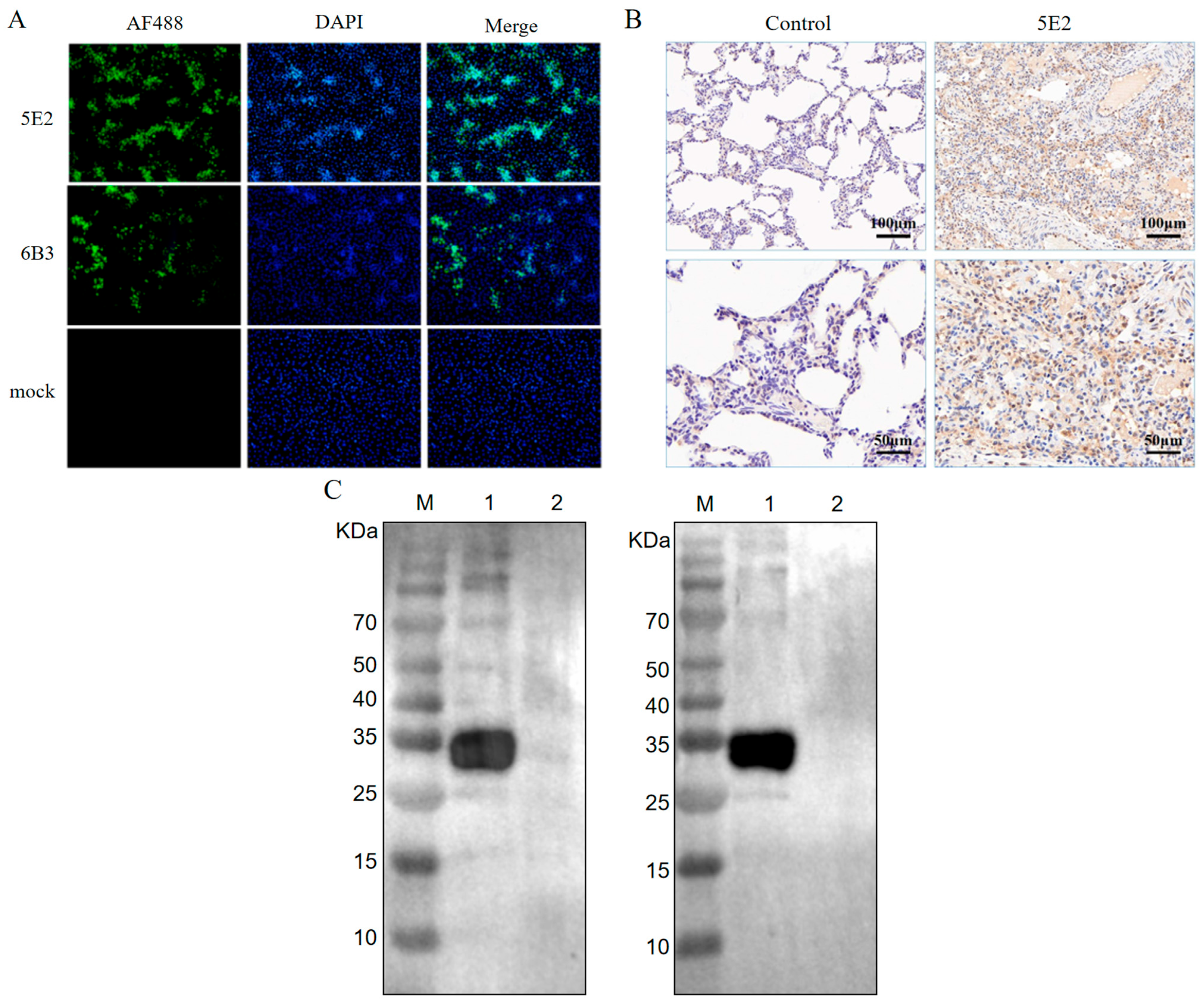
Mice were immunized with the NSP7 protein. Cell fusion was performed when the antibody titer reached 1:100,000 or higher. Two positive clones specific to the NSP7 protein, designated 6B3 and 5E2, were obtained and subcloned three times using the limiting dilution method. The two mAbs were further verified by Western blotting and IFA. The purified NSP7 protein was used as the experimental group, while the pET-28a empty vector served as the negative control. HRP-conjugated mAbs 5E2 and 6B3 (1:10,000 dilution) were used as the detection antibodies for Western blotting. As shown in Figure 2C, distinct specific bands at 35 kDa were observed, while no bands were detected in the negative control. Indirect immunofluorescence assays reflected the interaction between monoclonal antibodies and cells after PRRSV infection. MARC-145 cells were infected with PRRSV for 12–15 h and then incubated with purified ascites fluid at a 1:1000 dilution ratio as the primary antibody, with FITC-labeled goat anti-mouse IgG as the secondary antibody. Mouse serum diluted 1:1000 served as the positive control. As shown in Figure 2A,, both the positive control wells and the wells treated with the two antibodies exhibited distinct green fluorescence signals distributed in the nucleus and cytoplasm of the cells, whereas the negative control wells showed no fluorescence signal. Laboratory -preserved lung tissue sections and paraffin blocks from the PRRSV animal infection experiment were sent to Wuhan Baiqiangdu Biotechnology Co., Ltd., for immunohistochemical analysis. The HRP-conjugated monoclonal antibody 5E2 was used as the detection antibody. As shown in Figure 2B, the infected samples showed distinct brown positive signals, whereas the negative control group showed no positive signals.
Figure 2.
Reactivity of the anti-NSP7 MAb. (A) Indirect immunofluorescence analysis of PRRSV-infected MARC-145 cells using mAbs 5E2 and 6B3. Green fluorescence was detected in infected cells but not in uninfected controls. (B) Immunohistochemical analysis using HRP-conjugated mAb 5E2. Brown staining was observed in infected tissue but not in the negative control. (C) Western blots probed with mAb 6B3 (left) or 5E2 (right). M, molecular weight marker; lane 1, NSP7; lane 2, negative control.
3.3. Optimization of the NSP7-Based Blocking
Assay conditions were optimized by comparing PI values. Checkerboard titration identified an optimal antigen-coating concentration of 0.5 μg/mL and a serum dilution of 1:1. Optimal blocking was achieved with 5% skim milk for 2 h at 37 °C. The selected incubation times were 2 h at 37 °C for serum and 1 h at 37 °C for HRP-conjugated mAb 5E2 (Table 1).
Table 1.
Optimized conditions for the NSP7-based blocking ELISA.
3.4. Establishment of the ROC Curve
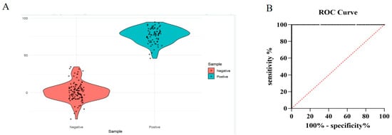
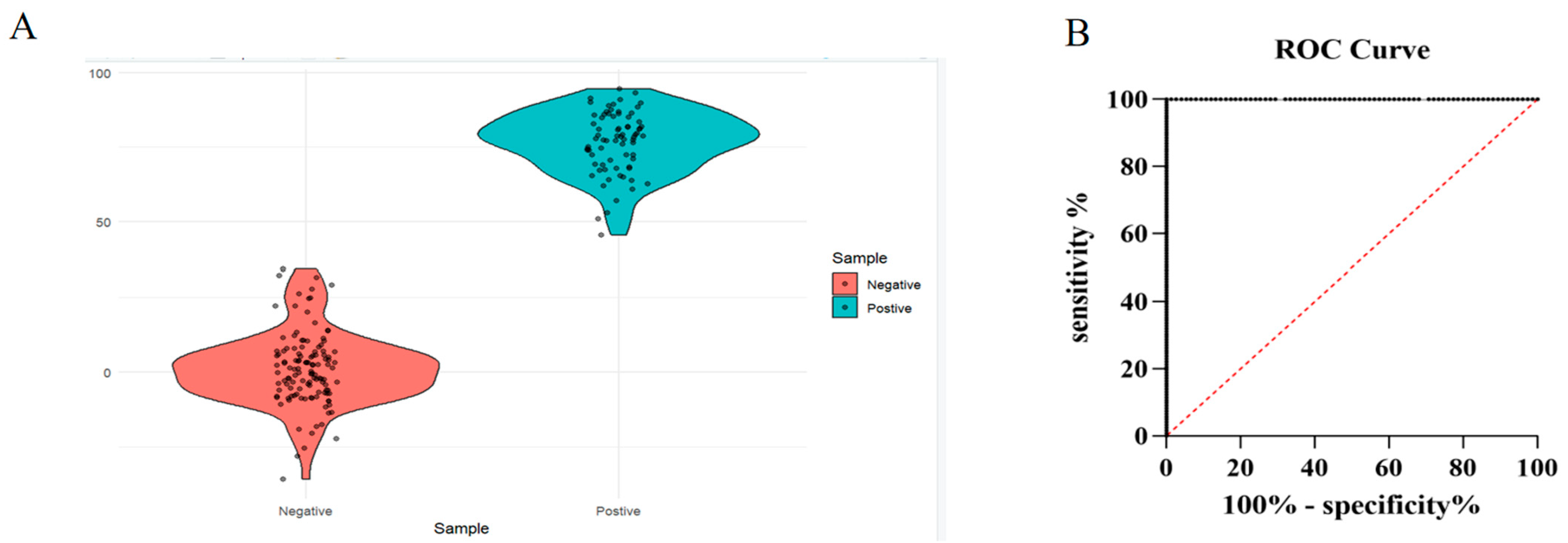
A total of 198 serum samples collected from experimentally infected animals, including 123 negative and 75 positive samples as determined using a commercial assay kit, were analyzed (Figure 3A). ROC curve analysis was performed using IBM SPSS Statistics 21, with the commercial assay kit results serving as the reference method. The established PRRSV NSP7 blocking ELISA showed excellent discriminatory performance, with an area under the ROC curve (AUC) of 1.000 (95% confidence interval [CI]: 1.000–1.000). Based on the maximum Youden index, the optimal cutoff value was determined to be 39.98647% PI. For practical convenience, this value was rounded to 40% PI and used as the final cutoff. Samples with PI values ≥ 40% were classified as positive for PRRSV NSP7 antibodies, whereas those with PI values < 40% were classified as negative. At this cutoff, the assay achieved a sensitivity of 100% (95% CI: 95.20–100%) and a specificity of 100% (95% CI: 97.05–100%) compared with the commercial assay kit (Figure 3B)
Figure 3.
Determination of the assay cutoff. (A) PI values for 198 serum samples classified as positive (n = 75) or negative (n = 123) by the commercial assay. (B) ROC analysis. The calculated cutoff of 39.98647% PI was rounded to 40% for sample classification.
3.5. Detection of Diluted Positive Sera and Assessment of Cross-Reactivity
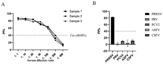
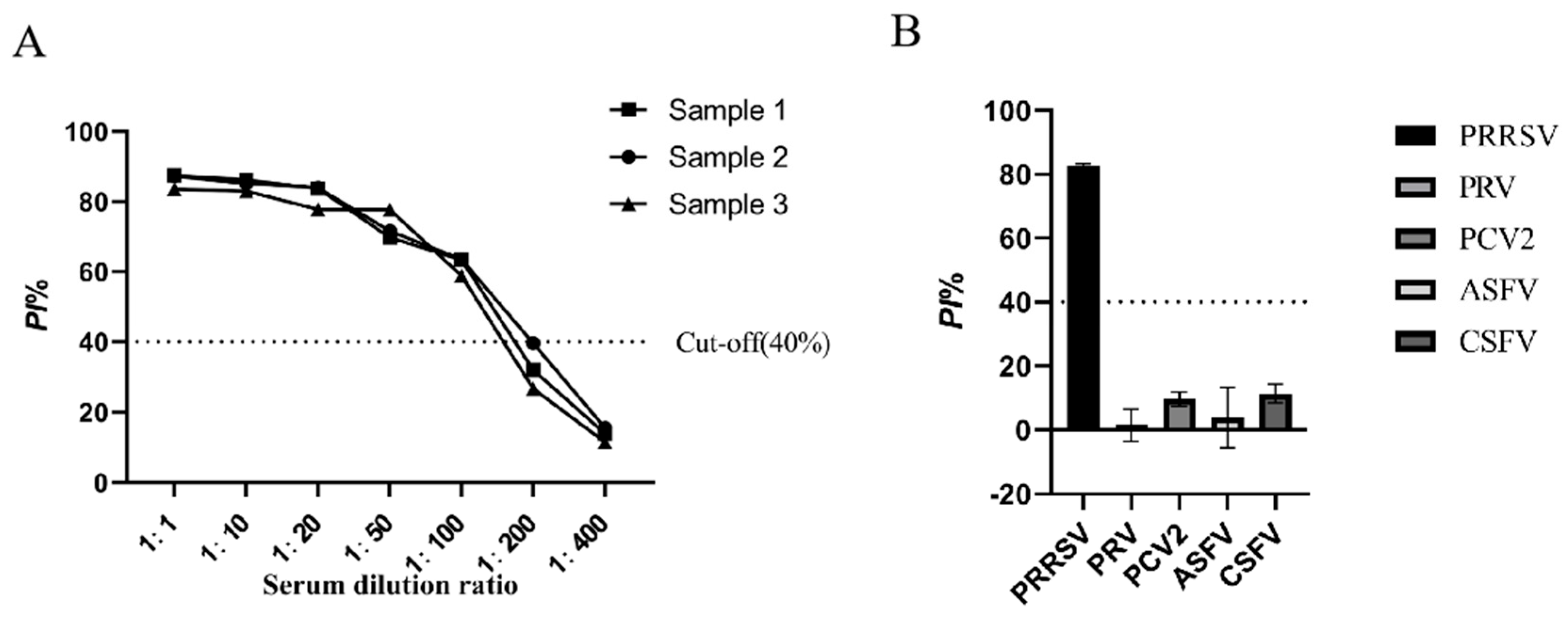
Three PRRS-positive serum samples stored in the laboratory were diluted in phosphate-buffered saline (PBS) at 1:50, 1:100, 1:200, and 1:400 and tested using the NSP7-based bELISA. The highest dilution yielding a positive result was 1:100 for all three samples (Figure 4A).
Figure 4.
Validation of the blocking ELISA. (A) Detection of PRRSV-positive sera diluted from 1:1 to 1:400. (B) Cross-reactivity testing using sera positive for PRRSV, ASFV, PRV, PCV2, or CSFV.
To evaluate the specificity of the established PRRSV NSP7 blocking ELISA, cross-reactivity was assessed using PRV-, PCV2-, ASFV-, and CSFV-positive sera. The mean PI values for PRV, PCV2, ASFV, and CSFV were 1.53 ± 5.11%, 9.70 ± 2.21%, 3.84 ± 9.53%, and 11.39 ± 2.98%, respectively. All values were below the established positive cutoff of 40%. In contrast, PRRSV-positive sera showed a mean PI of 82.58 ± 0.72%. These results indicated that the established PRRSV NSP7 blocking ELISA showed no detectable cross-reactivity with the tested heterologous pathogens (Figure 4B).
3.6. Assay Repeatability
To evaluate the repeatability of the blocking ELISA, three PRRSV antibody-positive and three negative serum samples were tested under intra- and interassay conditions. The intra-assay repeatability test showed that the coefficients of variation (CVs) of the samples ranged from 1.965% to 5.315%, and the CVs of all three positive and three negative serum samples were less than 10%, indicating that the PRRSV NSP7 blocking ELISA has good intra-assay repeatability (Table 2). The interassay repeatability test showed that the CVs of the samples ranged from 0.248% to 5.130%, and the CVs of all three positive and three negative serum samples were less than 10%, indicating that the PRRSV NSP7 blocking ELISA has good interassay repeatability (Table 3).
Table 2.
Intra-batch experimental testing of six serum samples.
Table 3.
Inter-batch experimental testing results of six serum samples.
3.7. Comparison with a Commercial ELISA
The NSP7-based bELISA was compared with the PRRS XR ELISA SK112 (BioChek) using 94 clinical serum samples, including 56 positive and 38 negative samples according to the commercial assay. One sample was negative by the bELISA but positive by the commercial assay, and three were positive by the bELISA but negative by the commercial assay. Overall agreement was 95.74% (90/94; Table 4).
Table 4.
Agreement between the NSP7-based bELISA and the commercial ELISA.
3.8. Detection of Antibodies in Experimentally Infected Pigs
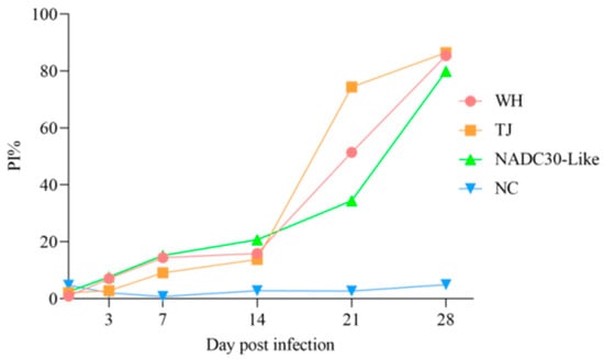
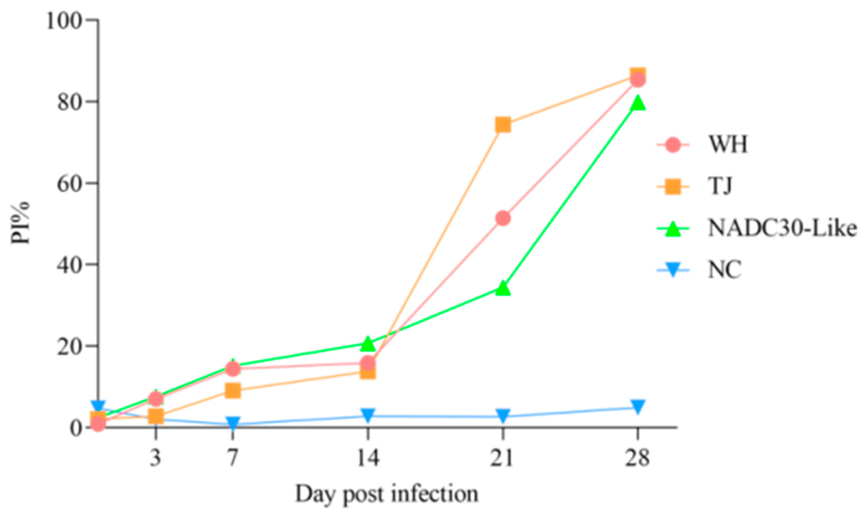
Sera from pigs experimentally infected with the highly pathogenic TJ or WH strain or a NADC30-like strain were tested using the NSP7-based bELISA. Samples were collected at 0, 3, 7, 14, 21, and 28 days postinfection (dpi). Seroconversion was detected at 21 dpi in the TJ and WH groups and at 28 dpi in the NADC30-like group (Figure 5).
Figure 5.
Detection of antibody responses in experimentally infected pigs. Seroconversion was detected at 21 days postinfection (dpi) in the groups infected with the highly pathogenic WH and TJ strains and at 28 dpi in the group infected with the NADC30-like strain.
3.9. Detection of Antibodies in Vaccinated Pigs
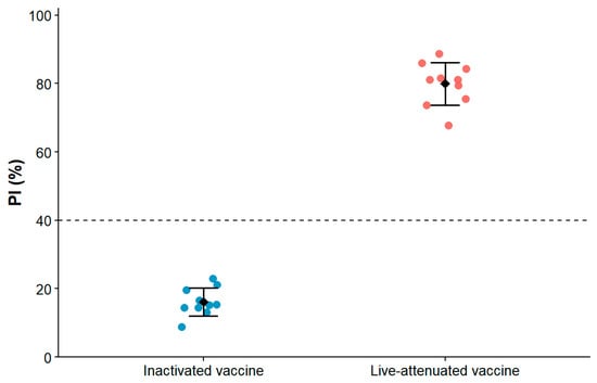
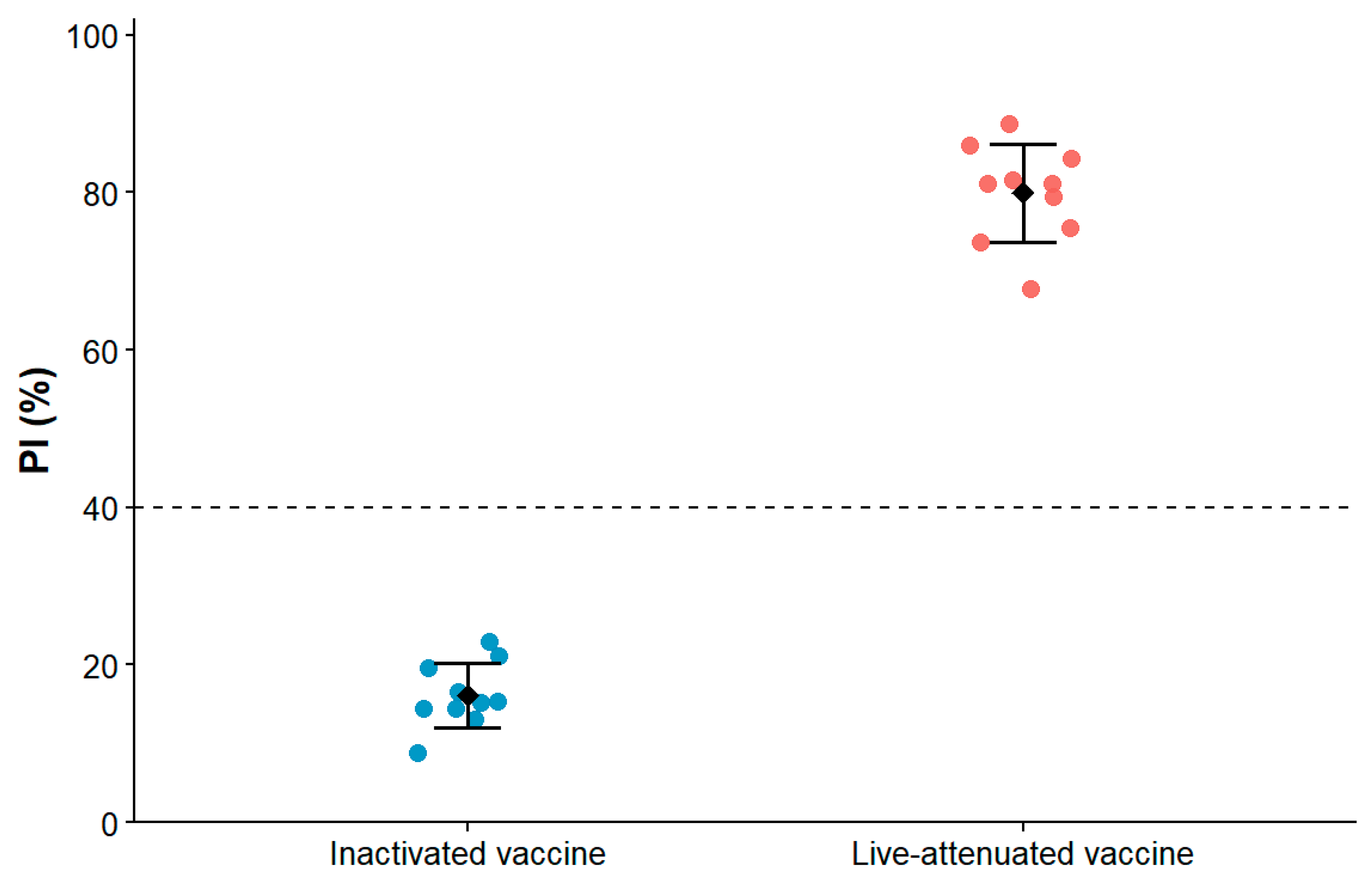
Sera from pigs immunized with an inactivated PRRSV vaccine (CH-1a) or a live attenuated vaccine (CH-1R) were tested, with 10 samples per group. A second dose was administered 14 days after the first, and sera were collected 56 days after the booster. All samples from the inactivated-vaccine group tested negative, whereas all samples from the live attenuated-vaccine group tested positive (Figure 6). Thus, the assay distinguished responses to these two vaccine preparations under the conditions tested.
Figure 6.
Detection of NSP7-specific antibodies in pigs immunized with inactivated or live attenuated PRRSV vaccines.
3.10. Testing of Field Serum Samples
The established PRRSV NSP7 blocking ELISA was used to test 308 clinical serum samples collected from six pig farms in different provinces of China (Table 5). Positive samples were detected on five of the six farms, with farm-level positivity rates ranging from 18.181% to 74.286%. No positive samples were detected on the remaining farm. These results demonstrate detection of NSP7-specific antibodies in field sera.
Table 5.
Results of testing field serum samples.
4. Discussion
In this study, recombinant NSP7 protein was expressed in E. coli and recovered from the soluble fraction after bacterial disruption. The purified protein retained antibody reactivity and was used to generate monoclonal antibodies and develop a blocking ELISA. Bacterial expression offers practical advantages for antigen production, including relatively low cost and straightforward procedures [17].
NSP7 was selected as the target protein for establishing the blocking ELISA because of its relatively high conservation among PRRSV genotypes, making it a promising candidate for broad-spectrum serological detection [18]. Brown reported strong antibody responses to NSP1, NSP2, and NSP7, with NSP7-specific antibodies detected from 14 to 202 days after inoculation [16]. These findings support its use in serological assays, although they do not establish that NSP7 is superior to every alternative antigen. Conservation within a PRRSV type also does not establish equivalent assay performance across PRRSV-1 and PRRSV-2; this requires direct evaluation.
The NSP7-based bELISA detected all three tested positive sera at a dilution of 1:100 and showed no detectable cross-reactivity with the heterologous sera examined. Agreement with a commercial ELISA was 95.74% among 94 clinical samples. For cutoff selection, 198 samples from experimentally infected animals were classified using a commercial antibody ELISA. The resulting performance estimates therefore describe agreement with that reference assay within the evaluated sample set. Antibody responses in field populations may differ because of variation in infection history, vaccination, and sampling time. The 40% PI cutoff and diagnostic performance should consequently be validated in larger, independent clinical sample panels. Such studies may help optimize the cutoff value and provide a more comprehensive assessment of the assay’s performance under field conditions.
Serological assays can complement vaccination in animal disease control. For example, antibodies to the foot-and-mouth disease virus nonstructural polyprotein 3ABC can help identify infection in populations vaccinated with inactivated vaccines [19]. Routine PRRSV serological diagnosis mainly relies on N-protein-based indirect ELISA methods, which generally do not distinguish vaccination from natural infection. The NSP7-based bELISA may provide additional information when interpreting responses to inactivated vaccination. However, the assay was also positive after live attenuated vaccination and therefore cannot distinguish that response from natural infection. Vaccination history must be considered when interpreting positive results.
Overall, this study developed an NSP7-based blocking ELISA using monoclonal antibody 5E2. The assay showed repeatable measurements, no detectable cross-reactivity with the heterologous sera tested, and 95.74% agreement with a commercial ELISA. It detected antibody responses following experimental infection and live attenuated vaccination, whereas samples from pigs given the tested inactivated vaccine were negative. These findings support further evaluation of the assay for PRRSV antibody monitoring. Its current application does not include distinguishing natural infection from live attenuated vaccination, and diagnostic performance across PRRSV genotypes remains unestablished. Independent validation across diverse viral genotypes and vaccination histories will help define its role in serological surveillances.
Supplementary Materials
The following supporting information can be downloaded at https://www.mdpi.com/article/10.3390/vetsci13111220/s1, Figure S1: Original uncropped Western blot image.
Author Contributions
X.J.: Writing—original draft, methodology, data curation, investigation, writing—review and editing, visualization, conceptualization. W.C.: Investigation, writing—review and editing, data curation, conceptualization. X.L.: Validation, methodology, investigation, formal analysis, data curation. X.G.: Investigation, writing—review and editing, data curation, validation. S.R.: Resources, methodology, data curation, supervision. M.Z.: Supervision, project administration, writing—review and editing, methodology. B.Y.: Resources, methodology, investigation, data curation, visualization. A.H.G.: Writing—review and editing, data curation, methodology, supervision. H.Z.: Writing—review and editing, supervision, conceptualization, project administration. Q.H.: Writing—review and editing, supervision, resources, funding acquisition, conceptualization, project administration. W.L.: Writing—review and editing, supervision, funding acquisition, conceptualization, project administration, formal analysis, data curation. All authors have read and agreed to the published version of the manuscript.
Funding
This work was supported by the Key Research and Development Program of Jiangxi Province (20261BCF320004), Hubei Jiangxia Laboratory (2025JXQJKKF017), China Agriculture Research System of MOF and MARA (CARS-34), Fundamental Research Funds for the Central Universities (2662026DKPY005), and the Self-designed research project for Hainan Institute of Huazhong Agricultural University (2024HZAUHNZS005).
Institutional Review Board Statement
The animal study protocol was approved by the Laboratory Animal Ethics Committee of Huazhong Agricultural University (protocol HZAUMO-2023-0246; approved 6 May 2023).
Informed Consent Statement
Not applicable. Informed consent was not applicable because all four- to six-week-old BALB/c mice used in this study were purchased from the Laboratory Animal Center of Huazhong Agricultural University and were not privately owned.
Data Availability Statement
The original contributions presented in this study are included in the article. Further inquiries can be directed to the corresponding authors.
Acknowledgments
We would like to thank the State Key Laboratory of Agricultural Microbiology Core Facility (http://hzaml.hzau.edu.cn/facility/index.htm; accessed on 8 November 2026) for providing essential support.
Conflicts of Interest
The authors declare no conflicts of interest.
References
- Albina, E. Epidemiology of porcine reproductive and respiratory syndrome (PRRS): An overview. Vet. Microbiol. 1997, 55, 309–316. [Google Scholar] [CrossRef] [Scilit] [PubMed]
- Zhao, H.; Zhou, J.; Zhang, M.; Yang, X.; Xi, Z.; Gao, Q.; Fang, R.; Zhao, P.; Zhao, J. Epidemiological surveillance and economic impact analysis of different porcine reproductive and respiratory syndrome virus infection statuses in 23 breeding pig farms in Hubei, China. Anim. Dis. 2024, 4, 47. [Google Scholar] [CrossRef] [Scilit]
- Qiu, Y.; Qiu, M.; Li, S.; Li, S.; Zhu, J.; Tian, K.; Chen, N. Emergence, prevalence and evolution of porcine reproductive and respiratory syndrome virus 1 in China from 1994 to 2024. Virology 2025, 605, 110457. [Google Scholar] [CrossRef] [Scilit] [PubMed]
- Van Noort, A.; Nelsen, A.; Pillatzki, A.E.; Diel, D.G.; Li, F.; Nelson, E.; Wang, X. Intranasal immunization of pigs with porcine reproductive and respiratory syndrome virus-like particles plus 2′, 3′-cGAMP VacciGrade™ adjuvant exacerbates viremia after virus challenge. Virol. J. 2017, 14, 76. [Google Scholar] [CrossRef] [Scilit] [PubMed]
- Ruan, S.; Yu, X.; Wu, H.; Lei, M.; Ku, X.; Ghonaim, A.H.; Li, W.; Jiang, Y.; He, Q. Assessing the antiviral activity of antimicrobial peptides Caerin1.1 against PRRSV in Vitro and in Vivo. Vet. Microbiol. 2024, 297, 110210, Corrigendum in Vet. Microbiol. 2026, in press. [Google Scholar] [CrossRef] [Scilit] [PubMed]
- Nan, Y.; Wu, C.; Gu, G.; Sun, W.; Zhang, Y.J.; Zhou, E.M. Improved Vaccine against PRRSV: Current Progress and Future Perspective. Front. Microbiol. 2017, 8, 1635. [Google Scholar] [CrossRef] [Scilit] [PubMed]
- Fu, C.; Shao, Q.; Zhang, L.; Cui, X.; Chen, T.; Tian, C.; Qian, F.; Chu, X.; Li, Y.; Yang, P.; et al. Development of an enzyme-linked immunosorbent assay using a monoclonal antibody to a dominant epitope in non-structural protein 4 of porcine reproductive and respiratory syndrome virus. J. Immunol. Methods 2024, 530, 113697. [Google Scholar] [CrossRef] [Scilit] [PubMed]
- Pan, J.; Zeng, M.; Zhao, M.; Huang, L. Research Progress on the detection methods of porcine reproductive and respiratory syndrome virus. Front. Microbiol. 2023, 14, 1097905. [Google Scholar] [CrossRef] [Scilit] [PubMed]
- Sun, J.; Wang, J.; Li, K.; Wei, D.; Li, J.; Sun, P.; Li, D.; Li, P.; Cao, Y.; Tang, L.; et al. Generation and characterization of a PRRSV-2 DIVA marker virus by deletion of a novel epitope in the nucleocapsid protein. J. Virol. 2026, 100, e0125926. [Google Scholar] [CrossRef] [Scilit] [PubMed]
- Zhang, H.; Sha, H.; Qin, L.; Wang, N.; Kong, W.; Huang, L.; Zhao, M. Research Progress in Porcine Reproductive and Respiratory Syndrome Virus-Host Protein Interactions. Animals 2022, 12, 1381. [Google Scholar] [CrossRef] [Scilit] [PubMed]
- Chen, J.; Xu, X.; Tao, H.; Li, Y.; Nan, H.; Wang, Y.; Tian, M.; Chen, H. Structural Analysis of Porcine Reproductive and Respiratory Syndrome Virus Non-structural Protein 7α (NSP7α) and Identification of Its Interaction with NSP9. Front. Microbiol. 2017, 8, 853. [Google Scholar] [CrossRef] [Scilit] [PubMed]
- Liu, K.; Ma, G.; Liu, X.; Lu, Y.; Xi, S.; Ou, A.; Wei, J.; Li, B.; Shao, D.; Li, Y.; et al. Porcine reproductive and respiratory syndrome virus counteracts type I interferon-induced early antiviral state by interfering IRF7 activity. Vet. Microbiol. 2019, 229, 28–38. [Google Scholar] [CrossRef] [Scilit] [PubMed]
- Song, J.; Li, K.; Li, T.; Zhao, G.; Zhou, S.; Li, H.; Li, J.; Weng, C. Screening of PRRSV- and ASFV-encoded proteins involved in the inflammatory response using a porcine iGLuc reporter. J. Virol. Methods 2020, 285, 113958. [Google Scholar] [CrossRef] [Scilit] [PubMed]
- Wang, H.; Liu, R.; Cui, J.; Deng, S.; Xie, J.; Nin, Z.; Zhang, G. Characterization and utility of phages bearing peptides with affinity to porcine reproductive and respiratory syndrome virus nsp7 protein. J. Virol. Methods 2015, 222, 231–241. [Google Scholar] [CrossRef] [Scilit] [PubMed]
- Fang, Y.; Schneider, P.; Zhang, W.P.; Faaberg, K.S.; Nelson, E.A.; Rowland, R.R. Diversity and evolution of a newly emerged North American Type 1 porcine arterivirus: Analysis of isolates collected between 1999 and 2004. Arch. Virol. 2007, 152, 1009–1017. [Google Scholar] [CrossRef] [Scilit] [PubMed]
- Brown, E.; Lawson, S.; Welbon, C.; Gnanandarajah, J.; Li, J.; Murtaugh, M.P.; Nelson, E.A.; Molina, R.M.; Zimmerman, J.J.; Rowland, R.R.R.; et al. Antibody response to porcine reproductive and respiratory syndrome virus (PRRSV) nonstructural proteins and implications for diagnostic detection and differentiation of PRRSV types I and II. Clin. Vaccine Immunol. 2009, 16, 628–635. [Google Scholar] [CrossRef] [Scilit] [PubMed]
- Nuc, P.; Nuc, K. Recombinant protein production in Escherichia coli. Postep. Biochem. 2026, 52, 448–456. [Google Scholar]
- Li, K.; Bai, J.; Du, L.; Wang, X.; Ke, C.; Yan, W.; Li, C.; Ren, L.; Han, H.; Zhao, Y. Generation of porcine monoclonal antibodies based on single cell technologies. Vet. Immunol. Immunopathol. 2019, 215, 109913. [Google Scholar] [CrossRef] [Scilit] [PubMed]
- De Diego, M.; Brocchi, E.; Mackay, D.; De Simone, F. The non-structural polyprotein 3ABC of foot-and-mouth disease virus as a diagnostic antigen in ELISA to differentiate infected from vaccinated cattle. Arch. Virol. 1997, 142, 2021–2033. [Google Scholar] [CrossRef] [Scilit] [PubMed]

Disclaimer/Publisher’s Note: The statements, opinions and data contained in all publications are solely those of the individual author(s) and contributor(s) and not of MDPI and/or the editor(s). MDPI and/or the editor(s) disclaim responsibility for any injury to people or property resulting from any ideas, methods, instructions or products referred to in the content. |
© 2026 by the authors. Licensee MDPI, Basel, Switzerland. This article is an open access article distributed under the terms and conditions of the Creative Commons Attribution (CC BY) license.