


Simple Summary
Nipah virus is a dangerous germ that can kill 40 to 75 out of every 100 infected people, and no approved medicine exists to treat it. The virus uses a protein on its surface to enter human cells, which makes this protein a good target for new treatments. In this study, we aimed to create small antibody pieces that could block this protein. We injected part of the Nipah virus surface protein into two alpacas and used blood cells from the one that responded better to build a large collection of antibody pieces. After several rounds of selection, we found seven unique antibody pieces that could recognize the virus protein and stop a model virus from entering cells. Two of them worked best, and making them larger and longer-lasting (by fusing them with an Fc fragment) improved their strength further. Computer modeling predicted that these two antibodies attach to non-overlapping spots on the viral protein, raising the possibility that a tandem format could improve neutralization; however, our experimental tests did not show this synergy. Our results offer a promising starting point for developing antibody-based medicines against Nipah virus, but more testing with the real virus and in animals is needed before they can help people.
Abstract
Nipah virus (NiV) is a WHO priority pathogen with case-fatality rates of 40–75%, and no antiviral therapeutic is licensed for human use. The attachment glycoprotein (G) mediates receptor engagement and is a validated target for neutralizing antibodies. Here, we aimed to isolate alpaca-derived single-domain antibodies (VHHs) against the NiV G head domain. Two alpacas were immunized with a baculovirus-expressed, truncated NiV G ectodomain (residues 170–575); peripheral blood mononuclear cells from the higher-responding animal were used to build an immune phage display library of 3.2 × 10⁸ transformants. Four rounds of biopanning enriched antigen-specific phages approximately 120-fold, and sequencing of 183 reactive clones yielded seven unique VHHs. All seven bound to recombinant NiV G, observed by ELISA, and inhibited entry of a VSV-based NiV pseudovirus. VHH-0G8 and VHH-1B6 were most active, with fitted IC50 values of 1.34 and 1.76 μg/mL (≈89 and 118 nM). Reformatting the two leads as human IgG1 Fc-YTE fusions lowered the fitted IC50 to 0.97 and 1.09 μg/mL (≈12 and 13 nM); because a wild-type Fc comparator was not included, this reflects the Fc-YTE format (M252Y/S254T/T256E, which enhances binding to FcRn to extend serum half-life) as a whole. A tandem 0G8/1B6 construct did not show a statistically significant improvement in neutralization compared to the Fc fusions (IC50 = 1.08 μg/mL, ≈36 nM). Computational docking placed the two VHHs on non-overlapping surfaces of the G head, raising the hypothesis that a tandem format could enhance neutralization; however, this synergy was not experimentally observed and requires further format optimization. These VHHs provide starting points for NiV antibody development and require authentic virus and in vivo evaluation.
1. Introduction
Nipah virus is a type of paramyxovirus and belongs to the same family as Hendra virus and Cedar virus. As a high-risk virus, it can be transmitted either through direct contact with infected bats, sick pigs, or patients or through contaminated food and water sources, with the latter being the main transmission route [1]. The World Health Organization (WHO) has repeatedly listed Nipah virus as one of the top ten high-risk infectious diseases. There have been reports of outbreaks in Southeast Asian countries such as India, Malaysia, and Bangladesh, with a mortality rate reaching up to 60% [2]. From July to December 2025, Nipah virus reappeared in India and Bangladesh, triggering an outbreak with a smaller geographic scope but a markedly higher case fatality rate [3]. The wide host tropism of Nipah virus has raised concerns about the outbreak of epidemics caused by contact with diseased livestock, especially its high mortality rate after infection, highlighting the urgent need to develop related vaccines and antibodies.
Nipah virus was named after its initial outbreak in Nipah village in Malaysia. As a high-risk virus, it can not only be transmitted through direct contact with infected bats, pigs, or patients but also through contaminated food and water, which is considered to be the main transmission route [4]. The glycoprotein of Nipah virus is a key target of the antibody defense system. Ortega et al. discovered that Nipah virus enters host cells through the synergistic action of the G and F proteins [5]. The seven consecutive residues on the top of the spherical head of the G protein mediate interaction with the EFNB2 receptor. The interaction with receptors exposes the stem domain of the G protein and induces a conformational change in the F protein: from the pre-fusion conformation to the pre-hairpin intermediate and, finally, to the post-fusion form capable of membrane fusion [6]. Currently, there are many antibodies that may be effective against Nipah virus and target the G protein, such as m102.4, NIV41, and their derivative antibodies [7,8].
Nanobodies (also known as VHHs) are variable regions of camel heavy-chain antibodies with a molecular weight of approximately 15 kDa, and they have been proven to be versatile molecular binding agents for structural and cell biology research [9,10]. This is mainly because VHH has excellent properties; compared with the traditional single-chain variable region (ScFv), which has a size of approximately 30 kDa, VHH is characterized by a relatively small size; low production costs; easy modification; high tissue permeability; rapid clearance in circulation; high solubility; a high stability index; and high specificity, sensitivity, and affinity [11,12,13]. It is worth noting that nanobody engineering technology has demonstrated significant potential in the development of antibodies for various severe zoonotic diseases such as Middle East Respiratory Syndrome [14], highlighting its potential application value in the prevention and treatment of Nipah virus. In this study, alpacas were specifically chosen because they naturally produce heavy-chain-only antibodies, providing a direct and efficient source for isolating highly stable, small-molecule VHHs.
The main objective of this study was to isolate Nipah virus-specific VHHs using phage display technology. To achieve this, a rational and stepwise experimental design was adopted. An immunized VHH gene library was constructed, as VHHs offer unique advantages such as superior tissue penetration and access to cryptic epitopes due to their small size. After multiple rounds of careful screening, seven Nipah virus-specific VHHs were isolated. Subsequently, these candidates were reformatted into Fc-fusion proteins to overcome the short half-life of monomeric VHHs and to restore bivalent binding, which is crucial for enhancing their neutralizing potency. Finally, a comprehensive evaluation of their in vitro properties was performed using multiple assays (e.g., ELISA, SPR, and pseudovirus neutralization) to ensure that the selected candidates possess both strong binding affinity and functional efficacy. Specifically, while ELISA and SPR were used to assess binding affinity and kinetics, a VSV-based pseudovirus system was employed for neutralization assays. This system not only provides superior sensitivity, high-throughput quantification, and reliable kinetic estimations but also circumvents the stringent BSL-4 containment requirements and operational limitations associated with authentic NiV. It was ultimately demonstrated that the selected VHHs have high neutralizing activity and have the potential to be used in the development of various therapeutic approaches, such as antibody–drug conjugates.
2. Materials and Methods
2.1. Cells, Proteins, Reagents, and Animals
HEK 293T and 293F cells were maintained by the Changchun Veterinary Research Institute. The cells were maintained in DMEM (C11965500BT, Gibco, Thermo Fisher Scientific Inc., Waltham, MA, USA) supplemented with 10% fetal bovine serum (A5256701, Gibco, USA) and 1% penicillin–streptomycin at 37 °C with 5% CO2. 293F cells were used for expression of Fc-containing constructs. Routine mycoplasma screening was performed. A recombinant NiV-G preparation produced in an E. coli expression system and horse anti-NiV serum were maintained by the Changchun Veterinary Research Institute under license. For phage precipitation, a 20% PEG-8000/2.5 M NaCl solution was prepared in ddH2O, filter-sterilized through a 0.22 μm membrane, and stored at 4 °C until use.
Ten female BALB/c mice, randomly assigned to two groups of five, were obtained from Jintaimeidi Biotechnology Co., Ltd. (Changchun, China). Three horses (two immunized and one adjuvant-only control) and three alpacas were maintained at the Changchun Veterinary Research Institute, Chinese Academy of Agricultural Sciences.
2.2. Expression and Purification of Truncated NiV-G
The ectodomain of the NiV G sequence (GenBank accession NC_002728) was truncated based on the literature [15], retaining amino acid residues 170–575. The truncated gene was cloned into the pFastBac1 vector with an N-terminal HBM-6×His tag. The recombinant plasmid was transformed into DH10Bac competent cells for transposition. The recombinant bacmid was identified by PCR using pUC-M13 forward and reverse primers and gene-specific primers (F: 5′-CGGGATCCGCCACCATGAAATTCTTAGTCAA-3′; R: 5′-CCCAAGCTTTTATATGCACCAAATCTTAT-3′) to confirm successful integration. The recombinant baculovirus was generated using the Bac-to-Bac baculovirus expression system in Sf9 cells. The recombinant NiV-G protein was expressed in serum-free suspension Sf9 cells and subsequently purified from the cell pellet via His-tag affinity chromatography. The purified protein was dialyzed against PBS, concentrated, and quantified using a BCA assay. The identity and purity of the truncated NiV-G protein were confirmed by Western blot (WB) using a mouse anti-His monoclonal antibody (AH367, Beyotime, Shanghai, China) as the primary antibody and an HRP-conjugated goat anti-mouse antibody (CW0102S, CWBIO, Beijing, China) as the secondary antibody. Detailed procedures for baculovirus passaging, cell lysis, ultrasonication, and chromatography are provided in the Supplementary Methods.
2.3. Mouse and Horse Immunization
To evaluate the immunogenicity of the truncated G protein of Nipah virus expressed by a baculovirus system, we immunized BALB/c mice and horses. Horses were specifically selected because they are a natural host for NiV and highly susceptible to the virus, providing a biologically relevant model to evaluate the authentic conformational antigenicity of the recombinant glycoprotein. Female BALB/c mice (6–8 weeks old, 18–22 g) were divided into two groups of five animals each: Group A served as the experimental group, and Group B as the control group. The mice in Group A received 0.1 mg per animal per immunization, with the first dose emulsified in complete Freund’s adjuvant (FCA), and the second and third doses in incomplete Freund’s adjuvant (FIA), administered at 0, 7, and 14 days post-initial immunization (dpi). Blood samples were collected before the primary immunization and at 7 days after each immunization. For horses, two 3-year-old individuals (No. 10 and No. 46) and one control horse were selected. The immunogen was injected subcutaneously at multiple sites on the back at 0, 7, 14, 21, and 28 days, with escalating doses of 1.5, 2.5, 3.0, 5.0, and 5.0 mg per horse, respectively. The first two immunizations employed FCA for emulsification, while the subsequent three used FIA. Pre-immune sera served as autologous controls, and the control horse received adjuvant-only injections as a concurrent negative control.
2.4. Indirect Immunofluorescence
Sf9 cells infected with recombinant NiV-G baculovirus or the corresponding empty-vector control were fixed with cold acetone, followed by blocking with PBS containing 1% BSA for 30 min at room temperature. Horse anti-NiV serum or mouse anti-His monoclonal antibody was used as the primary antibody and incubated overnight at 4 °C. After washing with PBS, the corresponding FITC-conjugated secondary antibody (anti-horse: bs-0308Rs, Bioss, Beijing, China; anti-mouse: bs-0296G, Bioss, China; both at 1:1000) was added and incubated for 1 h at room temperature in the dark. Following another PBS wash, Evans blue (G1810, Solarbio, Beijing, China) (1:300) was used as a counterstain for 10 min at room temperature. Finally, after a final PBS wash, fluorescence was examined by fluorescence microscopy. All fluorescence images were acquired with identical exposure parameters, which were determined in preliminary experiments and kept constant throughout.
2.5. Alpaca Immunization
Two healthy adult alpacas (Lama pacos, aged 2 years) were immunized with baculovirus-expressed purified truncated NiV-G protein, and one additional alpaca served as a blank control. The two immunized animals were designated as Alpaca W and Alpaca B. Each animal received 0.5 mg of antigen per immunization. The antigen was mixed with Freund’s complete adjuvant for the first and second immunizations and with Freund’s incomplete adjuvant for subsequent immunizations. Immunizations were administered subcutaneously at multiple sites on days 0, 7, 14, 21, 35, and 49. Serum collected after the final immunization was evaluated for NiV-G-specific IgG by ELISA using HRP-conjugated goat anti-llama IgG H&L (ab112786, Abcam, Cambridge, UK).
2.6. PBMC Isolation
Alpaca blood was collected into anticoagulant tubes and diluted with an equal volume of sample diluent. Peripheral blood mononuclear cells (PBMCs) were isolated using SepMate-50 tubes and density gradient separation medium (LTS1077, TBD Science, Tianjin, China). After centrifugation, the PBMC fraction was collected, washed at 300× g for 8 min at room temperature, counted, and aliquoted. Approximately 1 × 10⁷ cells were collected per aliquot and lysed in TRIzol for subsequent RNA extraction.
2.7. Construction of the Phage-Displayed VHH Library
Total RNA was isolated from PBMCs using TRIzol (15596026CN, Invitrogen, Thermo Fisher Scientific, Waltham, MA, USA), and cDNA was generated using TransScript One-Step gDNA Removal and cDNA Synthesis SuperMix (AE311-02, TransGen, Beijing, China). VHH-encoding sequences were amplified by two rounds of PCR. The first reaction used primers AlprH-LD (CTTGGTGGTCCTGGCTGC) and CH2-R (GGTACGTGCTGTTGAACTGTTCC) to generate an approximately 700 bp product. A second PCR used VHH-F (CGAGCTCGAGTCTGGAGGRRGCTTGGTGCA) and VHH-R (CGGACTAGTTGAGGAGACGRTGACSTSGGTC) to obtain an approximately 400 bp VHH fragment. The purified product was digested with SpeI and SacI, cloned into pComb3XSS, and transformed into E. coli TG1. Library size was estimated by serial dilution and colony counting. Helper phage VCSM13 was used for phage rescue, followed by precipitation with 20% PEG-8000/2.5 M NaCl.
2.8. Phage Library Screening
The volume of the bacterial library to be added to 100 mL of 2 × YT liquid medium was calculated based on the OD600nm of the bacterial library so that the initial OD600nm was 0.1:
V (μL) = 104/OD600
Based on the calculated result, the bacterial library was inoculated into 100 mL of 2 × YT liquid medium (containing 100 μg/mL Amp) and cultured at 37 °C and 250 rpm until the OD600nm reached 0.5–0.55 (1 OD ≈ 2 × 10⁹ cfu/mL). According to the helper phage titer, helper phage was added at a ratio of 1:20 (bacteria:phage), followed by incubation at 37 °C and 250 rpm for 30 min. Kana was added to a final concentration of 50 μg/mL, and the culture was incubated overnight at 30 °C and 250 rpm. The overnight culture was centrifuged at 4 °C and 4000 rpm for 20 min, and the supernatant was transferred to a new 50 mL centrifuge tube. One-fourth volume of pre-chilled PEG/NaCl was added, mixed thoroughly, and incubated on ice for at least 30 min. After centrifugation at 4 °C and 4000 rpm for 20 min, the supernatant was discarded, and the tube was inverted for 2 min. The pellet was resuspended in 1 mL of PBS and transferred to a new centrifuge tube, followed by centrifugation at 4 °C and 12,000 rpm for 10 min. After centrifugation, the supernatant was transferred to a new centrifuge tube, and one-fourth volume of pre-chilled PEG/NaCl stock solution was added again. The mixture was mixed thoroughly to form a white suspension and incubated on ice for at least 10 min. After centrifugation at 4 °C and 12,000 rpm for 10 min, the supernatant was discarded, and the pellet was resuspended in 1 mL of PBS, followed by centrifugation at 12,000 rpm for 10 min. The supernatant was transferred to a new centrifuge tube and stored at −80 °C, which constituted the purified phage library.
Biopanning was performed against the baculovirus-expressed truncated NiV-G protein immobilized in MaxiSorp immunotubes. In each round, antigen was coated in 2 mL of PBS overnight at 4 °C at 50, 50, 25, and 10 μg per tube for rounds 1–4, respectively, and the tube was then blocked with 5% (w/v) skim milk in PBS for 2 h at 37 °C. To deplete binders directed against plastic and blocking agent, the phage input for each round (corresponding to at least 100-fold coverage of the library) was first incubated for 1 h at room temperature in a blocked, antigen-free immunotube, and only the unbound fraction was transferred to the antigen-coated tube. Phages were allowed to bind at room temperature with gentle rotation for 0.5 h, followed by static incubation for 1.5 h. Unbound phages were removed by washing with PBS containing 0.1% (v/v) Tween-20 (PBST), with three washes in each round (2 mL per wash, 5 min contact time), followed by three washes with PBS. Bound phages were eluted with 2 mL of 100 mM triethylamine for 10 min at room temperature and immediately neutralized with 2 mL of 1 M Tris-HCl (pH 7.4). Input and output titers were determined in every round by infecting log-phase E. coli TG1 with serial dilutions of the corresponding phage preparation and counting ampicillin-resistant colonies; the recovery rate was calculated as output titer divided by input titer. Eluted phages were used to reinfect log-phase E. coli TG1, rescued with VCSM13 helper phages and precipitated with 20% (w/v) PEG-8000/2.5 M NaCl as described in Section 2.7, and the amplified stock served as the input for the next round. Four rounds of selection were performed.
2.9. Preparation of Monoclonal Supernatants and Phage ELISA
The phage library of the fourth-round nanobody screening was inoculated into 2 × YT medium (Amp + 2% Glu) and cultured in a constant-temperature shaker at 37 °C and 220 rpm for approximately 2 h. An aliquot of 100 μL of the bacterial culture was serially diluted with medium (10−2, 10−3, 10−4, 10−5), and 100 μL of each dilution was evenly spread onto 2 × YT (Amp + 2% Glu) solid medium plates. The plates were placed upright for 10 min and then inverted and incubated overnight at 37 °C in a biological incubator. Single colonies were randomly picked from the solid medium plates and inoculated into 5 mL of 2 × YT (Amp + 2% Glu) medium, followed by overnight shaking culture at 37 °C and 220 rpm. The monoclonal bacterial cultures were temporarily stored at 4 °C (or supplemented with glycerol and stored at −80 °C). An aliquot of 50 μL of each stored monoclonal culture was inoculated into 1 mL of 2 × YT (Amp + 2% Glu) medium and cultured at 37 °C and 220 rpm for 2 h, and the bacterial concentration (OD600nm) was measured. Helper phage VCSM13 was added to each tube at a helper phage: E. coli ratio of 20:1; the tubes were kept static in a 37 °C water bath for 45 min and then transferred to a 37 °C constant-temperature shaker and cultured at 220 rpm for 30 min. The bacterial culture was centrifuged at 1000× g for 10 min, the supernatant was discarded, and the pellet was collected. Each tube was resuspended in 1 mL of 2 × YT medium (Amp + Kana) and cultured overnight at 30 °C and 220 rpm. The next day, the bacterial culture was centrifuged at 1000× g and 4 °C for 10 min, and the supernatant was collected into a new EP tube, which constituted the monoclonal phage supernatant, and stored at 4 °C until use.
To identify positive clones specifically binding to NiV G from the fourth-round biopanning library, individual colonies were picked to prepare monoclonal phage supernatants, and phage ELISA was performed using E. coli-expressed NiV G at 10 μg/mL overnight at 4 °C as the coating antigen. Bound phages were detected with an HRP-conjugated anti-M13 monoclonal antibody, and positive clones were determined based on the OD450nm absorbance values. Clones with higher absorbance values were selected for sequencing. The detailed procedure was as follows: Coating: A 96-well enzyme-linked immunosorbent assay (ELISA) plate was coated with 100 μL per well of E. coli-expressed NiV G (10 μg/mL) overnight at 4 °C. Washing: The next day, the coating solution was discarded, and the plate was washed 3 times with PBST buffer at 650 r/min and then 3 times with PBS at 650 r/min, followed by tapping dry (this washing procedure was used for all subsequent washing steps). Each well was blocked with 100 μL of 1% skim milk blocking solution at 37 °C for 2 h. During blocking, blocking solution was mixed with phage supernatant at a volume ratio of 1:4 and incubated at room temperature for 20 min to remove interference. The liquid in the 96-well plate was discarded, and after washing and tapping dry, the treated monoclonal phage supernatant was added to the experimental wells at 100 μL per well and incubated at 37 °C for 2 h. Meanwhile, a positive control group was set up, in which phages were disrupted by ultrasonication at 400 W with 5 s on and 5 s off, followed by centrifugation at 1000× g and 4 °C for 10 min; eight replicate wells were set up, and 100 μL of the resulting supernatant was added to each well and incubated at 37 °C for 2 h (or co-coated with the protein at 4 °C overnight). The liquid in the 96-well plate was discarded, and the plate was washed and tapped dry. HRP-conjugated anti-M13 monoclonal antibody (27-9421-01, GE Healthcare, Chicago, IL, USA) was used as the secondary antibody at a 1:5000 dilution, and 100 μL was added to each well and incubated at 37 °C for 1 h. The liquid in the 96-well plate was discarded, and the plate was washed and tapped dry. Then, 100 μL of TMB single-component substrate solution was added to each well, and the plate was incubated at 37 °C in the dark for approximately 30 min. After incubation, 50 μL of ELISA stop solution was quickly added to each well to terminate the color reaction, and the absorbance at OD450nm was immediately read on a microplate reader. The monoclonal strains were ranked by absorbance value, and those with higher absorbance values were selected for sequencing.
2.10. Expression and Purification of VHHs
Selected VHH sequences were cloned into a modified pET-30a(+) vector containing a C-terminal 6 × His tag and transformed into E. coli BL21(DE3). This prokaryotic expression system was chosen for its cost-effectiveness and scalability [16]. Cultures were grown to an OD600 of 0.6–0.8 and induced with 0.5 mM IPTG at 30 °C for 12–16 h. Cells were harvested, disrupted by sonication, and centrifuged at 8000× g for 30 min at 4 °C. VHH-containing insoluble fractions were solubilized and purified by Ni-IDA 6FF affinity chromatography. Purified proteins were dialyzed against PBS to permit refolding, concentrated by ultrafiltration, quantified by BCA, and stored at −80 °C. The VHHs were then identified by Western blot (WB) using a mouse anti-His monoclonal antibody (AH367, Beyotime) as the primary antibody and a goat anti-mouse HRP-conjugated antibody (CW0102S, CWBIO) as the secondary antibody.
2.11. Construction and Expression of Fc-Fused and Tandem VHHs
Human IgG1 Fc containing the YTE substitutions M252Y/S254T/T256E was used to generate Fc-containing formats of VHH-0G8 and VHH-1B6. A tandem construct containing VHH-0G8 and VHH-1B6 separated by a GS linker was also generated. The corresponding expression cassettes were cloned into pcDNA3.1(+), transfected into Expi293F cells, and harvested after 72 h. His-tagged recombinant proteins were purified by Ni-IDA 6FF affinity chromatography, concentrated, quantified, and stored at −80 °C. Because a wild-type Fc comparator was not included, this design evaluates the Fc-YTE format as a whole and does not isolate the specific contribution of the YTE substitutions. The VHHs were then identified by Western blot (WB) using a mouse anti-His monoclonal antibody (AH367, Beyotime) as the primary antibody and a goat anti-mouse HRP-conjugated antibody (CW0102S, CWBIO) as the secondary antibody.
2.12. Indirect ELISA
Binding of recombinant VHHs to NiV-G was evaluated by indirect ELISA. Recombinant NiV-G produced in a prokaryotic expression system was coated onto 96-well plates at 10 μg/mL overnight at 4 °C. This prokaryotically expressed truncated NiV-G has been previously validated to retain authentic conformational epitopes and exhibit excellent antigenicity in serological assays [16]. Plates were blocked with 1% skim milk for 2 h at 37 °C. Purified VHHs were added as twofold serial dilutions starting from 100 μg/mL and incubated for 2 h at 37 °C. Bound VHH was detected using HRP-conjugated rabbit anti-VHH antibody (A001861-200, GenScript, Nanjing, China), followed by TMB substrate. Reactions were stopped with 0.5 mol/L H2SO4, and absorbance was measured at 450 nm. EC50 values were estimated by four-parameter logistic regression (quaternion-related).
2.13. VSV-Based NiV Pseudovirus Neutralization Assay
Neutralizing activity was measured with a replication-incompetent VSV-based pseudovirus bearing the NiV Malaysia F and G glycoproteins and encoding firefly luciferase (PNFGM-VLD076, ACROBiosystems, Beijing, China). The pseudovirus stock was titrated on HEK293T cells to determine the 50% tissue culture infectious dose (TCID50); the virus dose yielding approximately 31,000 relative light units (RLU) in the virus-only control (defined as 100 TCID50) was used throughout the assay.
HEK293T cells were seeded in white 96-well plates at 5 × 10⁵ cells/mL (100 μL per well) in DMEM supplemented with 10% fetal bovine serum and 1% penicillin–streptomycin and cultured for 24 h at 37 °C and 5% CO2 to approximately 70–80% confluence. Antibodies were diluted in complete medium in a twofold series of 8 points from a starting concentration of 25 μg/mL (1.67 μM for the monomeric VHHs and 313 nM for the Fc fusions), mixed with an equal volume of pseudovirus and incubated for 1 h at 37 °C. The antibody–virus mixtures (100 μL per well) were then transferred onto the cell monolayers and incubated for 24 h at 37 °C and 5% CO2, after which luciferase activity was measured with a Bio-Lumi™ Firefly Luciferase Reporter Gene Assay Kit (Beyotime, Shanghai, China; Cat. No. RG042M) on a Tecan luminometer using a 1000 ms integration time. Every plate included virus-only wells defining 0% inhibition and cell-only wells defining the background RLU (typically ~200 RLU). Percentage inhibition was calculated as [1 − (RLU_sample − RLU_cell-only)/(RLU_virus-only − RLU_cell-only)] × 100. IC50 values were obtained in GraphPad Prism 9.5.1 by fitting log10[antibody] against percentage inhibition with a four-parameter logistic model (variable slope), with the bottom constrained to 0% and the top left unconstrained; R2 and the 95% confidence interval of each IC50 are reported. Assays were performed in 3 independent experiments, each with 3 technical replicates, and data are presented as the mean ± SD of the independent experiments.
2.14. SPR (Surface Plasmon Resonance)
SPR was used as orthogonal evidence of binding. Binding kinetics were determined on a Biacore T200 instrument (Cytiva, Marlborough, MA, USA) at 25 °C. To better mimic physiological ionic conditions, 1.0 × PBS-P + buffer (20 mM phosphate, 2.7 mM KCl, 137 mM NaCl, 0.05% Tween 20, pH 7.4; Cytiva) supplemented with 1% DMSO was used as the running buffer. Since the E. coli-expressed NiV-G is oligomeric in solution, it was immobilized on the sensor chip to avoid avidity artifacts, while the monovalent VHHs were injected as the analyte. NiV-G was coupled to channel 2 of a CM5 sensor chip by standard amine chemistry (EDC/NHS activation, injection of 20–80 μg/mL protein in 10 mM sodium acetate pH 4.5, followed by ethanolamine blocking) at 10 μL/min; the channel was activated and blocked in the absence of protein and served as a reference. VHH-0G8 and VHH-1B6 were tested in independent experiments. For each run, the corresponding VHH was injected at 30 μL/min in a twofold concentration series of 9 points spanning 80–20,000 nM, with a 120 s association phase and a 60 s dissociation phase. Sensorgrams were double-referenced by subtracting both the reference flow cell signal and buffer blank injections. Kinetic constants (ka, kd, and KD) were obtained by global fitting to a 1:1 Langmuir binding model in Biacore Insight Evaluation Software 6.0. Because NiV-G may be oligomeric on the sensor surface, the 1:1 Langmuir-derived KD is reported as an apparent value.
2.15. Molecular Docking
Molecular docking between VHH-0G8 or VHH-1B6 and NiV-G was performed using the ClusPro web server (https://cluspro.org (accessed on 12 June 2026)). The protein structures were uploaded in PDB format, and the docking was run using the “balanced” scoring function, which accounts for van der Waals, electrostatic, and desolvation energy contributions. The docked complexes were clustered using pairwise RMSD, and the top-ranked clusters were selected based on cluster size and energy scores. The binding free energy (ΔG) of the top-ranked complex for each VHH was calculated using the ClusPro energy scoring function.
2.16. Data Analysis and Reporting
Dose–response curves for ELISA and pseudovirus neutralization were fitted by nonlinear regression in GraphPad Prism. EC50 and IC50 values are reported as fitted point estimates from the corresponding experiments. Because the study was designed as an exploratory in vitro characterization and does not include a formal inferential comparison between antibody formats, the results are interpreted descriptively. Mass concentration IC50 values are reported as measured; direct comparison across monomeric and Fc-fused formats should also consider differences in molecular mass and valency.
3. Results
3.1. Expression, Purification, and Immunogenicity Evaluation of the Truncated Nipah Virus G Protein
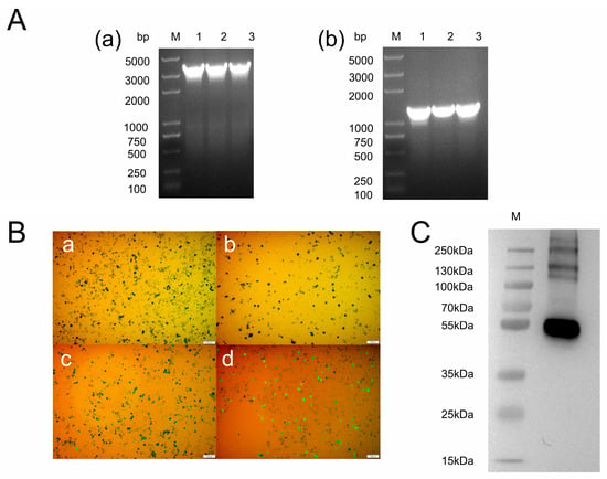
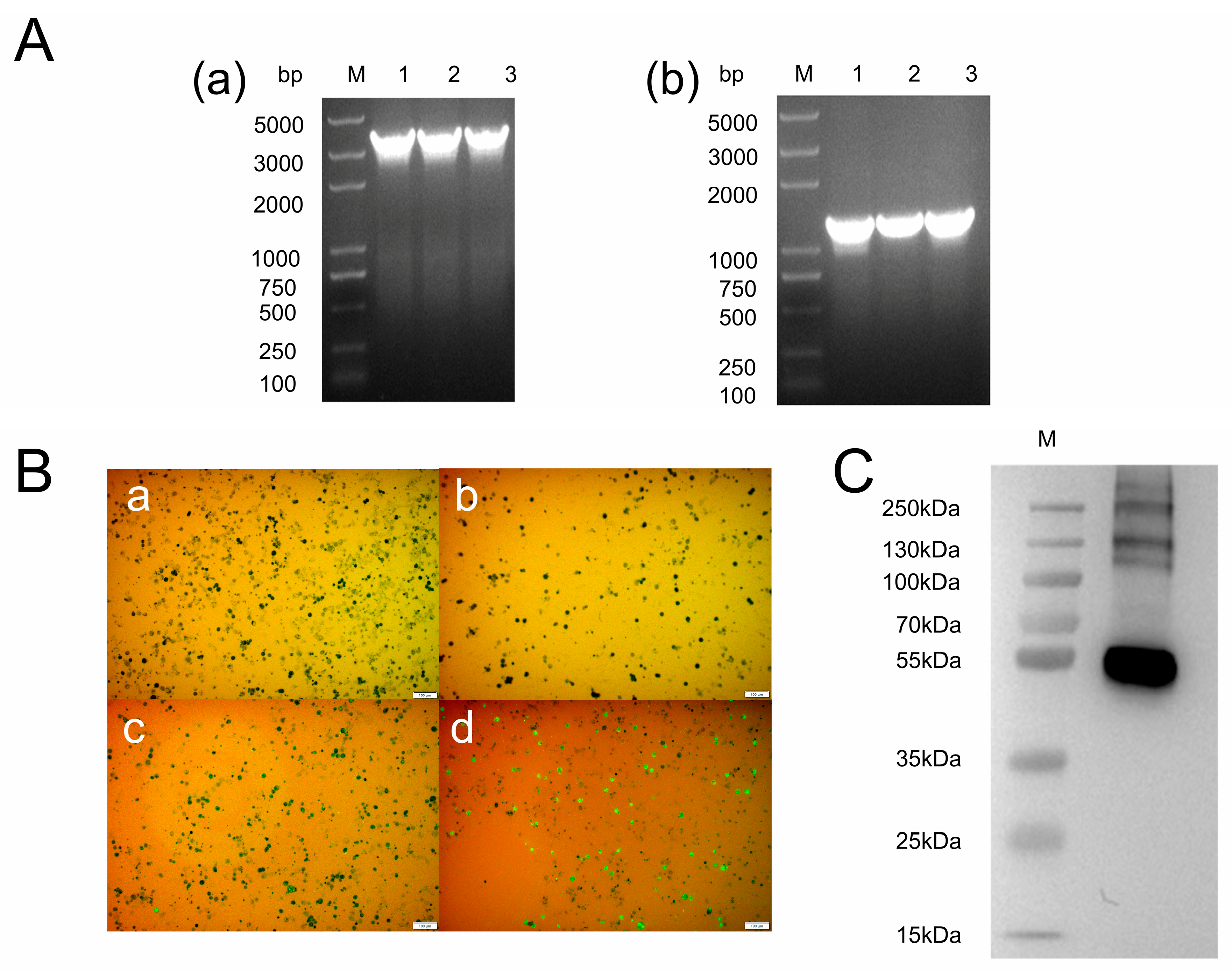
The truncated NiV-G gene was cloned into the pFastBac1 vector and transformed into DH10Bac competent cells for transposition. The recombinant bacmid was identified by PCR using pUC-M13 forward and reverse primers, which yielded a ~1400 bp fragment (Figure 1A(a)), as well as using gene-specific primers, which yielded a ~3200 bp fragment (Figure 1A(b)), confirming the successful integration of the target gene into the bacmid. The truncated NiV-G protein was then expressed using the Sf9 baculovirus expression system, and the recombinant plasmid was designated as pFastBac1-HBM-6His-niVG. Immunofluorescence analysis was performed using an anti-NiV-G serum. Sf9 cells infected with the recombinant baculovirus showed severe cell detachment and strong green fluorescence, whereas no fluorescence was observed in cells infected with the empty baculovirus or in normal adherent cells (Figure 1B). After purification, the protein was analyzed by Western blotting using an anti-His antibody. A band at approximately 55 kDa was detected (Figure 1C). Given that the truncated NiV-G construct comprises residues 170–575 (406 aa) with a calculated molecular mass of ~45 kDa, the observed ~10 kDa increase is consistent with N-linked glycosylation in Sf9 cells. These results confirmed the successful expression of the truncated NiV-G protein.
Figure 1.
Expression and identification of the truncated NiV-G protein. (A) PCR identification of the recombinant bacmid using gene-specific and pUC-M13 primers. (a) pUC-M13 primer amplification. (b) Specific primer amplification. (B) Indirect immunofluorescence analysis of Sf9 cells, including negative/empty-vector controls and recombinant NiV-G-expressing cells. (a) Mock, (b) empty vector, (c) horse anti-NiV serum, (d) mouse anti-His mAb, (C) reducing SDS-PAGE (10% gel) followed by anti-His Western blot analysis of purified truncated NiV-G, showing a predominant band at approximately 55 kDa.
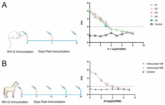
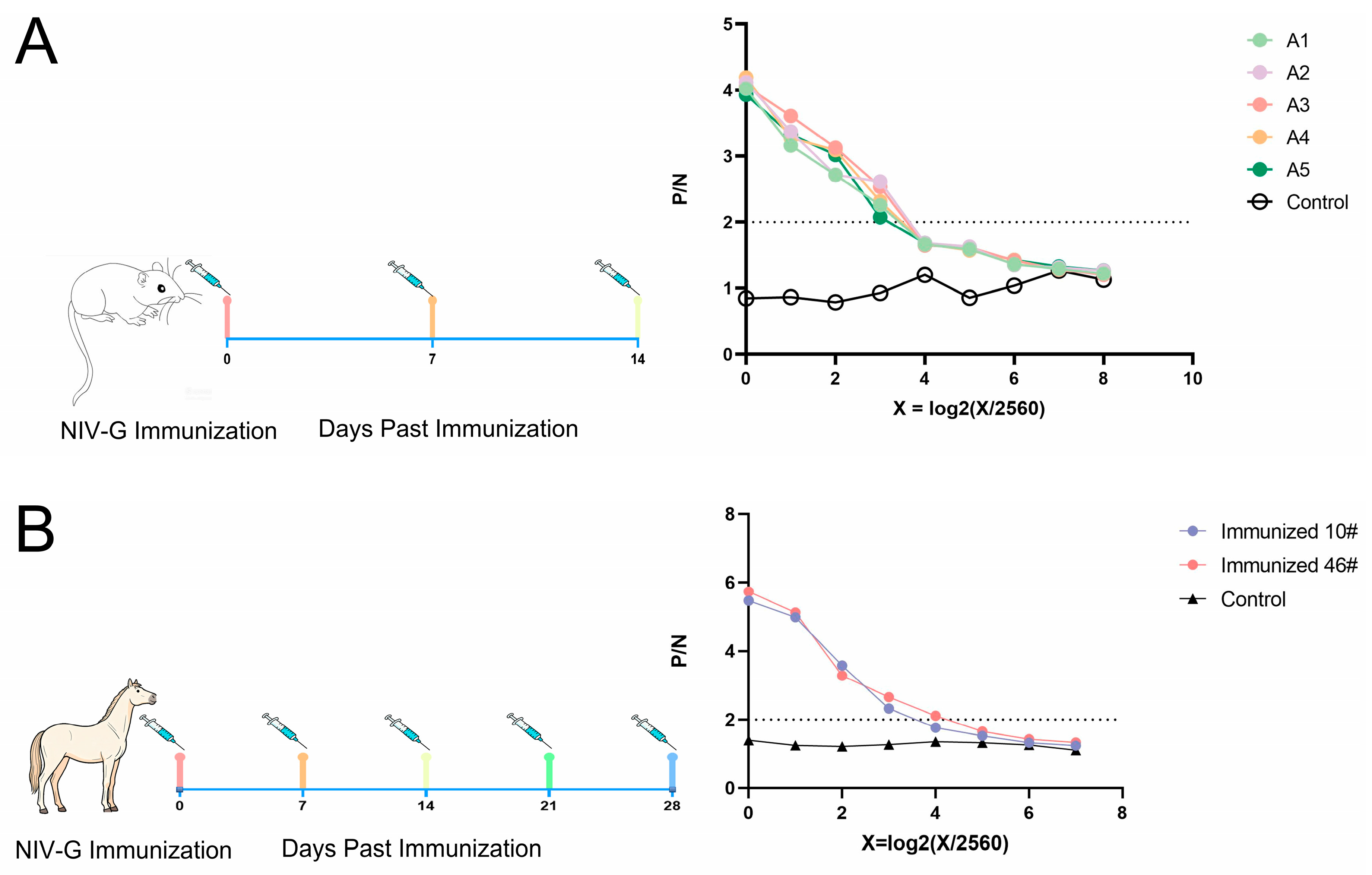
To evaluate the immunogenicity of the purified truncated NiV-G protein, BALB/c mice and horses were immunized separately, and serum antibody titers were measured by ELISA to assess the induced humoral immune response. For mice after the third immunization (Figure 2A), all immunized individuals (A1–A5) exhibited high P/N ratios at the initial dilution, which decreased in a dose-dependent manner as the serum was further diluted. At a dilution of 1:20,480 (log2 = 3), the P/N values for all immunized mice remained around the positive cut-off threshold of 2.0, whereas the control group maintained baseline P/N levels (~1.0) throughout the dilution series, indicating a robust antibody response following three immunizations. Similarly, for horses after the fifth immunization (Figure 2B), both immunized horses (10# and 46#) showed strong initial P/N values (approximately 5.8 and 5.5 at 1:2560, respectively). Based on the cut-off threshold of P/N = 2.0, horse 10# reached an endpoint titer of 1:20,480 (log2 = 3), while horse 46# exhibited a slightly higher titer of 1:40,960 (log2 = 4). The control group remained at baseline levels (~1.2) across all dilutions. Overall, these results demonstrate that the immunization protocols successfully elicited strong and specific humoral immune responses in both species, with endpoint titers reaching approximately 1:40,960 in mice and 1:20,480–1:40,960 in horses, indicating that the recombinant protein is immunogenic across different species. Study limitations include small sample sizes (horses, n = 2; horse control, n = 1), absence of neutralization/challenge data, use of Freund’s adjuvants that are not suitable for translational applications, and the exploratory nature of the alpaca experiment. Following the evaluation in mice and horses, the same antigen was subsequently used to immunize alpacas for the construction of a VHH phage display library.
Figure 2.
Serum antibody titers were determined by ELISA. (A) Balb/c mice; (B) horse. Dotted line indicates the cut-off value (P/N = 2). The titers reached 1:20,480 and 1:40,960, respectively, indicating that the recombinant protein elicited strong humoral immune responses in both species.
3.2. Construction and Screening of the Anti-NIV VHH Library
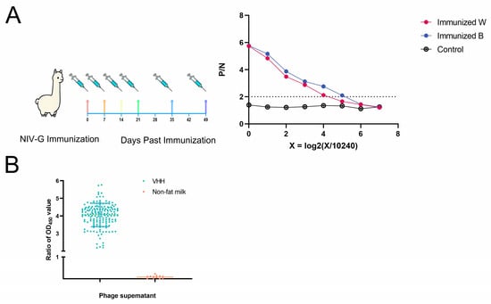
To generate the library, the alpaca with the higher serum ELISA titer was selected from the two immunized alpacas and immunized with the truncated NiV-G protein expressed in the insect cell system. Serum samples collected before and after immunization were analyzed by ELISA to monitor the immune response. The results showed that the post-immune serum specifically bound to the NiV-G protein, with a P/N ratio > 2 at a dilution of 1:327,680, confirming a robust humoral immune response in the immunized alpaca (Figure 3A). Following booster immunization, peripheral blood mononuclear cells (PBMCs) were isolated, and RNA was extracted. The VHH-encoding fragments were amplified and cloned into the pComb3xss phage display vector. The resulting VHH library was constructed by electroporation, with a library size of 3.2 × 10⁸ cfu and a phage display titer of 1 × 10¹³ pfu/mL.
Figure 3.
Construction and screening of the anti−NiV−G VHH library. (A) ELISA assessment of the post-immunization alpaca serum response. Dotted line indicates the cut−off value (P/N = 2). (B) Monoclonal phage ELISA of clones selected after biopanning.
To monitor the enrichment of NiV-G-specific phages during biopanning, the phage output after each round was titrated. As shown in Table 1, the output phage titers increased progressively from 1.6 × 10⁵ pfu/mL in the first round to 1.9 × 10⁷ pfu/mL in the fourth round, with a corresponding increase in recovery rates from 4.0 × 10⁻⁶ to 4.75 × 10⁻⁴, indicating successful enrichment of specific binders. After four rounds of biopanning against the immobilized truncated NiV-G protein, 183 positive clones were sequenced, yielding seven unique VHH sequences (Figure 3B and Table 2).
Table 1.
Enrichment of phage clones during biopanning.
Table 2.
Germline gene analysis of the seven unique VHH sequences. Nucleic acid sequences were analyzed using IMGT/V-QUEST.
3.3. Characteristics of NIV-Specific VHH Antibodies
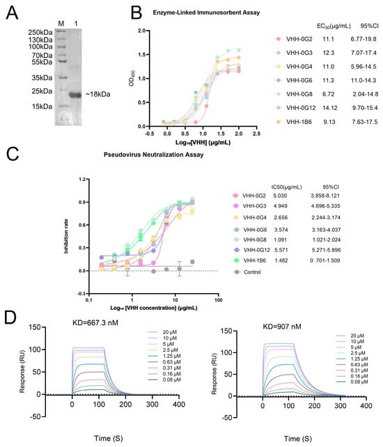
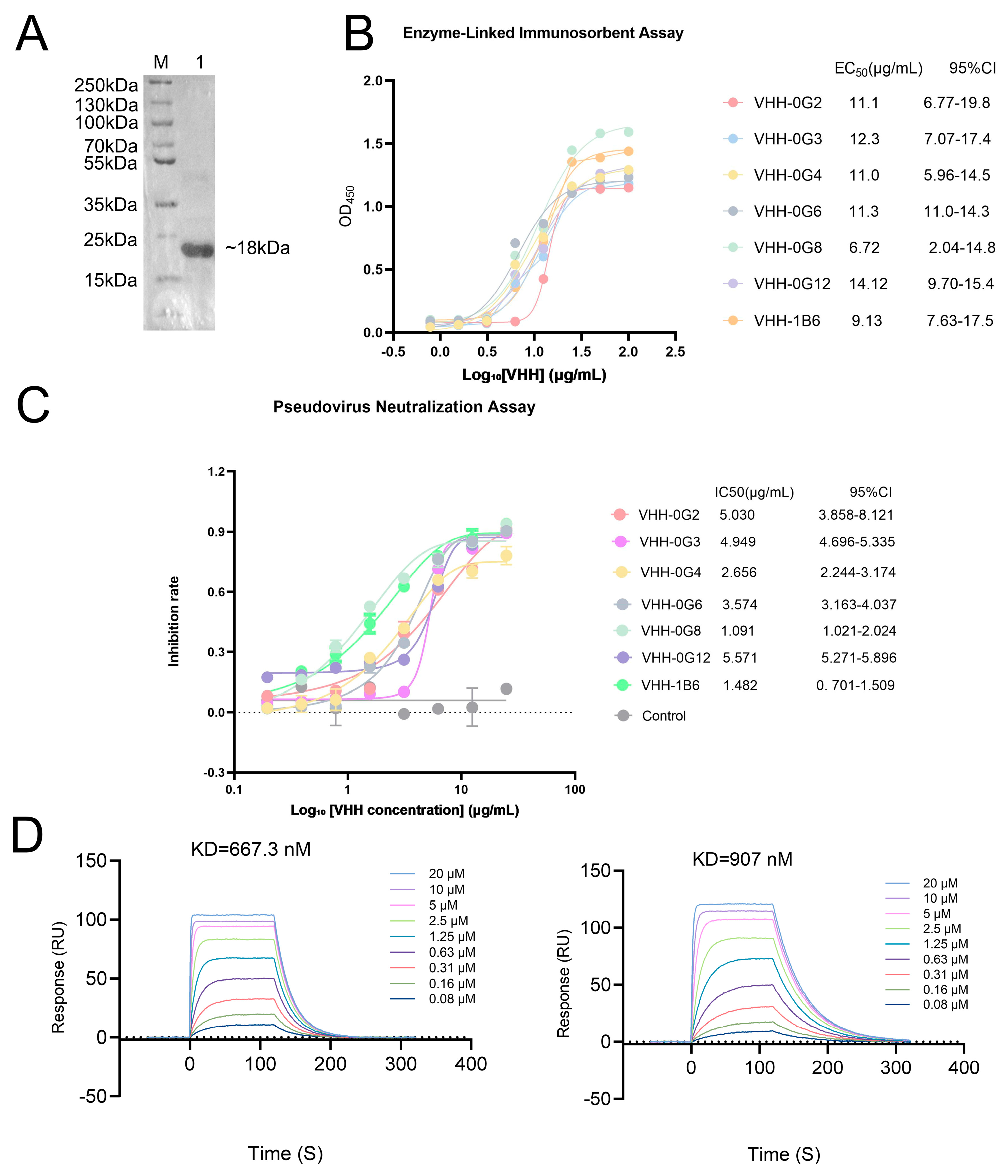
The seven unique VHHs were expressed in E. coli and purified by His-tag affinity chromatography. The final yield of the refolded VHHs ranged from 10 mg/L of bacterial culture. Western blot under reducing conditions showed a predominant band at approximately 18 kDa (Figure 4A), which is slightly higher than the calculated mass of the tagged monomeric VHH (14.7 kDa). Due to their compact globular structure and highly charged framework regions, VHHs often bind less to SDS than standard globular proteins, leading to an anomalous, slower migration on SDS-PAGE. This discrepancy is likely attributed to the intrinsic properties of the VHH sequence, leading to an anomalous migration in SDS-PAGE. This robust yield and high purity from a prokaryotic system highlight the cost-effectiveness and ease of scale-up for VHH production. ELISA demonstrated binding of the candidates to recombinant NiV-G. It should be noted that a lower EC50 value indicates stronger binding affinity. VHH-0G8 and VHH-1B6 showed the lowest fitted EC50 point estimates in the panel, approximately 6.72 and 9.13 μg/mL, although their 95% confidence intervals overlapped those of the remaining candidates (Figure 4B).
Figure 4.
Characterization of NiV−specific VHHs. (A) Reducing SDS−PAGE (10% gel) followed by anti-His Western blot of a representative purified VHH (M, molecular weight marker). (B) NiV−G binding measured by indirect ELISA; note that a lower EC50 value indicates stronger binding affinity. (C) Neutralizing activity against VSV-based NiV pseudovirus; note that a lower IC50 value indicates more potent neutralizing activity. VHH-0G8 and VHH−1B6 showed the lowest fitted IC50 values in the initial screening experiment. (D) SPR sensorgrams for VHH−0G8 and VHH−1B6 binding to truncated NiV−G, fitted to a 1:1 Langmuir model (apparent KD = 9.07 × 10⁻⁷ M for VHH-0G8 and 6.67 × 10⁻⁷ M for VHH−1B6). Analyte concentrations and the number of injections are given in Section 2.13.
The candidates were next evaluated in the VSV-based NiV pseudovirus neutralization assay. Here, a lower IC50 value indicates more potent neutralizing activity. VHH-0G8 and VHH-1B6 showed the strongest activity in the initial screening experiment, with fitted IC50 values of 1.091 and 1.482 μg/mL, respectively (Figure 4C). Consequently, VHH-0G8 and VHH-1B6, which exhibited the most potent binding and neutralizing activities among the seven candidates, were selected as lead candidates for further engineering and formatting. Because these values are derived from an in vitro pseudovirus assay and no established neutralizing antibody was tested side by side as a benchmark, they are interpreted as evidence of strong activity within the present assay rather than as a direct potency ranking against published antibodies.
To obtain orthogonal evidence of binding, SPR experiments were performed. VHH-0G8 and VHH-1B6 both exhibited sub-micromolar binding affinities to NiV-G, with apparent KD values of 9.07 × 10⁻⁷ M and 6.67 × 10⁻⁷ M, respectively (Figure 4D). Notably, the apparent binding affinities measured by SPR were 10–100-fold weaker than the corresponding pseudovirus neutralizing potencies (IC50 values in the nanomolar range, Figure 4C). This discrepancy likely reflects differences in antigen format and binding valency between the two assays, as discussed below.
3.4. Effect of VHH Reformatting on In Vitro Activity
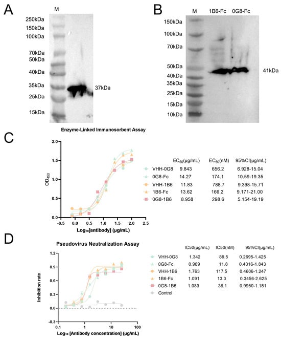
VHH-0G8 and VHH-1B6 were reformatted as Fc-YTE fusions, and a tandem construct containing both VHHs was generated. Western blot analysis confirmed expression of the tandem and Fc-containing proteins (Figure 5A,B). The tandem 0G8/1B6 construct showed a fitted ELISA EC50 of 8.958 μg/mL and a fitted pseudovirus neutralization IC50 of 1.083 μg/mL in the engineering comparison, which did not show a statistically significant improvement in functional neutralization relative to the monomeric formats tested in the same experiment.
Figure 5.
Effect of VHH reformatting on binding and pseudovirus neutralization. (A) Reducing SDS−PAGE (10% gel) followed by anti-His Western blot of the tandem 0G8/1B6 construct. (B) Reducing SDS−PAGE (10% gel) followed by anti-His Western blot of the Fc−YTE fusion constructs. (C) Fitted EC50 values from indirect ELISA. (D) Fitted IC50 values from VSV-based NiV pseudovirus neutralization. Values shown in this figure were obtained in the engineering comparison and are interpreted within that experiment. Based on nM calculations, Fc-fusion reformatting improved apparent binding (EC50) by 3.8−fold (0G8) and 4.7−fold (1B6), and neutralization (IC50) by 7.6−fold (0G8) and 44.7−fold (1B6) relative to their respective monomers. The relative contributions of the YTE mutation versus Fc bivalency cannot be distinguished without a wild-type Fc control.
When comparing within the same experimental batch, the Fc-YTE fusion constructs demonstrated significantly enhanced potency compared to their corresponding monomeric VHHs when evaluated on a molar basis (nM). Specifically, Fc-YTE reformatting improved the apparent binding (EC50) by 3.8-fold for 0G8 (174.1 nM vs. 656.2 nM) and 4.7-fold for 1B6 (166.2 nM vs. 788.7 nM). More importantly, the neutralizing potency (IC50) drastically improved by 7.6-fold for 0G8-Fc (11.8 nM vs. 89.5 nM) and 44.7-fold for 1B6-Fc (2.6 nM vs. 117.5 nM) relative to the monomers. These enhancements were statistically supported, as the 95% CIs of the IC50 values did not overlap between the monomers and their respective Fc-fused derivatives (Figure 5C,D). The present design does not distinguish whether this difference is attributable to Fc-mediated bivalency, the YTE substitutions, other format-dependent effects, or a combination of these factors because a wild-type Fc control was not included. Accordingly, the data support an effect of the Fc-YTE format as a whole rather than a specific YTE-mediated enhancement of neutralization.
It should be noted that the EC50 and IC50 values of VHH-0G8 and VHH-1B6 differed between the initial screening and the engineering comparison experiments (e.g., IC50: 1.091 vs. 1.342 μg/mL for VHH-0G8 and 1.482 vs. 1.091 μg/mL for VHH-1B6; EC50: 6.72 vs. 9.843 μg/mL for VHH-0G8 and 9.13 vs. 11.83 μg/mL for VHH-1B6), which may be attributable to differences in cell batches and other experiment-specific factors.
3.5. Computational Docking Models of NiV-G in Complex with VHH-0G8 and VHH-1B6
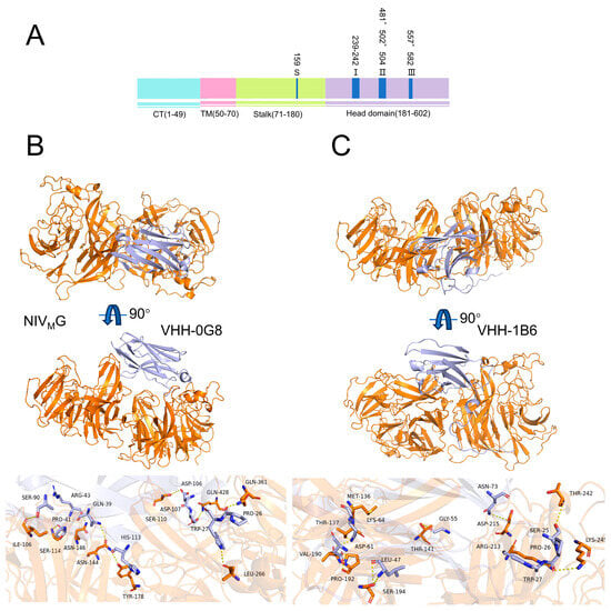
Figure 6A shows the classical antigen–antibody binding epitope of Nipah virus. To further characterize the interaction between the nanobodies and the G protein of Nipah virus, molecular docking was performed with ClusPro, and the resulting complexes were visualized in PyMOL.
Figure 6.
Computational docking analysis of NiV-G in complex with VHH−0G8 and VHH−1B6. (A) Domain organization of the NiV G protein (CT, cytoplasmic tail; TM, transmembrane domain; stalk; head domain), with previously described antigenic sites I−III indicated. (B) Predicted overall binding mode and detailed binding interface of VHH−0G8 with NiV-G. (C) Predicted overall binding mode and detailed binding interface of VHH−1B6 with NiV−G. NiV−G is colored orange and the VHH light blue. Hydrogen bonds are shown as yellow dashed lines and interacting side chains as sticks; key residues are labeled. Top−ranked ClusPro cluster scores were −11.7 kcal/mol for VHH−0G8 and −10.9 kcal/mol for VHH−1B6 These are unitless ranking metrics reflecting docking affinity rather than experimental binding free energies and are not directly comparable to SPR−derived KD values. The structures were generated by ClusPro and visualized with PyMOL. These models represent a structural hypothesis and require experimental validation.
To evaluate the binding ability of the nanobodies to the G protein of Nipah virus, molecular docking was performed with ClusPro, and the resulting complexes were visualized in PyMOL. The top-ranked balanced model cluster scores were −11.7 for VHH-0G8 and −10.9 for VHH-1B6 kcal/mol. These are unitless ranking metrics reflecting docking affinity rather than experimentally determined binding free energies, and they are not directly comparable with the SPR-derived KD.
The computational docking model of VHH-0G8 with NiV-G (Figure 6B) revealed that the nanobody forms a well-defined binding interface with the G protein. In this predicted model, contacts on the NiV-G side of the predicted interface involve head-domain residues, including Tyr178, Leu266, Gln361, and Gln428 (the figure confirms that these are orange NiV-G residues), with the aromatic ring of Tyr178 positioned for π–π stacking and the amide side chain of Gln428 contributing additional polar contacts. Because no competition or epitope-binning experiment was performed and no benchmark antibody was tested here, this predicted interface remains a computational hypothesis requiring experimental validation.
The docking model of VHH-1B6 with the G protein (Figure 6C) predicts interface mediated by hydrogen bonds and hydrophobic interactions. The predicted hydrogen-bonding network involves CDR residues of VHH-1B6 together with head-domain residues of NiV-G, including Thr141, Val190, Pro192, Arg213, Asp215, Thr242, and Lys245 (the figure shows orange NiV-G residues such as Thr242, Lys245, Asp215, Val190, Pro192, and Arg213, and blue VHH residues such as Thr137, Ser25, and Gly55, which were not distinguished in the original text). The hydrophobic interaction is mediated by Met136, Val190, Ser194, Thr242, Thr141, Asp215, and Lys245, among which Val190 is located in the stem–head connection region, and Asp215 and Lys245 are located in the head β-spiral domain. These hydrophobic residues spatially surround the polar interaction residues.
4. Discussion
This study used an immune phage display strategy to identify NiV-G-specific alpaca VHHs and then characterized their in vitro binding and pseudovirus-neutralizing activity. Seven unique VHH sequences were recovered, of which VHH-0G8 and VHH-1B6 showed the strongest neutralization in the initial VSV-based NiV pseudovirus screen. These findings add two additional G-directed VHH candidates to a growing set of antibody-based approaches targeting henipavirus entry [17,18,19].
The recombinant truncated G antigen supported efficient immune library generation, with a phage library size of 3.2 × 10⁸ cfu and progressive enrichment across four rounds of biopanning. The selected VHHs were readily produced in E. coli, and the two lead candidates showed measurable antigen binding by ELISA and strong activity in the pseudovirus assay. Importantly, ELISA EC50, SPR KD, and pseudovirus IC50 quantify different properties and should not be interpreted as interchangeable measures of affinity or potency. In particular, VHH-0G8 showed a micromolar range KD by SPR despite a much lower mass concentration IC50 in the pseudovirus assay. The present data do not establish the mechanism underlying this difference, and additional biophysical and functional studies would be required to determine how epitope accessibility, antigen presentation, antibody state, or assay configuration contribute to the observed values.
Reformatting VHH-0G8 and VHH-1B6 as Fc-YTE fusions was associated with lower fitted pseudovirus IC50 values than those of the monomeric VHHs in the engineering comparison. This observation is consistent with a format-dependent gain in functional activity [20], but the current study cannot assign that gain specifically to the YTE substitutions. Fc fusion changes molecular size and valency and may alter apparent functional avidity, whereas YTE substitutions are primarily intended to modify FcRn interactions and pharmacokinetic behavior. Because the study did not include matched wild-type Fc controls, the contribution of Fc-mediated bivalency cannot be separated from that of YTE or other format-dependent effects. The conclusion is therefore restricted to the performance of the Fc-YTE format as tested.
The tandem 0G8/1B6 construct gave a nominally lower ELISA EC50 than either monomer in the engineering experiment, although the 95% confidence intervals overlapped substantially, indicating no statistically significant improvement in binding affinity. Similarly, it produced only a modest change in pseudovirus neutralization on a mass basis, and this did not reach statistical significance. This result illustrates that increased binding in an immobilized antigen assay does not necessarily translate into proportionally greater neutralization of membrane-displayed viral glycoproteins [21]. The failure of the tandem format to achieve synergy can be attributed to several format-dependent factors. First, the molecular reach—the maximum antigen separation permitting simultaneous bivalent engagement—is a critical determinant of functional potency that can vary considerably across antibody formats [22]. If the two epitopes on the trimeric NiV-G are separated beyond the optimal reach of the 0G8/1B6 tandem, cooperative binding cannot occur. Second, the length and flexibility of the inter-VHH linker are known to profoundly influence the expression, stability, and functional activity of multivalent nanobody constructs [23]. A suboptimal linker may impose geometric constraints that prevent both VHH domains from engaging their targets simultaneously. Third, the VHH arrangement order (0G8-1B6 versus 1B6-0G8) could alter binding kinetics and epitope accessibility. Fourth, steric hindrance on the membrane-proximal G protein array may impede the tandem construct from adopting the orientation required for effective neutralization, even when individual binding events occur. Finally, the discrepancy between ELISA and neutralization assays highlights that antigen capture on a plastic surface does not replicate the dynamic and spatially constrained context of viral entry. Notably, the lack of additive or synergistic effects in tandem antibody formats is not unique to this study; similar format-dependent neutralization profiles have been observed for bispecific nanobodies targeting other viruses, such as HIV-1, where linking two binding domains does not always yield the expected potency gain due to altered structural dynamics or epitope accessibility [24]. In the Nipah virus field, a bispecific antibody (DS90-m102.4) was reported to have nearly identical neutralization potency to its parental counterpart, with the ~2-fold difference explicitly considered of little biological significance [25]. Optimization of VHH order, linker architecture, and epitope pairing should therefore be systematically explored in future studies. The present data do not establish synergistic neutralization by the tandem format; rather, they suggest that the computational prediction of non-overlapping epitopes is insufficient to guarantee functional synergy without format optimization.
Several limitations should be considered. First, due to biosafety restrictions (BSL-4 containment requirements), all neutralization assays were performed using a VSV-based NiV pseudovirus system rather than authentic NiV, and in vivo protective efficacy was not assessed. This pseudovirus system is a well-established, safe alternative that mimics viral entry, but future studies using live virus and animal models are still required. Second, our pseudovirus assays did not include a well-characterized NiV-neutralizing monoclonal antibody as a side-by-side positive benchmark [26,27,28], which precludes a direct potency comparison with established therapeutics; this should be addressed in future comparative studies. Third, although both lead VHHs exhibited sub-micromolar binding affinities to NiV-G by SPR (KD ≈ 0.9 and 0.7 μM), these monovalent affinities are 10–100-fold weaker than their neutralizing potencies (IC50 in the nanomolar range). This discrepancy likely reflects the avidity effects of the trimeric viral spike and bivalent Fc formats, as well as the different conformational states of the truncated antigen used in SPR. Fourth, the monomeric VHHs were recovered from the insoluble fraction of E. coli and refolded by dialysis, so the proportion of correctly folded, active protein is unknown, and the reported concentrations may overstate active antibody. Fifth, because the Fc-fused and tandem constructs were instead secreted from mammalian cells, the comparison between formats is confounded by expression system and folding state, as well as by format. Sixth, the Fc-fusion comparison did not include a wild-type Fc control, precluding attribution of the observed functional change specifically to YTE. Furthermore, the tandem 0G8/1B6 construct did not show a statistically significant enhancement in neutralization, likely due to spatial constraints or epitope proximity. Seventh, the E. coli-expressed G protein used in this study was not intended to replicate the glycosylation pattern of native G protein but rather to provide a consistent and validated antigen format for relative antibody quantification. Importantly, our previous study already systematically validated this prokaryotically expressed truncated NiV-G. In that work, we demonstrated that the antigen retains authentic conformational epitopes, achieving a remarkably high sensitivity (titer of 1:20,480) and excellent specificity in detecting NiV-positive sera [16]. Furthermore, while non-glycosylated antigens might theoretically bias epitope selection, the potent neutralizing activity of VHH-0G8 and VHH-1B6 against the VSV-based pseudovirus displaying fully glycosylated NiV-G confirms that their binding epitopes are accessible on the native glycoprotein. Therefore, an additional insect cell-derived G protein ELISA was not required for the conclusions drawn here.
Finally, developability, pharmacokinetics, tissue distribution, and viral escape were not evaluated. These limitations define the present work as an antibody discovery and in vitro characterization study rather than a demonstration of therapeutic efficacy.
In summary, we identified two alpaca-derived NiV-G-specific VHHs, VHH-0G8 and VHH-1B6, with strong neutralizing activity in a VSV-based NiV pseudovirus assay. Fc-YTE reformatting was associated with lower fitted IC50 values in vitro, whereas the tandem 0G8/1B6 format provided only a modest functional change. The monomeric VHHs were produced in E. coli, while Fc-containing derivatives were expressed in mammalian cells, demonstrating a scalable and cost-effective production pipeline. These candidates warrant further mechanistic, biophysical, and preclinical evaluation before conclusions regarding therapeutic efficacy or clinical translation can be drawn.
5. Conclusions
In this study, we isolated two alpaca-derived single-domain antibodies, VHH-0G8 and VHH-1B6, that bind to the head domain of the Nipah virus G glycoprotein and neutralize a VSV-based NiV pseudovirus, with fitted IC50 values of approximately 0.97 and 1.76 μg/mL (≈65 and 118 nM). Reformatting both led to Fc-YTE-containing formats expressed in Expi293F cells and was associated with fitted IC50 values of 1.34 and 1.09 μg/mL (≈16 and 13 nM), an improvement of roughly an order of magnitude on a molar basis, whereas a tandem 0G8/1B6 construct appeared to give only a marginal gain (IC50 = 1.08 μg/mL, ≈36 nM). Because no wild-type Fc comparator was included, the contribution of the YTE substitutions could not be fully separated from that of Fc-mediated bivalency. These results suggest that bivalent Fc display may represent a more productive reformatting route for this antibody pair and could provide a basis for epitope mapping, pharmacokinetic analysis, authentic virus neutralization, and in vivo evaluation of 0G8-Fc and 1B6-Fc.
Supplementary Materials
The following supporting information can be downloaded at: https://www.mdpi.com/article/10.3390/vetsci13111209/s1, Supplementary Methods S1. Detailed protocol for NiV-G expression and purification; Supplementary Figure S1. Overall schematic diagram of the study design.
Author Contributions
Z.P.: conceptualization, supervision; Y.Z.: conceptualization, supervision; N.F.: formal analysis; F.Y.: project administration; T.W.: resources; R.M.: methodology; Q.W.: methodology; H.Z.: validation; T.Y.: data curation; M.L.: data curation; B.L.: data curation; J.X.: methodology, writing—original draft; all authors: writing—review and editing. All authors have read and agreed to the published version of the manuscript.
Funding
This research was funded by the National Key Research and Development Program of China (grant 2022YFC2305005).
Institutional Review Board Statement
All animal experiments were reviewed and approved by the relevant institutional animal welfare and ethics committees, including the Changchun Veterinary Research Institute and Chinese Academy of Agricultural Sciences (approval numbers IACUC-OF-AMMS-11-2025-135, as applicable; approval on 1 April 2025). All procedures were conducted in accordance with the Guidelines for Ethical Review of Animal Welfare (GB/T 35892-2018) and relevant AAALAC standards. Experimental procedures were designed to minimize animal discomfort, pain, and distress. To minimize distress, mice were anesthetized with isoflurane during blood collection; all injections were subcutaneous with minimal invasiveness, and experienced handlers restrained horses and the alpaca. No adverse events occurred; only mild transient swelling at FCA injection sites was noted, resolving spontaneously within 3–5 days. Humane endpoints (severe lethargy, anorexia > 24 h, >20% weight loss in mice or >10% in horses/alpaca, injection site ulceration/necrosis, or systemic illness) were predefined; animals were monitored daily, with extra checks within 24 h post-immunization. No animal reached any endpoint.
Informed Consent Statement
Not applicable.
Data Availability Statement
The data presented in this study are available on request from the corresponding author. The data are not publicly available due to privacy restrictions.
Acknowledgments
The authors thank Zhenshan Wang (Jilin Agricultural University) and Yangyi Chen (Jilin Agricultural University) for technical assistance and guidance during this study.
Conflicts of Interest
The authors declare no conflicts of interest.
Abbreviations
The following abbreviations are used in this manuscript:
| NIV | Nipah virus |
| VHH | Variable domain of heavy chain of heavy-chain antibody |
| WHO | World Health Organization |
| ELISA | Enzyme-linked immunosorbent assay |
| SPR | Surface Plasmon Resonance |
| VSV | Vesicular Stomatitis Virus |
| VH | Variable domain of the heavy chain |
| JH | Joining region of the heavy chain |
| HCDR | Heavy chain complementarity-determining region |
References
- Branda, F.; Ceccarelli, G.; Giovanetti, M.; Albanese, M.; Binetti, E.; Ciccozzi, M.; Scarpa, F. Nipah Virus: A Zoonotic Threat Re-Emerging in the Wake of Global Public Health Challenges. Microorganisms 2025, 13, 124. [Google Scholar] [CrossRef] [Scilit] [PubMed]
- Dsouza, N.N.; Chellasamy, S.K. A comparative genomic approach to decipher the mutations associated with Nipah viral human isolates from southeast Asia. Iran. J. Microbiol. 2024, 16, 104–113. [Google Scholar] [CrossRef] [Scilit] [PubMed]
- Ali, M.A.; Sajjad, F.; Bacha, Z.; Abid, H.; Ullah, M.; Kamil, K.A. Nipah virus outbreak in Bangladesh: An enduring zoonotic threat. Ann. Med. Surg. 2026, 88, 959–960. [Google Scholar] [CrossRef] [Scilit] [PubMed]
- Bonaparte, M.I.; Dimitrov, A.S.; Bossart, K.N.; Crameri, G.; Mungall, B.A.; Bishop, K.A.; Choudhry, V.; Dimitrov, D.S.; Wang, L.F.; Eaton, B.T.; et al. Ephrin-B2 ligand is a functional receptor for Hendra virus and Nipah virus. Proc. Natl. Acad. Sci. USA 2005, 102, 10652–10657. [Google Scholar] [CrossRef] [Scilit] [PubMed]
- Ortega, V.; Zamora, J.L.R.; Monreal, I.A.; Hoffman, D.T.; Ezzatpour, S.; Johnston, G.P.; Contreras, E.M.; Vilchez-Delgado, F.J.; Aguilar, H.C. Novel Roles of the Nipah Virus Attachment Glycoprotein and Its Mobility in Early and Late Membrane Fusion Steps. mBio 2022, 13, e0322221. [Google Scholar] [CrossRef] [Scilit] [PubMed]
- Playford, E.G.; Munro, T.; Mahler, S.M.; Elliott, S.; Gerometta, M.; Hoger, K.L.; Jones, M.L.; Griffin, P.; Lynch, K.D.; Carroll, H.; et al. Safety, tolerability, pharmacokinetics, and immunogenicity of a human monoclonal antibody targeting the G glycoprotein of henipaviruses in healthy adults: A first-in-human, randomised, controlled, phase 1 study. Lancet Infect. Dis. 2020, 20, 445–454. [Google Scholar] [CrossRef] [Scilit] [PubMed]
- Wang, Z.; Amaya, M.; Addetia, A.; Dang, H.V.; Reggiano, G.; Yan, L.; Hickey, A.C.; DiMaio, F.; Broder, C.C.; Veesler, D. Architecture and antigenicity of the Nipah virus attachment glycoprotein. Science 2022, 375, 1373–1378. [Google Scholar] [CrossRef] [Scilit] [PubMed]
- Chen, L.; Sun, M.; Zhang, H.; Zhang, X.; Yao, Y.; Li, M.; Li, K.; Fan, P.; Zhang, H.; Qin, Y.; et al. Potent human neutralizing antibodies against Nipah virus derived from two ancestral antibody heavy chains. Nat. Commun. 2024, 15, 2987. [Google Scholar] [CrossRef] [Scilit] [PubMed]
- Desmyter, A.; Farenc, C.; Mahony, J.; Spinelli, S.; Bebeacua, C.; Blangy, S.; Veesler, D.; van Sinderen, D.; Cambillau, C. Viral infection modulation and neutralization by camelid nanobodies. Proc. Natl. Acad. Sci. USA 2013, 110, E1371–E1379. [Google Scholar] [CrossRef] [Scilit] [PubMed]
- Muyldermans, S. Applications of Nanobodies. Annu. Rev. Anim. Biosci. 2021, 9, 401–421. [Google Scholar] [CrossRef] [Scilit] [PubMed]
- Vincke, C.; Loris, R.; Saerens, D.; Martinez-Rodriguez, S.; Muyldermans, S.; Conrath, K. General Strategy to Humanize a Camelid Single-domain Antibody and Identification of a Universal Humanized Nanobody Scaffold. J. Biol. Chem. 2009, 284, 3273–3284. [Google Scholar] [CrossRef] [Scilit] [PubMed]
- Vaneycken, I.; Govaert, J.; Vincke, C.; Caveliers, V.; Lahoutte, T.; De Baetselier, P.; Raes, G.; Bossuyt, A.; Muyldermans, S.; Devoogdt, N. In vitro analysis and in vivo tumor targeting of a humanized, grafted nanobody in mice using pinhole SPECT/micro-CT. J. Nucl. Med. 2010, 51, 1099–1106. [Google Scholar] [CrossRef] [Scilit] [PubMed]
- Fang, T.; Lu, X.; Berger, D.; Gmeiner, C.; Cho, J.; Schalek, R.; Ploegh, H.; Lichtman, J. Nanobody immunostaining for correlated light and electron microscopy with preservation of ultrastructure. Nat. Methods 2018, 15, 1029–1032. [Google Scholar] [CrossRef] [Scilit] [PubMed]
- Ma, S.; Zhang, D.; Wang, Q.; Zhu, L.; Wu, X.; Ye, S.; Wang, Y. Structure defining of ultrapotent neutralizing nanobodies against MERS-CoV with novel epitopes on receptor binding domain. PLoS Pathog. 2024, 20, e1012438. [Google Scholar] [CrossRef] [Scilit] [PubMed]
- Salleh, M.Z. Structural biology of Nipah virus G and F glycoproteins: Insights into therapeutic and vaccine development. Eur. J. Microbiol. Immunol. (BP) 2025, 15, 83–93. [Google Scholar] [CrossRef] [Scilit] [PubMed]
- Wang, Q.; Mo, R.; Zhang, Y.; Feng, N.; Yan, F.H.; Wang, T.C. Prokaryotic expression of truncated G protein of Nipah virus and establishment of indirect ELISA method. Chin. Vet. Sci. 2023, 53, 1223–1231. [Google Scholar]
- Wang, Z. Architecture and antigenicity of the nipah virus attachment glycoprotein. Biophys. J. 2022, 121, 29a. [Google Scholar] [CrossRef] [Scilit]
- Wang, Y.; Sun, Y.; Shen, Z.; Wang, C.; Qian, J.; Mao, Q.; Wang, Y.; Song, W.; Kong, Y.; Zhan, C.; et al. Fully human single-domain antibody targeting a highly conserved cryptic epitope on the Nipah virus G protein. Nat. Commun. 2024, 15, 6892. [Google Scholar] [CrossRef] [Scilit] [PubMed]
- Mire, C.E.; Chan, Y.P.; Borisevich, V.; Cross, R.W.; Yan, L.; Agans, K.N.; Dang, H.V.; Veesler, D.; Fenton, K.A.; Geisbert, T.W.; et al. A Cross-Reactive Humanized Monoclonal Antibody Targeting Fusion Glycoprotein Function Protects Ferrets Against Lethal Nipah Virus and Hendra Virus Infection. J. Infect. Dis. 2020, 221, S471–S479. [Google Scholar] [CrossRef] [Scilit] [PubMed]
- Vaskuri, G.J.; Ye, G.; Bu, F.; Yang, D.; Jonsson, C.B.; Turner-Hubbard, H.; Winecke, S.; Mendoza, A.; Li, F.; Tan, C. Long-Circulating Nanobody Confers Durable Prophylaxis against Severe Acute Respiratory Syndrome Coronavirus 2 Omicron Infection. Adv. Nanobiomed Res. 2025, 5, 2400214. [Google Scholar] [CrossRef] [Scilit] [PubMed]
- Kremer, E.; Hultberg, A.; Temperton, N.J.; Rosseels, V.; Koenders, M.; Gonzalez-Pajuelo, M.; Schepens, B.; Ibañez, L.I.; Vanlandschoot, P.; Schillemans, J.; et al. Llama-Derived Single Domain Antibodies to Build Multivalent, Superpotent and Broadened Neutralizing Anti-Viral Molecules. PLoS ONE 2011, 6, e17665. [Google Scholar] [CrossRef] [Scilit] [PubMed]
- Huhn, A.; Nissley, D.A.; Wilson, D.B.; Kutuzov, M.A.; Donat, R.; Tan, T.K.; Zhang, Y.; Barton, M.I.; Liu, C.; Dejnirattisai, W.; et al. Analysis of emergent bivalent antibody binding identifies the molecular reach as a critical determinant of SARS-CoV-2 neutralisation potency. bioRxiv 2024. [Google Scholar] [CrossRef] [Scilit]
- van Rosmalen, M.; Krom, M.; Merkx, M. Tuning the Flexibility of Glycine-Serine Linkers To Allow Rational Design of Multidomain Proteins. Biochemistry 2017, 56, 6565–6574. [Google Scholar] [CrossRef] [Scilit] [PubMed]
- Weiss, R.A.; Verrips, C.T. Nanobodies that Neutralize HIV. Vaccines 2019, 7, 77. [Google Scholar] [CrossRef] [Scilit] [PubMed]
- Isaacs, A.; Nieto, G.V.; Zhang, X.; Modhiran, N.; Barr, J.; Thakur, N.; Low, Y.S.; Parry, R.H.; Barnes, J.B.; Jara, R.; et al. A nanobody-based therapeutic targeting Nipah virus limits viral escape. Nat. Struct. Mol. Biol. 2025, 32, 1920–1931. [Google Scholar] [CrossRef] [Scilit] [PubMed]
- Wang, S.; Rao, G.; Zhang, X.; Yao, Y.; Chen, L.; Chen, S.; Zhang, H.; Liu, H.; Chen, M.; Li, W.; et al. Highly potent C-type nanoantibodies neutralize Nipah and Hendra viruses by cavity filling on fusion glycoprotein. Nat. Commun. 2026, 17, 6978. [Google Scholar] [CrossRef] [Scilit] [PubMed]
- Fan, P.; Sun, M.; Zhang, X.; Zhang, H.; Liu, Y.; Yao, Y.; Li, M.; Fang, T.; Sun, B.; Chen, Z.; et al. A potent Henipavirus cross-neutralizing antibody reveals a dynamic fusion-triggering pattern of the G-tetramer. Nat. Commun. 2024, 15, 4330. [Google Scholar] [CrossRef] [Scilit] [PubMed]
- Dong, J.; Cross, R.W.; Doyle, M.P.; Kose, N.; Mousa, J.J.; Annand, E.J.; Borisevich, V.; Agans, K.N.; Sutton, R.; Nargi, R.; et al. Potent Henipavirus Neutralization by Antibodies Recognizing Diverse Sites on Hendra and Nipah Virus Receptor Binding Protein. Cell 2020, 183, 1536–1550.e17. [Google Scholar] [CrossRef] [Scilit] [PubMed]

Disclaimer/Publisher’s Note: The statements, opinions and data contained in all publications are solely those of the individual author(s) and contributor(s) and not of MDPI and/or the editor(s). MDPI and/or the editor(s) disclaim responsibility for any injury to people or property resulting from any ideas, methods, instructions or products referred to in the content. |
© 2026 by the authors. Licensee MDPI, Basel, Switzerland. This article is an open access article distributed under the terms and conditions of the Creative Commons Attribution (CC BY) license.