


Simple Summary
During pregnancy, microbes in the rumen and intestine can make or modify molecules that may influence the placenta and developing fetus. The review considers four linked questions. Was the molecule produced or transformed by microbes? Was it measured in the mother’s circulation? Did it engage a placental target or cross into the fetus? Did that process contribute to a fetal or offspring outcome? Studies in cattle, sheep, goats, and yaks document microbial changes during gestation, while nutritional experiments demonstrate that the placenta and fetus respond to altered maternal metabolism. These observations do not necessarily establish that microbes caused the downstream effect. In the ruminant studies assessed, no single experiment established all four links for the same signal. Findings from mouse experiments offer useful ways to test this question but are not direct evidence in pregnant ruminants. Better source tracing, measured maternal exposure, functional target tests, and offspring follow-up could eventually distinguish microbiome-mediated effects from direct dietary effects. Distinguishing these pathways matters for animal health, pregnancy management, and evidence-based feeding strategies; a clinical intervention has not yet been established.
Abstract
Maternal rumen–gut microbes transform nutrients, but their specific contribution to placental signaling and offspring development remains unresolved. This focused narrative review assesses primary ruminant studies and selected non-ruminant mechanisms through four causal gates: microbial production or transformation (G1), quantified maternal circulatory exposure (G2), route-appropriate placental or fetal target engagement (G3), and developmental effects attributable to the specified upstream step (G4). Candidate signals include fermentation acids, succinate, amino-acid and one-carbon metabolites, tryptophan derivatives, and bile acid- and lipid-related molecules. Ruminant studies document gestational microbial remodeling, maternal–fetal metabolic responses, and placental or fetal effects of nutritional interventions. However, paired microbiome–metabolome profiles do not establish microbial source-to-blood transfer; placental tissue accumulation is not systemic exposure; and altered transporter expression is not target dependence. Ewe-derived gastrointestinal material transferred to antibiotic-treated pregnant mice conveys biological activity without isolating a metabolite-specific mechanism in pregnant ruminants. Among ruminant experiments critically assessed, none established all four gates for the same defined microbial signal. Future work requires source-resolved tracing, quantitative compartment sampling, functional target perturbation, and longitudinal offspring follow-up with postnatal exposure controls. The framework separates testable microbiome-mediated mechanisms from direct dietary effects and developmental-programming claims that remain unsupported.
1. Introduction
Ruminant developmental programming has a substantial experimental basis [1,2]. In sheep and cattle, maternal nutrition and heat stress can alter placental development, fetal nutrient supply, tissue growth, metabolism, and offspring performance [1,2,3]. Ruminant foregut fermentation adds a source-attribution challenge because circulating acetate, amino acids, and one-carbon intermediates can arise from dietary inputs, microbial synthesis or transformation, and host metabolism. In Small-tailed Han sheep, a stage-comparison study sampled rumen fluid and serum in 15 ewes before breeding, during pregnancy, and during lactation. It identified changes in ruminal communities, metabolites, and circulating metabolic profiles, but did not track a labeled metabolite from the rumen into blood [4]. In periparturient dairy cows, organism-resolved metagenomics and in vitro fermentation implicated Ruminococcus_E bovis in alanine production; the associated functional assays did not test placental or fetal outcomes [5]. These studies make an upstream microbial contribution plausible, but paired measurements and microbial production experiments alone do not establish a continuous source-to-fetus pathway.
Maternal gut microbiome-metabolite-placenta-fetus relationships have been considered from several complementary perspectives. Recent reviews have addressed maternal nutrition and the gut microbiome during pregnancy [6], gut microbial metabolites in fetal development [7], circulating microbial mediators at the maternal–fetal interface [8], livestock nutritional programming [9,10,11], maternal dietary programming of the young-ruminant gut-microbiome-immune axis [12], placental autophagy and ferroptosis as microbiome-responsive pathways [13], and the bovine maternal microbiome in fetal programming and early microbial colonization [14] (Table 1). These syntheses establish biological plausibility and clarify important nutritional, placental, microbial, and developmental mechanisms. A narrower question remains unresolved: whether a maternal circulating signal proposed to be microbial can be assigned to a defined microbial production or transformation step, quantified as a maternal exposure, linked to route-appropriate placental or fetal target engagement, and connected to a developmental consequence. The closest conceptual overlap is the 2026 structured review of maternal dietary programming in young ruminants [12], which begins with a defined maternal diet and follows offspring gastrointestinal, microbial, immune, and feed-related development. The present review instead begins with source attribution and follows the proposed signal toward the maternal–fetal interface. We therefore organize the evidence using four causal gates. Gate 1 is microbial source or transformation. Gate 2 is quantitative maternal exposure. Gate 3 is route-appropriate target engagement, with separate placental sensing/handling and transplacental-transfer routes. Gate 4 is developmental consequence. This framework is tailored to ruminant physiology because foregut fermentation, first-pass metabolism, and species-specific placental organization complicate direct extrapolation from mouse or human pregnancy [3,14].
Table 1.
Positioning of the present review relative to selected relevant reviews published from 2022 to 2026.
Review Scope, Literature Search, and Evidence Framework
Microbial source attribution and causal evidence are assessed separately. The term microbiome-derived is reserved for molecules or precursors with a demonstrated microbial production or transformation step; host–microbial co-metabolites require both microbial and host steps. Microbiome-associated denotes an established microbial association without resolved origin, while diet- or host-derived denotes a demonstrated predominantly nonmicrobial source. This is a focused narrative review, not a systematic review or a meta-analysis. The initial PubMed and Web of Science searches emphasized publications from 2020 through 19 September 2026, with earlier physiological and tracer experiments retained where indispensable. For the revision, bibliographic exports supplied for PubMed, Scopus, and Web of Science were reconciled alongside documented, repeatable query strategies covering ruminant species, gestation, the rumen/gut microbiome, the placenta or offspring, and metabolic transfer. The three supplied exports contained 133, 247, and 244 records, respectively, which yielded 326 distinct bibliographic records after DOI-first and normalized-title deduplication. Because the exports lack verifiable query histories and include off-topic records, these counts describe a bibliographic inventory, not validated hits from the listed exact queries or 326 eligible studies. Primary ruminant reports were prioritized if they experimentally informed microbial origin, maternal exposure, placental/fetal handling, or an offspring endpoint; direct dietary interventions were retained as downstream comparators but were never coded as proof of microbial mediation. Non-ruminant studies were treated separately when they offered depletion, transplantation, rescue, or receptor-perturbation mechanisms that could not be assumed to apply to ruminants. Eligibility judgments considered species, gestational versus postpartum context, sampling compartment, source attribution, measurement type, controls, and availability of interpretable experimental detail. Borderline reports were retained as explicit partial-chain or contextual evidence when they clarified a limiting step, rather than silently discarded or promoted to a complete causal mechanism. Supplementary Table S1 provides 32 purposively selected ruminant evidence anchors, identified through focused searches and citation tracing rather than an exhaustive 326-to-32 eligibility screen; Supplementary Note S1 documents the search expressions, bibliographic inventory, selection criteria and limitations. Because selection was not exhaustive, statements about missing evidence apply only to the studies assessed.
2. Maternal Rumen-Gut Remodeling During Gestation: Upstream Evidence
Evidence that gestation and superimposed nutritional pressures remodel the maternal gastrointestinal ecosystem is the first requirement for a maternal microbiome-metabolite-placenta model. In ruminants, foregut fermentation continuously shapes the maternal metabolome before nutrients and microbial products reach systemic circulation. Current studies support substantial pregnancy-associated remodeling of rumen or hindgut communities and metabolic outputs, but not a single conserved trajectory across cattle, sheep, goats, and yak. The key question is which changes are reproducible, nutritionally modifiable, connected to circulating metabolites, and traceable toward a placental mechanism.
2.1. Gestational Remodeling of the Maternal Rumen-Gut Ecosystem
Gestational remodeling varies across species and anatomical compartments. In five Ashidan yaks followed across reproductive stages, ruminal fermentation products and microbial/metabolomic profiles changed; the small sample and lack of matched placental data limit inference beyond ruminal adaptation [15]. In Small-tailed Han sheep, ruminal Prevotella abundance and carbohydrate-related predicted functions differed by reproductive stage [4]. These community-based annotations predict functional potential; they do not measure enzyme flux or delivery into the blood. Goats also displayed gestational shifts in fecal community profiles and metabolites, including indole-related signals [16]. These observations identify candidates for targeted assays but do not establish a reproducible microbial-to-placental causal route.
Remodeling is not uniform across ruminant populations. Hu and Suffolk ewes show breed-dependent changes in rumen microbial functions during pregnancy, while goat data associate reproductive load with differences in rumen microbial structure and metabolomic profiles [17,18]. A 2026 sheep multi-omics study also linked several rumen bacterial features with reproductive performance [19]. Together, these findings argue against a single conserved ‘pregnancy microbiome’ and support prospective control for species, breed, litter size, diet, and gestational stage.
Hindgut profiles complement rumen measurements but do not directly measure rumen production. In pregnant goats, fecal microbial changes accompanied reproductive-hormone patterns and serum metabolite shifts [16]. A Hu sheep study compared 24 animals across four independent reproductive-stage groups (six per group), measuring fecal microbial communities and serum metabolomics [20]. This was a cross-sectional stage comparison, not repeated longitudinal blood sampling of the same ewes. Functional pathway assignments derived from community profiling should be described as predicted rather than directly measured microbial metabolic activity. These studies show correlations but cannot determine whether circulating compounds originate from the hindgut, rumen, diet, or host tissues.
Prenatal exposure is distinct from postnatal transfer. Maternal gastrointestinal communities are associated with neonatal intestinal communities, microbial functions, and resistome composition in cow–calf studies [14,21,22]. Prenatal vitamin and mineral supplementation can also alter microbiota measured in newborn calves [23], but the intervention occurred before birth whereas microbiota profiling occurred after birth. Such findings may reflect prenatal influences, birth-related contact, milk, or other postnatal exposures. They do not demonstrate transfer of a defined microbial metabolite across the placenta.
2.2. Nutritional Perturbation of the Maternal Microbiome-Metabolome
Maternal undernutrition provides experimental evidence that the gestational rumen ecosystem is nutritionally plastic, but the strongest mechanistic example uses an intentionally severe challenge. In late-gestation sheep, feed intake was restricted to 30% of ad libitum intake for 15 days [24]. This severe restriction reduced ruminal volatile fatty acids and microbial protein, altered bacterial community composition and predicted metabolic functions, and reduced ruminal papillary dimensions together with epithelial signaling and cell-renewal programs [24]. Because the intervention represents marked undernutrition rather than routine gestational feeding variation, it is most informative as a proof-of-principle for microbiome-host plasticity under nutritional stress, not as a direct model of moderate production-level dietary restriction.
The effect of this severe nutritional challenge was not confined to rumen. In the matched colon study, the 30% feeding level increased colonic propionate, isobutyrate, butyrate, isovalerate, and valerate, reduced the acetate proportion, altered bacterial communities, and disrupted epithelial morphology [25]. A subsequent cecal study of undernourished pregnant sheep reported altered microbial diversity and composition, increased volatile fatty acid and microbial-protein concentrations, and inhibition of extracellular-matrix-receptor and phosphoinositide 3-kinase (PI3K) signaling together with impaired epithelial renewal and immune functions [26]. These data argue against treating the maternal gastrointestinal tract as a single homogeneous compartment: the rumen, cecum, and colon can display distinct ecological and metabolic responses to the same nutritional stress.
Nutritional insufficiency can coincide with placental dysfunction without proving microbiome mediation. In perinatal ewes, negative energy balance altered rumen morphology, microbial structure, maternal performance, and placental function [27]. This integrated phenotype links upstream gastrointestinal and downstream placental abnormalities in one model, but without source tracing, microbial perturbation, or mediator rescue it cannot distinguish microbial mediation from direct nutrient deficiency or endocrine-metabolic adaptation.
Targeted nutritional interventions also alter maternal microbial-metabolic states. Restricted gain during early bovine gestation changed ruminal community structure and interaction networks, whereas one-carbon supplementation had little effect on the ruminal community [28]. Energy and protein supply altered rumen microbiota and blood metabolic indices in gestating yaks [29]. In ewes, rumen-protected glutamine, resveratrol, and roughage source modified combinations of microbial, metabolic, immune, or antioxidant profiles [30,31,32]. These studies show that the maternal microbiome is manipulable, but some interventions primarily alter microbial substrate supply whereas others may act directly on host tissues and only secondarily affect the microbiome.
Offspring associations make a biological link plausible but do not establish mediation. Maternal rumen microbiota and metabolite profiles have been associated with lamb birth weight, body size, and intestinal immune status [17], while maternal gut microbial and metabolomic signatures were associated with calf birth weight in a pilot dairy-cow study [33]. Maternal protein supplementation also improves birth and postnatal growth in cattle [34]. A peripartum ewe study of omega-fatty-acid sources and essential oils also measured ruminal microbial patterns and blood metabolites [35], illustrating dietary perturbation rather than microbial source tracing. These observations support two separate statements, that maternal nutrition changes the microbiome and that maternal nutrition changes offspring growth. They do not establish the third, that the microbiome mediates the fetal effect.
2.3. From Rumen/Gut Metabolites to Maternal Circulation
Linking ruminal or intestinal metabolites to maternal circulation requires measurements from the relevant compartments. In 15 Small-tailed Han ewes, rumen fluid, microbial composition, fermentation products, and serum metabolic profiles were compared across reproductive stages in the same animals [4]. This design identifies stage-related co-variation, not labeled microbial flux into blood. Studies pairing fecal microbiota with serum metabolomics similarly show cross-compartment association without demonstrating a rumen origin [20]. By contrast, a preliminary dairy-cow study related maternal fecal microbial and metabolomic features to calf birth weight in only ten cows [33]. Its metabolite measurements were fecal, not quantitative maternal circulatory exposure, so they cannot satisfy Gate 2. A stronger test would quantify the same chemically identified metabolite in matched luminal, maternal blood, and placental compartments while distinguishing microbial generation from dietary absorption and host biotransformation.
Cross-compartment concordance supports metabolic coupling but not directional flux [4,20]. A metabolite found in both rumen and blood may reflect microbial synthesis, dietary absorption, host metabolism, or transformation after absorption, while a microbial precursor may be converted before entering systemic circulation. Correlation networks can prioritize candidates when taxa, pathway genes, luminal metabolites, and serum metabolites covary, but they cannot establish the source, magnitude, or direction of transfer. For the maternal–fetal axis, the relevant exposure is the circulating signal that can reach or act on the placenta.
These ruminant studies chiefly report cross-compartment associations; none traces a defined rumen or gut product into maternal blood. Amino acids, one-carbon intermediates, tryptophan derivatives, and lipids particularly require distinction among dietary, microbial, and host sources.
2.4. Source Attribution: When Can a Metabolite Be Called Microbiome-Derived?
Source attribution is the central methodological problem in a ruminant maternal microbiome-metabolite model. Four source categories are distinguished in this review. A microbiome-derived signal has been demonstrated to arise from microbial contributions to production or transformation. A host-microbial co-metabolite contains an identifiable microbial step but reaches the circulation only after host biotransformation. A microbiome-associated signal is linked statistically or experimentally to microbial composition or function without source-resolving evidence. A diet- or host-derived metabolite is demonstrated to originate primarily from non-microbial sources, even if the microbiome modifies its abundance or downstream chemistry. Collapsing these categories would artificially strengthen the evidence chain.
A recent dairy-cow study provides a useful technical proof of principle for how microbial source attribution can be strengthened in ruminants. By integrating metagenomics, metabolomics, and transcriptomics in periparturient cows, investigators identified a deficiency of alanine in microbial protein in animals with energy-metabolism disorders [5]. Metagenomic binning and in vitro rumen fermentation implicated Ruminococcus_E bovis in alanine biosynthesis, and alanine supplementation reduced ketogenesis in a hepatic cell model by supporting gluconeogenesis [5]. Although this periparturient study was not designed around placental or fetal programming, it demonstrates that ruminant research can move beyond microbe-metabolite correlation toward a defined organism-to-metabolite functional relationship.
The cited experiments inform different parts of the proposed pathway: organism-resolved alanine production without pregnancy targets [5], stage-matched rumen and serum profiling without tracer evidence [4], and transferable bioactivity of complex ewe-derived material tested in mice [36,37]. None of these designs establishes a continuous metabolite-specific pathway in pregnant ruminants.
3. Candidate Metabolic Signals: From Association to Biological Plausibility
The five metabolite classes differ in the evidence available for source, exposure, and target action. Fermentation acids and succinate offer biochemical or receptor-level anchors; amino-acid and one-carbon pathways offer maternal–fetal physiology but limited microbial attribution; tryptophan derivatives rely heavily on non-ruminant mechanistic bridges; and bile acid/lipid studies mainly inform compartmental handling and developmental associations. These categories are illustrative, not exhaustive. Table 2 identifies the strongest measured link and the missing experiment for each class.
Table 2.
Candidate metabolite classes mapped across source, exposure and target evidence in ruminants. Abbreviations: AHR, aryl hydrocarbon receptor; mTOR, mechanistic target of rapamycin; SCFA, short-chain fatty acid; SUCNR1, succinate receptor 1; VFA, volatile fatty acid.
3.1. Volatile Fatty Acids (VFAs), Short-Chain Fatty Acids (SCFAs), and Succinate: Substrates Versus Signals
Volatile fatty acids are obvious candidates for a rumen-to-placenta signaling model, but they are also conceptually difficult because they are central fermentation products and major host energy substrates in ruminants. Pregnancy-associated changes in ruminal acetate and other fermentation products have been documented in sheep and yak [4,15], and severe feed restriction can substantially reconfigure ruminal VFA production [24]. A change in maternal VFA concentration can therefore reflect altered substrate intake, microbial community structure, rumen absorption, hepatic utilization, or systemic energy demand. The designation ‘microbial signal’ therefore requires more than showing that a VFA changed during pregnancy.
Non-ruminant studies provide the strongest causal template for SCFA signaling. Maternal microbiota depletion in mice impairs placental growth and fetoplacental vascularization, and SCFA replacement partially rescues vascular defects [38]. A separate low-protein intrauterine growth restriction (IUGR) model linked Lactobacillus enrichment, increased circulating SCFAs, microbiota transfer, and SCFA rescue to improved placental efficiency and fetal development [39]. These experiments demonstrate the type of perturbation-rescue logic needed for causality, but they remain mechanistic bridges rather than direct evidence that rumen-derived SCFAs drive placental programming in cattle or sheep.
Interpretation of succinate depends on the sampled compartment and experimental context. In peripartal dairy cows with retained placenta, succinate was elevated locally in caruncular tissue, and cell experiments implicated SUCNR1-related PI3K signaling in macrophage polarization and macrophage–trophoblast communication [40]. SUCNR1 knockdown weakened the cell response, supplying an experimentally supported local receptor mechanism. However, placental-tissue accumulation was not a measurement of maternal systemic succinate concentration or source-to-blood kinetics. Neither microbial production of the relevant succinate nor fetal developmental programming was tested. Thus, the mechanism is supported in a specific postpartum pathological setting, not as evidence that rumen-derived succinate mediates normal gestational programming.
The evidence differs by candidate and setting. Fermentation acids have plausible microbial sources but limited ruminant placental target tests; SUCNR1-mediated succinate responses were tested locally in a postpartum retained-placenta setting [40], without source-resolved gestational exposure or fetal programming.
3.2. Amino-Acid and One-Carbon-Related Signals
Amino-acid and one-carbon pathways provide some of the strongest evidence that maternal metabolic signals can alter placental transport and fetal growth in ruminants, but they also exemplify the danger of conflating nutritional supplementation with microbial mediation. Supplementation with one-carbon metabolites, including methionine, choline, folate, and vitamin B12, changes maternal and fetal one-carbon metabolite pools in beef heifers [41]. Related work shows nutrition-dependent differences in maternal serum and fetal-fluid vitamin B12 and folate concentrations, together with hepatic one-carbon-pathway responses [42]. These studies demonstrate that these molecules and their downstream intermediates are accessible to the maternal–fetal unit. Because the compounds are supplied directly, however, these experiments test nutrient and metabolic programming rather than microbial source pathways.
Nutritional interventions provide downstream measurements but do not establish metabolite-specific microbial causality. Mid-gestation methionine supply was associated with changes in bovine placental mTOR-related markers, nutrient-transporter expression, and sex-dependent birth-weight outcomes [43]. Rumen-protected arginine or N-carbamylglutamate modified cotyledonary amino-acid profiles and transporter- or angiogenesis-associated markers in undernourished ewes [44]. Late-gestation N-carbamylglutamate in dairy cows altered placentome metabolic and gene-expression profiles and increased calf birth weight [45]. These studies show responses to defined maternal nutrient interventions. They do not establish that the nutrient originated in microbes or that the developmental outcome required the measured mTOR or transporter change. Gene or protein abundance, transport flux, receptor occupancy, and target dependence are distinct experimental endpoints.
The same evidence, however, cannot be cited as proof that rumen microbes generated the active signal. Methionine, arginine, folate-related intermediates, and other amino-acid derivatives can be supplied by diet, released by microbial protein, generated by host tissues, or transformed at multiple sites after absorption. These studies are therefore best used as a mechanistic analog: they show what the placenta can sense and how fetal nutrient supply can be altered once a relevant maternal metabolite changes, while leaving the upstream source unresolved. This distinction is especially important for rumen-protected supplements, whose formulation is specifically designed to reduce ruminal transformation [43,44].
The Ruminococcus_E bovis-alanine study provides a bridge between these evidence domains. It demonstrates that a specific rumen organism can be linked to a defined amino-acid product using metagenomic reconstruction and in vitro fermentation, and that the product can exert a measurable host metabolic effect [5]. The study did not involve a placental endpoint, but it shows that microbial source attribution is technically feasible in ruminants. Existing maternal–fetal studies also demonstrate that one-carbon-related metabolites can be measured across maternal and fetal compartments [41]. Across this class, the strongest ruminant evidence lies downstream, particularly in Gate 2 exposure or handling and Gate 3 placental sensing, with Gate 4 supported in selected supplementation models. The limiting step is Gate 1 because current pregnancy studies generally use defined dietary or rumen-protected nutrient exposures rather than source-resolved microbial production. Combining source-resolved microbial validation with matched compartmental measurements and placental signaling assays would convert amino-acid programming from a nutrient-association model into a testable microbiome-metabolite mechanism.
3.3. Tryptophan-Indole-Kynurenine Axis
In sheep, maternal intravenous administration of 100 mg/kg tryptophan provides a quantitative example in which compartment, units, and dose are specified [46]. At 95–98 days of gestation, fetal plasma kynurenine rose from 5.7 ± 1.2 to 247.9 ± 86.7 µmol/L, compared with maternal plasma values of 4.6 ± 0.8 to 118.0 ± 79.7 µmol/L; at 135–138 days, fetal values rose from 9.0 ± 2.3 to 289.0 ± 194.0 µmol/L while maternal values rose from 4.8 ± 2.9 to 98.3 ± 67.8 µmol/L. Fetal kynurenine also declined more slowly. These experimental peak concentrations were produced by an intravenous precursor load and must not be interpreted as physiological concentrations of a rumen-derived indole. The experiment established responsive maternal–fetal host metabolism, not microbial origin or a fetal receptor-dependent developmental consequence.
Non-ruminant studies describe distinct mechanisms that should not be conflated. Other mouse studies document microbiota-dependent placental and fetal metabolic responses [47,48,49,50]. In pregnant mice, prenatal environmental enrichment increased maternal Lactobacillus and indole-3-propionic acid (IPA) detected in maternal serum and fetal brain [51]. In experimental neural progenitor systems, IPA engaged the aryl hydrocarbon receptor (AHR), followed by Src and ERK1/2 pathway activation and neurodevelopmental effects; inhibitory experiments supported pathway involvement [51]. A different mouse model reported that prenatal IPA supplementation modified the later intestinal phenotype of offspring exposed to maternal immune activation, involving intestinal signaling rather than a validated ruminant placental AHR pathway [52]. These findings support a plausible candidate axis but cannot be assembled into proof of one continuous, receptor-dependent ruminant maternal–fetal mechanism.
The strongest directly relevant sheep finding is altered fetal kynurenine handling after maternal intravenous tryptophan administration [46]. Because the precursor was administered experimentally, this reflects maternal–fetal substrate metabolism rather than a microbial-origin signal; fetal exposure or production does not itself establish fetal receptor dependence. Detection of tryptophan derivatives in untargeted metabolomics alone is not sufficient even for a microbiome-associated designation: that label requires an independently documented relationship to microbial community composition or activity. Without that association, the finding is best described as a detected or putative metabolite feature of unresolved origin. IPA–AHR–Src–ERK1/2 signaling remains a non-ruminant mechanistic template and a hypothesis for targeted experiments in pregnant ruminants.
3.4. Bile Acid- and Lipid-Related Signals
The ovine taurocholate tracer study measured transfer direction but did not establish microbial origin [53]. In near-term sheep, separate maternal-to-fetal (n = 3) and fetal-to-maternal (n = 5) tracer experiments found no detectable maternal-to-fetal radiolabel transfer under the studied conditions. The reported mean fetal-to-maternal transfer was approximately 2.6 µmol per 24 h, or 2% of the fetal taurocholate pool, while maternal-to-fetal transfer was considered below 1 µmol per 24 h. These are directional tracer flux estimates rather than steady-state maternal plasma concentrations. Small cohorts and near-term sampling limit extrapolation to earlier gestation or to microbially transformed bile acids.
Maternal nutrient restriction changes bile-acid and lipid profiles alongside fetal growth. In the ovine SGA study [54], mean maternal plasma total bile acids across the reported gestational sampling days were 14.59 ± 3.12 µmol/L in the adequately nourished (100% NRC) group and 23.16 ± 3.00 µmol/L in the nutrient-restricted SGA subgroup (Table 1 of [54]). These maternal plasma concentrations were measured under nutritional perturbation and do not establish microbial production or mediation. Maternal and fetal bile acids and allantoic-fluid lipids also differed, while placentomal SLC27A6 transcript and protein abundance were reduced at gestational day 70 in nutrient-restricted SGA pregnancies [54]. SLC27A6 is a fatty-acid transporter, not evidence that bile acids were transported through this protein. In 29 late-gestation Brangus heifers, maternal jugular-plasma fatty acids were quantified by GC-MS at gestational day 240 [55]. Table 1 of [55] reports higher total monounsaturated fatty acids under nutrient restriction (RES 50.91 vs. ADQ 36.97 µg/mL; p = 0.001), and higher palmitic acid (C16:0; 57.45 vs. 52.96 µg/mL; p = 0.02). Melatonin showed no significant maternal-plasma fatty-acid effect. These measured circulating pools, together with placental transporter-transcript and fetal/amniotic profiles, do not establish microbial origin, transporter flux or target-dependent developmental causality [54,55].
Metabolomics of fetal fluids adds another layer of evidence. In multifetal ewe pregnancies, allantoic-fluid lipidomics identified lipid signatures associated with partial litter loss, together with differences in maternal metabolic indices and placental gene expression [56]. These observations identify lipid-related features associated with adverse pregnancy outcome, but they do not demonstrate that the lipids are causal signals or that they are microbially produced. Across this class, ruminant evidence is strongest for Gate 2 exposure or compartmental handling and partial Gate 3 placental or transfer readouts, whereas Gate 4 remains mainly associative. The major bottleneck is Gate 1 because microbial contribution to the measured bile-acid or lipid pools has not been resolved, and target-dependent developmental causality has not been demonstrated. The defensible current label is therefore ruminant association with unresolved microbial contribution. A comparative summary of these candidate metabolite classes is provided in Figure 1 and Table 2.
Figure 1.
Representative signal classes and their current evidence boundaries across the proposed maternal rumen–gut microbiome, circulation, placental interface, and fetal/offspring axis. The upper pathway denotes biological compartments, not an experimentally demonstrated complete route. The lower comparison summarizes five candidate classes and the main gaps in ruminant evidence. Study-specific assays and evidence codes are reported in Table 2 and Supplementary Table S1. SCFAs, short-chain fatty acids; AHR, aryl hydrocarbon receptor; SUCNR1, succinate receptor 1. All illustrations were manually assembled by the author team using licensed BioGDP graphical elements [57]. Created with BioGDP.com.
3.5. Parallel Non-Metabolite Microbial Signals
Non-metabolite cargo represents a different biological route and should not be treated as a metabolite-specific chain. In mice, maternal microbiota-associated extracellular vesicles have been reported in fetal compartments [58]. In separate ewe models, processed maternal gastrointestinal material transferred to antibiotic-treated pregnant mice reproduced some placental stress or growth phenotypes [36,37]. These complex preparations can carry viable microbes and noncellular constituents, and antibiotic pretreatment may also alter recipient physiology. The experiments therefore show transferable biological activity under the tested cross-species conditions, not a proven microbial metabolite mediator in pregnant ruminants.
4. Placental Sensing and Transduction: The Mechanistic Bottleneck
The placenta is the decisive filter in any maternal microbiome-metabolite-fetus pathway. A circulating metabolite may fail to affect fetal development because placental uptake, metabolism, efflux, or compensatory transport limits exposure. Conversely, a metabolite need not cross the placenta if it acts on placental vascular, immune, or trophoblast compartments and alters nutrient transfer, endocrine function, redox balance, or placental efficiency. The mechanistic question is therefore whether the ruminant placenta or fetus engages the signal in a way that changes developmental biology.
4.1. Access to and Partitioning Across the Ruminant Maternal-Placental Interface
Maternal plasma levels, placental tissue abundance, and fetal delivery are distinct measurements. Classical ovine flux studies quantified nutrient partitioning between placenta and fetus [59]; nutrient interventions in sheep and cattle also altered maternal/fetal metabolite levels or placental development [60,61]. Such data demonstrate nutrient handling, not necessarily transfer of a microbially produced signal. For Gate 2, an absolute concentration must be attributed to a specified maternal circulatory compartment and sampling stage; a relative untargeted peak or local placental accumulation is insufficient. For Gate 3, transporter expression is a response marker, whereas actual transport or ligand-evoked cellular effects require functional testing, and a dependence claim requires perturbation of the proposed target.
Placental access is also adaptive. Maternal nutrient manipulation and amino-acid-related supplementation can modify placental growth, transporter abundance, and placental development [62,63,64]. Maternal hemodynamics and parity are also associated with changes in circulating nutrient context and maternal–fetal nutritional relationships [65,66]. Thus, a convincing microbiome-metabolite study should measure a placental response or transplacental flux directly rather than infer target exposure from systemic maternal metabolomics alone.
Placental readouts are highly context dependent. In bovine placentomes, local vascular features and transcriptomic responses vary with placentome characteristics and maternal nutrient restriction [67]. More broadly, gestational stage, fetal number, maternal energy status, and sampling region are plausible sources of biological heterogeneity and should be treated as design factors in mechanistic microbiome studies rather than as background covariates.
4.2. Receptor and Nutrient-Sensing Modules
Among ruminant studies, mTOR-related nutrient sensing and transporter expression are the most frequently reported placental response modules. Methionine, arginine-related compounds, and N-carbamylglutamate have been associated with altered placental pathway readouts and, in selected experiments, birth-weight differences [43,44,45]. These are nutritional responses, not evidence of microbial mediation. In Hu sheep, rumen-protected leucine supplementation also changed placental transporter and birth-weight readouts [68], without isolating a microbial mediator. A meaningful target-dependence claim would require receptor, signaling-node, or transporter perturbation that changes the relevant functional placental output, rather than expression changes alone. Vascularity and nutrient transfer should likewise be assessed as distinct outcomes, since histologic vessel density does not quantify actual perfusion or exchange.
The same placental outputs respond to broader maternal physiology. Energy supply, parity-linked nutrient status, and late-gestation heat stress alter placental growth, vascular or transporter programs, or structure [66,69,70]. Because unrelated upstream perturbations converge on these outputs, a candidate microbial metabolite requires source-specific evidence rather than support based only on a familiar placental response.
The succinate/SUCNR1 study provides a complementary receptor-level example in a postpartum retained-placenta model [40]. Local caruncular succinate accumulation, macrophage polarization, and trophoblast responses were accompanied by SUCNR1- and PI3K-linked cell perturbation evidence. This supports a context-specific local immune–trophoblast signaling mechanism. The design did not demonstrate maternal systemic succinate exposure, ruminal source attribution, or fetal developmental programming. It should not be extrapolated as a normal-gestation ruminant microbiome-to-fetus pathway.
Receptor-level evidence for other candidate microbial metabolites remains limited in ruminant placenta. Mouse studies support SCFA-sensitive vascular mechanisms [38] and IPA-AHR signaling in fetal tissues [51], but these remain hypotheses for ruminants until ligand exposure, receptor localization, and functional perturbation are demonstrated. Conserved receptor expression alone does not establish comparable exposure or physiological consequence across species.
4.3. Functional Outputs: Vascularity, Transport and Endocrine/Metabolic Adaptation
Placental vascularity and transport are useful mechanistic readouts because they are measurable and perturbable. One-carbon metabolites, selenium, and selected bioactive interventions can modify placental vascularization or uteroplacental perfusion [71,72,73], and maternal nutrition alters placental development in sheep [60,64]. Negative results are also informative. Citrulline supplementation did not reverse heat-stress-associated feto-placental effects in ewes [74], constraining rather than expanding the causal model.
Placental transport capacity responds in parallel to maternal metabolic conditions. Nutrient restriction, maternal energy supply, methionine and arginine-related interventions, one-carbon supplementation, and melatonin in a nutrient-restricted bovine model alter combinations of placental nutrient transporters, amino-acid availability, or maternal–fetal metabolite partitioning [43,44,63,69,75]. Because these outputs recur across unrelated interventions, transporter changes are informative placental readouts but are not source-specific fingerprints of microbial signaling.
Placental endocrine and transcriptional programs are also context dependent. Preconceptional nutritional state and fetal number alter pregnancy-associated glycoprotein or progesterone profiles, while body condition, severe nutrient restriction, and small-for-gestational-age pregnancy influence one-carbon metabolism, stress signaling, transcriptional adaptation, and thyroid-hormone dynamics [76,77,78,79]. These readouts are informative only when linked to a defined upstream signal.
Ruminant studies report placental nutrient and vascular responses that must be distinguished from experiments testing target dependence. mTOR or transporter abundance indicates tissue responsiveness; an SUCNR1 perturbation identifies a local receptor mechanism in postpartum pathological tissue [40]; and vascular or endocrine measures indicate downstream organ function [43,44,45,68,71,72,73,74,75]. None of these outputs alone identifies the microbial origin of a maternal circulating ligand. These measurements inform different links; they are not interchangeable evidence for one established causal pathway.
4.4. Stress-Response Nodes: Mitochondria, Redox, Autophagy and Ferroptosis
In this review, dysbiosis refers to a study-specific change in intestinal community composition relative to a defined control, not a separate disease diagnosis. In bisphenol-A (BPA)-exposed pregnant ewes, the altered gut profile coincided with placental oxidative and mitochondrial stress [36]. Processed donor gastrointestinal material transferred corresponding responses to antibiotic-pretreated pregnant mice, but the experiment did not isolate a microbial metabolite or exclude other components of the inoculum.
A maternal testosterone-exposure model of pregnant ewes linked gut community alterations to placental mitochondrial stress, autophagy, ferroptosis, and fetal growth restriction [37]. After transfer of processed intestinal material to antibiotic-pretreated pregnant mice, related phenotypes were observed in recipients. As in the bisphenol-A study [36], the recipient species, prior antibiotic exposure, viable versus nonviable inoculum constituents, and possible original-exposure carryover constrain causal inference. The findings support biological activity transferred by the preparation, not identification of a necessary microbial species or metabolite.
The next critical experiment is to identify the active mediator rather than add more redox-pathway associations. Contextual reports on human fetal growth restriction [80] and murine cadmium or cholestasis models [81,82] also link microbial shifts to maternal–placental stress, but none is direct evidence that a single microbially produced metabolite mediates normal ovine pregnancy. These species and exposure differences preclude treating their mechanisms as interchangeable.
These experiments distinguish two testable questions. Did a complex maternal gastrointestinal preparation transfer a phenotype to a susceptible recipient? For the cited ewe-to-mouse experiments, the answer was yes within the experimental comparisons [36,37]. Did a specified microbially produced molecule at a measured maternal concentration require a placental or fetal receptor to cause the phenotype in pregnant ruminants? That more restrictive question remains unanswered in the studies examined. These are distinct levels of evidence: transferring a complex preparation does not establish a complete metabolite-specific mechanism.
4.5. Central Causal Gap: The Unclosed Microbe-Metabolite-Maternal–Fetal Link
The representative studies have different evidentiary gaps. The organism-resolved alanine experiment supports microbial source production but lacks a pregnant placental endpoint [5]. In the retained-placenta cow model, SUCNR1 perturbation supports a local receptor mechanism without demonstrated microbial origin or systemic maternal exposure [40]. Sheep taurocholate experiments resolve transfer direction but do not concern a microbial precursor [53]. Ewe-to-mouse transfers identify bioactivity of complex preparations rather than an isolated mediator [36,37]. Table 3 uses these differences to identify the next discriminating experiment for each candidate class.
Table 3.
Representative ruminant and mechanistic-bridge primary studies mapped to the four evidence gates (additional key ruminant studies are summarized in Supplementary Table S1). Abbreviations: BPA, bisphenol A; FMT, fecal microbiota transplantation; MFGM, milk fat globule membrane; mTOR, mechanistic target of rapamycin; PI3K, phosphoinositide 3-kinase; SCFA, short-chain fatty acid; SUCNR1, succinate receptor 1; L. murinus, Lactobacillus murinus.
Table 3 compares observed functional links, missing experiments, and the limits of cross-species inference rather than repeating the four-gate checklist.
Each candidate class calls for a different follow-up experiment. Fermentation products need source-resolved maternal blood kinetics; amino acids require disentangling direct nutrient supply from microbial production; indoles require in-species receptor and dose testing; and bile-acid/lipid candidates require route and compartment confirmation. Figure 2 highlights where the existing evidence is discontinuous, while Figure 3 is retained as a prospective experimental design rather than a second summary of the same studies.
Figure 2.
Evidence-discontinuity map for the proposed source-to-development pathway. The upper four-gate schematic defines the inferential requirements, with Gate 3 distinguishing local placental target testing (3A) from transplacental transfer followed by fetal target engagement (3B). The lower examples come from separate species and experimental contexts and do not form a continuous evidence chain. The bovine SUCNR1 mechanism concerns postpartum retained placenta [40], whereas fetal-growth and rescue models in mice are non-ruminant mechanistic illustrations [38,39]. No juxtaposition of boxes implies a complete source-resolved pathway in a single pregnant-ruminant experiment. Figure elements were manually assembled using licensed BioGDP resources [57]. Created with BioGDP.com.
Figure 3.
Proposed study design and criteria for testing a source-resolved microbial signal in pregnant ruminants. (A) Select intervention windows before and during gestation, followed by fetal and offspring phenotyping. (B) Use stage-matched samples from rumen or hindgut, maternal blood, uteroplacental circulation, placenta, fetal compartments, and offspring. (C) Test G1 microbial source with organism- or isotope-resolved validation; G2 maternal systemic exposure with absolute quantification and tracing; G3A placental ligand/target dependence or G3B transplacental passage plus fetal target dependence; and G4 an outcome attributable to manipulation of the specified upstream step. (D) The five inference boxes show prospective evidence thresholds; no included ruminant study completed the full sequence. Passing G4 establishes a microbial-mediated developmental effect; the stronger term persistent developmental programming additionally requires temporally defined offspring follow-up and control of postnatal exposures. The figure was manually assembled by the authors using licensed BioGDP graphical elements [57]. Created with BioGDP.com.
5. Downstream Developmental Endpoints That a Microbial Mechanism Must Explain
Section 5 frames Gate 4 around developmental consequences that a source-resolved microbial mechanism can reproduce, prevent, or rescue. Ruminant studies already establish that maternal metabolic perturbation can alter fetal growth, nutrient partitioning, tissue metabolism, molecular programming, and postnatal performance. The key question is which endpoints can be causally linked to an upstream microbial source, maternal exposure, and a validated placental or fetal target.
5.1. Fetal Growth and Maternal–Fetal Nutrient Partitioning
Birth weight and placental size are useful integrated outcomes but cannot distinguish microbial mediation from direct nutritional or endocrine effects [43,45,60,64,83,84]. More informative designs pair these outcomes with compartment-resolved measurements across maternal serum, placental tissue, fetal fluids, and fetal tissues [41,42,61,85]. This allows a source-attributed signal to be linked to the compartments relevant to developmental change.
5.2. Tissue-Specific Metabolic, Mitochondrial and Molecular Programming
Maternal restriction, refeeding, and one-carbon interventions alter fetal tissue growth, metabolomic and lipid profiles, mitochondrial function, vascular or proliferative phenotypes, cardiac development, and transcriptional or epigenetic programs [86,87,88,89,90,91,92,93,94,95,96,97]. These tissue-resolved outcomes provide greater mechanistic resolution than integrated measures such as birth weight. However, they become informative Gate 4 endpoints only when connected to source-resolved maternal exposure and a specified placental or fetal target. Without that linkage, they remain evidence of nutritional or metabolic programming.
5.3. Persistence into Neonatal and Postnatal Phenotypes
Fetal tissue expression, birth weight, and persistent postnatal programming represent different levels of outcome evidence. Changes measured during gestation demonstrate fetal responses; birth-weight differences are outcomes at delivery, not proof of an enduring developmental trajectory. Postnatal metabolomic and ruminal profiles following prenatal feeding interventions have been described in cattle [98,99,100,101,102,103,104], including multi-omics data measured many months after birth [100]. Even long follow-up cannot attribute persistent effects to prenatal microbial exposure unless lactation, maternal contact, weaning, and postnatal nutrition are distinguished experimentally. Future programming claims therefore require a specified follow-up interval and independent control of these later exposures, as well as a causal link to the proposed upstream microbial or metabolite step.
For Gate 4, a reproducible fetal or offspring consequence must be causally linked to the tested upstream step; persistence and prenatal specificity are additional claims requiring appropriately controlled follow-up.
6. Nutritional Perturbations as Causal Probes and Intervention Strategies
Nutritional intervention is a practical way to perturb the proposed pathway, but it is also a major source of confounding. Diet simultaneously changes microbial substrate supply, maternal endocrine and hepatic metabolism, placental nutrient availability, and fetal growth. Its causal value therefore depends on whether the design can distinguish competing routes rather than simply associate a supplement with a microbiome shift and an offspring phenotype.
6.1. Nutritional Perturbations as Discovery Tools
Across the evidence summarized above, global nutritional manipulations and targeted supplements perturb different combinations of maternal microbial ecology, systemic metabolism, placental physiology, and offspring phenotypes [24,27,29,30,31,32,34,35,41,43,44,45,68,73,85,88,103,104,105]. Their value in this section is not to catalog nutritional responsiveness again, but to define candidate exposures and downstream targets that can be tested for mediation.
The main limitation is that these interventions have low mediation specificity. Rumen-protected formulations reduce ruminal transformation, many one-carbon and bioactive compounds act directly on host tissues, and similar placental outputs can arise from unrelated interventions [28,41,43,44,45]. Nutritional perturbation should therefore be treated as a discovery tool that defines physiologic exposure ranges and target biology, not as evidence that the microbiome generated or mediated the active signal.
6.2. Testing Microbiome Mediation Against Direct Nutrient Effects
A causal nutritional design should treat direct nutrient programming and microbiome mediation as competing models. First, the intervention should reproducibly alter a microbial function or transformation step together with a candidate maternal exposure. Second, microbial depletion, targeted inhibition, or transfer should test whether the altered microbial state is necessary or sufficient. Third, physiologic metabolite supplementation or blockade should rescue, phenocopy, or interrupt the relevant placental/fetal effect. Fourth, target dependence should be tested through placental handling, receptor or pathway perturbation, or, where the signal crosses the placenta, fetal target manipulation. Statistical mediation can prioritize candidates but cannot replace these experiments [38,106].
Ruminant studies have begun to enter this causal space through ewe-derived microbiota transfer, but the decisive intervention still occurs in mice, and the active mediator remains unresolved [36,37]. Future trials should therefore pre-specify which causal model is being tested and report negative links explicitly. A diet that changes the microbiome without changing the candidate exposure, or a placental/fetal response that persists after the microbial step is removed, is informative evidence against microbiome mediation rather than a failed experiment.
7. Discussion and Future Directions: Causal Inference and Experimental Roadmap
7.1. What Microbiota-Transfer Studies Show, and What Remains Unresolved
Both ewe-to-mouse transfer studies compared exposed and control donors and included matched recipient controls. In the BPA study [36], control (vehicle) and BPA-exposed pregnant ewes (n = 8/group) supplied separately pooled colonic preparations; both GMT(CON) and GMT(BPA) recipient groups comprised 10 pregnant mice after the same antibiotic pretreatment. In the testosterone study [37], control and testosterone-treated twin-bearing ewes (n = 10/group) provided processed colonic suspensions to matched antibiotic-pretreated mouse groups (GMT(CON) and GMT(T), n = 10/group). Both protocols used centrifugation and 70 µm filtration before inoculation; the mouse recipients were assessed during pregnancy. These comparisons support treatment-associated transferable biological activity of the donor preparations, not a specific causal microbial metabolite in pregnant sheep.
Because both recipient groups received the same antibiotic pretreatment in each study, differential antibiotic administration alone does not explain their comparison [36,37]. Nevertheless, filtration at 70 µm does not yield a metabolite-purified or bacteria-free fraction. Neither design separated viable-cell activity from soluble constituents by matched sterilized or cell-free fractions, established the absence of carried-over BPA/testosterone in the inocula, or identified a single necessary microbial product. These limitations concern the identity of the transferred mediator and extrapolation from mice to sheep; the studies did include comparator groups.
7.2. Source Tracing and Metabolic Flux Across Compartments
Metabolite-level mediation requires quantitative source attribution. The Ruminococcus_E bovis-alanine study provides a useful template: metagenomic reconstruction nominated a producer, rumen fermentation supported alanine biosynthesis, and host assays demonstrated metabolic activity [5]. Pregnancy studies must add maternal-compartment and placental steps. Stable-isotope substrates, isotope-resolved metabolomics, microbial gene or strain tracking, and repeated sampling could test whether microbial carbon or nitrogen enters a candidate maternal metabolite and at what rate.
Compartmental sampling is equally important. Rumen fluid, portal or mesenteric blood where feasible, systemic maternal blood, uterine inflow and venous outflow, placentome tissue, fetal blood, and fetal fluids are successive filters rather than interchangeable samples. Paired rumen-serum metabolomics [4] and maternal–fetal one-carbon measurements [41,42] demonstrate feasibility; temporal gradients and isotopic enrichment would provide stronger evidence than cross-sectional concentration correlations.
Four experimental links are needed to establish a microbiome-mediated developmental mechanism. Gate 1 (G1) tests microbial production or transformation of a chemically defined molecule, illustrated by organism-resolved alanine fermentation in cattle [5]. Gate 2 (G2) requires a quantified metabolite in maternal systemic or uteroplacental circulation at a defined gestational stage; maternal serum one-carbon metabolites [41], amino-acid measurements [43,61,75], maternal plasma bile acids [54], and GC-MS-quantified maternal fatty acids [55] provide examples of measured maternal pools but not proof of microbial origin. Gate 3 has two possible routes: G3A requires a ligand-responsive placental process with functional target perturbation (the bovine succinate/SUCNR1 example is confined to postpartum retained-placenta pathology [40]); G3B requires demonstrated passage followed by functional fetal target engagement (ovine tryptophan and taurocholate transport studies [46,53] inform handling but do not complete fetal target dependence). G4 requires a reproducible developmental response attributable to manipulation of the specific microbial or metabolite step; nutritional birth-weight outcomes [43,44,45,68] and later offspring multi-omics [100] do not by themselves satisfy that microbial-mediation criterion. Fetal responses, birth outcomes, and persistent postnatal phenotypes are reported separately. Supplementary Table S1 codes D only when the study meets the gate-specific criterion in its tested context, P for incomplete functional evidence, A for association, and NT for an untested or uninterpretable link. A D code at one gate indicates neither a continuous source-to-outcome chain nor an overall study-quality score. Figure 3 illustrates prospective experiments that could close these gaps.
7.3. Placental Handling and Placental/Fetal Target Validation in Ruminants
After source attribution and maternal exposure are established, target validation should follow the biological route. Placenta-mediated mechanisms require localization of candidate receptors, transporters, enzymes, or responsive cell populations, followed by physiologic ligand exposure, and receptor or pathway perturbation in relevant placental systems. For signals that cross the placenta without requiring placental signaling, transplacental flux and fetal target dependence should be tested directly. This two-route design preserves a target-engagement requirement without forcing every mechanism through a placenta-specific signaling event.
Available experiments address separate parts of the pathway. Cell-level SUCNR1 perturbation informs local bovine retained-placenta pathology after birth, without a validated G3A mechanism in normal gestation [40]; mouse models illustrate microbiota depletion, metabolite supplementation, or rescue [38,39]; and sheep maternal tryptophan loading informs fetal metabolic response without demonstrating fetal receptor dependence [46]. None integrates microbial source attribution, maternal exposure, route-specific target necessity, and developmental rescue within pregnant ruminants.
7.4. Longitudinal Multi-Omics and Standardized Study Design
Causal studies also require longitudinal design because maternal microbial composition, placental function, and fetal development change across gestation [2,3,14]. Early placental development, mid-gestation growth, and late-gestation nutrient demand should be treated as distinct windows. Species, breed, parity, fetal number, body condition, housing, basal diet, and antibiotic or probiotic history should be reported as potential sources of heterogeneity.
Multi-omics should be organized around causal questions rather than data accumulation. A useful design would combine microbial metagenomics or metatranscriptomics, rumen and maternal-plasma metabolomics, placental molecular and histological endpoints, and fetal phenotyping in the same animals. Spatial placental assays and targeted microbial culture or fermentation can identify responding compartments and validate production, while longitudinal offspring follow-up can separate prenatal mechanisms from postnatal colonization.
Negative links should be reported as explicitly as positive ones. If microbiome change is not accompanied by the candidate metabolite, if exposure changes without target engagement, or if placental responses persist after the microbial step is removed, the causal model should be narrowed. Such falsification-oriented designs are essential for moving from plausible networks to testable mechanisms.
7.5. Limitations of the Present Narrative Synthesis
This selective narrative review neither systematically enumerates the literature nor estimates pooled effects. Source and outcome comparisons are limited by species, breed, pregnancy stage, maternal diet, microbiome compartment, assay platform, control groups, and analytic sample size. Supplementary Table S1 reports which outcome was actually measured, the experimental unit where identifiable, and major reliability constraints; missing information is distinguished from demonstrated absence of a method. An untargeted relative metabolite signal is not an absolute plasma concentration, predicted microbial gene functions are not measured enzyme flux, and a transporter-expression change is not a causal target test. The documented query expressions and bibliographic inventory in Supplementary Note S1 clarify the search approach, but the supplied exports cannot establish an exhaustive eligibility screen or independently verified query histories.
Within the selected and critically assessed primary studies, no single pregnant-ruminant experiment was identified that traced one defined microbial product through quantitative maternal blood exposure and route-appropriate target dependence to a causally attributable developmental effect. This conclusion is limited to the studies assessed and does not imply that the pathway is biologically impossible. Newly published, poorly indexed, or terminology-divergent work may have been missed; cross-species rescue models cannot fill a ruminant-specific causal gap. The current evidence base also does not show that an observed birth or fetal response necessarily persists after postnatal exposures are accounted for. Supplementary Table S1 therefore records specific evidence anchors and their limitations rather than an exhaustive study-quality ranking.
8. Conclusions
The rumen provides a high-flux microbial metabolic system during a period in which the placenta is highly responsive to maternal nutritional conditions. The studies assessed here support different parts of a proposed microbial–maternal–placental pathway rather than one established mechanism. Fermentation and organism-specific metabolic studies clarify potential sources; nutritional interventions reveal placental responses; fetal-tracer physiology specifies movement between compartments; and postnatal follow-up identifies possible downstream consequences. The inference that a microbial signal mediates persistent offspring programming is stronger than the available ruminant evidence currently allows.
Each unresolved link requires a different experimental test. Stable-isotope tracing and organism-resolved fermentation should establish source, while absolute maternal blood concentrations and stage-matched kinetics should establish exposure. Placental perfusion, transporter or receptor perturbation, and fetal compartment assays can then test target dependence, followed by rescue or blockade experiments and offspring assessments that separate prenatal influences from lactational and postnatal effects. This sequence can distinguish microbiome-derived products, host–microbial co-metabolites, and directly supplied nutrients. Its practical value is to identify which future feeding or microbiome interventions are justified by mechanistic evidence rather than association alone.
Supplementary Materials
The following supporting information can be downloaded at: https://www.mdpi.com/article/10.3390/vetsci13111191/s1. A single supplementary file contains Supplementary Table S1 (Panels A–C), covering gate-specific evidence coding, study-level critical appraisal and experimental substantiation, followed by Supplementary Note S1 describing literature-search expressions, bibliographic record handling, study selection and their limitations.
Author Contributions
Conceptualization, X.Y., L.H. and W.Z.; investigation, M.M. and J.H.; formal analysis, M.M., X.Y. and X.L.; visualization, Y.S. and W.Z.; writing—original draft preparation, X.Y., Y.S. and L.H.; writing—review and editing, M.M. and N.L.; supervision, X.C. and N.L.; X.Y., L.H. and W.Z. contributed equally to this work. All authors have read and agreed to the published version of the manuscript.
Funding
This research was funded by the Guizhou Provincial Basic Research Program (Natural Science), Youth Guidance Project, grant number Qiankehe-Jichu [2024] Youth Guidance-312; the Science and Technology Project of Guizhou Provincial Health Commission, grant number 2026GZWJKJXM1350; the Scientific and Technological Research Project of Traditional Chinese Medicine and Ethnic Medicine of Guizhou Provincial Administration of Traditional Chinese Medicine, grant number QZYY-2025-063; and the National Undergraduate Training Program for Innovation and Entrepreneurship, grant numbers ZYDC202202190, ZYDC202402174, and ZYDC202402014.
Institutional Review Board Statement
Not applicable.
Informed Consent Statement
Not applicable.
Data Availability Statement
No new animal experiments were conducted, and no original biological datasets were generated for this narrative review. The study-level evidence appraisal and a summary of the search strategy, record handling and selection limitations are provided in the single Supplementary Materials file (Supplementary Table S1 and Supplementary Note S1).
Conflicts of Interest
The authors declare no conflicts of interest.
Abbreviations
The following abbreviations are used in this manuscript:
| AHR | aryl hydrocarbon receptor |
| BPA | bisphenol A |
| ERK | extracellular signal-regulated kinase |
| FMT | fecal microbiota transplantation |
| IPA | indole-3-propionic acid |
| IUGR | intrauterine growth restriction |
| MFGM | milk fat globule membrane |
| mTOR | mechanistic target of rapamycin |
| PI3K | phosphoinositide 3-kinase |
| SCFA | short-chain fatty acid |
| SGA | small for gestational age |
| SLC27A6 | solute carrier family 27 member 6 |
| SUCNR1 | succinate receptor 1 |
| VFA | volatile fatty acid |
References
- Caton, J.S.; Crouse, M.S.; Dahlen, C.R.; Ward, A.K.; Diniz, W.J.S.; Hammer, C.J.; Swanson, R.M.; Hauxwell, K.M.; Reynolds, L.P. Review: Micronutrient supply, developmental programming, and strategic supplementation in ruminant livestock. Animal 2025, 19, 101563. [Google Scholar] [CrossRef] [Scilit] [PubMed]
- Caton, J.S.; Crouse, M.S.; Dahlen, C.R.; Ward, A.K.; Diniz, W.J.S.; Hammer, C.J.; Swanson, R.M.; Hauxwell, K.M.; Syring, J.G.; Safain, K.S.; et al. International Symposium on Ruminant Physiology: Maternal nutrient supply-Impacts on physiological and whole-animal outcomes in offspring. J. Dairy Sci. 2025, 108, 7696–7709. [Google Scholar] [CrossRef] [Scilit] [PubMed]
- Casarotto, L.T.; Jones, H.N.; Chavatte-Palmer, P.; Dahl, G.E. Placental physiology and fetal programming in ruminants under heat stress. Biol. Reprod. 2026, 115, 323–331. [Google Scholar] [CrossRef] [Scilit] [PubMed]
- Sha, Y.Z.; Liu, X.; Pu, X.N.; He, Y.Y.; Wang, J.Q.; Zhao, S.G.; Shao, P.Y.; Wang, F.X.; Xie, Z.H.; Chen, X.W.; et al. Characterizing the dynamics of the rumen microbiota, its metabolites, and blood metabolites across reproductive stages in Small-tailed Han sheep. Microbiol. Spectr. 2023, 11, e02867-23. [Google Scholar] [CrossRef] [Scilit] [PubMed]
- Kong, F.L.; Wang, S.; Zhang, Y.J.; Li, C.; Dai, D.W.; Wang, Y.J.; Cao, Z.J.; Yang, H.J.; Li, S.L.; Wang, W. Alanine Derived from Ruminococcus_E bovis Alleviates Energy Metabolic Disorders during the Peripartum Period by Providing Glucogenic Precursors. Research 2025, 8, 0682. [Google Scholar] [CrossRef] [Scilit] [PubMed]
- Barrientos, G.; Ronchi, F.; Conrad, M.L. Nutrition during pregnancy: Influence on the gut microbiome and fetal development. Am. J. Reprod. Immunol. 2024, 91, e13802. [Google Scholar] [CrossRef] [Scilit] [PubMed]
- Zhang, H.; Ni, T.; Zha, X.; Elsabagh, M.; Wang, M. The Involvement of Gut Microbiota and Their Key Metabolites in Regulating Fetal Development via the Gut-Placental Axis. Cell. Reprogram. 2026, 28, 75–88. [Google Scholar] [CrossRef] [Scilit] [PubMed]
- Barrientos, G.; Fahlbusch, F.B.; Conrad, M.L. Microbes, molecules, and the maternal–fetal interface: Rethinking the gut–placenta axis. Trends Immunol. 2026, 47, 810–823. [Google Scholar] [CrossRef] [Scilit] [PubMed]
- Swanson, R.M.; Dávila Ruiz, B.J.; Hurlbert, J.L.; Diniz, W.J.S.; Banerjee, P.; Dahlen, C.; Crouse, M.S.; Hauxwell, K.M.; Caton, J.S.; Reynolds, L. Livestock models of maternal nutrition and developmental programming. J. Dev. Orig. Health Dis. 2026, 17, e14. [Google Scholar] [CrossRef] [Scilit] [PubMed]
- Safain, K.S.; Swanson, K.C.; Caton, J.S. The Interplay of One-Carbon Metabolism, Mitochondrial Function, and Developmental Programming in Ruminant Livestock. J. Dev. Biol. 2026, 14, 3. [Google Scholar] [CrossRef] [Scilit] [PubMed]
- Oladele, A.M.; Gobikrushanth, M.; Oba, M.; Ambrose, D.J. Prenatal determinants of fetal programming in dairy cattle: A mini-review. JDS Commun. 2026, 7, 365–370. [Google Scholar] [CrossRef] [Scilit] [PubMed]
- Zhang, Y.; Li, Y.; Zhang, Y.; Ma, Y. Maternal dietary programming in young ruminants: Developmental regulation of the gut–microbiome–immune axis. Anim. Feed Sci. Technol. 2026, 344, 116980. [Google Scholar] [CrossRef] [Scilit]
- Du, X.; Elsabagh, M.; He, F.; Wu, H.; Zhang, B.; Fan, K.; Wang, M.; Zhang, H. Gut Microbiota and Its Metabolites Modulate Pregnancy Outcomes by Regulating Placental Autophagy and Ferroptosis. Antioxidants 2025, 14, 970. [Google Scholar] [CrossRef] [Scilit] [PubMed]
- Amat, S.; Dahlen, C.R.; Swanson, K.C.; Ward, A.K.; Reynolds, L.P.; Caton, J.S. Bovine Animal Model for Studying the Maternal Microbiome, in utero Microbial Colonization and Their Role in Offspring Development and Fetal Programming. Front. Microbiol. 2022, 13, 854453. [Google Scholar] [CrossRef] [Scilit] [PubMed]
- Liu, X.H.; Zhang, J.X.; Niu, Y.M.; Bai, Y.B.; Jia, X.; Cai, S.Y.; Wang, Y.C.; Zhang, X.L.; Shi, B.G.; Hu, J.; et al. Dynamic changes in rumen fermentation, microbial communities, and metabolite profiles of non-pregnant and gestational Ashidan yaks. Genomics 2026, 118, 111205. [Google Scholar] [CrossRef] [Scilit] [PubMed]
- Zhang, K.; Liu, G.W.; Wu, Y.J.; Zhang, T.; Guo, M.M.; Lei, Y.; Cao, X.; Suo, L.D.; Brugger, D.; Wang, X.L.; et al. Gut Microbial Succession Patterns and Metabolic Profiling during Pregnancy and Lactation in a Goat Model. Microbiol. Spectr. 2023, 11, e02955-22. [Google Scholar] [CrossRef] [Scilit] [PubMed]
- Sha, Y.Z.; Liu, X.; Li, X.X.; Wang, Z.W.; Shao, P.Y.; Jiao, T.; He, Y.Y.; Zhao, S.G. Succession of rumen microbiota and metabolites across different reproductive periods in different sheep breeds and their impact on the growth and development of offspring lambs. Microbiome 2024, 12, 172. [Google Scholar] [CrossRef] [Scilit] [PubMed]
- Raza, S.H.A.; Khan, M.; Ni, X.J.; Zhao, X.Q.; Yang, H.Y.; Jiang, Y.T.; Danzeng, B.; Ouyang, Y.N.; Pant, S.D.; Zhong, R.M.; et al. Association of litter size with the ruminal microbiome structure and metabolomic profile in goats. Sci. Rep. 2024, 14, 15476. [Google Scholar] [CrossRef] [Scilit] [PubMed]
- Zhao, L.R.; Wu, L.H.; Yin, S.; Gao, W.; Xiang, X.; Xie, Y.S.; Guo, Y.J.; Wang, Z.G. Multi-omics reveals effects of several rumen bacteria on reproductive performance of sheep. Microbiome 2026, 14, 180. [Google Scholar] [CrossRef] [Scilit] [PubMed]
- Li, L.L.; Shi, H.B.; Wang, S.Q.; Dou, X.R.; Wang, P.C.; Fan, H.; Liu, X.C.; Zhang, X.Y.; Quan, K.; Wang, Y.Q. Characterization of Fecal Microbiota and Serum Metabolome Variations Across Different Gestational Stages in Hu Sheep. Animals 2026, 16, 2149. [Google Scholar] [CrossRef] [Scilit] [PubMed]
- Zhuang, Y.M.; Liu, S.; Gao, D.; Xu, Y.M.; Jiang, W.; Hou, G.B.; Li, S.M.; Zhao, X.J.; Chen, T.Y.; Li, S.R.; et al. Maternal gastrointestinal microbiome shapes gut microbial function and resistome of newborns in a cow-to-calf model. Microbiome 2024, 12, 216. [Google Scholar] [CrossRef] [Scilit] [PubMed]
- Jin, S.W.; Zhang, Z.; Zhang, G.H.; He, B.; Qin, Y.L.; Yang, B.; Yu, Z.T.; Wang, J.K. Maternal Rumen Bacteriota Shapes the Offspring Rumen Bacteriota, Affecting the Development of Young Ruminants. Microbiol. Spectr. 2023, 11, e03590-22. [Google Scholar] [CrossRef] [Scilit] [PubMed]
- Luecke, S.M.; Holman, D.B.; Schmidt, K.N.; Gzyl, K.E.; Hurlbert, J.L.; Menezes, A.C.B.; Bochantin, K.A.; Kirsch, J.D.; Baumgaertner, F.; Sedivec, K.K.; et al. Whole-body microbiota of newborn calves and their response to prenatal vitamin and mineral supplementation. Front. Microbiol. 2023, 14, 1207601. [Google Scholar] [CrossRef] [Scilit] [PubMed]
- Xue, Y.F.; Lin, L.M.; Hu, F.; Zhu, W.Y.; Mao, S.Y. Disruption of ruminal homeostasis by malnutrition involved in systemic ruminal microbiota-host interactions in a pregnant sheep model. Microbiome 2020, 8, 138. [Google Scholar] [CrossRef] [Scilit] [PubMed]
- Xue, Y.F.; Hu, F.; Guo, C.Z.; Mei, S.J.; Xie, F.; Zeng, H.B.; Mao, S.Y. Undernutrition shifted colonic fermentation and digest-associated bacterial communities in pregnant ewes. Appl. Microbiol. Biotechnol. 2020, 104, 5973–5984. [Google Scholar] [CrossRef] [Scilit] [PubMed]
- Wu, W.B.; Lu, H.Z.; Cheng, J.B.; Geng, Z.Y.; Mao, S.Y.; Xue, Y.F. Undernutrition Disrupts Cecal Microbiota and Epithelium Interactions, Epithelial Metabolism, and Immune Responses in a Pregnant Sheep Model. Microbiol. Spectr. 2023, 11, e05320-22. [Google Scholar] [CrossRef] [Scilit] [PubMed]
- Wang, Z.B.; Chen, P.Y.; Liang, Y.X.; Wang, F.; Zhang, Y.L. Negative energy balance affects perinatal ewe performance, rumen morphology, rumen flora structure, and placental function. J. Anim. Physiol. Anim. Nutr. 2024, 108, 1747–1760. [Google Scholar] [CrossRef] [Scilit] [PubMed]
- Luecke, S.M.; Aryee, G.; Holman, D.B.; Schmidt, K.N.; King, L.E.; Crouse, M.S.; Ward, A.K.; Dahlen, C.R.; Caton, J.S.; Amat, S. Effects of dietary restriction and one-carbon metabolite supplementation during the first 63 days of gestation on the maternal gut, vaginal, and blood microbiota in cattle. Anim. Microbiome 2024, 6, 48. [Google Scholar] [CrossRef] [Scilit] [PubMed]
- Zhu, Z.Y.; Zhang, Q.Y.; Chen, F.J.; Yang, Y.K.; Bai, B.Q.; Bai, Y.; Yang, D.Y.; Ding, C.X.; Hao, L.Z.; Zhang, J.B. Effects of Concentrates with Different Energy and Protein Levels on Growth Performance, Blood Metabolism, and Rumen Microbiota of Housed Pregnant Yaks During the Warm Season. Microorganisms 2026, 14, 1506. [Google Scholar] [CrossRef] [Scilit] [PubMed]
- Nie, Y.F.; Peng, X.J.; Li, J.H.; Gao, Z.T.; Zhang, F.; Jing, W.; Liu, Y.F.; Nie, C.X. Appropriate Rumen-Protected Glutamine Supplementation During Late Gestation in Ewes Promotes Lamb Growth and Improves Maternal and Neonatal Metabolic, Immune and Microbiota Functions. Animals 2026, 16, 2. [Google Scholar] [CrossRef] [Scilit] [PubMed]
- Li, X.X.; Sha, Y.Z.; Li, S.Y.; Wang, Z.W.; Yang, Y.A.; Jiao, T.; Zhao, S.G. Dietary resveratrol improves immunity and antioxidant defense in ewes by regulating the rumen microbiome and metabolome across different reproductive stages. Front. Immunol. 2024, 15, 1462805. [Google Scholar] [CrossRef] [Scilit] [PubMed]
- Du, H.D.; Li, K.N.; Guo, W.L.; Na, M.L.; Zhang, J.; Na, R.H. Roughage Sources During Late Gestation and Lactation Alter Metabolism, Immune Function and Rumen Microbiota in Ewes and Their Offsprings. Microorganisms 2025, 13, 394. [Google Scholar] [CrossRef] [Scilit] [PubMed]
- Zhang, H.Q.; Wu, Y.M.; Li, Y.Y.; Xue, Y.H.; Jiao, Y.C.; Sun, R.G.; Ma, Y.N.; Kulyar, M.F.; Li, M.; Xu, H.F.; et al. Maternal Gut Microbiota and Metabolomic Signatures Are Potential Indicators of Neonatal Calf Birth Weight: A Preliminary Pilot Study. FASEB J. 2025, 39, e71089. [Google Scholar] [CrossRef] [Scilit] [PubMed]
- Nascimento, K.B.; Galvao, M.C.; Meneses, J.A.M.; Ramírez-Zamudio, G.D.; Pereira, D.G.; Paulino, P.V.R.; Casagrande, D.R.; Gionbelli, T.R.S.; Ladeira, M.M.; Duarte, M.S.; et al. Maternal protein supplementation during mid-gestation improves offspring performance and metabolism in beef cows. J. Anim. Sci. 2024, 102, skae058. [Google Scholar] [CrossRef] [Scilit] [PubMed]
- Karimian, M.; Riasi, A.; Ghasemi, E.; Mahyari, S.A. Effect of omega fatty acids sources alone or combined with essential oils on feed intake, apparent digestibility, rumen microbiome and blood metabolites during peripartum period of ewes. Small Rumin. Res. 2024, 231, 107201. [Google Scholar] [CrossRef] [Scilit]
- Zhang, H.; Zha, X.; Zhang, B.; Zheng, Y.; Elsabagh, M.; Wang, H.R.; Wang, M.Z. Gut microbiota contributes to bisphenol A-induced maternal intestinal and placental apoptosis, oxidative stress, and fetal growth restriction in pregnant ewe model by regulating gut-placental axis. Microbiome 2024, 12, 28. [Google Scholar] [CrossRef] [Scilit] [PubMed]
- He, F.Y.; Liu, G.; Wu, H.S.; Elsabagh, M.; Huang, Y.M.; Wang, J.N.; Wang, M.Z.; Zhang, H. Maternal intestinal and placental mitochondrial dysfunction, autophagy, and ferroptosis involving intestinal microbiota by gut microbiota transplantation from sheep to mice. Biol. Reprod. 2026, 114, 1030–1044. [Google Scholar] [CrossRef] [Scilit] [PubMed]
- Pronovost, G.N.; Yu, K.B.; Coley-O’Rourke, E.J.L.; Telang, S.S.; Chen, A.S.; Vuong, H.E.; Williams, D.W.; Chandra, A.; Rendon, T.K.; Paramo, J.; et al. The maternal microbiome promotes placental development in mice. Sci. Adv. 2023, 9, eadk1887. [Google Scholar] [CrossRef] [Scilit] [PubMed]
- Feng, C.; Wu, Y.; Zhang, X.; Wang, S.; Wang, J.; Yang, H. Maternal milk fat globule membrane enriched gut L. murinus and circulating SCFAs to improve placental efficiency and fetal development in intrauterine growth restricted mice model. Gut Microbes 2025, 17, 2449095. [Google Scholar] [CrossRef] [Scilit] [PubMed]
- Zhang, H.J.; Li, M.; Zhao, B.C.; Chang, R.X.; Wang, J.Y.; Huang, Q.N.; Song, S.H.; Wu, W.D.; Loor, J.J.; Xu, C. Succinate/SUCNR1-mediated regulation of macrophage polarization reduces trophoblast apoptosis: A potential cause of retained placenta in peripartal dairy cows. J. Dairy Sci. 2026, 109, 9910–9930. [Google Scholar] [CrossRef] [Scilit] [PubMed]
- Syring, J.G.; Crouse, M.S.; Entzie, Y.L.; King, L.E.; Hirchert, M.R.; Ward, A.K.; Reynolds, L.P.; Borowicz, P.P.; Dahlen, C.R.; Caton, J.S. One-carbon metabolite supplementation increases vitamin B12, folate, and methionine cycle metabolites in beef heifers and fetuses in an energy dependent manner at day 63 of gestation. J. Anim. Sci. 2024, 102, skae202. [Google Scholar] [CrossRef] [Scilit] [PubMed]
- Syring, J.G.; Crouse, M.S.; Neville, T.L.; Ward, A.K.; Dahlen, C.R.; Reynolds, L.P.; Borowicz, P.P.; McLean, K.J.; Neville, B.W.; Caton, J.S. Concentrations of vitamin B12 and folate in maternal serum and fetal fluids, metabolite interrelationships, and hepatic transcript abundance of key folate and methionine cycle genes: The impacts of maternal nutrition during the first 50 d of gestation. J. Anim. Sci. 2023, 101, skad139. [Google Scholar] [CrossRef] [Scilit] [PubMed]
- Gouvêa, V.N.; Smithyman, M.M.; Hentz, F.; Bagheri, N.; Batistel, F. Methionine supply during mid-gestation modulates the bovine placental mTOR pathway, nutrient transporters, and offspring birth weight in a sex-specific manner. J. Anim. Sci. 2024, 102, skae305. [Google Scholar] [CrossRef] [Scilit] [PubMed]
- Zhang, H.; Zha, X.; Zhang, B.; Zheng, Y.; Liu, X.Y.; Elsabagh, M.; Ma, Y.; Wang, H.R.; Shu, G.H.; Wang, M.Z. Dietary rumen-protected L-arginine or N-carbamylglutamate enhances placental amino acid transport and suppresses angiogenesis and steroid anabolism in underfed pregnant ewes. Anim. Nutr. 2023, 15, 149–158. [Google Scholar] [CrossRef] [Scilit] [PubMed]
- Gu, F.F.; Jiang, L.Y.; Xie, L.Y.; Wang, D.M.; Zhao, F.Q.; Liu, J.X. Supplementing N-carbamoylglutamate in late gestation increases newborn calf weight by enhanced placental expression of mTOR and angiogenesis factor genes in dairy cows. Anim. Nutr. 2021, 7, 981–988. [Google Scholar] [CrossRef] [Scilit] [PubMed]
- Nicholls, T.; Nitsos, I.; Walker, D.W. Tryptophan metabolism in pregnant sheep: Increased fetal kynurenine production in response to maternal tryptophan loading. Am. J. Obstet. Gynecol. 1999, 181, 1452–1460. [Google Scholar] [CrossRef] [Scilit] [PubMed]
- Vuong, H.E.; Pronovost, G.N.; Williams, D.W.; Coley, E.J.L.; Siegler, E.L.; Qiu, A.; Kazantsev, M.; Wilson, C.J.; Rendon, T.; Hsiao, E.Y. The maternal microbiome modulates fetal neurodevelopment in mice. Nature 2020, 586, 281–286. [Google Scholar] [CrossRef] [Scilit] [PubMed]
- Husso, A.; Pessa-Morikawa, T.; Koistinen, V.M.; Kärkkäinen, O.; Kwon, H.N.; Lahti, L.; Iivanainen, A.; Hanhineva, K.; Niku, M. Impacts of maternal microbiota and microbial metabolites on fetal intestine, brain, and placenta. BMC Biol. 2023, 21, 207. [Google Scholar] [CrossRef] [Scilit] [PubMed]
- Herrington, R.T.B.; Lyu, Z.; Raman, R.; Seda, S.E.; Snyder, C.; Bivens, N.J.; Lei, Z.; Islam, T.; Sumner, L.W.; Joshi, T.; et al. Disturbances of the Placenta-Brain Axis in Germ-Free Pregnant Mice. Biol. Reprod. 2026, 115, 890–910. [Google Scholar] [CrossRef] [Scilit] [PubMed]
- Pessa-Morikawa, T.; Husso, A.; Kärkkäinen, O.; Koistinen, V.; Hanhineva, K.; Iivanainen, A.; Niku, M. Maternal microbiota-derived metabolic profile in fetal murine intestine, brain and placenta. BMC Microbiol. 2022, 22, 46. [Google Scholar] [CrossRef] [Scilit] [PubMed]
- Liu, Y.; Huang, C.; Cheng, X.; Qu, W.; Wang, X.; Liu, S.; Zhang, J.; Li, Y.; Huang, X.; Zhu, J.; et al. Exposure to the prenatal enriched environment alters maternal gut microbiota and promotes embryonic neurodevelopment via activating the AHR-Src pathway. Sci. China Life Sci. 2025, 68, 1964–1979. [Google Scholar] [CrossRef] [Scilit] [PubMed]
- He, Y.; Ding, C.; Zhang, B.; Bao, B.; Liu, Y.; Yun, D.; Chen, X.; Guo, R.; She, Y.; Liu, Z. Prenatal supplementation with the gut-derived tryptophan metabolite indole-3-propionic acid alleviates colitis susceptibility in maternal immune-activated offspring mice. J. Adv. Res. 2026, 81, 211–222. [Google Scholar] [CrossRef] [Scilit] [PubMed]
- Sewell, R.B.; Hardy, K.J.; Smallwood, R.A.; Hoffman, N.E. Fetal bile salt metabolism: Placental transfer of taurocholate in sheep. Am. J. Physiol. 1980, 239, G354–G357. [Google Scholar] [CrossRef] [Scilit] [PubMed]
- Steinhauser, C.B.; Askelson, K.; Lambo, C.A.; Hobbs, K.C.; Bazer, F.W.; Satterfield, M.C. Lipid metabolism is altered in maternal, placental, and fetal tissues of ewes with small for gestational age fetuses. Biol. Reprod. 2021, 104, 170–180. [Google Scholar] [CrossRef] [Scilit] [PubMed]
- Kennedy, K.M.; Contreras-Correa, Z.; Messman, R.D.; Swanson, R.M.; Sidelinger, D.R.; King, H.; Wang, S.; Dinh, T.; Lemley, C.O. Maternal melatonin supplementation during late-gestation nutrient restriction alters placental fatty acid transporter expression and fetal fatty acid profiles in beef heifers. J. Anim. Sci. 2026, 104, skag090. [Google Scholar] [CrossRef] [Scilit] [PubMed]
- Alon, T.; Ross, M.; Spirer, J.; Silva, P.D.; Rosov, A.; Lifshitz, L.; Kra, G.; Gershoni, M.; Malitsky, S.; Itkin, M.; et al. Lipidomics of allantoic fluid collected by allantocentesis indicates a possible mechanism underlying partial litter loss in multifetal pregnancies of ewes. Sci. Rep. 2026, 16, 14002. [Google Scholar] [CrossRef] [Scilit] [PubMed]
- Jiang, S.; Li, H.; Zhang, L.; Mu, W.; Zhang, Y.; Chen, T.; Wu, J.; Tang, H.; Zheng, S.; Liu, Y.; et al. Generic Diagramming Platform (GDP): A comprehensive database of high-quality biomedical graphics. Nucleic Acids Res. 2025, 53, D1670–D1676. [Google Scholar] [CrossRef] [Scilit] [PubMed]
- Kaisanlahti, A.; Turunen, J.; Byts, N.; Samoylenko, A.; Bart, G.; Virtanen, N.; Tejesvi, M.V.; Zhyvolozhnyi, A.; Sarfraz, S.; Kumpula, S.; et al. Maternal microbiota communicates with the fetus through microbiota-derived extracellular vesicles. Microbiome 2023, 11, 249. [Google Scholar] [CrossRef] [Scilit] [PubMed]
- Sparks, J.W.; Hay, W.W., Jr.; Meschia, G.; Battaglia, F.C. Partition of maternal nutrients to the placenta and fetus in the sheep. Eur. J. Obstet. Gynecol. Reprod. Biol. 1983, 14, 331–340. [Google Scholar] [CrossRef] [Scilit] [PubMed]
- Clarke, L.; Heasman, L.; Juniper, D.T.; Symonds, M.E. Maternal nutrition in early-mid gestation and placental size in sheep. Br. J. Nutr. 1998, 79, 359–364. [Google Scholar] [CrossRef] [Scilit] [PubMed]
- Crouse, M.S.; Greseth, N.P.; McLean, K.J.; Crosswhite, M.R.; Pereira, N.N.; Ward, A.K.; Reynolds, L.P.; Dahlen, C.R.; Neville, B.W.; Borowicz, P.P.; et al. Maternal nutrition and stage of early pregnancy in beef heifers: Impacts on hexose and AA concentrations in maternal and fetal fluids. J. Anim. Sci. 2019, 97, 1296–1316. [Google Scholar] [CrossRef] [Scilit] [PubMed]
- Dandrea, J.; Wilson, V.; Gopalakrishnan, G.; Heasman, L.; Budge, H.; Stephenson, T.; Symonds, M.E. Maternal nutritional manipulation of placental growth and glucose transporter 1 (GLUT-1) abundance in sheep. Reproduction 2001, 122, 793–800. [Google Scholar] [CrossRef] [Scilit]
- Edwards, A.K.; McKnight, S.M.; Askelson, K.; McKnight, J.R.; Dunlap, K.A.; Satterfield, M.C. Adaptive responses to maternal nutrient restriction alter placental transport in ewes. Placenta 2020, 96, 1–9. [Google Scholar] [CrossRef] [Scilit] [PubMed]
- Zhang, H.; Sun, L.; Wang, Z.; Deng, M.; Nie, H.; Zhang, G.; Ma, T. N-carbamylglutamate and L-arginine improved maternal and placental development in underfed ewes. Reproduction 2016, 151, 623–635. [Google Scholar] [CrossRef] [Scilit] [PubMed]
- Peine, J.L.; Neville, T.L.; Jia, G.; Van Emon, M.L.; Kirsch, J.D.; Hammer, C.J.; Meyer, A.M.; O’Rourke, S.T.; Reynolds, L.P.; Caton, J.S. Effects of maternal nutrition and rumen-protected arginine supplementation on maternal carotid artery hemodynamics and circulating amino acids of ewes and offspring. J. Anim. Sci. 2021, 99, skab201. [Google Scholar] [CrossRef] [Scilit] [PubMed]
- Mashimo, R.; Ohban, H.; Kumazaki, Y.; Ito, S.; Katagiri, T.; Kusaba, N.; Kawashima, C. Dairy cow parity affects relationships among nutritional parameters in the blood of dams, umbilical cords, and calves and placental development at calving. J. Reprod. Dev. 2024, 70, 264–271. [Google Scholar] [CrossRef] [Scilit] [PubMed]
- Reid, D.S.; Burnett, D.D.; Contreras-Correa, Z.E.; Lemley, C.O. Differences in bovine placentome blood vessel density and transcriptomics in a mid to late-gestating maternal nutrient restriction model. Placenta 2022, 117, 122–130. [Google Scholar] [CrossRef] [Scilit] [PubMed]
- Gao, Q.; Yuan, C.; Li, S.L.; Yang, H.; Wei, Z.Y.; Zhang, Y.L. Maternal Rumen-Protected Leucine Supplementation Enhances Placental Nutrient Transport Capacity and Increases Birth Weight in Hu Sheep. Vet. Sci. 2026, 13, 592. [Google Scholar] [CrossRef] [Scilit] [PubMed]
- Kang, K.; Zeng, L.; Ma, J.; Shi, L.Y.; Hu, R.; Zou, H.W.; Peng, Q.H.; Wang, L.Z.; Xue, B.; Wang, Z.S. High energy diet of beef cows during gestation promoted growth performance of calves by improving placental nutrients transport. Front. Vet. Sci. 2022, 9, 1053730. [Google Scholar] [CrossRef] [Scilit] [PubMed]
- Casarotto, L.T.; Jones, H.N.; Chavatte-Palmer, P.; Laporta, J.; Peñagaricano, F.; Ouellet, V.; Bromfield, J.; Dahl, G.E. Late-gestation heat stress alters placental structure and function in multiparous dairy cows. J. Dairy Sci. 2025, 108, 1125–1137. [Google Scholar] [CrossRef] [Scilit] [PubMed]
- Kanjanaruch, C.; Bochantin, K.A.; Dávila Ruiz, B.J.; Syring, J.; Entzie, Y.; King, L.; Borowicz, P.P.; Crouse, M.S.; Caton, J.S.; Dahlen, C.R.; et al. One-carbon metabolite supplementation to nutrient-restricted beef heifers affects placental vascularity during early pregnancy. J. Anim. Sci. 2024, 102, skae044. [Google Scholar] [CrossRef] [Scilit] [PubMed]
- Zhang, Y.; Niu, H.Z.; Li, J.; Dong, J.N.; Zhang, C.X.; Ren, Y.S.; Shi, L. Effects of dietary selenium on placental development, antioxidant status and the expression of vascular endothelial growth factors in sheep. Anim. Feed Sci. Technol. 2025, 330, 116561. [Google Scholar] [CrossRef] [Scilit]
- El-Sherbiny, H.R.; Samir, H.; Youssef, F.S.; Mohamed, G.G.; Ismail, S.H.; El-Shahat, K.H.; Aboelmaaty, A.M.; Mahrous, K.F.; Al Syaad, K.M.; Ahmed, A.E.; et al. Maternal supplementation of curcumin-olive oil nanocomposite improves uteroplacental blood flow, placental growth and antioxidant capacity in goats. J. Anim. Physiol. Anim. Nutr. 2024, 108, 839–853. [Google Scholar] [CrossRef] [Scilit] [PubMed]
- Casarotto, L.T.; Richard, C.; Gelin, V.; Aguirre-Lavin, T.; Lasserre, O.; Dahl, G.E.; Chavatte-Palmer, P. Citrulline supplementation does not reverse the effects of late gestation heat stress in ewes on feto-placental development. Theriogenology 2026, 249, 117656. [Google Scholar] [CrossRef] [Scilit] [PubMed]
- Swanson, R.; Contreras-Correa, Z.; Dinh, T.; King, H.; Sidelinger, D.; Burnett, D.; Lemley, C. Melatonin Supplementation Alters Maternal and Fetal Amino Acid Concentrations and Placental Nutrient Transporters in a Nutrient Restriction Bovine Model. Metabolites 2022, 12, 1208. [Google Scholar] [CrossRef] [Scilit] [PubMed]
- Rosales-Nieto, C.A.; Ehrhardt, R.; Mantey, A.; Makela, B.; Byrem, T.; Veiga-Lopez, A. Preconceptional diet manipulation and fetus number can influence placenta endocrine function in sheep. Domest. Anim. Endocrinol. 2021, 74, 106577. [Google Scholar] [CrossRef] [Scilit] [PubMed]
- Aboragah, A.A.; Alharthi, A.S.; Wichasit, N.; Loor, J.J. Body condition prepartum and its association with term placentome nutrient transporters, one-carbon metabolism pathway activity, and intermediate metabolites in Holstein cows. Res. Vet. Sci. 2023, 162, 104956. [Google Scholar] [CrossRef] [Scilit] [PubMed]
- Wu, W.B.; Xu, Y.; Liu, P.P.; Yang, Y.T.; Lu, H.Z.; Rahim, M.A.; Li, B.Y.; Liu, S.; Mao, S.Y.; Cheng, J.B.; et al. Nutritional restriction during late gestation disrupts placental transcriptional programs related to metabolism, transport, and immune function in a sheep model. BMC Genom. 2026, 27, 631. [Google Scholar] [CrossRef] [Scilit] [PubMed]
- Steinhauser, C.B.; Askelson, K.; Hobbs, K.C.; Bazer, F.W.; Satterfield, M.C. Maternal nutrient restriction alters thyroid hormone dynamics in placentae of sheep having small for gestational age fetuses. Domest. Anim. Endocrinol. 2021, 77, 106632. [Google Scholar] [CrossRef] [Scilit] [PubMed]
- Tao, Z.; Chen, Y.; He, F.; Tang, J.; Zhan, L.; Hu, H.; Ding, Z.; Ruan, S.; Chen, Y.; Chen, B.; et al. Alterations in the Gut Microbiome and Metabolisms in Pregnancies with Fetal Growth Restriction. Microbiol. Spectr. 2023, 11, e00076-23. [Google Scholar] [CrossRef] [Scilit] [PubMed]
- Zhang, H.; Liu, X.; Zheng, Y.; Zha, X.; Elsabagh, M.; Zhang, Y.; Ma, Y.; Loor, J.J.; Wang, M.; Wang, H. Effects of the maternal gut microbiome and gut-placental axis on melatonin efficacy in alleviating cadmium-induced fetal growth restriction. Ecotoxicol. Environ. Saf. 2022, 237, 113550. [Google Scholar] [CrossRef] [Scilit] [PubMed]
- Xue, Z.; Han, Q.; Han, H.; Yan, L.; Ma, X.; Ji, P.; Wang, B.; Zhang, L.; Wang, L.; Liu, G. Mechanisms of fetal growth restriction in gestational cholestasis: Role of gut microbiota and placental redox. Front. Vet. Sci. 2026, 13, 1815498. [Google Scholar] [CrossRef] [Scilit] [PubMed]
- Redifer, C.A.; Rathert-Williams, A.R.; Meyer, A.M. Late gestational nutrient restriction decreases placental size and calf birth weight without altering uterine blood flow in primiparous beef females. J. Anim. Sci. 2025, 103, skaf163. [Google Scholar] [CrossRef] [Scilit] [PubMed]
- Heasman, L.; Clarke, L.; Firth, K.; Stephenson, T.; Symonds, M.E. Influence of restricted maternal nutrition in early to mid gestation on placental and fetal development at term in sheep. Pediatr. Res. 1998, 44, 546–551. [Google Scholar] [CrossRef] [Scilit] [PubMed]
- Redifer, C.A.; Wichman, L.G.; Rathert-Williams, A.R.; Freetly, H.C.; Meyer, A.M. Late gestational nutrient restriction in primiparous beef females: Nutrient partitioning among the dam, fetus, and colostrum during gestation. J. Anim. Sci. 2023, 101, skad195. [Google Scholar] [CrossRef] [Scilit] [PubMed]
- Smith, B.I.; Liefeld, A.; Vásquez-Hidalgo, M.A.; Vonnahme, K.A.; Grazul-Bilska, A.T.; Swanson, K.C.; Mishra, N.; Reed, S.A.; Zinn, S.A.; Govoni, K.E. Mid- to late-gestational maternal nutrient restriction followed by realimentation alters development and lipid composition of liver and skeletal muscles in ovine fetuses. J. Anim. Sci. 2021, 99, skab299. [Google Scholar] [CrossRef] [Scilit] [PubMed]
- Smith, B.I.; Vásquez-Hidalgo, M.A.; Li, X.M.; Vonnahme, K.A.; Grazul-Bilska, A.T.; Swanson, K.C.; Moore, T.E.; Reed, S.A.; Govoni, K.E. The Effects of Maternal Nutrient Restriction during Mid to Late Gestation with Realimentation on Fetal Metabolic Profiles in the Liver, Skeletal Muscle, and Blood in Sheep. Metabolites 2024, 14, 465. [Google Scholar] [CrossRef] [Scilit] [PubMed]
- Safain, K.S.; Crouse, M.S.; Syring, J.G.; Entzie, Y.L.; King, L.E.; Hirchert, M.R.; Ward, A.K.; Reynolds, L.P.; Borowicz, P.P.; Dahlen, C.R.; et al. One-carbon metabolites supplementation and nutrient restriction alter the fetal liver metabolomic profile during early gestation in beef heifers. J. Anim. Sci. 2024, 102, skae258. [Google Scholar] [CrossRef] [Scilit] [PubMed]
- Safain, K.S.; Crouse, M.S.; Hirchert, M.R.; Entzie, Y.L.; Syring, J.G.; Daneshi, M.; Anas, M.; King, L.E.; Reynolds, L.P.; Borowicz, P.P.; et al. Tissue-Specific Mitochondrial Functionality and Mitochondrial-Related Gene Profiles in Response to Maternal Nutrition and One-Carbon Metabolite Supplementation During Early Pregnancy in Heifers. Animals 2025, 15, 2689. [Google Scholar] [CrossRef] [Scilit] [PubMed]
- Safain, K.S.; Crouse, M.S.; Syring, J.G.; Entzie, Y.L.; King, L.E.; Ward, A.K.; Reynolds, L.P.; Borowicz, P.P.; Dahlen, C.R.; Swanson, K.C.; et al. Early Gestational Hepatic Lipidomic Profiles Are Modulated by One-Carbon Metabolite Supplementation and Nutrient Restriction in Beef Heifers and Fetuses. Metabolites 2025, 15, 302. [Google Scholar] [CrossRef] [Scilit] [PubMed]
- Daneshi, M.; Borowicz, P.P.; Entzie, Y.L.; Syring, J.G.; King, L.E.; Safain, K.S.; Anas, M.; Reynolds, L.P.; Ward, A.K.; Dahlen, C.R.; et al. Influence of Maternal Nutrition and One-Carbon Metabolites Supplementation during Early Pregnancy on Bovine Fetal Small Intestine Vascularity and Cell Proliferation. Vet. Sci. 2024, 11, 146. [Google Scholar] [CrossRef] [Scilit] [PubMed]
- Zhang, S.; Lock, M.C.; Tie, M.; McMillen, I.C.; Botting, K.J.; Morrison, J.L. Cardiac programming in the placentally restricted sheep fetus in early gestation. J. Physiol. 2024, 602, 3815–3832. [Google Scholar] [CrossRef] [Scilit] [PubMed]
- Zhang, Y.; Otomaru, K.; Oshima, K.; Goto, Y.; Oshima, I.; Muroya, S.; Sano, M.; Saneshima, R.; Nagao, Y.; Kinoshita, A.; et al. Effects of low and high levels of maternal nutrition consumed for the entirety of gestation on the development of muscle, adipose tissue, bone, and the organs of Wagyu cattle fetuses. Anim. Sci. J. 2021, 92, e13600. [Google Scholar] [CrossRef] [Scilit] [PubMed]
- Miller, M.E.; Baker, E.C.; Satterfield, M.C. Maternal Nutrient Restriction Programs Fetal Hepatic DNA Methylation in Ovine Monozygotic Twins. Int. J. Mol. Sci. 2026, 27, 1553. [Google Scholar] [CrossRef] [Scilit] [PubMed]
- Wang, X.; Shang, M.; Hu, W.; Zhang, L. Multi-Omics Analysis Reveals the Potential Effects of Maternal Dietary Restriction on Fetal Muscle Growth and Development. Nutrients 2023, 15, 1051. [Google Scholar] [CrossRef] [Scilit] [PubMed]
- Peñagaricano, F.; Wang, X.; Rosa, G.J.M.; Radunz, A.E.; Khatib, H. Maternal nutrition induces gene expression changes in fetal muscle and adipose tissues in sheep. BMC Genom. 2014, 15, 1034. [Google Scholar] [CrossRef] [Scilit] [PubMed]
- Steinhauser, C.B.; Lambo, C.A.; Askelson, K.; Burns, G.W.; Behura, S.K.; Spencer, T.E.; Bazer, F.W.; Satterfield, M.C. Placental Transcriptome Adaptations to Maternal Nutrient Restriction in Sheep. Int. J. Mol. Sci. 2021, 22, 7654. [Google Scholar] [CrossRef] [Scilit] [PubMed]
- Dias, E.F.F.; de Carvalho, F.E.; Polizel, G.H.G.; Cançado, F.A.C.Q.; Furlan, É.; Fernandes, A.C.; Schalch Júnior, F.J.; Santos, G.E.C.; Ferraz, J.B.S.; Santana, M.H.d.A. Fetal Programming Influence on Microbiome Diversity and Ruminal and Cecal Epithelium in Beef Cattle. Animals 2024, 14, 870. [Google Scholar] [CrossRef] [Scilit] [PubMed]
- Furlan, É.; Polizel, G.H.G.; Fernandes, A.C.; Prati, B.C.T.; Pombo, G.d.V.; Ramírez-Zamudio, G.D.; Fukumasu, H.; Santana, M.H.d.A. The influence of prenatal nutrition on ruminal microbiome of beef cattle. Front. Genet. 2026, 16, 1708543. [Google Scholar] [CrossRef] [Scilit] [PubMed]
- Polizel, G.H.G.; Diniz, W.J.S.; Cesar, A.S.M.; Ramírez-Zamudio, G.D.; Cánovas, A.; Dias, E.F.F.; Fernandes, A.C.; Prati, B.C.T.; Furlan, É.; Pombo, G.d.V.; et al. Impacts of prenatal nutrition on metabolic pathways in beef cattle: An integrative approach using metabolomics and metagenomics. BMC Genom. 2025, 26, 359. [Google Scholar] [CrossRef] [Scilit] [PubMed]
- Schalch Junior, F.J.; Polizel, G.H.G.; Cançado, F.A.C.Q.; Fernandes, A.C.; Mortari, I.; Pires, P.R.L.; Fukumasu, H.; Santana, M.H.d.A.; Saran Netto, A. Prenatal Supplementation in Beef Cattle and Its Effects on Plasma Metabolome of Dams and Calves. Metabolites 2022, 12, 347. [Google Scholar] [CrossRef] [Scilit] [PubMed]
- Carlis, M.S.P.; Abitante, G.; Nishimura, T.K.; Gionbelli, T.R.S.; Oliveira, J.M.; Dahlen, C.R.; Zamudio, G.D.R.; Silva, S.L.; Santana, M.H.A.; Netto, A.S.; et al. Early weaning in pregnant Nelore cows as a foetal programming strategy: 1. Effects on cow-calf and backgrounding offspring growth, and skeletal muscle gene expression in beef cattle. Animal 2026, 20, 101735. [Google Scholar] [CrossRef] [Scilit] [PubMed]
- de Almeida, D.L.; Bezerra, H.V.A.; Moreira, F.M.; Leme, P.R.; Tedeschi, L.O.; Fukumasu, H.; Gallo, S.B. Influence of Chromium Propionate Supplementation in Ewes During the Gestational Phase on the Regulation of Genes Associated with Growth and Metabolism in the Offspring. Biol. Trace Elem. Res. 2026, 204, 7243–7250. [Google Scholar] [CrossRef] [Scilit] [PubMed]
- Roque-Jiménez, J.A.; Oviedo-Ojeda, M.F.; Whalin, M.; Lee-Rangel, H.A.; Relling, A.E. Ewe early gestation supplementation with eicosapentaenoic and docosahexaenoic acids affects the liver, muscle, and adipose tissue fatty acid profile and liver mRNA expression in the offspring. J. Anim. Sci. 2023, 101, skad144. [Google Scholar] [CrossRef] [Scilit] [PubMed]
- Kotsampasi, B.; Tsiplakou, E.; Karatzia, M.A.; Oikonomou, S.; Mitsiopoulou, C.; Kalogiannis, D.; Dovolou, E.; Lymperopoulos, A.; Sotirakoglou, K.; Anastasiadou, M.; et al. Effects of Rumen-Protected Methionine, Choline, and Betaine Supplementation on Ewes’ Pregnancy and Reproductive Outcomes. Vet. Sci. 2025, 12, 723. [Google Scholar] [CrossRef] [Scilit] [PubMed]
- Lopez-Tello, J.; Schofield, Z.; Kiu, R.; Dalby, M.J.; van Sinderen, D.; Le Gall, G.; Sferruzzi-Perri, A.N.; Hall, L.J. Maternal gut microbiota Bifidobacterium promotes placental morphogenesis, nutrient transport and fetal growth in mice. Cell. Mol. Life Sci. 2022, 79, 386. [Google Scholar] [CrossRef] [Scilit] [PubMed]
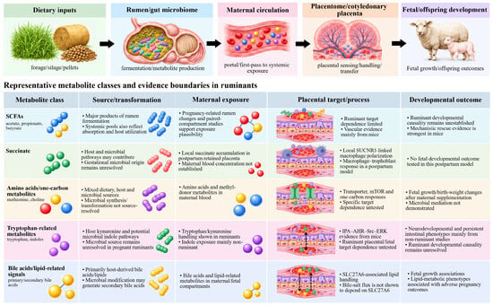
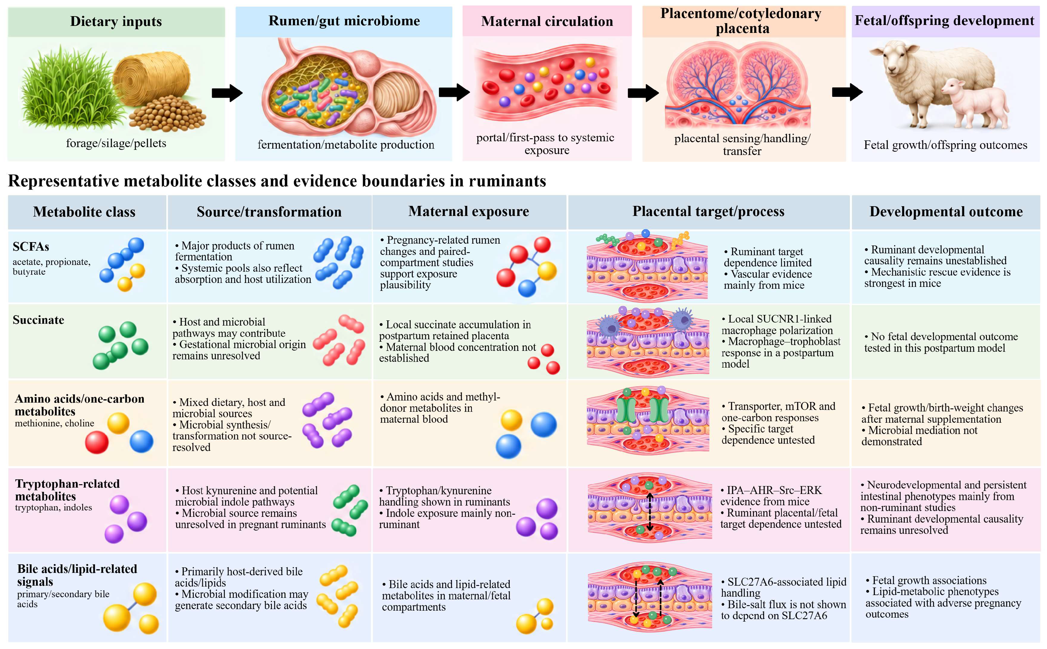
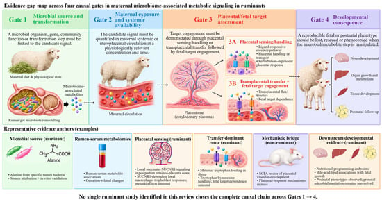
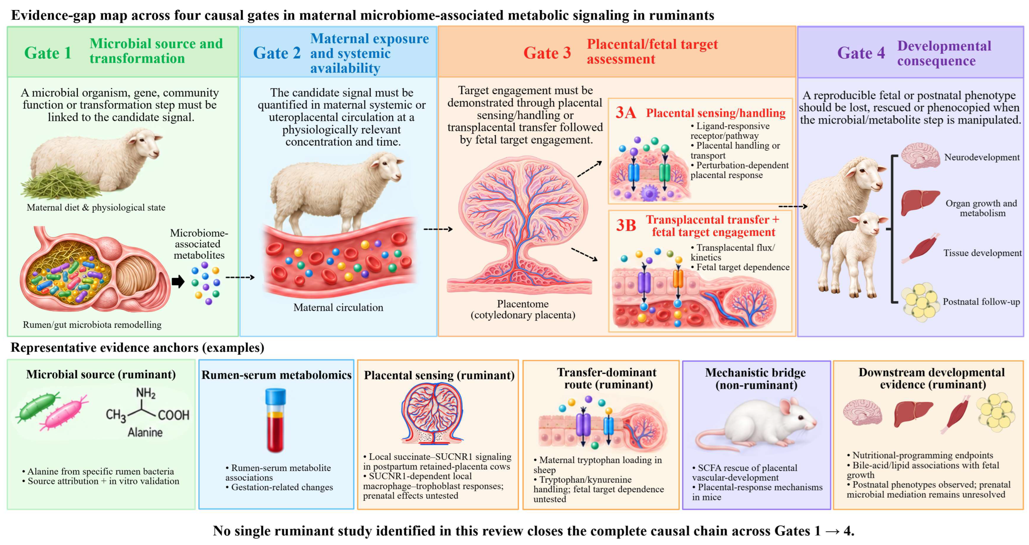
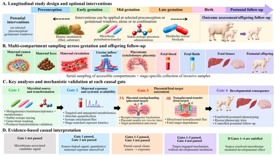
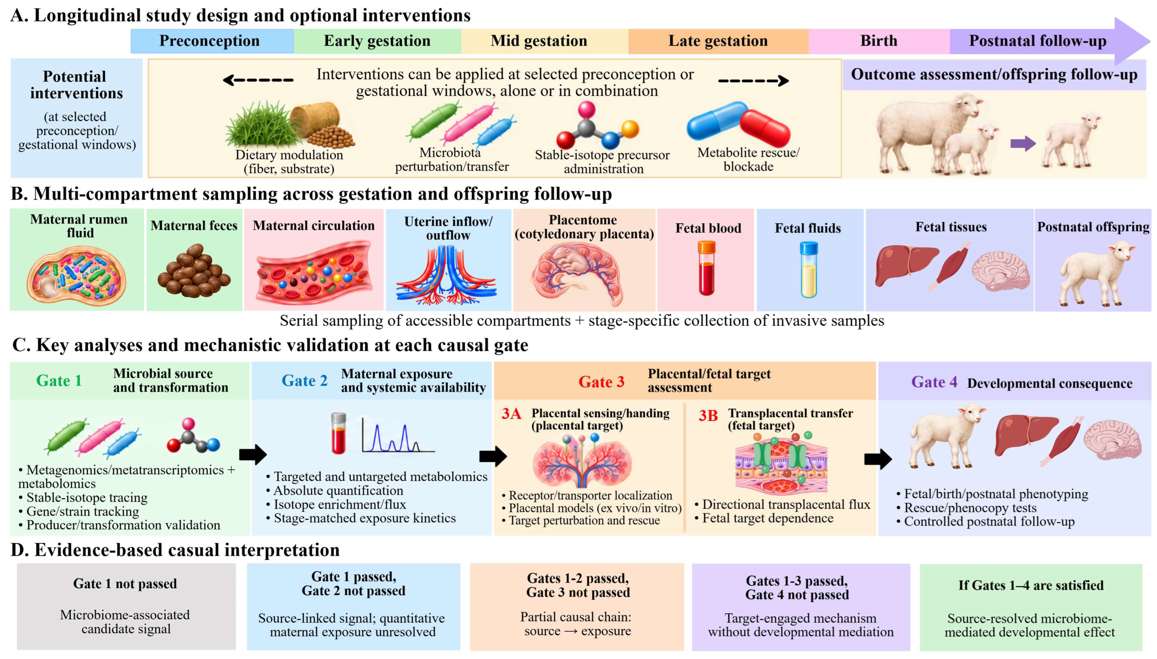

Disclaimer/Publisher’s Note: The statements, opinions and data contained in all publications are solely those of the individual author(s) and contributor(s) and not of MDPI and/or the editor(s). MDPI and/or the editor(s) disclaim responsibility for any injury to people or property resulting from any ideas, methods, instructions or products referred to in the content. |
© 2026 by the authors. Licensee MDPI, Basel, Switzerland. This article is an open access article distributed under the terms and conditions of the Creative Commons Attribution (CC BY) license.