


Simple Summary
Young, growing dogs that break both forearm bones sometimes develop a crooked leg as they continue to grow, because the injury can quietly damage the growth plate of one of these bones without this being visible on X-rays at the time of the fracture. Some surgeons remove a small piece of this bone during the repair surgery, hoping to prevent this crooked growth, but it was unclear whether this extra step actually helps. We reviewed the medical records of 28 small-breed puppies that had this type of fracture repaired, some with this extra surgical step and some without. To separate true abnormal growth from the puppies’ normal growth, we also measured the puppies’ healthy, uninjured leg for comparison. Puppies that had the extra surgical step showed less change in leg alignment than those that did not, even after accounting for normal growth. There was no clear difference between the two groups in how well the elbow joint fit together afterward. These findings suggest an association between this extra surgical step and less subsequent change in leg alignment, and support further research to determine whether the procedure itself accounts for this difference.
Abstract
This retrospective cohort study compared antebrachial angular change following radioulnar diaphyseal fracture repair in immature small-breed dogs treated with or without concurrent ulnar ostectomy (UO). Medical records of small-breed dogs younger than 12 months that underwent plate fixation for radioulnar diaphyseal fractures were reviewed, and dogs were divided into UO and non-UO groups. Joint orientation angles (medial proximal radial angle [MPRA], lateral distal radial angle [LDRA], valgus angle [VA], and sagittal plane angle [SPA]) were measured on the operated limb at surgery and at bone union, and contralateral-limb measurements were used to calculate growth-adjusted net change. Twenty-eight dogs were included (UO, n = 13; non-UO, n = 15), 24 of which had contralateral follow-up. Operated-limb angular change (Δoperated) differed significantly between groups for all four angles (Holm-adjusted p < 0.05); mean changes were larger in magnitude in the non-UO group. These between-group differences persisted in the growth-adjusted analysis (Holm-adjusted p < 0.05). Elbow incongruity did not differ significantly between groups (0/13 vs. 3/15; p = 0.23). Concurrent UO was associated with smaller magnitudes of Δoperated, and this association persisted after growth adjustment. Prospective studies controlling for potential confounding factors are needed to confirm this association.
1. Introduction
Radioulnar diaphyseal fractures are commonly encountered in skeletally immature dogs and are frequently managed using plate fixation techniques [1,2,3,4]. However, because the radius and ulna function as paired bones, disruption of normal ulnar growth following trauma may result in asynchronous growth and subsequent antebrachial angular deformity [5,6,7]. The distal ulnar physis is particularly vulnerable to premature closure, which can lead to progressive carpal valgus, radial procurvatum, and elbow incongruity [6,8]. Radiographic changes of the distal ulnar physis may not be evident immediately after injury [9], and progressive antebrachial angular deformity may develop even after successful fracture repair [3,10,11].
Ulnar ostectomy (UO) has traditionally been described as a treatment for established antebrachial angular deformity secondary to premature distal ulnar physeal closure [12]. Concurrent UO has also been described as an option during the initial surgical repair of radial fractures in young dogs, particularly when traumatic physeal compression is a concern [8]. The rationale for concurrent UO is to release potential ulnar restraint on continued radial growth if occult distal ulnar physeal injury subsequently results in premature closure. However, evidence regarding concurrent UO at the time of radioulnar fracture fixation remains limited, particularly in immature small-breed dogs, and clear indications for its use and its effect on subsequent angular change have not been established.
A separate challenge in interpreting angular change in the operated limb in this population is that immature dogs are still growing: some degree of change in joint orientation angles over the fracture-healing period may reflect normal antebrachial growth rather than a pathologic process. The contralateral, uninjured limb has been used as an individual reference for normal radial alignment in dogs with unilateral antebrachial angular deformity [13]. In the present study, the contralateral limb was used as an internal reference to account for physiologic angular change during growth.
The purpose of this study was to compare operated-limb angular change following radioulnar diaphyseal fracture repair in immature small-breed dogs with and without concurrent UO. As a secondary analysis, growth-adjusted net change was compared in dogs with available contralateral follow-up radiographs.
We hypothesized that dogs undergoing UO would show smaller magnitudes of operated-limb angular change than dogs treated without UO, and that this between-group difference would persist in growth-adjusted net change.
2. Materials and Methods
2.1. Animals
Medical records from four referral hospitals were retrospectively reviewed. Small-breed dogs younger than 12 months of age that underwent plate-and-screw fixation for radioulnar diaphyseal fractures were included; chondrodystrophic breeds were excluded. The age criterion of <12 months was chosen to capture the developmental window during which the distal ulnar physis remains open and susceptible to injury-related premature closure in small-breed dogs [6,8,12]. Signalment data (breed, sex, age, and body weight at surgery) were collected.
Cases were categorized into a UO group and a non-UO group based on whether a concurrent UO was performed during fracture repair. The decision to perform concurrent UO was made by the attending surgeon at each institution based on individual clinical judgment; no predefined protocol or objective cutoff was applied across the study period. Clinical considerations that may have influenced this decision included a distal fracture location or proximity to the distal ulnar physis, suspicion of occult physeal injury despite the absence of radiographically apparent physeal damage, and very young age with substantial remaining growth potential. These considerations were not consistently documented on a case-by-case basis in the retrospective medical records.
The length of the resected ulnar segment was not determined according to a standardized protocol or predefined criterion. The attending surgeon selected the segment length intraoperatively based on clinical judgment; however, the specific factors used to determine the length were not consistently documented in the medical records and therefore could not be reliably reconstructed retrospectively. Operative records documented resected segment lengths of approximately 6–12 mm, representing the minimum and maximum lengths recorded rather than a predefined target range. The ostectomy site was selected freehand according to fracture location rather than predefined anatomic landmarks. It was generally performed proximal to the fracture site; however, when the fracture was located near the mid-diaphysis, the ostectomy was performed distal to the fracture site to avoid excessive proximity to the elbow joint and potential reduction in proximal radioulnar-humeral stability. Postoperatively, external coaptation was not routinely used following plate fixation in either group, and no systematic group-specific immobilization protocol was applied.
2.2. Measurements
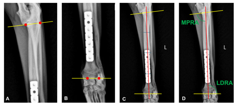
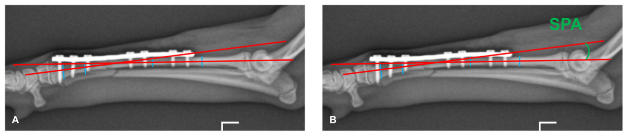
For both groups, radiographs obtained immediately postoperatively (day of operation, DO) and at bone union (BU) were compared. Bone union was defined as disappearance of the fracture line in the cortical bone on both frontal and sagittal radiographs. Radiographs were evaluated using RadiAnt DICOM Viewer (Medixant, Poznań, Poland). Measurements followed the center of rotation of angulation (CORA) methodology [13], as illustrated in Figure 1 and Figure 2.
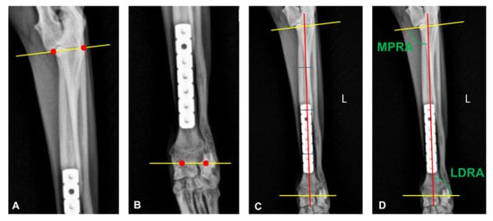
Figure 1.
Measurement of joint orientation angles in the frontal plane. (A) The elbow joint orientation line was drawn from the proximolateral aspect of the radial head to the medial coronoid process, or from the distal-most aspect of the humeral condylar capitulum to the trochlea. (B) The carpal joint orientation line was drawn from the most lateral aspect of the distal radial articular surface to the lateral aspect of the radial styloid process. (C) The radial anatomic axis was defined by a line passing through the centers at 25%, 50%, and 75% of the radial length. (D) The medial proximal radial angle (MPRA) and lateral distal radial angle (LDRA) were measured between the radial anatomic axis and the corresponding joint orientation lines. Red dots indicate the anatomical landmarks used to construct the joint orientation lines; yellow lines represent the elbow and carpal joint orientation lines; and the red line represents the radial anatomic axis.
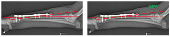
Figure 2.
Measurement of the sagittal plane angle (SPA). (A) The proximal and distal radial diaphyseal axes were defined by connecting the centers at one-third and two-thirds of each segment. (B) The SPA was measured between the axes of the proximal and the distal radial diaphysis. Red lines represent the proximal and distal radial diaphyseal axes; blue lines indicate the one-third and two-thirds reference levels used to define the centers of each segment.
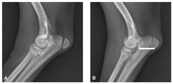
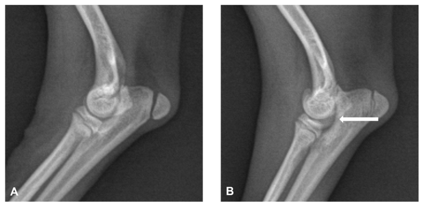
In the frontal plane, joint orientation lines were drawn at the elbow and carpal levels, and the radial anatomic axis was defined through the centers at 25%, 50%, and 75% of radial length (or 25% and 75% if the line did not pass through all three points). MPRA and LDRA were measured between the radial anatomic axis and the corresponding joint orientation lines, and VA was calculated as MPRA—LDRA. In the sagittal plane, SPA was measured between the proximal and distal radial diaphyseal axes, each defined by the centers at one-third and two-thirds of the corresponding segment. Elbow incongruity at BU was defined by step formation between the lateral coronoid process and radial head, altered humeroulnar articular spacing, elliptical appearance of the trochlear notch, or altered humeroradial joint space [14], as illustrated in Figure 3. Radiographic status of the radial and ulnar physes at BU was also recorded as open or closed. Antebrachial length was not analyzed in this study, as linear measurements on retrospectively collected multicenter radiographs could not be reliably corrected for inter-radiograph magnification differences in the absence of a calibration marker; angular measurements are unaffected by uniform magnification and were therefore considered appropriate outcome measures.
Figure 3.
Representative radiographs demonstrating elbow congruity. (A) Congruent elbow joint. (B) Incongruent elbow joint with step formation at the humeroradial articulation (arrow).
To account for angular change attributable to normal physiologic growth in immature dogs, radiographs of the contralateral (non-fractured) limb obtained at the same two time points (DO and BU) were evaluated using identical methodology, when available in the medical record. Contralateral-limb radiographs were not uniformly included in routine radiographic protocols across participating institutions and, in some cases, were declined by the owner.
2.3. Reliability Assessment
Intra-observer reliability was assessed for the angular measurements (MPRA, LDRA, VA, SPA) on operated-limb radiographs. A randomly selected, group-stratified subset of 15 of 28 dogs (54%; UO group, n = 7; non-UO group, n = 8) was re-measured at both DO and BU by a single observer (H.K., a veterinarian with a master’s degree in veterinary surgery, currently pursuing a PhD, with approximately 12 years of clinical experience at the time of measurement), blinded to dog identity, group, and time point, with measurements presented in randomized order. A second round of measurements was performed ≥2 weeks after the first, in a re-randomized order, with the observer blinded to the results of the first round. Intraclass correlation coefficients (ICC) were calculated using a two-way random-effects model with absolute agreement (ICC(2,1)), with values interpreted as poor (<0.50), moderate (0.50–0.75), good (0.75–0.90), or excellent (>0.90) [15].
2.4. Definitions of Angular Change Outcomes
For each angular measurement, operated-limb angular change (Δoperated) was defined as the value at bone union (BU) minus the value on the day of operation (DO). When contralateral radiographs were available at both time points, contralateral-limb angular change (Δcontralateral) was defined analogously as the value at BU minus the value at DO. Growth-adjusted net change was defined as Δoperated − Δcontralateral and was calculated only for dogs with contralateral radiographs available at both time points.
2.5. Statistical Analysis
Statistical analyses were performed using commercial software (SPSS Statistics version 24.0; IBM Corp., Armonk, NY, USA) and Python (version 3.13.16; Python Software Foundation, Wilmington, DE, USA) with the SciPy library (version 1.18.1). The normality of continuous variables was assessed using the Shapiro–Wilk test. Because the distributions of the variables did not satisfy the assumption of normality, non-parametric statistical tests were used. Categorical variables were compared using the Pearson chi-square test or Fisher’s exact test, as appropriate. Continuous signalment variables were compared using the Mann–Whitney U test. The interval from DO to BU was summarized as the median and range for each group and compared between the UO and non-UO groups using the Mann–Whitney U test. Within-group change in joint orientation angles from DO to BU was evaluated using the Wilcoxon signed-rank test. The primary between-group comparison was Δoperated for each measurement, compared between the UO and non-UO groups using the Mann–Whitney U test. The secondary between-group comparison was growth-adjusted net change, compared between groups using the Mann–Whitney U test in the subset of dogs with available contralateral follow-up radiographs. The incidence of elbow incongruity between groups was analyzed using Fisher’s exact test. A p-value < 0.05 was considered statistically significant. To account for multiple comparisons across the four angular outcomes (MPRA, LDRA, VA, and SPA), Holm–Bonferroni correction was applied separately to the primary operated-limb comparisons and the growth-adjusted analyses. Because VA was derived from MPRA and LDRA (VA = MPRA − LDRA), the four angular outcomes were not statistically independent; Holm–Bonferroni correction remains valid under such dependence.
A generative artificial intelligence tool, Claude (Claude Opus 5.5; Anthropic, San Francisco, CA, USA), was used to (1) assist in drafting and debugging analysis code for the contralateral limb growth-adjusted analysis comparisons, as well as for ICC(2,1) calculation and between-group tests, and (ii) to support literature searching by identifying candidate references. All statistical outputs generated with the tool’s assistance were independently re-run by the authors in SPSS using the original dataset, and results were cross-checked against manual calculations. All candidate references were verified by the authors against the original full-text articles via PubMed/Google Scholar; references that could not be verified were excluded. The tool was not used to generate, modify, or interpret the raw data, and the authors take full responsibility for the accuracy of the analyses and the content of this manuscript.
3. Results
3.1. Study Population
Medical records of 28 dogs met the inclusion criteria (UO group, n = 13; non-UO group, n = 15). There was no significant difference in breed distribution, sex distribution, body weight, or age between groups. Signalment data are summarized in Table 1. The median interval from DO to BU was 44 days (range, 30–90 days) in the UO group and 48 days (range, 25–84 days) in the non-UO group. The interval did not differ significantly between groups (Mann–Whitney U test, p = 0.963).
Table 1.
Signalment data of dogs included in the study.
3.2. Operated-Limb Angular Change
Values at DO and BU for each group are summarized in Table 2. Comparing Δoperated directly between groups, significant between-group differences were observed for MPRA, LDRA, VA, and SPA (Table 3). All between-group differences in the primary operated-limb analysis remained statistically significant after Holm–Bonferroni correction.
Table 2.
Operated-limb measurements at DO and BU, by group.
Table 3.
Operated-limb angular change (Δoperated = BU − DO), UO vs. non-UO group.
3.3. Growth-Adjusted Net Change
Contralateral-limb radiographs at both DO and BU were available for 24 of 28 dogs (UO group, n = 12; non-UO group, n = 11–12 depending on measurement). In this subset, growth-adjusted net change differed significantly between the UO and non-UO groups for MPRA, LDRA, VA, and SPA (Table 4). Mean values in the UO group were closer to zero than those in the non-UO group across all four measurements. All between-group differences in the growth-adjusted analysis remained statistically significant after Holm–Bonferroni correction.
Table 4.
Growth-adjusted net change (Δoperated − Δcontralateral), UO vs. non-UO group.
3.4. Incongruity
Elbow incongruity was observed in 0 of 13 dogs in the UO group and 3 of 15 dogs in the non-UO group. This difference was not statistically significant (Fisher’s exact p = 0.226). Incongruity data are summarized in Table 5.
Table 5.
Incidence of elbow incongruity, UO vs. non-UO group.
3.5. Physeal Status at BU
At BU, the distal radial physis remained open in 7 of 13 dogs (53.8%) in the UO group and 9 of 15 dogs (60.0%) in the non-UO group. The distal ulnar physis remained open in 4 of 13 dogs (30.8%) and 6 of 15 dogs (40.0%), respectively. In all dogs with an open distal ulnar physis, the distal radial physis was also open.
3.6. Reliability
Intra-observer reliability, assessed using a two-way random-effects model with absolute agreement (ICC(2,1)) in the subset of 15 dogs (30 operated-limb radiographs), was moderate for MPRA (ICC = 0.66, 95% CI 0.35–0.83) and VA (ICC = 0.72, 95% CI 0.40–0.87), and good for LDRA (ICC = 0.80, 95% CI 0.63–0.90) and SPA (ICC = 0.80, 95% CI 0.53–0.91), by the criteria of Koo and Li [15]. All ICC values were statistically significant (p < 0.001).
4. Discussion
This retrospective cohort study showed significant differences in Δoperated between the UO and non-UO groups across all four angular measurements. Because all dogs were skeletally immature, part of the observed change could reflect physiologic growth. In dogs with contralateral follow-up radiographs, the between-group differences were also observed in the growth-adjusted net change analysis across all four measurements.
The joint orientation angles observed at BU can also be considered in the context of published reference values for skeletally mature, nonchondrodystrophic small-breed dogs, in which MPRA and LDRA have been reported as 80.86 ± 2.86° and 85.60 ± 1.74°, respectively [16]. Although these values were derived from mature dogs and are not directly interchangeable with measurements from the immature population studied here, they provide an approximate benchmark. Mean LDRA at BU in the non-UO group (80.9 ± 4.92°) was more than 2 SD below the reference mean, reflecting a change away from the reference value over the healing period, whereas mean LDRA in the UO group (84.3 ± 2.73°) remained within approximately 1 SD. Mean MPRA at BU remained within approximately 1 SD of the reference mean in both groups.
Continued radial growth in the presence of partial or complete ulnar growth arrest is a recognized mechanism for progressive carpal valgus and radial procurvatum [5,6,7,12,17]. The pattern of angular change observed in the non-UO group is consistent with this established mechanism, although the retrospective design of the present study does not establish causation. Elbow incongruity was observed only in the non-UO group (3/15 dogs), but the between-group difference was not statistically significant. Given the small number of events and limited sample size, this finding should be interpreted cautiously and does not allow conclusions regarding an association between concurrent UO and subsequent elbow congruity.
Growth-adjusted net changes were closer to zero in the UO group than in the non-UO group across all four measurements (Table 4). These findings provide supportive evidence that the greater magnitude of Δoperated observed in the non-UO group was not fully explained by physiologic growth alone.
Concurrent UO also warrants consideration from a fracture-healing perspective. Because the ulna in small-breed dogs is frequently too narrow to permit rigid internal fixation, the fractured ulna is commonly left without direct fixation; nevertheless, as it heals, it may contribute to antebrachial stability by acting as an internal splint for the plated radius, and resection of an ulnar segment removes this potential contribution. This is relevant because toy- and small-breed radioulnar fractures are already recognized as prone to delayed union owing to their limited blood supply [4,18]. The healing behavior of the ulna, whether the native fracture or the ostectomy gap, may also influence radial–ulnar mechanical interaction during growth. The approximately 6–12 mm resection lengths documented in the operative records fall within the range previously described for distal UO in juvenile dogs with discordant radial–ulnar physeal growth [12]; however, that range was reported for the treatment of established deformity rather than for use at the time of fracture repair, and a biomechanical rationale for segment length in this setting has not been established.
No dog had radiographic evidence of ulnar physeal injury at the time of fracture; however, occult physeal injury may have been a clinical consideration in the decision to perform concurrent UO. As described in Section 2.1, this decision was based on individual surgeon judgment, and the factors influencing it were not systematically documented. The two groups may therefore have differed in their baseline risk of antebrachial angular deformity, and the observed between-group differences cannot be attributed solely to UO. Pending prospective data, the additional surgical morbidity of UO should be weighed against the observed between-group difference in Δoperated on a case-by-case basis.
From a clinical perspective, once clinically relevant antebrachial angular deformity develops, corrective osteotomy may be required, increasing surgical complexity, cost, and potential morbidity [5,17]. There is currently no established threshold of change in MPRA, LDRA, VA, or SPA that defines clinical significance in immature small-breed dogs. Because no threshold of functional relevance was established in this study, the present findings do not support describing concurrent UO as prophylactic. The clinical relevance of the observed angular differences should be interpreted in relation to published reference values, the contralateral limb as an individual internal reference, and the presence of functional impairment rather than by a single angular cutoff. The lower mean LDRA at BU in the non-UO group relative to the published reference values may warrant clinical consideration, although its functional significance remains uncertain.
Limitations
Several limitations should be acknowledged. (1) This was a retrospective, multicenter study in which the decision to perform concurrent UO was based on individual surgeon judgment rather than randomization, and surgical technique, fixation implants, and the precise anatomic location of UO were not standardized across the four participating institutions. Growth adjustment using the contralateral limb addresses the possibility that observed differences reflect normal growth, but it does not address the possibility that dogs selected for UO differed systematically from those who were not (indication bias), nor can it account for between-institution variation in technique. (2) Only intra-observer reliability of operated-limb measurements was assessed, as a second independent observer was not available; inter-observer reliability and the reliability of contralateral-limb measurements used in the growth-adjustment analysis were not evaluated. These should be addressed before the magnitude of reported angular changes is used for clinical decision-making. The moderate reliability observed for MPRA and VA, despite the observer’s substantial clinical experience, suggests that these measurements may be inherently more susceptible to variability than LDRA and SPA. For MPRA, this may reflect sensitivity to small differences in placement of the proximal radial reference landmarks and construction of the radial anatomic axis. Because VA was derived from MPRA and LDRA, variability in MPRA measurement may also have contributed to the moderate reliability observed for VA. (3) The growth-adjusted analysis relied on the contralateral limb as an internal reference and therefore assumes broadly symmetric physiologic growth between limbs. Altered loading of the contralateral limb following injury could also have influenced angular change and cannot be excluded in this retrospective cohort. In addition, contralateral-limb follow-up radiographs were unavailable for 4 of 28 dogs, limiting the growth-adjusted analysis to 24 dogs. Although there was no clear evidence that missingness was outcome-dependent, selection bias resulting from exclusion of these four dogs cannot be excluded. (4) Ulnar healing patterns were not systematically documented or evaluated. In the non-UO group, healing of the native ulnar fracture may have resulted in variable restoration of ulnar continuity, whereas in the UO group, persistence or osseous bridging of the ostectomy gap may have altered the intended mechanical release between the radius and ulna. Because these healing patterns are closely related to the proposed mechanism of UO, their contribution to the angular outcomes could not be separately assessed. Future studies should therefore systematically document native ulnar fracture healing and ostectomy-gap healing. (5) The study population consisted of small-breed dogs, and these findings may not generalize to larger breeds. (6) Follow-up was limited to the period of bone union; long-term functional outcomes and the clinical significance of the angular differences observed were not evaluated. At BU, at least one distal antebrachial physis remained open in more than half of the dogs in both groups (UO, 7/13; non-UO, 9/15), indicating that growth was ongoing in many dogs at the end of follow-up. The angular values reported here therefore reflect alignment at the time of bone union rather than at skeletal maturity, and further angular change after BU cannot be excluded in either group. In addition, because physeal status was assessed only at BU, premature closure could not be distinguished from age-appropriate physiologic closure. (7) Although Holm–Bonferroni correction was applied to account for multiple comparisons across the four angular outcomes, the small sample size and exploratory nature of the analyses warrant cautious interpretation.
5. Conclusions
In this retrospective cohort of immature small-breed dogs, concurrent UO at the time of radioulnar diaphyseal fracture repair was associated with smaller magnitudes of Δoperated than in dogs treated without UO across the evaluated angular measurements. Between-group differences also persisted in the growth-adjusted net change analysis using the contralateral limb as an internal reference. No significant difference in elbow incongruity was detected between groups. Given the retrospective design, potential confounding by indication, and limited functional outcome data, these findings should be interpreted as hypothesis-generating and warrant confirmation in larger prospective studies.
Author Contributions
Conceptualization, H.K.; methodology, H.K.; formal analysis, H.K.; investigation, H.K.; resources, H.K. and J.C.; data curation, H.K.; writing—original draft preparation, H.K.; writing—review and editing, H.K. and J.C.; visualization, H.K.; supervision, J.C.; project administration, H.K. All authors have read and agreed to the published version of the manuscript.
Funding
This research received no external funding.
Institutional Review Board Statement
Based on confirmation from the Jeju National University Institutional Animal Care and Use Committee, separate ethical review was not required for this retrospective study because it involved only the analysis of existing clinical records and diagnostic images obtained during routine veterinary care, with no additional procedures or interventions performed on animals for research purposes.
Informed Consent Statement
Informed consent was obtained from the owners of all animals involved in this study for the use of their clinical data and radiographic images.
Data Availability Statement
The data presented in this study are available on request from the corresponding author. The data are not publicly available due to privacy restrictions related to client-owned animal records.
Acknowledgments
During the preparation of this manuscript, the authors used Claude (Claude Opus 5.5; Anthropic) for the purpose of translating, revising text, statistical analysis and literature searching. The authors have reviewed and edited all outputs and take full responsibility for the content of this publication.
Conflicts of Interest
The authors declare no conflicts of interest.
References
- Milovancev, M.; Ralphs, S.C. Radius/ulna fracture repair. Clin. Tech. Small Anim. Pract. 2004, 19, 128–133. [Google Scholar] [CrossRef] [Scilit]
- Boudrieau, R.J. Fractures of the radius and ulna. In Textbook of Small Animal Surgery, 3rd ed.; Slatter, D., Ed.; W.B. Saunders: Philadelphia, PA, USA, 2003; pp. 1953–1973. [Google Scholar]
- Woods, S.; Perry, K.L. Fractures of the Radius and Ulna. Companion Anim. 2017, 22, 670–680. [Google Scholar] [CrossRef] [Scilit]
- Muir, P. Distal antebrachial fractures in toy-breed dogs. Compend. Contin. Educ. Pract. Vet. 1997, 19, 137–145. [Google Scholar]
- DeCamp, C.E.; Johnston, S.A.; Déjardin, L.M.; Schaefer, S.L. Correction of abnormal bone growth and healing. In Brinker, Piermattei and Flo’s Handbook of Small Animal Orthopedics and Fracture Repair, 5th ed.; Saunders: St. Louis, MO, USA, 2016; pp. 791–820. [Google Scholar]
- Ramadan, R.O.; Vaughan, L.C. Premature Closure of the Distal Ulnar Growth Plate in Dogs—A Review of 58 Cases. J. Small Anim. Pract. 1978, 19, 647–667. [Google Scholar] [CrossRef] [Scilit]
- McKee, M. Growth Deformities of the Long Bones in Dogs. In Pract. 2010, 32, 282–291. [Google Scholar] [CrossRef] [Scilit]
- Johnson, A. Premature physeal closure after fracture. In Complications in Small Animal Surgery; Griffon, D., Hamaide, A., Eds.; John Wiley & Sons: Hoboken, NJ, USA, 2016; pp. 649–654. [Google Scholar]
- Jackson, L.C.; Pacchiana, P.D. Common Complications of Fracture Repair. Clin. Tech. Small Anim. Pract. 2004, 19, 168–179. [Google Scholar] [CrossRef] [Scilit]
- Toombs, J.P. Fractures of the radius. In AO Principles of Fracture Management in the Dog and Cat; Johnson, A.L., Houlton, J.E.F., Vannini, R., Eds.; AO Publishing: Davos, Switzerland, 2005; pp. 230–259. [Google Scholar]
- McLain, D.L.; Brown, S.G. Fixation of Radius and Ulna Fractures in the Immature Dog and Cat: A Review of Popular Techniques and a Report of Eight Cases Using Plate Fixation. Vet. Surg. 1982, 11, 140–145. [Google Scholar] [CrossRef] [Scilit]
- Christopher, S. Short Term Outcomes and Complications of Distal Ulnar Ostectomy in 23 Juvenile Dogs with Carpal Valgus Secondary to Discordant Radial-Ulnar Physeal Growth. Front. Vet. Sci. 2022, 9, 971527. [Google Scholar] [CrossRef] [Scilit]
- Fox, D.B.; Tomlinson, J.L.; Cook, J.L.; Breshears, L.M. Principles of Uniapical and Biapical Radial Deformity Correction Using Dome Osteotomies and the Center of Rotation of Angulation Methodology in Dogs. Vet. Surg. 2006, 35, 67–77. [Google Scholar] [CrossRef] [Scilit]
- Kirberger, R.M. The elbow joint. In BSAVA Manual of Canine and Feline Musculoskeletal Imaging, 2nd ed.; Kirberger, R.M., McEvoy, F.J., Eds.; British Small Animal Veterinary Association: Gloucester, UK, 2016; pp. 189–211. [Google Scholar]
- Koo, T.K.; Li, M.Y. A Guideline of Selecting and Reporting Intraclass Correlation Coefficients for Reliability Research. J. Chiropr. Med. 2016, 15, 155–163. [Google Scholar] [CrossRef] [Scilit]
- Duncan, K.L.; Mielke, B.A.; Phillips, A.; Pead, M.; Meeson, R.; Kaye, B.M. Assessment of Normal Radial Joint Orientation Angles in Nonchondrodystrophic Small-Breed Dogs. J. Am. Vet. Med. Assoc. 2022, 261, 1–4. [Google Scholar]
- Knapp, J.L.; Tomlinson, J.L.; Fox, D.B. Classification of Angular Limb Deformities Affecting the Canine Radius and Ulna Using the Center of Rotation of Angulation Method. Vet. Surg. 2016, 45, 295–302. [Google Scholar] [CrossRef] [Scilit]
- Probst, C.W. Radius and ulna: Repair of fractures of the radius and ulna. In Current Techniques in Small Animal Surgery, 5th ed.; Bojrab, M.J., Ed.; Teton NewMedia: Jackson, WY, USA, 2014; pp. 933–943. [Google Scholar]

Disclaimer/Publisher’s Note: The statements, opinions and data contained in all publications are solely those of the individual author(s) and contributor(s) and not of MDPI and/or the editor(s). MDPI and/or the editor(s) disclaim responsibility for any injury to people or property resulting from any ideas, methods, instructions or products referred to in the content. |
© 2026 by the authors. Licensee MDPI, Basel, Switzerland. This article is an open access article distributed under the terms and conditions of the Creative Commons Attribution (CC BY) license.