


Simple Summary
A dog can develop new wrist instability even after an earlier forearm fracture has healed. This report describes a Maltese dog with excessive bending of the left wrist following fracture surgery during growth. Moving X-ray imaging helped identify the unstable joint. The old forearm plate occupied the area needed for a new fixation plate. Removing all screws and the plate at once raised concern about temporarily weakening the bone because plate support would be lost while screw holes remained. The screws were therefore removed in stages, leaving two screws in each plate temporarily before plate removal 28 days later. Surgery to stabilise all wrist joints was performed 68 days after plate removal. Limb function remained satisfactory for approximately four years, including after a late plate fracture was detected. This unusual case illustrates the importance of assessing wrist stability after fracture healing and planning the transition from existing implants to definitive fixation. The staged approach addressed a clinical concern; its effect on fracture prevention was not measured.
Abstract
Delayed carpal instability after healed juvenile antebrachial fractures presents a diagnostic and surgical challenge when retained implants occupy the site required for arthrodesis. A 3-year-old, 2.58 kg, neutered male Maltese dog presented with left forelimb lameness after bilateral radial–ulnar fracture repair during skeletal immaturity. Before implant removal, radiography and dynamic fluoroscopy localised hyperextension instability and subluxation to the antebrachiocarpal joint. Staged implant removal was selected because immediate loss of plate support and residual screw holes raised concern about temporary bone vulnerability. Intermediate screws were removed first, leaving one proximal and one distal screw in each plate. Both plates and all accessible remaining screws were removed 28 days later; contralateral removal was elective at the owner’s request. No perioperative antebrachial fracture was documented. A distal left screw head fractured during removal, leaving an intraosseous shaft that did not interfere with subsequent fixation. Left pancarpal arthrodesis was performed 68 days after the second removal procedure, with an immediate postoperative angle of approximately 12°. Gait lameness was absent by postoperative day 40. At day 75, intermittent limb elevation while standing and discomfort during manipulation persisted, although walking and trotting were free of lameness. Plate fracture was identified at approximately 38 months, with maintained alignment and apparent clinical stability; monitoring without revision was selected. Satisfactory limb function persisted at approximately 48 months. This unusual case documents localisation of postoperative carpal instability, the rationale for staged implant management, and long-term function after pancarpal arthrodesis. The contribution of staging to refracture prevention remains unquantified.
1. Introduction
Canine carpal hyperextension injuries result from failure or attenuation of the palmar ligamentous, fibrocartilaginous, and capsular structures that normally resist excessive extension. Depending on the direction and magnitude of loading, instability may occur at the antebrachiocarpal, middle carpal, intercarpal, or carpometacarpal level. Clinical manifestations range from subtle weight-bearing lameness to severe hyperextension and loss of functional palmar support [1,2,3,4,5,6].
Stress radiographs help identify the affected carpal joint level and guide the extent of arthrodesis [2,6]. Extension stress views assess hyperextension, whereas separate varus–valgus stress views assess collateral stability. Textbook guidance describes positioning and sedation or anaesthesia to facilitate this assessment [7]. Dynamic imaging can relate abnormal motion to clinical instability. In the present case, fluoroscopy during applied stress complemented orthogonal radiographs by demonstrating abnormal motion at the antebrachiocarpal joint.
Accurate localisation guides selection between partial carpal and pancarpal arthrodesis. Partial arthrodesis preserves antebrachiocarpal motion when that articulation remains stable, whereas involvement of the antebrachiocarpal joint generally favours pancarpal arthrodesis to stabilise the affected carpal levels [1,2,3,4,5,6]. The treatment goal is a stable, acceptably aligned limb with comfortable weight bearing. Working-dog series report generally satisfactory function, although some residual lameness and activity restrictions occur [8,9,10]. Reported complications include surgical-site infection, implant irritation, and subsequent implant removal [11,12,13,14,15,16,17], screw or plate failure and incomplete fusion [18], and metacarpal fracture [19]. These outcomes support assessment of both limb function and implant integrity during follow-up.
The development of carpal instability after antebrachial fracture healing presents an additional diagnostic and surgical problem. Distal radial or ulnar physeal growth disturbance can produce valgus, procurvatum, rotation, and adjacent-joint incongruity [20,21,22]. Carpal derangement with valgus and chronic lateral collateral ligament sprain with varus have also been described [23,24]. Together, these reports provide context for considering forelimb alignment and joint loading when evaluating new carpal dysfunction in a dog with previous fracture repair. The present case concerns delayed antebrachiocarpal instability after healed diaphyseal fractures, without documented physeal injury.
Long-standing antebrachial implants can obstruct placement of an arthrodesis construct. Three related but distinct concerns arise during removal: cortical remodelling or reduced density associated with altered load transfer (stress shielding) and, with some fixation modes, periosteal vascular effects [25,26]; local stress concentration at residual screw holes [27]; and the resulting period of mechanical vulnerability after plate support is withdrawn [28,29,30]. Experimental measurements of density, stress, or failure load do not directly quantify clinical refracture risk during weight bearing and activity. Staged removal has been described after treatment of septic antebrachial nonunion [31], but that setting differs from conversion to arthrodesis in a toy-breed dog with healed fractures and carpal instability.
The specific clinical problem is how to sequence implant removal and definitive carpal stabilisation when the existing plate occupies the required fixation site. The cited literature does not establish an optimal sequence for this combination of problems. The objective of this report is to describe diagnostic localisation, staged implant management, pancarpal arthrodesis, and approximately 4 years of follow-up in one Maltese dog. The case provides a longitudinal account of decisions and complications that may inform individual surgical planning; it does not determine a generally applicable removal schedule or demonstrate the superiority of staging.
2. Case Description
2.1. Signalment and Previous Orthopaedic History
A 3-year-old, 2.58 kg, neutered male Maltese dog was presented to the Veterinary Medical Teaching Hospital of Konkuk University for evaluation of left thoracic limb lameness in March 2016.
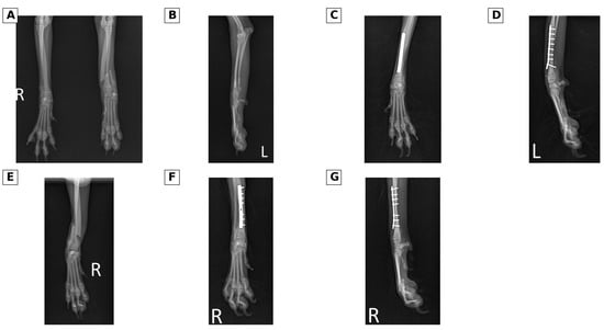
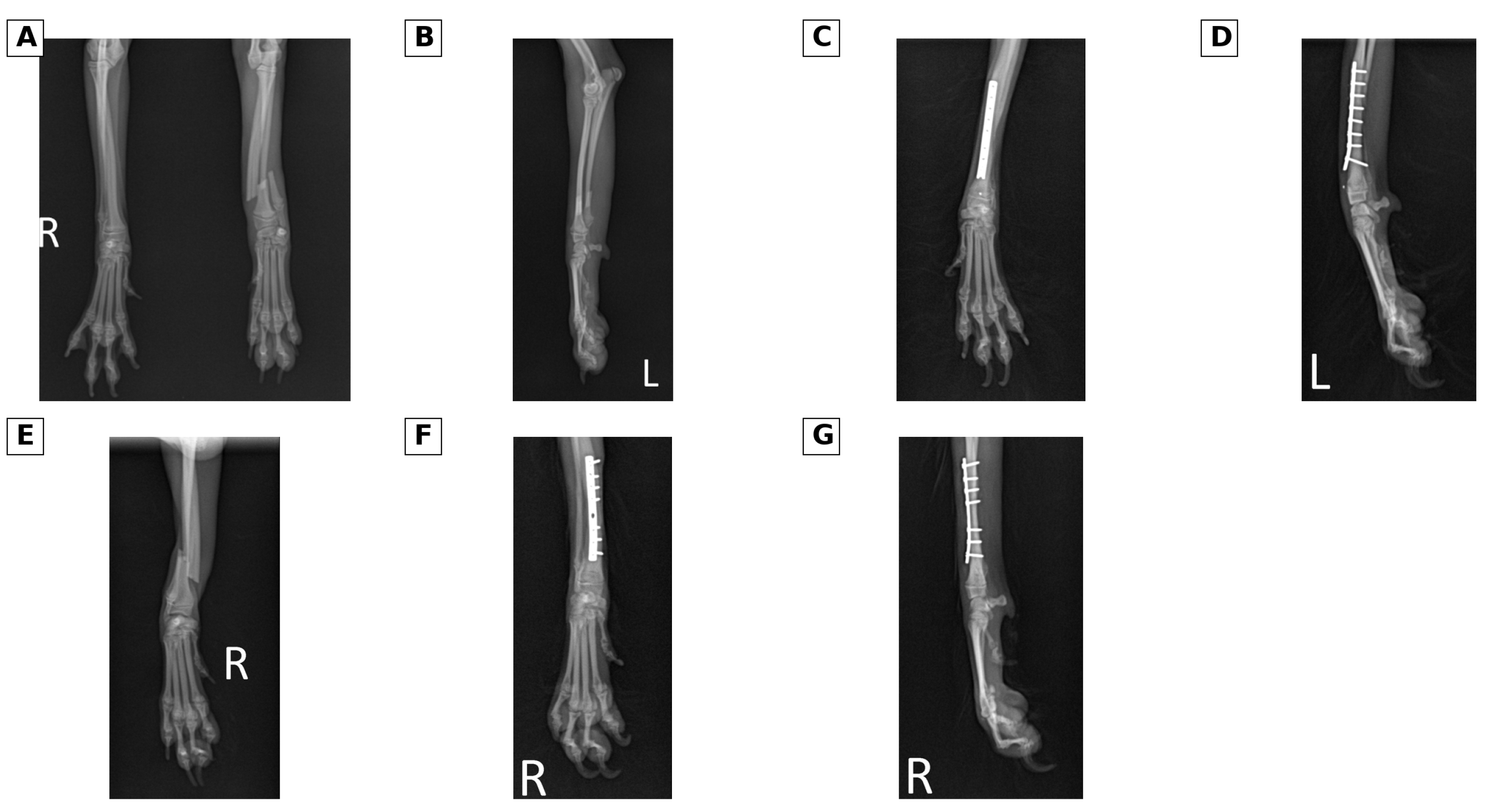
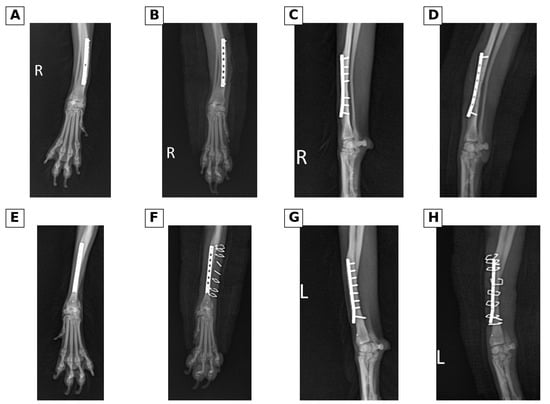
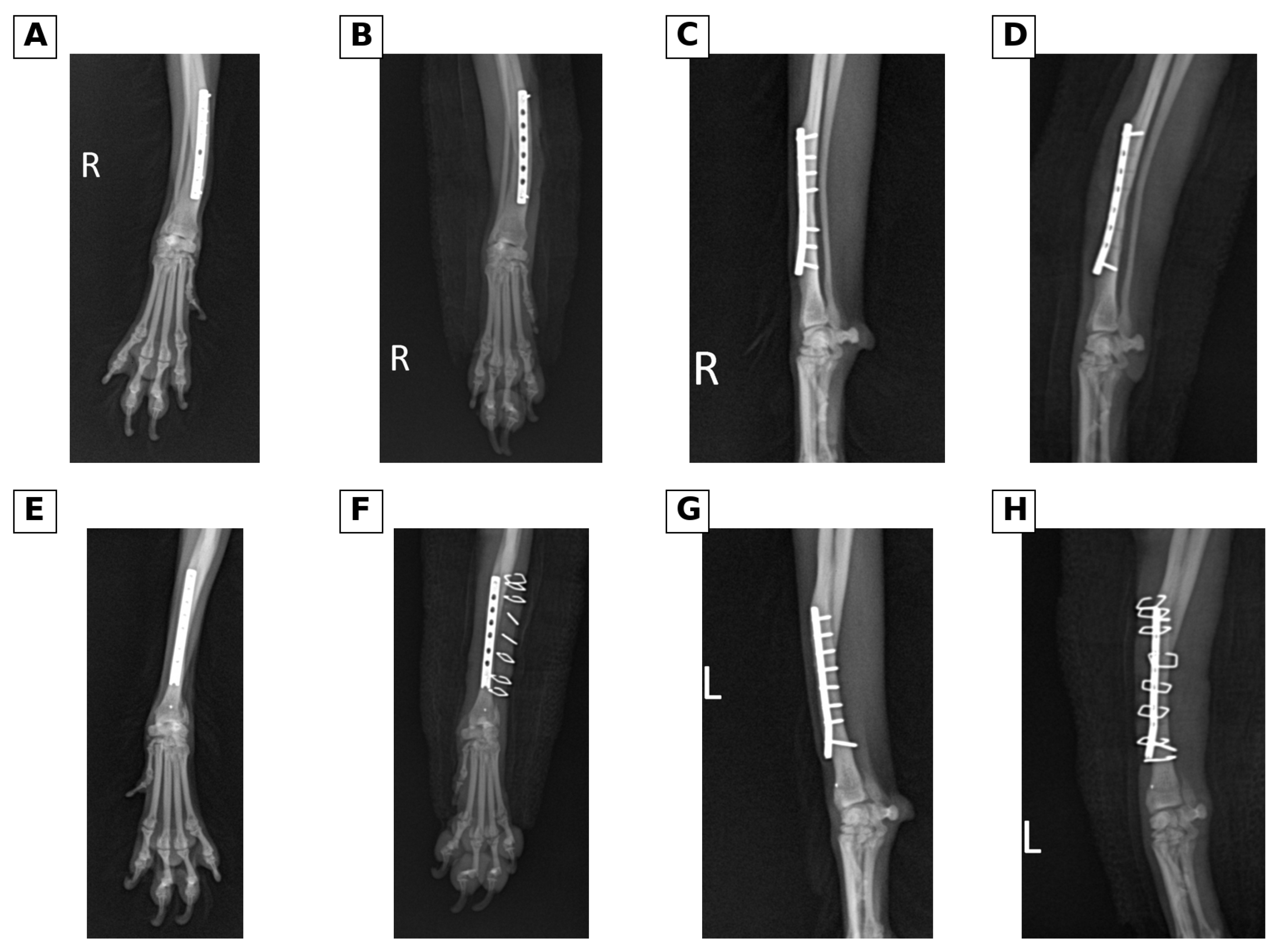
The dog had sustained bilateral radius-ulna fractures during skeletal immaturity. The left injury consisted of transverse fractures of the radius and ulna within the distal diaphysis, approximately the distal one-quarter to one-third of the antebrachium. Plate-and-screw fixation was performed on 17 October 2013, when the dog was approximately 6 months old and weighed 1.5 kg. A morphologically similar right distal radial and ulnar diaphyseal fracture was repaired on 19 December 2013, when the dog was approximately 8 months old and weighed 1.5 kg. Both antebrachial constructs remained in situ after fracture healing (Figure 1).
Figure 1.
Preoperative and immediate postoperative radiographs of the juvenile left and right distal radius-ulna fractures. (A–D) The left fracture series: (A) preoperative craniocaudal (CrCd) view including both distal antebrachia, (B) preoperative left mediolateral (ML) view, (C) immediate postoperative left craniocaudal (CrCd) view, and (D) immediate postoperative left mediolateral (ML) view. (E–G) The right fracture series: (E) preoperative right craniocaudal (CrCd) view, (F) immediate postoperative right craniocaudal (CrCd) view, and (G) immediate postoperative right mediolateral (ML) view. R and L indicate the right and left limbs, respectively.
The material available for retrospective review did not establish whether functional recovery after the juvenile fracture repairs had been complete or whether intermittent lameness preceded March 2016. Serial radiographs spanning the remaining growth period were not available for assessment, so earlier carpal congruity, degeneration, and alignment could not be characterised longitudinally. A detailed interval history of trauma or changes in activity was also unavailable. The absence of a witnessed inciting event at presentation should therefore not be interpreted as a documented trauma-free interval after fracture healing.
2.2. Presenting Complaint and Diagnostic Assessment
The owner first observed the left thoracic limb abnormality at approximately 21 March 2016. No single inciting traumatic event was witnessed. The dog resisted manipulation of the affected limb, had difficulty walking on slippery flooring, and intermittently carried the limb before paw placement. Once the paw was positioned, the dog could bear weight, and the abnormality was less apparent on a padded surface.
Orthopaedic examination revealed palpable crepitus and excessive hyperextension of the left carpus. Passive carpal range of motion (ROM) was assessed in lateral recumbency using a goniometer. The joint was moved through flexion and extension to identify its centre and anatomical landmarks. The elbow was extended during carpal extension assessment and flexed during flexion assessment to reduce the influence of multiarticular muscle tension. The goniometer axis was placed over the carpal joint centre. Its stationary arm was aligned using the lateral humeral epicondyle and the craniocaudal midpoint of the antebrachium at the level of the ulnar styloid process; its moving arm followed the long axis of metacarpal bones III and IV. The palmar-side joint angle was recorded at the passive flexion and extension endpoints, consistent with the described goniometric method [32]. Recorded extension and flexion angles were 195° and 25° on the right and 193° and 20° on the left, respectively.
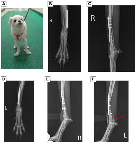
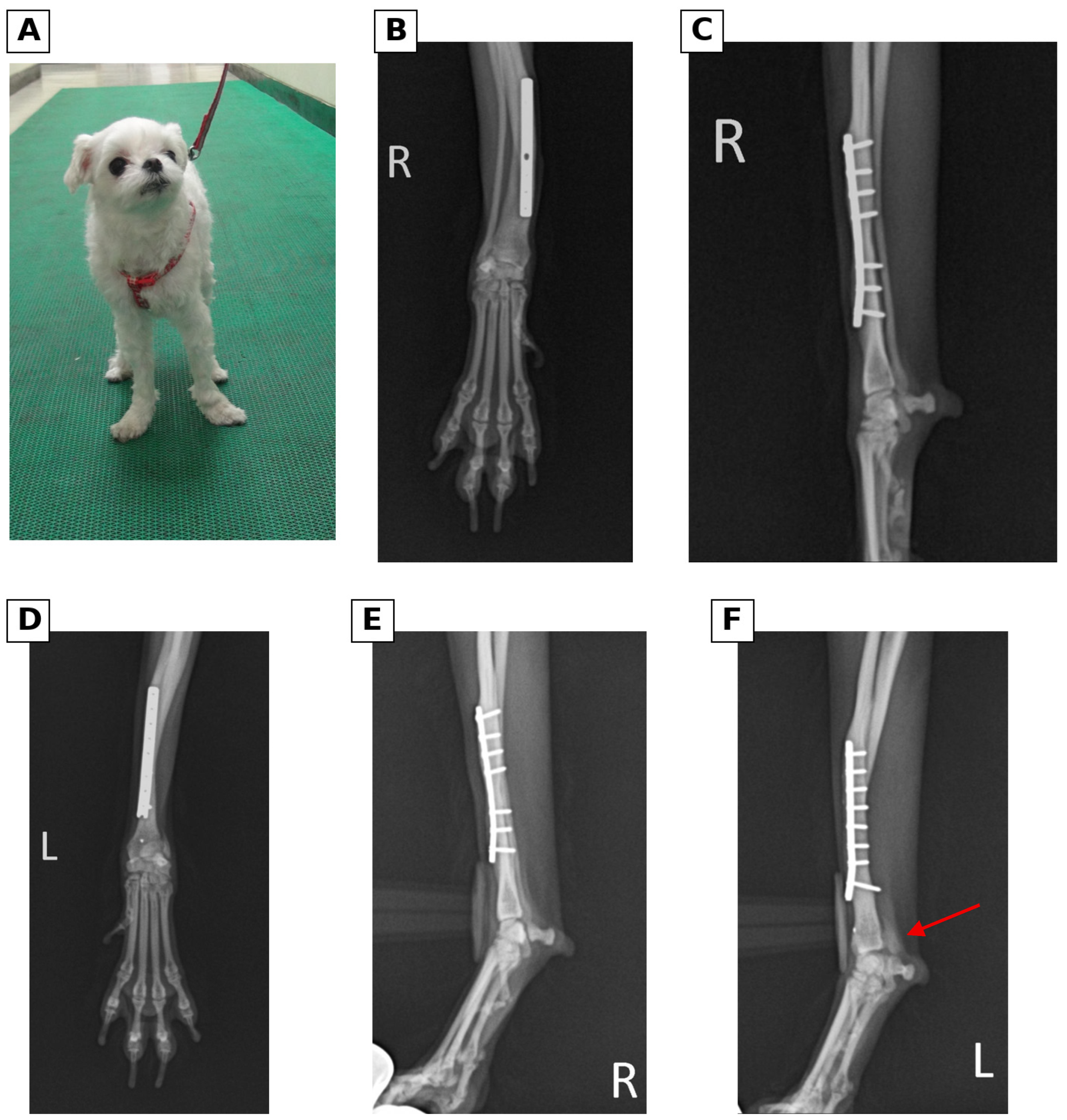
Orthogonal radiographs showed healed bilateral fractures and left antebrachiocarpal incongruity or subluxation. The clinical chart documented dorsopalmar and mediolateral (ML) stress fluoroscopic views. For extension stress assessment, the dog was positioned in lateral recumbency and the distal limb was manually extended while ML images were obtained. Dorsopalmar images were obtained during medial and lateral stress of the distal limb to assess collateral stability. These manoeuvres follow the principles described for stress radiography [7]. Fluoroscopic recordings were available for retrospective review. Dynamic fluoroscopy demonstrated abnormal motion between the distal antebrachium and proximal carpal row, localising the instability to the antebrachiocarpal joint before implant removal. The degree of stress-induced displacement was assessed qualitatively; no quantitative displacement measurements were obtained. The recorded ROM endpoint angles did not quantify displacement at the antebrachiocarpal joint. Applied force was not quantitatively measured, and whether sedation or anaesthesia was used could not be verified.
The presumptive diagnosis was antebrachiocarpal hyperextension instability associated with insufficiency of the palmar ligamentous and supporting structures; a specific ligament rupture was not directly confirmed. Because the fractures had occurred during skeletal immaturity, residual malalignment was considered as a possible contributor to altered carpal loading. Standard CrCd and ML views showed no gross clinically relevant frontal- or sagittal-plane angular deformity. Rotational alignment, distal radial joint orientation, and radial-ulnar length were not quantified by computed tomography or standardised measurements, and neither a concurrent physeal injury nor subsequent growth disturbance had been documented. Symptomatic hyperextension and demonstrable antebrachiocarpal instability guided the plan for pancarpal arthrodesis: partial carpal arthrodesis would have left the identified unstable articulation unfused (Figure 2).
Figure 2.
Standing posture and comparative neutral and stress radiographs of the right and left carpi before definitive treatment. (A) The standing posture of the dog. (B,C) Neutral craniocaudal (CrCd) and mediolateral (ML) views of the right antebrachium and carpus, respectively. (D) A neutral craniocaudal (CrCd) view of the left antebrachium and carpus. (E,F) Stress mediolateral (ML) views of the right and left carpi, respectively. The red arrow in (F) indicates the abnormal antebrachiocarpal joint space/alignment on the affected left side. R and L indicate the right and left limbs, respectively.
2.3. Staged Bilateral Antebrachial Implant Removal
The left antebrachial plate occupied the region required for subsequent arthrodesis plate placement. Immediate removal of the entire construct would eliminate plate support while leaving residual screw holes. Concern about temporary bone vulnerability, potentially compounded by cortical weakening beneath the long-standing plate, led to a two-stage removal plan. Intermediate screws were removed first while proximal and distal fixation was retained, with the intention of maintaining temporary support and allowing a more gradual return of load to the bone before plate removal. Contralateral implant removal was performed electively at the owner’s request as part of the bilateral implant-management plan; no right-sided clinical abnormality was documented.
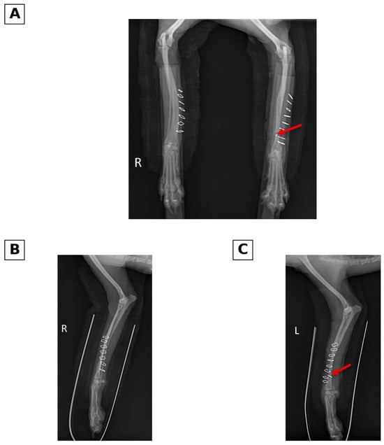
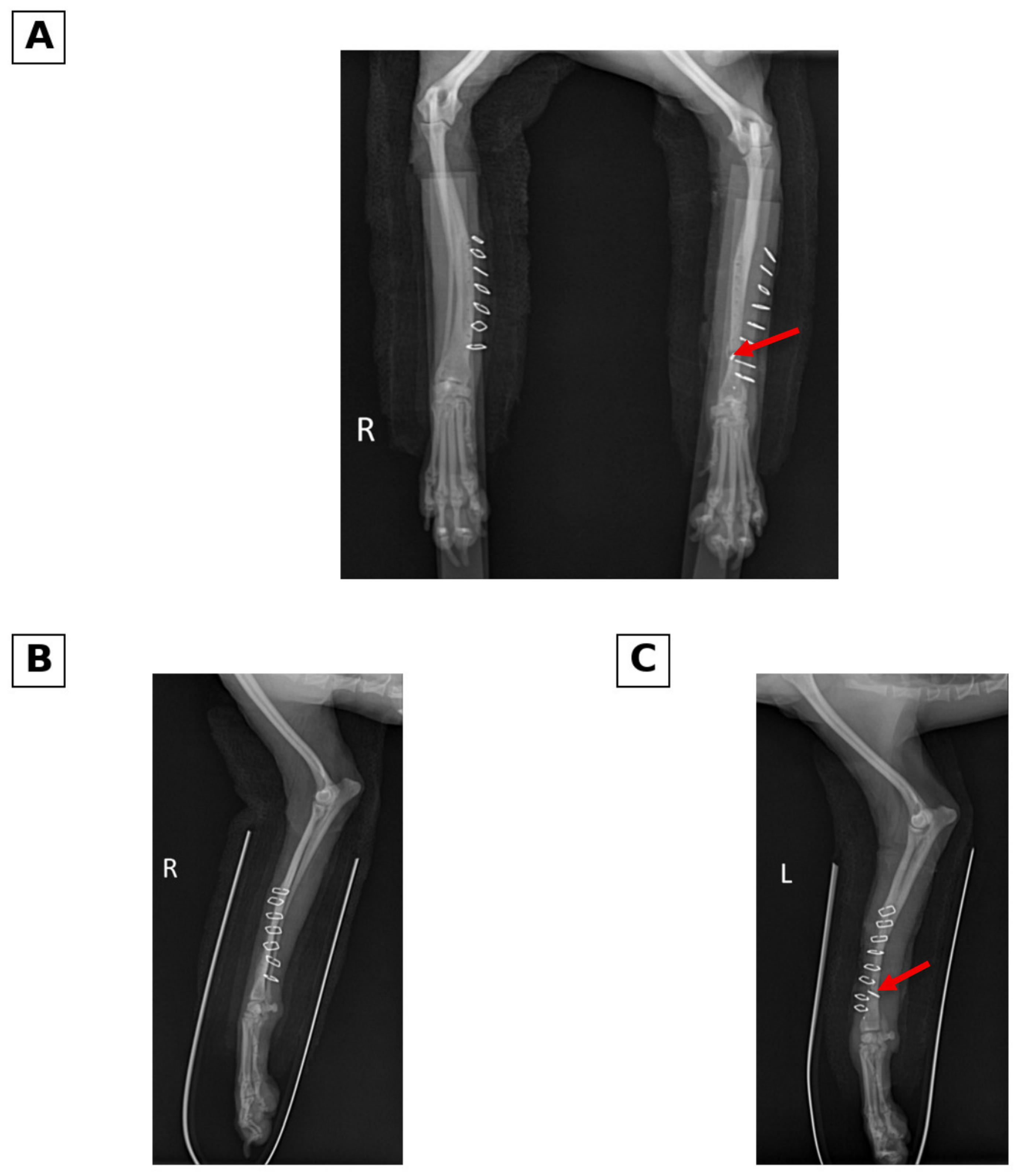
On 20 April 2016, all intermediate screws were removed while one proximal and one distal screw were retained in each plate. No perioperative antebrachial fracture was documented. On 18 May 2016, 28 days later, both plates and all accessible retained screws were removed. During removal of the left construct, the head of a distal screw fractured, and the residual shaft remained embedded within the left antebrachial bone. The fragment was not associated with an immediate clinical complication and did not interfere with the planned position or screw trajectories of the subsequent arthrodesis construct (Figure 3 and Figure 4).
Figure 3.
Radiographs obtained before and after the first-stage bilateral implant-removal procedure. (A,C) Preoperative craniocaudal (CrCd) and mediolateral (ML) views of the right antebrachium, respectively, and (B,D) the corresponding craniocaudal (CrCd) and mediolateral (ML) views after the first-stage screw-removal procedure. (E,G) Preoperative craniocaudal (CrCd) and mediolateral (ML) views of the left antebrachium, respectively, and (F,H) the corresponding craniocaudal (CrCd) and mediolateral (ML) views after the first-stage screw-removal procedure. During this stage, the intermediate screws were removed, while one proximal and one distal screw were retained in each plate. R and L indicate the right and left limbs, respectively.
Figure 4.
Radiographs obtained after the second-stage bilateral plate-and-screw removal procedure. (A) A bilateral craniocaudal (CrCd) view after complete removal of the antebrachial plates. (B,C) Right and left mediolateral (ML) views after the second-stage removal procedure, respectively. The red arrows in (A,C) identify the retained fragment of the distal left radial screw. R and L indicate the right and left limbs, respectively.
Left thoracic limb lameness persisted after implant removal. At reassessment on 8 July 2016, the lameness had mildly improved but remained evident. The treatment sequence and the two removal intervals are summarised in Table 1. Details of activity restriction or external support during the implant-removal intervals could not be reconstructed from the available records.
Table 1.
Chronology of diagnostic assessment and staged surgical treatment.
2.4. Pancarpal Arthrodesis and Postoperative Management
Premedication consisted of butorphanol (0.2 mg/kg IV) for analgesia and midazolam (0.3 mg/kg IV) for sedation. Anaesthesia was induced with propofol (6 mg/kg IV) and maintained with isoflurane. Bupivacaine (2 mg/kg) was administered by local injection for regional nerve blockade.
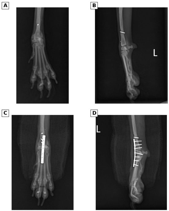
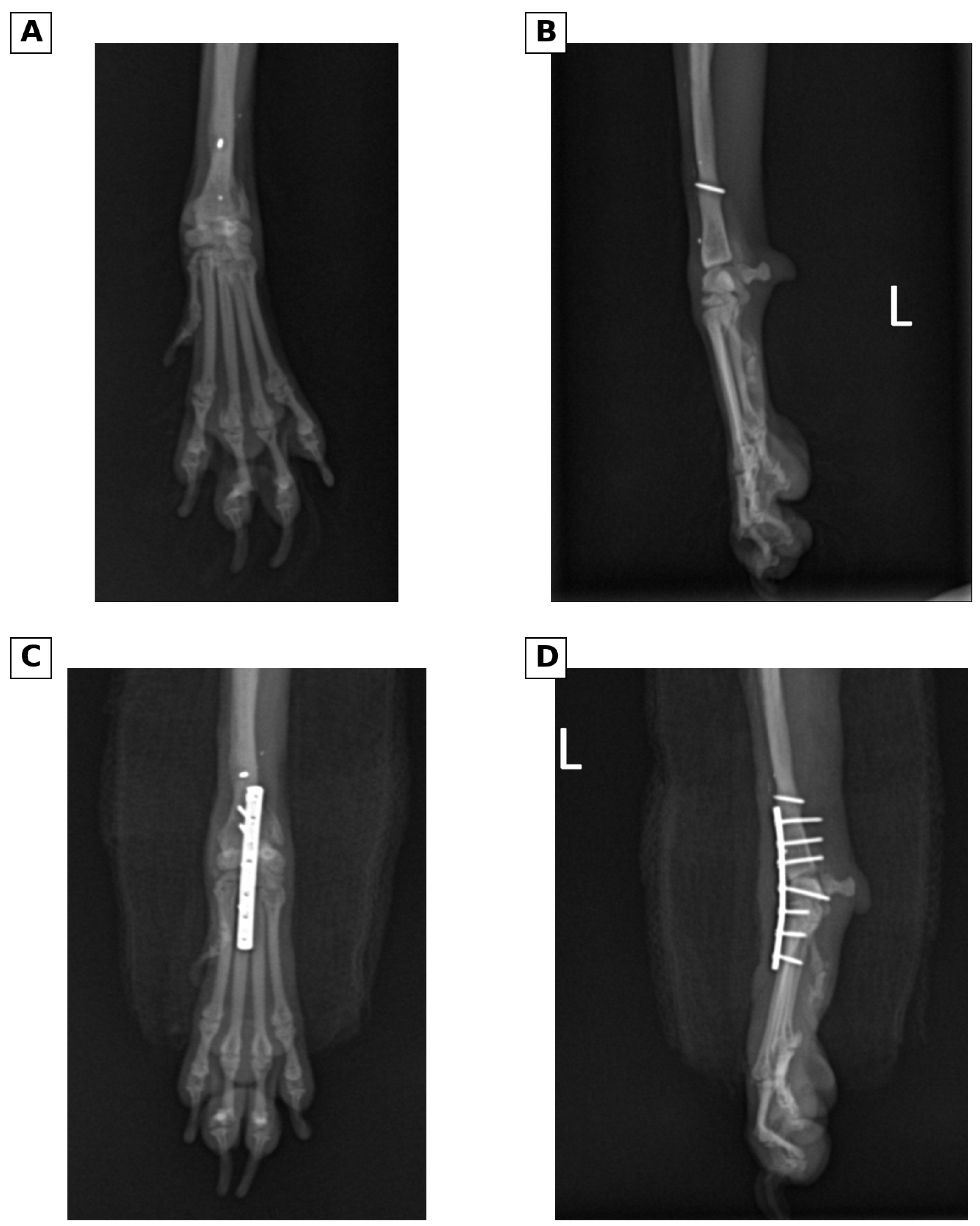
Left pancarpal arthrodesis was performed on 25 July 2016, 68 days after the second implant-removal procedure. The retained screw shaft did not interfere with placement of the definitive arthrodesis plate or screws. A slightly curved skin incision was made over the dorsal aspect of the carpus, extending from the distal radius to the proximal metacarpal region. The abductor pollicis longus tendon was identified and retracted, and the tendon sheaths of the extensor carpi radialis and common digital extensor tendons were opened longitudinally. The tendons were retracted to expose the antebrachiocarpal, middle carpal, and carpometacarpal joints. The joint capsules were incised, and the articular cartilage at the intended arthrodesis surfaces was removed to expose subchondral bone. The prepared joint spaces were thoroughly irrigated. The extent of cartilage removal and grafting status could not be verified from the available surgical documentation; neither autogenous grafting nor bone graft substitute use could be confirmed or excluded. The carpus was reduced and positioned in approximately 10–12° of extension, with care taken to avoid varus, valgus, and rotational malalignment. A 1.5 mm locking plate was contoured and positioned on the dorsal aspect of the distal radius, carpus, and third metacarpal bone. The construct was secured with 1.5 mm locking screws placed into the distal radius and third metacarpal bone, thereby stabilising the carpus across the intended arthrodesis levels. A reliable screw-by-screw inventory, including the count and length of screws in each bone and any carpal screw, could not be reconstructed from the available documentation. Alignment and implant positioning were assessed intraoperatively and confirmed on immediate postoperative orthogonal radiographs. The postoperative arthrodesis angle was approximately 12°. The surgical site was routinely closed in layers (Figure 5).
Figure 5.
Preoperative and immediate postoperative radiographs for left pancarpal arthrodesis. (A,B) Preoperative craniocaudal (CrCd) and mediolateral (ML) views of the left antebrachium and carpus, respectively, after plate removal and before arthrodesis; a distal screw shaft remained intraosseous. (C,D) Immediate postoperative craniocaudal (CrCd) and mediolateral (ML) views, respectively, after left pancarpal arthrodesis. The immediate postoperative arthrodesis angle was approximately 12°. L indicates the left limb.
The dog was hospitalised for 5 days. Postoperative inpatient medications included cefazolin at 22 mg/kg IV three times daily, famotidine at 0.5 mg/kg IV twice daily, carprofen at 2.2 mg/kg SC twice daily, and tramadol at 2 mg/kg IV twice daily. The limb was supported with a postoperative dressing and spica bandage. Serial assessments did not identify a clinically important pressure injury, wound dehiscence, swelling, heat, or purulent discharge.
At discharge on postoperative day 5, the dog was prescribed cephalexin at 22 mg/kg, celecoxib at 2.2 mg/kg, misoprostol at 5 µg/kg, and tramadol at 2 mg/kg, all orally twice daily for 7 days. Skin sutures were removed on postoperative day 12, and external coaptation was maintained until postoperative day 29. Exercise was restricted while external coaptation was maintained. Rehabilitation was undertaken after coaptation removal; the specific modalities, frequency, and duration could not be recovered from the current clinical records.
2.5. Follow-Up
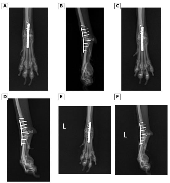
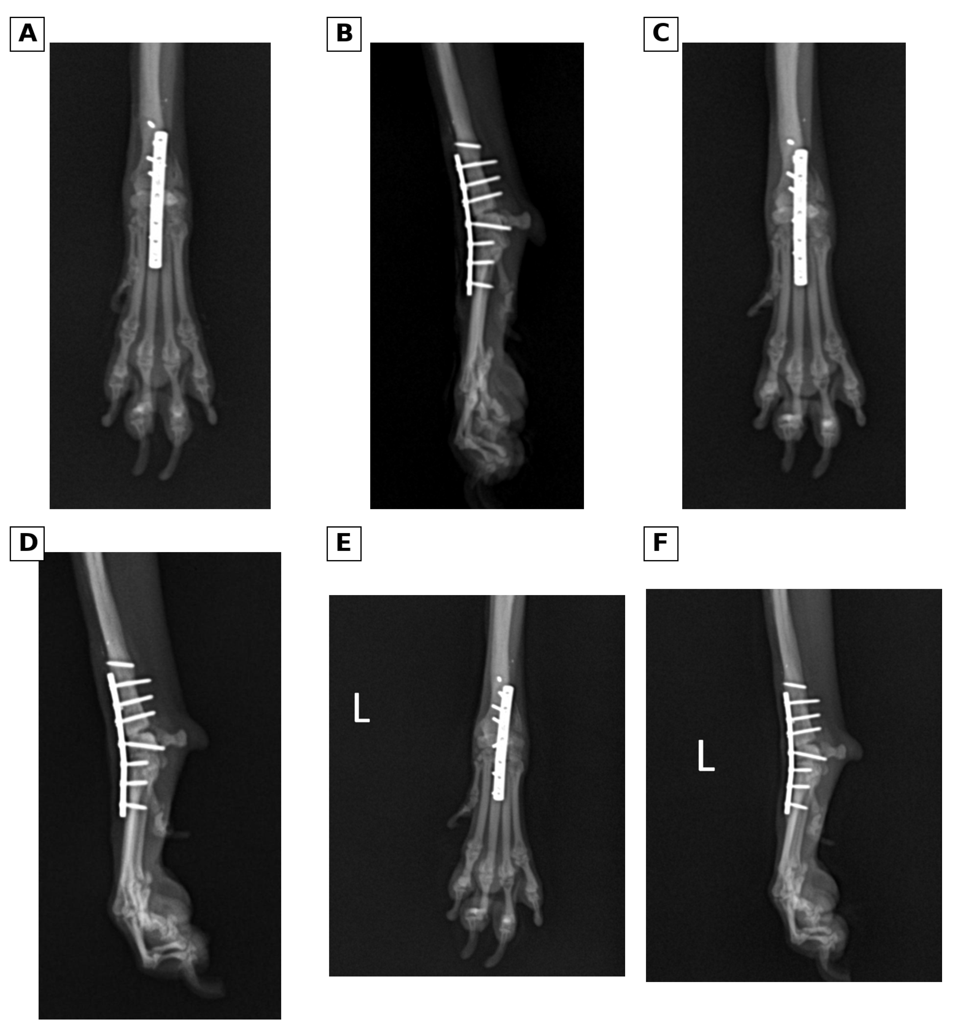
No left thoracic limb lameness was detected on postoperative day 40. At postoperative day 75, the owner reported occasional limb elevation while the dog was standing and intermittent discomfort during manipulation; however, no lameness was observed during walking or trotting (Figure 6). Follow-up assessment was descriptive; numerical pain or lameness scores were not available in the material reviewed.
Figure 6.
Serial postoperative follow-up radiographs obtained on days 29, 40, and 75 after pancarpal arthrodesis. (A,B) Craniocaudal (CrCd) and mediolateral (ML) views, respectively, on postoperative day 29. (C,D) Craniocaudal (CrCd) and mediolateral (ML) views, respectively, on postoperative day 40. (E,F) Craniocaudal (CrCd) and mediolateral (ML) views, respectively, on postoperative day 75. L indicates the left limb.
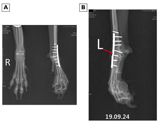
On 24 September 2019, approximately 38 months after arthrodesis, plate fracture was identified radiographically (Figure 7). The available radiographs showed maintained carpal alignment despite implant discontinuity; the clinical assessment recorded apparent stability and no clinically relevant gait abnormality or recurrent lameness. These findings supported monitoring without revision fixation or implant removal in this individual dog. No standardised stress assessment, quantified pain or stability score, or predefined radiographic threshold for observation was available. On 29 July 2020, approximately 48 months after arthrodesis, the dog remained free of recurrent left thoracic limb lameness. The date of fracture detection does not establish when the plate actually fractured.
Figure 7.
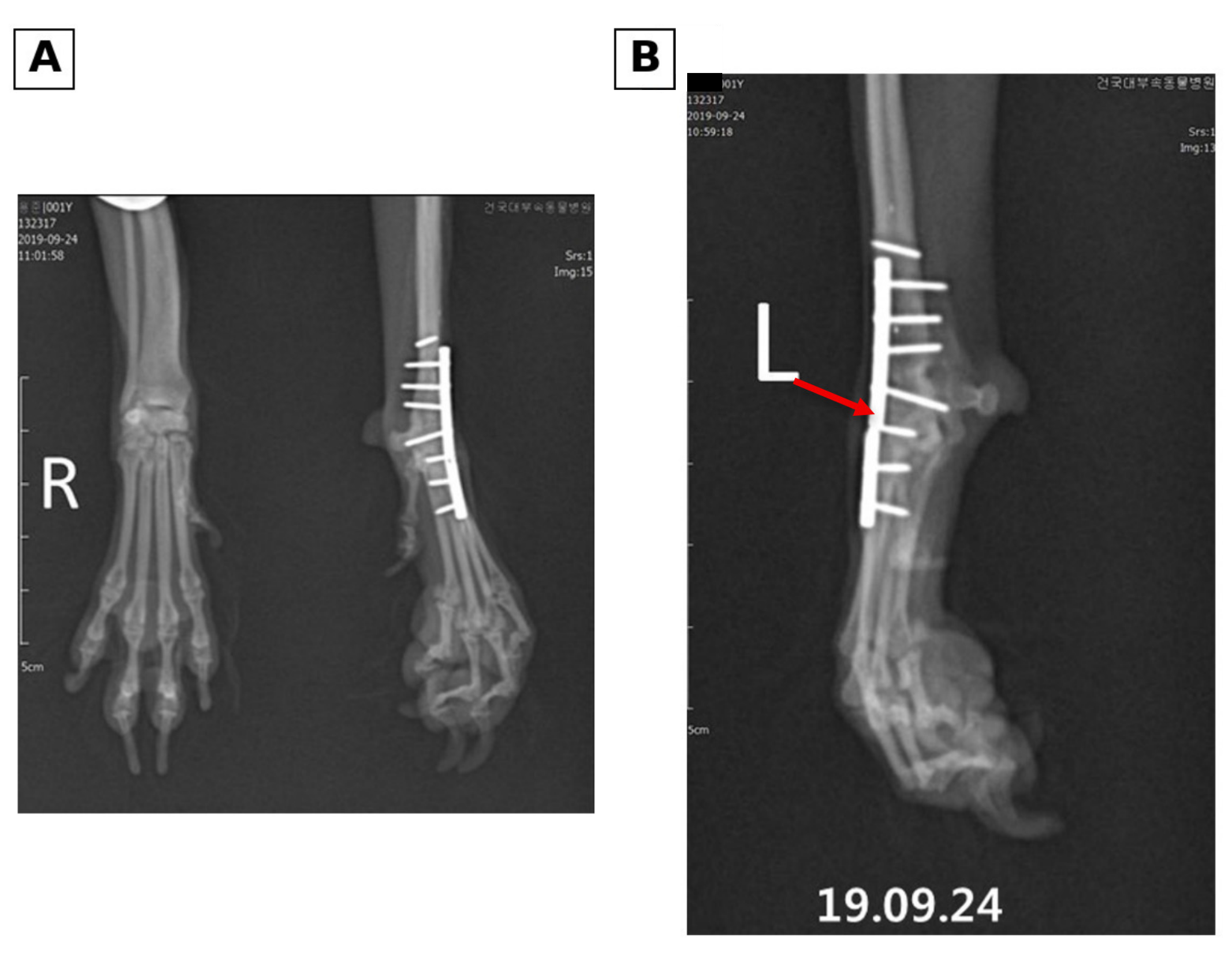
Long-term radiographs demonstrating late plate fracture identified on 24 September 2019. (A) Long-term craniocaudal (CrCd) and oblique/mediolateral (ML) follow-up views. (B) A mediolateral (ML) view from the same date. The red arrow in (B) indicates the plate step-off/fracture region around the third screw from the distal end. Despite implant breakage, carpal alignment and clinical limb function remained acceptable. R and L indicate the right and left limbs, respectively.
Fusion status remained incompletely characterised. No level-specific osseous-bridging assessment was documented for the antebrachiocarpal, middle carpal, or carpometacarpal articulations, and the available records and images did not establish complete osseous fusion at each intended level. No CT confirmation of fusion was available. Maintained alignment and clinical function are therefore reported separately from unconfirmed arthrodesis consolidation; the material also did not establish nonunion at a particular level.
3. Discussion
This case links diagnostic localisation of delayed carpal instability with management of pre-existing fixation and approximately 4 years of clinical follow-up. The documented dorsopalmar and mediolateral stress fluoroscopic views demonstrated abnormal motion between the distal antebrachium and proximal carpal row, complementing static radiographic incongruity and the separate passive ROM measurements. Together with symptomatic hyperextension, this identified an unstable antebrachiocarpal articulation that partial carpal arthrodesis would not stabilise. Pancarpal arthrodesis was therefore selected, consistent with reported indications [1,2,3,4,5,6]. This informed treatment planning for this patient; diagnostic accuracy or superiority over stress radiography was not tested. Fluoroscopic recordings were available, but applied force and stress-induced displacement were not quantified, limiting quantitative comparisons of instability.
New lameness after fracture healing warrants assessment of the adjacent carpal joints as well as the previous fracture site. The history of fractures during skeletal immaturity raised the possibility that residual changes in alignment or load transmission contributed to the later instability. Reports of juvenile growth disturbance describe effects on antebrachial alignment and adjacent-joint congruity [20,21,22], while carpal valgus and varus reports illustrate associations between joint alignment and ligament-related dysfunction [23,24]. In this dog, the original fractures were diaphyseal, no physeal disturbance was documented, and standard radiographs showed no gross angular deformity. Without serial growth-period imaging and a complete interval functional history, the onset and contribution of subtle malalignment, degeneration, or unrecognised intervening injury cannot be resolved. These remain possible explanations rather than established causes. Clinically, the demonstrable antebrachiocarpal instability provided a treatment target even though its precise origin could not be determined.
Cortical changes, screw-hole defects, and clinical refracture risk require separate interpretation. The canine plate studies assessed bone mineral density or radiographic pixel-value ratios (PVR), not the safety of this removal sequence [25,26]. Stress shielding and fixation-related vascular effects may contribute to remodelling, but reduced density was not measured in this patient. A clinical retrospective study associated reduced radial thickness and radiographic density with refracture after plate removal [33]; this supports assessing bone condition, without supplying a patient-specific risk estimate for the present Maltese dog.
Screw holes are discrete geometric defects that can concentrate stress even without diffuse cortical loss. A toy-poodle finite element model identified local stress concentrations [27], whereas rabbit testing compared screw-hole and cortical-atrophy effects [29]; neither design measured clinical refracture incidence in a dog undergoing this staged conversion. Persistent screw-hole defects in seven human patients [30] and a clinical approach combining plate removal with cancellous grafting [28] provide additional context, not interchangeable estimates of canine healing time. Clinical indications for implant removal also vary [34], limiting comparisons between removal cohorts.
The staged strategy retained proximal and distal fixation with the intention of providing temporary support until plate removal 28 days later. Actual stiffness, load sharing, and protection against refracture were not measured. Ferreira et al. described a different staged course after septic nonunion in a 47 kg dog [31]; this shows a related clinical strategy, not evidence of comparative efficacy here. Studies published after the 2016 procedures inform retrospective interpretation rather than the documented contemporary decision. Subsequent arthrodesis was achievable without a perioperative antebrachial fracture, but that observation alone cannot establish safety.
The additional 68-day interval provided time during which bone recovery could occur before reinstrumentation. York and Hutton found that all canine radii tested in torsion five weeks after plate removal failed through former screw holes despite histological bone formation [35]. This supports a rationale for allowing recovery, but does not validate either the 28-day or 68-day interval. Screw-hole filling and bone strength were not measured, and the exact scheduling rationale was not recoverable. Evaluating safety would require serial assessment of cortical condition and hole healing, documented weight bearing, activity restriction and external support, mechanical evaluation of retained fixation, and comparative clinical follow-up with defined refracture outcomes.
The retained distal screw shaft was an intraoperative complication of implant removal, but its intraosseous position and lack of interference with the planned arthrodesis construct allowed definitive fixation to proceed. No associated adverse effect was documented. This aspect of the case highlights the practical importance of evaluating a retained fragment in relation to the new plate position and screw trajectories when planning further fixation.
Functional recovery was evident as the absence of gait lameness by postoperative day 40. At day 75, occasional limb elevation while standing and discomfort during manipulation coexisted with the absence of lameness during walking or trotting, documenting residual positional discomfort alongside improvement in gait. The approximately 48-month follow-up adds a long-term clinical perspective to the generally satisfactory function reported after pancarpal arthrodesis in working dogs [8,9,10], while documenting the course of an individual toy-breed companion dog.
Structured functional assessment would improve comparison across reports. García et al. used the Canine Brief Pain Inventory for long-term follow-up in nine dogs after orthogonal-plate pancarpal arthrodesis [36]. Such owner-reported measures complement gait observation and may capture discomfort during activities that are not apparent during a clinic examination. In this case, the descriptive follow-up supports satisfactory clinical function but cannot be converted retrospectively into a validated pain or disability score.
Late plate fracture was a documented complication, not an uncomplicated endpoint. Plate fracture after carpal arthrodesis has been reported [4], including two of 15 limbs treated with a titanium hybrid locking plate by Chong et al. [37]. A different series using 3.5 mm/2.7 mm locking compression plates with additional internal fixation reported infection and screw loosening without implant failure [17]; construct and population differences preclude direct comparison. In this dog, maintained radiographic alignment, apparent clinical stability, and absence of recurrent gait lameness supported individualised observation, followed by satisfactory function at approximately 48 months. These findings did not confirm fusion at the antebrachiocarpal, middle carpal, or carpometacarpal levels. Persistent implant loading with incomplete fusion and cyclic fatigue are possible explanations for failure, but no level-specific fusion assessment or implant failure analysis established a mechanism. Without such information, the fracture cannot be classified as failure after confirmed complete fusion, nor can nonunion be diagnosed solely from implant breakage. The clinical course supports reporting preserved function during observation, not a general recommendation to leave fractured arthrodesis plates untreated.
Antimicrobial administration included cefazolin during the five-day postoperative hospitalisation and seven days of oral cephalexin prescribed at discharge. Evidence available near the time of treatment was inconsistent: Pratesi et al. reported fewer infections with seven-day postoperative oral therapy [38], whereas Aiken et al. found similar infection rates with and without a postoperative course [39]. Neither study evaluated this dog’s entire regimen, and a patient-specific justification for its duration could not be recovered from the available records. Current ENOVAT guidance recommends against postoperative antimicrobial prophylaxis for clean orthopaedic implant procedures [40]. The regimen is reported as treatment history, without implying that prolonged administration was necessary or should be routinely reproduced.
This retrospective single-case report cannot establish causation or comparative treatment effects. Neither the origin of delayed instability nor a protective effect of staging was demonstrated. The incomplete interval history and growth-period imaging limit reconstruction of disease development. The examination position and goniometric landmarks are described, and fluoroscopic recordings were available. However, unverified sedation or anaesthesia status, unmeasured applied force, absent quantitative alignment and stress-displacement measurements, and incomplete records of joint preparation, grafting, screw distribution, and rehabilitation limit reproducibility and quantitative assessment. Stress-induced displacement was assessed qualitatively. Fusion was not confirmed separately at all intended levels; numerical pain and lameness scores and kinetic testing were unavailable, and radiographic surveillance was intermittent. These limitations qualify the interpretation of late plate fracture while preserving the report’s descriptive clinical contribution.
4. Conclusions
This case documents diagnostic localisation and management of delayed antebrachiocarpal hyperextension instability in a Maltese dog with healed juvenile forearm fractures and retained implants. Pancarpal arthrodesis was followed by satisfactory limb function at approximately 48 months despite a retained screw fragment and late plate fracture; complete osseous fusion at each intended joint level was not established. Staged removal was selected with the intention of preserving temporary support, and its comparative benefit remains unknown. The case illustrates considerations for locating instability, planning around existing implants, and evaluating late implant complications alongside function. It does not establish a generally applicable removal interval, refracture-prevention effect, or therapeutic superiority.
Author Contributions
Conceptualization, M.-S.K., S.-W.K. and H.-Y.K.; methodology, S.-Y.M., S.-W.K. and H.-Y.K.; investigation, S.-Y.M., E.-J.J., Y.-U.C. and J.-M.K.; resources, S.-Y.M., E.-J.J., Y.-U.C., J.-M.K. and K.-D.E.; data curation, S.-Y.M., E.-J.J., Y.-U.C., J.-M.K. and K.-D.E.; writing—original draft preparation, M.-S.K.; writing—review and editing, M.-S.K., S.-W.K. and H.-Y.K.; supervision, S.-W.K. and H.-Y.K.; project administration, M.-S.K., S.-W.K. and H.-Y.K. All authors have read and agreed to the published version of the manuscript.
Funding
This research received no external funding.
Institutional Review Board Statement
Ethical review and approval were waived for this study due to its retrospective description of a single client-owned dog managed as part of routine clinical care at the Konkuk University Veterinary Teaching Hospital.
Informed Consent Statement
Written informed consent was obtained from the owner for the diagnostic and therapeutic procedures and for the publication of this case report and the accompanying images.
Data Availability Statement
The original contributions presented in this study are included in the article. Further inquiries can be directed to the corresponding authors.
Conflicts of Interest
The authors declare no conflicts of interest.
Abbreviations
| CrCd | Craniocaudal |
| IV | Intravenous |
| ML | Mediolateral |
| PVR | Pixel-value ratio |
| ROM | Range of motion |
| SC | Subcutaneous |
References
- Beierer, L.H. Canine carpal injuries: From fractures to hyperextension injuries. Vet. Clin. N. Am. Small Anim. Pract. 2021, 51, 285–303. [Google Scholar]
- Choi, C.J.; Balara, J.M.; Casale, S.A.; Wendelburg, K.L. Implant removal rate after partial carpal arthrodesis in dogs: A retrospective analysis of 22 cases. Front. Vet. Sci. 2023, 10, 1160129. [Google Scholar] [CrossRef] [Scilit] [PubMed]
- Denny, H.R.; Barr, A.R.S. Partial carpal and pancarpal arthrodesis in the dog: A review of 50 cases. J. Small Anim. Pract. 1991, 32, 329–334. [Google Scholar] [CrossRef] [Scilit]
- Johnson, K.A. Carpal arthrodesis in dogs. Aust. Vet. J. 1980, 56, 565–573. [Google Scholar] [CrossRef] [Scilit]
- Parker, R.B.; Brown, S.G.; Wind, A.P. Pancarpal arthrodesis in the dog: A review of forty-five cases. Vet. Surg. 1981, 10, 35–43. [Google Scholar] [CrossRef] [Scilit]
- Willer, R.L.; Johnson, K.A.; Turner, T.M.; Piermattei, D.L. Partial carpal arthrodesis for third degree carpal sprains: A review of 45 carpi. Vet. Surg. 1990, 19, 334–340. [Google Scholar] [CrossRef] [Scilit] [PubMed]
- Hayashi, K. (Ed.) Diagnosis of Lameness in Dogs; Wiley: Hoboken, NJ, USA, 2023; pp. 97–98. [Google Scholar] [CrossRef] [Scilit]
- Higgins, J.; Hayes, G. Owner satisfaction and prognosis for return to work after pancarpal arthrodesis in working dogs in the United Kingdom: A retrospective study (2011–2020). Acta Vet. Scand. 2024, 66, 49. [Google Scholar] [CrossRef] [Scilit] [PubMed]
- Jerram, R.M.; Walker, A.M.; Worth, A.J.; Kuipers von Lande, R.G. Prospective evaluation of pancarpal arthrodesis for carpal injuries in working dogs in New Zealand, using dorsal hybrid plating. N. Z. Vet. J. 2009, 57, 331–337. [Google Scholar] [CrossRef] [Scilit] [PubMed]
- Worth, A.J.; Bruce, W.J. Long-term assessment of pancarpal arthrodesis performed on working dogs in New Zealand. N. Z. Vet. J. 2008, 56, 78–84. [Google Scholar] [CrossRef] [Scilit] [PubMed]
- Bristow, P.C.; Meeson, R.L.; Thorne, R.M.; Butterworth, S.J.; Rutherford, S.; Renwick, A.I.; Wustefeld-Janssens, B.; Witte, P.G.; Woods, S.; Parsons, K.J.; et al. Clinical comparison of the hybrid dynamic compression plate and the CastLess plate for pancarpal arthrodesis in 219 dogs. Vet. Surg. 2015, 44, 70–77. [Google Scholar] [CrossRef] [Scilit] [PubMed]
- Clarke, S.P.; Ferguson, J.F.; Miller, A. Clinical evaluation of pancarpal arthrodesis using a CastLess plate in 11 dogs. Vet. Surg. 2009, 38, 852–860. [Google Scholar] [CrossRef] [Scilit] [PubMed]
- Dosseray, H.; Camilletti, P.; Elbaz, L.S.; Hanot, E.; Ragetly, G.; Pucheu, B.; Boland, L.; Minier, K. Implant removal rate and contributing factors following pancarpal arthrodesis in 42 dogs (52 cases): A multicentric retrospective study. Acta Vet. Scand. 2025, 67, 44. [Google Scholar] [CrossRef] [Scilit] [PubMed]
- Guerrero, T.G.; Montavon, P.M. Medial plating for carpal panarthrodesis. Vet. Surg. 2005, 34, 153–158. [Google Scholar] [CrossRef] [Scilit] [PubMed]
- Li, A.; Gibson, N.; Carmichael, S.; Bennett, D. Thirteen pancarpal arthrodeses using 2.7/3.5 mm hybrid dynamic compression plates. Vet. Comp. Orthop. Traumatol. 1999, 12, 102–107. [Google Scholar] [CrossRef] [Scilit]
- Ramirez, J.M.; Macias, C. Pancarpal arthrodesis without rigid coaptation using the hybrid dynamic compression plate in dogs. Vet. Surg. 2016, 45, 303–308. [Google Scholar] [CrossRef] [Scilit] [PubMed]
- Tuan, J.; Comas, N.; Solano, M. Clinical outcomes and complications of pancarpal arthrodesis stabilised with 3.5 mm/2.7 mm locking compression plates with internal additional fixation in 12 dogs. N. Z. Vet. J. 2019, 67, 270–276. [Google Scholar] [CrossRef] [Scilit] [PubMed]
- Díaz-Bertrana, C.; Darnaculleta, F.; Durall, I.; Franch, J.; Puchol, J.L.; Martinez, J.J.; Rubio, A. The stepped hybrid plate for carpal panarthrodesis—Part II: A multicentre study of 52 arthrodeses. Vet. Comp. Orthop. Traumatol. 2009, 22, 389–397. [Google Scholar] [PubMed]
- Whitelock, R.G.; Dyce, J.; Houlton, J.E.F. Metacarpal fractures associated with pancarpal arthrodesis in dogs. Vet. Surg. 1999, 28, 25–30. [Google Scholar] [CrossRef] [Scilit] [PubMed]
- Christopher, S. Short term outcomes and complications of distal ulnar ostectomy in 23 juvenile dogs with carpal valgus secondary to discordant radial-ulnar physeal growth. Front. Vet. Sci. 2022, 9, 971527. [Google Scholar] [CrossRef] [Scilit] [PubMed]
- O’Brien, T.R.; Morgan, J.P.; Suter, P.F. Epiphyseal plate injury in the dog: A radiographic study of growth disturbance in the forelimb. J. Small Anim. Pract. 1971, 12, 19–36. [Google Scholar] [CrossRef] [Scilit] [PubMed]
- Ramadan, R.O.; Vaughan, L.C. Premature closure of the distal ulnar growth plate in dogs: A review of 58 cases. J. Small Anim. Pract. 1978, 19, 647–667. [Google Scholar] [CrossRef] [Scilit] [PubMed]
- Comerford, E.J.; Doran, I.C.; Owen, M.R. Carpal derangement and associated carpal valgus in a dog. Vet. Comp. Orthop. Traumatol. 2006, 19, 113–116. [Google Scholar] [CrossRef] [Scilit]
- Langley-Hobbs, S.J.; Hamilton, M.H.; Pratt, J.N.J. Radiographic and clinical features of carpal varus associated with chronic sprain of the lateral collateral ligament complex in 10 dogs. Vet. Comp. Orthop. Traumatol. 2007, 20, 324–330. [Google Scholar] [CrossRef] [Scilit] [PubMed]
- Muroi, N.; Ochi, H.; Shimada, M.; Asou, Y.; Hara, Y. Effects of long-term plate fixation with different fixation modes on the radial cortical bone in dogs. PLoS ONE 2021, 16, e0247410. [Google Scholar] [CrossRef] [Scilit] [PubMed]
- Muroi, N.; Shimada, M.; Murakami, S.; Akagi, H.; Kanno, N.; Suzuki, S.; Harada, Y.; Orima, H.; Hara, Y. A retrospective study of postoperative development of implant-induced osteoporosis in radial-ulnar fractures in toy breed dogs treated with plate fixation. Vet. Comp. Orthop. Traumatol. 2021, 34, 375–385. [Google Scholar] [CrossRef] [Scilit] [PubMed]
- Anggoro, D.; Purba, M.S.; Jiang, F.; Nishida, N.; Itoh, H.; Itamoto, K.; Nemoto, Y.; Nakaichi, M.; Sunahara, H.; Tani, K. Finite element study on post-screw removal stress in toy poodle radius with different plate designs and screw arrangements. Open Vet. J. 2024, 14, 885–894. [Google Scholar] [CrossRef] [Scilit] [PubMed]
- Lesser, A.S. Cancellous bone grafting at plate removal to counteract stress protection. J. Am. Vet. Med. Assoc. 1986, 189, 696–699. [Google Scholar] [CrossRef] [Scilit]
- Rosson, J.; Egan, J.; Shearer, J.; Monro, P. Bone weakness after the removal of plates and screws: Cortical atrophy or screw holes? J. Bone Jt. Surg. Br. 1991, 73, 283–286. [Google Scholar] [CrossRef] [Scilit] [PubMed]
- Rosson, J.; Murphy, W.; Tonge, C.; Shearer, J. Healing of residual screw holes after plate removal. Injury 1991, 22, 383–384. [Google Scholar] [CrossRef] [Scilit] [PubMed]
- Ferreira, D.R.C.; Montanhim, G.L.; Sá, M.A.R.; Diogo, L.M.I.; Minto, B.W.; Lucena, D.V.F.; Moraes, P.C.; Dias, L.G.G.G. Septic nonunion in radius and ulna in a dog: Treatment with orthogonal plating associated with corticospongious bone autograft. Acta Sci. Vet. 2025, 53, 1022. [Google Scholar] [CrossRef] [Scilit]
- Duerr, F.M.; Elam, L.H. (Eds.) Canine Lameness, 2nd ed.; Wiley: Hoboken, NJ, USA, 2026; pp. 77–82. [Google Scholar] [CrossRef] [Scilit]
- Muroi, N.; Kanno, N.; Harada, Y.; Hara, Y. A retrospective study of risk factors associated with refracture after repair of radial-ulnar fractures in small-breed dogs. Vet. Comp. Orthop. Traumatol. 2025, 38, 77–86. [Google Scholar] [CrossRef] [Scilit] [PubMed]
- Emmerson, T.D.; Muir, P. Bone plate removal in dogs and cats. Vet. Comp. Orthop. Traumatol. 1999, 12, 74–77. [Google Scholar] [CrossRef] [Scilit]
- York, M.J.; Hutton, W.C. Treatment of screw hole defects using bone graft materials: A histologic and biomechanic study. J. South. Orthop. Assoc. 1996, 5, 5–12. [Google Scholar] [PubMed]
- García, A.R.; Brincin, C.; Craig, A. Outcome, Complications, and Follow-Up in Dogs Treated with Pancarpal Arthrodesis Stabilized with Orthogonal Plates. J. Am. Anim. Hosp. Assoc. 2024, 60, 252–264. [Google Scholar] [CrossRef] [Scilit] [PubMed]
- Chong, W.L.; Eckart, L.H.; Sul, R.; Jermyn, K.; Michelsen, J. Retrospective comparison of titanium hybrid locking plate with stainless steel hybrid dynamic compression plate for pancarpal arthrodesis: 23 dogs. Vet. Comp. Orthop. Traumatol. 2022, 35, 270–278. [Google Scholar] [CrossRef] [Scilit] [PubMed]
- Pratesi, A.; Moores, A.P.; Downes, C.; Grierson, J.; Maddox, T.W. Efficacy of Postoperative Antimicrobial Use for Clean Orthopedic Implant Surgery in Dogs: A Prospective Randomized Study in 100 Consecutive Cases. Vet. Surg. 2015, 44, 653–660. [Google Scholar] [CrossRef] [Scilit] [PubMed]
- Aiken, M.J.; Hughes, T.K.; Abercromby, R.H.; Holmes, M.A.; Anderson, A.A. Prospective, Randomized Comparison of the Effect of Two Antimicrobial Regimes on Surgical Site Infection Rate in Dogs Undergoing Orthopedic Implant Surgery. Vet. Surg. 2015, 44, 661–667. [Google Scholar] [CrossRef] [Scilit] [PubMed]
- Allerton, F.; Sørensen, T.M.; Scahill, K.; Ruperez, J.E.; Swinbourne, F.; Verwilghen, D.R.; Nolff, M.C.; Foroutan, F.; Baines, S.J.; Vilen, A.; et al. European Network for Optimization of Veterinary Antimicrobial Therapy (ENOVAT) 2025 guidelines for surgical antimicrobial prophylaxis in dogs and cats. J. Small Anim. Pract. 2026, 67, 383–405. [Google Scholar] [CrossRef] [Scilit] [PubMed]

Disclaimer/Publisher’s Note: The statements, opinions and data contained in all publications are solely those of the individual author(s) and contributor(s) and not of MDPI and/or the editor(s). MDPI and/or the editor(s) disclaim responsibility for any injury to people or property resulting from any ideas, methods, instructions or products referred to in the content. |
© 2026 by the authors. Licensee MDPI, Basel, Switzerland. This article is an open access article distributed under the terms and conditions of the Creative Commons Attribution (CC BY) license.