


Simple Summary
Ovarian hydrobursitis is a serious reproductive disorder that affects female dromedary camels, resulting in infertility, pregnancy loss, abortion, and culling. This review summarizes more than a decade of research into the occurrence, potential causes, diagnosis, treatment, and prevention of this condition. The distinct anatomy of the camel ovary appears to predispose females to fluid accumulation and progressive enlargement of the ovarian bursa. Several lines of evidence suggest a strong link between Chlamydia abortus (C. abortus) and ovarian hydrobursitis, though definitive causation has yet to be established. Ultrasound examination is an important diagnostic tool, and laboratory tests can reveal additional evidence of infection and inflammation. Treatment effectiveness is limited, especially in females with bilateral or advanced disease, who frequently have a poor reproductive prognosis. As a result, prevention is likely to be the most sustainable strategy for reducing disease-related reproductive losses. More research is needed to determine the role of C. abortus in disease development, as well as to develop effective preventive strategies for improving reproductive efficiency and camel sustainability.
Abstract
Ovarian hydrobursitis (OVHB) is a serious reproductive disorder that has been described predominantly in dromedary camels, characterized by fluid accumulation within the ovarian bursa and complete encapsulation of the ovary. Among female camels presented for reproductive examination, OVHB is a frequent diagnosis and has been associated with infertility, embryonic loss, abortion, and culling; however, these estimates derive from selected populations, and true population prevalence and attributable impact remain unknown. This review summarizes more than a decade of investigations into the epidemiology, pathogenesis, diagnosis, treatment, and prevention of OVHB. Among female camels presented for reproductive examination, OVHB accounts for approximately 16.5–33.8% of infertility cases, while slaughterhouse surveys report a prevalence of 4.8–6.5% in slaughtered females. These figures derive from different study populations and should not be interpreted as directly comparable estimates of population prevalence. The unique anatomy of the camel ovary, which is completely enclosed within a fused mesosalpinx, mesovarium, and proper ligament, appears to predispose affected animals to fluid retention within the bursa. Multiple lines of Serological, molecular, and microbiological evidence strongly implicate Chlamydia abortus (C. abortus) as the principal pathogen associated with OVHB; however, definitive causation remains unproven pending experimental infection studies. However, experimental reproduction of the disease has not yet been achieved, and definitive causality remains unconfirmed. Current treatment options are limited. Surgical ablation may restore fertility in selected unilateral cases, whereas medical therapy is effective only in small bursae. Bilateral and advanced cases generally have a poor prognosis and are commonly culled. Consequently, prevention is likely to represent the most sustainable long-term control strategy. Ongoing investigations of camel-derived C. abortus isolates are improving our understanding of pathogen diversity and may support the future development of preventive interventions. Further studies are required to clarify disease causation and establish effective control measures.
1. Introduction
Ovarian hydrobursitis (OVHB) is a serious, long-standing reproductive disorder that causes major economic losses in dromedary camel herds worldwide. The condition has been reported mainly in the dromedary, a species in which the ovary is completely enclosed within the ovarian bursa. While this anatomical feature is not exclusive to camels, it is particularly pronounced in this species and appears to predispose it to OVHB [1,2,3,4]. This anatomical feature, while normally protective, becomes a predisposing factor when inflammation or infection occurs: fluid accumulates inside the bursa, the ovary becomes encapsulated, and the animal develops persistent infertility [5,6,7,8,9,10,11,12].
The syndrome accounts for 16.5–33.8% of infertile female camels presented to veterinary clinics. In unilateral cases, conception may still occur through the unaffected bursa; however, when associated with Chlamydia abortus (formerly classified as Chlamydophila abortus and, before that, as Chlamydia psittaci serotype 1; the genus was historically known as Bedsonia) infection during pregnancy, embryonic loss or abortion may occur [6,8,13,14]. In some herds, multiple animals are affected simultaneously, suggesting an infectious or venereal aetiology [8,14,15].
The exact pathogenesis of OVHB has long remained unclear; however, accumulating serological, microbiological, and molecular evidence suggests that Chlamydia abortus (C. abortus; formerly Chlamydophila abortus and previously classified as Chlamydia psittaci serotype 1) is the principal pathogen associated with disease development. The current nomenclature (Chlamydia abortus) is used throughout this review; older names appear in some cited references and are retained in reference titles as originally published. Serological surveys have detected anti-C. abortus antibodies in 86.3% of affected camels, and the organism has been successfully isolated from both bursal fluid and bursal tissue of clinical cases [13,14,15]. Other bacteria (e.g., Trueperella pyogenes, Escherichia coli, Staphylococcus aureus) and protozoa (Trypanosoma evansi) have also been isolated from the bursal contents, but they appear to act as secondary or opportunistic pathogens rather than primary initiators [5,8]. Venereal transmission of C. abortus is considered the most plausible route of spread within camel herds. This hypothesis is supported by the occurrence of chlamydial infection in nulliparous females, the presence of multiple affected animals within the same herd, and the detection of Chlamydia in the genital tracts of male camels [16]. Under this model, venereal transmission of C. abortus leads to chlamydial infection of the reproductive tract, which may in turn cause bursitis and secondary fluid accumulation within the ovarian bursa (i.e., OVHB). It is important to emphasize that OVHB is not itself a transmissible disease; what may be transmitted is the infectious agent. C. abortus may contribute to the development of OVHB, but its precise causal role remains to be established.
Treatment options remain limited. Unilateral surgical ablation of the affected bursa and ovary can achieve conception in appropriately selected cases, but this approach is not feasible for bilateral disease [6,13,17]. A medical protocol combining oxytetracycline, intrauterine lotagen and cloprostenol has shown some success only when the bursa is small (<5 cm in diameter); medium and large bursae do not respond [13,14]. Consequently, many affected animals are culled, representing a substantial reproductive and economic loss. Given the failure of treatment in advanced cases and the possible contribution of infectious agents, prevention, including vaccination, represents a promising research direction that requires validation before it can be considered an established control measure [18,19,20].
The objective of this review is to synthesize current knowledge on ovarian hydrobursitis in dromedary camels, with particular emphasis on its epidemiology, pathogenesis, etiology, clinical presentation, diagnosis, treatment, and prevention. A further aim is to critically appraise the strength of the available evidence, to distinguish established findings from hypotheses, and to identify future research directions, particularly regarding the development of a vaccine against chlamydial reproductive disease in this species.
2. Review Methodology
This article is a narrative (non-systematic) review, prepared in accordance with the principles of transparent reporting for narrative reviews (SANRA guidelines; [21]). Given the rarity and a particularly pronounced anatomical feature of ovarian hydrobursitis in dromedary camels, the available literature is limited, heterogeneous, and derived predominantly from a small number of research groups and geographic regions. A formal systematic review with meta-analysis was therefore not feasible.
A comprehensive literature search was conducted in PubMed/MEDLINE, Scopus, Web of Science, Google Scholar, and the institutional repository of Qassim University. The search covered the period from 1987 (the year of the earliest detailed anatomical description of the camel ovarian bursa) to March 2026. The following search terms and Boolean strings were used: (“ovarian hydrobursitis” OR “ovario-bursal pathology” OR “bursal cyst” OR “ovarian bursa” AND “camel” OR “dromedary” OR “Camelus dromedarius”); (“Chlamydia abortus” OR “Chlamydophila abortus” AND “camel” OR “infertility” OR “reproductive disorder”); and (“camel” AND “reproductive pathology” AND “ultrasonography” OR “treatment” OR “vaccine”).
Peer-reviewed original research articles, case reports, case series, and reviews published in English were included if they reported on OVHB in dromedary camels or on C. abortus infection in camels or relevant comparative species (sheep, goats, koalas, mice, humans). Non-peer-reviewed literature and studies not reporting reproductive outcomes were excluded.
Titles and abstracts were screened independently by two authors (A.A. and D.R.D.). Full texts of potentially relevant articles were then reviewed against the inclusion criteria. Disagreements were resolved by consensus. Reference lists of all retrieved articles were additionally screened (snowballing) to identify further relevant studies.
Because of the paucity of controlled experimental studies and the predominance of observational and clinical reports, a formal GRADE or risk-of-bias assessment was not feasible. Instead, evidence was appraised narratively, with emphasis on consistency across independent studies, biological plausibility, strength of association, and the availability of confirmatory laboratory evidence (e.g., isolation, molecular detection, histopathology). Where evidence was limited or conflicting, this is explicitly acknowledged in the text. Throughout the review, we distinguish between findings that have been independently replicated, findings primarily reported by our own group, and hypotheses derived from these observations.
The studies included in this review are categorized in Table 1 as (A) direct OVHB studies, (B) broader reproductive pathology studies reporting OVHB or related lesions, and (C) conference proceedings. For each study, the country, number and type of animals examined, and main OVHB-relevant findings are indicated. Much of the available literature originates from a limited number of research groups and geographic regions, which may limit the generalizability of the conclusions. These limitations should be considered when interpreting the findings.
Table 1.
Categorization of studies included in this review, showing study type, country of origin, number and type of animals examined, study design, primary diagnostic method(s), major findings relevant to ovarian hydrobursitis, and major limitations.
3. Epidemiology and Prevalence
OVHB is consistently reported as a major cause of infertility in dromedary camels across different geographic regions and production systems. However, the available prevalence estimates derive from fundamentally different study populations, and it is essential to distinguish between them.
As noted in the Introduction, OVHB is a frequent finding among infertile female camels presented for reproductive examination at referral clinics. However, this referral-based estimate is subject to substantial selection bias, because only animals with reproductive failure are examined. It therefore represents the proportion of infertile females diagnosed with OVHB, not the proportion of all female camels affected by the condition.
In abattoir surveys of female camels slaughtered for meat production, the prevalence of OVHB at post-mortem examination ranges from 4.8% to 6.5% [7,8]. This population includes animals culled for various reasons, not only infertility, and may therefore better reflect the general population than referral-based figures. However, slaughterhouse surveys are also biased: severely affected animals with advanced disease are often removed from breeding herds and may not reach slaughter, potentially leading to underestimation.
The referral-based and slaughterhouse-based prevalence estimates describe different populations and cannot be directly compared or combined. Neither provides a true estimate of OVHB prevalence in the general camel population. The true population prevalence remains unknown.
Referral populations overestimate prevalence because they are selected for infertility. Slaughterhouse populations may underestimate prevalence because affected animals are often culled before slaughter. Neither study design captures the full spectrum of disease, including subclinical or early-stage cases that may not yet show bursal distension (see below).
An important consideration is the potential existence of an early stage of chlamydial infection in which OVHB remains clinically and ultrasonographically undetectable. During this phase, C. abortus may already be colonizing the ovarian bursa and adjacent reproductive tissues, initiating inflammatory and fibrotic changes before sufficient fluid accumulation occurs to allow diagnosis as OVHB. Two distinct but potentially overlapping phenomena should be considered when interpreting the absence of detectable bursal distension in C. abortus-infected camels: the prepatent period and a subclinical course of infection. The prepatent period refers to the interval between infection and the point at which bursal fluid accumulation becomes sufficient to be visualized ultrasonographically; during this phase, infection is present but OVHB is not yet detectable. A subclinical course refers to the presence of infection or early pathological change without recognizable clinical signs, which may persist without progression or may eventually evolve into clinically apparent disease. Because ultrasonographic diagnosis of OVHB depends on the detection of bursal fluid, animals in either the prepatent or subclinical phase may be misclassified as unaffected. Consequently, reliance solely on clinically apparent cases is likely to underestimate the true prevalence and impact of chlamydial infection on camel fertility and reproductive performance. Longitudinal studies with repeated ultrasonographic and molecular examinations are needed to distinguish these phases and to define the earliest detectable stage of OVHB.
Large-scale, population-based field studies using standardized diagnostic criteria are needed to accurately determine the true prevalence of OVHB and its associated risk factors under commercial production conditions. Such studies should include both clinically affected and subclinical cases and should be conducted across multiple geographic regions to account for potential variation in disease frequency.
4. Anatomy, Pathogenesis, and Predisposition
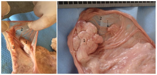
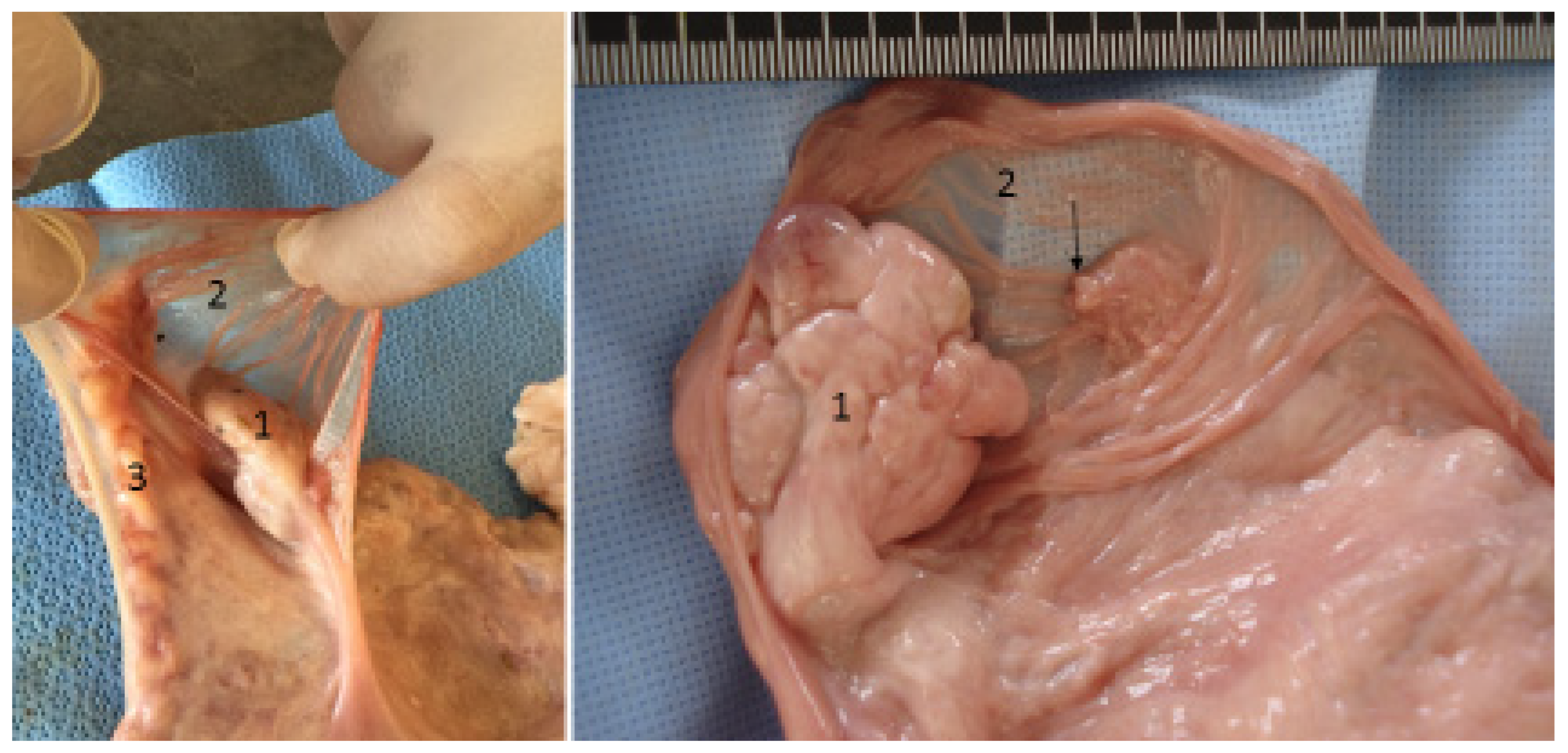
The functional significance of the ovarian bursa in camels is not fully understood, but it may provide protection and facilitate ovum capture by the fimbriae. The complete encapsulation of the ovary by the ovarian bursa is a predisposing anatomical factor for ovarian hydrobursitis, particularly when adhesions develop and the bursal orifice becomes closed (Figure 1). Fluid that accumulates within the bursa cannot easily escape, leading to progressive distension, ovarian encapsulation, and persistent infertility [7,9,10,11,12,17]. A possible relationship between recurrent anovulatory hemorrhagic follicles and bursal adhesions has been suggested [17].
Figure 1.
The anatomical relationship between the ovary (1), ovarian bursa (2), and fallopian tube (3) in camels: the ovarian bursa completely envelops the ovary, providing protection and facilitating the passage of oocytes to the ostium abdominalis, which is located at the base of the ovarian bursa (arrow).
The exact pathogenesis of OVHB has long remained unclear. Accumulating serological, microbiological, and molecular evidence strongly associates C. abortus with the condition; however, whether the organism acts as a primary initiating cause, a contributing factor, a secondary consequence of altered bursal anatomy, or one component of a multifactorial process remains unresolved. Ascending infection from the lower genital tract to the upper reproductive tract is a well-documented route for Chlamydia trachomatis in women [31,32] and for Chlamydia species in koalas and mice [33,34]. By analogy, it has been proposed that C. abortus may ascend from the lower genital tract to the uterus, oviduct, and ultimately the ovarian bursa in camels. However, direct evidence demonstrating this route in camels is currently lacking, and the organism has not been tracked sequentially from the vagina to the bursa in experimental studies.
OVHB is a serious reproductive disorder that has been described predominantly in dromedary camels. However, it should be noted that equivalent pathology has not been systematically investigated in other camelids (Bactrian camels, llamas, alpacas) or in other domestic species. The absence of reported cases in these species does not constitute evidence that the condition does not occur; it may simply reflect underdiagnosis or lack of systematic study. A similar syndrome has been described in koalas, which also possess a well-developed ovarian bursa and are susceptible to chlamydial infection, suggesting that comparable pathology can occur in other species [8,11,17]. It has been hypothesized that progressive fluid accumulation within the enclosed bursa may increase intrabursal pressure, potentially compressing the ovarian vasculature and leading to ischemia. This mechanism is biologically plausible, given the confined nature of the bursal space, and is consistent with the histopathological finding of hemosiderin-laden macrophages and capillary congestion observed in chronic cases [6,22]. However, direct measurements of intrabursal pressure or ovarian blood flow in affected camels have not been performed, and this mechanism therefore remains proposed rather than proven. Histopathological examination of chronically affected bursae has documented epithelial degeneration, vacuolation of lining cells, mononuclear cell infiltration, and hyperplasia [6]. These findings are consistent with chronic inflammation and tissue remodeling. It has been proposed that sustained distension and inflammation may lead to fibrosis and loss of ciliated epithelium, which could impair normal ovum transport and contribute to infertility. However, the sequence of events—whether fibrosis precedes or follows fluid accumulation—has not been established through longitudinal studies, and the extent of ciliated epithelium loss has not been quantified [6,35]. A similar syndrome has been described in koalas (Phascolarctos cinereus), which also possess a well-developed ovarian bursa and are susceptible to chlamydial infection [35,36,37,38]. However, this comparison should be regarded as a biological analogy rather than as supporting evidence for causality in camels. The chlamydial species involved in koala disease differ from C. abortus, and host–pathogen interactions are likely to differ between the two species.
It is important to distinguish between pathological changes that have been directly documented in affected camels and mechanistic steps that remain hypothetical. Documented findings include: (1) complete ovarian encapsulation within the bursa [1,4]; (2) accumulation of fluid of variable echogenicity within the bursa [22]; (3) histopathological changes including mononuclear cell infiltration, lymphoid aggregation, epithelial degeneration, hyperplasia, and hemosiderin-laden macrophages [6,35]; and (4) the presence of C. abortus in bursal tissue and fluid [15]. Proposed but unproven mechanisms include: (1) ascending infection from the lower genital tract to the bursa; (2) vascular compression and ischemia due to increased intrabursal pressure; (3) progression from chronic distension to fibrosis and ciliated epithelium loss; and (4) impairment of drug penetration by adhesions and fibrosis. These proposed mechanisms are biologically plausible and consistent with comparative evidence from other species, but they have not been directly demonstrated in camels (Table 2).
Table 2.
Level of evidence for proposed mechanistic steps in OVHB pathogenesis.
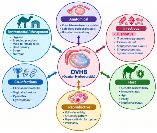
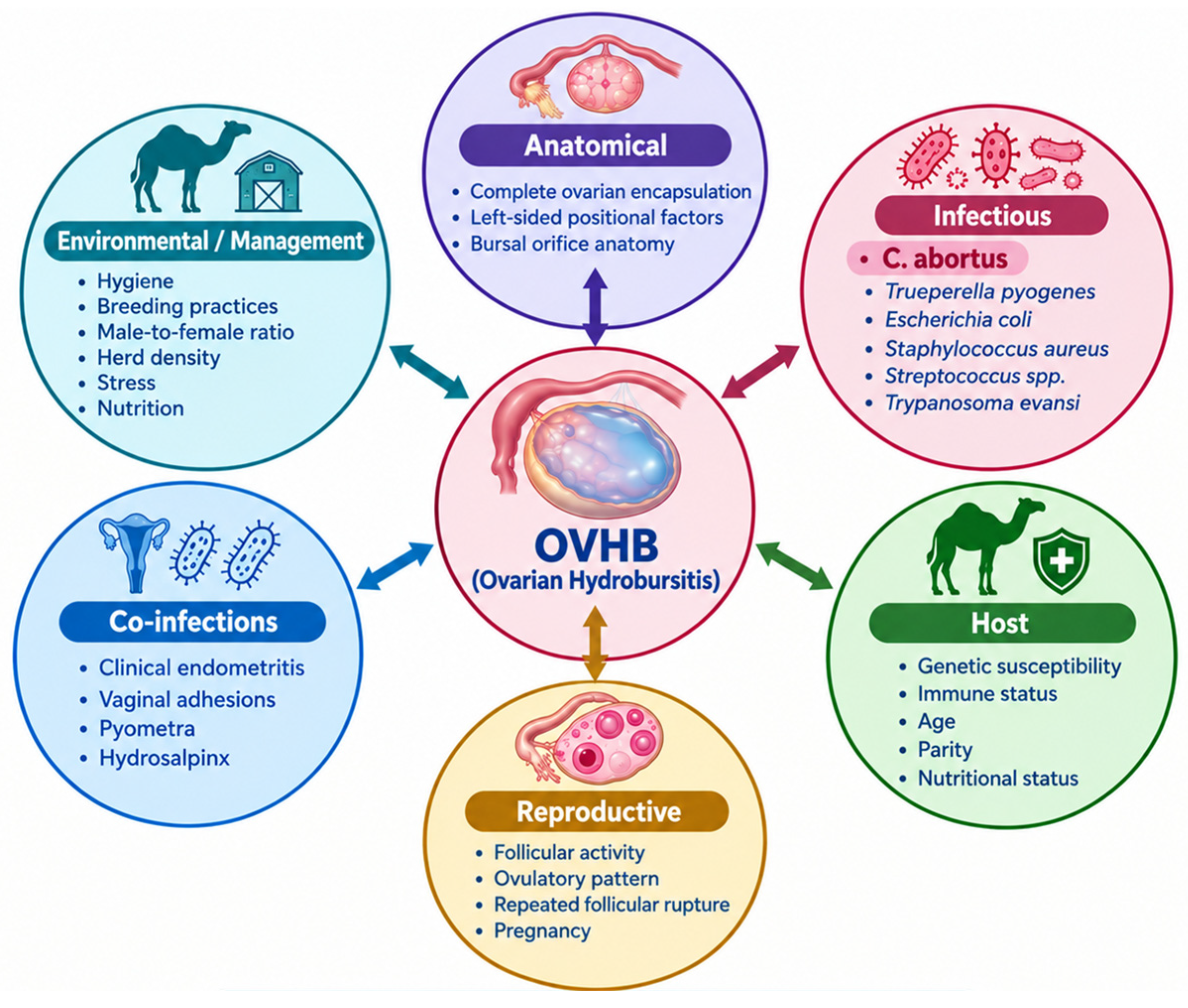
Although C. abortus is the infectious agent most consistently associated with OVHB, current evidence does not support its designation as the sole or exclusive cause. Rather, OVHB is best conceptualized as a multifactorial condition in which anatomical, infectious, host, and environmental factors interact to initiate and perpetuate disease (Figure 2).
Figure 2.
Ovarian hydrobursitis results from the interaction of multiple factors.
The complete encapsulation of the ovary within the ovarian bursa is a particularly pronounced anatomical feature in dromedary camels that appears to be a necessary, though not sufficient, predisposing factor. Without this anatomy, fluid accumulation and ovarian encapsulation would be less likely to occur. The left-sided predominance of OVHB suggests that positional factors—such as the greater size and weight of the left uterine horn, or compression by the distended urinary bladder—may further modulate risk.
C. abortus is the agent most strongly and consistently associated with OVHB. However, other bacteria (Trueperella pyogenes, Escherichia coli, Staphylococcus aureus, Streptococcus spp.) and protozoa (Trypanosoma evansi) have also been isolated from bursal contents. The role of these organisms—whether primary, contributory, or consequential—remains undetermined. It is equally plausible that they ascend secondarily from a concurrent endometritis, or that they act synergistically with C. abortus to exacerbate inflammation and fluid accumulation.
Not all C. abortus-infected camels develop OVHB. This observation suggests that host factors—including genetic susceptibility, immune status, age, parity, and nutritional status—may influence disease expression. Genome-wide association studies and immune profiling of affected versus unaffected animals are needed to explore this hypothesis.
The camel ovary is dynamically active, with waves of follicular growth, ovulation, and luteal formation. Repeated follicular rupture within the enclosed bursa may release fluid and cellular debris that contribute to bursal distension. Additionally, the induced ovulatory pattern of camels means that follicles may persist and grow abnormally in the absence of mating, potentially contributing to bursal fluid accumulation.
OVHB is frequently associated with clinical endometritis (21%), vaginal adhesions (8%), and pyometra. Whether these conditions predispose to OVHB, result from it, or share common risk factors remains unclear. Co-infections may amplify inflammation and impair local immune defenses, facilitating chlamydial colonization and persistence.
Herd-level factors—including hygiene, breeding practices, male-to-female ratios, nutrition, and stress—may influence the transmission and severity of OVHB. The observation that multiple animals within the same herd are often affected suggests that management practices contribute to disease spread.
In summary, OVHB is best understood as the outcome of interactions among multiple factors, with anatomical predisposition providing the necessary substrate, infectious agents (especially C. abortus) serving as important triggers or perpetuators, and host and environmental factors modulating disease expression. This multifactorial model has important implications for prevention: strategies that target only C. abortus may achieve partial control, but comprehensive prevention may require simultaneous attention to anatomy-associated risk, co-infections, and herd management practices.
A key unresolved question is why only some C. abortus-infected camels develop OVHB while others remain asymptomatic. Possible explanations include variation in bacterial strain virulence, host genetic susceptibility, the presence of co-infections, and the timing of infection relative to the reproductive cycle. These factors warrant further investigation.
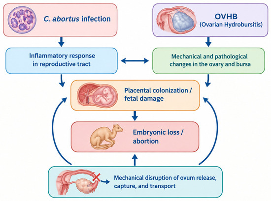
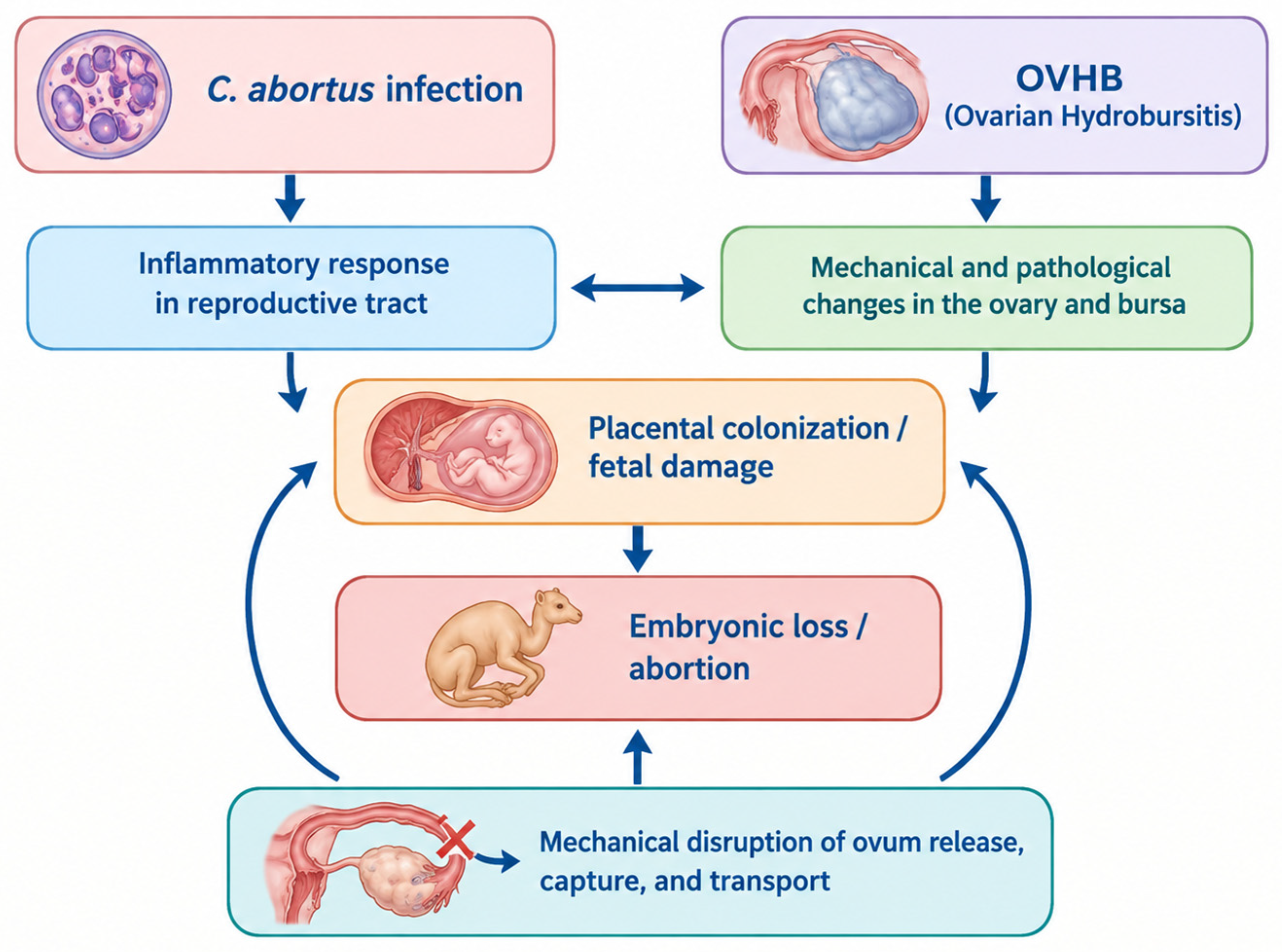
The association between OVHB and embryonic loss/abortion may involve two distinct but potentially overlapping mechanisms. OVHB is characterized by fluid accumulation within the ovarian bursa and encapsulation of the ovary. This anatomical disruption may impair fertility through several mechanisms: (1) interference with normal ovum release from the follicle; (2) obstruction of ovum capture by the fimbriae due to bursal distension; (3) disruption of normal oviductal transport; and (4) interference with corpus luteum function due to vascular compromise. These mechanical factors could lead to failure of conception or early embryonic loss. However, direct evidence demonstrating these mechanisms in camels is currently lacking, and this pathway remains hypothetical. C. abortus infection may independently cause embryonic loss or abortion through mechanisms documented in other species, including: (1) placental colonization and inflammation; (2) fetal infection and damage; (3) induction of pro-inflammatory cytokines that disrupt pregnancy maintenance; and (4) interference with progesterone signaling [32,33]. In sheep and goats, C. abortus causes late-term abortion, stillbirth, and weak offspring. Whether C. abortus causes similar outcomes in camels is unknown, but the organism has been associated with abortion in other camelid species [11]. Because OVHB and C. abortus infection frequently co-occur, their independent effects on pregnancy outcome are difficult to disentangle. It is plausible that both mechanisms operate simultaneously: C. abortus infection may initiate the inflammatory process that leads to OVHB, and the resulting anatomical disruption may further impair fertility and pregnancy maintenance. Alternatively, OVHB may develop as a consequence of C. abortus infection, and the same infection may independently cause abortion. Longitudinal studies comparing pregnancy outcomes in camels with OVHB alone, C. abortus infection alone, and both conditions together are needed to clarify these pathways (Figure 3).
Figure 3.
Proposed pathways linking OVHB and C. abortus infection to embryonic loss and abortion in dromedary camels.
5. Etiological Evidence Linking C. abortus with Ovarian Hydrobursitis
Multiple lines of evidence support an association between C. abortus infection and OVHB (Table 3). However, seropositivity indicates prior exposure or immune response rather than active infection, and PCR positivity indicates the presence of DNA rather than viable organisms or causation. The strongest evidence comes from isolation of viable organisms from affected tissues [15], although experimental reproduction of the disease has not yet been achieved. (Table 3). Serological, microbiological, molecular, pathological, and epidemiological findings collectively suggest that C. abortus plays a central role in disease pathogenesis. Nevertheless, experimental reproduction of the disease has not yet been achieved, and definitive proof of causation remains to be established [13]. In a cross-sectional study of 51 infertile female camels with confirmed OVHB (24 unilateral, 27 bilateral) from the Qassim region of Saudi Arabia, anti-C. abortus IgG antibodies were detected by ELISA in 86.3% (44/51) of affected animals [14]. Seropositivity indicates prior exposure or an immune response to C. abortus but does not necessarily indicate active infection or causation in a given animal [14]. Viable C. abortus was successfully isolated from bursal tissue and bursal fluid of affected camels using yolk sac inoculation of embryonated chicken eggs, with confirmation by Giemsa staining, Gimenez staining, and direct immunofluorescence [15]. Isolation provides the strongest available evidence of the presence of viable organisms in affected tissues, though it does not by itself establish causation [13]. Together, these findings provide the strongest available evidence implicating C. abortus in OVHB pathogenesis.
Table 3.
Context and denominators for key serological and molecular findings on C. abortus in dromedary camels with ovarian hydrobursitis.
Other bacteria (Trueperella pyogenes, Escherichia coli, Staphylococcus aureus, Streptococcus spp.) and protozoa (Trypanosoma evansi) have also been isolated from the bursal contents. Their role in pathogenesis—whether primary, contributory, or consequential—remains undetermined. It is plausible that they ascend secondarily from a concurrent endometritis, act synergistically with C. abortus, or contribute independently to bursal inflammation. Current evidence does not permit exclusion of any of these possibilities [5,8,13]. However, several observations indicate that these are secondary or opportunistic pathogens rather than primary initiators. First, C. abortus is consistently present in a much higher percentage of cases (86.3%) than any other organism. Second, the pathological lesions observed in affected bursae—mononuclear cell infiltration and lymphoid aggregation—are compatible with chronic chlamydial infection, as described in other species [33,34,39], but they are not specific for C. abortus [35,40,41]. Similar lesions can be observed in chronic inflammatory conditions caused by other bacteria, protozoa, or non-infectious agents. Therefore, histopathology alone cannot distinguish chlamydial from non-chlamydial inflammation. Third, standard bacteriological culture fails to detect any growth in 53.3% of bursal fluid samples, whereas C. abortus (an obligate intracellular bacterium) is detected by specialized methods in the same samples [5,13]. The presence of pyogenic bacteria likely reflects ascending infection from concurrent endometritis, which is a common comorbidity in OVHB cases (21% association; [8]).
Establishing a causal relationship between a microorganism and a disease requires more than detection of the organism in affected tissues.
It is important to distinguish clearly between OVHB as a clinical syndrome and C. abortus infection as a microbiological state. The two are associated but not synonymous: not all C. abortus-infected camels develop OVHB, and not all OVHB cases may be attributable to C. abortus alone. Current evidence does not permit a definitive conclusion regarding the precise role of C. abortus in OVHB pathogenesis. The organism may act as (1) a primary initiating cause, (2) a contributing factor that exacerbates a process initiated by other mechanisms, (3) a secondary consequence of altered bursal anatomy or pre-existing inflammation, or (4) one component of a multifactorial disease process involving anatomical, infectious, and host factors. This uncertainty has direct implications for prevention: the rationale for vaccination against C. abortus rests on the assumption that the organism is a primary or major contributing cause. If this assumption is incorrect, vaccine efficacy may be partial. Experimental infection studies are therefore essential to clarify the causal role of C. abortus before vaccine development proceeds to field trials.
Venereal transmission is considered plausible and is supported by indirect evidence, including the occurrence of multiple cases within herds [14], the prevalence of affected nulliparous females, and the detection of Chlamydia in male genital tracts [16]. However, no direct transmission studies have been conducted in camels, and other routes—including direct contact, aerosol, ingestion, iatrogenic, and vector-borne transmission—cannot be excluded. The relative importance of each route remains to be determined. In other species, C. abortus is known to cause prostatitis and epididymitis in males [42,43]. The venereal transmission pattern has important control implications: treating only females will not eliminate the infection from a herd if infected males continue to serve as reservoirs. While congenital reproductive anomalies and ovulatory disorders may contribute to infertility in individual animals, current evidence does not support these conditions as the primary explanation for the characteristic pathological features of OVHB.
The routes by which C. abortus is transmitted among dromedary camels, and how infection leads to OVHB, remain incompletely understood. Several observations are consistent with venereal transmission, but none provides definitive proof. Multiple affected females within the same herd (2–4 cases on 18 occasions; [14]) suggest a common source or contagious spread, and the high prevalence in nulliparous females (37.1%; [8]), which have never calved and thus have not been exposed to postpartum contamination, further supports the possibility of mating-related transmission. Chlamydia has also been detected in the genital tracts of breeding male camels [16], and in other species C. abortus causes prostatitis and epididymitis in males [42,43], while venereal transmission is well documented for Chlamydia trachomatis in humans [31]. Nevertheless, the venereal hypothesis has important limitations. No direct transmission studies have been conducted in camels, the organism has not been isolated from semen or preputial washings of male camels, and the mere presence of Chlamydia in male genital tracts does not prove that transmission occurs during mating. Moreover, multiple affected females in a herd could reflect a common environmental source rather than venereal spread.
Alternative or additional transmission routes therefore warrant consideration. C. abortus is shed in uterine discharges, aborted material, and placental tissues, and direct contact with these materials—or with contaminated bedding, feed, or water—could facilitate transmission, particularly in confined or densely stocked herds [44]. Inhalation of aerosols from contaminated environments, particularly in lambing pens, is an established route in sheep [45], although whether this route contributes to OVHB in camels is unknown. The fecal–oral route has been suggested for some Chlamydia species, though its relevance to C. abortus in camels is unclear. Iatrogenic transmission via contaminated instruments, gloves, or semen used during gynecological examination or artificial insemination could potentially occur, but this route has not been investigated in camels. Mechanical transmission by flies or other arthropods has been suggested for Trypanosoma evansi [5] and could theoretically contribute to C. abortus spread, though no evidence exists to support this route. Finally, transplacental or perinatal transmission of C. abortus is well documented in sheep [46] and could theoretically occur in camels, although this route would not explain the high prevalence in nulliparous females. In conclusion, venereal transmission is plausible and supported by indirect evidence, but it has not been definitively demonstrated. Other routes—particularly direct contact with infected reproductive tissues or contaminated environments—may also contribute, and the relative importance of each route remains unknown. Direct transmission studies are urgently needed, and until such studies are conducted, prevention strategies should not assume that venereal transmission is the sole or predominant route.
Although the available evidence strongly implicates C. abortus in the pathogenesis of OVHB, several criteria for definitive causation remain unmet. Experimental infection studies reproducing OVHB under controlled conditions have not yet been conducted, and the temporal relationship between infection and disease development has not been established through longitudinal studies. Consequently, C. abortus should presently be regarded as the pathogen most strongly associated with OVHB rather than a definitively proven sole cause.
6. Clinical Presentation and Diagnosis
Affected animals typically present with long-standing infertility (1–4 years), early embryonic death, abortion, repeat breeding, or refusal to mate [6,8]. The association between OVHB and early embryonic death was based on the reproductive history of affected females. Conception was established by successful mating followed by pregnancy diagnosis using the tail-cocking behavior test, performed approximately 15–20 days after mating, a method with approximately 80% accuracy in dromedary camels [47]. When available, pregnancy was confirmed by transrectal ultrasonography performed approximately 20–30 days after mating, at which time the presence of an embryonic vesicle and fetal heartbeat was assessed. Early embryonic death was recorded when a female initially diagnosed as pregnant subsequently returned to estrus, or when ultrasonographic examination at approximately 30 days after mating revealed the absence of an embryo or heartbeat despite a prior positive diagnosis. In the dromedary camel, the embryonic period extends from conception to approximately day 30–35 of gestation, after which the conceptus enters the fetal period. Early embryonic loss was therefore defined as the return to estrus after an initial positive pregnancy diagnosis, with no evidence of an embryonic vesicle or embryonic heartbeat on subsequent examination before approximately day 30–35 of gestation. Importantly, affected camels are systemically healthy: they show no fever, reduced appetite, or abnormal heart or respiratory rates. This absence of systemic signs means that diagnosis depends entirely on specific gynecological examination.
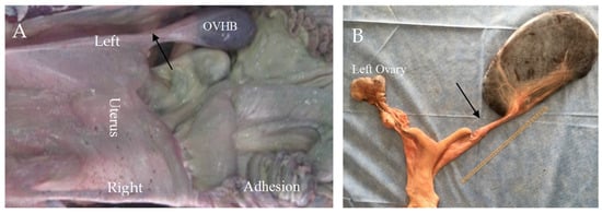
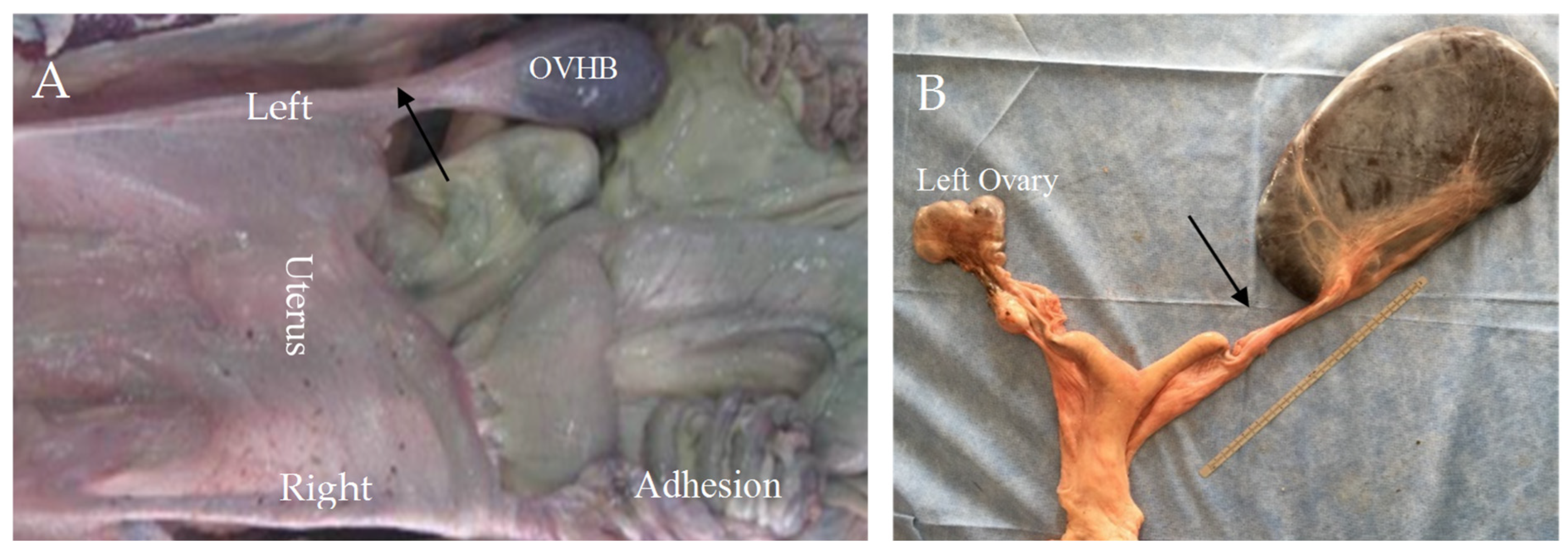
On transrectal palpation, OVHB is suspected when the genital tract is difficult to retract and a cord-like structure is felt between the uterine horn and a non-palpable ovary [6,19]. The encapsulated ovary itself cannot be grasped freely. This finding is highly suggestive but not definitive (Figure 4).
Figure 4.
Topography of the reproductive tract in a dromedary camel with ovarian hydrobursitis (OVHB). (A) The reproductive tract is retracted into the abdominal cavity and stretched by the weight of the fluid-filled bursa and associated adhesions, making ovarian palpation difficult because the ovary is entrapped within the bursa. (B) A cord-like structure (black arrow) is palpated between the uterine horn and the non-palpable ovary.
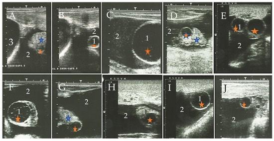
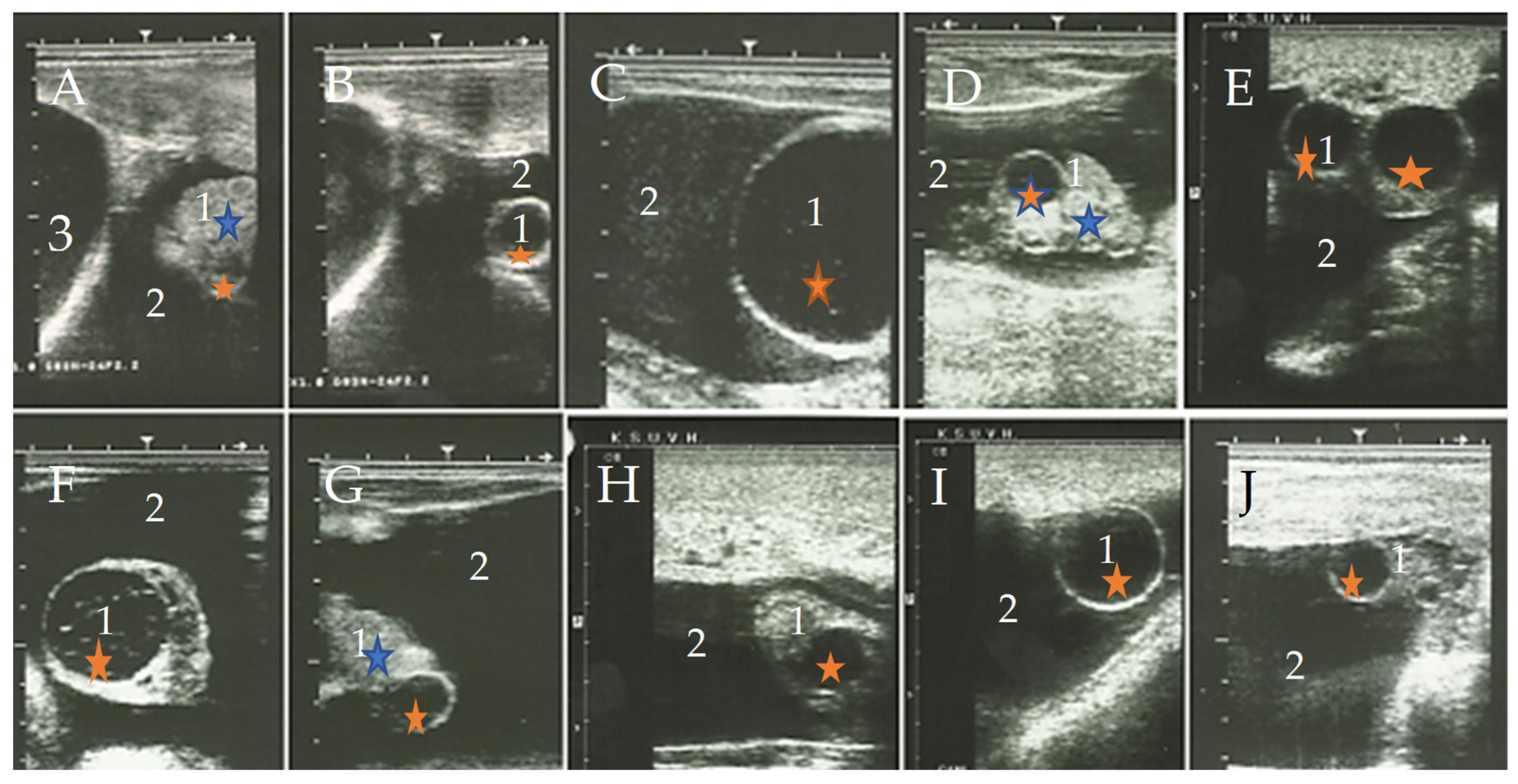
Ultrasonography is the most informative and practical diagnostic modality currently available for OVHB and is considered the diagnostic method of choice in clinical practice [22]. However, it is important to note that no formal validation studies have been conducted to determine the diagnostic accuracy (sensitivity, specificity, predictive values) of ultrasonography for OVHB against an accepted reference standard. The diagnostic criteria described in the literature have not been standardized or systematically validated, and misdiagnosis—particularly confusion with hydrosalpinx, paraovarian cysts, or other fluid-filled structures—remains possible [22]. The characteristic finding is a large, well-demarcated, hypoechogenic sac lateral to or beneath the corresponding uterine horn, with the ovary visualized as a floating echogenic mass within the bursal fluid (Figure 5). The appearance varies with disease duration: (1) early cases (<6 months): clear, homogeneous, non-echogenic fluid with a freely floating ovary; (2) chronic cases (>6 months): increasingly echogenic fluid with ovarian adherence to the bursal wall; (3) long-standing cases: thoroughly echogenic content with thick walls and clotted blood. The affected bursae can reach enormous sizes, with dimensions up to 48 × 15 cm and weights up to 11 kg in extreme cases [22].
Figure 5.
Ultrasonographic appearance of ovarian hydrobursitis (OVHB) in dromedary camels at different stages of disease. (A–C) Early-stage disease: the bursal fluid (2) is clear, homogeneous, and non-echogenic, and the ovary (1) floats freely within the distended bursa; the urinary bladder (3) is visible in panel A. (D–F) Chronic-stage disease: the bursal fluid becomes increasingly echogenic, and the ovary (1) may adhere to the bursal wall. (G–I) Long-standing disease: the bursal content is thoroughly echogenic with a thickened bursal wall, and clotted blood may be present within the bursal lumen. (J) A large OVHB adjacent to the urinary bladder (3), illustrating the spatial relationship between the affected bursa and adjacent pelvic structures. Measurement scales are provided in representative panels. All images are original and were acquired by the authors during the studies described in this review. 1: ovary; 2: ovarian bursa filled with fluid of varying echogenicity; 3: urinary bladder. Blue star: corpus luteum; orange star: follicle.
The diagnostic performance of ultrasonography is likely to be substantially lower in early or subclinical stages of OVHB than in advanced disease. The characteristic finding of a large, well-demarcated, hypoechogenic sac encapsulating the ovary depends on sufficient fluid accumulation within the bursa to produce a detectable acoustic window. In the earliest stages of infection, before appreciable fluid accumulates, the bursa may appear ultrasonographically normal or show only subtle, non-specific changes (e.g., mild thickening of the bursal wall or a small amount of anechoic fluid). Such findings may be indistinguishable from normal physiological fluid, which can be present in the vicinity of the ovarian bursa around the time of ovulation [8,11,15,22]. Consequently, ultrasonography may fail to detect early or subclinical OVHB, leading to false-negative diagnoses and underestimation of true disease prevalence. Furthermore, because the diagnostic criteria used in published studies were derived primarily from animals with clinically apparent, advanced disease, their applicability to early-stage disease has not been established. Prospective longitudinal studies using repeated ultrasonographic examinations in at-risk animals are needed to characterize the earliest detectable ultrasonographic changes and to determine the sensitivity of ultrasonography at different stages of disease.
7. Differential Diagnosis
Several reproductive and non-reproductive conditions may mimic the clinical and ultrasonographic appearance of OVHB, and accurate differentiation is essential for appropriate case management and prognosis (Table 4).
Table 4.
Differential diagnosis of ovarian hydrobursitis in dromedary camels.
The single most important diagnostic feature of OVHB is the presence of a large, well-demarcated, hypoechogenic sac encapsulating the ovary, with the ovary visualized as a floating echogenic mass within the bursal fluid. In all other conditions listed above, the ovary is either separate from the fluid-filled structure (hydrosalpinx, pyosalpinx, hydrometra, pyometra), contains the fluid within its parenchyma (ovarian cysts), or is part of a complex mass without bursal encapsulation (tubo-ovarian abscess, neoplasia).
Ultrasonography is the diagnostic method of choice for differentiating OVHB from other conditions, although its accuracy has not been formally validated against an accepted reference standard. Transrectal palpation alone is insufficient because the encapsulated ovary cannot be grasped freely and may be confused with other fluctuant structures. When the diagnosis remains uncertain, additional diagnostic procedures—including laparoscopy, laparotomy, or post-mortem examination—may be necessary. Measurement of serum anti-C. abortus antibodies or molecular detection of the organism in bursal fluid (if aspirated) may provide supportive evidence but should not be used as the sole basis for diagnosis.
A common pitfall is to mistake a large hydrosalpinx for OVHB, particularly when the distended fallopian tube is palpated near the ovary. Careful ultrasonographic examination to confirm whether the ovary is inside or outside the fluid-filled structure is essential. Another pitfall is to misinterpret a large ovarian follicular cyst as an encapsulated ovary; however, in OVHB, the ovary is seen floating within the bursa, whereas in follicular cysts, the cyst is within the ovary. Finally, the coexistence of OVHB with other conditions—particularly endometritis, hydrosalpinx, and adhesions—should be considered, as these may complicate the clinical picture.
8. Treatment and Reproductive Outcomes
For unilaterally affected camels, surgical ablation of the affected bursa and ovary achieved a 90-day non-return rate of 64.3% (9/14) and a calving rate of 50% (7/14) in a retrospective case series [6,17]. However, these findings are based on a small sample size (n = 14) and a retrospective design without randomization and should be interpreted with caution. Larger, prospective studies are needed to confirm these outcomes [9,17].
Right-sided ablation was associated with a higher observed calving rate (3/3, 100%) than left-sided ablation (4/11, 36.4%) [6]. It has been hypothesized that this difference reflects the greater functional importance of the left ovary during pregnancy in camels, such that its removal may have a greater impact on future fertility than removal of the right ovary [4,22]. This hypothesis is consistent with the observation that pregnancy occurs almost exclusively in the left uterine horn and that the left ovary is more commonly the source of the ovum and corpus luteum of pregnancy [4,22]. However, the comparison is based on a very small number of animals (n = 3 for right-sided ablation), and the difference may be due to chance, confounding, or differences in disease severity, duration, or concurrent genital pathology between the two groups—factors that were not controlled for in this retrospective series. The greater functional importance of the left ovary should therefore be regarded as a hypothesis requiring confirmation rather than an established mechanism. Testing this hypothesis will require prospective studies with standardized surgical protocols, pre- and post-operative assessment of ovarian function, and long-term reproductive follow-up in a large number of animals. Surgical ablation is not applicable to bilateral disease.
Medical treatment combining long-acting oxytetracycline (20 mg/kg, IM, 7 days), intrauterine lotagen 4%, and cloprostenol (500 μg, IM) was effective only for small bursae (<5 cm diameter), achieving a 73.3% (11/15) non-return rate and 60% (9/15) calving rate in a clinical trial that included an untreated control group [14]. Medium (5–7 cm) and large (>7 cm) bursae did not respond (0% calving rate). However, the number of animals in the medium (n = 7) and large (n = 8) subgroups was small, and the trial was not blinded. These limitations should be considered when interpreting the results. The failure of medical treatment in medium and large bursae [14] may be explained by several factors, including the physical barrier posed by a thickened, fibrotic bursal wall, the dilution of antimicrobial agents within a large volume of accumulated fluid, and the presence of dense adhesions between the ovary and bursal wall. However, the relative contribution of each factor has not been systematically investigated, and no studies have measured drug concentrations within the bursal fluid or wall of affected camels. This mechanism therefore remains plausible but unproven.
In a limited number of severe bilateral cases where conventional treatment was not feasible, transabdominal ultrasound-guided ovarian bursocentesis and laparotomy-assisted bursal drainage with lavage were performed as salvage procedures [13]. The number of treated animals was too small to permit meaningful assessment of efficacy, and no control group was included. These approaches should therefore be regarded as experimental and not as established treatments. Table 5 summarizes the reported treatment outcomes.
Table 5.
Summary of treatment outcomes for ovarian hydrobursitis in dromedary camels, with sample size and study design limitations.
9. Future Perspectives for Prevention and Vaccine Development
Given the limitations of current treatment options—surgery being applicable only to unilateral cases [6,17] medical therapy showing limited success in advanced disease [14], and the absence of effective treatment for bilateral OVHB—prevention represents the most sustainable long-term control strategy. This rationale is supported by the demonstrated efficacy of C. abortus vaccines in sheep, where both inactivated and modified live vaccines have been successfully used to control enzootic abortion [48,49], and by the venereal and within-herd transmission pattern of OVHB, which predisposes even successfully treated animals to reinfection [14,16]. However, the rationale for vaccination is based on the assumption that C. abortus is a primary or major contributing cause of OVHB. If the organism instead acts as a secondary invader or as one component of a multifactorial process, vaccine efficacy may be partial. This uncertainty underscores the importance of experimental infection studies to clarify causation before vaccine development proceeds to field trials. Additional factors supporting the development of preventive measures include: (1) the strong association of C. abortus with OVHB; (2) the apparent venereal transmission pattern, which may predispose successfully treated animals to reinfection; (3) the relatively high prevalence of the disease among infertile female camels, making herd-level prevention more cost-effective than individual treatment; and (4) the zoonotic importance of C. abortus.
Commercial inactivated and modified live vaccines against C. abortus are available for the control of enzootic abortion in sheep, and subcellular vaccines have shown protective efficacy in pregnant sheep challenge models [48,49,50,51,52]. However, their efficacy and safety have not been evaluated in dromedary camels, and no vaccine is currently licensed for this species. Consequently, efforts toward vaccine development should begin with a detailed characterization of camel-derived C. abortus isolates. In our laboratory, ongoing studies are investigating the molecular and proteomic characteristics of these isolates to improve understanding of their antigenic diversity and identify immunologically relevant proteins that may support future preventive strategies. Further research is required to characterize these proteins, evaluate their immunogenicity, and determine whether existing ovine vaccines or camel-specific vaccine formulations can provide effective protection against chlamydial reproductive disease in camels.
10. Limitations of Current Knowledge
Despite substantial advances in understanding OVHB, several limitations remain. A limitation of the current evidence base is that much of the published literature on ovarian hydrobursitis originates from a limited number of research groups and geographic regions, reflecting the relative scarcity of focused investigations on this condition. Recent reviews of reproductive disorders in camelids have likewise emphasized that several aspects of camel reproductive pathology remain incompletely understood and that further investigation is needed [11,53]. Independent studies from additional camel-producing countries are therefore needed to validate existing findings and improve the generalizability of current knowledge. Although Chlamydia abortus DNA has been detected in affected bursae and viable organisms have been isolated from bursal tissue and fluid, experimental reproduction of OVHB has not yet been reported, and the precise role of C. abortus in disease development remains to be established [15]. Independent investigations of C. abortus infection in dromedary camels from other geographic regions, such as Selmi et al. in Tunisia [54], Selim et al. in Egypt [55], Hussein et al. [56] and Khalifa et al. [57] in eastern Saudi Arabia, Benaissa et al. in Algeria [7], Elzlitne and Elhafi in Libya [58], and Pourfattahi et al. in Iran [59], further demonstrate the value of expanding epidemiological investigations beyond the populations in which OVHB has been primarily studied. Furthermore, molecular characterization of camel-derived C. abortus isolates remains limited, and no vaccine efficacy trials against OVHB have been reported in dromedary camels. The identification and validation of protective antigens therefore remain important areas for future investigation. Addressing these limitations should be a priority for future research.
A summary of the principal knowledge gaps and recommended research priorities is provided in Table 6.
Table 6.
Knowledge gaps and research priorities for ovarian hydrobursitis (OVHB) in dromedary camels.
11. Conclusions
Ovarian hydrobursitis (OVHB) is a prevalent reproductive disorder among female dromedary camels in referral populations, though its true population prevalence remains unknown. Its pathogenesis appears linked to the particularly pronounced anatomical anatomy of the ovarian bursa and is strongly associated with Chlamydia abortus infection, although definitive causation remains unproven.
Current treatment options are limited, with surgical ablation applicable only to selected unilateral cases and medical therapy effective only for small bursae. Bilateral and advanced cases have a poor prognosis.
Vaccine development remains a future research direction rather than an established strategy, given the absence of efficacy data in camels. Further studies are needed to clarify causation, validate diagnostic criteria, determine true prevalence, and develop effective preventive measures.
Author Contributions
All authors contributed to the study conception and design. Material preparation, data collection and analysis were performed by A.A., D.R.D. and S.A.A. The first draft of the manuscript was written by A.A. and all authors commented on previous versions of the manuscript. All authors have read and agreed to the published version of the manuscript.
Funding
The researchers would like to thank the Deanship of Graduate Studies and Scientific Research at Qassim University (www.qu.edu.sa) for financial support (QU-APC-2026).
Institutional Review Board Statement
Not applicable.
Informed Consent Statement
Not applicable.
Data Availability Statement
The original contributions presented in this study are included in the article. Further inquiries can be directed to the corresponding author(s).
Conflicts of Interest
The authors declare no conflicts of interest.
References
- Smuts, M.M.S.; Bezuidenhout, R.J.; Bezuidenhout, A.J. Anatomy of the Dromedary; Oxford University Press: Oxford, UK, 1987. [Google Scholar]
- Srikandakumar, A.; Johnson, E.H.; Mahgoub, O.; Kadim, I.T.; Al-Ajmi, D.S. Anatomy and histology of the female reproductive tract of the Arabian camel. Emir. J. Food Agric. 2001, 13, 23–26. [Google Scholar] [CrossRef] [Scilit]
- Salari, E.; Raji, A.R.; Farzaneh, N. Comparative study of anatomy and histology on the ovary and oviduct in camel (Camelus dromedarius) and cow. J. Camel Pract. Res. 2011, 18, 115–118. [Google Scholar]
- Ali, A.; Derar, D.R. Ovary and ovarian bursa in dromedary camels: Clinical relevance of the topographical features. Anat. Histol. Embryol. 2020, 49, 325–332. [Google Scholar] [CrossRef] [Scilit] [PubMed]
- Ali, A.; Al-Sobayil, F.A.; Tharwat, M.; Hassanein, K.M. Ovarian hydrobursitis in female camels (Camelus dromedarius): Biochemical, bacterial and protozoal evaluation. Theriogenology 2011, 75, 734–741. [Google Scholar] [CrossRef] [Scilit] [PubMed]
- Ali, A.; Mehana, E.E.; Ahmed, A.F.; El-Tookhy, O.; Al-Sobayil, A.; Al-Hawas, A. Ovarian hydrobursitis in female camels (Camelus dromedarius): Clinical findings, histopathology and fertility after unilateral surgical ablation. Theriogenology 2011, 76, 492–499. [Google Scholar] [CrossRef] [Scilit] [PubMed]
- Benaissa, M.H.; Faye, B.; Kaidi, R. Ovarian hydrobursitis in slaughtered female camels (Camelus dromedarius) in Southeast Algeria. Emir. J. Food Agric. 2014, 26, 915–920. [Google Scholar] [CrossRef] [Scilit]
- Ali, A.; Derar, R.; Al-Sobayil, F.; Al-Hawas, A.; Hassanein, K. A retrospective study on clinical findings of 7300 cases (2007–2014) of barren female dromedaries. Theriogenology 2015, 84, 452–456. [Google Scholar] [CrossRef] [Scilit] [PubMed]
- Ali, A.; Derar, D.R.; Almundarji, T.I. Infertility in female dromedary camels. J. Camel Pract. Res. 2021, 28, 267–276. [Google Scholar] [CrossRef] [Scilit]
- Al-Eknah, M.M.; Ali, A.M.A. Infundibular cysts jeopardize reproduction in female camels (Camelus dromedarius). Emir. J. Agric. Sci. 2001, 13, 52–56. [Google Scholar] [CrossRef] [Scilit]
- Sghiri, A.; Ciccarelli, M.; Waqas, M.S.; Anouassi, A.; Tibary, A. Advances in the diagnosis of reproductive disorders in female camelids. Animals 2025, 15, 2902. [Google Scholar] [CrossRef] [Scilit] [PubMed]
- Nourani, H.; Khodakaram-Tafti, A. Pathological study of ovaries of nonpregnant camels (Camelus dromedarius) slaughtered in Iran. J. Camel Pract. Res. 2004, 11, 109–113. [Google Scholar]
- Ali, A.; Derar, D.R.; Osman, S.A.; Allam, S.A. Ovarian hydrobursitis and chlamydial infection in dromedary camels: Diagnosis, impact, and management. Reprod. Domest. Anim. 2026, 61, e70147. [Google Scholar]
- Ali, A.; Al-Sobayil, F.A.; Hassanein, K.M.; Al-Hawas, A. Ovarian hydrobursitis in female camels (Camelus dromedarius): The role of Chlamydophila abortus and a trial for medical treatment. Theriogenology 2012, 77, 1754–1758. [Google Scholar] [CrossRef] [Scilit] [PubMed]
- Ali, A.; Derar, D.R.; Mousa, H.A.; Osman, S.A.; Refaai, W.; Almundarij, T.I.; Al-Dubaib, M.A.; Allam, S.A. First report on the isolation of Chlamydia abortus from female dromedary camels with ovarian hydrobursitis. Theriogenology 2022, 191, 102–108. [Google Scholar] [CrossRef] [Scilit] [PubMed]
- Derar, D.R.; Ali, A.; Osman, S.A.; Al-Sobayil, F.A.; Saeed, E.M.; Hassanein, K.; Al-Hawas, A.-A. Potential pathogens in infertile male dromedary camels and their association with the spermogram and clinical findings. Comp. Clin. Pathol. 2017, 26, 965–970. [Google Scholar] [CrossRef] [Scilit]
- Tibary, A.; Anouassi, A. Retrospective study on an unusual form of ovario-bursal pathology in the camel (Camelus dromedarius). Theriogenology 2001, 56, 415–424. [Google Scholar] [CrossRef] [Scilit] [PubMed]
- Debrine, A.M.; Karplus, P.A.; Rockey, D.D. Structural foundation for studying chlamydial polymorphic membrane proteins. Microbiol. Spectr. 2023, 11, e03242-23. [Google Scholar] [CrossRef] [Scilit] [PubMed]
- Zhang, H.; Li, Z.; Li, W.; Jin, Y.; Li, Y.; Xiao, Q.; Tong, D.; Zhou, J. Comparison of the immune effects of the Chlamydia abortus MOMP antigen displayed in different parts of bacterial ghosts. Front. Microbiol. 2024, 15, 1349746. [Google Scholar] [CrossRef] [Scilit] [PubMed]
- Guerra-Infante, F.M.; de Haro-Cruz, M.J.; López-Hurtado, M.; De la Rosa-Ramos, M.A.; Díaz-Aparicio, E.; Arellano-Reynoso, B. Comparative evaluation of recombinant Chlamydia abortus and Chlamydia trachomatis major outer membrane proteins for diagnosing human chlamydial infection. Microbiol. Res. 2025, 16, 159. [Google Scholar] [CrossRef] [Scilit]
- Baethge, C.; Goldbeck-Wood, S.; Mertens, S. SANRA—A scale for the quality assessment of narrative review articles. Res. Integr. Peer Rev. 2019, 4, 5. [Google Scholar] [CrossRef] [Scilit] [PubMed]
- Ali, A.; Derar, D.; Alsamri, A.; Al Sobayil, F. Echography of clinically relevant disorders in the genital tract of female dromedary camels. Anim. Reprod. Sci. 2017, 182, 123–133. [Google Scholar] [CrossRef] [Scilit] [PubMed]
- Hussein, F.M.; Hassieb, M.M. Morphopathological studies on the ovaries, oviducts and uteri from camels with cystic ovaries. Assiut Vet. Med. J. 1989, 21, 184–189. [Google Scholar] [CrossRef] [Scilit]
- El-Khouly, A.B.; El-Nasr, A.; Ontabli, A.; Gadir, F.A. Some pathologic affections of camel ovaries in U.A.E. Zagazig Vet. J. 1990, 18, 210–217. [Google Scholar]
- Belina, D.; Eshetu, A.; Alemu, S.; Shasho, B.; Mohammed, T.; Mohammed, A.; Mummed, B.; Regassa, D. Reproductive diseases and disorders of female camels: An assessment and pathological and bacteriological study in Eastern Ethiopia. Vet. Med. Int. 2021, 2021, 6641361. [Google Scholar] [CrossRef] [Scilit] [PubMed]
- Getahun, A.M.; Hunderra, G.C.; Gebrezihar, T.G.; Boru, B.G.; Desta, N.T.; Ayana, T.D. Comparative study on lesions of reproductive disorders of cows and female dromedary camels slaughtered at Addis Ababa, Adama and Akaki abattoirs with bacterial isolation and characterization. BMC Vet. Res. 2021, 17, 134. [Google Scholar] [CrossRef] [Scilit] [PubMed]
- Alharbi, Y.M. Associations between ubiquitin, follicle-stimulating hormone, and sex steroid hormones in the failed to conceive female dromedary camels raised in hot climates. Vet. World 2022, 15, 2046–2051. [Google Scholar] [CrossRef] [Scilit] [PubMed]
- Gherissi, D.E.; Ben Ali, Y.; Rahmoun, D.E.; Bouzebda-Afri, F.; Bouzebda, Z. Pathological findings on genital abnormalities in female camel in the El Oued region, Algeria. Vet. Stanica 2022, 53, 663–675. [Google Scholar] [CrossRef] [Scilit]
- Qureshi, Z.I.; Al-Jabouri, A. Clinical prevalence of reproductive disorders in dromedary camel (Camelus dromedarius) under field conditions. In Proceedings of the Second Conference of the International Society of Camelid Research and Development (ISOCARD), Djerba, Tunisia, 12–14 March 2009; Abstract No. 169; ISOCARD/IRA: Djerba, Tunisia, 2009; p. 136. [Google Scholar]
- Al-Eknah, M.M.; Ramadan, R.O.; Almubarak, A.I.; Al Mohsen, F.; Al-Salman, M.; Ali, A.M. Verification of ovarian hydro-bursitis syndrome in the dromedary camels and its surgical ablation. In Proceedings of the ICAR Satellite Meeting on Camelid Reproduction, Vancouver, BC, Canada, 5 August 2012; pp. 42–46. [Google Scholar]
- Faber, M.T.M.; Nielsen, A.M.; Nygård, M.; Sparén, P.; Tryggvadottir, L.M.; Hansen, B.T.; Liaw, K.-L.; Kjaer, S.K.M. Genital chlamydia, genital herpes, Trichomonas vaginalis and gonorrhea prevalence, and risk factors among nearly 70,000 randomly selected women in 4 Nordic countries. Sex. Transm. Dis. 2011, 38, 727–734. [Google Scholar] [CrossRef] [Scilit] [PubMed]
- Shao, R.; Wang, X.; Wang, W.; Stener-Victorin, E.; Mallard, C.; Brännström, M.; Billig, H. From mice to women and back again: Causalities and clues for Chlamydia-induced tubal ectopic pregnancy. Fertil. Steril. 2012, 98, 1175–1185. [Google Scholar] [CrossRef] [Scilit] [PubMed]
- Obendorf, D.L.; Handasyde, K.A. Pathology of chlamydial infection in the reproductive tract of the female koala. In Biology of the Koala; Lee, A.K., Handasyde, K.A., Sanson, G.D., Eds.; Surrey Beatty and Sons: Sydney, Australia, 1990; pp. 273–289. [Google Scholar]
- Swenson, C.E.; Donegan, E.; Schachter, J. Chlamydia trachomatis-induced salpingitis in mice. J. Infect. Dis. 1983, 148, 1101–1107. [Google Scholar] [CrossRef] [Scilit] [PubMed]
- Zachary, J.F. Pathologic Basis of Veterinary Disease, 7th ed.; Elsevier: St. Louis, MI, USA, 2022. [Google Scholar]
- Obendorf, D.L. Pathology of the female reproductive tract in the koala, Phascolarctos cinereus (Goldfuss), from Victoria, Australia. J. Wildl. Dis. 1981, 17, 587–592. [Google Scholar] [CrossRef] [Scilit] [PubMed]
- Fabijan, J.; Caraguel, C.; Jelocnik, M.; Polkinghorne, A.; Boardman, W.S.J.; Nishimoto, E.; Johnsson, G.; Molsher, R.; Woolford, L.; Timms, P.; et al. Chlamydia pecorum prevalence in South Australian koala (Phascolarctos cinereus) populations: Identification and modelling of a population free from infection. Sci. Rep. 2019, 9, 6261. [Google Scholar] [CrossRef] [Scilit] [PubMed]
- Kramer, G.; Duffy, J.; Mella, V.S.A. Preliminary evidence of chlamydiosis in koalas of the Greater Geelong Region, Victoria: A potential emerging threat? Animals 2025, 15, 2048. [Google Scholar] [CrossRef] [Scilit] [PubMed]
- Khamesipour, A.; Pal, S.; Peterson, E.M.; de la Maza, L.M. Induction of infertility by the Chlamydia trachomatis mouse pneumonitis biovar in strains of mice that differ in their response to the 60 kDa heat shock protein. J. Reprod. Fertil. 1994, 101, 287–294. [Google Scholar] [CrossRef] [Scilit] [PubMed]
- Williams, E.S.; Barker, I.K. Infectious Diseases of Wild Mammals, 3rd ed.; Iowa State University Press: Ames, IA, USA, 2001. [Google Scholar]
- LaVerda, D.; Albanese, L.N.; Ruther, P.E.; Morrison, S.G.; Morrison, R.P.; Ault, K.A.; Byrne, G.I. Seroreactivity to Chlamydia trachomatis Hsp10 correlates with severity of human genital tract disease. Infect. Immun. 2000, 68, 303–309. [Google Scholar] [CrossRef] [Scilit] [PubMed]
- Wagenlehner, F.M.; Naber, K.G.; Weidner, W. Chlamydial infections and prostatitis in men. BJU Int. 2006, 97, 687–690. [Google Scholar] [CrossRef] [Scilit] [PubMed]
- Teankum, K.; Pospischil, A.; Janett, F.; Brugnera, E.; Hoelzle, L.E.; Hoelzle, K.; Weilenmann, R.; Zimmermann, D.R.; Gerber, A.; Polkinghorne, A.; et al. Prevalence of chlamydiae in semen and genital tracts of bulls, rams and bucks. Theriogenology 2007, 67, 303–310. [Google Scholar] [CrossRef] [Scilit] [PubMed]
- Cheong, H.C.; Lee, C.Y.Q.; Cheok, Y.Y.; Tan, G.M.Y.; Looi, C.Y.; Wong, W.F. Chlamydiaceae: Diseases in primary hosts and zoonosis. Microorganisms 2019, 7, 146. [Google Scholar] [CrossRef] [Scilit] [PubMed]
- Essig, A.; Longbottom, D. Chlamydia abortus: New aspects of infectious abortion in sheep and potential risk for pregnant women. Curr. Clin. Microbiol. Rep. 2015, 2, 22–34. [Google Scholar] [CrossRef] [Scilit]
- Longbottom, D.; Livingstone, M.; Maley, S.; van der Zon, A.; Rocchi, M.; Wilson, K.; Wheelhouse, N.; Dagleish, M.; Aitchison, K.; Wattegedera, S.; et al. Intranasal infection with Chlamydia abortus induces dose-dependent latency and abortion in sheep. PLoS ONE 2013, 8, e57950. [Google Scholar] [CrossRef] [Scilit] [PubMed]
- Deen, A. Evaluation of tail curling test for diagnosis of pregnancy in female camels through scientific means. J. Camel Pract. Res. 2008, 15, 219–222. [Google Scholar]
- Livingstone, M.; Wattegedera, S.R.; Aitchison, K.; Corbett, C.; Sait, M.; Wilson, K.; Chianini, F.; Rocchi, M.S.; Wheelhouse, N.; Entrican, G.; et al. Efficacy of Two Chlamydia abortus Subcellular Vaccines in a Pregnant Ewe Challenge Model for Ovine Enzootic Abortion. Vaccines 2021, 9, 898. [Google Scholar] [CrossRef] [Scilit] [PubMed]
- Livingstone, M.; Aitchison, K.; Palarea-Albaladejo, J.; Chianini, F.; Rocchi, M.S.; Caspe, S.G.; Underwood, C.; Flockhart, A.; Wheelhouse, N.; Entrican, G.; et al. Evaluation of the protective efficacy of different doses of a Chlamydia abortus subcellular vaccine in a pregnant sheep challenge model for ovine enzootic abortion. Animals 2024, 14, 3004. [Google Scholar] [CrossRef] [Scilit] [PubMed]
- Montbrau, C.; Fontseca, M.; March, R.; Sitja, M.; Benavides, J.; Ortega, N.; Caro, M.R.; Salinas, J. Evaluation of the efficacy of a new commercially available inactivated vaccine against ovine enzootic abortion. Front. Vet. Sci. 2024, 11, 593. [Google Scholar]
- Livingstone, M.; Aitchison, K.; Palarea-Albaladejo, J.; Ciampi, F.; Underwood, C.; Paladino, A.; Chianini, F.; Entrican, G.; Wattegedera, S.R.; Longbottom, D. Protective efficacy of decreasing antigen doses of a Chlamydia abortus subcellular vaccine against ovine enzootic abortion in a pregnant sheep challenge model. Vaccines 2025, 13, 89. [Google Scholar] [CrossRef] [Scilit] [PubMed]
- Livingstone, M.; Aitchison, K.; Palarea-Albaladejo, J.; Caspe, S.G.; Underwood, C.; Hill, H.; Cunnea, C.; Stronach, K.; Chianini, F.; Entrican, G.; et al. Effects of different adjuvants on the protective efficacy of a subcellular vaccine against Chlamydia abortus infection in sheep. Vaccines 2025, 13, 609. [Google Scholar] [CrossRef] [Scilit] [PubMed]
- Arroyo, E.; Laquiz-Silva, N. Ovarian Dynamics and Pathological Conditions in Camelids. Reprod. Domest. Anim. 2025, 60, e70054. [Google Scholar] [CrossRef] [Scilit] [PubMed]
- Selmi, R.; Mamlouk, A.; Belkahia, H.; Ben Yahia, H.; Abdelaali, H.; Jemli, M.-H.; Ben Said, M.; Messadi, L. Serological and Molecular Survey of Brucellosis and Chlamydiosis in Dromedary Camels from Tunisia. Comp. Immunol. Microbiol. Infect. Dis. 2024, 104, 102098. [Google Scholar] [CrossRef] [Scilit] [PubMed]
- Selim, A.; Marzok, M.; Gattan, H.S.; Hereba, A.M. Serosurvey and associated risk factors for Chlamydia abortus infection in dromedary camels in Egypt. Trop. Anim. Health Prod. 2024, 56, 188. [Google Scholar] [CrossRef] [Scilit] [PubMed]
- Hussein, M.F.; Alshaikh, M.; Gad El-Rab, M.O.; Aljumaah, R.S.; Gar El Nabi, A.R.; Abdel Bagi, A.M. Serological prevalence of Q fever and chlamydiosis in camels in Saudi Arabia. J. Anim. Vet. Adv. 2008, 7, 685–688. [Google Scholar]
- Khalifa, I.A.; Alshaikh, M.A.; Aljumaah, R.S.; Jarelnabi, A.; Hussein, M.F. Serological prevalence of abortifacient agents in female Mijaheem camels (Camelus dromedarius) in Saudi Arabia. J. Anim. Res. 2018, 8, 335–343. [Google Scholar] [CrossRef] [Scilit]
- Elzlitne, R.; Elhafi, G. Seroprevalence of Chlamydia abortus in camel in the western region of Libya. J. Adv. Vet. Anim. Res. 2016, 3, 178–183. [Google Scholar] [CrossRef] [Scilit]
- Pourfattahi, M.; Mohammadi, E.; Golchin, M. The first evaluation of Chlamydia abortus infestation in the Iranian dromedary camel population. Iran. J. Vet. Sci. Technol. 2024, 16, 44–49. [Google Scholar]

Disclaimer/Publisher’s Note: The statements, opinions and data contained in all publications are solely those of the individual author(s) and contributor(s) and not of MDPI and/or the editor(s). MDPI and/or the editor(s) disclaim responsibility for any injury to people or property resulting from any ideas, methods, instructions or products referred to in the content. |
© 2026 by the authors. Licensee MDPI, Basel, Switzerland. This article is an open access article distributed under the terms and conditions of the Creative Commons Attribution (CC BY) license.