


Simple Summary
Multilobular osteochondrosarcoma is a rare type of bone tumor that most commonly develops in the flat bones of a dog’s skull. Tumors of this type arising from the ribs are extremely uncommon, and only a small number of cases have been described in dogs. This report describes the treatment of a 13-year-old dog with a tumor that had returned at the original site in the ribs one year after its first surgery and had also spread to the lung. The recurrent tumor was removed together with three affected ribs. Because this created a large opening in the chest wall, the diaphragm was used to repair the defect instead of an artificial mesh. The lung tumor was then removed by surgery. Tumor tissue was also tested to determine which anticancer drugs were most likely to be effective, and the results were used to select an oral treatment after surgery. The dog recovered well from both operations. At the 3-month postoperative follow-up, no local recurrence or new lung lesions visible on chest radiographs were identified. Because tumors of this type originating from the ribs are rarely reported, this case adds to the limited information available on surgical treatment of recurrent tumors that have spread to the lung.
Abstract
Multilobular osteochondrosarcoma (MLO) is an uncommon canine primary bone tumor that typically arises from the flat bones of the skull, whereas rib involvement is rarely reported. A 13-year-old castrated male mixed-breed dog was referred for recurrence of a previously resected grade 2 MLO originating from the left 12th rib. Imaging revealed the recurrent rib mass together with a 2.1 × 1.5 × 1.7 cm mass in the right caudal lung lobe and additional pulmonary micronodules. The dog underwent combined surgical treatment consisting of en bloc resection of the recurrent mass with the 11th–13th ribs, thoracic wall reconstruction by diaphragmatic advancement, and complete lobectomy of the affected lung lobe. Histopathology confirmed grade 3 MLO with pulmonary metastasis. Postoperative anticancer drug sensitivity testing guided the selection of an oral tyrosine kinase inhibitor. The dog recovered without major complications, and at the 3-month postoperative follow-up, physical examination revealed no evidence of local recurrence, while thoracic radiography showed no new radiographically detectable pulmonary lesions. This case describes en bloc resection of recurrent rib-origin MLO combined with resection of the dominant pulmonary metastatic lesion in a dog, with no clinically detectable local recurrence or new radiographically detectable pulmonary lesions during the 3-month follow-up period. Although the short follow-up precludes conclusions regarding long-term efficacy, this report contributes clinically relevant information on the surgical management and short-term outcome of this rare condition.
1. Introduction
Primary rib tumors are uncommon in dogs. Among the histologic types reported to affect the ribs, osteosarcoma is the most frequently diagnosed, followed by chondrosarcoma, fibrosarcoma, hemangiosarcoma, multilobular osteochondrosarcoma (MLO), and mast cell tumor [1,2,3,4,5,6]. Rib tumors most often develop in the distal third of the rib, adjacent to the costochondral junction, and pulmonary metastasis is common at the time of diagnosis or during follow-up; in MLO specifically, metastatic rates as high as 58% have been reported [7]. Regardless of histologic type, completeness of the surgical margin is considered one of the most important prognostic indicators for rib tumors [7].
MLO is an uncommon canine primary bone tumor that can arise from bone, connective tissue, or cartilage [1,8]. The terms “chondroma rodens”, “cartilage analogue of fibromatosis”, “calcifying aponeurotic fibroma”, “multilobular osteoma”, “multilobular chondroma”, and “multilobular tumor of bone” are used synonymously [8]. These tumors are typically slow-growing, locally invasive tumors that most commonly affect the membranous or flat bones of the skull, pelvis, or ribs [8,9]. More specifically, MLO is an uncommon tumor of the flat bone of the canine skull, with rare reports of sites other than the skull [7].
Clinically, MLO typically presents as a firm, fixed, palpable mass [8]. Signalment of affected dogs varies, with a possible prevalence for middle-aged to older, medium-to-large dog breeds, although reports of young and small dogs exist, with no apparent breed or sex predilection [9]. Radiographically, MLO characteristically produces a “popcorn”-like appearance with a well-defined border and a lobulated internal pattern [8,9,10,11]. Histologically, the tumor forms a multilobular mass in which individual lobules are demarcated by fibrovascular septa; each lobule typically contains a central area of cartilage and bone, a middle zone of plump spindle-to-ovoid cells, and a peripheral zone of fibrous tissue [8].
Surgical management of thoracic wall tumors, including rib-origin MLO, generally involves resection of the mass with an adequate margin, followed by reconstruction of the thoracic wall when the resulting defect is large. Reconstructive options include prosthetic (mesh) reconstruction, diaphragmatic advancement, and autogenous muscle flaps such as the latissimus dorsi flap [12,13,14].
Although most previous reports of MLO have involved cranial lesions, only one documented case of rib-origin MLO in a dog (described as chondroma rodens) could be identified in the available literature [15]. To our knowledge, no canine case of rib-origin MLO with both local recurrence and pulmonary metastasis has been reported. This report describes the clinical presentation, surgical treatment, and short-term outcome of a dog with recurrent rib-origin MLO and pulmonary metastasis treated with en bloc rib resection, thoracic wall reconstruction, resection of the dominant pulmonary metastatic lesion, and postoperative tyrosine kinase inhibitor therapy selected on the basis of drug sensitivity testing.
2. Case Description
A 13-year-old, 5.4-kg, castrated male mixed-breed dog was referred to Konkuk University Animal Hospital for evaluation of a recurrent mass at the site of a previous rib tumor resection. One year prior to referral, the dog had undergone resection of a mass involving the left 12th rib at a local veterinary hospital; histopathologic examination of the resected mass confirmed a diagnosis of grade 2 MLO with complete excision, with no neoplastic cells at the surgical margins. The dog subsequently received four cycles of carboplatin-based injectable chemotherapy at 3-week intervals. Five months after the initial surgery, pulmonary micronodules were identified on CT. Eleven months after the initial surgery, a recurrent mass was palpated at the previous surgical site, and a pulmonary mass was identified on thoracic radiographs, prompting referral for further evaluation and management.
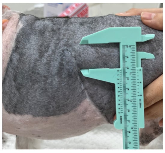
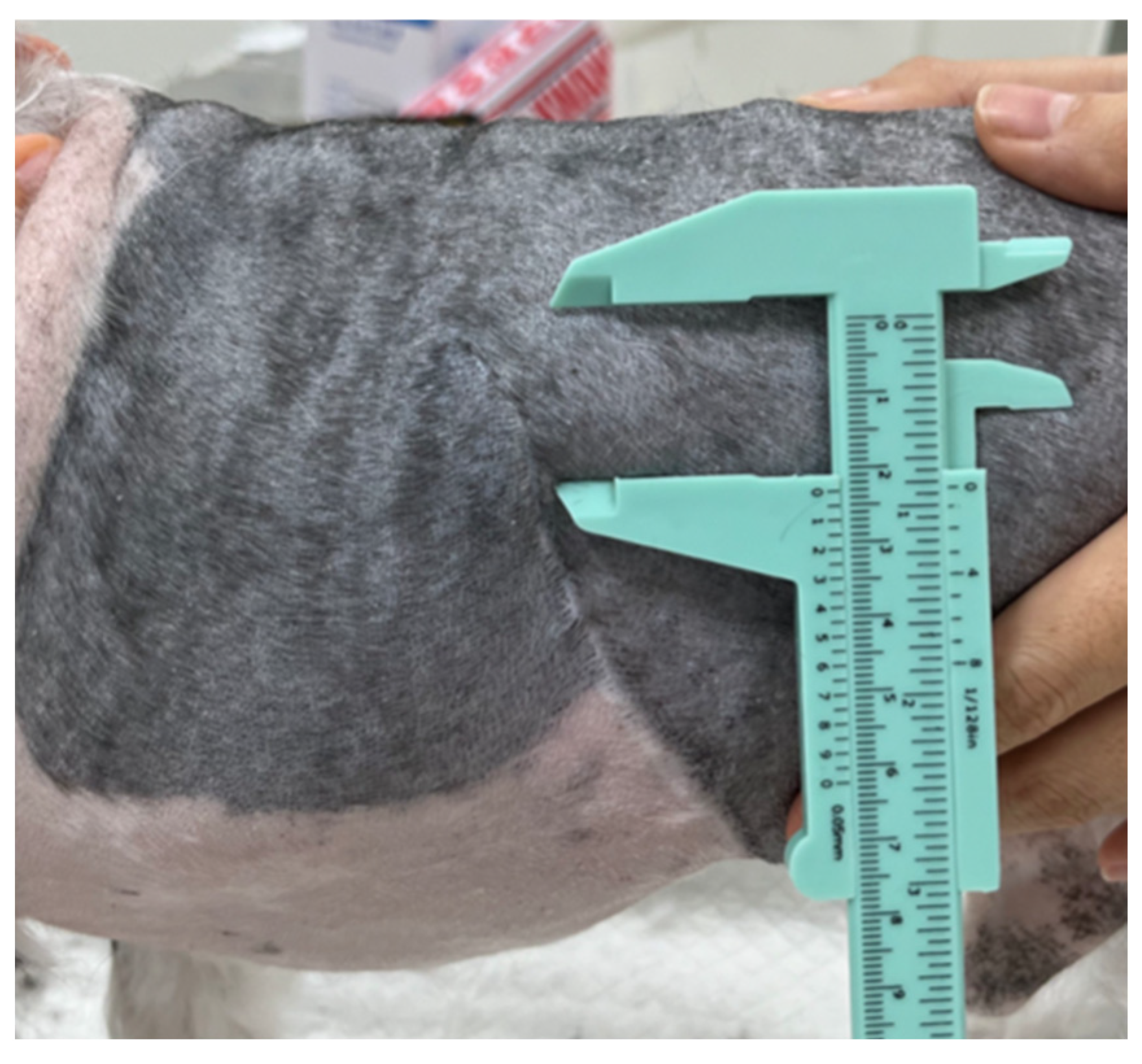
On physical examination, a firm, fixed mass measuring 2 × 2 × 0.5 cm (length × width × height) was palpated on the left chest wall at the level of the 12th rib (Figure 1). The mass had an irregular contour and showed no overt signs of inflammation, including heat, erythema, erosion, or pain on palpation.
Figure 1.
Gross appearance of the recurrent mass overlying the left 12th rib at presentation.
A complete blood count, serum biochemistry panel, electrolyte analysis, C-reactive protein, coagulation profile, D-dimer assay, and urinalysis revealed no clinically significant abnormalities that would preclude general anesthesia or surgery.
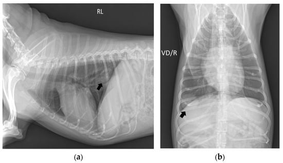
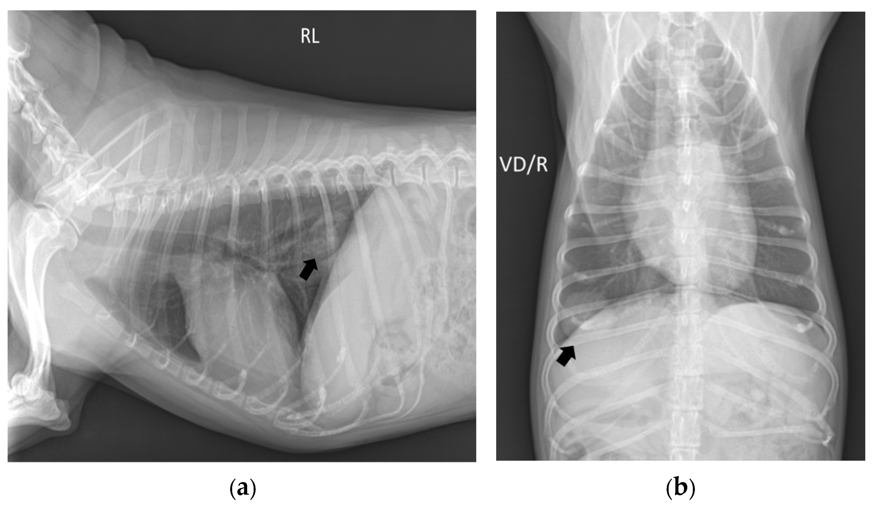
Thoracic radiography identified a pulmonary mass measuring 2.1 × 1.9 cm in the right caudal lung lobe (Figure 2). Abdominal ultrasonography performed for staging revealed hyperechoic nodules in the liver, gallbladder wall, and spleen, as well as gallbladder sludge. These lesions were considered nonspecific and not highly suspicious for metastatic disease based on their ultrasonographic appearance. However, because cytologic or histopathologic confirmation was not performed, metastatic involvement could not be definitively excluded.
Figure 2.
Thoracic radiographs showing a pulmonary mass in the right caudal lung lobe (arrow): (a) right lateral view; (b) ventrodorsal view.
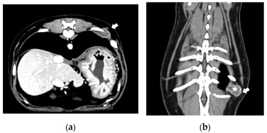
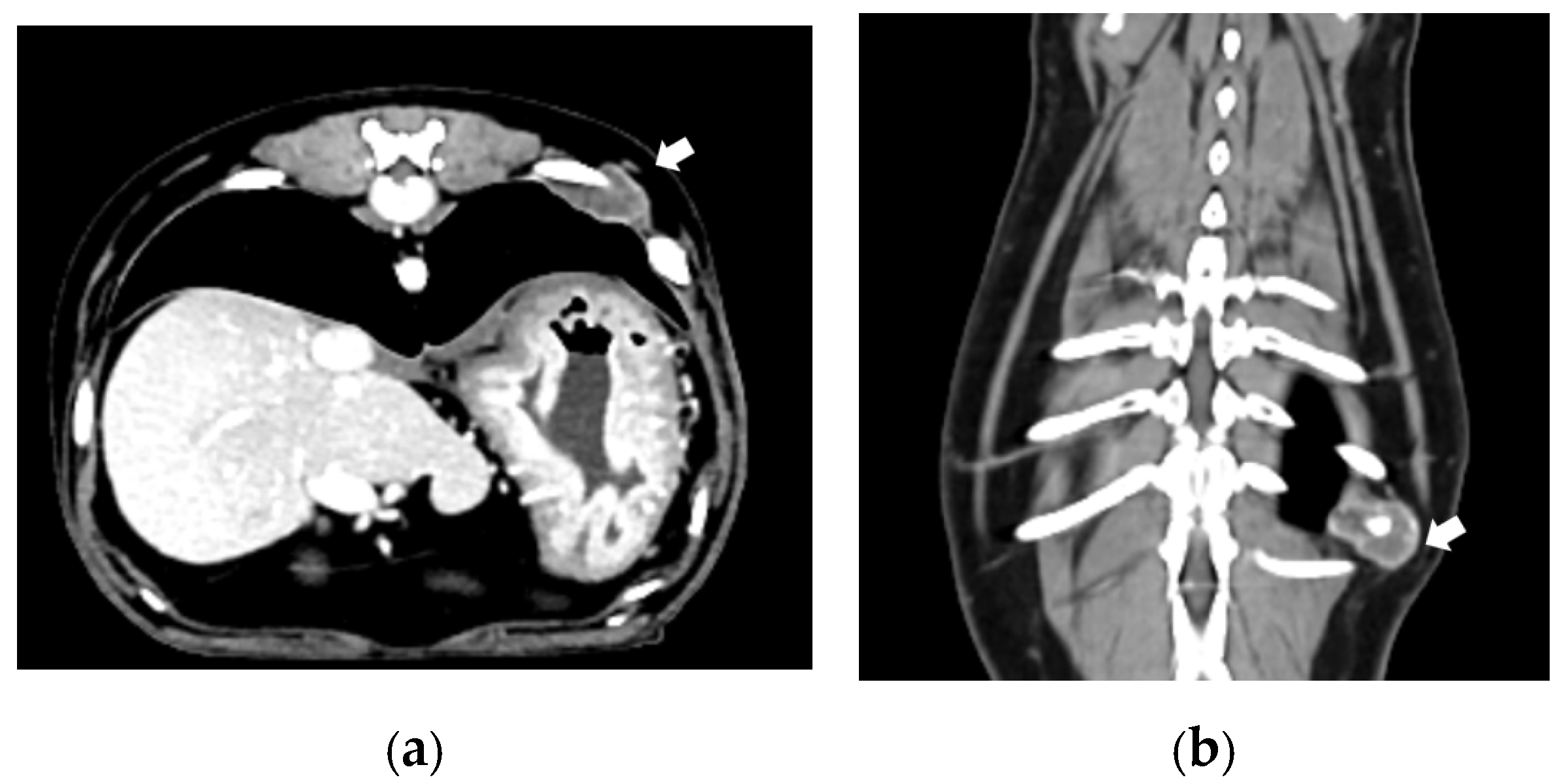
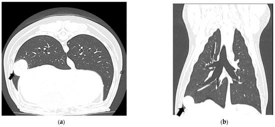
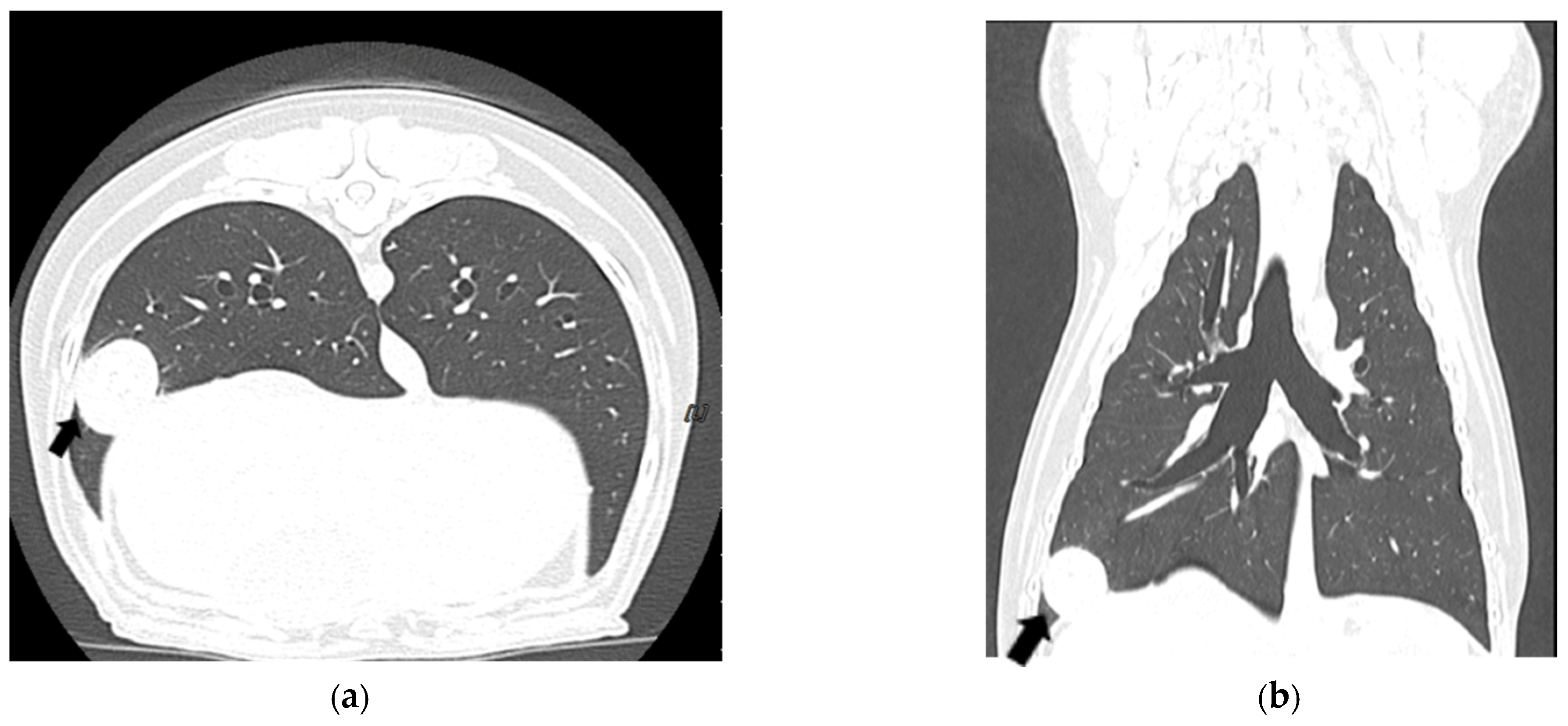
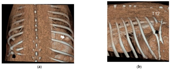
Thoracic CT (Figure 3) identified a mass centered on the left 12th rib, measuring 1.5 × 1.4 × 1.1 cm (length × width × height). The adjacent 11th and 13th ribs were closely apposed to the lesion and showed irregular margins, suggesting possible local extension or adhesion. A 2.1 × 1.5 × 1.7 cm mass was also identified in the right caudal lung lobe, with central hyperattenuation; diaphragmatic invasion could not be definitively determined. Additional pulmonary micronodules were scattered throughout the lung fields (Figure 4). Three-dimensional CT reconstruction (Figure 5) further delineated the rib and pulmonary lesions and showed healed fractures with bony union of the 11th and 13th ribs at the previous surgical site.
Figure 3.
Computed tomography images of the recurrent rib mass centered on the left 12th rib (white arrow): (a) transverse view; (b) dorsal view.
Figure 4.
Computed tomography images showing the pulmonary mass (black arrow) and pulmonary micronodules: (a) transverse view; (b) dorsal view.
Figure 5.
Three-dimensional reconstruction of the rib mass (black arrow) and pulmonary mass (white arrow): (a) dorsal view; (b) sagittal view.
Based on diagnostic imaging findings, two sequential surgical procedures were planned: (1) resection of the recurrent rib mass with thoracic wall reconstruction and (2) complete right caudal lung lobectomy for removal of the dominant pulmonary mass.
Amoxicillin–clavulanate (13.75 mg/kg) was administered for perioperative antibiotic prophylaxis. The dog was premedicated with midazolam (0.2 mg/kg IV), fentanyl (2 μg/kg IV), and maropitant (1 mg/kg SC). General anesthesia was induced with propofol (6 mg/kg IV) and maintained with isoflurane. Perioperative analgesia included meloxicam (0.2 mg/kg SC), epidural bupivacaine, an intercostal nerve block with lidocaine, and a fentanyl constant rate infusion.
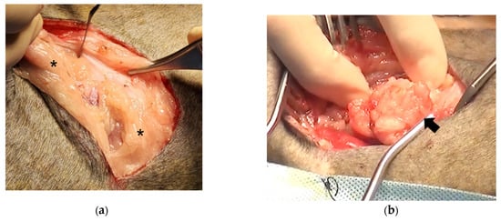
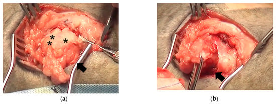
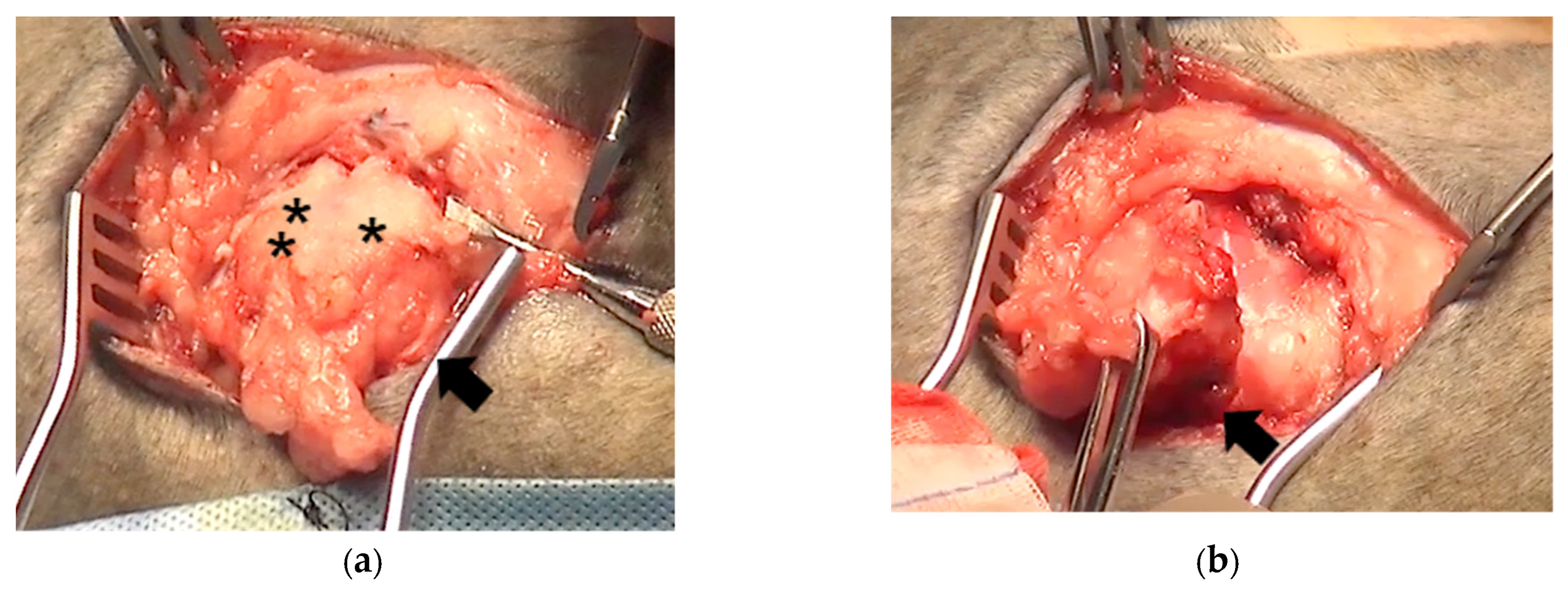
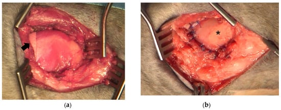
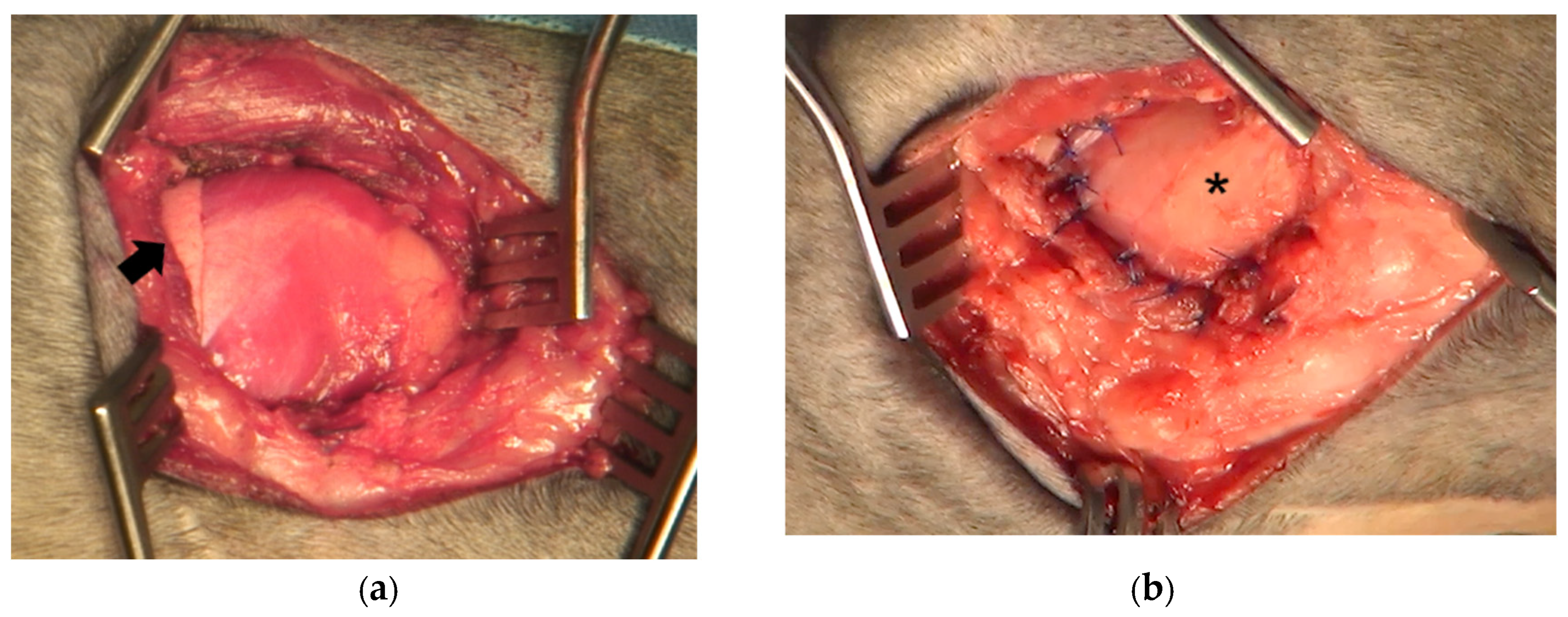
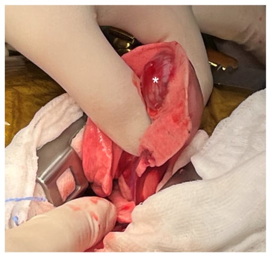
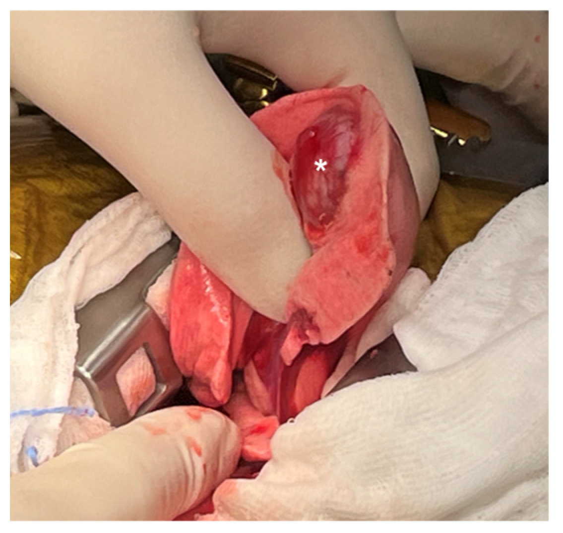
With the dog positioned in right lateral recumbency, a skin incision was made caudal to the palpable mass. On dissection, the mass was surrounded by fibrotic, desmoplastic tissue (Figure 6). The thoracic wall musculature was divided to isolate the mass, which was adherent to the adjacent 11th and 13th ribs in addition to the 12th rib of origin (Figure 7). Using an oscillating saw, the 11th, 12th, and 13th ribs were transected approximately 3 cm dorsal and ventral to the mass, and the intercostal musculature was divided to expose the pleura. The mass, together with segments of the 11th–13th ribs, was removed en bloc. Following resection, the caudal lung lobe and diaphragm were visible through the resulting thoracic wall defect (Figure 8). The defect was reconstructed by diaphragmatic advancement, with the mobilized diaphragm sutured to the intercostal musculature using 2-0 polydioxanone (PDS). The thoracic wall musculature was then closed in layers over the reconstructed diaphragm using 2-0 PDS, the subcutaneous tissues were closed using 4-0 PDS, and the skin was closed using skin staples.
Figure 6.
Intraoperative appearance of the recurrent rib mass: (a) the recurrent mass surrounded by fibrotic, desmoplastic tissue (asterisks); (b) the mass (black arrow) after further isolation from the adjacent soft tissues.
Figure 7.
Intraoperative appearance of the recurrent rib tumor and adjacent ribs: (a) the tumor (black arrow) arising from the 12th rib (double asterisks) was adherent to the 11th rib (asterisk) and the 13th rib; (b) the 11th–13th ribs were dissected to isolate the tumor (black arrow).
Figure 8.
Intraoperative reconstruction of the thoracic wall defect: (a) the caudal lung lobe (black arrow) and diaphragm were visible after tumor resection; (b) the diaphragm (asterisk) was sutured to the intercostal musculature to cover the thoracic wall defect.
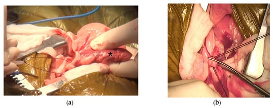
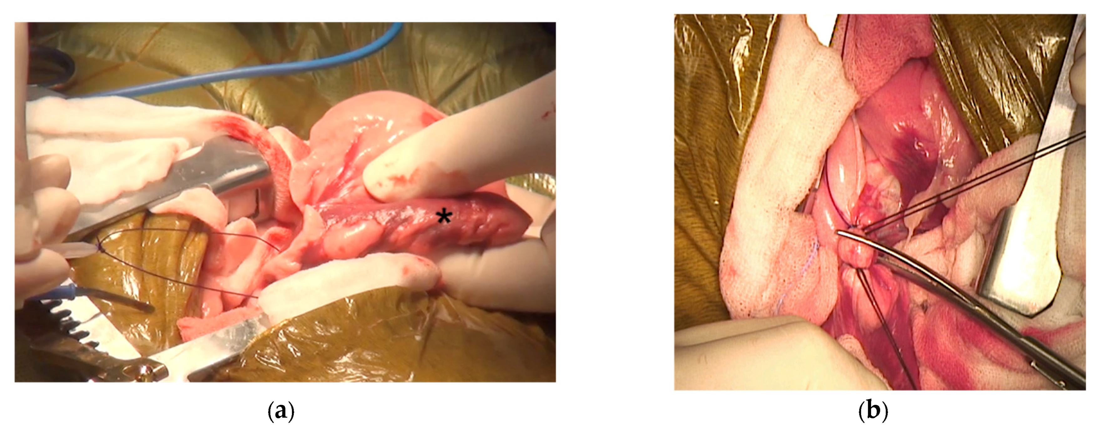
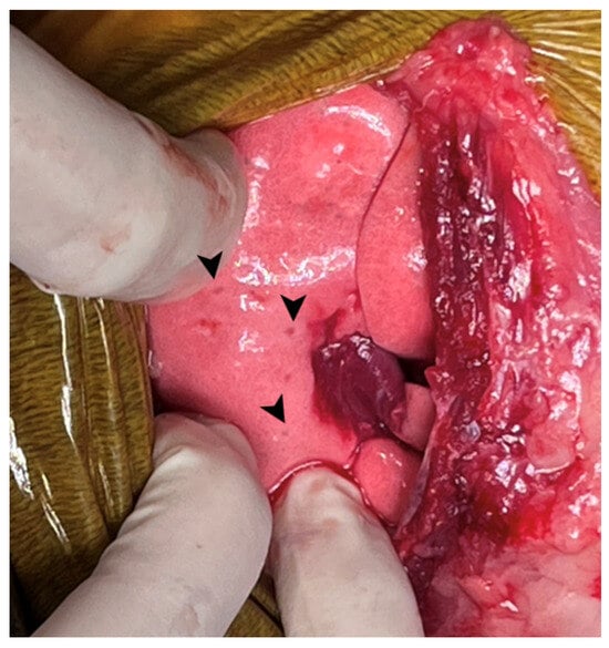
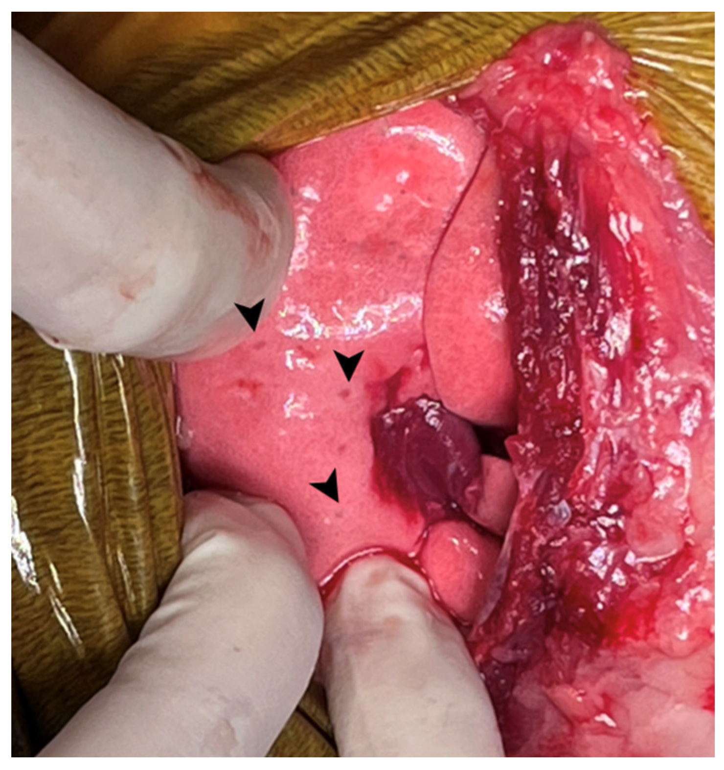
The dog was then repositioned in left lateral recumbency for the second procedure. The thoracic cavity was approached through a right sixth intercostal thoracotomy, and the right caudal lung lobe containing the previously identified pulmonary mass was isolated (Figure 9). The pulmonary artery, pulmonary vein, and bronchus were individually triple-ligated using pre-tied Endoloop ligatures and 2-0 black silk sutures, then transected with scissors to complete the lobectomy (Figure 10). The bronchial stump was tested for air leakage by submerging it in sterile saline, and no leakage was detected. Numerous micronodules were grossly visible throughout the remaining lung parenchyma (Figure 11). A thoracic drain was not placed because no air leakage was detected following lung lobectomy, and continued pleural drainage was not considered necessary based on intraoperative assessment.
Figure 9.
A pulmonary mass (asterisk) was identified in the right caudal lung lobe.
Figure 10.
Intraoperative appearance of the right caudal lung lobe and pulmonary vessels: (a) the pulmonary mass (asterisk) was identified within the right caudal lung lobe; (b) the pulmonary vessels and bronchus were ligated using Endoloop ligatures and silk sutures before transection.
Figure 11.
Multiple pulmonary micronodules (arrowheads) were identified grossly throughout the remaining lung parenchyma.
The thoracic wall was then closed using preplaced circumcostal sutures of 2-0 PDS. The serratus ventralis was closed in a simple continuous pattern using 2-0 PDS, followed by separate closure of the latissimus dorsi and scalenus muscles using 2-0 PDS. The subcutaneous tissue was closed using 4-0 PDS, and the skin was closed using skin staples. No postoperative respiratory compromise was observed, and the respiratory rate remained stable at approximately 28 breaths/min during hospitalization.
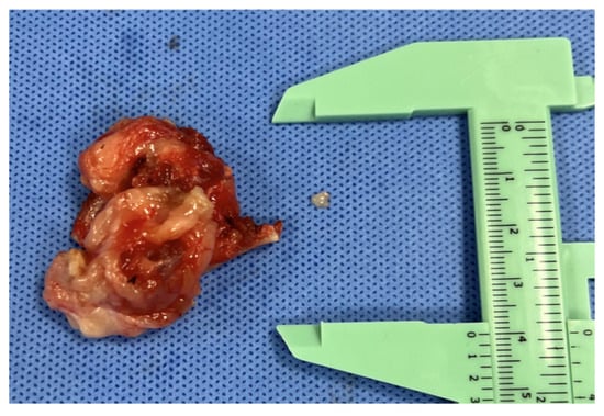
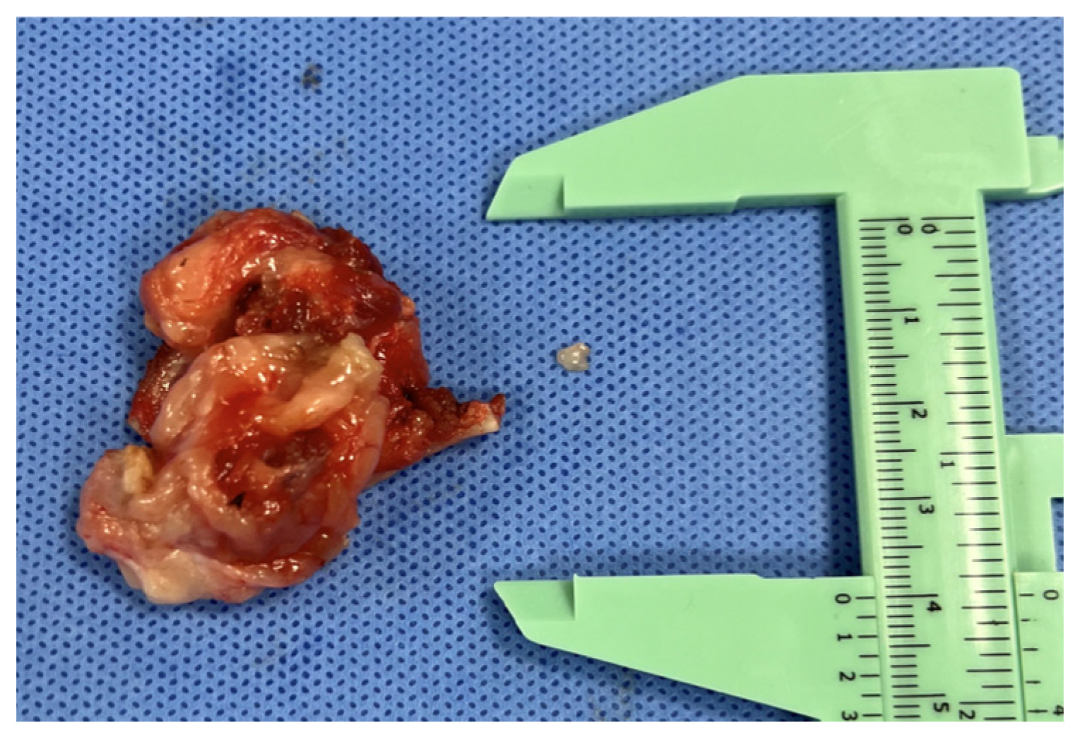
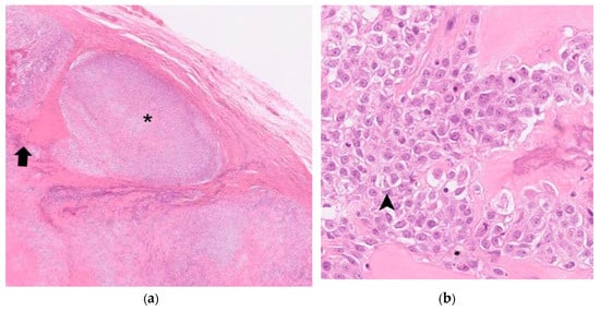
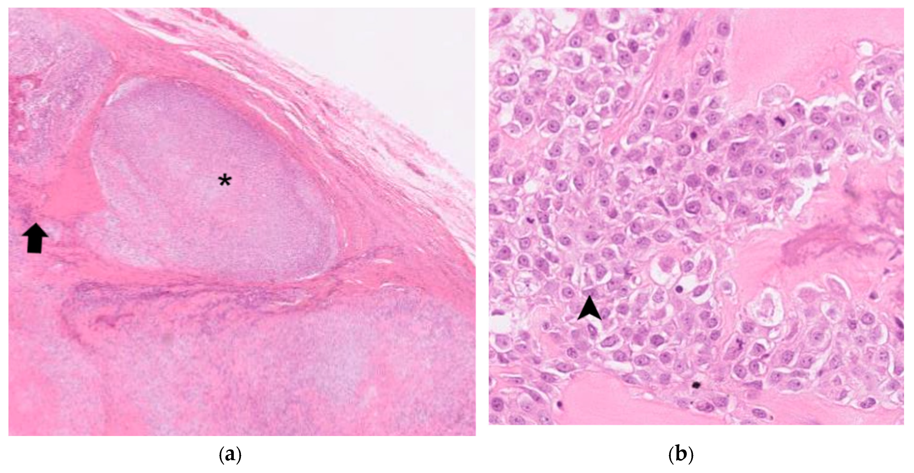
Histopathologic examination of the resected rib mass (Figure 12) confirmed grade 3 MLO, characterized by a multilobular architecture with neoplastic mesenchymal cells surrounding chondroid and osseous matrix (Figure 13). According to the previously described histologic grading system for canine MLO [8], the tumor had a total score of 13, based on an invasive border, large lobule size, moderate organization and cellular pleomorphism, high mitotic activity, and the presence of necrosis, corresponding to grade 3. The examined surgical margins were free of neoplastic cells, with the narrowest histologic margin measuring 0.37 mm, and no angiolymphatic invasion was identified.
Figure 12.
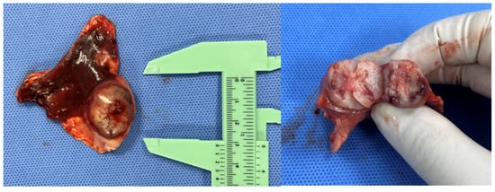
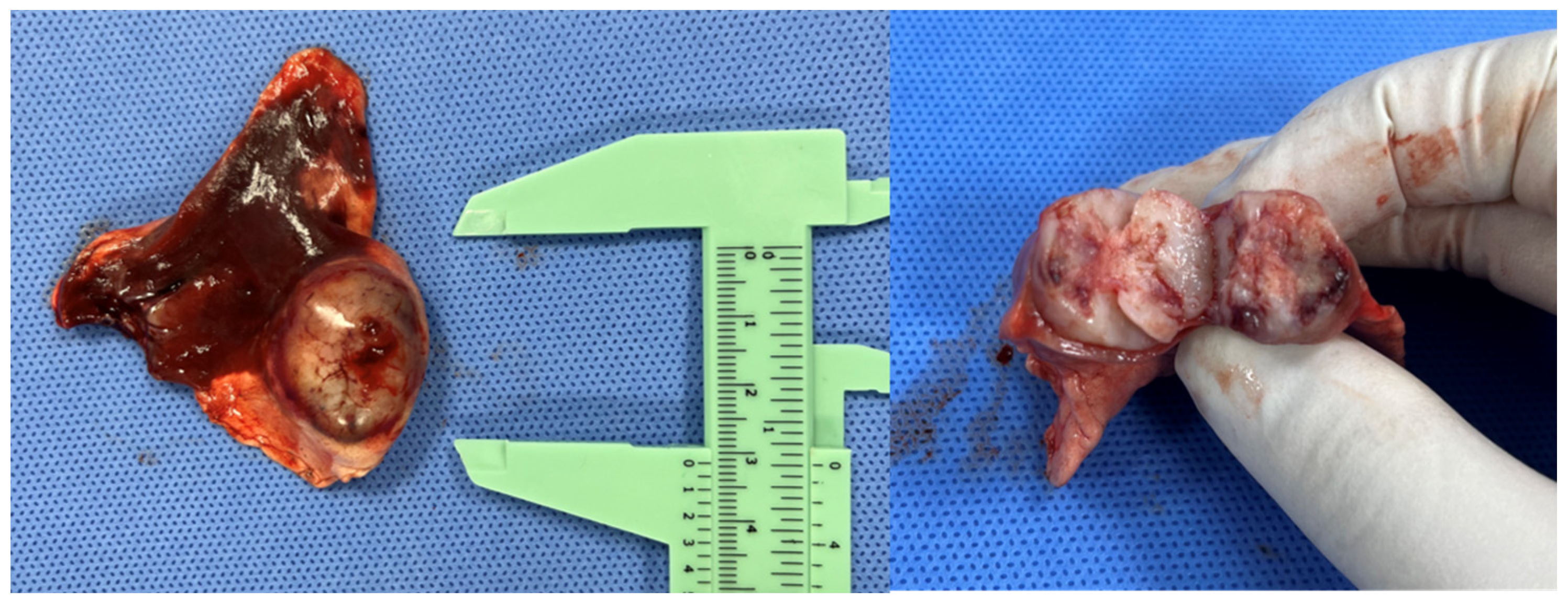
Gross appearance of the surgically excised rib mass.
Figure 13.
Histopathological evaluation of the resected rib mass: (a) Multinodular architecture characterized by confluent lobules of neoplastic mesenchymal cells surrounding chondroid matrix (asterisk) and mineralized bone (black arrow). Hematoxylin and eosin (H&E) stain, original magnification ×40. (b) Higher-magnification view showing pleomorphic polygonal to spindle-shaped neoplastic cells with moderate anisocytosis and anisokaryosis (arrowhead), supporting a diagnosis of grade 3 multilobular osteochondrosarcoma. H&E stain, original magnification ×400.
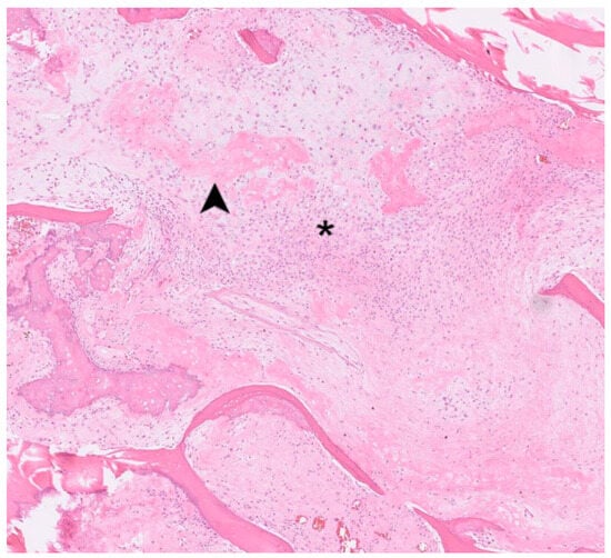
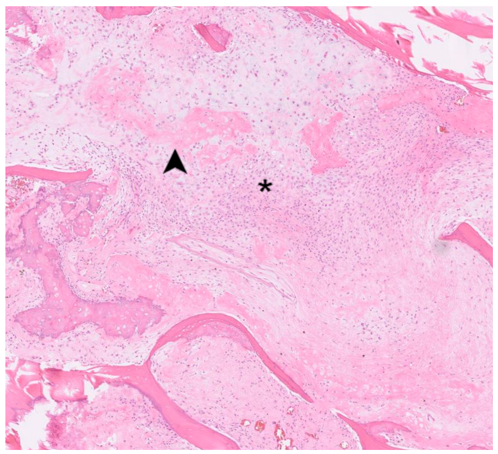
Histopathologic examination of the resected pulmonary mass, the center of which was grossly ossified (Figure 14), was consistent with pulmonary metastasis from the rib-origin MLO, although the metastatic lesion showed osteosarcomatous morphology indistinguishable from chondroblastic osteosarcoma (Figure 15).
Figure 14.
Gross appearance of the surgically excised pulmonary mass. The center of the mass was ossified.
Figure 15.
Histopathology of the resected pulmonary mass showing nodular proliferation of ovoid-to-spindle neoplastic cells (asterisk) arranged in streams around islands of mineralized osteoid. Central areas contained cartilaginous matrix with ossification and ischemic necrosis (arrowhead), consistent with pulmonary metastasis from the rib-origin MLO, with osteosarcomatous morphology. Hematoxylin and eosin (H&E) stain, original magnification ×40.
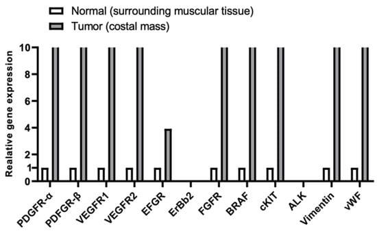
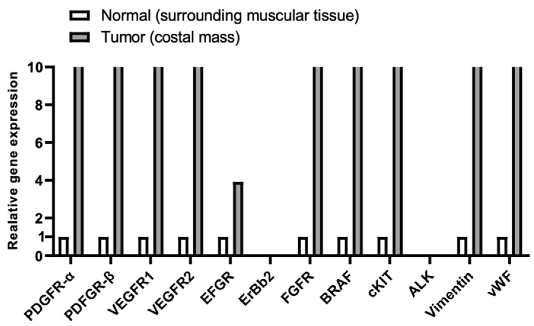
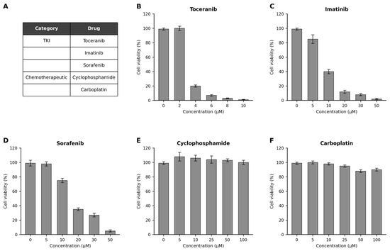
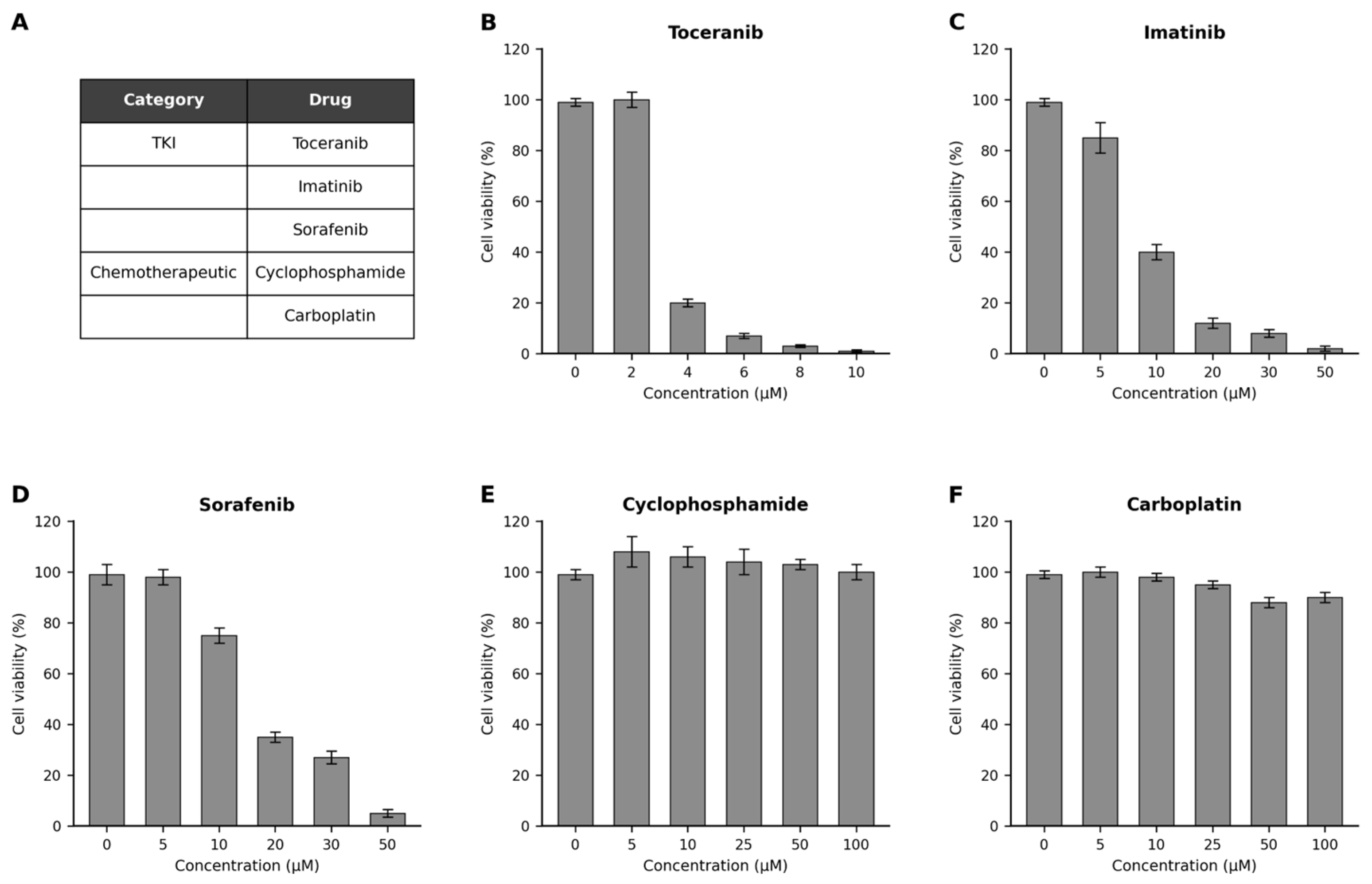
To further characterize the tumor and guide adjuvant therapy, a cancer gene expression panel and an in vitro anticancer drug-sensitivity assay were performed on the resected rib mass using previously described protocols [16,17]. Gene expression was assessed by semi-quantitative RT-PCR using adjacent normal muscle as the reference tissue. The gene expression panel (Figure 16) showed marked upregulation of PDGFR-α, PDGFR-β, VEGFR1, VEGFR2, FGFR, BRAF, cKIT, vimentin, and vWF, with a smaller increase in EGFR; ERBB2 and ALK were not upregulated. In short-term cultured tumor-derived cells, toceranib, imatinib, and sorafenib produced concentration-dependent reductions in cell viability, whereas cyclophosphamide and carboplatin showed minimal cytotoxic effects (Figure 17). Carboplatin had previously been included in the postoperative chemotherapy protocol following the initial surgery.
Figure 16.
Cancer gene expression profile of the costal tumor compared with surrounding normal muscle tissue.
Figure 17.
In vitro anticancer drug sensitivity assay of tumor cells derived from the resected costal mass. (A) Drugs evaluated in the assay. Cell viability after treatment with increasing concentrations of (B) toceranib, (C) imatinib, (D) sorafenib, (E) cyclophosphamide, and (F) carboplatin.
The dog was discharged 3 days after surgery without major postoperative complications. Based on the results of the anticancer drug sensitivity assay, adjuvant treatment with oral imatinib (10 mg/kg PO SID) was initiated [16]. At the 3-month postoperative follow-up, physical examination revealed no clinically detectable local recurrence, and thoracic radiography showed no new radiographically detectable pulmonary lesions while the dog was receiving imatinib.
3. Discussion
Multilobular osteochondrosarcoma (MLO), also referred to as multilobular osteosarcoma or multilobular tumor of bone, is an uncommon, slow-growing, and locally invasive bone tumor that most frequently arises from the flat bones of the canine skull. Although MLO can metastasize, extracranial involvement is uncommon, and rib-origin MLO is particularly rare. To our knowledge, only one previous canine case of rib-origin MLO has been documented [7,8,9,15]. In the present case, the initially resected tumor originated from the 12th rib and was diagnosed as grade 2 MLO with complete histologic excision. Despite complete excision, pulmonary micronodules suspicious of metastatic disease were identified approximately 5 months after the initial surgery, and local recurrence accompanied by a gross pulmonary mass was identified approximately 11 months after surgery. Histopathologic examination of the recurrent rib mass subsequently demonstrated grade 3 MLO. The increase in histologic grade from grade 2 in the primary tumor to grade 3 in the recurrent lesion may suggest biological progression toward a more aggressive phenotype; however, intratumoral heterogeneity or differences in tissue sampling between the two specimens cannot be excluded.
Histologic grade is considered an important prognostic factor in canine MLO. Reported local recurrence rates are approximately 30%, 47%, and 78% for grade 1, 2, and 3 tumors, respectively, while reported metastatic rates are approximately 30%, 60%, and 78% [7,8,9]. Surgical treatment is associated with substantially longer survival than no treatment, with reported median survival times of approximately 24 days in untreated dogs and 669–797 days in surgically treated dogs [8,9]. Survival also appears to vary according to histologic grade and tumor location. Reported median survival times are greater than 897 days for grade 1 tumors, 520 days for grade 2 tumors, and 405 days for grade 3 tumors. Mandibular MLO has been associated with a median survival time of 1487 days, compared with 528 days for non-mandibular cranial sites, while the reported median survival after diagnosis of recurrent or metastatic disease is 239 days [7,8]. However, most available data on MLO are based on tumors arising from the skull, and little is known about the clinical behavior and prognosis of rib-origin MLO. In this case, local recurrence and pulmonary metastasis developed despite histologically tumor-free margins after excision of the initial grade 2 tumor, indicating that complete histologic margins do not necessarily rule out subsequent disease progression. The higher histologic grade of the recurrent tumor may reflect more aggressive biological behavior at recurrence.
Wide excision of a rib tumor can leave a large thoracic wall defect that requires reconstruction to maintain thoracic stability and normal respiratory function. In this case, the recurrent mass was resected together with the 11th, 12th, and 13th ribs, and the defect was closed by diaphragmatic advancement. This technique makes use of the marked convexity of the canine diaphragm and has been associated with a low complication rate and rapid postoperative recovery [14]. In contrast, prosthetic reconstruction has been reported to have complication rates as high as 66.7% [13]. Autogenous reconstruction techniques are generally considered appropriate when up to three ribs are resected [13]. Because ribs 9–13 do not attach directly to the sternum, the diaphragm can be advanced cranially to the level of the eighth intercostal space without significantly compromising lung expansion [14]. In the present case, the caudal location of the defect, its proximity to the diaphragm, and the resection of three contiguous ribs made diaphragmatic advancement an appropriate autogenous reconstructive option. Diaphragmatic advancement allowed tension-free closure, and no major postoperative respiratory complications occurred.
The dominant pulmonary lesion was resected because of the presence of a gross mass in the right caudal lung lobe. The generally proposed criteria for pulmonary metastasectomy, which are derived largely from human thoracic oncology, include (1) an acceptable surgical risk, (2) control of the primary tumor, (3) absence of uncontrolled extrapulmonary metastatic disease, and (4) the possibility of complete resection of the pulmonary metastases [18]. In this dog, three of these four criteria were met. The dog was considered an acceptable anesthetic candidate, the recurrent primary tumor was considered surgically controllable and was resected en bloc during the same anesthetic episode, and no uncontrolled extrapulmonary metastatic disease was identified. However, complete resection of the pulmonary metastatic disease was not possible because CT revealed multiple additional pulmonary micronodules.
Despite this limitation, removal of the dominant pulmonary lesion was considered worthwhile. MLO generally has a relatively slow clinical course, with metastasis often occurring late and survival after metastatic disease sometimes being prolonged [9]. In this case, the dominant pulmonary mass was substantially larger than the remaining nodules and could be completely removed by lobectomy. Its removal also allowed histopathological confirmation of metastatic pulmonary disease, which had previously been suspected only based on imaging findings. Pulmonary metastasectomy has been associated with longer survival in selected dogs with osteosarcoma [19,20], although whether removal of a dominant lesion in the presence of unresected pulmonary nodules provides a similar benefit is unknown. Accordingly, the procedure was undertaken to remove the major macroscopic pulmonary lesion and obtain a definitive histopathological diagnosis, rather than to achieve complete clearance of pulmonary metastatic disease.
The role of systemic therapy in canine MLO remains poorly defined. Conventional chemotherapy has not been shown to provide a clear survival benefit over surgery alone, and evidence supporting targeted therapy is limited [9]. Imatinib has previously been used in a dog with an incompletely resected multilobular tumor of bone, in which a reduced rate of tumor growth was reported during treatment with imatinib and carprofen [16]. In the present case, in vitro drug-sensitivity testing showed concentration-dependent cytotoxic activity with toceranib, imatinib, and sorafenib, whereas carboplatin and cyclophosphamide produced little reduction in tumor-cell viability. Carboplatin had already been used in the initial chemotherapy protocol. Given the observed in vitro sensitivity and the previous report of imatinib use in canine MLO, postoperative treatment with imatinib was initiated at 10 mg/kg PO SID.
The dog recovered without major postoperative complications and was discharged 3 days after surgery. At the 3-month postoperative follow-up, no local recurrence or new radiographically detectable pulmonary lesions were identified on physical examination and thoracic radiography while the dog was receiving imatinib.
This case has several limitations. The postoperative follow-up period was limited to 3 months, which is insufficient to assess long-term disease control in a tumor with a relatively slow clinical course. In addition, postoperative surveillance was based on thoracic radiography rather than CT, and the previously identified pulmonary micronodules could therefore not be reliably reassessed. The effects of resection of the dominant pulmonary lesion and postoperative imatinib therapy require further evaluation in additional cases with longer follow-up.
4. Conclusions
This report describes the surgical management of recurrent rib-origin grade 3 MLO with pulmonary metastasis using en bloc resection of three caudal ribs, diaphragmatic advancement for thoracic wall reconstruction, resection of the dominant pulmonary metastatic lesion, and postoperative sensitivity-guided imatinib therapy. Rib-origin MLO is rarely reported, and its prognosis and optimal treatment have not been well established. This case demonstrates that combined surgical management of both local recurrence and a resectable dominant pulmonary metastatic lesion was technically feasible, even when multiple caudal ribs are involved. However, the short follow-up period and residual pulmonary micronodules preclude conclusions regarding long-term efficacy, and additional cases will be needed to clarify the roles of resection of the dominant pulmonary metastatic lesion and targeted systemic therapy in dogs with rib-origin MLO.
Author Contributions
Conceptualization, H.L. and J.-M.K.; investigation, J.P. and H.-Y.K.; resources, J.P.; writing—original draft preparation, H.L.; writing—review and editing, J.-M.K. and H.-Y.K.; visualization, J.K. and K.E.; supervision, H.-Y.K. and J.-M.K.; project administration, J.-M.K. All authors have read and agreed to the published version of the manuscript.
Funding
This research received no external funding.
Institutional Review Board Statement
Ethical review and approval were waived for this study because it describes a single retrospective clinical case managed according to standard diagnostic and therapeutic procedures. No experimental interventions were performed, and all procedures were conducted as part of routine veterinary care.
Informed Consent Statement
Informed consent was obtained from the owner of the animal involved in the study.
Data Availability Statement
The original contributions presented in this study are included in the article. Further inquiries can be directed to the corresponding author.
Conflicts of Interest
The authors declare no conflicts of interest.
Abbreviations
The following abbreviations are used in this manuscript:
| ALK | Anaplastic lymphoma kinase |
| BRAF | B-Raf proto-oncogene, serine/threonine kinase |
| CT | Computed tomography |
| EGFR | Epidermal growth factor receptor |
| ERBB2 | Erb-B2 receptor tyrosine kinase 2 |
| FGFR | Fibroblast growth factor receptor |
| IV | Intravenous |
| MLO | Multilobular osteochondrosarcoma |
| PDGFR-α | Platelet-derived growth factor receptor alpha |
| PDGFR-β | Platelet-derived growth factor receptor beta |
| PO | Per os (orally) |
| SC | Subcutaneous |
| SID | Once daily |
| TKI | Tyrosine kinase inhibitor |
| VEGFR1 | Vascular endothelial growth factor receptor 1 |
| VEGFR2 | Vascular endothelial growth factor receptor 2 |
| cKIT | KIT proto-oncogene receptor tyrosine kinase |
| vWF | von Willebrand factor |
References
- Dernell, W.S.; Ehrhart, N.P.; Straw, R.C.; Vail, D.M. Tumors of the skeletal system. In Withrow and MacEwen’s Small Animal Clinical Oncology, 4th ed.; Withrow, S.J., Vail, D.M., Eds.; W.B. Saunders: St. Louis, MO, USA, 2007; pp. 540–582. [Google Scholar]
- Heyman, S.J.; Diefenderfer, D.L.; Goldschmidt, M.H.; Newton, C.D. Canine axial skeletal osteosarcoma: A retrospective study of 116 cases (1986 to 1989). Vet. Surg. 1992, 21, 304–310. [Google Scholar] [CrossRef] [Scilit] [PubMed]
- Hammer, A.S.; Weeren, F.R.; Weisbrode, S.E.; Padgett, S.L. Prognostic factors in dogs with osteosarcomas of the flat or irregular bones. J. Am. Anim. Hosp. Assoc. 1995, 31, 321–326. [Google Scholar] [CrossRef] [Scilit] [PubMed]
- Matthiesen, D.T.; Clark, G.N.; Orsher, R.J.; Pardo, A.O.; Glennon, J.; Patnaik, A.K. En bloc resection of primary rib tumors in 40 dogs. Vet. Surg. 1992, 21, 201–204. [Google Scholar] [CrossRef] [Scilit] [PubMed]
- Pirkey-Ehrhart, N.; Withrow, S.J.; Straw, R.C.; Ehrhart, E.J.; Page, R.L.; Hottinger, H.L.; Hahn, K.A.; Morrison, W.B.; Albrecht, M.R.; Hedlund, C.S.; et al. Primary rib tumors in 54 dogs. J. Am. Anim. Hosp. Assoc. 1995, 31, 65–69. [Google Scholar] [CrossRef] [Scilit] [PubMed]
- Liptak, J.M.; Kamstock, D.A.; Dernell, W.S.; Monteith, G.J.; Rizzo, S.A.; Withrow, S.J. Oncologic outcome after curative-intent treatment in 39 dogs with primary chest wall tumors (1992–2005). Vet. Surg. 2008, 37, 488–496. [Google Scholar] [CrossRef] [Scilit] [PubMed]
- Ettinger, S.J.; Feldman, E.C. Textbook of Veterinary Internal Medicine, 7th ed.; Saunders Elsevier: St. Louis, MO, USA, 2010. [Google Scholar]
- Dernell, W.S.; Straw, R.C.; Cooper, M.F.; Powers, B.E.; LaRue, S.M.; Withrow, S.J. Multilobular osteochondrosarcoma in 39 dogs: 1979–1993. J. Am. Anim. Hosp. Assoc. 1998, 34, 11–18. [Google Scholar] [CrossRef] [Scilit] [PubMed]
- Straw, R.C.; LeCouteur, R.A.; Powers, B.E.; Withrow, S.J. Multilobular osteochondrosarcoma of the canine skull: 16 cases (1978–1988). J. Am. Vet. Med. Assoc. 1989, 195, 1764–1769. [Google Scholar] [CrossRef] [Scilit]
- Hathcock, J.T.; Newton, J.C. Computed tomographic characteristics of multilobular tumor of bone involving the cranium in 7 dogs and zygomatic arch in 2 dogs. Vet. Radiol. Ultrasound 2000, 41, 214–217. [Google Scholar] [CrossRef] [Scilit] [PubMed]
- Lipsitz, D.; Levitski, R.E.; Berry, W.L. Magnetic resonance imaging features of multilobular osteochondrosarcoma in 3 dogs. Vet. Radiol. Ultrasound 2001, 42, 14–19. [Google Scholar] [CrossRef] [Scilit] [PubMed]
- Orton, E.C.; Monnet, E. Small Animal Thoracic Surgery, 2nd ed.; Wiley-Blackwell: Ames, IA, USA, 2018. [Google Scholar]
- Liptak, J.M.; Dernell, W.S.; Rizzo, S.A.; Monteith, G.J.; Kamstock, D.A.; Withrow, S.J. Reconstruction of chest wall defects after rib tumor resection: A comparison of autogenous, prosthetic, and composite techniques in 44 dogs. Vet. Surg. 2008, 37, 479–487. [Google Scholar] [CrossRef] [Scilit] [PubMed]
- Aronsohn, M. Diaphragmatic advancement for defects of the caudal thoracic wall in the dog. Vet. Surg. 1984, 13, 26–28. [Google Scholar] [CrossRef] [Scilit]
- Jacobson, S.A. The Comparative Pathology of the Tumors of Bone; Charles C. Thomas: Springfield, IL, USA, 1971; pp. 102–109. [Google Scholar]
- Lee, S.H.; Kim, D.H.; Bae, K.; Jeong, S.W.; Yoon, K.A.; Yoon, H.Y.; Do, S.H.; Kim, B.; Kim, J.H. Adjuvant therapy with imatinib for an incompletely resected multilobular tumour of bone in a dog. Vet. Med. 2022, 67, 590–597. [Google Scholar] [CrossRef] [Scilit] [PubMed]
- Kim, Y.R.; Bae, K.; Lee, J.Y.; Jeong, S.W.; Yoon, H.Y.; Han, H.J.; Hyun, J.E.; Nam, A.; Park, J.H.; Yoon, K.A.; et al. Clinical utility of patient-derived cell-based in vitro drug sensitivity testing for optimizing adjuvant therapy in dogs with solid tumors: A retrospective study (2019–2023). Animals 2025, 15, 1146. [Google Scholar] [CrossRef] [Scilit] [PubMed]
- Kanzaki, R.; Fukui, E.; Kanou, T.; Ose, N.; Funaki, S.; Minami, M.; Shintani, Y.; Okumura, M. Preoperative evaluation and indications for pulmonary metastasectomy. J. Thorac. Dis. 2021, 13, 2590–2602. [Google Scholar] [CrossRef] [Scilit] [PubMed]
- O’Brien, M.G.; Straw, R.C.; Withrow, S.J.; Powers, B.E.; Jameson, V.J.; Lafferty, M.; Ogilvie, G.K.; LaRue, S.M. Resection of pulmonary metastases in canine osteosarcoma: 36 cases (1983–1992). Vet. Surg. 1993, 22, 105–109. [Google Scholar] [CrossRef] [Scilit] [PubMed]
- Turner, H.; Séguin, B.; Worley, D.R.; Ehrhart, N.P.; Lafferty, M.H.; Withrow, S.J.; Selmic, L.E. Prognosis for dogs with stage III osteosarcoma following treatment with amputation and chemotherapy with and without metastasectomy. J. Am. Vet. Med. Assoc. 2017, 251, 1293–1305. [Google Scholar] [CrossRef] [Scilit] [PubMed]
Disclaimer/Publisher’s Note: The statements, opinions and data contained in all publications are solely those of the individual author(s) and contributor(s) and not of MDPI and/or the editor(s). MDPI and/or the editor(s) disclaim responsibility for any injury to people or property resulting from any ideas, methods, instructions or products referred to in the content. |
© 2026 by the authors. Licensee MDPI, Basel, Switzerland. This article is an open access article distributed under the terms and conditions of the Creative Commons Attribution (CC BY) license.