


Abstract
Hereditary breast cancer (BC) remains unexplained in a substantial proportion of families who test negative for BRCA1/2 and other known susceptibility genes. To contribute to the genomic characterization of these unresolved cases, we generated a whole-genome sequencing (WGS) dataset from six women belonging to two unrelated high-risk families, each comprising three sisters diagnosed with BC. All participants had previously received negative results in conventional multigene panel testing. WGS was performed on peripheral blood DNA using the Illumina NovaSeq platform, followed by variant calling against GRCh38 and the comprehensive annotation of single-nucleotide variants, indels, and structural variants. For each family, we identified shared ClinVar-annotated variants, rare exonic or splice-site alterations, and intronic variants located within a curated set of 286 cancer-related genes. The dataset includes per-patient VCF files, copy number variation annotations, and family-level variant summaries. Raw and processed data are publicly available through the Sequence Read Archive and Zenodo. This resource supports variant reinterpretation, exploration of regulatory and intronic regions, and methodological benchmarking in the study of familial BC beyond established susceptibility genes.
Dataset: The datasets are available at NCBI SRA under BioProject PRJNA1268872 and at Zenodo (DOI: 10.5281/zenodo.15534738).
Dataset License: CC-BY 4.0
1. Summary
Hereditary breast cancer (BC) accounts for approximately 5–10% of all BC cases, with germline pathogenic variants in BRCA1 and BRCA2 being the most well-characterized contributors to familial risk [1,2]. Studies have identified additional moderate- and high-penetrance genes, such as PALB2, CHEK2, ATM, BARD1, RAD51C/D, TP53, and CDH1, which are implicated in BC susceptibility [3,4]. In addition, it is estimated that 18% of the BC genetic risk is explained by a combination of low-risk common variants [5]. To date, more than 2000 single-nucleotide polymorphisms (SNPs) have been associated with BC risk in different Genome-wide Association Studies (GWAS) [6], and a subset of 313 of those common variants has been suggested to have risk-predictive value [7].
In clinical practice, next-generation sequencing (NGS) technologies are routinely applied to investigate hereditary BC using targeted multigene panels, whole-exome sequencing (WES), or whole-genome sequencing (WGS), depending on clinical indication and available resources. While gene panels are widely used for diagnostics due to their cost-effectiveness and focused scope, WES and WGS provide a broader view of the genome, enabling the detection of rare variants, structural rearrangements, and non-coding alterations that may contribute to unresolved familial cases [8].
Beyond rare pathogenic variants, the cumulative contribution of common SNPs to BC susceptibility can be estimated through polygenic risk scores (PRSs), which quantify an individual’s genetic predisposition based on the weighted sum of multiple low-risk alleles [7,9,10]. PRSs are increasingly being investigated for their potential clinical application in personalized risk prediction, complementing established hereditary factors such as BRCA1/2 mutations [11].
The advent of WGS has proven instrumental in uncovering both coding and non-coding variants, structural rearrangements, and mutational signatures that characterize tumor genomes and inform cancer biology [12,13,14]. Beyond its applications in sporadic tumors, WGS is increasingly being applied to germline analyses in familial BC unexplained by BRCA1/2 mutations, enabling the identification of novel susceptibility genes and variants in regulatory or intronic regions [15]. Notably, increasing evidence indicates that non-coding regulatory variants, such as promoter alterations in FOXA1 and MDM2 in BC, may influence gene expression and tumorigenesis [16,17,18]. These findings further underscore the value of WGS in revealing mechanisms that extend beyond protein-coding alterations.
In this study, WGS was performed on peripheral blood samples from six patients belonging to two unrelated families with a history of familial BC. All participants had previously tested negative for pathogenic variants in BRCA1, BRCA2, and other routinely screened BC susceptibility genes. The resulting dataset provides a comprehensive catalog of shared and individual germline variants for each family, enabling future re-annotation, comparison, and reuse. All raw and processed WGS files, together with derived variant summaries, have been deposited in publicly accessible repositories to support transparency, reproducibility, and downstream analyses.
We acknowledge that the small sample size represents an inherent limitation of this dataset; however, as a Data Descriptor, this work aims to share a genomic resource to support future reuse, reanalysis, and variant reinterpretation rather than to establish definitive clinical or epidemiological conclusions.
2. Data Description
This dataset contains WGS data and processed genomic annotations from six women belonging to two unrelated families with familial BC. All files have been deposited in public repositories to facilitate transparent reuse. The raw FASTQ files are available in the NCBI Sequence Read Archive under BioProject PRJNA1268872 (BioSamples SAMN48764618–SAMN48764623), while all processed data, including VCFs, structural variant annotations, and shared-variant tables, are hosted in Zenodo under DOI 10.5281/zenodo.15534738.
Each sample corresponds to one patient, and this mapping is essential for interpreting the per-patient genomic files. The sample IDs and their corresponding individuals are as follows: 21ID01084 corresponds to Family1_Pt1; 21ID01083 to Family1_Pt2; 21ID01085 to Family1_Pt3; 21ID01086 to Family2_Pt4; 21ID01138 to Family2_Pt5; and 21ID01087 to Family2_Pt6. This mapping applies consistently across the VCF files and the CNV annotation files.
For each of the six patients, a hard-filtered VCF file (.hard-filtered.vcf.gz) is provided. These files contain single-nucleotide variants and small insertions or deletions aligned to the GRCh38 reference genome and reflect the full set of germline variants retained after quality filtering. In parallel, structural variants for every patient are supplied as AnnotSV-generated TSV files (_AnnotSV_CNV.tsv), which include genomic coordinates, affected genes, ClinGen dosage sensitivity annotations, population frequencies, regulatory element overlaps, and the ACMG/ClinGen-based AnnotSV classification score.
To contextualize the shared genetic profiles that motivated the study, several summary tables are provided for each family. These include lists of variants shared by the three sisters in each family that are annotated in ClinVar (clinvar.tsv), as well as independent files summarizing rare exonic or splice-site variants (latest_exonic.tsv). Another set of summary files contains the shared variants located in a curated panel of 286 cancer-related genes (geneFilter.tsv). Together, these tables allow users to explore patterns of shared variation at multiple levels, ClinVar interpretation, coding variation, and cancer-gene relevance, without needing to manually re-derive these intersections.
The variants highlighted below are representative examples; the complete variant lists are available in the deposited dataset.
2.1. Family 1—Shared Germline Variants
WGS revealed several ClinVar-annotated variants shared among the three sisters in Family 1. A frameshift deletion in DUOX2 and a stop-gain variant in FCGR1A were classified by ClinVar as pathogenic or likely pathogenic. Additional shared pathogenic variants included a 5′UTR variant in HBG1 and an intronic variant in EHBP1. Several shared variants were annotated as risk-factor variants, including entries in IL13 and CDKAL1 (Family1_Clinvar.tsv, Zenodo).
Rare exonic variants not reported in ClinVar but predicted to be deleterious were identified in genes such as PIDD1, FAM219B, and MRPL43, and are listed in Family1_latest_exonic.tsv. Within the curated panel of 286 cancer-related genes, shared intronic variants were detected in genes frequently examined in cancer genomics, including RAD51, PIK3CA, MSH3, and MAP3K1 (Family1_geneFilter.tsv, Zenodo).
Although some CNVs classified as pathogenic (AnnotSV ranking = 5) were identified in Family 1 (e.g., deletions involving CFHR1/CFHR3 and ARHGAP22/ARHGAP22-IT1), these were not shared across all three sisters (AnnotSV_CNV.tsv, Zenodo).
2.2. Family 2—Shared Germline Variants
No shared ClinVar-annotated variants classified as pathogenic or likely pathogenic were identified among the three sisters in Family 2. Shared ClinVar entries mainly consisted of variants categorized as risk factors, variants of uncertain significance, or variants with conflicting interpretation, including entries in RNASEL, IL13, and IFITM3. Additional ClinVar risk-factor annotations were found in genes such as SLC30A8, NPSR1, and variants within TNF-related regulatory regions (Family2_Clinvar.tsv, Zenodo).
Rare exonic or splice-site variants not listed in ClinVar but predicted to be deleterious were also identified. These included variants in FAN1, NDUFB1, MRPS9, SLC5A10, and LRRC30 (Family2_latest_exonic.tsv, Zenodo).
Within the curated 286-gene cancer panel, several shared intronic variants were observed, including entries in RAD51, PALB2, NF1, MAP2K2, ERBB4, ABL1, and CDKN2A. (Family2_geneFilter.tsv, Zenodo).
Family 2 did not present any CNVs classified as pathogenic (AnnotSV ranking = 5), (AnnotSV_CNV.tsv, Zenodo).
A summary of the variants mentioned in the text has been added as Table S2. It should be noted that ClinVar classifications reflect general pathogenicity assessments and do not establish a direct causal association with BC susceptibility in these families; their biological relevance in the context of familial BC risk requires further functional validation.
3. Methods
3.1. Patients
Six women from two unrelated families, each with three sisters diagnosed with BC, were included. All had previously tested negative for pathogenic variants in a multigene panel covering BRCA1, BRCA2, ATM, CHEK2, PALB2, BRIP1, RAD51C, RAD51D, MLH1, MSH2, MSH6, CDH1, EPCAM, NBN, PMS2, PTEN, STK11, and TP53, with variant classification performed according to ACMG guidelines. Clinical data such as diagnosis, age, tumor histology, molecular subtype, and tumor, node, and metastasis (TNM) staging were retrieved from medical records.
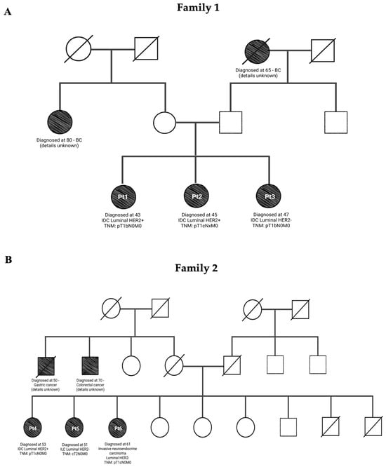
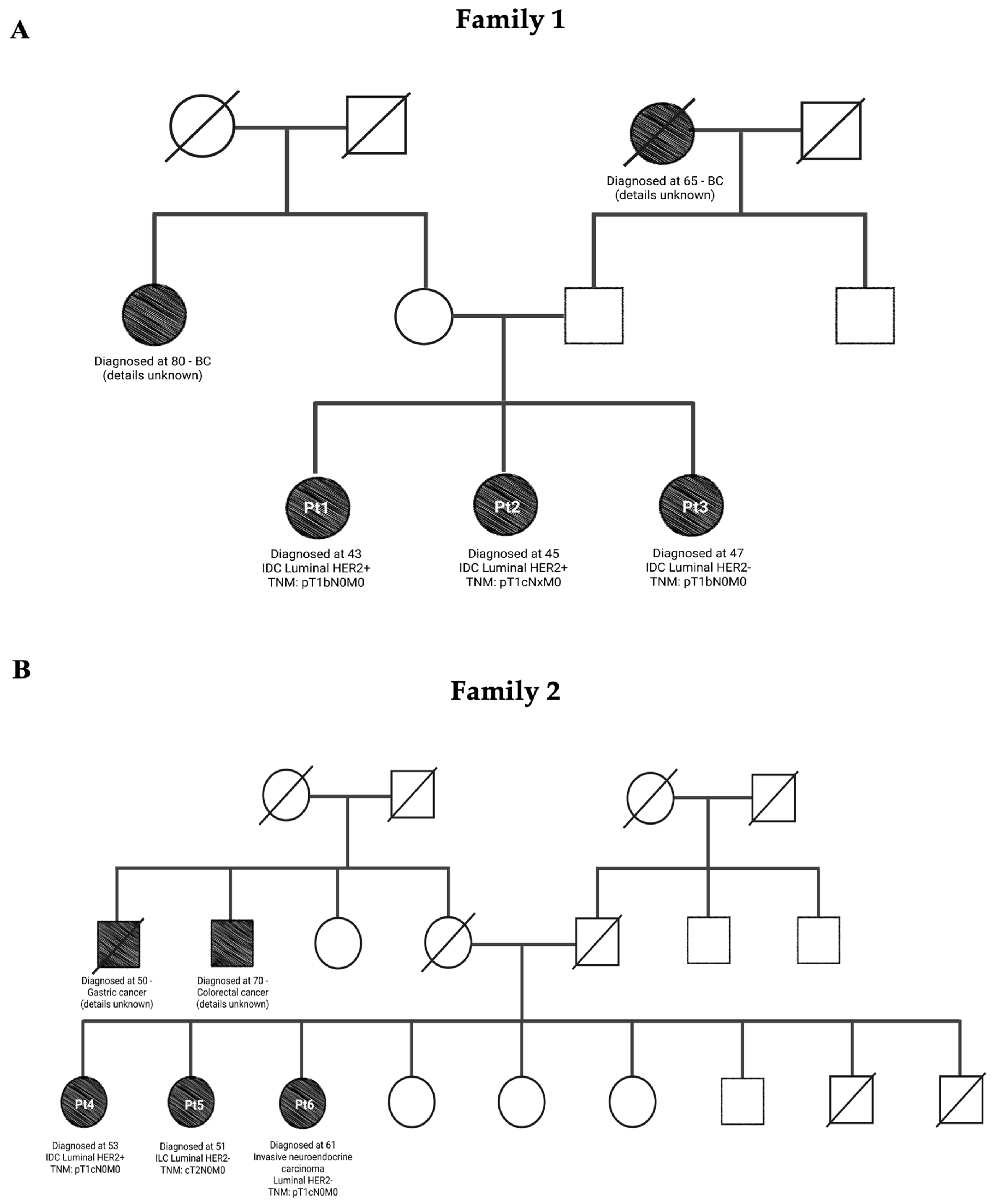
Table 1 summarizes the clinicopathological characteristics of the six patients analyzed. In Family 1, all three sisters were diagnosed with invasive ductal carcinoma (IDC), two of them being luminal HER2-positive and one being luminal HER2-negative. In Family 2, the tumor types were more heterogeneous, including invasive lobular carcinoma (ILC) and invasive neuroendocrine carcinoma, with two of them being luminal HER2-negative and one being luminal HER2-positive. The ages at diagnosis ranged from 43 to 47 years old in Family 1 and 51 to 61 years old in Family 2. Most tumors were early-stage (T1) and node-negative at diagnosis, although some had missing lymph node data (Nx). The three-generation pedigrees of each family are illustrated in Figure 1, providing a visual overview of familial aggregation and affected individuals.
Table 1.
Clinicopathological features of the six BC patients included in the study.
Figure 1.
Family pedigrees of study participants with breast cancer. Circles indicate females; squares indicate males. Shaded symbols represent individuals affected by cancer. A diagonal line denotes deceased individuals. (A) Pedigree Family 1: Pedigree showing three generations affected by BC. In the third generation, all sisters (Pt1, Pt2, and Pt3) were diagnosed with IDC BC before age 50. (B) Pedigree Family 2: Pedigree showing aggregation of gastrointestinal tumors and BC. Second-generation cases include gastric cancer and colorectal cancer. In the third generation, patients Pt4, Pt5, and Pt6 were diagnosed with BC. Other siblings have no documented cancer. (Created in BioRender.com.).
3.2. Samples
Peripheral blood samples were collected in EDTA-containing tubes at room temperature and processed within ≤24 h of collection. Genomic DNA was extracted using QIAamp DNA Blood Mini Kit (QIAGEN, Hilden, Germany). DNA purity was assessed using NanoDrop spectrophotometry, requiring A260/A280 ratios of 1.6–2.2 and A260/A230 ratios of 1.6–3. DNA concentration was quantified using the Qubit fluorometric system (Thermo Fisher Scientific, Waltham, MA, USA), and DNA integrity was evaluated on the TapeStation system (Agilent Technologies, Santa Clara, CA, USA), requiring the fragments to exceed 20 kb, indicating minimal degradation.
3.3. Library Preparation and Quality Control
Genomic DNA was fragmented to an average size of ~350 bp using acoustic shearing on a Covaris system (Covaris, Brighton, UK). Libraries were prepared using the TruSeq DNA PCR-Free Library Prep Kit (Illumina, San Diego, CA, USA), which eliminates PCR amplification to reduce coverage bias.
Library construction included end-repair, A-tailing, adapter ligation, and clean-up steps, following Illumina’s recommended workflow. Final libraries underwent quality control to ensure suitability for WGS. Fragment size distribution and library integrity were assessed using the Agilent TapeStation system, and library concentration was determined using the Qubit fluorometer.
3.4. WGS and Variant Analysis Workflow
Libraries were normalized and pooled at equimolar concentrations to ensure optimal cluster density on the flow cell. Sequencing was performed on the Illumina NovaSeq 6000 platform using paired-end 2 × 150 bp reads. Samples were multiplexed using unique dual indexes, and pooled libraries were loaded following Illumina’s standard workflow for high-throughput WGSs.
WGS was performed to an average depth of approximately 30×. Raw reads were aligned to the GRCh38 reference genome using BWA-MEM (v0.7.18) with default parameters. SNVs and small insertions/deletions were identified using GATK HaplotypeCaller following GATK Best Practices for germline variant calling, with hard filtering applied to the resulting calls. Variants were annotated against ClinVar and gnomAD to assess pathogenicity and population frequency. All raw and annotated variant files deposited in the public repository contain the full, unfiltered set of calls (Data Availability).
Variants were organized into three categories: (1) ClinVar-annotated variants shared by all three sisters; (2) shared exonic or splice-site variants, regardless of public annotation; and (3) shared variants located in 286 cancer-related genes from a curated “All-in-One” panel (Table S1), covering both exonic and intronic regions. These groupings were applied only to structure the summary tables in this manuscript, and the corresponding tables are also included in the deposited dataset (Data Availability).
Structural variants, including copy number variations (CNVs), were annotated using AnnotSV v3.3.6. Input structural variant data were processed to incorporate gene coordinates, dosage annotations, and database frequency metrics. Variants were then ranked using the AnnotSV classification system based on ACMG/ClinGen guidelines. All structural variant calls are provided individually for each participant in the corresponding AnnotSV TSV files.
Supplementary Materials
The following supporting information can be downloaded at: https://www.mdpi.com/article/10.3390/data11050099/s1, Table S1: “All-in-One” panel genes; Table S2: Summary of the germline variants mentioned in the manuscript.
Author Contributions
S.G.-M.: Conceptualization, Formal Analysis, Investigation, Writing—original draft. A.R.A.: Pedigree construction, Writing—review. J.M.P.-G.: Patient recruitment and inclusion, Writing—review. J.P., B.P.-M., J.R., L.G., J.B., D.C., S.Í.-M. and D.M.M.: Writing—review. J.C.: Conceptualization, Patient recruitment and inclusion, Funding acquisition, Writing—review and editing. All authors have read and agreed to the published version of the manuscript.
Funding
This research was funded by Contigo Contra el Cáncer de la Mujer Foundation.
Institutional Review Board Statement
This study was conducted in accordance with the ethical guidelines of University Ramón y Cajal Hospital (Madrid, Spain). As this work is considered a case-based report involving a small number of patients, institutional policy does not require the assignment of an ethics committee project code. All participants provided written informed consent for genetic testing and for the publication of anonymized genomic data, and all procedures adhered to the Declaration of Helsinki.
Informed Consent Statement
Informed consent was obtained from all subjects involved in the study.
Data Availability Statement
The raw sequencing data (FASTQ files) from WGS of six patients have been deposited in the NCBI Sequence Read Archive (SRA) under BioSample accessions: SAMN48764618, SAMN48764619, SAMN48764620, SAMN48764621, SAMN48764622, SAMN48764623, associated with the BioProject PRJNA1268872. Processed variant files, including VCFs, Annotated CNVs (AnnotSV_CNV.tsv), shared ClinVar variants (Clinvar.tsv), exonic/splicing variants (latest_exonic.tsv) and variants in a curated 286-gene cancer panel (geneFilter.tsv), have been deposited in Zenodo under DOI 10.5281/zenodo.15534738.
Acknowledgments
We thank the six patients for their invaluable participation in this study. Their contribution made the generation and sharing of this dataset possible.
Conflicts of Interest
J.C. reports the following: Consulting/Advisor: Roche, AstraZeneca, Seattle Genetics, Daiichi Sankyo, Lilly, Merck Sharp&Dohme, Leuko, Bioasis, Clovis Oncology, Boehringer Ingelheim, Ellipses, Hibercell, BioInvent, Gemoab, Gilead, Menarini, Zymeworks, Reveal Genomics, Scorpion Therapeutics, Expres2ion Biotechnologies, Jazz Pharmaceuticals, Abbvie, BridgeBio, Biontech. Honoraria: Roche, Novartis, Eisai, Pfizer, Lilly, Merck Sharp&Dohme, Daiichi Sankyo, Astrazeneca, Gilead, Steamline Therapeutics. Research funding to the Institution: Roche, Ariad pharmaceuticals, AstraZeneca, Baxalta GMBH/Servier Affaires, Bayer healthcare, Eisai, F.Hoffman-La Roche, Guardanth health, Merck Sharp&Dohme, Pfizer, Piqur Therapeutics, Iqvia, Queen Mary University of London. Stock: MAJ3 Capital, Leuko relative. Travel, accommodation, expenses: Roche, Novartis, Eisai, Pfizer, Daiichi Sankyo, Astrazeneca, Gilead, Merck Sharp&Dhome, Steamline Therapeutics.
Abbreviations
The following abbreviations are used in this manuscript:
| BC | Breast cancer |
| WGS | Whole-genome sequencing |
| SNP | Single-nucleotide polymorphisms |
| GWAS | Genome-wide association studies |
| NGS | Next-generation sequencing |
| WES | Whole-exome sequencing |
| PRS | Polygenic risk scores |
| TNM | Tumor, node, metastasis |
| IDC | Invasive ductal carcinoma |
| ILC | Invasive lobular carcinoma |
| IHC | Immunohistochemistry |
| CNVs | Copy number variations |
| SRA | Sequence Read Archive |
References
- Kuchenbaecker, K.B.; Hopper, J.L.; Barnes, D.R.; Phillips, K.-A.; Mooij, T.M.; Roos-Blom, M.-J.; Jervis, S.; Van Leeuwen, F.E.; Milne, R.L.; Andrieu, N.; et al. Risks of Breast, Ovarian, and Contralateral Breast Cancer for BRCA1 and BRCA2 Mutation Carriers. JAMA 2017, 317, 2402. [Google Scholar] [CrossRef] [Scilit] [PubMed]
- Easton, D.F.; Pharoah, P.D.P.; Antoniou, A.C.; Tischkowitz, M.; Tavtigian, S.V.; Nathanson, K.L.; Devilee, P.; Meindl, A.; Couch, F.J.; Southey, M.; et al. Gene-Panel Sequencing and the Prediction of Breast-Cancer Risk. N. Engl. J. Med. 2015, 372, 2243–2257. [Google Scholar] [CrossRef] [Scilit] [PubMed]
- Weitzel, J.N.; Neuhausen, S.L.; Adamson, A.; Tao, S.; Ricker, C.; Maoz, A.; Rosenblatt, M.; Nehoray, B.; Sand, S.; Steele, L.; et al. Pathogenic and likely pathogenic variants in PALB2, CHEK2, and other known breast cancer susceptibility genes among 1054 BRCA-negative Hispanics with breast cancer. Cancer 2019, 125, 2829–2836. [Google Scholar] [CrossRef] [Scilit] [PubMed]
- Walsh, T.; Casadei, S.; Lee, M.K.; Pennil, C.C.; Nord, A.S.; Thornton, A.M.; Roeb, W.; Agnew, K.J.; Stray, S.M.; Wickramanayake, A.; et al. Mutations in 12 genes for inherited ovarian, fallopian tube, and peritoneal carcinoma identified by massively parallel sequencing. Proc. Natl. Acad. Sci. USA 2011, 108, 18032–18037. [Google Scholar] [CrossRef] [Scilit] [PubMed]
- Lilyquist, J.; Ruddy, K.J.; Vachon, C.M.; Couch, F.J. Common Genetic Variation and Breast Cancer Risk—Past, Present, and Future. Cancer Epidemiol. Biomark. Prev. 2018, 27, 380–394. [Google Scholar] [CrossRef] [Scilit] [PubMed]
- Sollis, E.; Mosaku, A.; Abid, A.; Buniello, A.; Cerezo, M.; Gil, L.; Groza, T.; Güneş, O.; Hall, P.; Hayhurst, J.; et al. The NHGRI-EBI GWAS Catalog: Knowledgebase and deposition resource. Nucleic Acids Res. 2023, 51, D977–D985. [Google Scholar] [CrossRef] [Scilit] [PubMed]
- Mavaddat, N.; Michailidou, K.; Dennis, J.; Lush, M.; Fachal, L.; Lee, A.; Tyrer, J.P.; Chen, T.-H.; Wang, Q.; Bolla, M.K.; et al. Polygenic Risk Scores for Prediction of Breast Cancer and Breast Cancer Subtypes. Am. J. Hum. Genet. 2019, 104, 21–34. [Google Scholar] [CrossRef] [Scilit] [PubMed]
- Brancato, D.; Treccarichi, S.; Bruno, F.; Coniglio, E.; Vinci, M.; Saccone, S.; Calì, F.; Federico, C. NGS Approaches in Clinical Diagnostics: From Workflow to Disease-Specific Applications. Int. J. Mol. Sci. 2025, 26, 9597. [Google Scholar] [CrossRef] [Scilit] [PubMed]
- Yanes, T.; Young, M.-A.; Meiser, B.; James, P.A. Clinical applications of polygenic breast cancer risk: A critical review and perspectives of an emerging field. Breast Cancer Res. 2020, 22, 21. [Google Scholar] [CrossRef] [Scilit] [PubMed]
- Yanes, T.; McInerney-Leo, A.M.; Law, M.H.; Cummings, S. The emerging field of polygenic risk scores and perspective for use in clinical care. Hum. Mol. Genet. 2020, 29, R165–R176. [Google Scholar] [CrossRef] [Scilit] [PubMed]
- Kapoor, P.M.; Mavaddat, N.; Choudhury, P.P.; Wilcox, A.N.; Lindström, S.; Behrens, S.; Michailidou, K.; Dennis, J.; Bolla, M.K.; Wang, Q.; et al. Combined Associations of a Polygenic Risk Score and Classical Risk Factors With Breast Cancer Risk. J. Natl. Cancer Inst. 2021, 113, 329–337. [Google Scholar] [CrossRef] [Scilit] [PubMed]
- Koboldt, D.C.; Steinberg, K.M.; Larson, D.E.; Wilson, R.K.; Mardis, E.R. The Next-Generation Sequencing Revolution and Its Impact on Genomics. Cell 2013, 155, 27–38. [Google Scholar] [CrossRef] [Scilit] [PubMed]
- Goodwin, S.; McPherson, J.D.; McCombie, W.R. Coming of age: Ten years of next-generation sequencing technologies. Nat. Rev. Genet. 2016, 17, 333–351. [Google Scholar] [CrossRef] [Scilit] [PubMed]
- Deng, N.; Minoche, A.; Harvey, K.; Li, M.; Winkler, J.; Goga, A.; Swarbrick, A. Deep whole genome sequencing identifies recurrent genomic alterations in commonly used breast cancer cell lines and patient-derived xenograft models. Breast Cancer Res. 2022, 24, 63. [Google Scholar] [CrossRef] [Scilit] [PubMed]
- Zelli, V.; Compagnoni, C.; Cannita, K.; Capelli, R.; Capalbo, C.; Di Vito Nolfi, M.; Alesse, E.; Zazzeroni, F.; Tessitore, A. Applications of Next Generation Sequencing to the Analysis of Familial Breast/Ovarian Cancer. High-Throughput 2020, 9, 1. [Google Scholar] [CrossRef] [Scilit] [PubMed]
- Knappskog, S.; Bjørnslett, M.; Myklebust, L.M.; Huijts, P.E.A.; Vreeswijk, M.P.; Edvardsen, H.; Guo, Y.; Zhang, X.; Yang, M.; Ylisaukko-oja, S.K.; et al. The MDM2 Promoter SNP285C/309G Haplotype Diminishes Sp1 Transcription Factor Binding and Reduces Risk for Breast and Ovarian Cancer in Caucasians. Cancer Cell 2011, 19, 273–282. [Google Scholar] [CrossRef] [Scilit] [PubMed]
- Rheinbay, E.; Parasuraman, P.; Grimsby, J.; Tiao, G.; Engreitz, J.M.; Kim, J.; Lawrence, M.S.; Taylor-Weiner, A.; Rodriguez-Cuevas, S.; Rosenberg, M.; et al. Recurrent and functional regulatory mutations in breast cancer. Nature 2017, 547, 55–60. [Google Scholar] [CrossRef] [Scilit] [PubMed]
- Iñiguez-Muñoz, S.; Llinàs-Arias, P.; Ensenyat-Mendez, M.; Bedoya-López, A.F.; Orozco, J.I.J.; Cortés, J.; Roy, A.; Forsberg-Nilsson, K.; DiNome, M.L.; Marzese, D.M. Hidden secrets of the cancer genome: Unlocking the impact of non-coding mutations in gene regulatory elements. Cell. Mol. Life Sci. 2024, 81, 274. [Google Scholar] [CrossRef] [Scilit] [PubMed]

Disclaimer/Publisher’s Note: The statements, opinions and data contained in all publications are solely those of the individual author(s) and contributor(s) and not of MDPI and/or the editor(s). MDPI and/or the editor(s) disclaim responsibility for any injury to people or property resulting from any ideas, methods, instructions or products referred to in the content. |
© 2026 by the authors. Licensee MDPI, Basel, Switzerland. This article is an open access article distributed under the terms and conditions of the Creative Commons Attribution (CC BY) license.