


Abstract
Clear-cell renal cell carcinoma (ccRCC) accounts for the majority of kidney cancer diagnoses and exhibits widely variable clinical behaviour. The dataset described here was generated to support the discovery of robust biomarkers of tumour cell-cycle arrest and to inform the risk-stratified management of ccRCC. We assembled four independent cohorts including 480 patients from the UK arm of the SORCE adjuvant trial, 300 patients from a surgically treated series in Korea, 120 patients from a retrospective Scottish cohort, and a paired primary–metastatic cohort comprising 62 patients. Formalin-fixed paraffin-embedded nephrectomy specimens were processed for routine hematoxylin and eosin (H&E) histology, and for multiplex immunofluorescence (mIF). The mIF panels detect the cyclin-dependent kinase inhibitor p21CDKN1a, the DNA replication licencing factor MCM2, endoglin/CD105, Lamin B1 and nuclear DNA (Hoechst). Whole-slide images (WSIs) were acquired at high resolution, and artificial-intelligence pipelines were used to segment nuclei, classify individual cells into arrested phenotypes, and calculate the fraction of cells. Accompanying metadata include demographics, tumour stage, grade, Leibovich score, treatment arm (sorafenib/placebo), relapse events, and disease-free survival. All images and derived tables are released under a CC0 licence via the BioImage Archive, ensuring unrestricted reuse. This multi-cohort dataset provides a rich resource for studying cell-cycle arrest and proliferation markers, training image-analysis algorithms, and developing prognostic signatures in RCC.
Dataset: The dataset described is publicly available in the BioImage Archive. The SORCE cohort is available under accession S-BIAD2453 (DOI: 10.6019/S-BIAD2453); the Korean ccRCC cohort under S-BIAD2452 (DOI: 10.6019/S-BIAD2452); the Scottish ccRCC cohort under S-BIAD2454 (DOI: 10.6019/S-BIAD2454); and the metastatic cohort under S-BIAD2455 (DOI: 10.6019/S-BIAD2455).
Dataset Licence: All four datasets were made available under the CC0 licence.
1. Summary
Renal cell carcinoma (RCC) is the most common form of kidney cancer in adults, accounting for roughly 2% of all cancer cases and deaths worldwide [1]. The clear-cell subtype (ccRCC) makes up the majority of diagnoses and is notable for its highly variable clinical course, ranging from curable localized disease to aggressive metastatic progression [2]. Outcomes differ dramatically, with five-year disease-free survival reaching around 93% in early-stage tumours but falling to about 12% once metastases are present [3]. Predicting which patients are likely to relapse remains a key challenge in guiding adjuvant treatment decisions.
Cell-cycle regulators offer valuable insight into tumour biology [4]; The cyclin-dependent kinase inhibitor p21, for instance, is a well-known marker of senescence that halts the G1/S transition under p53 control [5] and has been associated with reduced proliferative activity and, in some contexts, with a more favourable prognosis [6]. Similarly, loss of Lamin B1, a structural component of the nuclear envelope, signals cell-cycle arrest and nuclear remodelling [7]. In contrast, MCM2, a component of the mini-chromosome maintenance complex, marks cells with replicative potential [8]. Increased expression of proliferation-associated proteins, such as MCM2, has been linked to higher-grade and adverse outcomes [9]. Lastly, because angiogenesis plays a central role in ccRCC progression, endothelial markers such as CD105 are also routinely used to highlight vascular structures within the tumour microenvironment [10].
To address the difficulty of predicting which RCC patients relapse or progress, we created a multi-cohort dataset of histopathology and multiplex immunofluorescence (mIF) images focused on cell cycle–related markers from four independent patient series: 480 (433 ccRCC, 37 papillary RCC) from the UK arm of the SORCE adjuvant trial of sorafenib against placebo [11], 300 from a Korean surgical cohort, 120 from a Scottish retrospective cohort, and lastly a matched primary–metastatic pairs cohort comprising 62 patients. Our mIF panel specifically focused on cell-cycle regulators and proliferation markers. This targeted approach was chosen to explore tumour cell-cycle dynamics (arrest and replication). The selected markers (p21, MCM2, Lamin B1, and CD105) directly inform on cellular senescence, proliferation, and tumour vasculature, aligning with our goal to investigate how cell-cycle state may underpin prognostic differences. The datasets include whole-slide images, multiplex immunofluorescence output tables, and anonymised clinical metadata. All datasets are released under a CC0 licence to support open reuse (Table 1). The resource is intended to facilitate biomarker discovery, digital pathology algorithm development, and integrative prognostic modelling.
Table 1.
Summary of datasets deposited in the BioImage Archive (EMBL-EBI). All datasets are made available under a CC0 licence.
2. Data Description
The dataset contains four BioImage Archive submissions, summarized below in Table 2. Each submission comprises two major components: (i) raw imaging data, and (ii) derived feature tables.
Table 2.
Overview of the three patient cohorts included in the dataset. For each cohort, the sample size, available data types, study notes, and ethical approval details are provided. H&E, hematoxylin and eosin; WSIs, whole-slide images; IF, immunofluorescence; IHC, immunohistochemistry.
2.1. Haematoxylin and Eosin (H&E) Whole-Slide Images
Brightfield whole-slide H&E-stained images were acquired at ×20 or ×40 magnification using a Zeiss Axio Scan.Z1 scanner and saved in CZI format. These images provide high-resolution visualization of tissue morphology and histoarchitectural features, enabling detailed assessment of cellular organization, nuclear morphology, stromal composition, and tumour–microenvironment interactions. The images can be accessed and analyzed using the open-access software ZEISS ZEN lite, available for download at https://www.zeiss.com/microscopy/en/products/software/zeiss-zen-lite.html (accessed on 22 December 2025).
The dataset includes a total of 1024 H&E-stained whole-slide images obtained across four cohorts. This collection comprises samples from patients who received sorafenib adjuvant treatment as part of the UK arm of the SORCE cohort, as well as paired primary and metastatic tissue sections from the same individuals in a Scottish cohort. The inclusion of both primary and metastatic lesions, along with treated and untreated samples, provides a rich and diverse histopathological resource. This dataset can support a wide range of computational pathology and translational research applications, including morphometric analysis, tissue segmentation, tumour grading, biomarker discovery, and machine learning model development for renal cell carcinoma characterization.
2.2. Multiplex Immunofluorescence WSIs
When sufficient tissue was available, multiplex immunofluorescence was performed to enable simultaneous visualization of multiple biomarkers within the same tissue section. Multi-spectral whole-slide images for 627 sections were acquired at ×20 magnification using a Zeiss Axio Scan.Z1 scanner. Multiplex IF images were acquired for cases studied by mIF, which included 80% of cases in the UK SORCE cohort (382/480 patients), 24% of the Korean cohort (71/300), 73% of the Scottish cohort (88/120), and 71% of the metastatic cohort (44/62).
The fluorescence channels were p21 (Cy3, yellow), MCM2 (FITC, green), and nuclear DNA (DAPI/Hoechst). In addition, the SORCE and metastatic cohorts included staining for CD105 (Cy5, red), whereas the Korean and Scottish cohorts utilized the Cy5 channel to capture Lamin B1 expression.
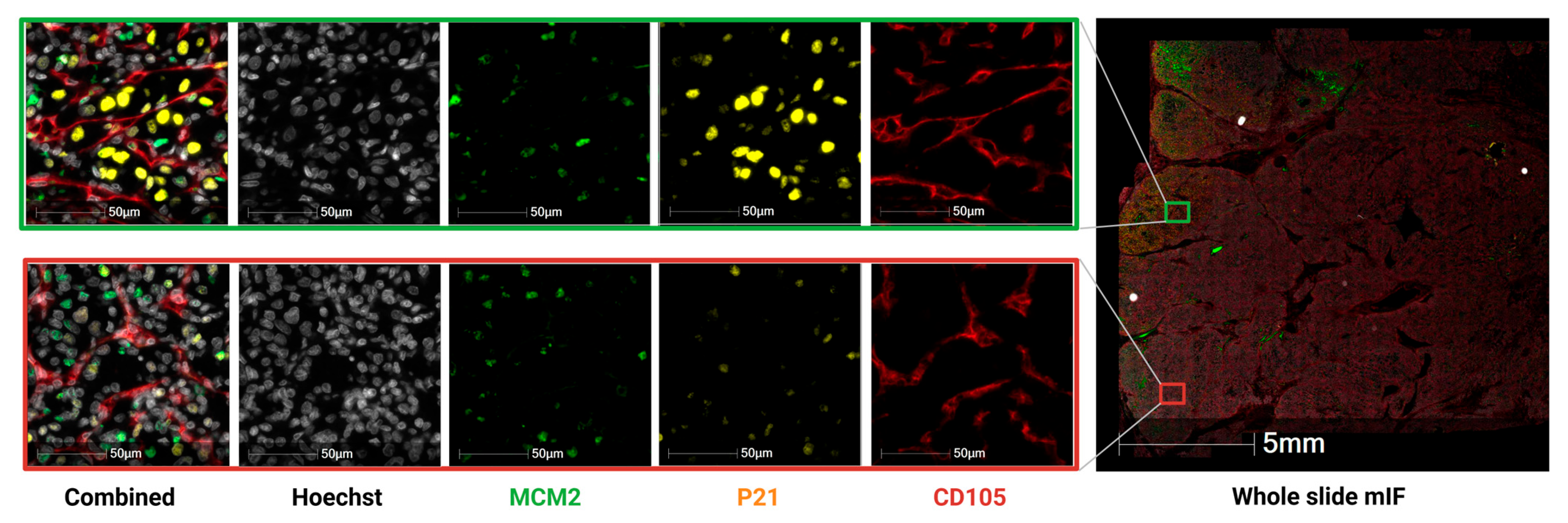
All images are provided as multi-channel CZI files, preserving spatial resolution for downstream quantitative analysis. An example of multiplex immunofluorescence, showing the individual fluorescence channels (Hoechst, MCM2, p21, and CD105) and the corresponding whole-slide image, is illustrated in Figure 1. These multiplexed datasets enable detailed exploration of cell cycle regulation, endothelial marker expression, and nuclear integrity.
Figure 1.
Multiplex immunofluorescence of ccRCC. Representative multiplex immunofluorescence images showing nuclear counterstain (Hoechst), MCM2 (green), p21 (yellow), and CD105 (red) expression. Left panels display individual marker channels and the combined overlay for two regions at higher magnification (scale bar = 50 μm). The right panel shows the corresponding whole-slide section with the highlighted regions of interest (scale bar = 5 mm).
The datasets may be analyzed jointly, as staining protocols, imaging parameters, and image-analysis workflows were deliberately standardized across all cohorts. However, because the data were generated in multiple batches and across different study periods, users are advised to verify staining performance using the cohort-specific positive controls provided, in order to ensure comparability when combining datasets.
2.3. Immunhistochemistry Images
For the Korean cohort, single-plex chromogenic immunohistochemistry for CD105 (DAB) was performed after multiplex immunofluorescence was completed on the same tissue sections. Data is available as CZI files for 71 patients.
2.4. Cell Phenotype Tables
CSV files containing quantitative data derived from image analysis. Each file includes phenotype counts for p21+/MCM2− cells, CD105+/p21+/MCM2− cells, and morphometric measurements summarizing all detected cells within the analyzed regions. Reported morphometric parameters include average cell area (μm2), average cytoplasm area (μm2), average nucleus area (μm2), average nucleus perimeter (μm), and average nucleus roundness. Together, these variables provide both phenotypic and structural information for each patient.
2.5. Patient-Level Metadata
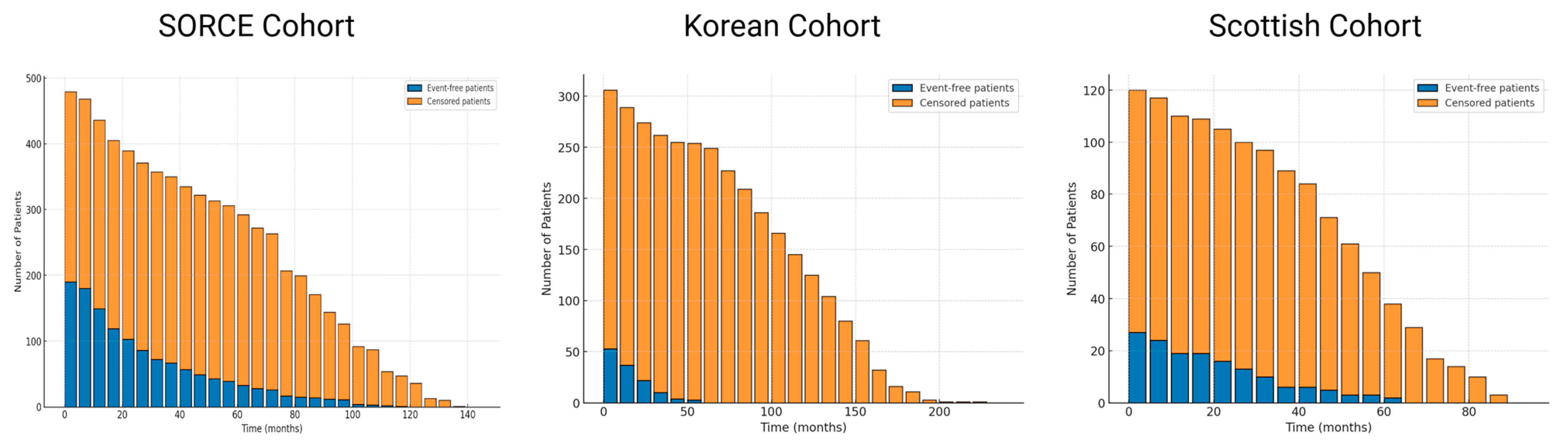
De-identified spreadsheets containing clinicopathological variables are provided, including age, sex, tumour stage, ISUP nuclear grade, Fuhrman grade, Leibovich score, treatment arm (for the SORCE cohort), node status, necrosis, relapse status, disease-free survival, and overall survival for each patient. In the SORCE cohort, the treatment arm is defined as follows: Arm A- placebo, Arm B- one year of adjuvant Sorafenib, and Arm C- three years of adjuvant Sorafenib. This dataset can be utilized in various ways to explore clinical and prognostic patterns. For example, to analyze recurrence dynamics, compare cohort-specific outcomes, or visualize event-free and censored patient distributions, as demonstrated in Figure 2.
Figure 2.
Distribution of event-free and censored patients across three renal cell carcinoma cohorts. Bar plots display the number of patients over time (in months) for the UK arm of SORCE, Korean, and Scottish cohorts. Blue bars represent patients who were event-free (recurrence) at that specific time point but later experienced recurrence, whereas orange bars represent censored patients who remained event-free at their last follow-up.
3. Methods
3.1. Sample Collection and Preparation
This study included four independent cohorts of patients with renal cell carcinoma (ccRCC). The UK arm of the SORCE trial cohort (n = 480; ClinicalTrials.gov NCT00492258) comprised UK patients from a phase III adjuvant sorafenib versus placebo study. A second cohort (n = 300) of ccRCC patients was collected at Seoul National University Hospital, South Korea (IRB H-1908-133-1057). A retrospective Scottish cohort (n = 120) consisted of nephrectomy samples obtained prior to systemic therapy with approval from NHS Lothian Bioresource (IRAS 15/ES/0094). Finally, a paired cohort (n = 62) from Edinburgh included matched primary and metastatic tumours from patients with vascular invasion or metastases, approved by the Lothian Regional Ethics Committee (IRAS 15/ES/0094).
3.2. Histology and Multiplex Immunofluorescence Staining
Formalin-fixed paraffin-embedded (FFPE) tissue blocks were sectioned at 3 μm and baked at 65 °C. H&E staining was performed on selected sections to assess tissue architecture. Following dewaxing and rehydration, the slides were rinsed with distilled water and stained with Mayer’s hematoxylin for 3 min, followed by a gentle wash under running tap water. Sections were then counterstained with eosin for 10 s, dehydrated through a graded ethanol series (50%, 80%, and 100%), cleared in xylene, and mounted using DPF medium.
Adjacent sections were processed for multiplex immunofluorescence. After dewaxing and rehydration, antigen retrieval was carried out in a pressure cooker for 5 min using sodium citrate buffer (pH 6.0), followed by washes in TBST (0.1% Tween® 20). Endogenous peroxidase was quenched with 3% hydrogen peroxide (Sigma, St. Louis, MO, USA; #H1009), and non-specific binding was minimized with a serum-free protein block (Agilent Santa Clara, CA, USA; #X090930-2). Sections were incubated with MCM2 (Cell Signalling, Danvers, MA, USA; #4007; 1:500, 1 h, room temperature), then treated with anti-rabbit HRP (Agilent, #K400311-2; 30 min) and visualized using TSA FITC (Akoya, Marlborough, MA, USA; #NEL741001KT; 1:50). Antibodies were stripped by microwaving in sodium citrate buffer (pH 6.0) for 17 min prior to sequential labelling with p21 (Abcam, Cambridge, UK; #ab109520; 1:400) and either Lamin B1 (Novus Biologicals, Centennial, CO, USA; #NBP1-42594; 1:2000) or CD105 (Atlas Antibodies, Stockholm, Sweden; HPA067440; 1:600), depending on the study cohort. Lamin B1 was visualized with TSA Cyanine 5 (Akoya, #NEL745001KT; 1:50) in the Scottish and Korean cohorts, whereas CD105 was detected with TSA Cyanine 5 (Akoya, #NEL745001KT; 1:50) in the SORCE and metastatic cohorts. Nuclei were counterstained with Hoechst 33,342 (ThermoFisher, Waltham, MA, USA; #H3570; 1:100), and slides were mounted with ProLong Gold Antifade (ThermoFisher, #P36930). To ensure consistency and reproducibility across staining runs, each batch included both positive and negative controls.
3.3. Immunohistochemistry
CD105 Immunohistochemistry was performed for the Korean cohort. After multiplex immunofluorescence (mIF) imaging, coverslips were carefully removed, and the same tissue sections were reprocessed for immunohistochemistry (IHC). Antigen retrieval was performed in citrate buffer (pH 6.0) for 5 min, after which slides were incubated with a rabbit polyclonal anti-CD105 antibody (Atlas Antibodies, #HPA067440; 1:600 dilution). Detection was carried out using the HRP-based EnVision system (Agilent), and colour development was achieved with DAB chromogen (DAKO, Agilent, Manchester, UK). Slides were then counterstained with hematoxylin, dehydrated, cleared in xylene, mounted with DPX mounting medium (Sigma-Aldrich, Gillingham, UK), and re-imaged under brightfield conditions.
3.4. Image Acquisition
Brightfield whole-slide images (WSIs) were acquired at ×20 or ×40 magnification, while multiplex immunofluorescence whole slides were scanned at ×20 magnification using a Zeiss Axio Scan Z1 scanner and stored in CZI format. Image quality was assessed for focus, exposure, and staining uniformity, and slides not meeting these criteria were either re-scanned or excluded from analysis.
3.5. Image Analysis and Cell Phenotyping
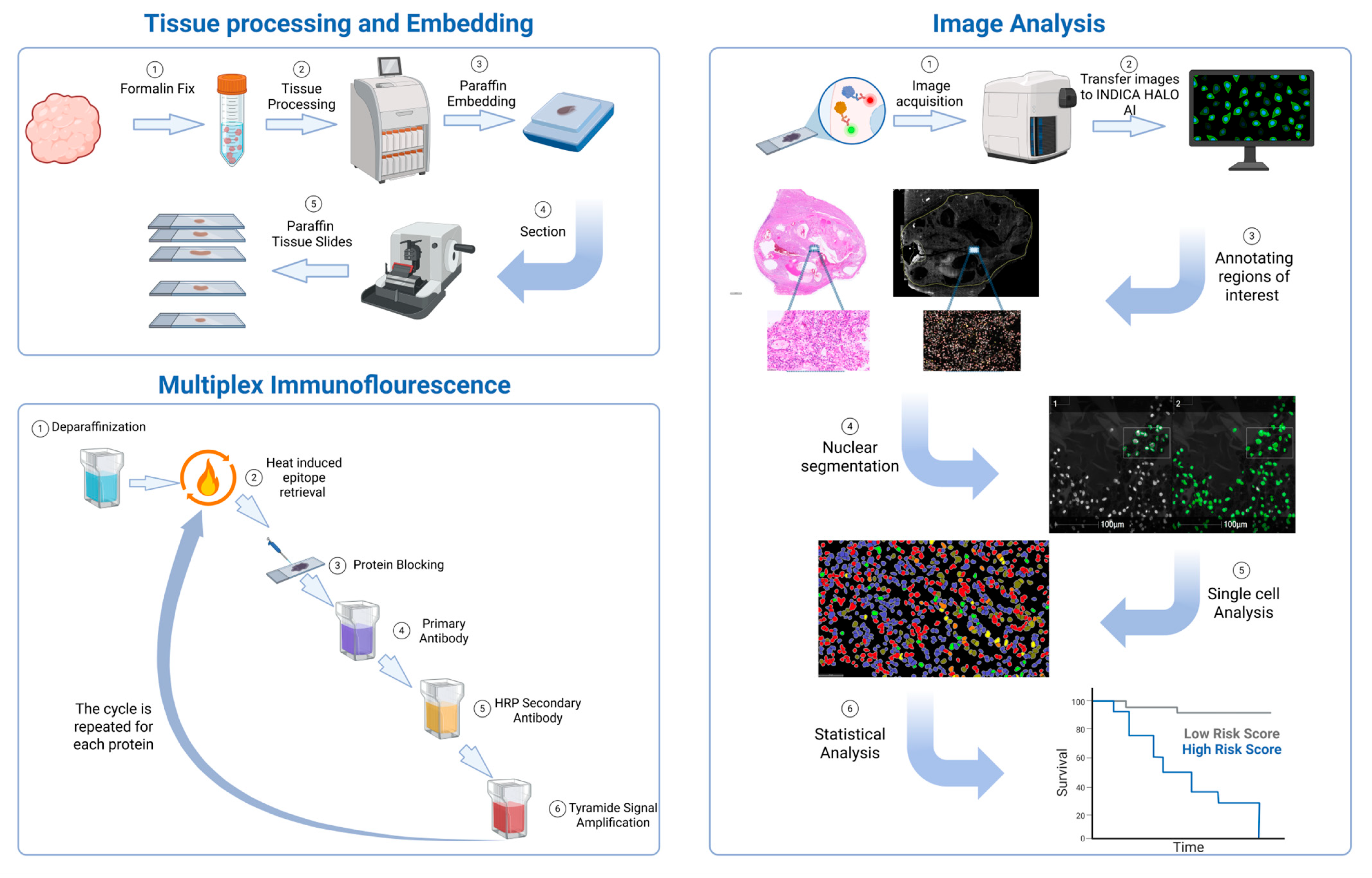
Image analysis was performed using the Indica HALO High Plex FL module as shown in Figure 3. Tumour regions were manually annotated by trained observers (HA, IHU, DJH) after reviewing matched H&E whole-slide images (WSIs) and transferred to the corresponding mIF slides. Nuclear segmentation was carried out on the Hoechst channel using a customized segmentation classifier created with HALO AI™ (Indica Labs, Albuquerque, NM, USA; https://www.indicalab.com/halo-ai accessed on 21 December 2025), which incorporates deep-learning neural network algorithms. For each segmented cell, fluorescence intensities of p21, MCM2, CD105, and Lamin B1 were quantified. Marker positivity thresholds were established using control tissues and applied consistently across all cohorts to ensure comparability. Based on p21 and MCM2 co-expression patterns, cells were assigned to specific phenotypic subsets, and CD105 expression was subsequently overlaid to define CD105+ subpopulations within these groups. Each patient includes quantified counts of p21+/MCM2− and CD105+/p21+/MCM2− cell populations, together with morphometric metrics summarizing all cells identified within the analyzed regions. The recorded morphometric features comprise mean cell area (μm2), mean cytoplasmic area (μm2), mean nuclear area (μm2), mean nuclear perimeter (μm), and mean nuclear roundness.
Figure 3.
Overview of the experimental workflow for tissue processing, multiplex immunofluorescence, and image analysis. Formalin-fixed tissues were processed, embedded in paraffin, sectioned, and mounted onto slides (top left). Multiplex immunofluorescence was performed through iterative cycles of deparaffinisation, antigen retrieval, protein blocking, primary and HRP-conjugated secondary antibody incubation, and tyramide signal amplification (bottom left). Slides were scanned, and digital images were analyzed using HALO AI software version 4.2 (right panel). Regions of interest were annotated, nuclei were segmented, and single-cell features were quantified to enable statistical and survival analyses.
3.6. Clinical Variables
For each patient, demographic and clinical data were extracted from electronic health records or trial databases. Variables include age at surgery, sex, necrosis, node status, pathological stage (pT), nuclear grade (Fuhrman and ISUP), Leibovich score, treatment arm (sorafenib/placebo in the SORCE cohort), recurrence status, and disease-free survival (months). Data were de-identified before release and linked to imaging files through anonymous identifiers. No personally identifiable information is included.
4. User Notes
All imaging data and derived feature tables are available through the BioImage Archive at EMBL-EBI under the accession numbers listed in Table 1. Whole-slide image files are large (often >1 GB), so users should ensure adequate storage and bandwidth before downloading. CZI files can be viewed using open-source software such as QuPath, Fiji, or ZEISS ZEN lite (available at https://www.zeiss.com/microscopy/en/products/software/zeiss-zen-lite.html (accessed on 22 December 2025)). Because all data are released under a CC0 licence, users are free to reuse, modify, and redistribute the material without restriction, and this Data Descriptor should be cited.
This dataset is anchored by a large collection of high-resolution hematoxylin and eosin whole-slide images that capture the full histological diversity of renal cell carcinoma. These slides can be used to study tumour architecture, cellular morphology, and stromal patterns, as well as to develop and validate computational tools for tissue segmentation, grading, and feature extraction. The accompanying multiplex immunofluorescence images for p21, MCM2, Lamin B1, and CD105 provide complementary molecular information that enables investigation of cell-cycle dynamics, proliferation, and angiogenesis within the same specimens. De-identified clinical and pathological data allow integration of image-based features with patient outcomes, including stage, grade, treatment arm, recurrence, and survival. Altogether, the resource offers a versatile foundation for studying renal cancer biology, testing analytic pipelines, and identifying potential prognostic markers.
Limitations
Several technical and methodological limitations should be considered when reusing this dataset. First, the whole-slide images and multiplex immunofluorescence files are provided at full resolution and are large in size (often exceeding 1 GB per file), which may limit accessibility for users without high-performance computing resources, sufficient local storage, or fast network bandwidth. While open-source platforms such as QuPath, Fiji, and ZEISS ZEN lite can be used to visualize and analyze the data, performance limitations or lag may occur when handling large image volumes or performing computationally intensive tasks such as whole-slide segmentation and single-cell phenotyping. For large-scale or high-throughput analyses, the use of specialized digital pathology software with optimized image handling and parallel processing capabilities (e.g., Indica Labs HALO™ version 4.2) may therefore be advantageous.
In addition, multiplex immunofluorescence was not available for all cases, and the marker composition differed slightly between cohorts, with CD105 included in the SORCE and metastatic cohorts and Lamin B1 included in the Korean and Scottish cohorts. This may limit direct cross-cohort comparisons for specific biomarkers. Finally, tumour regions were manually annotated prior to image analysis, introducing a degree of observer dependency, although annotations were performed and reviewed by trained investigators to minimize variability.
Author Contributions
Conceptualization, H.A. and D.J.H.; methodology, H.A. and I.H.U.; software, H.A.; validation, H.A.; formal analysis, H.A. and D.J.H.; investigation, H.A. and D.J.H.; resources, H.A., D.J.H., G.D.S., A.L., K.K., C.W.J., C.K., K.C.M., T.E., E.F., A.W., A.M. and TranSORCE Team; data curation, H.A.; writing—original draft preparation, H.A. and D.J.H.; writing—review and editing, H.A., I.H.U. and D.J.H.; visualization, H.A.; supervision, D.J.H.; project administration, D.J.H.; funding acquisition, D.J.H. TransSORCE Team contributed patients to the SORCE Clinical Trial. All authors have read and agreed to the published version of the manuscript.
Funding
This work was supported by the Industrial Centre for AI Research in Digital Diagnostics (iCAIRD), funded by Innovate UK on behalf of UK Research and Innovation (UKRI) [Project No. 104690]. Additional support was provided by the KATY project, which received funding from the European Union’s Horizon 2020 research and innovation programme under grant agreement No. 101017453. H.A. acknowledges support from the University of St Andrews Sanctuary Scholarship.
Institutional Review Board Statement
The SORCE trial cohort was approved by the SORCE trial ethics committee (ClinicalTrials.gov NCT00492258). The Korean cohort was approved by the Institutional Review Board of Seoul National University Hospital (H-1908-133-1057). The Scottish and metastatic cohorts were approved by the Lothian regional ethics committees (IRAS 15/ES/0094: 15/ES/0094, 08/S1101/41, 10/S1402/33).
Informed Consent Statement
Informed consent was obtained from all subjects involved in the study.
Data Availability Statement
The imaging data and derived feature tables described in this article are available in the BioImage Archive at EMBL-EBI under the accession numbers indicated in the Dataset table. Clinical metadata (age, sex, stage, grade, Leibovich score, treatment, relapse status, and survival) are available as de-identified CSV files accompanying each dataset. There are no restrictions on data reuse.
Acknowledgments
TransSORCE Team includes James Larkin, Axel Bex, Jim Barber, Janet Brown, Naveen S Vasudev, Rhona McMenemin, Paul Nathan, Lisa M Pickering, and Max Parmer. Thank the patients and clinical teams involved in the SORCE trial, Seoul National University Hospital, and NHS Lothian for providing tissue samples and clinical data. We acknowledge support from the University of St Andrews School of Medicine.
Conflicts of Interest
TE served as a Trustee of Macmillan Cancer Support for 10 years until 2021, is a non-executive director of Cambridge University Health Partners, Director of DeviceChooser Ltd., and Senior Partner at ALTIMIM LLP. He holds stock in AstraZeneca (self and spouse) and Roche (self), has received research support from AstraZeneca, and was employed as Vice President of Oncology Early Clinical Development at AstraZeneca until March 2020; he is currently Vice President of GU and GI Oncology Late Clinical Development at Roche. GDS has received educational grants from AstraZeneca (to the institution), consultancy fees from Evinova and Qurin, and travel expenses from MSD; he is Clinical Lead (urology) for the National Kidney Cancer Audit and Topic Advisor for the NICE kidney cancer guideline. GDS is also supported by The Mark Foundation for Cancer Research (RG95043), the Cancer Research UK Cambridge Centre (C9685/A25177 and CTRQQR-2021\100012), and the NIHR Cambridge Biomedical Research Centre (NIHR203312). The views expressed are those of the authors and not necessarily those of the NIHR or the Department of Health and Social Care. DJH receives research funding from the MRC, Melville Trust, Medical Research Scotland, and the EU, and is a part-time employee of NuCana plc. AL has received a travel grant and speaker’s fees from MSD, as well as research funding from the MRC, the EC Horizon2020 Fund, and CSO Scotland. All remaining authors have declared no conflicts of interest.
References
- Hsieh, J.J.; Purdue, M.P.; Signoretti, S.; Swanton, C.; Albiges, L.; Schmidinger, M.; Heng, D.Y.; Larkin, J.; Ficarra, V. Renal Cell Carcinoma. Nat. Rev. Dis. Primers 2017, 3, 17009. [Google Scholar] [CrossRef] [Scilit] [PubMed]
- Schiavoni, V.; Campagna, R.; Pozzi, V.; Cecati, M.; Milanese, G.; Sartini, D.; Salvolini, E.; Galosi, A.B.; Emanuelli, M. Recent Advances in the Management of Clear Cell Renal Cell Carcinoma: Novel Biomarkers and Targeted Therapies. Cancers 2023, 15, 3207. [Google Scholar] [CrossRef] [Scilit]
- Padala, S.A.; Barsouk, A.; Thandra, K.C.; Saginala, K.; Mohammed, A.; Vakiti, A.; Rawla, P.; Barsouk, A. Epidemiology of Renal Cell Carcinoma. World J. Oncol. 2020, 11, 79–87. [Google Scholar] [CrossRef] [Scilit] [PubMed]
- Leal-Esteban, L.C.; Fajas, L. Cell Cycle Regulators in Cancer Cell Metabolism. Biochim. Biophys. Acta (BBA)—Mol. Basis Dis. 2020, 1866, 165715. [Google Scholar] [CrossRef] [Scilit] [PubMed]
- Yan, J.; Chen, S.; Yi, Z.; Zhao, R.; Zhu, J.; Ding, S.; Wu, J. The Role of P21 in Cellular Senescence and Aging-Related Diseases. Mol. Cells 2024, 47, 100113. [Google Scholar] [CrossRef] [Scilit] [PubMed]
- Weiss, R.H.; Borowsky, A.D.; Seligson, D.; Lin, P.-Y.; Dillard-Telm, L.; Belldegrun, A.S.; Figlin, R.A.; Pantuck, A.D. P21 Is a Prognostic Marker for Renal Cell Carcinoma: Implications for Novel Therapeutic Approaches. J. Urol. 2007, 177, 63–68; discussion 68–69. [Google Scholar] [CrossRef] [Scilit]
- López-Otín, C.; Blasco, M.A.; Partridge, L.; Serrano, M.; Kroemer, G. Hallmarks of Aging: An Expanding Universe. Cell 2023, 186, 243–278. [Google Scholar] [CrossRef] [Scilit] [PubMed]
- Sun, Y.; Cheng, Z.; Liu, S. MCM2 in Human Cancer: Functions, Mechanisms, and Clinical Significance. Mol. Med. 2022, 28, 128. [Google Scholar] [CrossRef] [Scilit] [PubMed]
- Zhang, J.; Zhang, H.; Wang, Y.; Wang, Q. MCM2-7 in Clear Cell Renal Cell Carcinoma: MCM7 Promotes Tumor Cell Proliferation. Front. Oncol. 2021, 11, 782755. [Google Scholar] [CrossRef] [Scilit] [PubMed]
- Oladejo, M.; Nguyen, H.-M.; Wood, L. CD105 in the Progression and Therapy of Renal Cell Carcinoma. Cancer Lett. 2023, 570, 216327. [Google Scholar] [CrossRef] [Scilit] [PubMed]
- Eisen, T.; Frangou, E.; Oza, B.; Ritchie, A.W.S.; Smith, B.; Kaplan, R.; Davis, I.D.; Stockler, M.R.; Albiges, L.; Escudier, B.; et al. Adjuvant Sorafenib for Renal Cell Carcinoma at Intermediate or High Risk of Relapse: Results From the SORCE Randomized Phase III Intergroup Trial. J. Clin. Oncol. 2020, 38, 4064–4075. [Google Scholar] [CrossRef] [Scilit] [PubMed]

Disclaimer/Publisher’s Note: The statements, opinions and data contained in all publications are solely those of the individual author(s) and contributor(s) and not of MDPI and/or the editor(s). MDPI and/or the editor(s) disclaim responsibility for any injury to people or property resulting from any ideas, methods, instructions or products referred to in the content. |
© 2026 by the authors. Licensee MDPI, Basel, Switzerland. This article is an open access article distributed under the terms and conditions of the Creative Commons Attribution (CC BY) license.