


Abstract
The purpose of this perspective paper is to present a translational approach aimed at identifying the critical periods in brain development during which to provide neuroprotective and neurorestorative strategies, followed by prehensile training, to minimize hemiparesis and foster skill acquisition after asymmetrical perinatal brain injury (APBI). We contend that hemiparesis could be minimized after APBI through a multimodal sequential approach based on timely implementation of evidence-based intervention strategies. If APBI is identified early, neuroprotective techniques can be used in the first 10 days of life to minimize cell death. This can be followed in the next 3 months by the provision of timely neurorestorative strategies to enhance neuron survival and promote brain growth. Ideally these strategies will prime the system for targeted prehensile training from 3 months and beyond. Building on animal work, scientifically sound pilot work in human infants is needed to determine the best timing and therapy dose verified with sensitive outcome measures available before widespread implementation. Strategic planning among a multidisciplinary group working in basic science, medicine, and rehabilitation will inform efforts to bring this translational approach to clinical fruition.
1. Introduction
Asymmetrical perinatal brain injury (APBI) can be described as unequal or one-sided brain injury before, during, or within 4 weeks of birth. This condition is typically diagnosed in the NICU based on magnetic resonance imaging (MRI) and neurobehavioral signs. APBI can stem from perinatal arterial ischemic stroke (PAIS) in infants born at term and periventricular hemorrhagic infarction (PVHI) in infants born preterm [1,2]. White matter injury or parenchymal hemorrhages can also lead to APBI. Neonates who are at high risk for unilateral cerebral palsy (UCP) or one-sided weakness and incoordination, after APBI may present with injury to the corticospinal tract or basal ganglia/thalamus [3,4], leading to hemiparesis.
Hemiparesis secondary to APBI often impedes the development and execution of prehensile skills, including performance of upper limb reach to grasp tasks [3,4,5,6]. Historically, hand and arm function have been found to improve after intensive training in children with UCP and subsequent hemiparesis [7,8,9,10,11,12,13,14,15]. Yet, despite training, impaired dexterity and function often persist [12,16,17], suggesting that training may have been provided too late. To examine the benefit of earlier training, constraint-induced movement therapy (CIMT), bimanual training, and combined approaches have been examined in infants as young as 3 months corrected age [18,19,20,21,22,23], yet later outcomes for these children have not yet been determined. While a positive trend toward ongoing improvement is anticipated, it is also possible that even with early training, the neural substrate may be insufficient to allow full recovery [22,23].
We present a perspective paper that proposes that, to prevent or reduce childhood hemiparesis, there should be complimentary approaches with greater utilization of innovative strategies to prevent cell death and promote brain growth in early infancy before neuronal loss or impaired connectivity within the corticospinal system occurs. These strategies, applied in early infancy to promote neuronal survival and connectivity, can be followed by hand and arm training in infancy and childhood. The paper presents a translational multimodal approach aimed at reviewing the best time periods in brain development to provide neuroprotection and promote brain growth, followed by prehensile training, in an effort to minimize hemiparesis and foster skill acquisition after APBI in infants at risk for UCP.
2. Brain Development and Neural Injury
A multimodal approach aimed at minimizing hemiparesis after early brain injury requires close examination of the neural processes associated with postnatal brain development to determine which processes can be modified with specific intervention(s) and at what time point [23,24,25,26,27,28]. Select interventions focusing on the prevention of cell death or promoting brain growth have been supported in animal work, with some gaining support for use with human infants [29,30,31,32,33,34,35,36,37]. Moving key research from animal work to human infants is feasible through collaboration between professionals in basic research, medicine, and rehabilitation [38,39]. Innovative methods to promote brain growth are expanding [40,41,42,43].
2.1. Neural Processes Associated with Brain Development
Cerebral development occurs in an orderly systematic fashion [26,27,28,29,42,43,44], with the embryonic brain present by 2–3 months of gestation [26]. Neuronal proliferation and migration follow over the next 2 months, followed by neuronal organization and myelination that extend years postnatally. Some brain maturation and developmental processes continue into adolescence and adulthood [26,43,44,45]. Neural injury alters this trajectory and activates expansion of select cell populations [46,47,48,49,50]. Table 1 illustrates these key processes, including (1) proliferation, (2) migration, (3) synaptogenesis, (4) selective pruning, and (5) myelination [26,27,43,44,45]. In addition, critical events shown to be direct or indirect mediators of hemiparesis are highlighted. While not all undesirable consequences can be negated, some interventions may prevent apoptosis or neural degradation caused by these events, whereas other methods may foster neural growth and connectively.
Table 1.
Association of neural processes with hemiparesis.
Proliferation is the process in which neurons and glial cells are formed by symmetrical and asymmetrical division of neural progenitor cells [26,27]. This is a robustly active process that results in the development of nearly 100 billion nerve cells. Proliferation can be disrupted by genetic causes or teratogenic etiologies such as radiation, infections (e.g., rubella, cytomegalovirus, zika virus), or metabolic or toxic factors (e.g., alcohol, hyperphenylalaninemia) [27]. Disruption to proliferation most commonly results in microcephaly. Hemiparesis resulting from viruses has been reported in older children [50,51], though this stems from injury rather than disruption of proliferation.
Neural migration occurs when newly formed neurons are relocated from the ventricular and subventricular zones to the region where they will permanently reside and function [27]. The peak period for migration is 3–5 months of gestation, with the cerebral cortex and cerebellum being key structures formed during this process. When neurons fail to migrate to their proper location, malformations such as polymicrogyria, heterotopias, and cortical dysplasia result, which are commonly associated with the development of seizures [23]. Hemiparesis may result from malformations along the motor strip [52,53,54]. Krsek et al. [52] found that 25% to 37% of infants with focal dysplasia did develop hemiparesis. Seizures associated with focal dysplasia are typically treated with pharmaceuticals and, in refractory cases, surgery.
Synaptogenesis is the formation of functional connections (synapses) between neurons, is ongoing throughout life, and supports learning and neuroplasticity. A related process is neurite outgrowth in which the number of dendritic spines, axons, and synaptic contacts rises, contributing to central nervous system functional capacity [26,51,55]. Dendritic and axon length increase 5–6 fold in the first 6 months postnatally [26]. Apoptosis limits the number of neurons capable of synaptogenesis and neurite outgrowth, thus indirectly leading to hemiparesis. If neuroprotective strategies can minimize apoptosis in the neonatal period, this may enhance the CNS’s capacity for synaptogenesis.
Selective pruning is the phase in which unused, weak or obsolete connections are eliminated, while those that undergo repeated use are strengthened [56,57] which may be a key process in the development of hemiparesis. The ipsilateral corticospinal tract (CST) connections present at birth begin to be eliminated 6 months postnatally [58] so that by 24 months of age, many ipsilateral CST connections are gone. After unilateral perinatal injury to the primary motor cortex (M1), the risk of hemiplegia in the contralateral limb increases [59,60]. Outward signs of hemiplegia may be revealed once ipsilateral pathways from the intact M1 region begin to be eliminated, reducing the neural substrate. This period of elimination coincides with the expansion of age-appropriate prehensile skills. Training in unilateral and bilateral hand/arm use in early infancy, prior to and during selective pruning is critical to delay or minimize pruning of ipsilateral CST tracts.
Myelination is the process by which myelin, a fatty insulating sheath, begins to surround the axon [26,61], accelerating nerve conduction velocity. The neonatal brain is predominantly unmyelinated [62], with postnatal myelination occurring rapidly in the first year and continuing slowly into the 3rd decade [63]. Myelin is produced by oligodendrocytes, which are generated from preoligodendrocyte progenitor cells [64]. Preoligodendrocytes are sensitive to oxidative and inflammatory injury, leading to impaired myelination [40,49,65]. Preoligodendrocyte injury leading to dysmaturation is a component of underlying neuropathologic process seen in preterm white matter injury, including periventricular leukomalacia (PVL), which can lead to hemiparesis [66,67,68]. Tracking of brain development has been done with neuroimaging, adding valuable insight into select conditions [69]. Thomas et al. [68] used diffusion tensor imaging (DTI) to examine five children with unilateral periventricular white matter injury and unilateral cerebral palsy (CP). They found a significant reduction in CST fiber count linked with the paretic limb and evidence of CST reorganization associated with the non-paretic limb. Secondary degeneration was also found in the brainstem, corpus collosum, and deep brain nuclei linked with the paretic limb. Environmental enrichment in the neonatal period [70], including methods to foster motor skill learning, have been proposed as a way to increase the production of oligodendrocytes. The methods reviewed below, such as positive sensory exposure [41,70,71] and training using the paradigm of contingent reinforcement [72,73,74], may help foster the production of oligodendrocytes and subsequent myelination through activity-based regeneration.
2.2. Neurotransmitters and Networks
Brain development relies on the influence of neurotransmitters and the strengthening of neural networks. Abundant neurotransmitters available at birth aid the neonate’s transition to extrauterine life and are crucial to activity-dependent plasticity [75]. Glutamate regulates neurogenesis, neurite outgrowth, synaptogenesis, and apoptosis in the developing brain [76]. Brain-derived neurotrophic factor (BDNF), glial cell line-derived neurotrophic factor and other growth factors are influenced by glutamate. BDNF levels increase from infancy to young adulthood and correspond with structural maturity [77]. Full-term neonates [78] and breastfed infants [79] have higher BNDF levels. The period of significant synaptogenesis, 2–4 months postnatally, may be the best time to influence BDNF production and synaptic plasticity through environmental enrichment [70,80] in preparation for prehensile training. As with adults, BDNF levels may increase in response to motor activity [81], but this needs further study in young infants.
2.3. Remodeling
Remapping of the motor or somatosensory cortex and re-establishment of vital neural networks are key factors in recovery in infants with APBI [33,62,82]. Yap and colleagues [62] examined brain development from 2 weeks to 2 years of age with diffusion tensor imaging (DTI) using a three-dimensional modeling technique, fiber tractography. They found that small local networks evolved into large, distributed networks that operate in a functional manner. Insufficient control of spinal motor circuits [31,60] may account for why signs of hemiparesis become apparent late and evolve over the first 2 years of life [60,83]. Evidence from Friel et al. [33] suggests that tailored prehensile training programs in a feline model conducted in a timely manner promote cortical mapping and repair of spinal motor circuitry to re-establish neural networks. To enhance success, motor training should be introduced in early infancy to induce activity-based competition for neural resources between the CSTs of both hemispheres to prevent childhood hemiparesis [33,84,85].
3. Translational Multimodal Approach to Minimize Hemiparesis
A translational multimodal approach based on evidence would include three key areas: (1) neuroprotection; (2) neurorestoration; and (3) prehensile training (Figure 1). A combined approach optimizing neuroprotection, neurorestoration with brain growth, and motor training is vital, given that up to 50–70% of infants who sustain perinatal arterial ischemic stroke (PAIS) later display evidence of upper limb hemiparesis [86,87,88]. Since significant synaptogenesis occurs from about 28 weeks’ gestation to 3 months postnatally, it is crucial to preserve as many cells and synapses as possible to ensure that the infant retains sufficient neural substrate needed for brain growth and prehension development. A multimodal approach would require collaboration among professionals from birth through the first year of life and beyond.
Figure 1.
Multimodal approach timeline from birth to childhood.
3.1. Neuroprotection
Preserving the integrity of the cerebral mantle of newborns after injury is essential. Different approaches are necessary to address the varied etiologies of brain injury that occur in the preterm vs. the term infant. Here we review non-pharmaceutical and pharmaceutical modes of neuroprotection which vary regionally and by age of the infant. Suggested non-pharmaceutical methods used in most NICUs include therapeutic hypothermia, adequate nutrition, physiologic support, and kangaroo care. Recommended pharmaceuticals progressively change as research findings are published. All methods are now reviewed.
Therapeutic hypothermia. This form of neuroprotection is considered standard of care in term-born newborn infants with hypoxic-ischemic encephalopathy (HIE) to prevent apoptosis or cell death [89,90,91]. Immediately after an episode of hypoxia-ischemia, the neonatal brain enters a phase of “primary cell injury” [91] in which high-energy metabolites are depleted, cells begin to swell and extracellular amino acids accumulate. In the next “latent phase” there is recovery of cerebral circulation and oxidative metabolism. Yet 6 to 15 h after the initial episode of hypoxia-ischemia, secondary deterioration may begin as noted by seizures, resumed cell swelling, inflammation, abnormal receptor activity and apoptosis. Hypothermia administered within 6 h of birth or during the “latent phase” of HIE can lead to a reduction in the incidence of neurological deficits and long-term disability stemming from secondary deterioration [91,92].
We assessed developmental outcome after hypothermia treatment with a CoolCap® in infants with HIE [93] during the neonatal period and early infancy (2–4 months of age) based on the Test of Infant Motor Performance (TIMP) [94] and again at one year of age with the BSID III [95]. As shown in Figure 2, we found neuromotor performance improved based on the TIMP with 32% scoring in the average range in the neonatal period and 88% scoring in the average range by 2–4 months. By one year of age, 92% of infants performed in the average range on cognitive and language subtests and 76% were in the average range on motor subtests of the BSITD III. Retention in neuromotor performance from early infancy to one year of age implies that hypothermia may have prevented apoptosis, preserving neurons and synaptic connections and allowing sufficient development of neural networks.
Figure 2.
Study with HIE and hypothermia: (a) CoolCap® in place on the head of a full-term infants with HIE. The cap provides a form of hypothermia for 72° in infants with HIE when placed within 6° of birth. (b) Developmental outcome with TIMP results on the left from the Neonatal and Early Infancy time periods (blue) and results from the BSID III on the right (yellow). The improvement in sensorimotor outcome from the neonatal period to early infancy based on the TIMP was maintained at one year of age based on the BSID III [93].
The onset of injury in HIE during the birth process allows timely use of therapeutic hypothermia within the first 6 h of birth for maximal efficacy [90]. Yet most newborn infants with an APBI or stroke are not diagnosed immediately after birth, therefore missing the window for hypothermia to be effective. One study examined the effect of whole-body hypothermia in a small group of infants with perinatal stroke [96]. The authors found that five out of 15 infants diagnosed with perinatal stroke based on MRI received hypothermia and did not show evidence of seizures in the neonatal period whereas seven of 10 untreated infants did. While limited studies have investigated hypothermia following APBI [39,96,97], it is not considered standard practice and thus requires further investigation.
Adequate nutrition. Digestion of key nutrients is an essential component of fetal growth and neonatal care. It provides the foundation for ongoing brain development, health, and neuroplastic changes [98]. Optimal nutrition can support ongoing development of neural processes from proliferation to myelination [99]. Essential nutrients with a neuroprotective influence for the neonate with brain injury include docosahexaenoic acid (DHA), choline, iron, and protein [100,101,102,103]. These nutrients support gray and white matter growth, prevent injury to cells and promote development in the neonatal period [98,99,102,103,104,105,106,107]. Neonates can receive maternal milk, donor milk, or formula orally or through gastric systems [108]. Some infants may require parenteral nutrition which bypasses the digestive system. Fortunately, infant formula is designed to supply sufficient nutrients to neonates and infants who sustain APBI [109,110,111]. This area of clinical research is progressing rapidly; thus, nutrition should continue to play a strong role in neuroprotection of infants at risk.
Physiologic support. Maintaining good neuro-supportive care includes normotension, normoglycemia, normocarbia [49,112], and avoidance of physiologic swings, as undertreating and overtreating physiologic disturbances are associated with worse outcomes in those with brain injury and in the preterm population.
Kangaroo care. To achieve skin to skin contact or kangaroo care, mom or dad holds their baby upright, belly-down directly against their bare chest, while the infant wears only a hat and diaper, covered in a blanket for warmth. It was developed from the work of Edgar Ray, MD and Hector Martinez, MD from Bogotá, Colombia [113,114]. Kangaroo care (KC) has been found to stabilize an infant’s vitals, reduce stress, and release oxytocin for bonding [115,116]. An extension of KC is kangaroo mother care (KMC) which also includes exclusive breastfeeding, early discharge, and close follow-up [115]. The benefits of KMC beyond KC include a reduction in neonatal sepsis, hypothermia, hypoglycemia, lower mean respiratory rate and pain, higher oxygen saturation, temperature, and head circumference growth [117,118]. Implementation of KC or KMC varies depending on the setting, sociocultural context, and constructs of parental roles. In addition, other responsibilities and stressors can influence the ability of the staff and parents to carry out KC or KMC successfully in the NICU and home [119].
Pharmaceutical therapies have been investigated for use in neonatal stroke, white matter injury of prematurity, intraventricular hemorrhage, and as an adjunct to therapeutic hypothermia in HIE for their neuroprotective effects, though none are clinically available at this time. Common targeted mechanisms include anti-inflammatory (e.g., azithromycin), anti-apoptotic, and anti-excitatory, with some agents having multiple mechanisms of action (e.g., melatonin, erythropoietin). See Table 2 for a list of pharmaceuticals. Injury-specific mechanisms and targets are also being explored, such as fibrinolytics to dissolve clots and restore blood flow in acute ischemic stroke, as is currently done in adults, and to dissolve clots in post-hemorrhagic ventricular dilatation. Iron chelation is being explored in intraventricular hemorrhage. The goal is to minimize tissue loss after injury. The preclinical evidence of these pharmaceuticals provides hope, but their benefit has yet to be established in human clinical trials. The list of potential pharmaceuticals continues to expand, yet further research is needed to prove their safety and efficacy in human infants and to determine the optimal timing and dosing frequency and duration [120,121,122,123,124,125].
Table 2.
Neuroprotection and neurorestorative pharmaceuticals and other interventions.
3.2. Neurorestorative Strategies
After perinatal neural injury, methods to restore the neural substrate are of interest. Animal studies reveal that select pharmaceuticals and other interventions contribute to brain growth and repair. While some procedures are supported with research findings, others require further investigation. Select methods will be reviewed here.
Pharmaceuticals to promote neuronal repair are being explored in both adult and infant brain injury; none are clinically available yet [121,126]. These agents act by promoting neurogenesis, angiogenesis, and synaptic plasticity. Many of them also have neuroprotective properties (e.g., erythropoietin). Biologics are also being explored, including stem cells, exosomes, and growth factors. There are additional agents like hydrogels that are in preclinical investigation and can serve as substrate for delivery of therapies in addition to providing physical support, preventing cyst formation, and encouraging regrowth. In addition, preclinical work is exploring the targeting of innate pathway activation to promote oligodendrocyte precursor cells after injury to improve myelination [35,70]. Further study is needed to determine the optimal intervention, route of administration, timing, dosing, and frequency to promote neurorestoration after perinatal brain injury. See Table 2 for summary of pharmaceuticals and other agents being explored for neurorestoration.
Environmental enrichment. In preclinical work, enrichment of the environment has been shown to contribute to neural recovery. While brain injury delays the sequential development of oligodendrocytes, important to the production of the myelin sheath, Gallo and colleagues [70] have shown that environmental enrichment (EE) promotes myelination through endogenous processes. In the rat model, exposure in a natural habitat with novel stimuli, social interactions, and voluntary physical activity are encouraged [116].
Clinicians working with newborn infants have drawn upon the preclinical work of Gallo and colleagues [127,128,129] and propose that there are critical periods and elements essential to enriching the environment. They suggest that EE should begin immediately after birth with positive sensory exposure [71,128] as reviewed below and continue through later infancy and childhood. Importantly, all components are necessary to promote functional recovery (motor, sensory, and cognitive) [129]. Current early interventions may not be optimal since they do not specifically target the risk each particular infant has. Imaging could be used to individualize the intervention, so that it is adequately directed and target specific risk factors prior to onset of symptoms. As suggested in this paper, targeting motor therapies for those at risk for hemiparesis (injury to PLIC, basal ganglia or motor cerebral cortex) or vision therapy for those at risk for cortical vision impairment (injury to occipital lobes or optic radiations) is critical. The authors recommend that rehabilitation promoting motor learning, active movement, engagement of parents, and modifications to the environment may be best to foster early achievement of skill.
Positive sensory exposure. An association has been established between positive sensory exposure and brain growth [36,71,130]. The ideal NICU environment has yet to be fully recognized, as private rooms limit sensory stimuli for infants, including both noxious noise and developmentally supportive exposure to human voice, such as language exposure [131,132], highlighting that the ideal NICU environment may vary based on social circumstances, including family availability [133]. Exposure to noxious stimuli like painful procedures remains high, thus the need for positive sensory stimulation is vital. Pineda et al. [41,71] examined and implemented a program entitled SENSE—supporting and enhancing neonatal intensive care experiences. This program provides evidence-based multi-sensory experiences for infants at risk while engaging parents in the process. Interventions focus on rocking for vestibular input, soft music for auditory input, and holding with skin-to-skin care and massage as further reviewed below. Initial findings from this program conducted with preterm infants support improvements in parent confidence, neurobehavior, feeding, and communication at one year of age. The SENSE program has been implemented worldwide and studies providing further support are ongoing. Such interventions in the setting of APBI have not been fully established.
Massage is a positive exposure that may influence brain growth and neural connectivity [130,134]. Guzzetta et al. [130] conducted massage for 10 days (with a 2-day interval), 3 times/day, in 10-day-old preterm infants 30–33 weeks’ gestation. They found that massage modulated the level of endogenous or insulin-like factors (IGF-1) that aid in the regulation of brain growth and improved visual acuity by 3 months of age when compared to controls (45% greater). Although the authors found that by 7 months of age, visual acuity was similar between the groups, the rate of change and the period of heightened arousal may have had positive benefits on brain growth and function.
The effects of positive sensory exposure are short-lived by their nature. However, the sensory experience may increase state regulation [36], helping to prepare the CNS and neuromuscular system for prehensile training. Evidence suggests that attentive infants in the awake-alert state learn more quickly than infants in any other level of state control [36,135]. Methods such as massage may serve to modulate levels of neurotransmitters and promote an awake-alert state as described in the Neonatal Behavioral Assessment Scale [136], in preparation for interventions after neural injury. While evidence-based sensory supports have not been specifically studied in infants with APBI, these methods warrant further investigation to see if these techniques alter sensorimotor behavior.
3.3. Neuroimaging Identification of Risk for Hemiplegia
Identification of the population at risk for hemiplegia is key for determining who should receive targeted therapy. As discussed above, hemiparesis is associated with asymmetrical brain injury, which may be vascular or infectious, but hemiparesis can also occur in the setting of developmental malformations, such as cortical dysplasias, along the motor tracts. MRI findings involving the posterior limb of the internal capsule and/or basal ganglia and/or cerebral cortex after perinatal arterial ischemic stroke are predictors for later hemiparesis [137,138].
3.4. Prehensile Training
Prehension involves locating, reaching, grasping, manipulating and releasing toys or objects of any kind. Prehensile training could focus on any of those components, yet for the purpose of this paper we will be focusing on reaching and grasping with the upper limb. We will introduce successful animal and human models of CIMT and then the paradigm of contingent reinforcement.
Targeted prehensile training programs aimed at strengthening neural connectivity and motor control reportedly prevent hemiparesis in the animal model and are slowly being translated to human infants. Animal models have also shown that we can take advantage of activity-dependent competition for neural resources to aid neural recovery post-injury [33]. Specifically, the integrity of the contralateral CST and the associated spinal circuitry from the impaired primary motor cortex (M1) could be reinforced through timely skill training to prevent the development of dominant connections from the intact M1, thereby enhancing contralesional motor function [139]. Further, evidence suggests that skill training can be used to positively reinforce redundant corticospinal pathways available before selective pruning subsides [56,60].
Constraint-induced movement therapy (CIMT). This form of prehensile training involves restricting movement of the upper limb that is not at risk for hemiparesis in a constraint that prevents movement, while the hand and arm at risk are encouraged to perform motor actions such as reaching and grasping. The feline model of prehensile training from Friel et al. [33] is an excellent template from which to design translational studies. Friel et al. [33] used CIMT to aid in the neural repair and restoration of function following inactivation of M1 between postnatal weeks (PW) 5–7. They compared the outcome of early vs. late 4-week-long training in a forelimb reaching task, during which kittens wore a restraint on the ipsilesional forelimb, with those of a control group that only wore a restraint and did not receive training (see Figure 3). The early training and control group wore the restraint from PW 8–12. The delayed training group wore the restraint from PW 20–24. As shown in Figure 4, the results indicate that early training restored CST connections and the M1 motor map, increased the number of cholinergic spinal neurons contralaterally, and prevented impairment in forelimb motor control during reaching and horizontal ladder walking. Delayed training restored only connections of the CST and the motor map of M1, and only CST connections were restored in the control group. Reportedly, the restraint alone was insufficient to recover the M1 motor representation [33]. As demonstrated in the primate stroke model [140], training was deemed essential to restore spinal circuitry, aided by an increase in choline acetyltransferase expression in spinal interneurons. Thus, restoration of CST connectivity and the M1 motor map and an increase in the numbers of cholinergic spinal interneurons are critical for gaining motor control and function following injury to the corticospinal system.
Figure 3.
The timeline for the experimental design used in the training study with cats [33]. During weeks 5–7, the left M1 was inactivated and a tracer was injected 2 days later to track the CST connections. All three groups wore a restraint for 4 weeks. The early training plus restraint group and the restraint-alone group began at PW 8 and ended at PW 13. The delayed training group began at PW20 and ended at PW 24. All of the cats were sacrificed to examine the brain and the spinal cord histologically. PW = postnatal week; ET = early training; RA = restraint alone; DT = delayed training.
Figure 4.
ChAT-positive cells distributed in the lower cervical spinal cord. (A–C) Density maps in the cervical enlargement for ChAT-positive cells. Motoneurons are predominantly depicted by the large dense clusters of cells in the ventral horn. For all panels, the color scale is the same (red_1.25_10 -5 cells/square_m). Scale bar, 1 mm. (D) Ratios of ChAT label on the affected side of the spinal cord (left) to the contralateral (right). The early training group had a much greater ratio of ChAT-positive cells in the intermediate zone (laminae I–VII) on the affected/trained side than the untrained side. The two sides of all groups were symmetrical with regard to the ratio of ChAT label in motor nuclei [33].
Studies examining the benefits of CIMT and bimanual training have found these training programs to be efficacious for children 6 to 20 years of age [11,12,13,14,15,16,17,20,21]. Research examining CIMT and bimanual training in infants less than one year of age [18,22] has shown that improvements can be made if the dosage is high enough. Evidence reveals that the potential to develop useful prehensile skill may be greater if training is conducted at younger ages [18,19,20,21,22,141]. This potential may be even greater if training is initiated by 3 months of age given the dynamic and redundant neural processes present in early infancy before selective pruning begins.
Contingent reinforcement. Self-initiated reaching is one of the hallmarks of early infancy. Typically developing (TD) infants initially display flapping movements, yet by 3 to 5 months of age these movements are refined to purposeful reaching [142]. One reward infants can receive for reaching successfully is having the arms activate a musical mobile or toy [72,73,74]. By 3 to 4 months of age, TD infants display signs of associative learning and memory in the paradigm known as contingent reinforcement (CR), in which an infant’s leg is tethered to an overhead mobile such that leg kicks result in proportional mobile movement [73,74]. In the study by Heathcock et al. [73], the authors studied the benefit of CR on TD and preterm infants and found that preterm infants did not show the associative learning, as noted by an increase in kicking rate, found in TD infants. They suggested that a suboptimal arousal level may have influenced the findings and that future work may determine the best timing and form of CR for this group of infants.
With advances in technology, methods to employ the CR paradigm can now be tailored to the primary dysfunction an infant at risk presents with and provide higher dosage and intensity of targeted intervention. Using surface electromyography, we have shown that muscle activation above a pre-set threshold could trigger a video to play in infants who sustain a birth-related injury to the brachial plexus [72]. While this system is effective for infants with limited muscle activation, some infants tend to disuse the arm(s) or display maladaptive patterns. For them, a movement-based system may be easier to learn and could be more effective. Sargent and colleagues [143,144] used a three-dimensional motion analysis system to quantify the spatial exploration of the feet, as they attempted to activate an overhead mobile. Infants in those studies were typically developing (TD) at 4 months of age [143] and preterm at 4 months corrected age [143]. Both groups increased their spatial exploration, yet the preterm infants displayed less task specificity perhaps due to variations in perception, strength, and postural control. We have designed and tested a system of CR which uses wrist-based inertial sensors to trigger a mobile or toy to play when acceleration exceeds a set threshold [145]. This system has been found feasible for infants less than 6 months of age, including an infant who sustained a perinatal stroke.
Infants with APBI may need activity-dependent competition to enhance neural circuitry. Thus, combining CIMT with strategies involving contingent reinforcement may be an ideal way to induce repetitive reaching in the limb most at risk for hemiplegia, fostering motor learning in this group [18,19,22,33,84]. Because contingent reinforcement is an age-appropriate paradigm for young infants, and CIMT leads to gains in active movement, this combination may take advantage of neuroplasticity to foster early motor skills in infants with APBI.
3.4.1. Practical Recommendations
A multimodal approach to minimize hemiparesis requires inclusion of all three components: neuroprotection, neurorestoration, and rehabilitation. It is feasible for medical and rehabilitation personnel to employ all three components as the infant moves from one setting to the next. For neuroprotection, it is recommended that therapeutic hypothermia [89,90,91,92,96,97] be used by NICU personnel if the diagnosis of APBI is made early enough. In addition, it is essential to provide sufficient physiologic support [112] and administer targeted pharmaceuticals [120,121,122,123,124,125] during the neonatal period. We also recommend that adequate nutrition be provided immediately by NICU personnel and continued by parents in the home [98,99,100,101,102,103,104,105,106,107,108,109,110,111]. Kangaroo care is another method of neuroprotection that could be initiated by parents with guidance from NICU staff then continued in the home after discharge [113,114,115,116,117,118]. In terms of neurorestoration strategies, we highly recommend ongoing administration of key pharmaceuticals [120,121,122,123,124,125] and provision of environmental enrichment with positive sensory exposure as outlined in the SENSE program [127,128,129,130,131,132,133,134]. SENSE techniques begun by NICU personnel and parents include positioning to allow limb movement, simple massage, easy rocking, soft singing, or playing of classical music. Positioning the infant in right and left sidelying, as well as supine with receiving blankets for support, should allow movement of the upper and lower limbs most at risk for hemiparesis without limitations. As upper limb active movement increases, CIMT could be employed by lightly restricting motion in the arm not at risk while fostering reaching in the arm at risk. These reaching or reach-to-grasp actions could be done with the infant in sidelying so gravity is minimized as the infant attempts to reach toward a mirror secured to the crib rails or nearby toys. In the supine position, the infant would be moving against gravity, as the infant attempts to reach for hanging toys. The key to success is to ensure that the infant is attentive and capable of moving the arm and leg at risk since repetitive, purposeful action enhances neuroplasticity and motor learning. As this translational approach becomes more utilized in the NICU and the home, we may begin to see signs of hemiparesis lessening in infants with APBI.
3.4.2. Case Example
Our group worked with an infant who sustained perinatal ischemic stroke in the right middle cerebral artery (MCA) territory and experienced all three phases of our proposed intervention to minimize hemiparesis: neuroprotection in the neonatal period, neurorestorative strategies in the NICU, and contingent reinforcement at about 6 months of age. Thus, he may serve as a great example of how this program could benefit infants at risk. This case infant was born at 36 weeks and 1 day by emergency c-section for a drop in fetal heart tones and failed vacuum delivery. The Apgar scores were 1, 3, and 4 at 1 min, 5 min, and 10 min of life. Neuroprotection provided in the form of therapeutic hypothermia was provided for 3 days. Seizures were noted arising from the right central area on days 1 and 2 of life and responded to phenobarbital. The infant was transitioned to levetiracetam monotherapy for maintenance anti-seizure medication. An MRI following rewarming on day 4 demonstrated diffusion restriction in the right MCA territory, while follow-up imaging on day 17 demonstrated evolution of the infarct with encephalomalacia (Figure 5 and Figure 6). Neurorestorative strategies in the form of positive sensory exposure were provided starting in the NICU and continued in the home by his parents and included reading to him, skin-to-skin contact, and visual stimulation. At one month of age, he was weaned off levetiracetam and began outpatient physical and occupational therapy, each twice a week. At about 6 months of age, as part of a training study using the paradigm of contingent reinforcement, he demonstrated the ability to repetitively activate an overhead mobile with his arm at risk for hemiplegia. Now at 2 years of age, he is developing well with equal use of both hands, and fine/gross motor skills within normal limits for his age (Bayley-4 motor standard score of 96, in the 39th percentile). He wears glasses for astigmatism and is receiving speech therapy for stuttering.
Figure 5.
Case: MRI day 4. (A,B) diffusion-weighted images (A. SB100 and B. ADC) demonstrating right middle cerebral artery territory infarct. (C) T2 image demonstrating corresponding edema. (D) T1 image with hyperintensity in the left basal ganglia, most consistent with hemorrhage.
Figure 6.
Case: MRI day 17. (A). T2 imaging demonstrating evolution of the right middle cerebral artery infarct with encephalomalacia. (B). T1 imaging demonstrating development of right-sided laminar necrosis vs. microhemorrhage and resolution of previously seen left basal ganglia hemorrhage.
4. Conclusions
The methods to provide neuroprotection early after APBI followed by neurorestorative strategies may preserve and enhance the neural substrate essential to recovery. With this neural base, the CNS could be ready to refine the sensorimotor map and neural circuitry to minimize hemiparesis, allowing skill development [138]. The results from Friel et al. [33] suggest that to minimize hemiparesis and foster prehensile skills, training must begin early postnatally, well before the initiation of neuronal pruning in human infants at 6 months [27,58]. To enhance the long-term success of targeted training, our proposed translational multimodal approach may be best (see Figure 1). Yet are we ready for this exciting and important translational research opportunity? We propose that preliminary evidence is there, and it may be time to investigate the efficacy of this multimodal approach.
Author Contributions
All authors contributed to essential features of this paper. S.V.D. contributed to clinical findings reported for therapeutic hypothermia, and S.V.D. and T.I. contributed to the clinical findings for the case report. All authors have read and agreed to the published version of the manuscript.
Funding
This work was partially supported by the CSO Small Grants Program sponsored by The CHOC Children’s Research Institute and the Faculty Opportunity Fund at Chapman University.
Institutional Review Board Statement
The two studies referenced were conducted in accordance with the Declaration of Helsinki and approved separately by the Institutional Review Board (IRB) of Thomas Jefferson University (IRB#06U-455R) and the IRB of Chapman University (IRB-18-83).
Informed Consent Statement
Informed consent was obtained from one parent of all subjects involved in this study.
Data Availability Statement
The original contributions presented in this study are included in the article. Further inquiries can be directed to the corresponding author.
Acknowledgments
We would like to acknowledge Bobbie Pineda, PhD, OTR/L, CNT for providing information on the SENSE program and implementing this mode of intervention to the infant in our case example. The findings presented from prior research are published [33] or not currently available [93].
Conflicts of Interest
The authors declare no conflicts of interest.
Abbreviations
The following abbreviations are used in this manuscript:
| APBI | Asymmetrical perinatal brain injury |
| CIMT | Constraint-induced movement therapy |
| CP | Cerebral palsy |
| CNS | Central nervous system |
| CST | Corticospinal tract |
| PVL | Periventricular leukomalacia |
| DTI | Diffusion tensor imaging |
| BDNF | Brain-derived neurotrophic factor |
| HIE | Hypoxic-ischemic encephalopathy |
| TIMP | Test of Infant Motor Performance |
| BSITD III | Bayley Test of Infant Toddler Development, 3rd edition |
| MSCs | Mesenchymal stem cells |
| EPO | Erythropoietin |
| MRI | Magnetic resonance imaging |
| EE | Environmental enrichment |
| PLIC | Posterior limb of the internal capsule |
| NICU | Neonatal Intensive Care Unit |
| SENSE | Supporting and enhancing neonatal intensive care unit sensory experiences |
| IGF-1 | Insulin-like Growth Factor 1 |
| PW | Post natal weeks |
| TD | Typically developing |
| CR | Contingent reinforcement |
| MCA | Middle cerebral artery |
| APGARS | Appearance, pulse, grimace, activity, respiration |
References
- Dunbar, M.; Kirton, A. Perinatal Stroke: From Epidemiology to Optimized Outcomes. Pediatr. Clin. N. Am. 2025, 72, 957–974. [Google Scholar] [CrossRef] [Scilit] [PubMed]
- Ilves, N.; Metsvaht, T.; Laugesaar, R.; Rull, K.; Lintrop, M.; Laan, M.; Loorits, D.; Kool, P.; Ilves, P. Periventricular hemorrhagic infarction in preterm neonates: Etiology and time of development. J. Neonatal Perinat. Med. 2024, 17, 111–121. [Google Scholar] [CrossRef] [Scilit] [PubMed]
- Ryll, U.C.; Krumlinde-Sundholm, L.; Verhage, C.H.; Sicola, E.; Sgandurra, G.; Bastiaenen, C.H.; Eliasson, A.C. Predictive validity of the Hand Assessment for Infants in infants at risk of unilateral cerebral palsy. Dev. Med. Child Neurol. 2021, 63, 436–443. [Google Scholar] [CrossRef] [Scilit] [PubMed] [PubMed Central]
- Sutter, E.N.; Guerrero-Gonzalez, J.; Casey, C.P.; Dean DC3rd de Abreu EGouvea, A.; Peyton, C.; McAdams, R.M.; Gillick, B.T. White-Matter Connectivity and General Movements in Infants with Perinatal Brain Injury. Brain Sci. 2025, 15, 341. [Google Scholar] [CrossRef] [Scilit] [PubMed] [PubMed Central]
- Chen, C.Y.; Lo, W.D.; Heathcock, J.C. Neonatal stroke causes poor midline motor behaviors and poor fine and gross motor skills during early infancy. Res. Dev. Disabil. 2013, 34, 1011–1017. [Google Scholar] [CrossRef] [Scilit]
- Eliasson, A.C.; Nordstrand, L.; Backheden, M.; Holmefur, M. Longitudinal development of hand use in children with unilateral spastic cerebral palsy from 18 months to 18 years. Dev. Med. Child Neurol. 2023, 65, 376–384. [Google Scholar] [CrossRef] [Scilit] [PubMed] [PubMed Central]
- Holmefur, M.; Krumlinde-Sundholm, L.; Bergström, J.; Eliasson, A.C. Longitudinal development of hand function in children with unilateral cerebral palsy. Dev. Med. Child Neurol. 2010, 52, 352–357. [Google Scholar] [CrossRef] [Scilit] [PubMed]
- Sakzewski, L.; Sicola, E.; Verhage, C.H.; Sgandurra, G.; Eliasson, A.C. Development of hand function during the first year of life in children with unilateral cerebral palsy. Dev. Med. Child Neurol. 2019, 61, 563–569. [Google Scholar] [CrossRef] [Scilit] [PubMed]
- Sakzewski, L.; Greaves, S.; Eliasson, A.C.; Wallen, M.; Novak, I.; Ware, R.S.; Heathcock, J.; Maitre, N.; Boyd, R.N. Early developmental trajectories of the impaired hand in infants with unilateral cerebral palsy. Dev. Med. Child Neurol. 2025, 67, 901–909. [Google Scholar] [CrossRef] [Scilit] [PubMed] [PubMed Central]
- Steenbergen, B.; van der Kamp, J. Control of prehension in hemiparetic cerebral palsy: Similarities and differences between the ipsi- and contra-lesional sides of the body. Dev. Med. Child Neurol. 2004, 46, 325–332. [Google Scholar] [CrossRef] [Scilit]
- Araneda, R.; Klöcker, A.; Ebner-Karestinos, D.; Sogbossi, E.S.; Renders, A.; Saussez, G.; Paradis, J.; Bleyenheuft, Y. Feasibility and effectiveness of HABIT-ILE in children aged 1 to 4 years with cerebral palsy: A pilot study. Ann. Phys. Rehabil. Med. 2021, 64, 101381. [Google Scholar] [CrossRef] [Scilit] [PubMed]
- Araneda, R.; Ebner-Karestinos, D.; Paradis, J.; Klöcker, A.; Saussez, G.; Demas, J.; Bailly, R.; Bouvier, S.; Carton de Tournai, A.; Herman, E.; et al. Changes Induced by Early Hand-Arm Bimanual Intensive Therapy Including Lower Extremities in Young Children With Unilateral Cerebral Palsy: A Randomized Clinical Trial. JAMA Pediatr. 2024, 178, 19–28, Erratum in JAMA Pediatr. 2024, 178, 206. https://doi.org/10.1001/jamapediatrics.2023.6170. [Google Scholar] [CrossRef] [Scilit] [PubMed] [PubMed Central]
- Bleyenheuft, Y.; Arnould, C.; Brandao, M.B.; Bleyenheuft, C.; Gordon, A.M. Hand and Arm Bimanual Intensive Therapy Including Lower Extremity (HABIT-ILE) in Children with Unilateral Spastic Cerebral Palsy: A Randomized Trial. Neurorehabil. Neural Repair 2015, 29, 645–657. [Google Scholar] [CrossRef] [Scilit] [PubMed]
- Friel, K.M.; Ferre, C.L.; Brandao, M.; Kuo, H.C.; Chin, K.; Hung, Y.C.; Robert, M.T.; Flamand, V.H.; Smorenburg, A.; Bleyenheuft, Y.; et al. Improvements in Upper Extremity Function Following Intensive Training Are Independent of Corticospinal Tract Organization in Children With Unilateral Spastic Cerebral Palsy: A Clinical Randomized Trial. Front Neurol. 2021, 12, 660780. [Google Scholar] [CrossRef] [Scilit] [PubMed] [PubMed Central]
- Charles, J.R.; Wolf, S.L.; Schneider, J.A.; Gordon, A.M. Efficacy of a child-friendly form of constraint-induced movement therapy in hemiplegic cerebral palsy: A randomized control trial. Dev. Med. Child Neurol. 2006, 48, 635–642. [Google Scholar] [CrossRef] [Scilit]
- Gordon, A.M.; Charles, J.; Wolf, S.L. Efficacy of constraint-induced movement therapy on involved upper-extremity use in children with hemiplegic cerebral palsy is not age-dependent. Pediatrics 2006, 117, e363–e373. [Google Scholar] [CrossRef] [Scilit]
- Gordon, A.M.; Schneider, J.A.; Chinnan, A.; Charles, J.R. Efficacy of a hand-arm bimanual intensive therapy (HABIT) in children with hemiplegic cerebral palsy: A randomized control trial. Dev. Med. Child Neurol. 2007, 49, 830–838. [Google Scholar] [CrossRef] [Scilit]
- Eliasson, A.C.; Krumlinde-Sundholm, L.; Shaw, K.; Wang, C. Effects of constraint-induced movement therapy in young children with hemiplegic cerebral palsy: An adapted model. Dev. Med. Child Neurol. 2005, 47, 266–275. [Google Scholar] [CrossRef] [Scilit]
- Massey, J.; Dreyer, L.C.; Molteni, E.; Siegle, B.; Arichi, T.; Gordon, A. Partnership delivery of evidence-based therapy intervention to improve upper-limb function: A retrospective analysis. BMJ Paediatr. Open 2025, 9, e003572. [Google Scholar] [CrossRef] [Scilit] [PubMed] [PubMed Central]
- Gordon, A.M.; Friel, K.M. Intensive training of upper extremity function in children with cerebral palsy. In Sensorimotor Control of Grasping: Physiology and Pathophysiology; Nowak, D.A., Hermsdorfer, J., Eds.; Cambridge University Press: Cambridge, UK, 2009; pp. 438–457. [Google Scholar]
- Hoare, B.J.; Wallen, M.A.; Thorley, M.N.; Jackman, M.L.; Carey, L.M.; Imms, C. Constraint-induced movement therapy in children with unilateral cerebral palsy. Cochrane Database Syst. Rev. 2019, 4, CD004149. [Google Scholar] [CrossRef] [Scilit] [PubMed] [PubMed Central]
- Boyd, R.N.; Greaves, S.; Ziviani, J.; Novak, I.; Badawi, N.; Pannek, K.; Elliott, C.; Wallen, M.; Morgan, C.; Valentine, J.; et al. Randomized Comparison Trial of Rehabilitation Very Early for Infants with Congenital Hemiplegia. J. Pediatr. 2025, 277, 114381. [Google Scholar] [CrossRef] [Scilit] [PubMed]
- Eliasson, A.C.; Nordstrand, L.; Ek, L.; Lennartsson, F.; Sjöstrand, L.; Tedroff, K.; Krumlinde-Sundholm, L. The effectiveness of Baby-CIMT in infants younger than 12 months with clinical signs of unilateral-cerebral palsy; an explorative study with randomized design. Res. Dev. Disabil. 2018, 72, 191–201. [Google Scholar] [CrossRef] [Scilit] [PubMed]
- Maitre, N.L.; Jeanvoine, A.; Yoder, P.J.; Key, A.P.; Slaughter, J.C.; Carey, H.; Needham, A.; Murray, M.M.; Heathcock, J.; BBOP group. Kinematic and Somatosensory Gains in Infants with Cerebral Palsy After a Multi-Component Upper-Extremity Intervention: A Randomized Controlled Trial. Brain Topogr. 2020, 33, 751–766. [Google Scholar] [CrossRef] [Scilit] [PubMed]
- Ramey, S.L.; Lo, W.D.; DeLuca, S.C.; Heathcock, J.C.; Darragh, A.R.; Ramey, C.T.; Wintermark, M.; Martin, R.H.; Conaway, M.R.; Wolf, S.L.; et al. Perinatal Arterial Ischemic Stroke Phase 3 Trial Protocol for Intensive Infant-Toddler Rehabilitation (I-ACQUIRE). Stroke 2026, 57, 614–621. [Google Scholar] [CrossRef] [Scilit] [PubMed] [PubMed Central]
- de Graaf-Peters, V.B.; Hadders-Algra, M. Ontogeny of the human central nervous system: What is happening when? Early Hum. Dev. 2006, 82, 257–266. [Google Scholar] [CrossRef] [Scilit]
- du Plessis, A.J.; Volpe, J.J. Neural tube development. In Neurology of the Newborn, 7th ed.; Volpe, J.J., Ed.; Elsevier: Philadelphia, PA, USA, 2025; pp. 3–37, 37.e1–37.e11. [Google Scholar]
- Eyre, J.A.; Miller, S.; Clowry, G.J.; Conway, E.A.; Watts, C. Functional corticospinal projections are established prenatally in the human foetus permitting involvement in the development of spinal motor centres. Brain 2000, 123, 51–64. [Google Scholar] [CrossRef] [Scilit]
- Hadders-Algra, M. Early human motor development: From variation to the ability to vary and adapt. Neurosci. Biobehav. Rev. 2018, 90, 411–427. [Google Scholar] [CrossRef] [Scilit] [PubMed]
- Kowalski, J.L.; Nemanich, S.T.; Nawshin, T.; Chen, M.; Peyton, C.; Zorn, E.; Hickey, M.; Rao, R.; Georgieff, M.; Rudser, K.; et al. Motor Evoked Potentials as Potential Biomarkers of Early Atypical Corticospinal Tract Development in Infants with Perinatal Stroke. J. Clin. Med. 2019, 8, 1208. [Google Scholar] [CrossRef] [Scilit] [PubMed] [PubMed Central]
- Martin, J.H.; Chakrabarty, S.; Friel, K.M. Harnessing activity-dependent plasticity to repair the damaged corticospinal tract in an animal model of cerebral palsy. Dev. Med. Child Neurol. 2011, 53, 9–13. [Google Scholar] [CrossRef] [Scilit]
- Saadani-Makki, F.; Hagmann, C.; Balédent, O.; Makki, M.I. Early assessment of lateralization and sex influences on the microstructure of the white matter corticospinal tract in healthy term neonates. J. Neurosci. Res. 2019, 97, 480–491. [Google Scholar] [CrossRef] [Scilit] [PubMed]
- Friel, K.; Chakrabarty, S.; Kuo, H.C.; Martin, J. Using motor behavior during an early critical period to restore skilled limb movement after damage to the corticospinal system during development. J. Neurosci. 2012, 32, 9265–9276. [Google Scholar] [CrossRef] [Scilit]
- Salimi, I.; Friel, K.M.; Martin, J.H. Pyramidal tract stimulation restores normal corticospinal tract connections and visuomotor skill after early postnatal motor cortex activity blockade. J. Neurosci. 2008, 28, 7426–7434. [Google Scholar] [CrossRef] [Scilit] [PubMed] [PubMed Central]
- Jinnou, H.; Rosko, L.M.; Yamashita, S.; Henmi, S.; Prasad, J.; Lam, V.K.; Agaronyan, A.; Tu, T.W.; Imamura, Y.; Kuboyama, K.; et al. Outer radial glia promotes white matter regeneration after neonatal brain injury. Cell Rep. Med. 2025, 6, 101986. [Google Scholar] [CrossRef] [Scilit] [PubMed] [PubMed Central]
- Als, H.; Duffy, F.H.; McAnulty, G.B.; Rivkin, M.J.; Vajapeyam, S.; Mulkern, R.V.; Warfield, S.K.; Huppi, P.S.; Butler, S.C.; Conneman, N.; et al. Early experience alters brain function and structure. Pediatrics 2004, 113, 846–857. [Google Scholar] [CrossRef] [Scilit] [PubMed]
- Gonzalez, F.F.; Monsell, S.E.; Cornet, M.C.; Glass, H.; Wisnowski, J.; Mathur, A.; McKinstry, R.; Li, Y.; Wu, T.W.; Mayock, D.E.; et al. Perinatal arterial ischemic stroke diagnosed in infants receiving therapeutic hypothermia for hypoxic-ischemic encephalopathy. Pediatr. Res. 2025, 97, 1140–1146. [Google Scholar] [CrossRef] [Scilit] [PubMed] [PubMed Central]
- Husain, M.; Macleod, M.; Nudo, R.; Rothwell, J.; Rudd, A.; Teo, J.; Ward, N.; Wolf, S. The future of restorative neurosciences in stroke: Driving the translational research pipeline from basic science to rehabilitation of people after stroke. Neurorehabil. Neural Repair 2009, 23, 97–107. [Google Scholar] [CrossRef] [Scilit] [PubMed] [PubMed Central]
- Dobkin, B.H. Collaborative models for translational neuroscience and rehabilitation research. Neurorehabil. Neural Repair 2009, 23, 633–640. [Google Scholar] [CrossRef] [Scilit]
- Weber, A.; Harrison, T.M. Reducing toxic stress in the neonatal intensive care unit to improve infant outcomes. Nurs. Outlook. 2019, 67, 169–189. [Google Scholar] [CrossRef] [Scilit] [PubMed] [PubMed Central]
- Pineda, R.; Kellner, P.; Ibrahim, C.; Sense Advisory Team Working Group; Smith, J. Supporting and enhancing NICU sensory experiences (SENSE), 2nd edition: An update on developmentally appropriate interventions for preterm infants. Children 2023, 10, 961. [Google Scholar] [CrossRef] [Scilit]
- Xiao, L.; Ohayon, D.; McKenzie, I.A.; Sinclair-Wilson, A.; Wright, J.L.; Fudge, A.D.; Emery, B.; Li, H.; Richardson, W.D. Rapid production of new oligodendrocytes is required in the earliest stages of motor-skill learning. Nat. Neurosci. 2016, 19, 1210–1217. [Google Scholar] [CrossRef] [Scilit] [PubMed] [PubMed Central]
- Cumberland Consensus Working Group; Cheeran, B.; Cohen, L.; Dobkin, B.; Ford, G.; Greenwood, R.; Howard, D.; Lebel, C.; Deoni, S. The development of brain white matter microstructure. Neuroimage 2018, 182, 207–218. [Google Scholar] [CrossRef] [Scilit]
- Chugani, H.T. A critical period of brain development: Studies of cerebral glucose utilization with PET. Prev. Med. 1998, 27, 184–188. [Google Scholar] [CrossRef] [Scilit]
- Lebel, C.; Walker, L.; Leemans, A.; Phillips, L.; Beaulieu, C. Microstructural maturation of the human brain from childhood to adulthood. Neuroimage 2008, 40, 1044–1055. [Google Scholar] [CrossRef] [Scilit] [PubMed]
- Cioni, G.; D’Acunto, G.; Guzzetta, A. Perinatal brain damage in children: Neuroplasticity, early intervention, and molecular mechanisms of recovery. Prog. Brain Res. 2011, 189, 139–154. [Google Scholar] [CrossRef] [Scilit] [PubMed]
- Volpe, J.J. Brain injury in premature infants: A complex amalgam of destructive and developmental disturbances. Lancet Neurol. 2009, 81, 110–124. [Google Scholar] [CrossRef] [Scilit]
- Volpe, J.J.; Kinney, H.C. Late development of the GABAergic system in the human cerebral cortex and white matter. J. Neuropathol. Exp. Neurol. 2011, 70, 841–858. [Google Scholar] [CrossRef] [Scilit] [PubMed] [PubMed Central]
- Inder, T.E.; Volpe, J.J.; Anderson, P.J. Defining the Neurologic Consequences of Preterm Birth. Reply. N. Engl. J. Med. 2023, 389, 1827. [Google Scholar] [CrossRef] [Scilit] [PubMed]
- Rack, A.L.; Grote, V.; Streng, A.; Belohradsky, B.H.; Heinen, F.; von Kries, R.; Liese, J.G. Neurologic varicella complications before routine immunization in Germany. Pediatr. Neurol. 2010, 42, 40–48. [Google Scholar] [CrossRef] [Scilit]
- Thomas, S.L.; Minassian, C.; Ganesan, V.; Langan, S.M.; Smeeth, L. Chickenpox and risk of stroke: A self-controlled case series analysis. Clin. Infect. Dis. 2014, 58, 61–68. [Google Scholar] [CrossRef] [Scilit] [PubMed] [PubMed Central]
- Krsek, P.; Pieper, T.; Karlmeier, A.; Hildebrandt, M.; Kolodziejczyk, D.; Winkler, P.; Pauli, E.; Blümcke, I.; Holthausen, H. Different presurgical characteristics and seizure outcomes in children with focal cortical dysplasia type I or II. Epilepsia 2009, 50, 125–137. [Google Scholar] [CrossRef] [Scilit]
- Agrba, S.B.; Kozlova, A.B.; Shishkina, L.V.; Vlasov, P.A.; Shevchenko, A.M.; Melikyan, A.G. Surgical treatment of epilepsy in children with focal cortical dysplasia in central gyri. Burdenko’s J. Neurosurg. 2023, 87, 17–21. [Google Scholar] [CrossRef] [Scilit] [PubMed]
- Mohsin, S.N.; Grezenko, H.; Khan, S.; Eshete, F.D.; Shrestha, S.; Kamran, M.; Affaf, M.; Jama, A.; Gasim, R.W.; Zubaer Ahmad, D.; et al. Bridging Development and Disruption: Comprehensive Insights into Focal Cortical Dysplasia and Epileptic Management. Cureus 2023, 15, e45996. [Google Scholar] [CrossRef] [Scilit] [PubMed] [PubMed Central]
- Eswaran, H.; Lowery, C.L.; Wilson, J.D.; Murphy, P.; Preissl, H. Functional development of the visual system in Human fetus using magnetoencephalography. Exp. Neurol. 2004, 190, S52–S58. [Google Scholar] [CrossRef] [Scilit]
- Chechik, G.; Meilijson, I.; Ruppin, E. Neuronal regulation: A mechanism for synaptic pruning during brain maturation. Neural. Comput. 1999, 11, 2061–2080. [Google Scholar] [CrossRef] [Scilit]
- Hebb, D.O. The Organization of Behavior: A Neuropsychological Theory; Wiley: New York, NY, USA, 1949. [Google Scholar]
- Eyre, J.A. Development and plasticity of the corticospinal system in man. Neural Plast. 2003, 10, 93–106. [Google Scholar] [CrossRef] [Scilit]
- Van der Aa, N.E.; Verhage, C.H.; Groenendaal, F.; Vermeulen, R.J.; de Bode, S.; van Nieuwenhuizen, O.; de Vries, L.S. Neonatal neuroimaging predicts recruitment of contralesional corticospinal tracts following perinatal brain injury. Dev. Med. Child Neurol. 2013, 55, 707–712. [Google Scholar] [CrossRef] [Scilit]
- Eyre, J.A.; Smith, M.; Dabydeen, L.; Clowry, G.J.; Petacchi, E.; Battini, R.; Guzzetta, A.; Cioni, G. Is hemiplegic cerebral palsy equivalent to amblyopia of the corticospinal system? Ann. Neurol. 2007, 62, 493–503. [Google Scholar] [CrossRef] [Scilit]
- Dubois, J.; Dehaene-Lambertz, G.; Kulikova, S.; Poupon, C.; Hüppi, P.S.; Hertz-Pannier, L. The early development of brain white matter: A review of imaging studies in fetuses, newborns and infants. Neuroscience 2014, 276, 48–71. [Google Scholar] [CrossRef] [Scilit] [PubMed]
- Yap, P.T.; Fan, Y.; Chen, Y.; Gilmore, J.H.; Lin, W.; Shen, D. Development trends of white matter connectivity in the first years of life. PLoS ONE 2011, 6, e24678. [Google Scholar] [CrossRef] [Scilit]
- Widjaja, E. Brain Myelination in Term-Born Infants Shapes Behaviors and Is Disrupted in Extremely Preterm Infants. Radiology 2025, 317, e253089. [Google Scholar] [CrossRef] [Scilit] [PubMed]
- Kuhn, S.; Gritti, L.; Crooks, D.; Dombrowski, Y. Oligodendrocytes in Development, Myelin Generation and Beyond. Cells 2019, 8, 1424. [Google Scholar] [CrossRef] [Scilit]
- Liu, X.B.; Shen, Y.; Plane, J.M.; Deng, W. Vulnerability of premyelinating oligodendrocytes to white-matter damage in neonatal brain injury. Neurosci. Bull. 2013, 29, 229–238. [Google Scholar] [CrossRef] [Scilit] [PubMed] [PubMed Central]
- Borkute, S.R.; Hullumani, S. Infantile hemiparesis secondary to periventricular leukomalacia: Clinical findings and multidisciplinary rehabilitation outcomes. BMJ Case Rep. 2025, 18, e268850. [Google Scholar] [CrossRef] [Scilit] [PubMed]
- Lasry, O.; Shevell, M.I.; Dagenais, L.; REPACQ Consortium. Cross-sectional comparison of periventricular leukomalacia in preterm and term children. Neurology 2010, 74, 1386–1391. [Google Scholar] [CrossRef] [Scilit]
- Thomas, B.; Eyssen, M.; Peeters, R.; Molenaers, G.; Van Hecke, P.; Cock, P.; Sunaert, S. Quantitative diffusion tensor imaging in cerebral palsy due to periventricular white matter injury. Brain 2005, 128, 2562–2577. [Google Scholar] [CrossRef] [Scilit]
- Zhang, Y.; Shi, J.; Wei, H.; Han, V.; Zhu, W.-Z.; Liu, C. Neonate and infant brain development from birth to 2 years assessed using MRI-based quantitative susceptibility mapping. Neuroimage 2019, 185, 349–360. [Google Scholar] [CrossRef] [Scilit]
- Goldstein, E.Z.; Pertsovskaya, V.; Forbes, T.A.; Dupree, J.L.; Gallo, V. Prolonged Environmental Enrichment Promotes Developmental Myelination. Front. Cell Dev. Biol. 2021, 9, 665409. [Google Scholar] [CrossRef] [Scilit] [PubMed] [PubMed Central]
- Pineda, R.; Misikoff, M.; Ghahramani, S.; Smith, J.; Mathur, A. Description and evidence on the supporting and enhancing neonatal intensive care unit sensory experiences (SENSE) program. Acta Paediatr. 2025, 114, 731–742. [Google Scholar] [CrossRef] [Scilit] [PubMed] [PubMed Central]
- Duff, S.V.; Sargent, B.; Kutch, J.J.; Berggren, J.; Leiby, B.E.; Fetters, L. Using Contingent Reinforcement to Augment Muscle Activation After Perinatal Brachial Plexus Injury: A Pilot Study. Phys. Occup. Ther. Pediatr. 2017, 37, 555–565. [Google Scholar] [CrossRef] [Scilit]
- Heathcock, J.C.; Bhat, A.N.; Lobo, M.A.; Galloway, J.C. The Performance of Infants Born Preterm and Full-term in the Mobile Paradigm: Learning and Memory. Phys. Ther. 2004, 84, 808–821. [Google Scholar] [CrossRef] [Scilit]
- Rovee, C.K.; Rovee, D.T. Conjugate reinforcement of infant exploratory behavior. J. Exp. Child Psychol. 1969, 8, 33–39. [Google Scholar] [CrossRef] [Scilit]
- Herlenius, E.; Lagercrantz, H. Neurotransmitters and neuromodulators during early human development. Early Hum. Dev. 2001, 65, 21–37. [Google Scholar] [CrossRef] [Scilit]
- Mattson, M.P. Glutamate and neurotrophic factors in neuronal plasticity and disease. Ann. N. Y. Acad. Sci. 2008, 1144, 97–112. [Google Scholar] [CrossRef] [Scilit]
- Webster, M.J.; Weickert, C.S.; Herman, M.M.; Kleinman, J.E. BDNF mRNA expression during postnatal development, maturation and aging of the human prefrontal cortex. Brain Res. Dev. Brain Res. 2002, 139, 139–150. [Google Scholar] [CrossRef] [Scilit]
- Malamitsi-Puchner, A.; Economou, E.; Rigopoulou, O.; Boutsikou, T. Perinatal changes of brain-derived neurotrophic factor in pre- and full term neonates. Early Hum. Dev. 2004, 76, 17–22. [Google Scholar] [CrossRef] [Scilit]
- Nassar, M.F.; Younis, N.T.; El-Arab, S.E.; Fawzi, F.A. Neuro-developmental outcome and brain-derived neurotrophic factor level in relation to feeding practice in early infancy. Matern. Child Nutr. 2011, 7, 188–197. [Google Scholar] [CrossRef] [Scilit]
- Miguel, P.M.; Pereira, L.O.; Silveira, P.P.; Meaney, M.J. Early environmental influences on the development of children’s brain structure and function. Dev. Med. Child Neurol. 2019, 61, 1127–1133. [Google Scholar] [CrossRef] [Scilit] [PubMed]
- Roig, M.; Skriver, K.; Lundbye-Jensen, J.; Kiens, B.; Nielsen, J.B. A single bout of exercise improves motor memory. PLoS ONE 2012, 7, e44594. [Google Scholar] [CrossRef] [Scilit]
- Wittenberg, G.F. Experience, cortical remapping, and recovery in brain disease. Neurobiol. Dis. 2010, 37, 252–258. [Google Scholar] [CrossRef] [Scilit]
- Bouza, H.; Dubowitz, L.M.; Rutherford, M.; Pennock, J.M. Prediction of outcome in children with congenital hemiplegia: A magnetic resonance imaging study. Neuropediatrics 1994, 25, 60–66. [Google Scholar] [CrossRef] [Scilit]
- Chakrabarty, S.; Friel, K.M.; Martin, J.H. Activity-dependent plasticity improves M1 motor representation and corticospinal tract connectivity. J. Neurophysiol. 2009, 101, 1283–1293. [Google Scholar] [CrossRef] [Scilit]
- Chakrabarty, S.; Martin, J.H. Postnatal development of a segmental switch enables corticospinal tract transmission to spinal forelimb motor circuits. J. Neurosci. 2010, 30, 2277–2288. [Google Scholar] [CrossRef] [Scilit] [PubMed] [PubMed Central]
- Dunbar, M.; Kirton, A. Perinatal Stroke. Semin. Pediatr. Neurol. 2019, 32, 100767. [Google Scholar] [CrossRef] [Scilit] [PubMed]
- Lee, J.; Croen, L.A.; Backstrand, K.H.; Yoshida, C.K.; Henning, L.H.; Lindan, C. Maternal and infant characteristics associated with perinatal arterial stroke in the infant. JAMA 2005, 293, 723–729. [Google Scholar] [CrossRef] [Scilit]
- Wu, Y.W.; Lynch, J.K.; Nelson, K.B. Perinatal arterial stroke: Understanding mechanisms and outcomes. Semin. Neurol. 2005, 25, 424–434. [Google Scholar] [CrossRef] [Scilit]
- Edwards, A.D.; Brocklehurst, P.; Gunn, A.J.; Halliday, H.; Juszczak, E.; Levene, M.; Strohm, B.; Thoresen, M.; Whitelaw, A.; Azzopardi, D. Neurological outcomes at 18 months of age after moderate hypothermia for perinatal hypoxic ischaemic encephalopathy: Synthesis and meta-analysis of trial data. BMJ 2010, 340, c363. [Google Scholar] [CrossRef] [Scilit]
- Gluckman, P.D.; Wyatt, J.S.; Azzopardi, D.; Ballard, R.; Edwards, A.D.; Ferriero, D.M.; Polin, R.A.; Robertson, C.M.; Thoresen, M.; Whitelaw, A.; et al. Selective head cooling with mild systemic hypothermia after neonatal encephalopathy: Multicentre randomised trial. Lancet 2005, 365, 663–670. [Google Scholar] [CrossRef] [Scilit] [PubMed]
- Gunn, A.J.; Gluckman, P.D. Head cooling for neonatal encephalopathy: The state of the art. Clin. Obstet. Gynecol. 2007, 50, 636–651. [Google Scholar] [CrossRef] [Scilit]
- Tagin, M.A.; Woolcott, C.G.; Vincer, M.J.; Whyte, R.K.; Stinson, D.A. Hypothermia for neonatal hypoxic ischemic encephalopathy: An updated systematic review and meta-analysis. Arch. Pediatr. Adolesc. Med. 2012, 166, 558–566. [Google Scholar] [CrossRef] [Scilit]
- Duff, S.V.; Adeniyi-Jones, S.; Gringlas, M.; Shah, K.; Aldridge, H.; Desai, S. Neural recovery in neonates treated with head cooling: Early motor skills and developmental assessment at 12 months. In Platform Presentation at the Eastern Society for Pediatric Research 22nd Annual Meeting; Eastern Society for Pediatric Research: Philadelphia, PA, USA, 2010. [Google Scholar]
- Campbell, S.; Girolami, G.L.; Kolobe, T.H.A.; Skold, A.; Wright, B.D.; Swanson, M. Test of Infant Motor Performance; Infant Motor Performance Scales, LLC: Chicago, IL, USA, 2001. [Google Scholar]
- Bayley, N. Bayley Scales of Infant Development, 3rd ed.; The Psychological Corp.: New York, NY, USA, 2006. [Google Scholar]
- Harbert, M.J.; Tam, E.W.Y.; Glass, H.C.; Bonifacio, S.L.; Haeusslein, L.A.; Barkovich, A.J.; Jeremy, R.J.; Rogers, E.E.; Glidden, D.V.; Ferriero, D.M. Hypothermia is correlated with seizure absence in perinatal stroke. J. Child Neurol. 2011, 26, 1126–1130. [Google Scholar] [CrossRef] [Scilit]
- Chevin, M.; Chabrier, S.; Dinomais, M.; Bedell, B.J.; Sébire, G. Benefits of hypothermia in neonatal arterial ischemic strokes: A preclinical study. Int. J. Dev. Neurosci. 2020, 80, 257–266. [Google Scholar] [CrossRef] [Scilit] [PubMed]
- Schirmbeck, G.H.; Sizonenko, S.; Sanches, E.F. Neuroprotective Role of Lactoferrin during Early Brain Development and Injury through Lifespan. Nutrients 2022, 14, 2923. [Google Scholar] [CrossRef] [Scilit]
- Prado, E.L.; Dewey, K.G. Nutrition and brain development in early life. Nutr. Rev. 2014, 72, 267–284. [Google Scholar] [CrossRef] [Scilit] [PubMed]
- Bernhard, W.; Böckmann, K.A.; Minarski, M.; Wiechers, C.; Busch, A.; Bach, D.; Poets, C.F.; Franz, A.R. Evidence and Perspectives for Choline Supplementation during Parenteral Nutrition-A Narrative Review. Nutrients 2024, 16, 1873. [Google Scholar] [CrossRef] [Scilit] [PubMed] [PubMed Central]
- Calder, P.C. Docosahexaenoic Acid. Ann. Nutr. Metab. 2016, 69, 7–21. [Google Scholar] [CrossRef] [Scilit] [PubMed]
- Gawlik, N.R.; Anderson, A.J.; Makrides, M.; Kettler, L.; Gould, J.F. The Influence of DHA on Language Development: A Review of Randomized Controlled Trials of DHA Supplementation in Pregnancy, the Neonatal Period, and Infancy. Nutrients 2020, 12, 3106. [Google Scholar] [CrossRef] [Scilit] [PubMed] [PubMed Central]
- German, K.; Georgieff, M.K. The importance of iron in neonatal and infant nutrition. Semin. Perinatol. 2026, 50, 152235. [Google Scholar] [CrossRef] [Scilit] [PubMed] [PubMed Central]
- Andrew, M.J.; Parr, J.R.; Montague-Johnson, C.; Laler, K.; Holmes, J.; Baker, B.; Sullivan, P.B. Neurodevelopmental outcome of nutritional intervention in newborn infants at risk of neurodevelopmental impairment: The Dolphin neonatal double-blind randomized controlled trial. Dev. Med. Child Neurol. 2018, 60, 897–905. [Google Scholar] [CrossRef] [Scilit] [PubMed]
- Derbyshire, E.; Obeid, R. Choline, Neurological Development and Brain Function: A Systematic Review Focusing on the First 1000 Days. Nutrients 2020, 12, 1731. [Google Scholar] [CrossRef] [Scilit] [PubMed] [PubMed Central]
- Janson, E.; Koolschijn, P.C.M.P.; Schipper, L.; Boerma, T.D.; Wijnen, F.N.K.; de Boode, W.P.; van den Akker, C.H.P.; Licht-van der Stap, R.G.; Nuytemans, D.H.G.M.; Onland, W.; et al. Dolphin CONTINUE: A multi-center randomized controlled trial to assess the effect of a nutritional intervention on brain development and long-term outcome in infants born before 30 weeks of gestation. BMC Pediatr. 2024, 24, 384. [Google Scholar] [CrossRef] [Scilit] [PubMed] [PubMed Central]
- Talebi, S.; Kianifar, H.R.; Mehdizadeh, A. Nutritional requirements in pregnancy and lactation. Clin. Nutr. ESPEN 2024, 64, 400–410. [Google Scholar] [CrossRef] [Scilit] [PubMed]
- Pineda, R.; Muñoz, R.; Chrzastowski, H.; Dunsirn-Baillie, S.; Wallendorf, M.; Smith, J. Maternal Milk and Relationships to Early Neurobehavioral Outcome in Preterm Infants. J. Perinat. Neonatal Nurs. 2020, 34, 72–79. [Google Scholar] [CrossRef] [Scilit] [PubMed]
- Ahern, G.J.; Hennessy, A.A.; Ryan, C.A.; Ross, R.P.; Stanton, C. Advances in Infant Formula Science. Annu. Rev. Food Sci. Technol. 2019, 10, 75–102. [Google Scholar] [CrossRef] [Scilit] [PubMed]
- Iqbal, A.; Harding, J.E.; Lin, L. Infant Formula for the Prevention and Treatment of Neonatal Hypoglycaemia: A Systematic Review and Meta-Analysis. Acta Paediatr. 2026, 115, 32–42. [Google Scholar] [CrossRef] [Scilit] [PubMed] [PubMed Central]
- Xie, M.; Yuan, W.; Tian, Z.; Tan, L.; Zhou, H.; Yang, Q.; Zhou, L.; Shan, W.; Luo, J.; Fan, X. Advances in isolation, composition, properties and applications of milk fat globule membrane proteins: A review. Food Res. Int. 2025, 221, 117422. [Google Scholar] [CrossRef] [Scilit] [PubMed]
- Volpe, J.J. (Ed.) Neurology of the Newborn, 7th ed.; Elsevier: Philadelphia, PA, USA, 2025. [Google Scholar]
- Han, Z.; Li, X.; Hu, F.; Yang, J. Meta-analysis of the Impact of Kangaroo Care on Physical Growth and Neurobehavioral Development in Premature Infants. Adv. Neonatal Care 2025, 25, 162–172. [Google Scholar] [CrossRef] [Scilit] [PubMed]
- Kostandy, R.R.; Ludington-Hoe, S.M. The evolution of the science of kangaroo (mother) care (skin-to-skin contact). Birth Defects Res. 2019, 111, 1032–1043. [Google Scholar] [CrossRef] [Scilit] [PubMed]
- Lundington-Hoe, S.M. Evidence-based review of physiologic effects of Kangaroo Care. Curr. Women’s Health Rev. 2011, 7, 243–253. [Google Scholar] [CrossRef] [Scilit]
- Shorey, S.; He, H.G.; Morelius, E. Skin-to-skin contact by fathers and the impact on infant and paternal outcomes: An integrative review. Midwifery 2016, 40, 207–217. [Google Scholar] [CrossRef] [Scilit] [PubMed]
- Boundy, E.O.; Dastjerdi, R.; Spiegelman, D.; Fawzi, W.W.; Missmer, S.A.; Lieberman, E.; Kajeepeta, S.; Wall, S.; Chan, G.J. Kangaroo Mother Care and Neonatal Outcomes: A Meta-analysis. Pediatrics 2016, 137, e20152238. [Google Scholar] [CrossRef] [Scilit]
- Charpak, N.; Ruiz, J.G.; Zupan, J.; Cattaneo, A.; Figueroa, Z.; Tessier, R.; Cristo, M.; Anderson, G.; Ludington, S.; Mendoza, S.; et al. Kangaroo mother care: 25 years after. Acta Paediatr. 2005, 94, 514–522. [Google Scholar] [CrossRef] [Scilit]
- Chan, G.J.; Labar, A.S.; Wall, S.; Atun, R. Kangaroo mother care: A systematic review of barriers and enablers. Bull. World Health Organ. 2016, 94, 130–141. [Google Scholar] [CrossRef] [Scilit]
- Dhillon, S.K.; Beacom, M.J.; King, V.J.; Zhou, K.Q.; Nakao, M.; Lear, C.A.; Lear, B.A.; Davidson, J.O.; Bennet, L.; Gunn, A.J. Emerging and investigational therapies for intraventricular haemorrhage in preterm infants. Semin. Fetal Neonatal Med. 2026, 31, 101732. [Google Scholar] [CrossRef] [Scilit] [PubMed]
- Dolan, F.; Wintermark, P. Updates in Treatment of Hypoxic-Ischemic Encephalopathy. Clin. Perinatol. 2025, 52, 321–343. [Google Scholar] [CrossRef] [Scilit] [PubMed]
- Molloy, E.J.; El-Dib, M.; Soul, J.; Juul, S.; Gunn, A.J.; Bender, M.; Gonzalez, F.; Bearer, C.; Wu, Y.; Robertson, N.J.; et al. Neuroprotective therapies in the NICU in preterm infants: Present and future (Neonatal Neurocritical Care Series). Pediatr. Res. 2024, 95, 1224–1236. [Google Scholar] [CrossRef] [Scilit] [PubMed] [PubMed Central]
- Mondal, V.; Ross-Munro, E.; Balasuriya, G.K.; Kumari, R.; Hossen, M.M.; Ageeli, M.; Firipis, K.; Nisbet, D.R.; King, G.F.; Williams, R.J.; et al. A Comprehensive Review of the Pathophysiology of Neonatal Stroke and a Critique of Current and Future Therapeutic Strategies. Cells 2025, 14, 910. [Google Scholar] [CrossRef] [Scilit] [PubMed] [PubMed Central]
- Yang, M.; Wang, K.; Liu, B.; Shen, Y.; Liu, G. Hypoxic-ischemic encephalopathy: Pathogenesis and promising therapies. Mol. Neurobiol. 2025, 62, 2105–2122. [Google Scholar] [CrossRef] [Scilit]
- Perrone, S.; Lembo, C.; Gironi, F.; Petrolini, C.; Catalucci, T.; Corbo, G.; Buonocore, G.; Gitto, E.; Esposito, S.M.R. Erythropoietin as a Neuroprotective Drug for Newborn Infants: Ten Years after the First Use. Antioxidants 2022, 11, 652. [Google Scholar] [CrossRef] [Scilit]
- Volpe, J.J. Commentary—Exosomes: Realization of the great therapeutic potential of stem cells. J. Neonatal Perinatal Med. 2020, 13, 287–291. [Google Scholar] [CrossRef] [Scilit] [PubMed] [PubMed Central]
- Lambert, K.; Hyer, M.; Bardi, M.; Rzucidlo, A.; Scott, S.; Terhune-Cotter, B.; Hazelgrove, A.; Silva, I.; Kinsley, C. Natural-enriched environments lead to enhanced environmental engagement and altered neurobiological resilience. Neuroscience 2016, 330, 386–394. [Google Scholar] [CrossRef] [Scilit]
- Erdei, C.; Gallo, V.; Maitre, N.L.; Spittle, A.; Inder, T.E. The Science of Neurorehabilitation and Neurodevelopmental Care for Infants with High-Risk Neonatal Illnesses. J. Pediatr. 2025, 282, 114582. [Google Scholar] [CrossRef] [Scilit] [PubMed]
- Forbes, T.A.; Goldstein, E.Z.; Dupree, J.L.; Jablonska, B.; Scafidi, J.; Adams, K.L.; Imamura, Y.; Hashimoto-Torii, K.; Gallo, V. Environmental enrichment ameliorates perinatal brain injury and promotes functional white matter recovery. Nat. Commun. 2020, 11, 964. [Google Scholar] [CrossRef] [Scilit]
- Guzzetta, A.; Baldini, S.; Bancale, A.; Baroncelli, L.; Ciucci, F.; Ghirri, P.; Putignano, E.; Sale, A.; Viegi, A.; Berardi, N.; et al. Massage accelerates brain development and the maturation of visual function. J. Neurosci. 2009, 29, 6042–6051. [Google Scholar] [CrossRef] [Scilit]
- Pineda, R.G.; Neil, J.; Dierker, D.; Smyser, C.D.; Wallendorf, M.; Kidokoro, H.; Reynolds, L.C.; Walker, S.; Rogers, C.; Mathur, A.M.; et al. Alterations in brain structure and neurodevelopmental outcome in preterm infants hospitalized in different neonatal intensive care unit environments. J. Pediatr. 2014, 164, 52–60.e2, Erratum in J. Pediatr. 2015, 166. [Google Scholar] [CrossRef] [Scilit] [PubMed] [PubMed Central]
- Pineda, R.; Durant, P.; Mathur, A.; Inder, T.; Wallendorf, M.; Schlaggar, B.L. Auditory Exposure in the Neonatal Intensive Care Unit: Room Type and Other Predictors. J. Pediatr. 2017, 183, 56–66.e3. [Google Scholar] [CrossRef] [Scilit] [PubMed] [PubMed Central]
- Inder, T.E. Turns out not where but who you’re with that really matters. Pediatr. Res. 2020, 88, 533–534. [Google Scholar] [CrossRef] [Scilit] [PubMed]
- McCarty, D.B.; Kellner, P.; Mauger, N.; Bradford, E.; Pineda, R. The state of infant massage use in neonatal intensive care units. J. Perinatol. 2026, 46, 445–453. [Google Scholar] [CrossRef] [Scilit]
- Hayes, L.A.; Ewy, R.D.; Watson, J.S. Attention as a predictor of learning in infants. J. Exp. Child Psychol. 1982, 34, 38–45. [Google Scholar] [CrossRef] [Scilit]
- Brazelton, T.B.; Nugent, J.K. Neonatal Behavioral Assessment Scale; Cambridge, University Press: Cambridge, UK, 1995. [Google Scholar]
- Baak, L.M.; van der Aa, N.E.; Verhagen, A.A.E.; Dudink, J.; Groenendaal, F.; Nijboer, C.H.A.; Benders, M.J.N.L.; Wagenaar, N. Early predictors of neurodevelopment after perinatal arterial ischemic stroke: A systematic review and meta-analysis. Pediatr. Res. 2023, 94, 20–33. [Google Scholar] [CrossRef] [Scilit]
- Boardman, J.P.; Ganesan, V.; Rutherford, M.A.; Saunders, D.E.; Mercuri, E.; Cowan, F. Magnetic resonance image correlates of hemiparesis after neonatal and childhood middle cerebral artery stroke. Pediatrics 2005, 115, 321–326. [Google Scholar] [CrossRef] [Scilit] [PubMed]
- Kirton, A.; Metzler, M.J.; Craig, B.T.; Hilderley, A.; Dunbar, M.; Giuffre, A.; Wrightson, J.; Zewdie, E.; Carlson, H.L. Perinatal stroke: Mapping and modulating developmental plasticity. Nat. Rev. Neurol. 2021, 17, 415–432. [Google Scholar] [CrossRef] [Scilit] [PubMed]
- Friel, K.M.; Heddings, A.A.; Nudo, R.J. Effects of postlesion experience on behavioral recovery and neurophysiologic reorganization after cortical injury in primates. Neurorehabil. Neural Repair 2000, 14, 187–198. [Google Scholar] [CrossRef] [Scilit] [PubMed]
- Bollea, L.; Di Rosa, G.; Gisondi, A.; Guidi, P.; Petrarca, M.; Giannarelli, P.; Castelli, E. Recovery from hemiparesis and unilateral spatial neglect after Neonatal stroke. Case report and rehabilitation of an infant. Brain Inj. 2007, 21, 81–91. [Google Scholar] [CrossRef] [Scilit]
- Thelen, E.; Corbetta, D.; Kamm, K.; Spencer, J.P.; Schneider, K.; Zernicke, R.F. The Transition to Reaching: Mapping Intention and Intrinsic Dynamics. Child Dev. 1993, 64, 1058–1098. [Google Scholar] [CrossRef] [Scilit]
- Kim, J.A.; Park, S.; Fetters, L.; Eckel, S.P.; Kubo, M.; Sargent, B. Quantifying Infant Exploratory Learning. J. Mot. Learn. Dev. 2022, 10, 167–183. [Google Scholar] [CrossRef] [Scilit] [PubMed] [PubMed Central]
- Kim, J.A.; Park, S.; Fetters, L.; Eckel, S.P.; Kubo, M.; Sargent, B. Infants born preterm demonstrate reduced task-specific exploration during the scaffolded kick-activated mobile task. J. Mot. Learn. Dev. 2022, 10, 429–448. [Google Scholar] [CrossRef] [Scilit]
- Duff, S.V.; Barragan, K.; Wade, E.; Howell, A.; Soangra, R. Rewarding Movement to Prevent Disuse. In Proceedings of the 78th Annual Meeting of the American Academy for Cerebral Palsy and Developmental Medicine, Quebec City, QC, Canada, 23–26 October 2024. [Google Scholar]
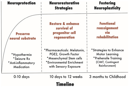
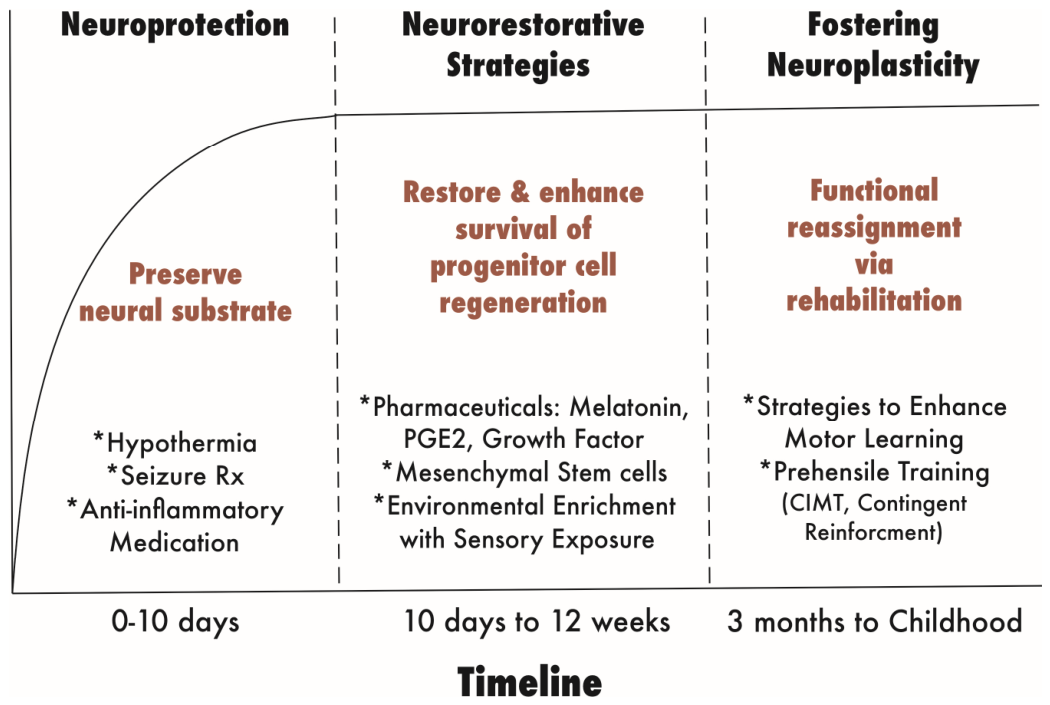
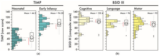
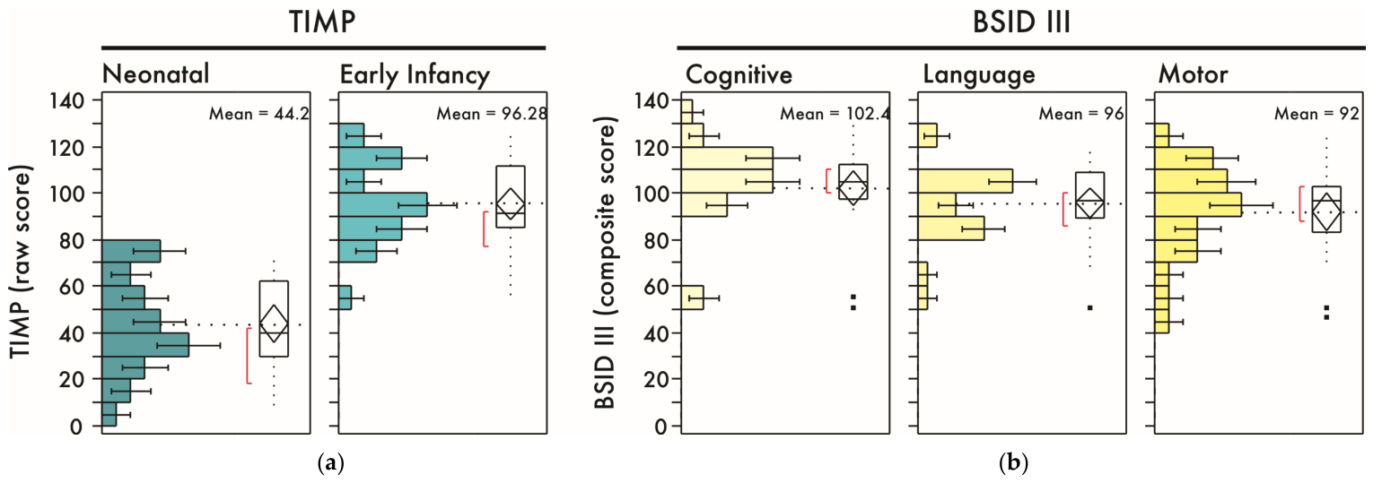
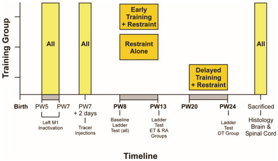
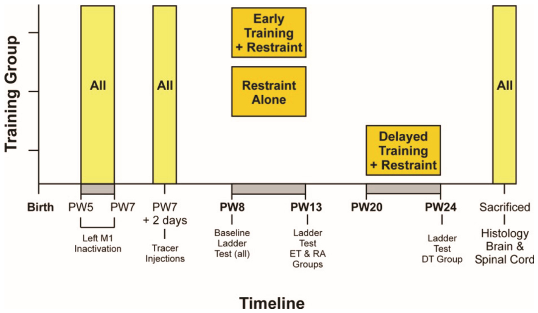
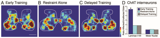
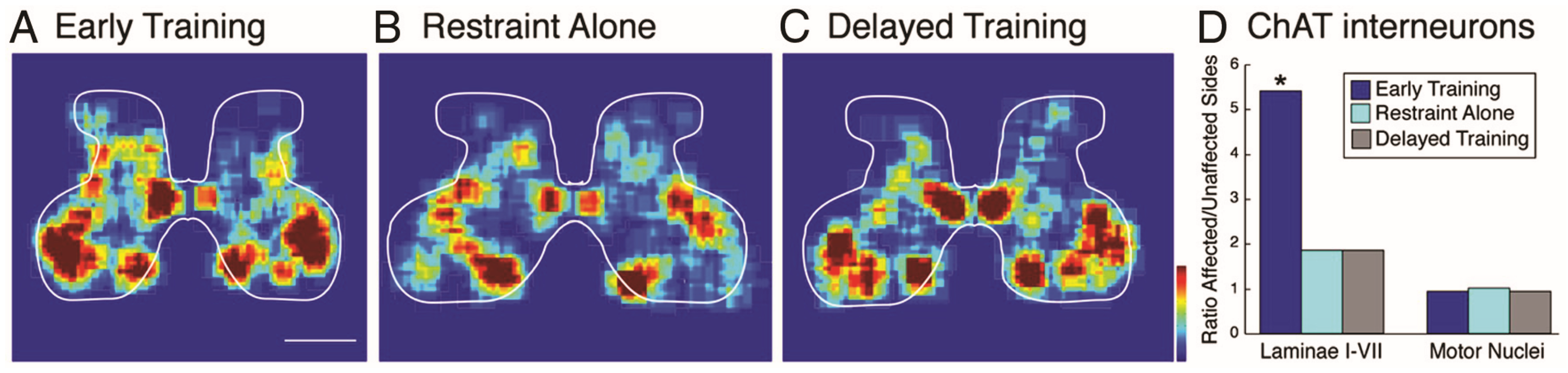
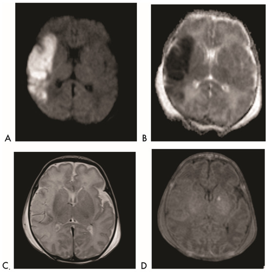
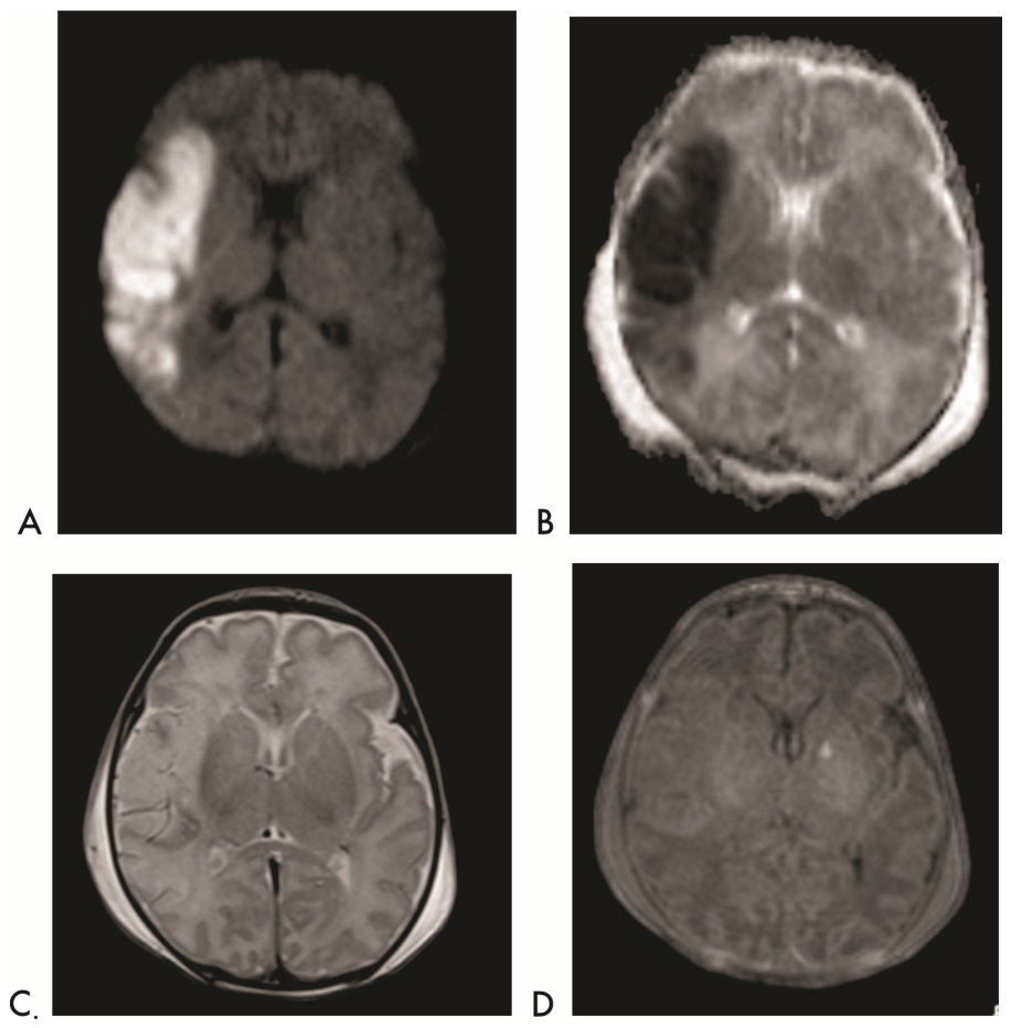
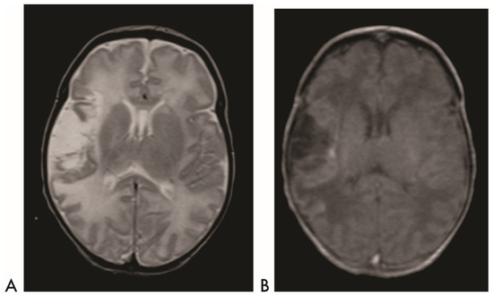
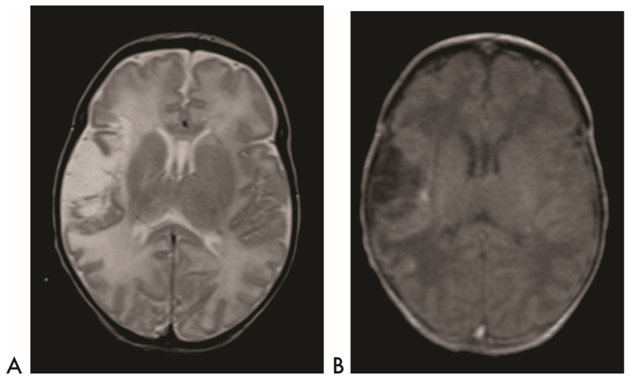

Disclaimer/Publisher’s Note: The statements, opinions and data contained in all publications are solely those of the individual author(s) and contributor(s) and not of MDPI and/or the editor(s). MDPI and/or the editor(s) disclaim responsibility for any injury to people or property resulting from any ideas, methods, instructions or products referred to in the content. |
© 2026 by the authors. Licensee MDPI, Basel, Switzerland. This article is an open access article distributed under the terms and conditions of the Creative Commons Attribution (CC BY) license.