


Abstract
Artificial intelligence has considerable potential to support dermoscopic skin cancer assessment; however, class imbalance, heterogeneous lesion morphology, indistinct lesion boundaries, and imaging variability can limit model generalizability. This study presents DeepSkin, a segmentation-guided deep learning framework integrating lesion segmentation, generative augmentation, and lesion-focused classification. Dermoscopic images were first segmented using a bidirectional ConvLSTM-based U-Net (BCDU-Net), and the resulting lesion regions were used for region-of-interest (ROI) extraction. During classifier development, a conditional generative adversarial network (cGAN) generated class-conditioned synthetic lesion ROIs to increase training-data diversity and mitigate class imbalance. Real and synthetic training ROIs were subsequently used to train a fine-tuned VGG16 network for binary benign–malignant classification. The framework was developed using the ISIC 2016 dataset, comprising 900 training and 379 test images, and independently evaluated on the PH2 dataset. On ISIC 2016, DeepSkin achieved a Dice coefficient of 0.937 and an intersection over union (IoU) of 0.882 for lesion segmentation, together with a classification accuracy of 96.6% and a receiver operating characteristic area under the curve (ROC-AUC) of 0.981. Cross-dataset external evaluation on the independent PH2 dataset, conducted without retraining or fine-tuning, yielded a classification accuracy of 95%. Ablation analysis further demonstrated incremental performance improvements associated with preprocessing, segmentation-guided ROI extraction, and generative augmentation. Grad-CAM visualizations provided qualitative insight into image regions contributing to classification decisions. These findings suggest that integrating generative augmentation with segmentation-guided lesion classification can support accurate dermoscopic image analysis across independent datasets. Further evaluation using larger, diverse, multi-center and prospective clinical datasets is required to establish broader generalizability and clinical applicability.
1. Introduction
Skin cancer represents a substantial global public health burden, with melanoma and non-melanoma skin cancers contributing to cancer incidence and mortality worldwide [1]. Ultraviolet (UV) radiation is a major environmental risk factor for skin cancer, with patterns of sun exposure and individual susceptibility, including skin phototype, contributing to variations in disease risk [2]. Although non-melanoma skin cancers, including basal cell carcinoma (BCC) and squamous cell carcinoma (SCC), account for a substantial proportion of skin cancer diagnoses, melanoma contributes disproportionately to skin cancer mortality because of its potential for invasion and metastasis [3]. Early recognition and accurate assessment of suspicious lesions are therefore important for improving patient outcomes.
Although melanoma incidence is generally higher among populations with lighter skin pigmentation, studies have reported later-stage diagnosis and poorer outcomes among certain racial and ethnic minority populations [4]. The underrepresentation of diverse skin tones in dermatological imaging datasets further raises concerns regarding the generalizability and equitable performance of AI-based diagnostic systems [5].
Differences in access to dermatological expertise and the underrepresentation of diverse populations in medical imaging datasets also present challenges for the development of broadly generalizable artificial intelligence (AI) systems. These considerations emphasize the need for objective and reproducible computational approaches that can support dermoscopic lesion assessment across heterogeneous populations and imaging conditions.
Dermoscopy is a non-invasive imaging technique that enables visualization of morphological structures that are not readily apparent to the unaided eye and can improve the assessment of suspicious skin lesions. Nevertheless, dermoscopic interpretation depends on clinical expertise and can be challenging for atypical or visually ambiguous lesions [6,7,8]. Histopathological examination remains the reference standard for definitive diagnosis when tissue evaluation is required [9]. Consequently, computer-aided analysis of dermoscopic images has attracted considerable interest as a means of supporting, rather than replacing, clinical decision-making [10].
Recent advances in AI, particularly deep learning, have substantially improved automated medical image analysis. Convolutional neural networks (CNNs) can learn hierarchical representations directly from dermoscopic images and have demonstrated strong performance in skin lesion classification tasks [11]. However, the performance and generalizability of such models can be affected by limited training data, class imbalance, heterogeneous lesion morphology, illumination differences, acquisition conditions, and imaging artefacts. These challenges are particularly important when models trained on one dataset are evaluated on images acquired under different conditions.
Artificial intelligence-based melanoma assessment extends beyond dermoscopic image classification to encompass complementary diagnostic and prognostic approaches operating at different biological and morphological scales. Veronesi et al. [12] investigated machine learning-based melanoma detection using nuclear morphology and spatial organization in histopathological images, highlighting the diagnostic relevance of cellular-level characteristics. Similarly, Venturi et al. [13] reviewed advances in digital pathology and artificial intelligence for melanoma diagnosis, emphasizing the potential of computational analysis of whole-slide images, tissue architecture, and histopathological features. At another level, Scotti et al. [14] examined non-invasive imaging modalities for estimating Breslow thickness, an important indicator of melanoma prognosis. Collectively, these studies demonstrate that dermoscopy, non-invasive depth assessment, and computational pathology provide complementary information throughout melanoma evaluation. Accordingly, dermoscopic AI systems should be regarded as potential decision-support tools for lesion assessment rather than substitutes for histopathological confirmation or established clinical staging procedures.
Lesion segmentation and data augmentation provide complementary strategies for addressing these limitations. Accurate segmentation can isolate lesion regions from surrounding skin and irrelevant background information, enabling subsequent classifiers to focus on lesion-specific features [15,16]. At the same time, generative models can increase training-data diversity and mitigate class imbalance beyond what can be achieved using conventional geometric transformations alone [17,18].
To address this gap, this study presents DeepSkin, a segmentation-guided deep learning framework that integrates conditional generative adversarial network (cGAN)-based augmentation, bidirectional ConvLSTM U-Net (BCDU-Net) lesion segmentation, region-of-interest (ROI) extraction, and VGG16-based classification. The framework is designed to mitigate class imbalance, capture spatial–contextual information for lesion localization, and use lesion-focused regions for binary benign–malignant classification. DeepSkin is evaluated on the ISIC 2016 dataset and externally assessed on independent dermoscopic data to investigate its generalization under dataset shift.
The main contributions of this study are: (1) a cGAN-based augmentation strategy designed to mitigate class imbalance and increase training-data diversity; (2) a BCDU-Net architecture for context-aware lesion segmentation and segmentation-guided ROI extraction; (3) a fine-tuned VGG16 network for binary benign–malignant classification of lesion-focused regions; and (4) a comprehensive experimental evaluation incorporating segmentation and classification metrics, ablation analysis, model interpretability, and external validation.
The remainder of this paper is organized as follows. Section 2 reviews related work on deep learning-based dermoscopic image classification, segmentation architectures, and generative augmentation. Section 3 describes the datasets, preprocessing procedures, proposed framework, training strategy, and evaluation protocol. Section 4 presents the experimental results, including component-wise ablation, computational performance, and external validation. Section 5 discusses the principal findings, generalization, limitations, and relation to existing approaches. Finally, Section 6 summarizes the main conclusions and directions for future research.
2. Related Work
The rapid development of AI in medical imaging has advanced automated dermoscopic image analysis, particularly in lesion classification, segmentation, and computer-aided clinical decision support. However, limited annotated datasets, class imbalance, heterogeneous lesion morphology, and differences in imaging conditions remain important challenges to model generalization. The literature relevant to DeepSkin can be broadly organized into three areas: deep learning-based skin lesion classification, lesion segmentation, and generative approaches for data augmentation, followed by the research gap motivating the proposed framework.
2.1. Deep Learning-Based Skin Lesion Classification
2.1.1. CNN-Based Classification
Deep learning has substantially advanced automated skin lesion classification by enabling discriminative features to be learned directly from dermoscopic images. The landmark study by Esteva et al. [19] demonstrated the potential of CNNs trained on large-scale image datasets for dermatologist-level classification of selected skin lesion categories [20]. Early convolutional neural network architectures, such as LeNet, established foundational principles for hierarchical feature extraction [21]. Subsequent developments have introduced deeper architectures, including VGGNet [22], ResNet [23], DenseNet [24], and Inception-v3 [25], for automated dermoscopic skin lesion classification. These architectures learn hierarchical feature representations that capture visual characteristics relevant to lesion assessment, including texture, color distribution, border irregularities, and structural patterns.
More recent studies have incorporated attention mechanisms and feature-fusion strategies to improve representation learning. For example, DSCIMABNet [26] combines depthwise separable convolutions with multi-head attention to enhance feature discrimination while considering computational efficiency. Tang et al. [27] proposed a joint–individual fusion framework incorporating multimodal attention mechanisms to integrate dermatological images and patient metadata, enabling the learning of complementary features for skin cancer classification. These developments demonstrate the increasing emphasis on directing model attention toward diagnostically relevant representations rather than relying exclusively on conventional convolutional feature extraction.
Despite these advances, CNN-based classifiers can remain sensitive to class imbalance, irrelevant background information, image artefacts, and dataset-specific characteristics. Full-image classification may additionally allow models to learn features outside the lesion region, motivating approaches that explicitly incorporate lesion localization into the classification pipeline.
2.1.2. Transfer Learning and Fine-Tuning
A major challenge in dermatological imaging is the limited availability of labeled datasets, particularly for malignant lesions, as dermoscopic image annotation requires expert dermatologists. Transfer learning (TL) has been widely used to address the scarcity of labeled dermoscopic data by adapting models pre-trained on large datasets such as ImageNet. This approach improves convergence and generalization in small medical datasets. For example, Alotaibi and AlSaeed [28] demonstrated that incorporating self-attention, soft-attention, and hard-attention mechanisms into a pretrained Xception architecture improved skin cancer classification performance compared with the baseline model. Deng et al. [29] developed a transfer learning-based multimodal framework combining pretrained DenseNet-121 with clinical information to improve malignant skin lesion identification from smartphone images.
Recent studies have also investigated computationally efficient transfer learning architectures for skin lesion classification. For example, Fathonah et al. [30] employed a modified EfficientNetV2B0 architecture to improve multiclass skin lesion classification while considering computational efficiency. Attention-based models have also been investigated to improve feature learning in dermoscopic image classification. For instance, Al-Waisy et al. [31] proposed Skin-DeepNet, which combines lesion segmentation, HRNet-based multiscale feature extraction, attention-based feature refinement, and decision fusion to enhance skin lesion classification.
To further enhance classification performance, ensemble learning approaches have been investigated to combine complementary feature representations from different deep learning architectures. For example, Perera et al. [32] integrated ResNet-101, DenseNet-121, and Inception-v3 through a weighted-average ensemble strategy for melanoma classification, incorporating explainable artificial intelligence techniques to improve the interpretability of model predictions. In addition, multimodal learning frameworks have gained attention in dermatological diagnosis. He et al. [33] proposed a Co-Attention Fusion Network that integrates dermoscopic and clinical images through cross-modal attention mechanisms to learn complementary feature representations. More recently, Tran-Van and Le [34] developed a multimodal framework incorporating cross-attention-based fusion of dermoscopic images and patient metadata to enhance skin lesion classification. Tan et al. [35] proposed MFF-CINet, a clinical-inspired skin lesion recognition framework that integrates multi-level feature fusion for hair segmentation, deep hair removal, and lesion-feature attention to improve dermoscopic lesion recognition.
Recent advancements in automated skin lesion classification have explored multiscale feature extraction, attention mechanisms, and optimization-based learning strategies to improve diagnostic performance. Mousa et al. [36] proposed a framework integrating late-stage discrete wavelet transform (DWT), pretrained convolutional neural networks, self-attention mechanisms, and swarm-based optimization algorithms to enhance feature representation and classification. Their approach reported classification accuracies of 98.11% and 97.95% on the ISIC 2016 and ISIC 2017 datasets, respectively, demonstrating the potential of wavelet-enhanced feature learning and optimization strategies for automated dermoscopic image classification. While their approach emphasizes feature extraction and optimization, other aspects of dermoscopic image analysis, including explicit lesion localization and training-data augmentation, remain important considerations in the development of classification frameworks. The integration of these complementary strategies warrants further investigation, particularly to determine whether lesion-focused representations and generative augmentation provide measurable benefits beyond those achieved through conventional transfer learning and feature optimization.
Despite these advancements, CNN-based skin lesion classification models continue to face challenges associated with imbalanced datasets and the underrepresentation of diverse skin tones, which may affect model generalizability [37]. Moreover, classification performance often depends on accurate lesion segmentation and balanced datasets, highlighting the need for integrated frameworks that combine segmentation, data augmentation, and classification to improve robustness and clinical applicability. However, transfer learning-based approaches often depend on pre-trained features that may not fully capture lesion-specific structures, particularly when segmentation is not explicitly incorporated into the learning pipeline.
2.2. Skin Lesion Segmentation Architectures
Accurate lesion segmentation is an important component of automated dermoscopic image analysis because it delineates lesion regions from surrounding skin and facilitates subsequent lesion-focused analysis [38]. U-Net, introduced by Ronneberger et al. [39], has become a foundational architecture for biomedical image segmentation. Its encoder–decoder structure and skip connections enable the integration of high-level semantic information with fine-grained spatial features. Subsequent extensions have been developed to improve feature fusion and localization. UNet++, proposed by Zhou et al. [40], introduces nested and densely connected skip pathways to reduce the semantic gap between encoder and decoder representations. Similarly, Attention-enhanced U-Net architectures have also been investigated for dermoscopic skin lesion segmentation. For example, Babu and Murali [41] proposed AttenUNeT X, which integrates attention mechanisms and iterative decoder feedback to emphasize lesion-relevant features and improve boundary delineation.
Further developments have focused on multi-scale feature extraction, contextual representation, and spatial feature integration. Basak et al. [42] proposed MFSNet, a multi-focus segmentation network that incorporates multi-scale feature representations, boundary attention, and reverse attention mechanisms for dermoscopic skin lesion segmentation. Azad et al. [43] introduced BCDU-Net, which integrates bidirectional convolutional long short-term memory (ConvLSTM) units and densely connected convolutions to improve feature propagation and the fusion of encoder–decoder representations. Alom et al. [44] developed R2U-Net, incorporating recurrent residual convolutional units to strengthen contextual feature representation. Building upon recurrent residual learning, Zuo et al. [45] proposed R2AU-Net, which incorporates attention gates to refine feature selection during medical image segmentation, including skin lesion segmentation.
More recently, Rout et al. [46] proposed DilatedSkinNet, an encoder–decoder segmentation framework incorporating lesion detail extraction and a fine feature aggregation module to preserve spatial information and enhance lesion boundary delineation. The framework was trained on ISIC 2016 and evaluated using ISIC 2016 and additional dermoscopic datasets, including ISIC 2017, ISIC 2018, and PH2. Although these approaches demonstrate advances in lesion segmentation, segmentation accuracy alone does not establish improvements in downstream classification.
2.3. Generative Adversarial Network for Data Augmentation
A major challenge in developing reliable skin cancer classification systems is the limited availability and class imbalance of annotated dermoscopic datasets, particularly for underrepresented lesion categories. Furthermore, obtaining expert-annotated dermoscopic images can be resource-intensive. Generative adversarial networks (GANs), originally introduced by Goodfellow et al. [47], provide a framework for generating synthetic images through adversarial learning. GANs consist of two competing neural networks: a generator that produces synthetic samples and a discriminator that distinguishes generated images from real images. Through iterative adversarial training, the generator learns to approximate the underlying data distribution. In dermatological imaging, GAN-based approaches have been investigated to expand training datasets, increase image diversity, and mitigate class imbalance [48].
Conditional generative adversarial networks (cGANs) extend this framework by incorporating auxiliary information, such as class labels or segmentation masks, to guide image generation [48,49]. This enables controlled image synthesis and provides opportunities to increase the representation of underrepresented lesion categories. Several studies have investigated GAN-based synthesis in dermoscopic image analysis. Behara et al. [50] proposed an improved deep convolutional generative adversarial network (DCGAN) for skin lesion image synthesis and binary classification, addressing challenges associated with data scarcity and class imbalance. More recently, Su et al. [18] developed a two-stage GAN-based augmentation framework that integrates shared and class-specific information to generate synthetic skin lesion images for imbalanced multiclass classification. Their findings demonstrate the potential of generative augmentation to support downstream classification when synthetic images are appropriately generated and evaluated.
Beyond data augmentation, GAN-based approaches have been investigated for image normalization, synthesis, and reconstruction. Salvi et al. [51] proposed a GAN-based color normalization framework that formulates color standardization as an image-to-image translation task in digital pathology and dermatology, addressing variability in image appearance. Sharafudeen et al. [52] developed Derm-CGAN to synthesize dermoscopic lesion images and evaluated Vision Transformer architectures for distinguishing synthetic images from real images. Their work highlights the importance of assessing the authenticity and characteristics of GAN-generated medical images. Furthermore, Veeramani and Jayaraman [53] proposed MELIIGAN, a GAN-based super-resolution reconstruction framework incorporating stacked residual blocks to recover fine-grained image details in dermoscopic images. In a broader medical imaging context, Pang et al. [49] reviewed generative approaches, including Pix2Pix and CycleGAN, for image creation and image-to-image translation.
Despite these advances, GAN-based approaches face several limitations, including training instability, mode collapse, insufficient sample diversity, and difficulties in preserving clinically meaningful image characteristics [48,49]. Although synthetic images may exhibit high visual similarity to real dermoscopic images, visual realism alone does not establish their diagnostic validity or their usefulness for downstream classification. Consequently, the effectiveness of generative augmentation should be evaluated in relation to both image quality and classification performance. These considerations motivate further investigation of approaches that combine lesion-focused image representations with controlled generative augmentation. Within this context, DeepSkin investigates the integration of segmentation-guided region-of-interest extraction and conditional generative augmentation to increase training-data diversity while retaining diagnostically relevant lesion information.
2.4. Research Gap and Motivation for DeepSkin
The reviewed literature demonstrates substantial progress in dermoscopic image classification, lesion segmentation, transfer learning, attention mechanisms, and generative augmentation. Nevertheless, these components are frequently developed and evaluated independently. Classification models operating on entire dermoscopic images may incorporate irrelevant background information, while standalone segmentation approaches do not necessarily exploit lesion localization for downstream classification. Similarly, generative augmentation can increase training-data diversity without being explicitly coupled with lesion-focused analysis.
A further challenge concerns generalization across datasets. Differences in image acquisition, lesion distribution, annotation protocols, and other dataset-specific characteristics can cause performance degradation when a model is evaluated outside its development dataset. Therefore, evaluation on independent dermoscopic datasets is important for assessing robustness to dataset shift, although such evaluation should not be interpreted as equivalent to prospective clinical validation.
Although recent studies have demonstrated strong classification performance through advanced feature extraction and optimization techniques, including the wavelet-enhanced approach proposed by Mousa et al. [36], further investigation of integrated segmentation-guided classification and generative augmentation remains relevant. High classification accuracy alone does not establish the contribution of lesion localization or training-data augmentation to downstream classification performance. In contrast to approaches primarily emphasizing feature optimization, DeepSkin combines BCDU-Net-based lesion segmentation, segmentation-guided ROI extraction, and class-conditioned generative augmentation with VGG16 classification. The principal contribution of the proposed framework lies in evaluating the complementary roles of lesion localization and generative augmentation within an integrated dermoscopic image-analysis pipeline. This integration is examined through segmentation and classification evaluation, component-wise ablation analysis, qualitative interpretability assessment, and independent cross-dataset testing.
To address these limitations, this study proposes DeepSkin, an integrated multi-stage deep learning framework combining cGAN-based generative augmentation, BCDU-Net lesion segmentation, segmentation-guided ROI extraction, and VGG16-based binary classification. The cGAN component is designed to mitigate class imbalance and increase training-data diversity, while BCDU-Net captures spatial–contextual information for lesion localization and boundary delineation. The resulting lesion-focused ROIs are subsequently used for benign–malignant classification. This integration enables the study to examine whether generative augmentation and lesion-focused analysis provide complementary benefits within a unified dermoscopic image-analysis framework.
3. Materials and Methods
This section describes the datasets, image preprocessing procedures, architecture, training strategy, and evaluation protocol used to develop and evaluate the proposed DeepSkin framework. DeepSkin is an integrated multi-stage deep learning framework combining context-aware lesion segmentation, segmentation-guided region-of-interest (ROI) extraction, conditional generative augmentation, and transfer learning-based classification. The framework was designed to address major challenges in dermoscopic image analysis, including lesion-boundary variability, class imbalance, and the potential influence of irrelevant background information on classification.
3.1. Overview of the Proposed DeepSkin Framework
The proposed DeepSkin framework consists of four principal computational stages following image preprocessing: lesion segmentation using a bidirectional ConvLSTM U-Net (BCDU-Net), segmentation-guided ROI extraction, conditional generative augmentation of training ROIs, and binary lesion classification using a fine-tuned VGG16 network.
Dermoscopic images are initially standardized through resizing, intensity normalization, and artefact reduction. The preprocessed images are subsequently processed by the BCDU-Net segmentation network to generate binary lesion masks. These masks are used to isolate lesion regions and extract standardized lesion-centered ROIs.
During model development, ROIs obtained from the training subset are used to train a conditional generative adversarial network (cGAN). The cGAN generates additional class-conditioned lesion ROIs to increase training-data diversity and mitigate class imbalance. The resulting synthetic ROIs are combined with real training ROIs and used to train the VGG16 classifier for binary benign–malignant discrimination.
The cGAN therefore functions exclusively as a training-time augmentation component and is not required during inference. For an unseen image, the operational inference pathway consists of preprocessing, BCDU-Net lesion segmentation, ROI extraction using the predicted mask, and classification of the resulting ROI using the trained VGG16 network. Figure 1 illustrates the overall architecture and distinguishes the training-specific augmentation stage from the inference pathway. The cGAN is used only during classifier training to augment lesion-centered ROIs; inference consists of preprocessing, BCDU-Net segmentation, ROI extraction, and VGG16-based classification.
Figure 1.
Overview of the proposed DeepSkin framework.
3.2. Dataset Description and Preprocessing
The DeepSkin framework was developed and evaluated using two publicly available dermoscopic image datasets: the ISIC 2016 Challenge dataset and the PH2 dataset. The ISIC 2016 dataset was used for model development and internal evaluation, whereas PH2 was reserved for independent external evaluation.
The ISIC 2016 Challenge dataset comprises 1279 dermoscopic images with corresponding ground-truth lesion segmentation masks and diagnostic annotations. The dataset follows the original predefined training and testing partitions, comprising 900 training images (70.4%) and 379 test images (29.6%), representing an approximately 70:30 training–testing distribution. The original training partition contains 727 benign and 173 malignant lesions, while the test partition consists of 304 benign and 75 malignant lesions.
To ensure consistency and traceability in the experimental evaluation, the official ISIC 2016 test partition and its corresponding diagnostic annotations were used as the reference for dataset verification. Each dermoscopic image is associated with a unique ISIC identifier, allowing its diagnostic ground-truth label to be matched with the corresponding model-generated classification output. This image-level correspondence provides the basis for constructing the confusion matrix and calculating classification performance metrics, including accuracy, precision, sensitivity, specificity, F1-score, and Matthews correlation coefficient (MCC). The experimental workflow was implemented in Python (version 3.10) using Google Colab and documented in the notebook Skin_UNET_GAN.ipynb. The predefined test partition serves as the reference for evaluating classification performance, while the training and validation data are used for model development.
Validation was conducted as part of model development using images from the original training partition, without altering the predefined 70:30 training–testing distribution. The validation subset was used to monitor model performance, guide early stopping, and support learning-rate adjustment. For BCDU-Net, validation involved dermoscopic images and their corresponding segmentation masks, whereas VGG16 validation involved lesion-focused ROIs and their associated diagnostic labels. The cGAN was employed for training-data augmentation, with synthetic images restricted to classifier training rather than independent evaluation.
The PH2 dataset comprises 200 high-resolution dermoscopic images, including 160 benign lesions and 40 melanoma cases. This dataset was reserved for independent external evaluation to investigate the generalizability of the proposed DeepSkin framework under different imaging conditions and lesion characteristics. No additional retraining or fine-tuning was performed using PH2 images.
Prior to model development, image preprocessing was performed to standardize image dimensions and pixel-intensity distributions. All dermoscopic images were resized to 256 × 256 pixels to ensure consistent input dimensions across the dataset. The corresponding segmentation masks were resized to the same spatial resolution to maintain pixel-level correspondence between the images and their reference lesion annotations.
Pixel intensities were standardized using z-score normalization:
where I denotes the input image, while μ and σ denote the mean and standard deviation of the image intensity distribution, respectively. This transformation standardizes the intensity distribution by centering the pixel values around zero and scaling them relative to their standard deviation.
Following z-score normalization, min–max scaling was applied to rescale the standardized intensity values to the range of 0–255, according to:
where Imin and Imax represent the minimum and maximum normalized intensity values, respectively.
Hair artefacts were addressed using a DullRazor-style preprocessing procedure involving morphological hair detection and image inpainting. Specifically, black-hat morphological filtering was applied using a 9 × 9 elliptical structuring element to enhance dark hair structures against the surrounding skin. Candidate hair pixels were identified using a threshold of 10 on the 8-bit grayscale black-hat response. The detected hair regions were subsequently reconstructed through image inpainting with a radius of three pixels. Following hair removal, morphological opening was applied using a 3 × 3 elliptical structuring element for one iteration to suppress small-scale residual artefacts and background noise. These preprocessing operations were intended to reduce image artefacts while preserving diagnostically relevant lesion characteristics, including pigmentation patterns and boundary irregularities.
3.3. Lesion Segmentation Using BCDU-Net
3.3.1. Network Architecture
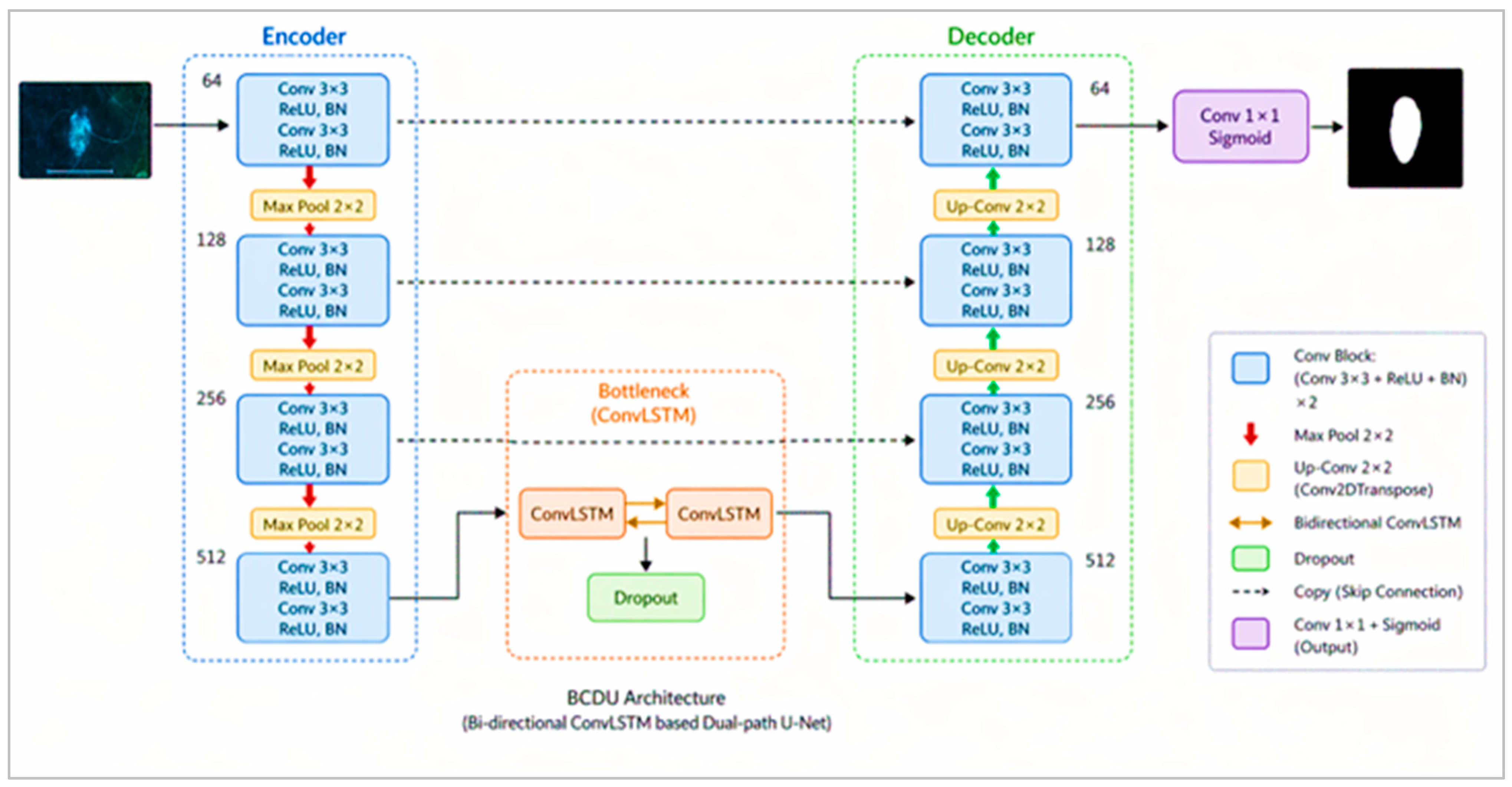
The lesion segmentation component of DeepSkin employs a BCDU-Net architecture that integrates bidirectional convolutional long short-term memory (ConvLSTM) units within a U-Net-based encoder–decoder framework, as shown in Figure 2. The architecture is intended to capture spatial–contextual dependencies between feature representations while retaining fine-grained spatial information required for lesion-boundary delineation.
The encoder performs hierarchical feature extraction using successive convolutional and downsampling operations. Each encoder block contains two 3 × 3 convolutional layers followed by rectified linear unit (ReLU) activation and batch normalization. A 2 × 2 max-pooling operation is used to progressively reduce spatial resolution and increase the receptive field.
Bidirectional ConvLSTM units are incorporated to integrate contextual information between feature representations. In contrast to conventional skip connections, the recurrent units enable information from complementary feature representations to be combined before decoding. This mechanism is useful for dermoscopic lesions characterized by irregular boundaries, heterogeneous pigmentation, low contrast, and imaging artefacts.
The decoder progressively restores spatial resolution using transposed convolutional layers. Skip connections transfer spatial information from corresponding encoder levels to the decoder, supporting recovery of lesion-boundary details. A final 1 × 1 convolution followed by sigmoid activation produces the binary lesion probability map. The BCDU-Net used in the study contains approximately 20.6 million parameters.
Figure 2.
Architecture of the BCDU-Net used for dermoscopic lesion segmentation.
3.3.2. ConvLSTM Formulation
Given an input tensor Xt and previous states (Ht−1,Ct−1), the ConvLSTM operations are defined as:
where * is convolution, and ⊙ denotes element-wise multiplication.
it = σ(Wxi × Xt + Whi × Ht−1 + bi,
ft = σ(Wxf ∗ Xt + Whf ∗ Ht−1 + bf,
ot = σ(Wxo ∗ Xt + Who ∗ Ht−1 + bo),
Ht = Ot⨀tanh(Ct)
Bidirectionality concatenates forward and backward hidden states:
where the bracket denotes concatenation of the forward and backward feature representations.
3.3.3. Segmentation Training
The BCDU-Net was trained using image–mask pairs from the ISIC 2016 training subset. The validation subset was used to monitor optimization and control overfitting. The segmentation network was optimized using Adam with a learning rate of 1 × 10⁻⁴, a batch size of 8, and a maximum of 100 epochs. Early stopping and adaptive learning-rate reduction were employed based on validation performance.
For an input image I, the predicted lesion mask is represented as:
where fθ denotes the BCDU-Net parameterized by θ, and σ denotes sigmoid activation.
A combined Dice–binary cross-entropy (BCE) loss was used for segmentation:
seg = BCE + Dice,
The BCE component penalizes pixel-wise prediction errors, whereas the Dice component promotes spatial overlap between predicted and reference masks. The combined objective therefore considers both pixel-level discrimination and lesion-region overlap.
3.4. Segmentation-Guided ROI Extraction
Following lesion segmentation using BCDU-Net, the predicted lesion probability maps were converted into binary segmentation masks using Otsu’s thresholding method. This adaptive thresholding technique automatically determines a threshold by maximizing the between-class variance of the predicted pixel values, thereby separating lesion regions from the surrounding background. The resulting binary masks were subsequently used to isolate lesion regions and suppress irrelevant background information, facilitating the extraction of lesion-focused regions of interest (ROIs). For a dermoscopic image I and its corresponding binary mask M, the lesion-focused image IROI was obtained through element-wise multiplication, expressed as:
where ⊙ denotes element-wise multiplication and M represents the binary lesion mask, with pixel values of 1 corresponding to the lesion region and 0 representing the background.
IROI = I⊙M,
For classifier development, ground-truth segmentation masks were used to extract ROIs from the training and validation images, whereas masks predicted by the trained BCDU-Net were used during testing. The application of predicted masks during testing was intended to reflect the inference scenario, in which ground-truth lesion annotations are unavailable for unseen dermoscopic images.
The use of ground-truth segmentation masks during classifier development was intended to provide accurately delineated lesion regions and minimize the influence of segmentation errors on feature learning. This approach allowed the VGG16 classifier to learn lesion-specific characteristics from consistently defined ROIs. However, during independent testing, lesion ROIs were generated using BCDU-Net-predicted masks to reproduce the fully automated inference pathway. This difference between ground-truth-based ROI extraction during classifier development and prediction-based ROI extraction during testing introduces a potential distribution mismatch, as inaccuracies in predicted lesion boundaries may alter the morphological and textural information available to the classifier. Consequently, segmentation errors may influence downstream classification performance, particularly for lesions with irregular boundaries or low contrast.
Following lesion masking, bounding-box cropping was applied to isolate the lesion regions while minimizing the inclusion of surrounding background information. Zero-padding was introduced where necessary to preserve the spatial proportions of the extracted lesions, and each ROI was resized to 256 × 256 pixels to ensure uniform input dimensions for the subsequent classification network. Gaussian smoothing was subsequently applied to reduce residual image artefacts and local intensity variations while maintaining relevant lesion structures and boundary characteristics.
The resulting standardized lesion-centered ROIs were subsequently used as inputs to the conditional generative adversarial network (cGAN) for training-data augmentation and to the fine-tuned VGG16 network for binary benign–malignant classification. This segmentation-guided extraction strategy was intended to emphasize lesion-specific morphological characteristics while reducing the influence of irrelevant background information on downstream classification.
3.5. Conditional Generative Data Augmentation
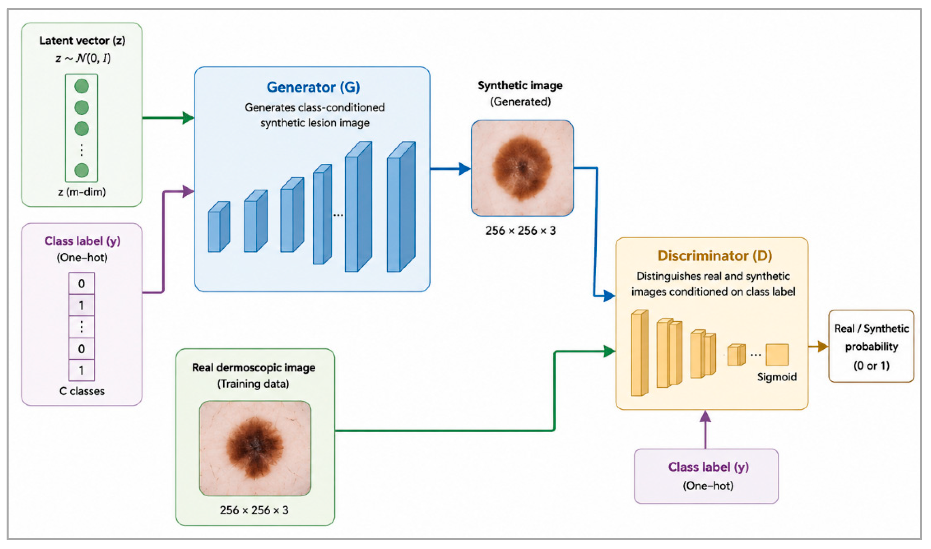
Class imbalance and limited sample diversity can affect the performance of supervised dermoscopic image classifiers. To address these issues, a cGAN was applied to the lesion ROIs obtained from the training subset, as shown in Figure 3. Importantly, the cGAN was used as a training-time augmentation strategy rather than as part of the inference pathway.
The cGAN was conditioned on binary lesion-class labels, with benign and malignant lesions encoded as class 0 and class 1, respectively. This class-conditioning mechanism enabled the generator to produce synthetic lesion ROIs corresponding to the specified diagnostic category, thereby supporting class-specific data augmentation.
The cGAN consists of two competing networks: a generator G and discriminator D. The generator receives a latent noise vector z, sampled from a Gaussian distribution, together with a class label y, and produces a class-conditioned synthetic lesion ROI G(z,y). The discriminator receives either a real or generated lesion sample together with the corresponding class condition and estimates whether the sample originates from the real training distribution.
The conditional adversarial objective is expressed as:
The generator was implemented as a fully convolutional network using transposed convolutional layers for progressive upsampling. The architecture incorporated a dense layer, reshaping operations, ConvTranspose2D layers, LeakyReLU activation (α = 0.2), and batch normalization, producing synthetic images with dimensions of 256 × 256 × 3.
The discriminator employed Conv2D and dropout layers with LeakyReLU activations, followed by sigmoid activation for real-versus-generated discrimination. The generator and discriminator were trained using Adam with a learning rate of 2 × 10⁻⁴, β1 = 0.5, a batch size of 32, and 200 epochs.
Synthetic lesion ROIs were generated separately for the benign and malignant classes using the trained cGAN, with a greater proportion of synthetic samples allocated to the malignant class to mitigate class imbalance and improve its representation during classifier training. Synthetic-image quality was evaluated using the Fréchet inception distance (FID), structural similarity index measure (SSIM), peak signal-to-noise ratio (PSNR), and image diversity measures. These metrics were used to characterize distributional similarity, structural consistency, image fidelity, and variability among the samples generated. The synthetic ROIs were subsequently combined with real training ROIs for VGG16 classification. Generative augmentation was restricted to the training dataset, while synthetic images were excluded from the independent test and external evaluation datasets.
Figure 3.
Architecture of the conditional generative adversarial network used for class-conditioned augmentation of training ROIs.
3.6. VGG16-Based Lesion Classification
The classification component of DeepSkin used the VGG16 architecture pretrained on ImageNet. Instead of processing complete dermoscopic images, the classifier used lesion-centered ROIs resized to 256 × 256 × 3 pixels.
During training, real lesion ROIs were combined with synthetic ROIs generated by the cGAN. The pretrained VGG16 network was used to extract features from the lesion images. During training, the first three convolutional blocks of VGG16 were frozen to preserve pretrained features, while the final two convolutional blocks and classification layers were trainable to adapt the model to dermoscopic images. Fully connected layers and a SoftMax output layer were used to classify lesions as benign or malignant. Dropout was applied to reduce overfitting. The classification architecture is illustrated in Figure 4.
Figure 4.
VGG16 architecture used for binary classification of segmentation-guided lesion ROIs.
The SoftMax output layer generated probabilities for the two classes. Categorical cross-entropy was used as the classification loss function, expressed as:
where yc denotes the ground-truth class indicator and denotes the predicted probability associated with class c.
The VGG16 classifier was optimized using Adam with a learning rate of 1 × 10⁻⁴, a batch size of 16, and a maximum of 50 epochs. ReduceLROnPlateau was employed to adaptively reduce the learning rate when validation performance plateaued.
During inference, an unseen dermoscopic image is preprocessed, segmented using the trained BCDU-Net, and converted into a lesion-centered ROI using the predicted segmentation mask. The resulting ROI is then passed directly to the trained VGG16 classifier. No cGAN-generated images are required during inference.
3.7. Evaluation Metrics
DeepSkin was evaluated separately for lesion segmentation and binary lesion classification to characterize the performance of the individual components and the integrated framework.
For lesion segmentation, performance was evaluated using the Dice coefficient, intersection over union (IoU), pixel accuracy, sensitivity, specificity, 95th-percentile Hausdorff distance (HD95), and average symmetric surface distance (ASSD). Dice and IoU quantify the spatial overlap between predicted and reference lesion masks, whereas HD95 and ASSD characterize boundary agreement.
The Dice coefficient was calculated as:
and IoU as:
For binary lesion classification, performance was evaluated using accuracy, precision, sensitivity (recall), specificity, F1-score, Matthew’s correlation coefficient (MCC), and the area under the receiver operating characteristic curve (ROC-AUC). Quantitative performance was evaluated using the following standard metrics [35]:
Here, TP, TN, FP, and FN denote true positives, true negatives, false positives, and false negatives, respectively. ROC-AUC was used to characterize the ability of the classifier to discriminate between the two diagnostic classes across decision thresholds.
3.8. External Validation
The PH2 dataset was reserved exclusively for external evaluation and was not used during model training, validation, model selection, or hyperparameter optimization. This evaluation strategy was designed to assess the generalizability of the DeepSkin framework by examining whether the model developed using the ISIC 2016 dataset maintained its classification performance when applied to an independent dermoscopic dataset. The use of PH2 enabled an assessment of model performance under differences in imaging conditions and lesion characteristics. No cGAN augmentation, classifier retraining, or model fine-tuning was performed using PH2 data. The same preprocessing procedure used for the ISIC 2016 dataset was applied to PH2 images to maintain consistency across the internal and external evaluation procedures.
3.9. Implementation Details and Reproducibility
The DeepSkin framework was implemented in Python 3.10 using TensorFlow 2.14 and Keras 3.0 with CUDA-enabled GPU acceleration. All experiments were conducted using Google Colab Pro+ equipped with an NVIDIA Tesla T4 GPU with 16 GB of VRAM and 25 GB of system RAM. Random seeds were fixed during experimentation to improve reproducibility across training runs.
The BCDU-Net segmentation model, cGAN, and VGG16 classifier were trained using batch sizes of 8, 32, and 16, respectively. The corresponding learning rates were 1 × 10⁻⁴ for BCDU-Net, 2 × 10⁻⁴ for the cGAN, and 1 × 10⁻⁴ for VGG16. Maximum training durations were set to 100 epochs for BCDU-Net, 200 epochs for the cGAN, and 50 epochs for VGG16.
Adam was used for optimization. For BCDU-Net and VGG16, the Adam parameters were set to β1 = 0.9 and β2 = 0.999, whereas cGAN training used β1 = 0.5 and β2 = 0.999 to support adversarial optimization. Regularization and training-control strategies included dropout, batch normalization, early stopping based on validation performance, and adaptive learning-rate reduction where applicable. Training progress was monitored using TensorBoard, while model checkpoints, trained weights, and experimental logs were retained in Google Drive to support experiment tracking and reproducibility.
The computational workflow was implemented and documented in the Python-based Google Colab notebook Skin_UNET_GAN.ipynb. The notebook contains executable code and recorded procedures associated with model development and experimentation. Together with the retained model checkpoints and training logs, these records provide a basis for examining the implementation and reproducing the computational procedures.
4. Experiments and Results
This section presents the experimental evaluation of the DeepSkin framework. Results are reported sequentially for preprocessing, lesion segmentation, segmentation-guided ROI extraction, generative augmentation, and binary classification. Component-wise ablation, computational performance, and external validation are subsequently examined to assess the contribution and robustness of the integrated framework. Additional analyses examine the contribution of individual framework components, computational efficiency, and performance on an independent external dataset.
4.1. Preprocessing Evaluation
Figure 5 illustrates the visual effect of the preprocessing pipeline on a representative dermoscopic image. Compared with the original image, the successive preprocessing stages produced noticeable changes in pixel-intensity distribution and reduced visible image artefacts.
Figure 5.
Effect of the preprocessing pipeline on a representative dermoscopic image: (a) original image; (b) z-score normalization; (c) min–max scaling; (d) hair removal; and (e) morphological filtering, with the corresponding pixel-intensity histograms.
Following z-score normalization and min–max scaling, the corresponding histograms show a more standardized distribution of pixel intensities. Hair removal visibly reduced hair occlusion over the lesion region, while morphological filtering further reduced small-scale residual artefacts and background irregularities. Across these stages, the lesion remained visually distinguishable for subsequent segmentation.
The accompanying intensity histograms illustrate the changes in pixel distribution associated with each preprocessing stage. Overall, the qualitative results indicate that preprocessing reduced prominent artefacts and standardized image intensity characteristics before lesion segmentation. The quantitative contribution of preprocessing to downstream classification performance is examined separately in the ablation analysis in Section 4.6.
4.2. BCDU-Net Lesion Segmentation Results
The segmentation performance of BCDU-Net was evaluated using region-overlap, pixel-level, and boundary-distance metrics. The quantitative results are summarized in Table 1.
Table 1.
Segmentation performance of BCDU-Net on the ISIC 2016 test subset.
BCDU-Net achieved a Dice coefficient of 0.937 ± 0.018 and an IoU of 0.882 ± 0.024, indicating substantial overlap between the predicted and reference lesion masks. Pixel accuracy reached 0.964 ± 0.014, while sensitivity and specificity were 0.921 ± 0.029 and 0.973 ± 0.012, respectively. The boundary-distance measures yielded an HD95 of 4.6 ± 1.3 pixels and an ASSD of 1.82 ± 0.64 pixels, indicating close correspondence between the predicted and reference lesion boundaries.
Figure 6 provides a representative qualitative example of the segmentation output. The predicted mask closely follows the overall shape of the ground-truth lesion region. The boundary overlay further shows close correspondence between the predicted and reference contours, with relatively small deviations occurring mainly along irregular portions of the lesion boundary.
Figure 6.
Representative BCDU-Net lesion segmentation result: (a) preprocessed dermoscopic image, (b) ground-truth lesion mask, (c) BCDU-Net-predicted lesion mask, and (d) overlay of the ground-truth (green dashed line) and predicted (red solid line) lesion boundaries.
The quantitative and qualitative results show that BCDU-Net provided lesion masks with high spatial overlap and relatively small boundary discrepancies. These predicted lesion regions were subsequently used for segmentation-guided ROI extraction.
4.2.1. Segmentation Training Convergence Analysis
Figure 7 presents the training and validation trajectories of the BCDU-Net segmentation model over 100 epochs. Both training and validation losses decreased progressively during optimization before approaching stable values toward the later epochs. A corresponding increase was observed in the accuracy curves, with both training and validation performance stabilizing above 0.92. The relatively close trajectories of the training and validation curves indicate stable convergence, without a pronounced divergence between model performance on the training and validation subsets.
Figure 7.
Training and validation accuracy and loss curves of the proposed BCDU-Net segmentation network over 100 training epochs.
4.2.2. Robustness Evaluation Under Challenging Dermoscopic Conditions
To examine segmentation behavior under more difficult dermoscopic conditions, performance was further analyzed across hair-occluded lesions, low-contrast images, small lesions (<10% lesion area), lesions with irregular borders. The corresponding Dice and IoU values are summarized in Table 2.
Table 2.
Robustness performance across challenging dermoscopic conditions.
Segmentation performance varied across the challenging categories, with the highest Dice and IoU values observed for lesions with irregular borders (0.931 ± 0.019 and 0.874 ± 0.022, respectively). Hair-occluded lesions also retained comparatively high segmentation performance, with a Dice coefficient of 0.921 ± 0.022. The largest reduction was observed for small lesions, for which the Dice coefficient and IoU decreased to 0.894 ± 0.028 and 0.821 ± 0.034, respectively. Low-contrast lesions similarly presented greater segmentation difficulty than the overall test set.
Across the challenging samples, 93.2% of cases achieved a Dice coefficient of at least 0.90. The observed failure cases were primarily associated with pronounced dermoscopic shadows, substantial illumination gradients, and very small lesion regions. Overall, the subgroup analysis indicates that segmentation performance was retained across several difficult imaging conditions, although reduced overlap for small and low-contrast lesions identifies conditions in which further improvement may be required.
Taken together, the overlap, boundary, convergence, and challenging-case analyses demonstrate consistent lesion localization by BCDU-Net across the evaluated dermoscopic conditions. Although reduced performance was observed for small and low-contrast lesions, the predicted masks generally maintained close spatial correspondence with reference annotations. These segmentation outputs subsequently provide the lesion localization required for segmentation-guided ROI extraction in the next stage of the DeepSkin framework.
4.3. Segmentation-Guided ROI Results
Following lesion localization, the segmentation outputs were used to isolate lesion-centered regions for subsequent augmentation and classification. Figure 8 illustrates the transition from lesion segmentation to ROI extraction, showing the predicted lesion mask, its correspondence with the dermoscopic image, the isolated lesion region, and the resulting cropped ROI.
Figure 8.
Representative segmentation-guided ROI extraction: (a) dermoscopic image, (b) lesion mask, (c) mask–image overlay, (d) isolated lesion region, and (e) cropped lesion-centered ROI.
The extracted ROIs substantially restricted the image field to the segmented lesion region, thereby reducing the amount of surrounding skin and unrelated background information presented to subsequent stages of the framework. The standardized lesion crops served as the lesion-focused representation used for generative augmentation and classifier development. The extracted lesion-centered ROIs were subsequently used as inputs for cGAN-based generative augmentation and CNN-based classifier (VGG16) within the proposed DeepSkin framework. By restricting the analysis to segmented lesion regions, the ROI extraction procedure was designed to emphasize diagnostically relevant characteristics, including pigmentation patterns, textural variations, and lesion boundary irregularities, while reducing the influence of surrounding background information.
The contribution of ROI extraction is also reflected in the ablation analysis presented in Section 4.6, where incorporation of the ROI stage increased classification accuracy relative to the preprocessing-only configuration.
4.4. cGAN-Based Augmentation Results
The cGAN augmentation stage was evaluated using qualitative and quantitative measures of the generated lesion samples. In accordance with the DeepSkin framework, the cGAN operated on training ROIs following lesion segmentation and extraction, with the generated samples incorporated into VGG16 classifier development.
Representative real and generated samples are presented in Figure 9. The synthetic ROIs exhibited variations in pigmentation, colour distribution, border configuration, and lesion geometry across benign and malignant categories.
Figure 9.
Representative real and cGAN-generated benign and malignant lesion ROIs used for classifier development.
The cGAN-based augmentation process incorporated 900 synthetic lesion ROIs into the training dataset, comprising 300 benign and 600 malignant samples. The original ISIC 2016 training dataset comprised 727 benign and 173 malignant images. The class distribution before and after augmentation is presented in Table 3.
Table 3.
Class distribution of the ISIC 2016 training dataset before and after cGAN augmentation.
Following augmentation, the proportion of malignant samples increased from 19.2% to 42.9%, while the proportion of benign samples decreased from 80.8% to 57.1%. This indicates a substantial reduction in class imbalance and improved representation of malignant lesions during classifier development.
Quantitative characteristics of the generated lesion ROIs were assessed using the Fréchet inception distance (FID), structural similarity index measure (SSIM), peak signal-to-noise ratio (PSNR), and image diversity measures. The corresponding results are presented in Table 4.
Table 4.
Quantitative evaluation of cGAN-generated lesion ROIs.
The mean FID was 14.2 ± 0.5, with similar values for benign (13.8) and malignant (14.6) samples. The mean SSIM and PSNR were 0.921 ± 0.017 and 31.9 ± 0.3 dB, respectively, while the diversity index was 0.50 ± 0.04. Together, these measures characterize the distributional, structural, and diversity properties of the generated samples.
Importantly, cGAN augmentation was confined to classifier development; synthetic samples were not introduced into the test or external evaluation sets, and the cGAN was not part of the inference pathway.
4.5. Classification Performance of the Proposed DeepSkin Framework
The classification performance of the proposed DeepSkin framework was evaluated using accuracy, precision, sensitivity, specificity, F1-score, MCC, and AUC. The complete framework integrates image preprocessing, BCDU-Net-based lesion segmentation, segmentation-guided region-of-interest (ROI) extraction, and a fine-tuned VGG16 classification network trained using real and cGAN-generated lesion ROIs. During inference, the trained VGG16 classifier operates on lesion-focused ROIs extracted using predicted segmentation masks, without requiring cGAN augmentation.
Table 5 summarizes the binary classification performance reported for the ISIC 2016 test subset. To assess the numerical consistency of these results, the performance metrics were recalculated from the classification outcomes presented in Figure 10, considering malignant lesions as the positive class. The confusion matrix contains 68 true positives, 298 true negatives, 6 false positives, and 7 false negatives. These outcomes correspond to 75 malignant and 304 benign lesions, respectively, and account for all 379 test images.
Table 5.
Binary classification performance of the proposed DeepSkin on the ISIC 2016 test subset.
Figure 10.
Confusion matrix for benign–malignant lesion classification using DeepSkin.
The confusion-matrix-derived results indicate an overall classification accuracy of 96.6% with precision of 91.9%, sensitivity of 90.7%, and specificity of 98.0%. The balanced accuracy indicates the average classification performance across the two diagnostic categories, accounting for differences in class representation. The F1-score was 91.3%, while the MCC reached 89.14%, indicating strong agreement between predicted and reference labels. The balanced accuracy is 94.3%, representing the average of sensitivity and specificity and accounting for differences in class representation.
The confusion matrix in Figure 10 shows that most lesions were correctly assigned to their respective diagnostic categories, with comparatively few false-positive and false-negative predictions. The corresponding ROC curve in Figure 11 illustrates the classifier’s discrimination across decision thresholds.
Figure 11.
Receiver operating characteristic curve for DeepSkin on the ISIC 2016 test subset.
The corresponding class-specific classification performance is summarized in Table 6.
Table 6.
Class-specific classification performance.
The class-specific analysis shows recall values of 98.0% for benign lesions and 90.7% for malignant lesions. The corresponding F1-scores are 97.9% and 91.3%, respectively. These results indicate higher classification performance for benign lesions, while malignant lesions exhibit comparatively lower sensitivity. This emphasizes the importance of reducing false-negative predictions when developing deep learning systems for computer-aided melanoma detection.
The ROC-AUC curve was used to evaluate the discriminative ability of the DeepSkin framework across different classification thresholds. As illustrated in Figure 11, the model achieved an area under the ROC curve of 0.981, indicating strong discrimination between benign and malignant skin lesions. This result complements the confusion-matrix-based evaluation by characterizing classification performance across varying decision thresholds rather than at a single operating threshold.
Grad-CAM visualizations were used to examine the image regions contributing to the classification decisions of DeepSkin. Figure 12 presents representative benign and malignant lesion examples. These visualizations provide qualitative insight into the spatial features influencing the model’s predictions and support the interpretation of its classification behaviour.
Figure 12.
Grad-CAM visualisations of representative benign and melanoma lesions using the VGG16 classification model. The input regions of interest (ROIs) are shown alongside their corresponding Grad-CAM heatmaps, illustrating the image regions that contribute to the model’s classification decisions.Red regions indicate the highest relative activation and strongest contribution to the predicted class, followed by yellow and green regions representing intermediate activation, while blue regions indicate the lowest activation and relatively limited contribution.The heatmaps provide a visual interpretation of the spatial regions emphasised by the model during classification.
4.6. Ablation Analysis
A component-wise ablation analysis was performed to quantify the incremental contribution of the principal stages of the DeepSkin framework. The evaluated configurations progressively incorporated preprocessing, segmentation-guided ROI extraction, and cGAN-based augmentation. The corresponding classification performance is summarized in Table 7.
Table 7.
Ablation analysis of DeepSkin pipeline components.
The ablation analysis employed VGG16 as the baseline classification architecture to evaluate the contribution of individual DeepSkin components. The baseline VGG16 model operated on the original dermoscopic images without additional preprocessing, segmentation-guided ROI extraction, or cGAN-based augmentation. Subsequent configurations progressively incorporated these components while maintaining the same VGG16 classification architecture, enabling their individual contributions to classification performance to be assessed.
As shown in Figure 13, preprocessing increased classification accuracy from 89.4% to 91.2%, accompanied by increases in F1-score from 88.1% to 89.7% and AUC from 91.0% to 93.2%. Incorporating segmentation-guided ROI extraction produced a further increase in accuracy to 94.1%, with an F1-score of 93.8% and AUC of 96.2%. These results suggest that preprocessing and segmentation-guided lesion localization contributed positively to classification performance.
Figure 13.
Component-wise ablation analysis of the DeepSkin framework.
The complete DeepSkin configuration incorporated cGAN-generated synthetic lesion ROIs during classifier training to improve training-data diversity and mitigate class imbalance. The final configuration achieved an accuracy of 96.6%, corresponding to an increase of 2.5 percentage points compared with the segmentation-guided VGG16 configuration and 7.2 percentage points compared with baseline VGG16. However, the final F1-score of 91.3% was lower than the preceding configuration’s F1-score of 93.8%, indicating that the higher overall accuracy did not correspond to an improvement in the balance between precision and recall.
The reported AUC increased from 96.2% for the segmentation-guided VGG16 configuration to 98.1% for the complete DeepSkin framework. Nevertheless, the final AUC requires confirmation from the model’s predicted class probabilities. Furthermore, the contribution of cGAN augmentation to malignant-class sensitivity cannot be established independently from the available ablation results. Additional class-specific comparisons would therefore be necessary to determine whether generative augmentation improves melanoma detection, particularly by reducing false-negative classifications.
The progressive changes across configurations demonstrate that preprocessing, lesion-focused ROI extraction, and generative augmentation each contributed to the reported improvement over the baseline classifier. However, these results represent the observed performance of the evaluated model configurations rather than statistically validated differences. As repeated training runs and bootstrap-based uncertainty estimation were not performed, the statistical significance of the observed improvements could not be established. Consequently, the findings should be interpreted as descriptive evidence of the potential contributions of the individual framework components, rather than conclusive evidence of their superiority. Further evaluation involving repeated experiments, confidence intervals, and appropriate statistical comparisons would be necessary to establish the reproducibility and statistical reliability of these improvements.
4.7. Computational Performance
The computational requirements of DeepSkin were evaluated in terms of training time and inference time per image. The results for the evaluated pipeline configurations are summarized in Table 8.
Table 8.
Computational performance of DeepSkin pipeline configurations.
The baseline classifier required the shortest training and inference times, whereas the progressive incorporation of preprocessing and segmentation-guided ROI extraction increased the computational requirements. The complete DeepSkin framework required 3 h 12 min of training time and achieved an inference time of 55 ms per image, compared with 48 min and 15 ms per image, respectively, for the baseline classifier.
A distinction was made between the model-development pipeline and the inference pipeline. During model development, the cGAN was used to generate synthetic lesion ROIs for augmentation of the classifier training data. In contrast, the cGAN was not executed during inference. For an unseen dermoscopic image, inference was performed sequentially as follows:
Preprocessing → BCDU-Net Segmentation → ROI Extraction → VGG16 Classification
Thus, the reported inference time of 55 ms/image corresponds to the operational pathway from preprocessing of an unseen image through lesion segmentation and ROI extraction to the final benign–malignant classification. The cGAN contributes to the overall model-development time but does not add computational overhead during deployment. The reported inference time reflects the experimental environment described in Section 3.
4.8. External Validation and Generalization Analysis
To assess the performance of the trained DeepSkin framework on an independent dermoscopic dataset, cross-dataset external evaluation was conducted using PH2 without retraining, fine-tuning, or hyperparameter adjustment. The PH2 images were evaluated using the same inference procedure described in Section 4.7, in which the trained BCDU-Net generated the lesion mask, the resulting mask guided ROI extraction, and the trained VGG16 produced the final benign–malignant classification. No cGAN-based augmentation was performed during external inference.
The segmentation and classification results obtained on PH2 were compared with the corresponding internal evaluation results on ISIC 2016, as summarized in Table 9.
Table 9.
Internal and external evaluation of DeepSkin.
On PH2, DeepSkin achieved a Dice coefficient of 0.921, an IoU of 0.865, and a classification accuracy of 95%. In comparison, the corresponding ISIC 2016 results were 0.937, 0.882, and 96.6%, respectively. External evaluation therefore resulted in reductions of 0.016 in Dice, 0.017 in IoU, and 1.6 percentage points in classification accuracy.
The confusion matrix presented in Figure 14 provides a detailed assessment of the classification outcomes on the PH2 dataset. Of the 160 benign lesions, 154 were correctly classified, while six were incorrectly classified as melanoma. Of the 40 melanoma lesions, 36 were correctly identified, while four were incorrectly classified as benign. Overall, the framework correctly classified 190 of the 200 PH2 images, corresponding to an accuracy of 95.0%.
Figure 14.
Confusion matrix and ROC for benign–malignant classification using DeepSkin on the independent PH2 dataset.
The corresponding classification performance metrics, together with 95% confidence intervals for accuracy, sensitivity, and specificity, are presented in Table 10. Confidence intervals were calculated using the Wilson score method based on the observed classification outcomes.
Table 10.
PH2 classification performance with 95% confidence intervals.
The external classification results demonstrate that DeepSkin achieved a sensitivity of 90.0% (95% CI: 76.9–96.0%) and a specificity of 96.3% (95% CI: 92.1–98.3%), indicating its ability to correctly identify melanoma and benign lesions, respectively. The overall classification accuracy was 95.0% (95% CI: 91.0–97.3%). Although the observed sensitivity was relatively high, its wider confidence interval reflects the limited number of melanoma cases (n = 40) in the PH2 dataset. This uncertainty should be considered when interpreting melanoma-specific detection performance. The reported ROC-AUC of 0.970 further indicates strong discrimination between the two diagnostic classes across classification thresholds.
Although modest reductions in segmentation and classification performance were observed relative to ISIC 2016, the results indicate that DeepSkin maintained substantial predictive performance on the independent PH2 dataset. In particular, the segmentation metrics suggest that BCDU-Net retained its ability to delineate lesion regions under different imaging conditions, while the classification results indicate that the segmentation-guided VGG16 classifier maintained strong discriminatory performance without additional model adaptation.
PH2 was used exclusively for cross-dataset external evaluation and was not incorporated into model training, validation, or generative augmentation. The results therefore provide evidence of DeepSkin’s performance on an independent, publicly available dermoscopic dataset under different imaging conditions. However, PH2 is a relatively small, curated dataset, and these findings should not be interpreted as evidence of prospective clinical validation or generalizability across real-world clinical populations. Further evaluation using larger, multi-centre datasets and prospective clinical studies is necessary to establish broader applicability.
5. Discussion
This study evaluated DeepSkin, an integrated multi-stage framework combining BCDU-Net lesion segmentation, segmentation-guided ROI extraction, cGAN-based augmentation of training ROIs, and VGG16-based binary classification. On ISIC 2016, the framework achieved strong segmentation performance (Dice = 0.937; IoU = 0.882) and classification performance (accuracy = 96.6%; ROC-AUC = 0.981). External evaluation on PH2 yielded a Dice coefficient of 0.921, an IoU of 0.865, and a classification accuracy of 95% without retraining or fine-tuning. These findings indicate that the framework retained comparatively consistent performance when transferred to an independent dermoscopic dataset.
5.1. Segmentation-Guided Classification and Generative Augmentation
A central feature of DeepSkin is the use of lesion segmentation to guide subsequent classification. BCDU-Net provided substantial spatial agreement with the reference lesion masks, while the boundary metrics indicated relatively small discrepancies between predicted and reference contours. This localization enables the classifier to operate on lesion-centered ROIs rather than the complete dermoscopic image, thereby reducing the contribution of surrounding skin and unrelated background regions.
The ablation results support this design. Classification accuracy increased from 89.4% for the baseline classifier to 91.2% after preprocessing and to 94.1% following ROI extraction. The complete DeepSkin framework achieved 96.6%. These results suggest complementary contributions from preprocessing, lesion-focused ROI extraction, and training-data augmentation.
Although the cGAN-generated lesion ROIs demonstrated favourable FID, SSIM, PSNR, and diversity measurements, these quantitative metrics primarily assess image-level similarity and variability and do not independently establish clinical realism. In particular, they cannot confirm whether the synthetic images accurately preserve diagnostically relevant melanoma characteristics. Dermatologist-led assessment of the generated images is therefore recommended in future studies to evaluate their clinical plausibility and diagnostic relevance.
5.2. External Generalization and Interpretability
External validation on PH2 resulted in only modest reductions relative to ISIC 2016: 0.016 in Dice, 0.017 in IoU, and 1.6 percentage points in classification accuracy. Importantly, these results were obtained without retraining or fine-tuning, using the same inference pathway of preprocessing, BCDU-Net segmentation, ROI extraction, and VGG16 classification. This provides evidence of cross-dataset generalization under the evaluated conditions, although validation on a single external dataset cannot establish broader clinical generalizability.
Grad-CAM provided qualitative insight into the spatial regions contributing to classification decisions, with activation predominantly localized within lesion-centered regions. However, such visualizations should be interpreted as supportive evidence rather than direct explanations of diagnostic reasoning. Further evaluation involving dermatologists would be necessary to determine whether highlighted regions correspond to clinically meaningful dermoscopic features.
5.3. Relation to Existing Approaches
The contribution of DeepSkin lies primarily in the integration of established techniques rather than the introduction of a new segmentation or classification architecture.
Within the broader melanoma diagnostic pathway, DeepSkin addresses the analysis of dermoscopic lesion morphology through segmentation-guided binary classification. Its role differs from computational pathology approaches that investigate cellular and tissue-level features. For example, Veronesi et al. [12] examined nuclear morphology and spatial organization for melanoma detection, while Venturi et al. [13] highlighted the expanding applications of digital pathology and artificial intelligence in histopathological melanoma assessment. Furthermore, Scotti et al. [14] reviewed non-invasive imaging methods for estimating Breslow thickness, illustrating the relevance of lesion-depth characterization beyond surface morphology. These approaches interrogate complementary aspects of melanoma biology and should not be regarded as directly interchangeable diagnostic methods. DeepSkin does not provide histopathological confirmation, nuclear-level characterization, or direct Breslow thickness estimation. Rather, its potential contribution lies in supporting dermoscopic assessment through automated lesion localization and benign–malignant classification, as one component of a broader clinical and pathological evaluation process.
Specifically, the framework combines BCDU-Net-based lesion segmentation, segmentation-guided ROI extraction, cGAN-based augmentation, and VGG16-based classification within a unified pipeline for dermoscopic skin lesion analysis.
Direct numerical comparisons with published studies should be interpreted cautiously because reported performance depends on dataset composition, class definitions, data partitioning, preprocessing, augmentation, and evaluation protocols. The internal ablation analysis therefore provides an assessment of the contribution of individual DeepSkin components, although the statistical significance of the observed improvements has not been established.
5.4. Limitations and Future Directions
Several limitations should be acknowledged. The evaluation was restricted to two publicly available dermoscopic datasets, ISIC 2016 and PH2, which may not adequately represent the diversity of skin phototypes, clinical populations, and imaging conditions encountered in clinical practice. External validation was limited to PH2, comprising 200 dermoscopic images, including only 40 melanoma cases. This relatively small sample size limits the statistical precision of melanoma-specific performance estimates and restricts conclusions regarding broader generalizability. Furthermore, the absence of standardized skin-phototype annotations in ISIC 2016 prevented a reliable evaluation of segmentation performance across different patient skin phototypes.
Future work will prioritize external validation using larger, independent, and multi-centre dermoscopic datasets with greater representation of melanoma cases and diverse skin phototypes. These evaluations will incorporate comprehensive class-specific performance metrics, including sensitivity, specificity, ROC-AUC, confusion matrices, and 95% confidence intervals, alongside systematic comparisons with existing methods under comparable experimental conditions.
The statistical significance of the improvements observed in the ablation analysis was not independently established. Future work will therefore incorporate repeated experimental runs, confidence intervals, and appropriate statistical tests to assess the reproducibility and statistical significance of the contributions of individual framework components.
DeepSkin currently addresses binary benign–malignant classification and should be extended to clinically relevant multiclass settings. In addition, segmentation errors, particularly for small and low-contrast lesions, may propagate to ROI extraction and subsequent classification. A further methodological limitation concerns the difference between the segmentation masks used during classifier development and those used during independent testing. Specifically, ground-truth segmentation masks were employed to extract training and validation ROIs, whereas BCDU-Net-predicted masks were used during testing to reproduce the fully automated inference pathway. Although reference masks provided accurately delineated lesion regions during classifier development, this difference may introduce a distribution mismatch, as segmentation inaccuracies encountered during inference are not fully represented in the training data. Consequently, downstream classification performance may be affected by imperfect lesion boundaries. Future investigations will therefore examine strategies for improving segmentation robustness and explore the use of out-of-fold BCDU-Net predictions to generate training ROIs. This approach would expose the classifier to realistic segmentation variability and more closely align classifier development with the fully automated inference pathway.
The clinical validity of cGAN-generated ROIs and Grad-CAM explanations was not independently assessed by dermatologists. Future studies will incorporate expert evaluation of synthetic lesions and model explanations to assess their clinical relevance. Furthermore, the framework has not undergone prospective clinical evaluation. Prospective studies involving diverse patient populations and clinical settings will therefore be necessary to determine its generalizability and potential utility in real-world dermatological decision-support applications.
Furthermore, although FID, SSIM, PSNR, and diversity measurements were used to evaluate the generated ROIs, these metrics do not establish whether clinically meaningful lesion characteristics were preserved. In particular, high SSIM and PSNR values may reflect close resemblance to reference training images, and the absence of a dedicated nearest-neighbour analysis means that potential memorization or near-duplication cannot be excluded. Future investigations should incorporate similarity-based screening between generated and real training images, together with dermatologist-led assessment of the clinical plausibility and diagnostic relevance of synthetic lesions.
6. Conclusions
This study presented DeepSkin, an integrated multi-stage framework for dermoscopic skin lesion analysis that combines BCDU-Net-based lesion segmentation, segmentation-guided ROI extraction, cGAN-based augmentation of training ROIs, and VGG16-based binary classification. On ISIC 2016, the framework achieved a Dice coefficient of 0.937 for lesion segmentation and a classification accuracy of 96.6%, demonstrating strong performance across the principal stages of the proposed approach. The ablation analysis further showed progressive improvements following the incorporation of preprocessing, lesion-focused ROI extraction, and generative augmentation.
Cross-dataset external evaluation on the independent PH2 dataset yielded a Dice coefficient of 0.921, an IoU of 0.865, and a classification accuracy of 95% without retraining or fine-tuning. These findings indicate that DeepSkin retained predictive performance on a second publicly available dermoscopic dataset; however, they do not establish prospective clinical validity or generalizability across diverse patient populations and clinical settings. The relatively modest reduction compared with ISIC 2016 provides evidence that the trained framework retained comparatively consistent performance across the evaluated datasets. Importantly, cGAN-based augmentation is restricted to classifier development and is not required during inference, where unseen images are processed through preprocessing, BCDU-Net segmentation, ROI extraction, and VGG16 classification.
The findings highlight the potential value of combining lesion localization, lesion-focused representation, and generative augmentation within a structured dermoscopic analysis pipeline. Nevertheless, the present study is limited by the number and diversity of datasets evaluated, the binary classification setting, and the absence of prospective clinical validation and independent expert assessment of the generated lesion ROIs and model explanations. Future work should therefore investigate larger multi-institutional datasets with broader representation of skin tones and lesion types, clinically relevant multiclass classification, expert evaluation of synthetic data and explainability outputs, and prospective validation under real-world clinical conditions.
Funding
This research received no external funding.
Institutional Review Board Statement
Not applicable.
Informed Consent Statement
Not applicable.
Data Availability Statement
The ISIC 2016 dataset used in this study is publicly available through the ISIC Challenge repository (https://challenge.isic-archive.com/(accessed on 26 January 2026)) and the Kaggle dataset repository (https://www.kaggle.com/datasets/mahmudulhasantasin/isic-2016-original-dataset (accessed on 26 January 2026)). The PH2 dataset is publicly available through its original institutional repository.
Acknowledgments
During the preparation of this manuscript, the author used Grammarly (version 1.2.305.1980) for Language and grammar check.
Conflicts of Interest
The author declares no conflicts of interest.
Abbreviations
The following abbreviations are used in this manuscript:
| BCDU-Net | Bidirectional ConvLSTM-based U-Net |
| ISIC | International Skin Imaging Collaboration |
| PH2 | Pedro Hispano Hospital |
| IoU | Intersection-over-Union |
| UV | Ultraviolet |
| BCC | Basal Cell Carcinoma |
| SCC | Squamous Cell Carcinoma |
| CNNs | Convolutional neural networks |
| GANs | Generative Adversarial Networks |
| cGANs | Conditional Generative Adversarial Networks |
| DL | Deep Learning |
| TL | Transfer Learning |
| ROI | Region of Interest |
| FID | Fréchet Inception Distance |
| SSIM | Structural Similarity Index |
| PSNR | Peak Signal-to-Noise Ratio |
| t-SNE | t-distributed Stochastic Neighbor Embedding |
| HD95 | Hausdorff Distance |
| ASSD | Average Symmetric Surface Distance |
| MCC | Matthews Correlation Coefficient |
| MMD | Maximum Mean Discrepancy |
References
- Sung, H.; Filho, A.M.; Laversanne, M.; Ferlay, J.; Siegel, R.L.; Soerjomataram, I.; Jemal, A.; Bray, F. Global cancer statistics 2024: GLOBOCAN estimates of incidence and mortality worldwide for 34 cancers in 186 countries. CA Cancer J. Clin. 2026, 76, e70090. [Google Scholar] [CrossRef] [Scilit]
- Modenese, A. Prevention of Health Risks Related to Occupational Solar Ultraviolet Radiation Exposure in Times of Climate Change and COVID-19 Pandemic. Atmosphere 2022, 13, 1147. [Google Scholar] [CrossRef] [Scilit]
- Long, G.V.; Swetter, S.M.; Menzies, A.M.; Gershenwald, J.E.; Scolyer, R.A. Cutaneous melanoma. Lancet 2023, 402, 485–502. [Google Scholar] [CrossRef] [Scilit]
- Joshi, P.; Ogobuiro, I.; Hernandez, A.; De La Cruz Ku, G.; Noe, D.G.; Feun, L.; Liu, Z.-J.; Möller, M.G. Racial and ethnic minorities in lower income brackets are associated with late-stage diagnosis of non-ocular melanoma. Cancer Epidemiol. 2023, 87, 102489. [Google Scholar] [CrossRef] [Scilit]
- Behara, K.; Bhero, E.; Agee, J.T. AI in dermatology: A comprehensive review into skin cancer detection. PeerJ Comput. Sci. 2024, 10, e2530. [Google Scholar] [CrossRef] [Scilit]
- Chen, J.Y.; Fernandez, K.; Fadadu, R.P.; Reddy, R.; Kim, M.-O.; Tan, J.; Wei, M.L. Skin Cancer Diagnosis by Lesion, Physician, and Examination Type. JAMA Dermatol. 2025, 161, 135. [Google Scholar] [CrossRef] [Scilit]
- Lalama, M.J.; Avila, A.; Jaimes, N. Dermoscopic structures and patterns used in melanoma detection. Ital. J. Dermatol. Venereol. 2024, 159, 294–302. [Google Scholar] [CrossRef] [Scilit]
- Behara, K.; Bhero, E.; Agee, J.T.; Gonela, V. Artificial intelligence in medical diagnostics: A review from a South African context. Sci. Afr. 2022, 17, e01360. [Google Scholar] [CrossRef] [Scilit]
- Garbe, C.; Amaral, T.; Peris, K.; Hauschild, A.; Arenberger, P.; Basset-Seguin, N.; Bastholt, L.; Bataille, V.; Brochez, L.; del Marmol, V.; et al. European consensus-based interdisciplinary guideline for melanoma. Part 1: Diagnostics—Update 2024. Eur. J. Cancer 2025, 215, 115152. [Google Scholar] [CrossRef] [Scilit]
- Salinas, M.P.; Sepúlveda, J.; Hidalgo, L.; Peirano, D.; Morel, M.; Uribe, P.; Rotemberg, V.; Briones, J.; Mery, D.; Navarrete-Dechent, C. A systematic review and meta-analysis of artificial intelligence versus clinicians for skin cancer diagnosis. npj Digit. Med. 2024, 7, 125. [Google Scholar] [CrossRef] [Scilit]
- Nawaz, K.; Zanib, A.; Shabir, I.; Li, J.; Wang, Y.; Mahmood, T.; Rehman, A. Skin cancer detection using dermoscopic images with convolutional neural network. Sci. Rep. 2025, 15, 7252. [Google Scholar] [CrossRef] [Scilit]
- Veronesi, G.; Curti, N.; Gardini, A.; Querzoli, G.; Castellani, G.; Dika, E. Machine learning to detect melanoma exploiting nuclei morphology and Spatial organization. Sci. Rep. 2025, 15, 21594. [Google Scholar] [CrossRef] [Scilit]
- Venturi, F.; Veronesi, G.; Gualandi, A.; Magnaterra, E.; Scotti, B.; Sotiri, I.; Baraldi, C.; Alessandrini, A.M.; Veneziano, L.; Vaccari, S.; et al. From Slide to Insight: The Emerging Alliance of Digital Pathology and AI in Melanoma Diagnostics. Cancers 2025, 17, 3696. [Google Scholar] [CrossRef] [Scilit]
- Scotti, B.; Lambertini, M.; Veneziano, L.; Venturi, F.; Magnaterra, E.; Baraldi, C.; Dika, E. Non-invasive Imaging Modalities for Breslow Thickness Estimation in Melanoma: A Scoping Review. Ski. Res. Technol. 2026, 32, e70392. [Google Scholar] [CrossRef] [Scilit]
- Fiaz, M.; Shoaib Khan, M.B.; Khan, A.H.; Bilal, A.; Abdullah, M.; Darem, A.A.; Sarwar, R. An explainable hybrid deep learning framework for precise skin lesion segmentation and multi-class classification. Front. Med. 2025, 12, 1681542. [Google Scholar] [CrossRef] [Scilit]
- Xu, Y.; Quan, R.; Xu, W.; Huang, Y.; Chen, X.; Liu, F. Advances in Medical Image Segmentation: A Comprehensive Review of Traditional, Deep Learning and Hybrid Approaches. Bioengineering 2024, 11, 1034. [Google Scholar] [CrossRef] [Scilit]
- Liu, R.; Chen, Z.; Yao, G.; Zhang, P. Exploring the challenge and value of deep learning in automated skin disease diagnosis. Biomed. Signal Process. Control 2026, 117, 109589. [Google Scholar] [CrossRef] [Scilit]
- Su, Q.; Hamed, H.N.A.; Isa, M.A.; Hao, X.; Dai, X. A GAN-Based Data Augmentation Method for Imbalanced Multi-Class Skin Lesion Classification. IEEE Access 2024, 12, 16498–16513. [Google Scholar] [CrossRef] [Scilit]
- Esteva, A.; Kuprel, B.; Novoa, R.A.; Ko, J.; Swetter, S.M.; Blau, H.M.; Thrun, S. Dermatologist-level classification of skin cancer with deep neural networks. Nature 2017, 542, 115–118. [Google Scholar] [CrossRef] [Scilit]
- Zbrzezny, A.M.; Krzywicki, T. Artificial Intelligence in Dermatology: A Review of Methods, Clinical Applications, and Perspectives. Appl. Sci. 2025, 15, 7856. [Google Scholar] [CrossRef] [Scilit]
- Taye, M.M. Theoretical Understanding of Convolutional Neural Network: Concepts, Architectures, Applications, Future Directions. Computation 2023, 11, 52. [Google Scholar] [CrossRef] [Scilit]
- Kapoor, D.; Gupta, D.; Gulzar, Y.; Alkanan, M.; Mir, M.S.; Soomro, A.B.; Uppal, M.; Reshma, R. Convolutional Neural Network-based Multi-Classification of Skin Disease with Fine-Tuned ResNet50 and VGG16. Open Bioinforma. J. 2025, 18, e18750362387304. [Google Scholar] [CrossRef] [Scilit]
- Alrabai, A.; Echtioui, A.; Kallel, F. Exploring Pre-Trained Models for Skin Cancer Classification. Appl. Syst. Innov. 2025, 8, 35. [Google Scholar] [CrossRef] [Scilit]
- Güler, F. Investigation of Binary and Multiclass Classification Performance of Skin Cancer Images Using Transfer Learning Methods. J. Clin. Pract. Res. 2025, 47, 235–245. [Google Scholar] [CrossRef] [Scilit]
- Nugroho, E.S.; Ardiyanto, I.; Nugroho, H.A. Enhancing dermoscopic pigmented skin lesion classification: A refined approach using the pre-trained Inception-V3 architecture. Narra J. 2025, 5, e1852. [Google Scholar] [CrossRef] [Scilit]
- Reis, H.C.; Turk, V. DSCIMABNet: A novel multi-head attention depthwise separable CNN model for skin cancer detection. Pattern Recognit. 2025, 159, 111182. [Google Scholar] [CrossRef] [Scilit]
- Tang, P.; Yan, X.; Nan, Y.; Hu, X.; Menze, B.H.; Krammer, S.; Lasser, T. Joint-individual fusion structure with fusion attention module for multi-modal skin cancer classification. Pattern Recognit. 2024, 154, 110604. [Google Scholar] [CrossRef] [Scilit]
- Alotaibi, A.; AlSaeed, D. Skin Cancer Detection Using Transfer Learning and Deep Attention Mechanisms. Diagnostics 2025, 15, 99. [Google Scholar] [CrossRef] [Scilit]
- Deng, J.; Guo, E.; Zhao, H.J.; Venugopal, K.; Moskalyk, M. Development of a Transfer Learning-Based, Multimodal Neural Network for Identifying Malignant Dermatological Lesions From Smartphone Images. Cancer Inform. 2025, 24, 11769351251349891. [Google Scholar] [CrossRef] [Scilit]
- Fathonah, Z.L.; Adi, P.W.; Wibawa, H.A. Optimized multiclass classification of skin lesions using a modified EfficientNetV2B0 with transfer learning. Discov. Artif. Intell. 2026, 6, 775. [Google Scholar] [CrossRef] [Scilit]
- Al-Waisy, A.S.; Al-Fahdawi, S.; Khalaf, M.I.; Mohammed, M.A.; Al-Attar, B.; Al-Andoli, M.N. A deep learning framework for automated early diagnosis and classification of skin cancer lesions in dermoscopy images. Sci. Rep. 2025, 15, 31234. [Google Scholar] [CrossRef] [Scilit]
- Perera, W.; Islam, A.; Pham, V.; An, M. Melanoma Classification Through Deep Ensemble Learning and Explainable AI. In Proceedings of the 17th International Joint Conference on Biomedical Engineering Systems and Technologies; SCITEPRESS—Science and Technology Publications: Setúbal, Portugal, 2024; pp. 263–274. [Google Scholar]
- He, X.; Wang, Y.; Zhao, S.; Chen, X. Co-Attention Fusion Network for Multimodal Skin Cancer Diagnosis. Pattern Recognit. 2023, 133, 108990. [Google Scholar] [CrossRef] [Scilit]
- Tran-Van, N.-Y.; Le, K.-H. A multimodal skin lesion classification through cross-attention fusion and collaborative edge computing. Comput. Med. Imaging Graph. 2025, 124, 102588. [Google Scholar] [CrossRef] [Scilit]
- Tan, L.; Wu, H.; Zhu, J.; Liang, Y.; Xia, J. Clinical-inspired skin lesions recognition based on deep hair removal with multi-level feature fusion. Pattern Recognit. 2025, 161, 111325. [Google Scholar] [CrossRef] [Scilit]
- Mousa, R.; Chamani, S.; Morsali, M.; Kazzazi, M.; Hatami, P.; Sarabi, S. Enhancing skin cancer diagnosis using late discrete wavelet transform and new swarm-based optimizers. Mach. Learn. Appl. 2026, 23, 100811. [Google Scholar] [CrossRef] [Scilit]
- Behara, K.; Behara, S. Explainable Artificial Intelligence in Dermatology: Overview of Data Scarcity for Skin Cancer Detection in Black Skin. In Measurement, Automation, and Control in Artificial Intelligence and Machine Learning; Springer Nature Switzerland: Cham, Switzerland, 2025. [Google Scholar]
- Manzoor, K.; Gilal, N.U.; Agus, M.; Schneider, J. Dual-stage segmentation and classification framework for skin lesion analysis using deep neural network. Digit. Heal. 2025, 11, page number. [Google Scholar] [CrossRef] [Scilit]
- Ronneberger, O.; Fischer, P.; Brox, T. U-Net: Convolutional Networks for Biomedical Image Segmentation. In Proceedings of the 4th International Workshop on Deep Learning in Medical Image Analysis, DLMIA 2018 and 8th International Workshop on Multimodal Learning for Clinical Decision Support, ML-CDS 2018 Held in Conjunction with MICCAI 2018, Granada, Spain, 20 September 2018. [Google Scholar]
- Zhou, Z.; Rahman Siddiquee, M.M.; Tajbakhsh, N.; Liang, J. UNet++: A Nested U-Net Architecture for Medical Image Segmentation. In Proceedings of the 4th International Workshop on Deep Learning in Medical Image Analysis, DLMIA 2018 and 8th International Workshop on Multimodal Learning for Clinical Decision Support, ML-CDS 2018 Held in Conjunction with MICCAI 2018, Granada, Spain, 20 September 2018. [Google Scholar]
- Babu, E.; Murali, S. AttenUNeT X with iterative feedback mechanisms for robust deep learning skin lesion segmentation. Sci. Rep. 2025, 15, 40690. [Google Scholar] [CrossRef] [Scilit]
- Basak, H.; Kundu, R.; Sarkar, R. MFSNet: A multi focus segmentation network for skin lesion segmentation. Pattern Recognit. 2022, 128, 108673. [Google Scholar] [CrossRef] [Scilit]
- Azad, R.; Asadi-Aghbolaghi, M.; Fathy, M.; Escalera, S. Bi-Directional ConvLSTM U-Net with Densley Connected Convolutions. In Proceedings of the 2019 IEEE/CVF International Conference on Computer Vision Workshop (ICCVW); IEEE: Piscataway, NJ, USA, 2019; pp. 406–415. [Google Scholar]
- Alom, M.Z.; Yakopcic, C.; Hasan, M.; Taha, T.M.; Asari, V.K. Recurrent residual U-Net for medical image segmentation. J. Med. Imaging 2019, 6, 014006. [Google Scholar] [CrossRef] [Scilit]
- Zuo, Q.; Chen, S.; Wang, Z. R2AU-Net: Attention Recurrent Residual Convolutional Neural Network for Multimodal Medical Image Segmentation. Secur. Commun. Netw. 2021, 2021, 6625688. [Google Scholar] [CrossRef] [Scilit]
- Rout, R.; Parida, P.; Panda, M.K.; Sahoo, A.K.; Bouwmans, T. DilatedSkinNet: A feature fusion induced intelligent framework for skin lesion extraction. Biomed. Signal Process. Control 2026, 112, 108836. [Google Scholar] [CrossRef] [Scilit]
- Goodfellow, I.; Pouget-Abadie, J.; Mirza, M.; Xu, B.; Warde-Farley, D.; Ozair, S.; Courville, A.; Bengio, Y. Generative adversarial networks. Commun. ACM 2020, 63, 139–144. [Google Scholar] [CrossRef] [Scilit]
- Izu-Belloso, R.M.; Ibarrola-Altuna, R.; Rodriguez-Alonso, A. Generative Adversarial Networks in Dermatology: A Narrative Review of Current Applications, Challenges, and Future Perspectives. Bioengineering 2025, 12, 1113. [Google Scholar] [CrossRef] [Scilit]
- Pang, H.; Zhang, T.; Wu, Y.; Chen, S.; Qian, W.; Yao, Y.; Ye, C.; Monkam, P.; Qi, S. Generative Models for Medical Image Creation and Translation: A Scoping Review. Sensors 2026, 26, 862. [Google Scholar] [CrossRef] [Scilit]
- Behara, K.; Bhero, E.; Agee, J.T. Skin Lesion Synthesis and Classification Using an Improved DCGAN Classifier. Diagnostics 2023, 13, 2635. [Google Scholar] [CrossRef] [Scilit]
- Salvi, M.; Branciforti, F.; Molinari, F.; Meiburger, K.M. Generative models for color normalization in digital pathology and dermatology: Advancing the learning paradigm. Expert Syst. Appl. 2024, 245, 123105. [Google Scholar] [CrossRef] [Scilit]
- Sharafudeen, M.; Andrew , J.; Vinod Chandra, S.S. Leveraging Vision Attention Transformers for Detection of Artificially Synthesized Dermoscopic Lesion Deepfakes Using Derm-CGAN. Diagnostics 2023, 13, 825. [Google Scholar] [CrossRef] [Scilit]
- Veeramani, N.; Jayaraman, P. A promising AI based super resolution image reconstruction technique for early diagnosis of skin cancer. Sci. Rep. 2025, 15, 5084. [Google Scholar] [CrossRef] [Scilit]
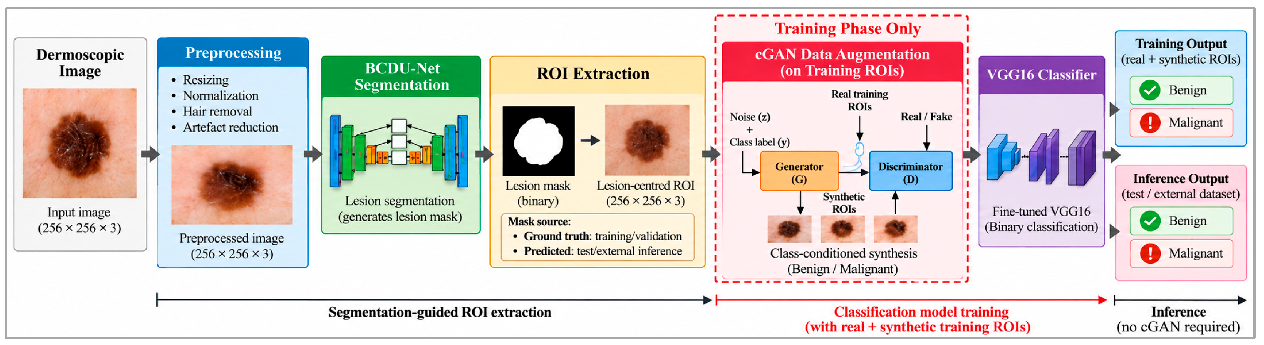
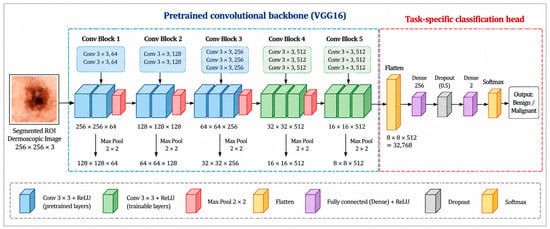
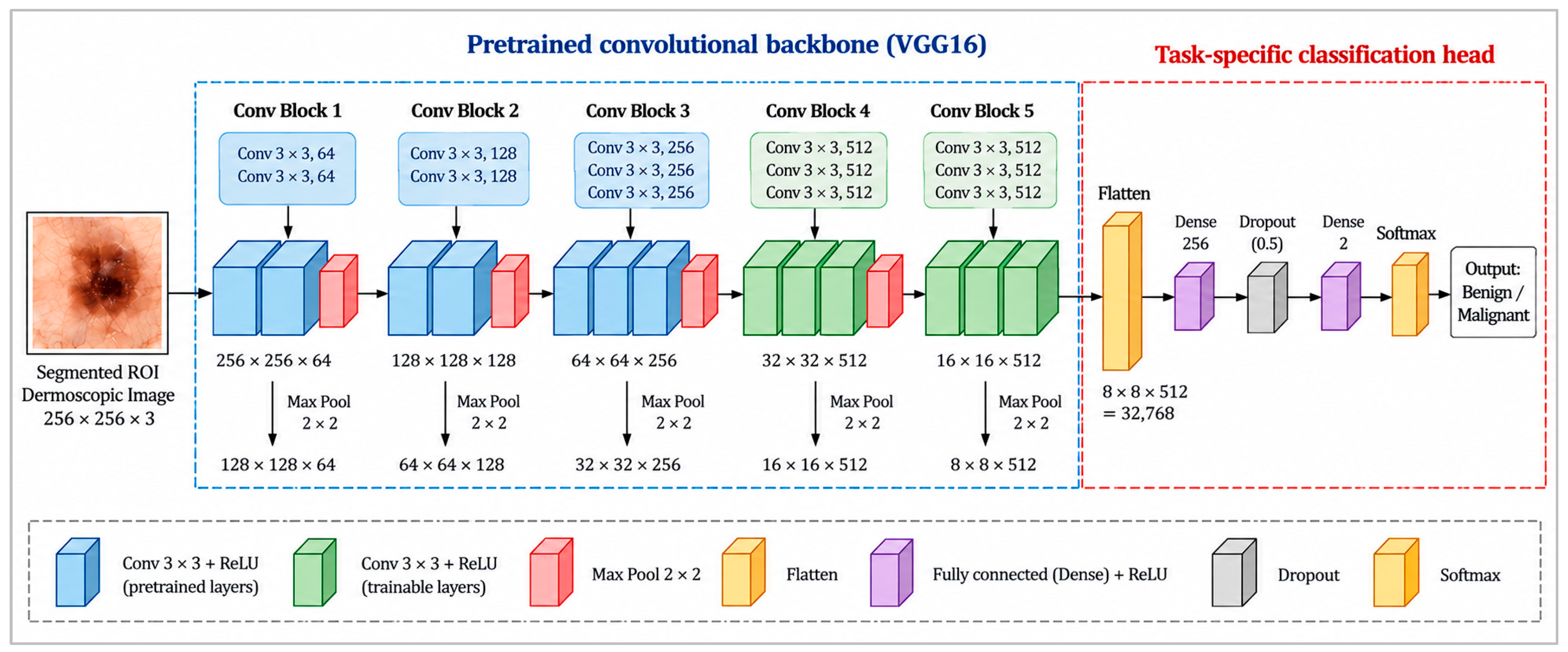
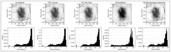
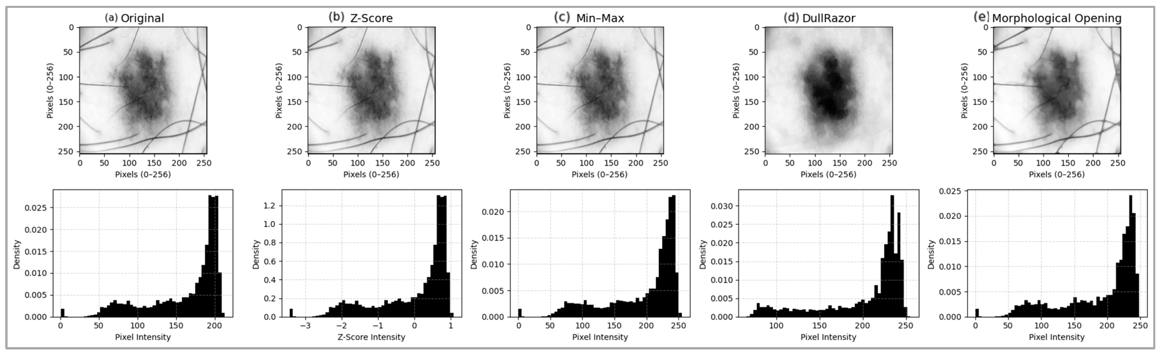
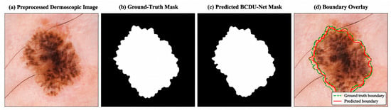
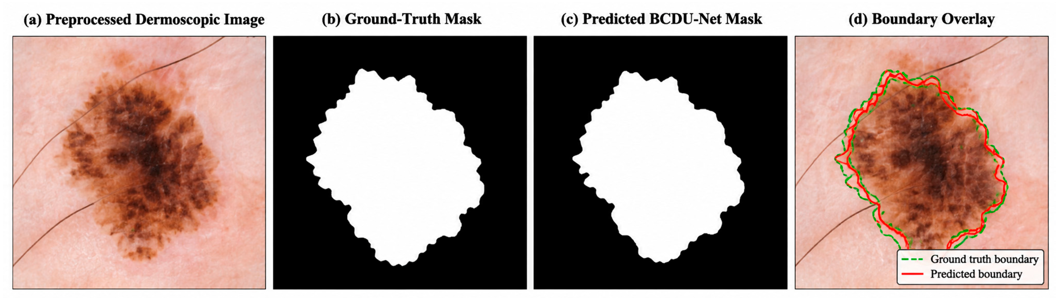
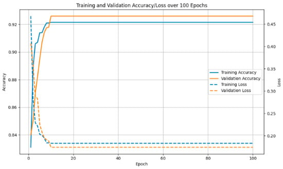
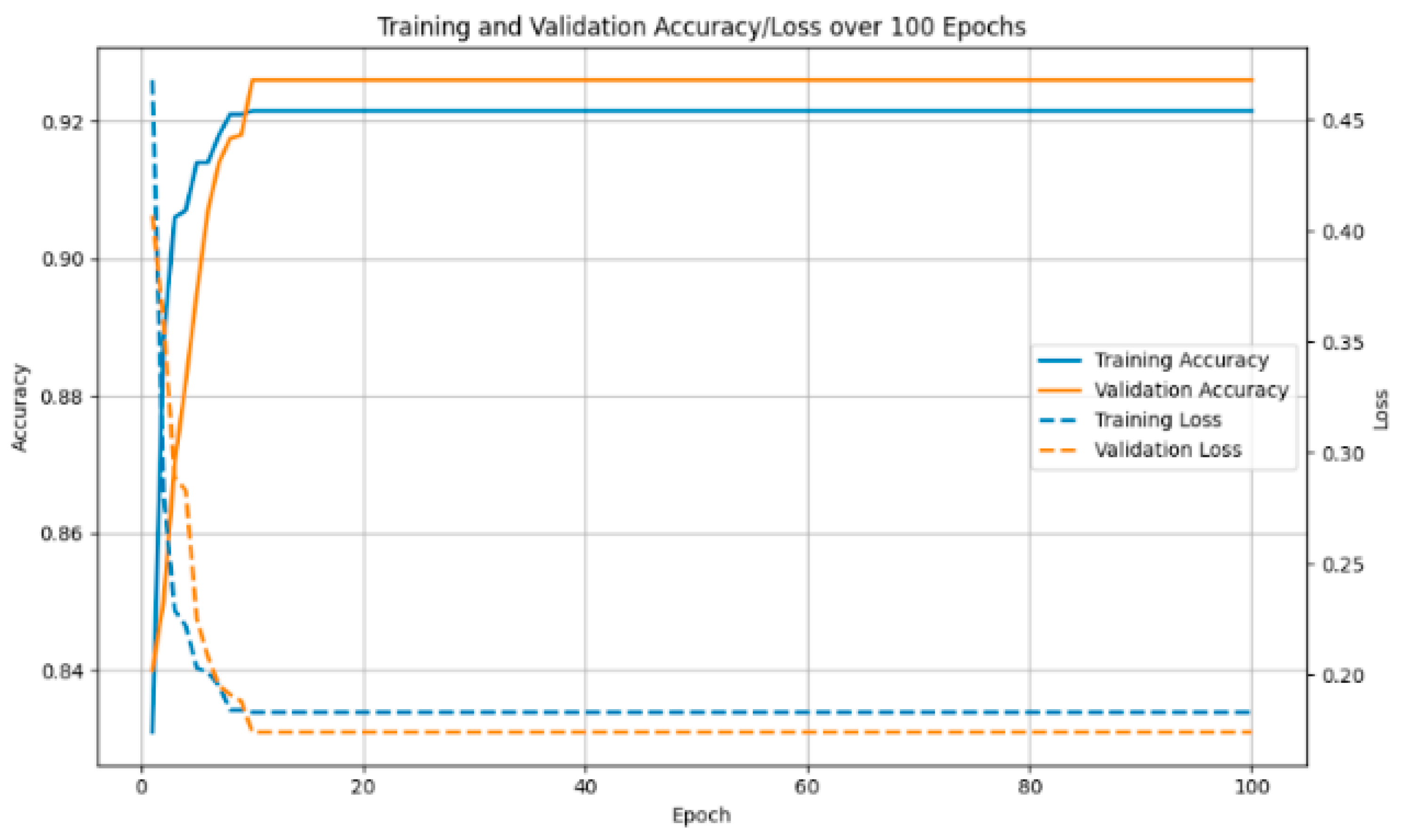
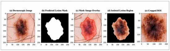
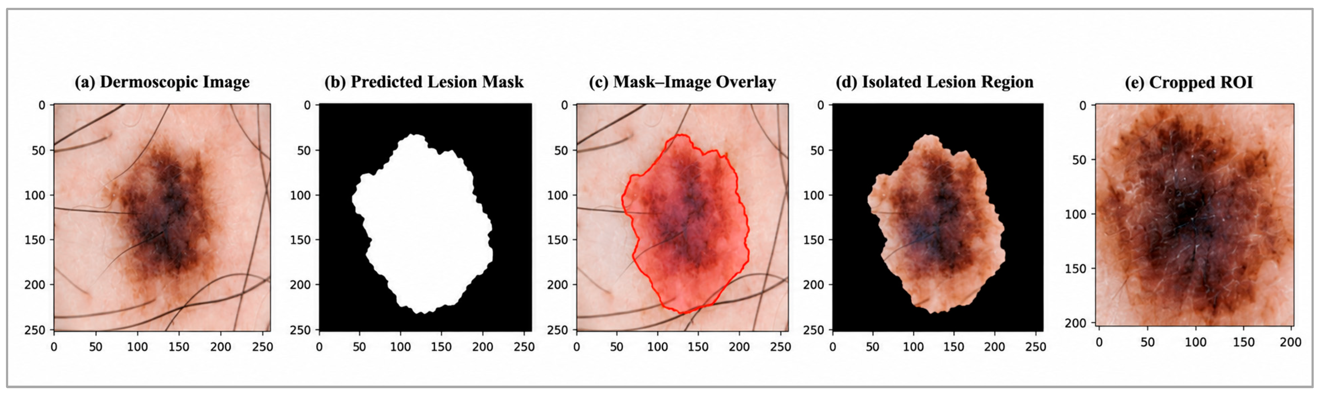
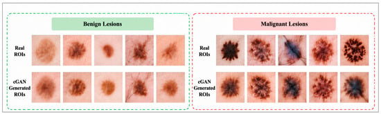
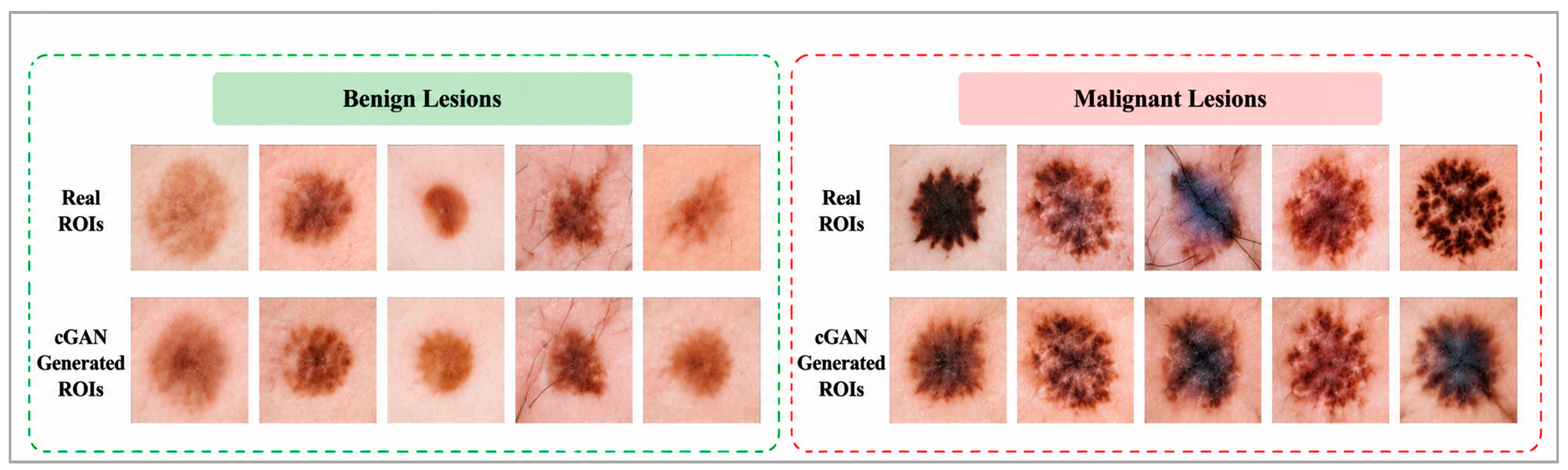
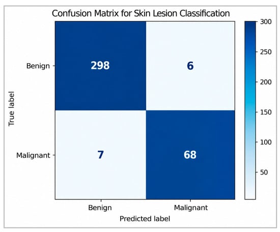
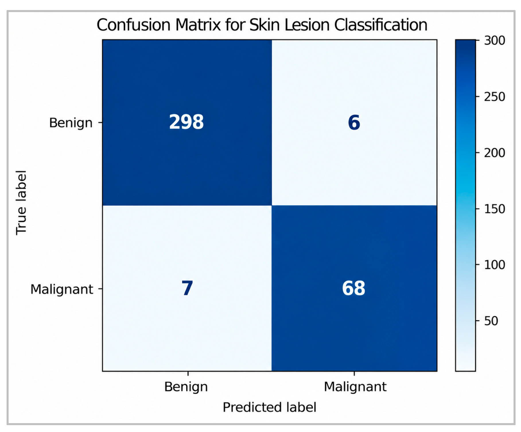
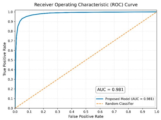
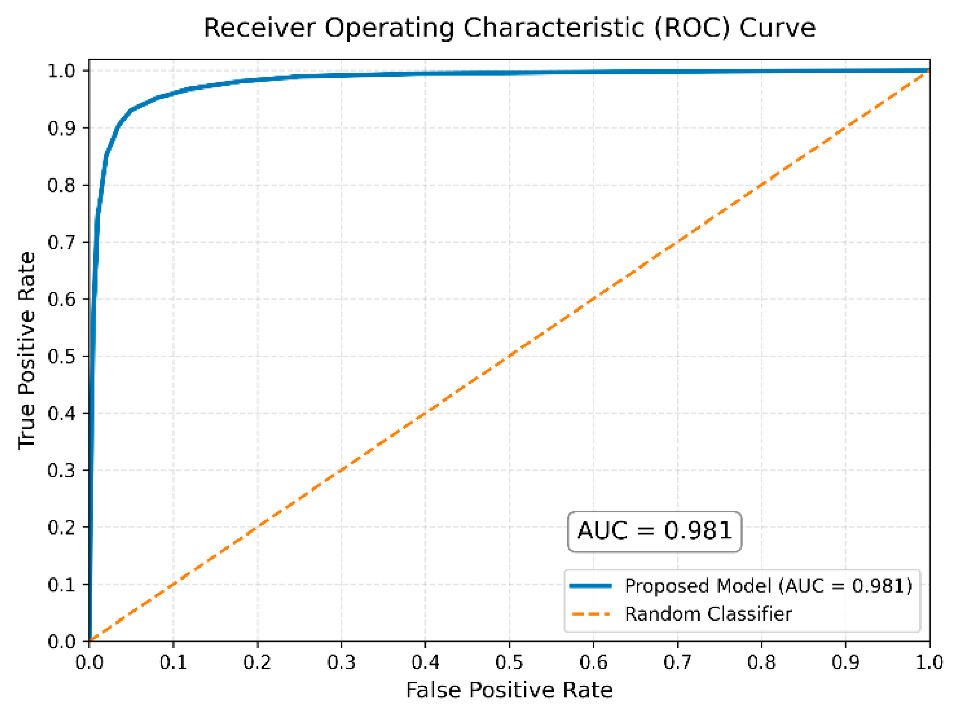
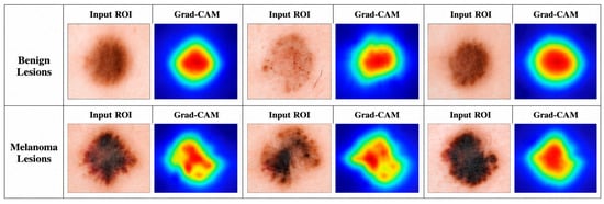
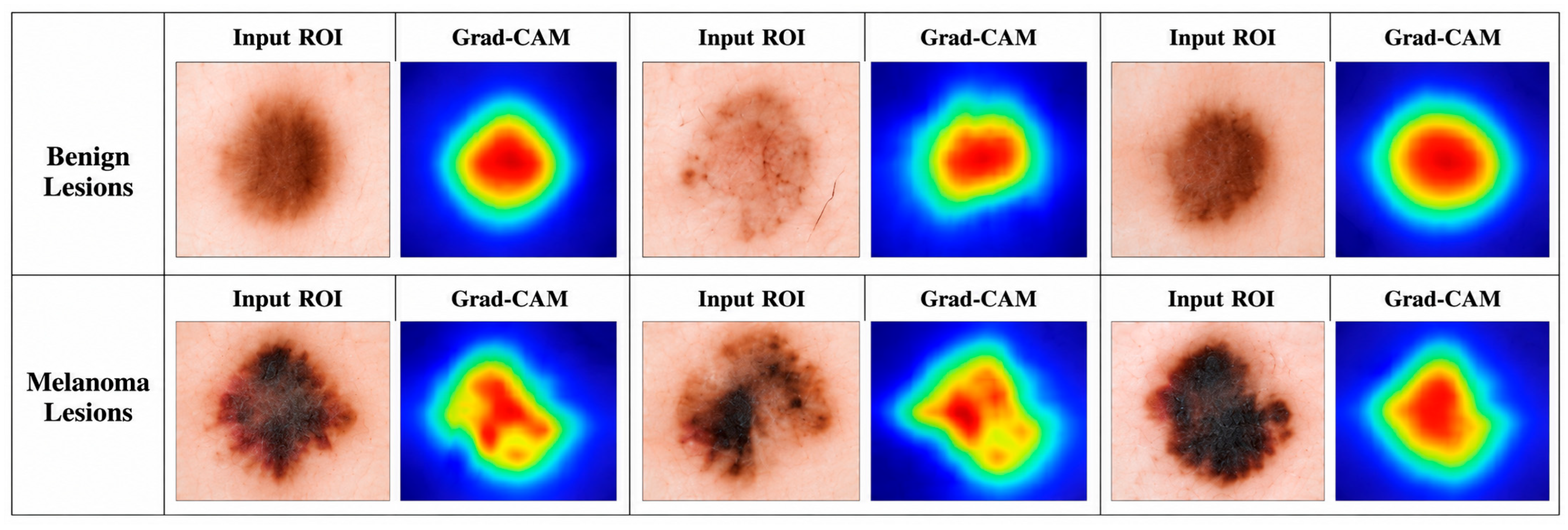
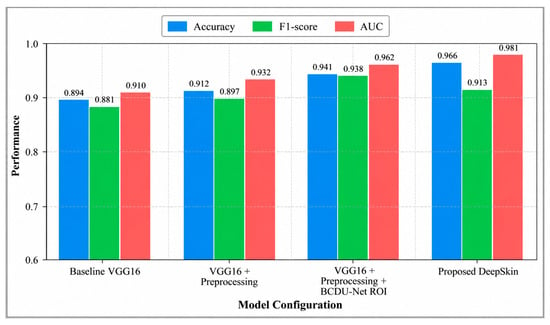
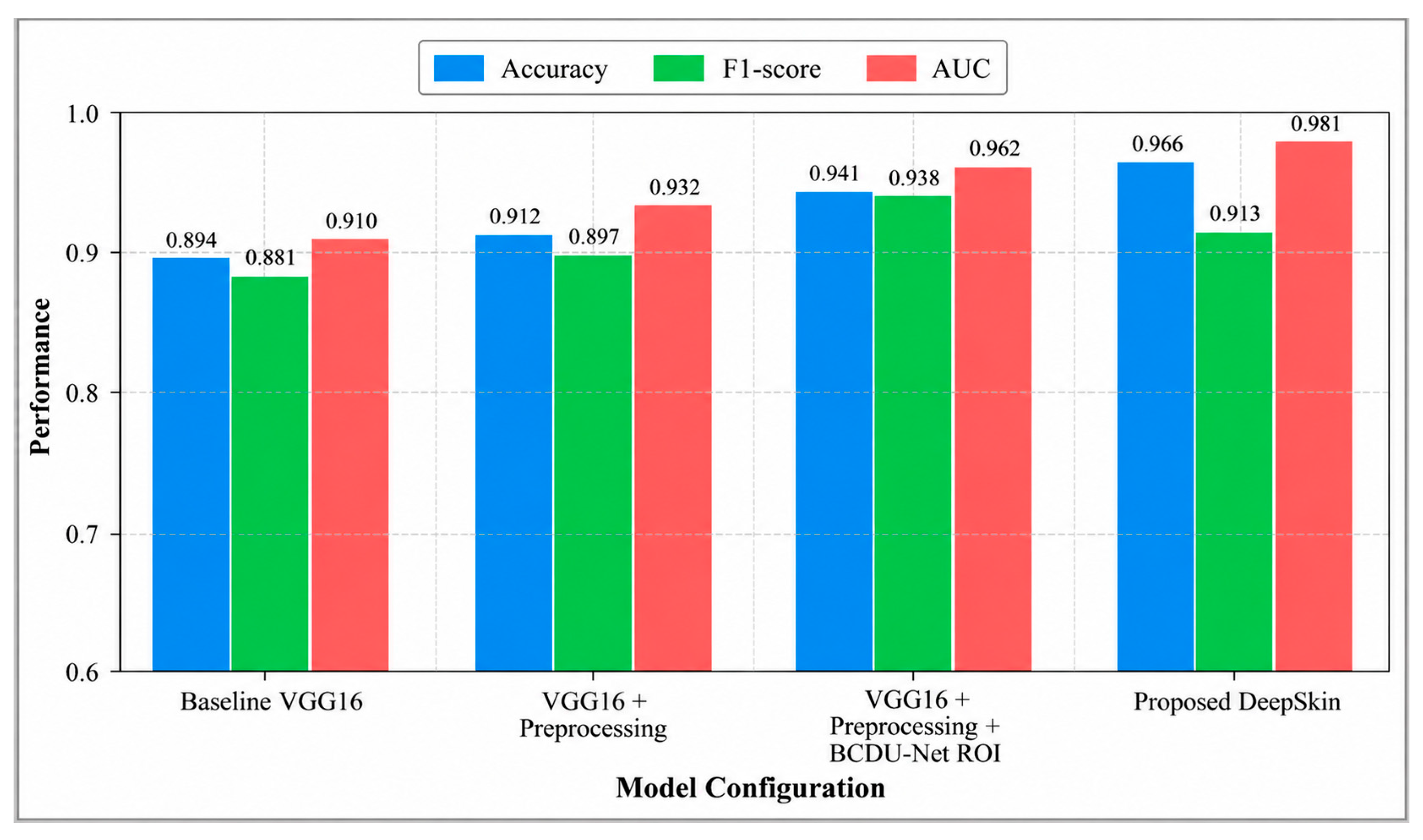
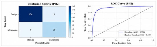
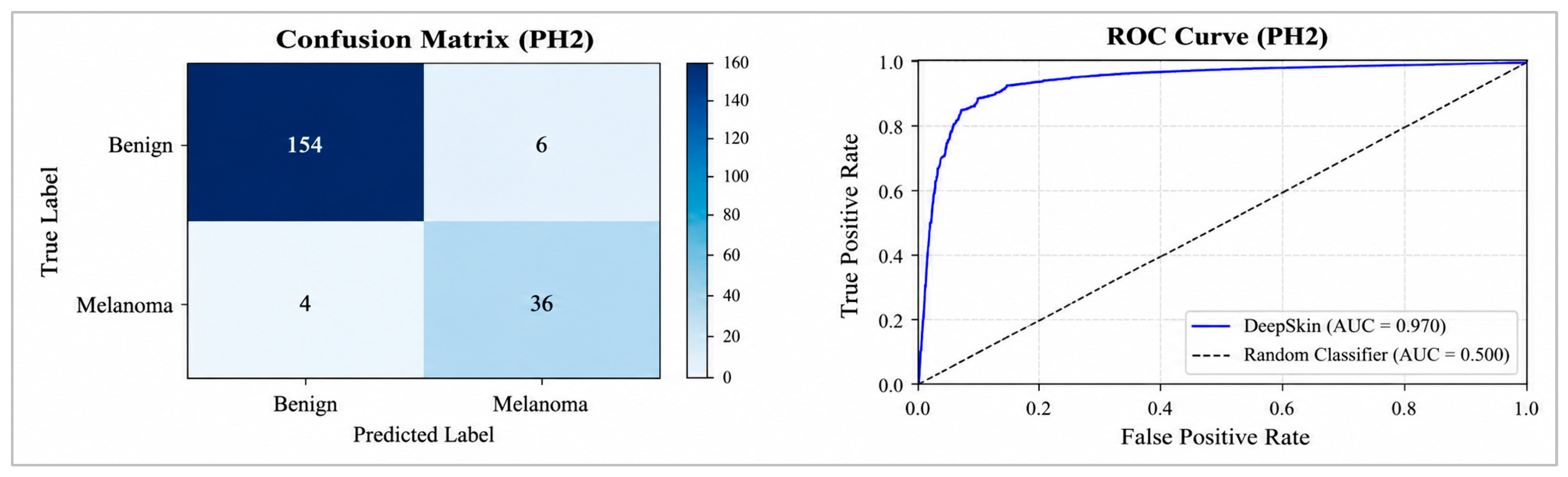

Disclaimer/Publisher’s Note: The statements, opinions and data contained in all publications are solely those of the individual author(s) and contributor(s) and not of MDPI and/or the editor(s). MDPI and/or the editor(s) disclaim responsibility for any injury to people or property resulting from any ideas, methods, instructions or products referred to in the content. |
© 2026 by the author. Licensee MDPI, Basel, Switzerland. This article is an open access article distributed under the terms and conditions of the Creative Commons Attribution (CC BY) license.