


Abstract
Progressive vision loss in adults is rising steadily, worldwide, alongside increased longevity and mounting chronic and metabolic disorders, such as age-related macular degeneration and diabetic retinopathy. Contemporary cell replacement therapy offers therapeutic promise for its potential to replace dystrophic and/or apoptotic photoreceptors with stem cell-derived neurons. However, the integration of transplanted retinal donor cells remains rare and difficult to manipulate, as low numbers of donor cells migrate appropriately within the damaged retinal host. The current study applied electric field stimulation to induce donor cell migration using alternating currents (ACs) applied in modern ophthalmic therapies for retinal disease. We extended our 3D electrotaxis assay, the BioEye, to apply minimal bursts of electric field stimulation across retinal facsimiles composed of established, biomimetic hydrogels. Tests used AC fields with biphasic and monophasic parameters of different waveforms. Results illustrate that positive, monophasic AC fields of triangle and square waveforms stimulated over 5-fold the number of motile cells than that of control (no EF), while similar biphasic fields produced over 2-fold the number of motile cells than control with consistent high cell viability in all tests. These findings suggest exciting and unexplored opportunities for EF-based strategies to help promote donor cell integration in retinal cell replacement therapy.
1. Introduction
Retinal degeneration is a leading cause of irreversible vision loss in working age and mature adults [1]. Over 470 million people worldwide are expected to experience moderate to severe vision impairment by 2050 from disorders such as age-related macular degeneration and retinitis pigmentosa [2]. These chronic pathologies lead to the death and/or dysfunction of photoreceptor cells that result in permanent and progressive vision loss [3,4]. The inability of retinal neurons to self-repair creates a dire need for biomedical treatments and therapeutic solutions [5,6,7].
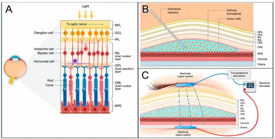
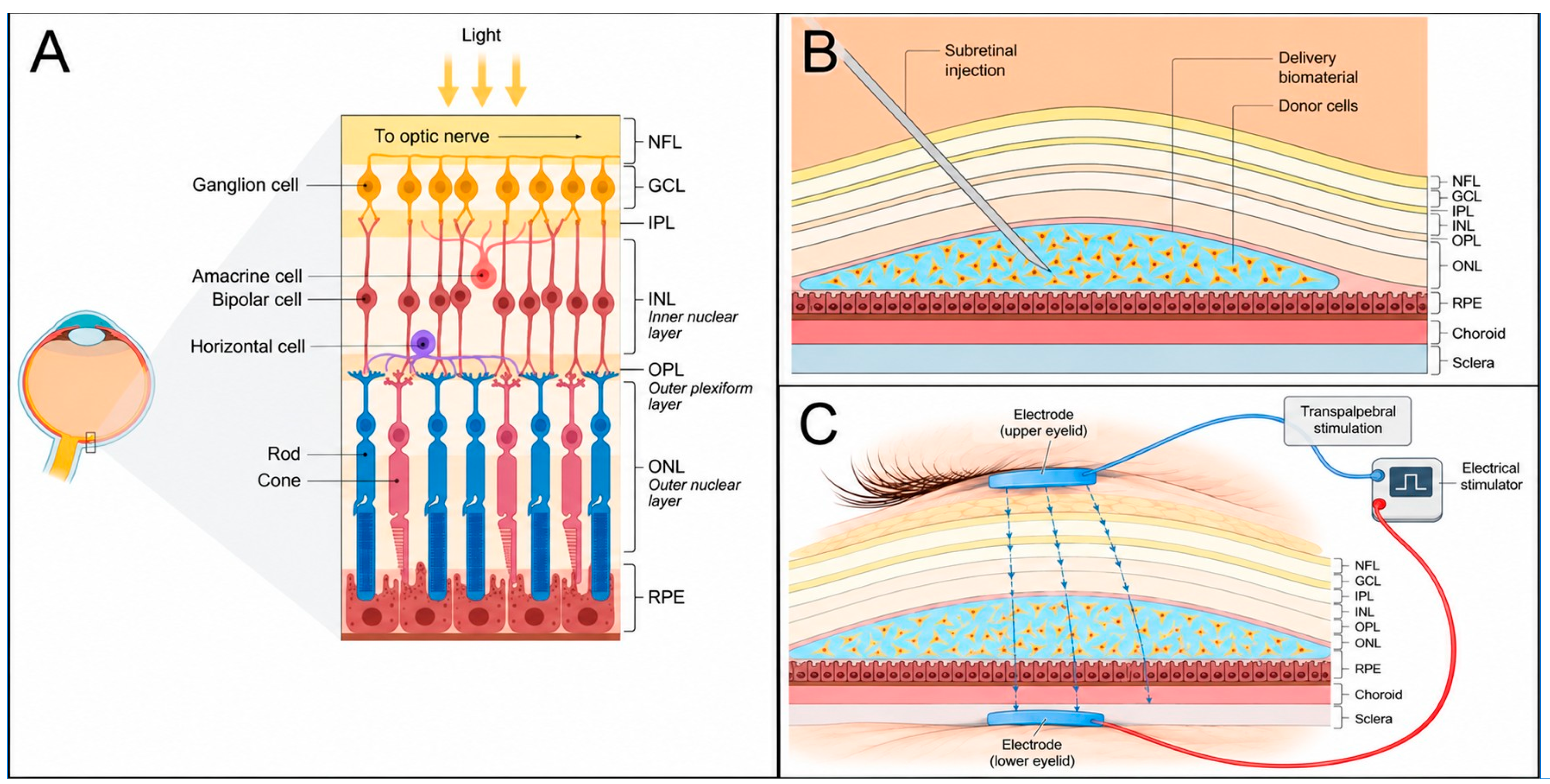
Cell replacement therapy offers great promise to treat vision loss for its potential to replace dystrophic photoreceptors with stem cell-derived neurons [8]. In an idealized model (Figure 1), donor cells are transplanted into the subretinal (SRS) space of host retina, often via injection within a delivery matrix to aid cell survival [9]. Viable donor cells then migrate out of the SRS and toward the neuroretina, where cells must navigate the restrictive intercellular spacing of the photoreceptor array to self-position within the outer nuclear layer (ONL). To restore vision, donor cells must then form appropriate synaptic connections with functional interneurons of the inner nuclear layer (INL), e.g., bipolar and horizontal cells, to transmit signals to the brain. Unfortunately, such functional integration remains elusive despite significant advances in stem cell biology and transplantable biomaterials [8,9,10]. This deficit is largely because viable donor cells fail to migrate appropriately into neuroretina, thereby impeding the formation of new synapses [11,12,13]. While numerous groups have derived donor cells with specialized identities to promote functional integration, [11,12] complementary strategies to guide transplanted cells to desired synaptic targets remain underexplored.
Figure 1.
Overview of healthy retina and cell replacement therapy in damaged tissue. (A) Schematic of a healthy adult retina with primary cell types and organized layers denoted from top to bottom as: nerve fiber layer (NFL), ganglion cell layer (GCL), inner nuclear layer (INL) (containing amacrine, bipolar, and horizontal cells), outer nuclear layer (ONL) (containing rod and cone photoreceptors), and the retinal pigment epithelium (RPE). (B) Schematic of typical injection used to deliver donor cells via biomaterial matrix (green) into the subretinal space adjacent the RPE. The choroid and sclera of the eye are marked on the right alongside defined retinal layers. (C) Schematic of trans-palpebral electrical stimulation (TpES) where an electrode is placed on one eyelid to be paired with a second electrode on a preocular location, e.g., second eyelid or orbit. Created in BioRender. Logan, A. (2026) https://BioRender.com/yg0vavd.
The migration of transplantable donor cells has been studied by our group and others toward gradients of targeted cytokines [14,15,16,17,18] using endogenous biomolecules known to guide stem and progenitor cell motility during neural development [19]. While bioelectric signals similarly regulate cell motility and tissue organization in the nervous system [20,21], electric fields (EFs) remain surprisingly underexplored for cell replacement therapy. Continued omission of EFs in transplantation is striking given the growing clinical success of electrical stimulation in modern ophthalmic approaches. Therapies such as trans-corneal electrical stimulation (TcES), trans-palpebral electrical stimulation (TpES), and trans-orbital alternating current stimulation (tACS) have applied EF stimulation to report enhanced retinal connectivity and neuronal communication in patients with degenerative retinal disease, as shown in Table 1. Each treatment has demonstrated neuroprotective effects and neuromodulation of retinal cells to delay degeneration, preserve photoreceptors, and in some cases improve visual function [22,23,24].
Table 1.
A summary of electric field (EF) parameters used for ophthalmic stimulation through trans-corneal electrical stimulation (TcES), trans-palpebral electrical stimulation (TpES), and trans-orbital alternating current stimulation (tACS). Studies are performed in patients with retinal disease via randomized controlled trial (RCT), pre-clinical trial, or pilot study. Note that burst trains are defined as periodic repetitions of a pulse at regular intervals.
Our group has previously applied EFs to induce donor cell migration in both microfluidic and tissue engineered systems [35,36]. Using direct current (DC) electric fields, we reported increased numbers of motile donor cells and their average displacement distances compared to chemotactic stimulation, alone, as well as control conditions (no EF) [35,36,37,38]. The current study evaluates the migratory responses of donor cells in response to clinical electrical stimulation using alternating current (AC) fields. We extend our prototype 3D electrotaxis assay [35], called the BioEye, to apply a minimal burst of EF stimulation across biomimetic retinal facsimiles using in vitro models of subretinal delivery. Donor cell migratory responses toward a panel of electrical stimulation fields were measured via confocal microscopy. Results illustrate significant increases in the numbers of motile cells toward both biphasic and monophasic AC fields using triangle and square waveforms, compared to the no EF control. Significantly, positive, monophasic AC fields stimulated five-fold the number of motile cells than control with sustained cell viability. These findings highlight the exciting and unexplored potential of EF-based strategies to stimulate donor cell migration as a step toward functional integration.
2. Methods and Materials
2.1. Culture of Donor Cell Models
Donor cells are represented by commercial retinal progenitor cells (R28; Cat. No. EUR201, Kerafest, Inc., Boston, MA, USA) used previously by our group [15,16] and many others [39]. Cells were cultured at 37 °C and 5% CO2 in media composed of 93% Dulbecco’s Modified Eagle Medium (DMEM; Cat No. 11885084 Thermo Fisher Scientific, Waltham, MA, USA), 5% Fetal Bovine Serum (FBS; Cat. No. 26140, Thermo Fisher Scientific, Waltham, MA, USA), 1% 100× Modified Eagle Medium (MEM; Cat. No. 11140050, Thermo Fisher Scientific, Waltham, MA, USA), and 1% 100 mg/mL penicillin–streptomycin (Cat. No. 15070063, Thermo Fisher Scientific, Waltham, MA, USA). Cells were passaged at 80–95% confluency in T-75 tissue culture flasks using trypsin solution (Cat. No 25200072, Thermo Fisher Scientific, Waltham, MA, USA) following a wash with Dulbecco’s Phosphate-Buffered Saline (1 × DPBS; Cat. No. 21–031-CV, VWR, Radnor, PA, USA). The trypsin was then neutralized with FBS and the resulting cell solution was centrifuged at 1500 RPM (433 RCF) for 3 min. After removing the supernatant, the cell pellet was resuspended in fresh media and the number of cells measured using the Countess II Automated Cell Counter (Cat. No. AMQAX1000, Invitrogen, Carlsbad, CA, USA). New flasks were seeded at 1.75 × 10⁵ cells for weekly passaging at 2.5 × 10⁶ cells to reach 85–90% confluency in 2 days. Cell viability was measured using live/dead assays (Thermo Fischer Scientific, Cat. No. R37601). Prior to usage in facsimiles, cells were pre-labeled with CellTracker Green CMFDA (5-chloromethylfluorescein diacetate) (Cat. No. C7025, Thermo Fisher Scientific, Waltham, MA, USA) at a working concentration of 15 μM, as described by the manufacturer. The dye was retained in cells for 72 h [40].
2.2. Synthesis and Rheology of Retinal Facsimiles and Cell Delivery Matrix
Alginate hydrogels were used as retinal facsimiles, as previously described by our group [35,37,41]. A 2% w/v alginate solution was prepared by dissolving sodium alginate (Cat. No. W201502, Sigma-Aldrich, St. Louis, MO, USA) in Dulbecco’s Phosphate-Buffered Saline (DPBS) on a stirring hot plate set to 400 RPM and 100 °C. The solution was supplemented with 1% penicillin–streptomycin to prevent bacterial contamination. A crosslinking solution of 5% w/v calcium chloride (Cat. No. C4901–100G, Sigma-Aldrich, St. Louis, MO, USA) in Dulbecco’s Phosphate-Buffered Saline (DPBS) was additionally prepared. Hydrogels were synthesized using 250 μL of alginate solution and 250 μL of calcium chloride (CaCl2) crosslinking solution, added dropwise to allow gels to solidify over 4–5 min. Cell delivery matrix was similarly synthesized using alginate to maintain 106 cells per mL in all experiments, as previously described by our group and others [16,37]. After formation, hydrogels were submerged in media to maintain hydration and sustain cells in the donor layer.
The storage and loss moduli of alginate hydrogels before and after electric field stimulus, were evaluated using oscillatory rheological measurements (Anton Paar MCR302e rheometer, Ashland, VA, USA). Crosslinked hydrogels were pipetted onto the lower geometry stage warmed to 37 °C with a 1 mm gap. A 0.1% oscillatory shear at 0.25 Hz was applied to the alginate over 7 min, by which time the hydrogel was considered fully crosslinked. Viscoelastic storage and loss moduli (G′ and G″) were recorded at 5 s intervals. An amplitude/strain sweep (0.1–10% strain) was performed for empirical verification and values were within torque resolution of the instrument. Note that the lower geometry stage reservoir was filled with water to prevent sample drying during testing.
2.3. Fabrication and Assembly of the BioEye Assay
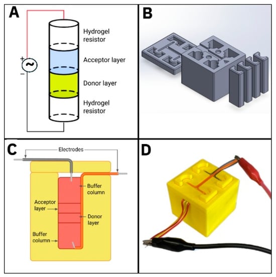
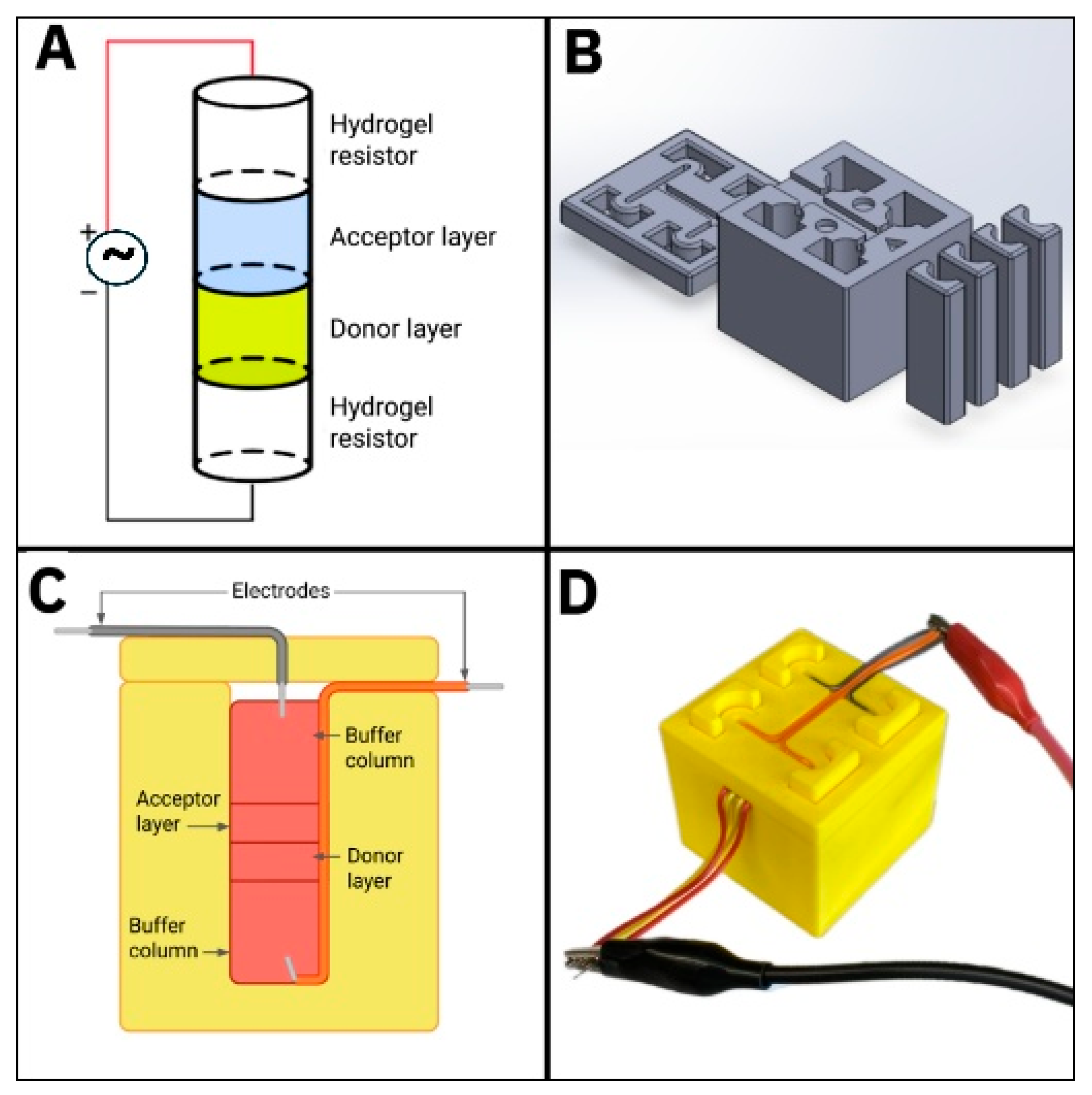
The BioEye was designed using computer-aided design software (SOLIDWORKS 2024; Dassault Systèmes SolidWorks Corp., Waltham, MA, USA) to represent the scale and key aspects of the cell transplantation process in the retina, as previously described. The device casing was 3D-printed (Bambu X1 Carbon, Bambu Lab, Shenzhen, China) using polylactic acid (PLA), and then cleaned, sprayed with 70% ethanol, and treated with a UV light lamp for 30 min (λUV = 360 nm, 180 mJ/cm2). The casing was then sterilized and placed in a tissue-culture hood prior to use. The BioEye casing was connected with tinned copper PVC-insulated jumper wires (22 AWG) such that one end faced upward perpendicular to the bottom surface, while the other end extended out of the device following alignment grooves on the top surface. The revised design used a column of 4 cylindrical hydrogels stacked into each well for testing (Figure 2). First, a hydrogel resistor (HGR-1) was used as an alginate buffer column to position and hold one electrode to reduce the effects of Joule heating on cell samples. Second, a hydrogel synthesized with donor cells and called the donor layer (DL) was placed adjacent to the hydrogel resistor. Third, an acellular hydrogel, called the acceptor layer (AL), was placed atop the donor layer (DL) to collect cells able to migrate within it during stimulation testing. Fourth, a second hydrogel resistor (HGR-2) was placed opposite the acceptor layer (AL) to position the complement electrode and similarly prevent Joule heating of the sample. Hydrogel resistors, cell donor layers, and cell acceptor layers were then stacked within 3D-printed cylindrical molds of the BioEye (10 mm diameter) for testing. These molds were then placed within the wells of the system, where the remaining well volume was filled with media for cell viability. As a final step, the electrodes were inserted into the top half of the device, such that each penetrated less than one fifth of each hydrogel resistor after positioning of the device lid. Note that the BioEye is used in this study to apply AC fields with and without DC offset.
Figure 2.
Summary of the revised BioEye electrotaxis assay. (A) Schematic illustration of a stacked column of 4 cylindrical hydrogels exposed to electric field stimulation to induce the migration of cells from the donor layer to the acellular acceptor layer. Cell donor and acceptor layers are sandwiched between two hydrogel resistors that hold positive and negative electrodes, respectively, to enable both direct and alternating current stimuli. (B) CAD design of the electrotaxis assay showing four cylindrical chambers, insert molds that hold the hydrogel columns, and device top cover. (C) Schematic diagram of the assembled BioEye assay used to apply electric field stimulation and (D) image of assay during testing.
2.4. Applied Electric Field (EF) Stimulation
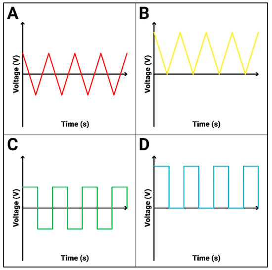
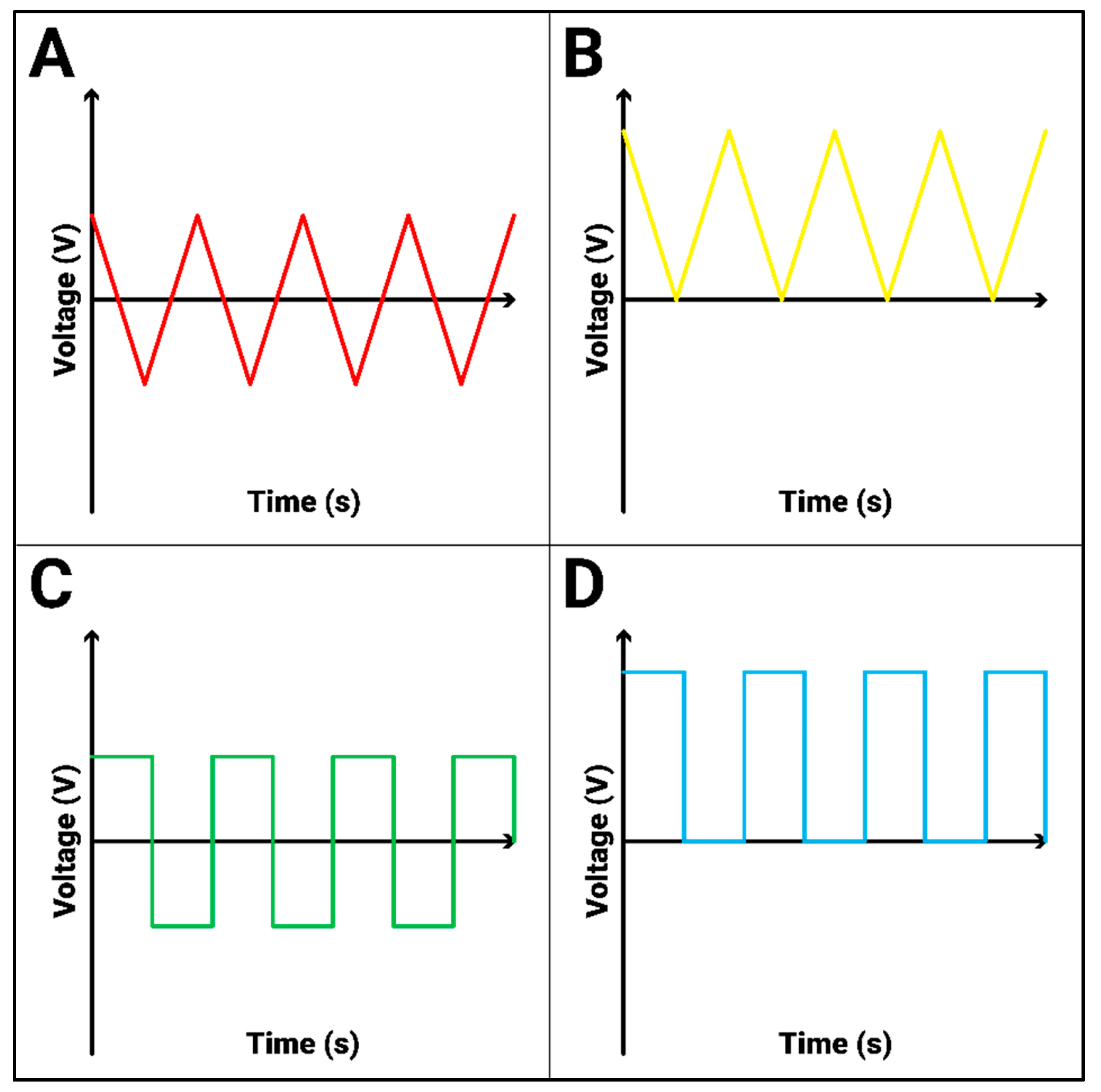
Tests applied 4 different stimulation fields for 10 min each at 100 Hz frequency using a function generator (Model No. GFG-8216A, GW Instek, New Taipei City, Taiwan) attached to the wires of the BioEye device (Figure 3). Tests exposed cells to 4 profiles: (a) biphasic EF with triangle waveform at 1.73 volts peak-to-peak (VP-P) and 0.500 root mean squared voltage (VRMS); (b) biphasic EF with square waveform at 1.73 VP-P (0.865 VRMS); (c) positive EF with triangle wave at 1.73 VP-P and a 0.865 V DC offset (1.000 VRMS); and (d) positive EF with square waveform at 1.73 VP-P and a 0.865 V DC offset (1.224 VRMS). All fields were measured and verified via multimeter (Model No. 34401A, Agilent, Santa Clara, CA, USA) and oscilloscope (Model No. GRS-6052A, GW Instek, New Taipei City, Taiwan), as per Figure 3. After EF stimulation, the BioEye device was placed inside a biological incubator at 37 °C and 5% CO2 to allow cell migration overnight. Acceptor and donor layers were then removed from the device and placed into individual well plates, ensuring that the donor–acceptor interface was facing downward and submerged in DPBS to maintain hydration.
Figure 3.
Overview of the electric field (EF) stimulation profiles applied during testing. All EFs were applied for 10 min using a 1.73 V peak-to-peak amplitude and frequency of 100 Hz. Schematics represent voltage applied over time for conditions of (A) biphasic triangle waveform; (B) monophasic positive triangle waveform (i.e., biphasic EF with full DC offset to provide positive voltage, only); (C) biphasic square waveform; and (D) monophasic positive square waveform (with full DC offset) during testing.
2.5. Electric Field Distribution Model
To characterize the spatial distribution of applied EFs within retinal facsimiles of the BioEye, a computational model was developed using MATLAB R2024b (Mathworks, Natick, MA, USA) and the Partial Differential Equation Toolbox. The model leveraged rotational symmetry of the facsimile system (10 mm diameter; 30 mm height) to apply a two-dimensional model that reduces simulation complexity. The geometry consisted of four distinct cylindrical hydrogels stacked into a column, described as follows: one hydrogel resistor (HGR-1) containing the negative electrode (elevation z = 0 to z = 10 mm); a donor layer (DL: elevation z = 10 to z = 15 mm); a cell acceptor layer (AL: z = 15 to z = 20 mm); and a second hydrogel resistor containing the positive electrode (HGR-2: elevation z = 20 to z = 30 mm). The electrodes were modeled as cylindrical conductors (0.5 mm diameter) that penetrate a maximum of 2 mm from the top and bottom surfaces of the hydrogel resistors along the system centerline.
The EF distribution was computed by solving the steady-state Laplace equation for electrical conduction, shown in Equation (1):
where V represents the electric potential (voltage) and σ (S/m) represents electrical conductivity. We note that capacitive effects were ignored in this study because the low AC frequency (100 Hz) permitted modeling of EF as a classical resistive conduction problem [42]. The material conductivities were assigned as per the literature of tinned copper electrodes (σ = 5 × 10⁷ S/m) and alginate (σ = 1 S/m) [43]. Dirichlet boundary conditions were applied at the electrode tips with the top electrode being the value of the applied waveform voltage and the bottom electrode being ground (0 V) [44]. All other boundary conditions were treated as insulation walls that prevented current leakage from the system.
For each applied EF, the root mean square (RMS) voltage was used as the boundary condition to represent the effective field strength experienced by cells in lieu of a time-averaged voltage. This was applied because the time-averaged voltage is equal to 0 for symmetric biphasic waveforms. All EFs maintained a peak-to-peak amplitude of 1.73 V with different VRMS values as listed previously: biphasic triangle (0.500 VRMS), biphasic square (0.865 VRMS), positive triangle (1.000 VRMS), and positive square (1.224 VRMS). A finite element method was used to discretize the geometry into triangular mesh elements that yielded approximately 50,000 to 100,000 elements per simulation. After the simulation converged on a solution, the EF magnitude was computed from the voltage gradient shown in Equation (2):
The radial electric field profiles were extracted at the interface of the donor and acceptor layers (elevation z = 15 mm) and axial profiles were extracted along the centerline (r = 0 mm) to characterize the uniformity of EFs applied across the hydrogels.
2.6. Microscopy and Image Analysis
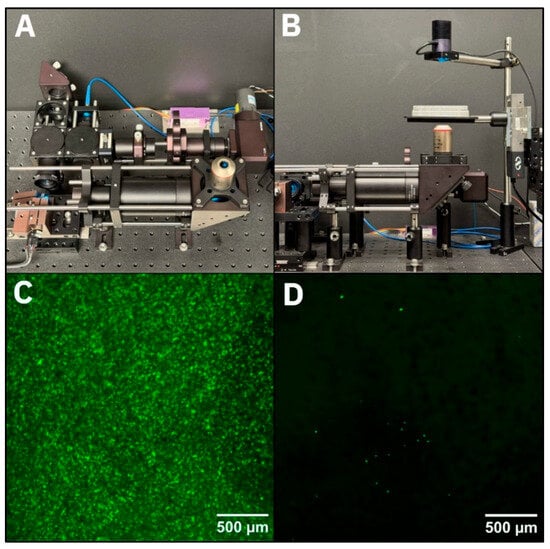
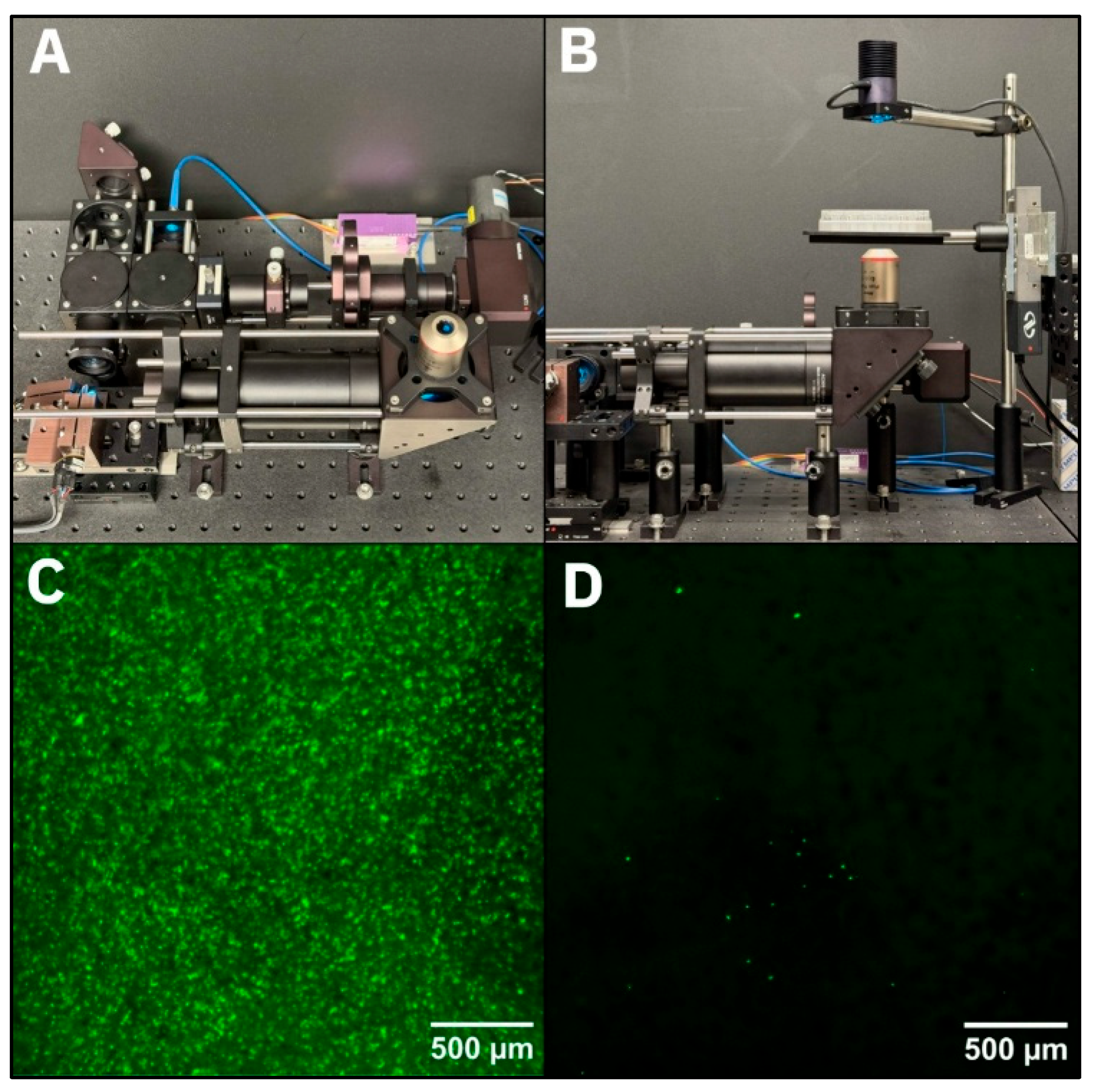
The spatial distribution of motile donor cells within acceptor layers after electrical stimulation was measured using a benchtop point-scanning confocal fluorescence microscope, previously described by our group [35]. Hydrogel samples were removed from the BioEye assay, placed into well plates, and mounted onto the microscope stage. A 4× objective lens (Plan Fluor 4×/0.13, Nikon Corporation, Tokyo, Japan) yielded a 2.5 mm × 2.5 mm field of view. Samples were illuminated with 488 nm excitation light with fluorescent emission light transmitted through a 506–594 nm bandpass filter (FF01-550/88-25, Semrock, West Henrietta, NY, USA) and 100 μm pinhole to a photomultiplier tube. The detector output was amplified and digitized at 2 MS/s for acquisition through LabView software developed in-house. Under the imaging conditions used (4×/0.13 NA objective and 100 µm pinhole), fluorescent polystyrene microspheres of 10 µm diameter and fluorescent cells were detected across an axial range of approximately 250 µm to denote migration within the average thickness of adult human retina [45] (Figure 4).
Figure 4.
Overview of the confocal system used to measure donor cell migration into acceptor layers of the BioEye assay. (A) A top-down view of the overall confocal setup. (B) A side view of the inverted confocal system with a well plate on the platform. (C) Representative fluorescence image of a cell-laden donor layer after electric field (EF) stimulation. (D) Representative image of an acceptor layer after EF stimulation thresholded for clarity. Scale bars as marked.
Samples were positioned such that the interface between the donor and acceptor layers (elevation z = 15 mm) was at the focal plane of the microscope. Multiple 2.5 mm × 2.5 mm images were acquired across the entire sample area to measure all motile cells. Imaging was used to distinguish cells as rounded and continuous regions of fluorescence that were distinct from the background and ranged in size from 10 μm diameter (e.g., single cells) to 50 μm diameter (e.g., clustered cells). The number of detected cells was tallied over all images per individual sample to produce cell counts for each EF stimulation profile and control (no EF).
2.7. Statistical Analysis
Each data set was collected from a sample of n = 3–5 independent experiments using the BioEye. Each experiment was performed using 4 replicate wells per device for a total of 12–20 data sets. The omission of minimal single outlier points (a total of 2) was satisfied by Grubbs’ outlier test [46] with alpha = 0.05. Statistical significance between experimental EF groups was evaluated using one-way ANOVA tests at a 95% confidence interval and with Dunnett’s T3 multiple comparisons test [47]. Cell count values were reported using mean and standard deviation. Significance was reported as *** for p < 0.001 and **** for p < 0.0001. Values with p > 0.05 were considered not significant.
3. Results
3.1. Viscoelastic Properties of Hydrogel Facsimiles Approach In Vivo Values of Adult Retina
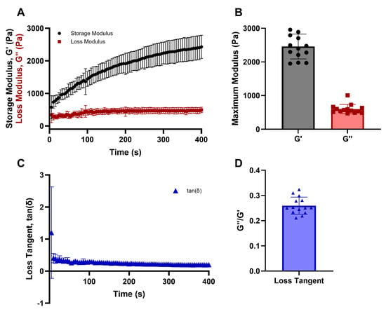
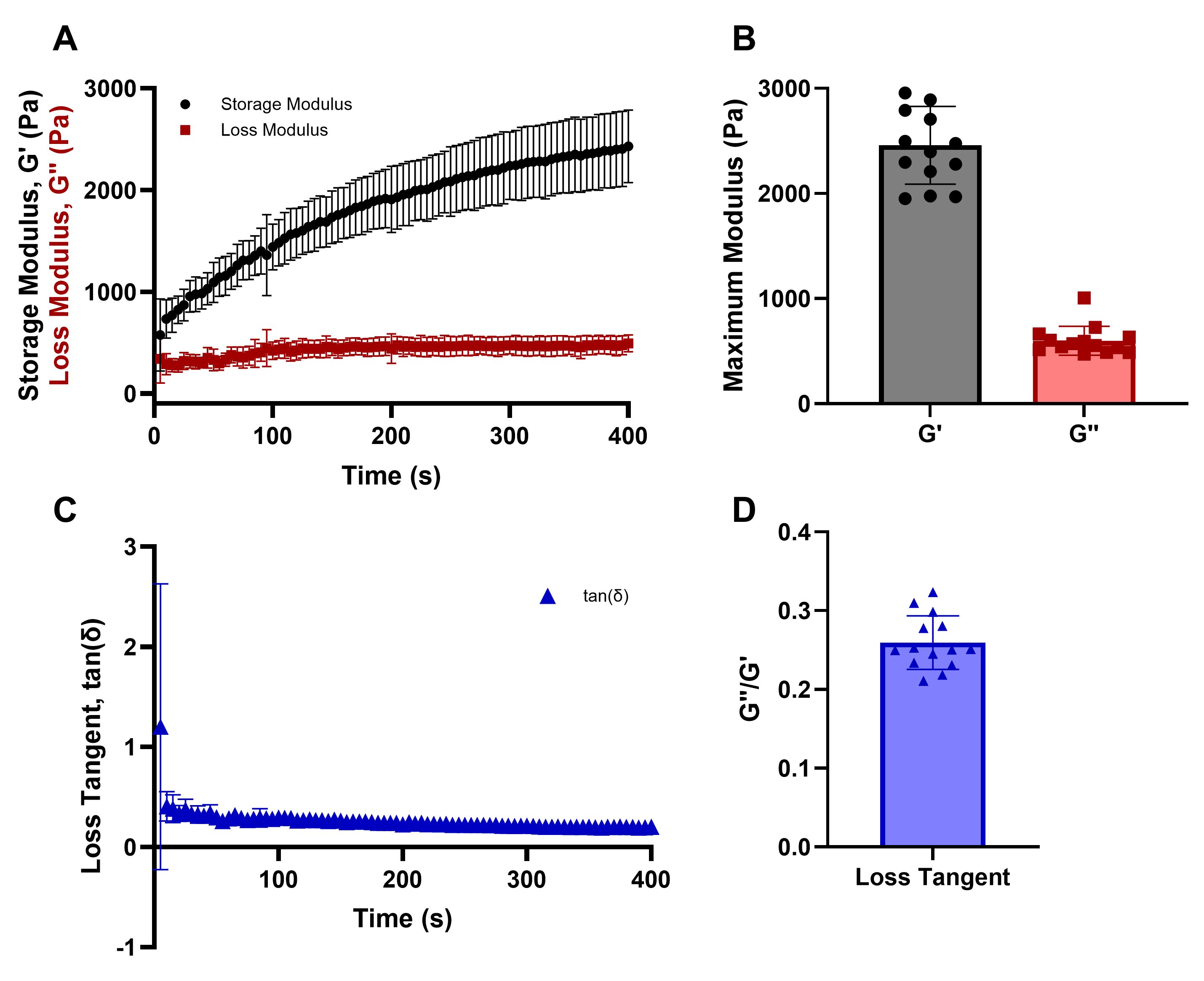
Viscoelastic properties of the hydrogels in the BioEye were measured via oscillatory rheology. Storage (G′) and loss (G″) moduli were recorded during crosslinking of the alginate hydrogels immediately following initiation with calcium chloride. As per Figure 5, the process of crosslinking rapidly increased hydrogel stiffness to reach a maximum mean storage modulus of G′ = 2458 ± 370.5 Pa (n = 14) that plateaued by ~400 s (6.5 min) to denote formation of hydrogels. Values of loss modulus followed a similar pattern with a maximum mean loss modulus of G″ = 567.5 ± 75.13 Pa (n = 14). These values were within those reported for adult mouse retina that is stiffened by diseases such as diabetic retinopathy and macular degeneration, i.e., G′ between 1.0 and 3.5 kPa and G″ between 0.25 and 0.65 kPa [48]. In addition, the values of loss tangent (tan (δ) = G″/G′) exhibited a flat profile, with an average value of δ = 0.26 ± 0.03 to indicate hydrogel stability during testing, as per the literature [48]. We note that no significant differences in viscoelastic properties were measured across hydrogels before and after electric field stimulation, as shown in Supplemental Figure S1.
Figure 5.
Measurements of viscoelastic properties of alginate hydrogels used as retinal facsimiles. (A) Storage modulus (G′) and loss modulus (G″) of alginate hydrogels throughout crosslinking initiated via calcium chloride. (B) Mean maximum storage and loss moduli after hydrogel formation. (C) Loss tangent (defined as tan(δ) = G″/G′) profile of alginate hydrogels throughout crosslinking. (D) Mean loss tangent of hydrogel formation. Data shown as mean ± standard deviation with n = 14.
3.2. Stimulation Profiles Generated Consistent EF Distributions Across Targeted Areas of Retinal Facsimiles
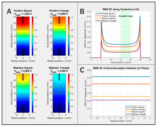
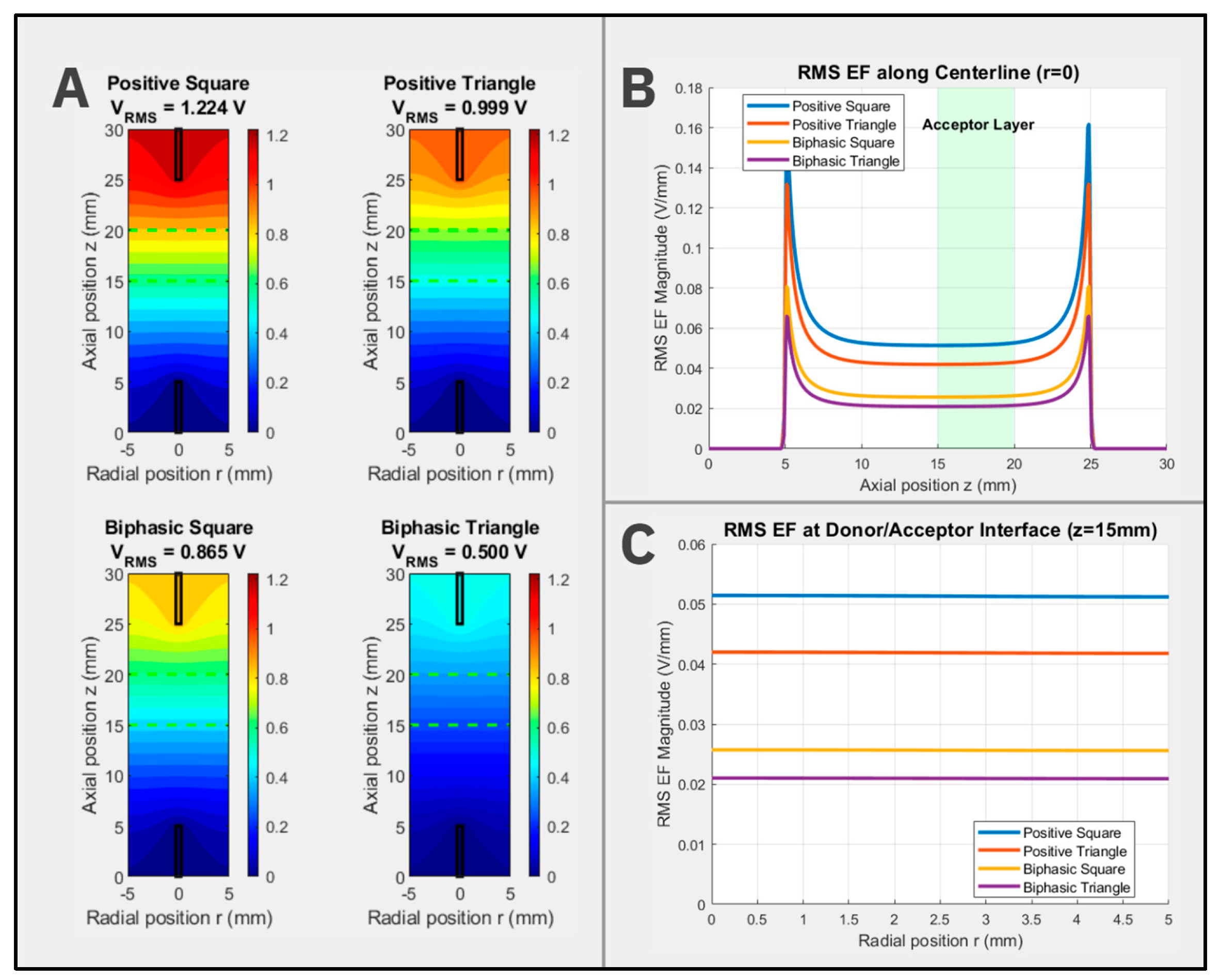
First, voltage distributions across facsimile cross-sections were computed for each applied stimulation field. The colormap in Figure 6A illustrates the voltage at axial positions (i.e., z-elevations) across layers of hydrogels positioned between the electrodes. The fixed color scale represents magnitudes of EF ranging from 1.224 V (red) to 0 V (blue). The EF profiles at the hydrogel centerline (r = 0 mm) are shown in Figure 6B, where the acceptor layer is shaded in green to denote the area studied for measurements of motile cells. The acceptor-layer hydrogel corresponds to axial positions with the BioEye that reflect elevations of zAL = 10–20 mm. As seen, the EF magnitude in this region is nearly constant along the axial direction for all EF stimulation profiles, indicating a spatially consistent stimulus regardless of cell axial position within donor and acceptor hydrogel layers. By contrast, the EF magnitude sharply increases near electrodes within the hydrogel resistor layers (zHGR-1 = 5–8 mm and zHGR-2 = 22–25 mm) to indicate significant edge effects. These findings support BioEye development of a uniform field between the donor and acceptor layers positioned farthest from electrodes.
Figure 6.
Computational modeling of voltage and electric field distributions across hydrogel facsimiles of the BioEye. (A) A 2D voltage distribution (V) of the RMS voltages of each field applied across facsimiles at different axial positions, i.e., z-elevations within BioEye wells. A fixed color scale represents magnitude of electric potential, ranging from red (1.224 V) to dark blue (0 V). The electrodes are represented by black rectangles, and the acceptor-layer region is outlined by green dashed lines. (B) Centerline distribution (r = 0 mm) of EF magnitude (V/mm) for each stimulation field applied. The acceptor-layer region is shaded in green. (C) Radial distribution of EF magnitude (V/mm) for each applied field is shown at the interface between the donor and acceptor layers (elevation z = 15 mm).
Second, the radial EF profile at the interface between donor and acceptor hydrogel layers (elevation z = 15 mm) is represented in Figure 6C. As seen, the EF magnitude was effectively constant from the hydrogel centerline (r = 0 mm) to the hydrogel edge (r = 5 mm) for all applied stimulation fields. Moreover, the maximum radial variation was 0.45% between the centerline and the periphery, indicating that cells at any radial position within the donor and acceptor layers experienced the same field strength.
3.3. Electrical Stimulation Applied Using Biphasic Fields Produced Large Increases in the Numbers of Motile Cells Compared to Control
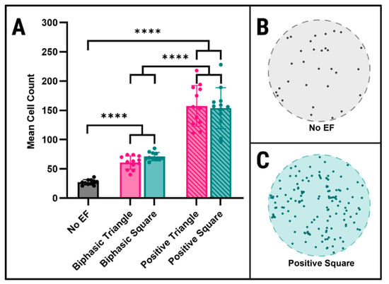
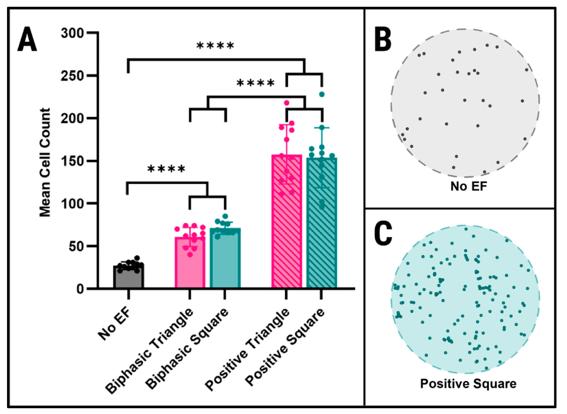
Experiments measured the numbers of fluorescently labeled cells detected within 250 µm distances from the interface of the donor and acceptor layers per stimulation condition. This limiting distance was chosen to represent the average width of adult retina, thereby capturing the maximum distance of physiological relevance [49]. As seen in Figure 7, the control group (CN; No EF) exhibited the lowest mean number of motile cells, i.e., NCN = 27.18 ± 4.51. The value represents the baseline chemokinetic, or non-directional, migration of cells in the absence of an applied electric field. By contrast, biphasic fields using different waveforms produced significant increases in numbers of motile cells that migrated within 250 μm distances of the donor layer–acceptor layer interface. As shown, stimulation with biphasic fields of triangle waveform (BT) produced a mean of NBT = 60.92 ± 11.23 cells (p < 0.001), while EF stimulation with biphasic fields of square waveforms (BS) induced a mean of NBS = 71.09 ± 7.13 cells (p < 0.001). No significant differences were measured (p > 0.05) between numbers of motile cells stimulated using triangle versus square waveforms of applied biphasic fields. We note that cell viability was >92% for all EF stimulation experiments.
Figure 7.
Numbers of motile cells measured within acceptor layers of the BioEye following EF stimulation via a panel of different field profiles. (A) Mean numbers of motile cells measured within hydrogel acceptor layers for each EF stimulation profile applied. (B) Representative midplane image from an acceptor layer in the control group (no EF) digitized to illustrate cell centroid position (diameter = 2.5 mm). (C) Representative image from an acceptor layer in the positive square EF group digitized to show cell centroid position. Significance bars **** represent p < 0.0001. Note that all measurements of motile cells were limited to a 250 μm distance of the acceptor layer closest to its interface with the adjacent donor layer to reflect physiological limits of migration distances within adult retina.
3.4. Applied Positive, Monophasic EF Stimulation Increased the Number of Motile Cells over Five-Fold That of Control
The largest numbers of motile cells were measured in response to EF stimulation using positive, monophasic fields, defined as AC fields with full DC offset to produce only positive voltages (Figure 4). As seen, positive triangle (PT) and positive square (PS) waveform stimulus fields produced average numbers of motile cells of NPT =157.58 ± 34.81 and NPS = 153.75 ± 35.24, respectively. These values were both a 2.2-fold increase over stimulation with biphasic fields (p < 0.001) and a 5.7-fold increase over the no-EF control (p < 0.0001). As with the previous measurements using biphasic fields, no significant differences were measured between the triangle and square waveforms (p > 0.05). We note that cell viability was >92% for all EF stimulation experiments.
4. Discussion
Progressive vision loss in maturing adults is rising steadily, worldwide, alongside increased longevity and mounting chronic and metabolic disorders. While contemporary cell replacement therapy offers exciting promise for treatment, the functional integration of transplanted donor cells remains extremely rare and difficult to manipulate. Despite the well-established roles of bioelectric signaling in cell motility for neural development, function, and plasticity [21], electric fields remain surprisingly unexplored for transplantation therapy. The current study evaluated the ability of electric field (EF) stimulation to induce and/or guide the migration of retinal donor cells toward distal sites of integration.
This bioengineering project is the first to enable systemic and rapid analysis of EF-induced motility in retinal donor cells via 3D electrotaxis assay. Our BioEye system expands upon prior migration study from our lab using panels of cytokines, extracellular matrix, and direct current (DC) electric fields [17,35,36,37,38,41]. Hydrogel samples tested within the 3D-printed system are directly mounted upon an optical microscope to facilitate rapid measurement of motile cell numbers and their average 3D displacement in response to EF stimulus (Figure 2). The BioEye assay enables analysis of cell migration within intact samples after as little as 12–24 h, in stark contrast to cryostat sectioning methods that prohibit rapid and iterative evaluation of cells within samples [42]. Tests used retinal facsimile hydrogels synthesized of alginate, a well-characterized and biocompatible material with established applications in ophthalmology [50,51,52]. Ionic crosslinking mechanisms of alginate facilitate rapid gelation under conditions that support cell viability, while its porous nature allows both nutrient diffusion and cell migration [43,50]. We applied these facsimiles in our in vitro assay because they reflect viscoelastic properties most relevant to cell migration. Measurements of these retinal facsimiles confirmed storage and loss moduli that were within the reported range of adult mouse retina that is stiffened by disease [53], indicating an appropriate 3D biomimetic matrix for donor cell study (Figure 5). Importantly, our modular design enables future study using facsimiles of increasingly complex 3D environments that more accurately represent the tortuosity of intracellular spacing in degenerated retina, e.g., biomaterials with defined pore sizes and interconnectivity or decellularized matrix [51,52].
EF stimulation was applied across retinal facsimiles using EFs applied in modern ophthalmic therapies for retinal disease. Importantly, EFs were applied for short, 10 min bursts to maintain cell viability during and after testing. First, RMS field strengths for the four applied stimulation profiles were chosen within the physiologically relevant range for electrotaxis [40], i.e., between ~21 mV/mm and 51 mV/mm (Figure 6). Second, applied fields remained spatially uniform by embedding electrodes within hydrogel resistors to prevent the edge effects and Joule heating known to alter cell viability and/or migration [53]. Third, the high spatial uniformity of axial EF profiles indicates that motile cells experienced comparable field strength regardless of 3D position within donor and acceptor hydrogels. These critical parameters enabled iterative testing of electrical stimulation to empower development of EF-based strategies for retinal cell replacement therapy. Moreover, the widening interfaces of bioengineering technologies with artificial intelligence (AI) suggest that AI-enriched targeting of EF profiles for testing can greatly aid development of optimized stimulation for therapeutic solutions [45].
Across a great variety of cell models, electrotaxis studies using DC fields have consistently reported significant increases in motility and directed cell displacement [54,55], but illustrated larger chemokinetic displacement with little impact on cell orientation when applying AC fields [20,56]. By contrast, recent projects using hybrid electric field conditions have reported that DC offsets with AC fields were able to both preserve cell alignment and increase cell migration speed [57]. Cell biology study has further suggested that hybrid EFs modulate cell migration via changes in signaling proteins [58], focal adhesions [59], charged glycoproteins [57,60], and voltage-gated signaling [61]. As a result, our project examined biphasic fields with and without targeted DC offset to produce positive, monophasic AC fields with the same frequency, RMS, and time duration. Field parameters were selected to represent current protocols of EF stimulation used in human patients to maintain safety considerations and relevance for retinal disease. A key finding was that the addition of a DC bias produced greater migration effect. Future study is needed to examine biological underpinnings in the context of contemporary theories of electrotaxis, including potential mechanisms of electro-osmosis, electro-chemical, and electro-mechanical [62].
The current study examined the numbers of EF-induced motile cells within a physiologically relevant distance of 250 μm, i.e., the average maximum thickness of neuroretina. This work builds upon our previous study that measured cell migration distance over time [35] to evaluate transplantation-relevant migration in response to a variety of clinical EF stimulation fields. Our findings illustrate that EF stimulation applied using biphasic fields increased numbers of motile cells by over 2-fold that of control (with no EF). This result is consistent with our previous work using DC fields in the BioEye, which reported electrotaxis 1.5-fold that of the no EF control [35,36,37]. Most interestingly, EF stimulation applied using positive monophasic fields, i.e., AC fields with full positive DC offset, increased the number of motile cells by over 5-fold compared to control. Further, the triangle versus square waveforms used here produced no significant changes in cell migration using either biphasic or monophasic EF stimulation. These data suggest that oscillatory voltage fields provide mechanical and/or signaling perturbations sufficient to increase migration activity. However, while these findings provide exciting opportunities for EF stimulation in transplantation, the community remains in dire need of mechanistic study to identify mechanisms of EF-induced migration in specialized retinal donor cells derived for cell replacement therapy. Moreover, our group has begun to examine how EF-induced motility is impacted by cytokines and other soluble factors for translational applications [37,63,64]. Future study will incorporate digitization of images to determine spatial distribution of EF-induced cells and their migration distances traveled within retinal facsimiles with and without additional stimuli.
The promise of cell replacement therapy to treat vision loss in aging adults has been limited by the unpredictable integration of transplanted donor cells. Our study demonstrates the abilities of ophthalmic EF stimulation to induce donor cell electrotaxis and significant opportunities for AI models to help researchers target therapeutic EF fields from a multitude of potential EF parameters. Taken together, these findings highlight exciting and unexplored opportunities of EF-based transplantation strategies in retina.
Supplementary Materials
The following supporting information can be downloaded at: https://www.mdpi.com/article/10.3390/bioengineering13111374/s1, Figure S1: Measurements of hydrogel viscoelastic properties after electric field (EF) stimulation. (A) Values of storage modulus (G′) between control (no EF) and EF treatment normalized to control. (B) Values of loss modulus (G″) between control (no EF) and EF treatment normalized to control. (C) Derived values of loss tangent (tan δ = G″/G) between control (no EF) and EF treatment. No significant differences were measured (p > 0.05) in any sets of tests (n = 4). Note that applied EF included positive offset triangle AC field used for experiments.
Author Contributions
Conceptualization, L.D. and M.V.; methodology, L.D., J.L. and A.L.; software, L.D. and M.P.; validation, L.D., J.L. and A.L.; formal analysis, L.D., M.P., and M.V.; investigation, L.D. and M.V.; resources, M.P. and M.V.; data curation, L.D., J.L. and A.L.; writing—original draft preparation, L.D. and M.V.; writing—review and editing, L.D., M.P., and M.V.; visualization, L.D. and J.L.; supervision, M.P. and M.V.; project administration, M.V.; funding acquisition, M.V. All authors have read and agreed to the published version of the manuscript.
Funding
This research was funded by the US National Science Foundation grant number [EEC-23-49225] And The APC was funded by [EEC-23-49225].
Institutional Review Board Statement
Not applicable.
Informed Consent Statement
Not applicable.
Data Availability Statement
Data is contained within the article or supplementary material.
Acknowledgments
The authors thank the Aresty Research Center at Rutgers University for undergraduate student support.
Conflicts of Interest
The authors declare no conflict of interest.
References
- Zhang, S.; Ren, J.; Chai, R.; Yuan, S.; Hao, Y. Global burden of low vision and blindness due to age-related macular degeneration from 1990 to 2021 and projections for 2050. BMC Public Health 2024, 24, 3510. [Google Scholar] [CrossRef] [Scilit] [PubMed]
- Blindness, G.B.D. Vision Impairment, and S. Vision Loss Expert Group of the Global Burden of Disease, Trends in prevalence of blindness and distance and near vision impairment over 30 years: An analysis for the Global Burden of Disease Study. Lancet Glob. Health 2021, 9, e130–e143. [Google Scholar] [CrossRef] [Scilit] [PubMed]
- Lin, J.B.; Apte, R.S. NAD(+) and sirtuins in retinal degenerative diseases: A look at future therapies. Prog. Retin Eye Res. 2018, 67, 118–129. [Google Scholar] [CrossRef] [Scilit] [PubMed]
- Agarwal, D.; Do, H.; Mazo, K.W.; Chopra, M.; Wahlin, K.J. Restoring vision and rebuilding the retina by Muller glial cell reprogramming. Stem Cell Res. 2023, 66, 103006. [Google Scholar] [CrossRef] [Scilit] [PubMed]
- Iwama, Y.; Masuda, T.; Maeyama, A.; Eade, K.T.; Friedlander, M.; Nishida, K.; Mandai, M. Cell-based regenerative therapy for retinal diseases: Challenges and emerging bioengineering strategies. Regen. Ther. 2025, 30, 1101–1112. [Google Scholar] [CrossRef] [Scilit] [PubMed]
- Radu, M.; Branisteanu, D.C.; Pirvulescu, R.A.; Dumitrescu, O.M.; Ionescu, M.A.; Zemba, M. Exploring Stem-Cell-Based Therapies for Retinal Regeneration. Life 2024, 14, 668. [Google Scholar] [CrossRef] [Scilit] [PubMed]
- Bhattacharya, S.; Gangaraju, R.; Chaum, E. Recent Advances in Retinal Stem Cell Therapy. Curr. Mol. Biol. Rep. 2017, 3, 172–182. [Google Scholar] [CrossRef] [Scilit]
- Warre-Cornish, K.; Barber, A.C.; Sowden, J.C.; Ali, R.R.; Pearson, R.A. Migration, integration and maturation of photoreceptor precursors following transplantation in the mouse retina. Stem Cells Dev. 2014, 23, 941–954. [Google Scholar] [CrossRef] [Scilit] [PubMed]
- Mathivanan, I.; Volker, E.; Thyagarajan, K.; Sivasubramanian, A.T.; Raju, K.; Thangaraj, A. Age-Related Macular Degeneration: Therapeutic Strategies Based on Stem Cells. Stem Cell Rev. Rep. 2025, 21, 1867–1882. [Google Scholar] [CrossRef] [Scilit] [PubMed]
- Sahoo, R.; Sanket, A.S.; Pattnaik, A.; Pany, S.; Pradhan, S.; Pati, S.; Haugen, H.J.; Puppi, D.; Samal, S.K. Designing of porous scaffolds for tissue engineering and regenerative medicine. J. Mater. Chem. B 2026, 14, 2733–2773. [Google Scholar] [CrossRef] [Scilit] [PubMed]
- Santos-Ferreira, T.; Llonch, S.; Borsch, O.; Postel, K.; Haas, J.; Ader, M. Retinal transplantation of photoreceptors results in donor-host cytoplasmic exchange. Nat. Commun. 2016, 7, 13028. [Google Scholar] [CrossRef] [Scilit] [PubMed]
- Van Gelder, R.N.; Chiang, M.F.; Dyer, M.A.; Greenwell, T.N.; Levin, L.A.; Wong, R.O.; Svendsen, C.N. Regenerative and restorative medicine for eye disease. Nat. Med. 2022, 28, 1149–1156. [Google Scholar] [CrossRef] [Scilit] [PubMed]
- Voisin, A.; Penaguin, A.; Gaillard, A.; Leveziel, N. Stem cell therapy in retinal diseases. Neural Regen. Res. 2023, 18, 1478–1485. [Google Scholar] [CrossRef] [Scilit]
- Markey, M.; Vazquez, M. Targeting collective behaviors of transplanted retinal cells as a strategy to improve cellular integration. Neural Regen. Res. 2022, 17, 1271–1272. [Google Scholar] [CrossRef] [Scilit] [PubMed]
- Markey, M.W.; Pena, C.D.; Venkatesh, T.; Cai, L.; Vazquez, M. Retinal Progenitor Cells Exhibit Cadherin-Dependent Chemotaxis across Transplantable Extracellular Matrix of In Vitro Developmental and Adult Models. J. Tissue Eng. Regen. Med. 2023, 2023, 1381620. [Google Scholar] [CrossRef] [Scilit] [PubMed]
- McCutcheon, S.; Unachukwu, U.; Thakur, A.; Majeska, R.; Redenti, S.; Vazquez, M. In vitro formation of neuroclusters in microfluidic devices and cell migration as a function of stromal-derived growth factor 1 gradients. Cell Adh Migr. 2017, 11, 1–12. [Google Scholar] [CrossRef] [Scilit] [PubMed]
- Mishra, S.; Thakur, A.; Redenti, S.; Vazquez, M. A model microfluidics-based system for the human and mouse retina. BioMed Microdevices 2015, 17, 107. [Google Scholar] [CrossRef] [Scilit] [PubMed]
- Klymenko, V.; Gonzalez Martinez, O.G.; Zarbin, M.A. Recent Progress in Photoreceptor Cell-Based Therapy for Degenerative Retinal Disease. Stem Cells Transl. Med. 2024, 13, 332–345. [Google Scholar] [CrossRef] [Scilit] [PubMed]
- Levin, M. Large-scale biophysics: Ion flows and regeneration. Trends Cell Biol. 2007, 17, 261–270. [Google Scholar] [CrossRef] [Scilit] [PubMed]
- Feng, J.F.; Liu, J.; Zhang, X.Z.; Zhang, L.; Jiang, J.Y.; Nolta, J.; Zhao, M. Guided migration of neural stem cells derived from human embryonic stem cells by an electric field. Stem Cells 2012, 30, 349–355. [Google Scholar] [CrossRef] [Scilit] [PubMed]
- Harris, M.P. Bioelectric signaling as a unique regulator of development and regeneration. Development 2021, 148, dev180794. [Google Scholar] [CrossRef] [Scilit] [PubMed]
- Miura, G.; Sugawara, T.; Kawasaki, Y.; Tatsumi, T.; Nizawa, T.; Baba, T.; Hanaoka, H.; Yamamoto, S. Clinical Trial to Evaluate Safety and Efficacy of Transdermal Electrical Stimulation on Visual Functions of Patients with Retinitis Pigmentosa. Sci. Rep. 2019, 9, 11668. [Google Scholar] [CrossRef] [Scilit] [PubMed]
- Li, J.; Zhou, W.; Liang, L.; Li, Y.; Xu, K.; Li, X.; Huang, Z.; Jin, Y. Noninvasive electrical stimulation as a neuroprotective strategy in retinal diseases: A systematic review of preclinical studies. J. Transl. Med. 2024, 22, 28. [Google Scholar] [CrossRef] [Scilit] [PubMed]
- Perin, C.; Vigano, B.; Piscitelli, D.; Matteo, B.M.; Meroni, R.; Cerri, C.G. Non-invasive current stimulation in vision recovery: A review of the literature. Restor. Neurol. Neurosci. 2020, 38, 239–250. [Google Scholar] [CrossRef] [Scilit] [PubMed]
- Naycheva, L.; Schatz, A.; Willmann, G.; Bartz-Schmidt, K.U.; Zrenner, E.; Rock, T.; Gekeler, F. Transcorneal electrical stimulation in patients with retinal artery occlusion: A prospective, randomized, sham-controlled pilot study. Ophthalmol. Ther. 2013, 2, 25–39. [Google Scholar] [CrossRef] [Scilit] [PubMed]
- Stett, A.; Schatz, A.; Gekeler, F.; Franklin, J. Transcorneal Electrical Stimulation Dose-Dependently Slows the Visual Field Loss in Retinitis Pigmentosa. Transl. Vis. Sci. Technol. 2023, 12, 29. [Google Scholar] [CrossRef] [Scilit] [PubMed]
- Schatz, A.; Rock, T.; Naycheva, L.; Willmann, G.; Wilhelm, B.; Peters, T.; Bartz-Schmidt, K.U.; Zrenner, E.; Messias, A.; Gekeler, F. Transcorneal electrical stimulation for patients with retinitis pigmentosa: A prospective, randomized, sham-controlled exploratory study. Investig. Ophthalmol. Vis. Sci. 2011, 52, 4485–4496. [Google Scholar] [CrossRef] [Scilit] [PubMed]
- Enayati, S.; Chang, K.; Lennikov, A.; Yang, M.; Lee, C.; Ashok, A.; Elzaridi, F.; Yen, C.; Gunes, K.; Xie, J.; et al. Optimal transcorneal electrical stimulation parameters for preserving photoreceptors in a mouse model of retinitis pigmentosa. Neural Regen. Res. 2024, 19, 2543–2552. [Google Scholar] [CrossRef] [Scilit] [PubMed]
- Zhou, W.; Huang, Z.; Xu, K.; Li, Y.; Li, X.; Li, J.; Jin, Y.; Snellingen, T.; Liang, L. Transpalpebral electrical stimulation for the treatment of retinitis pigmentosa: Study protocol for a series of N-of-1 single-blind, randomized controlled trial. Trials 2024, 25, 89. [Google Scholar] [CrossRef] [Scilit] [PubMed]
- Gunes, K.; Chang, K.; Lennikov, A.; Tai, W.L.; Chen, J.; ElZaridi, F.; Cho, K.S.; Utheim, T.P.; Feng, C.D. Preservation of vision by transpalpebral electrical stimulation in mice with inherited retinal degeneration. Front Cell Dev. Biol. 2024, 12, 1412909. [Google Scholar] [CrossRef] [Scilit] [PubMed]
- Gil-Carrasco, F.; Ochoa-Contreras, D.; Torres, M.A.; Santiago-Amaya, J.; Perez-Tovar, F.W.; Gonzalez-Salinas, R.; Nino-de-Rivera, L. Transpalpebral Electrical Stimulation as a Novel Therapeutic Approach to Decrease Intraocular Pressure for Open-Angle Glaucoma: A Pilot Study. J. Ophthalmol. 2018, 2018, 2930519. [Google Scholar] [CrossRef] [Scilit] [PubMed]
- Gall, C.; Schmidt, S.; Schittkowski, M.P.; Antal, A.; Ambrus, G.G.; Paulus, W.; Dannhauer, M.; Michalik, R.; Mante, A.; Bola, M.; et al. Alternating Current Stimulation for Vision Restoration after Optic Nerve Damage: A Randomized Clinical Trial. PLoS ONE 2016, 11, e0156134. [Google Scholar] [CrossRef] [Scilit] [PubMed]
- Gall, C.; Sgorzaly, S.; Schmidt, S.; Brandt, S.; Fedorov, A.; Sabel, B.A. Noninvasive transorbital alternating current stimulation improves subjective visual functioning and vision-related quality of life in optic neuropathy. Brain Stimul. 2011, 4, 175–188. [Google Scholar] [CrossRef] [Scilit] [PubMed]
- Schmidt, S.; Mante, A.; Ronnefarth, M.; Fleischmann, R.; Gall, C.; Brandt, S.A. Progressive enhancement of alpha activity and visual function in patients with optic neuropathy: A two-week repeated session alternating current stimulation study. Brain Stimul. 2013, 6, 87–93. [Google Scholar] [CrossRef] [Scilit] [PubMed]
- Koul, S.; Devecka, L.A.; Pierce, M.C.; Vazquez, M. A Microscale-Optical Interface to Examine Electric Field-Induced Cell Motility Within Whole-Eye Facsimiles. Micro 2025, 5, 10. [Google Scholar] [CrossRef] [Scilit] [PubMed]
- Mut, S.R.; Mishra, S.; Vazquez, M. A Microfluidic Eye Facsimile System to Examine the Migration of Stem-like Cells. Micromachines 2022, 13, 406. [Google Scholar] [CrossRef] [Scilit] [PubMed]
- Mishra, S.; Pena, J.S.; Redenti, S.; Vazquez, M. A novel electro-chemotactic approach to impact the directional migration of transplantable retinal progenitor cells. Exp. Eye Res. 2019, 185, 107688. [Google Scholar] [CrossRef] [Scilit] [PubMed]
- Mishra, S.; Vazquez, M. A Gal-MmicroS Device to Evaluate Cell Migratory Response to Combined Galvano-Chemotactic Fields. Biosensors 2017, 7, 54. [Google Scholar] [CrossRef] [Scilit] [PubMed]
- Seigel, G.M. Review: R28 retinal precursor cells: The first 20 years. Mol. Vis. 2014, 20, 301–306. [Google Scholar] [PubMed]
- Baker, G.R.; Sullam, P.M.; Levin, J. A simple, fluorescent method to internally label platelets suitable for physiological measurements. Am. J. Hematol. 1997, 56, 17–25. [Google Scholar] [CrossRef]
- Thakur, A.; Mishra, S.; Pena, J.; Zhou, J.; Redenti, S.; Majeska, R.; Vazquez, M. Collective adhesion and displacement of retinal progenitor cells upon extracellular matrix substrates of transplantable biomaterials. J. Tissue Eng. 2018, 9, 2041731417751286. [Google Scholar] [CrossRef] [Scilit] [PubMed]
- Paletzki, R.F.; Gerfen, C.R. Basic Neuroanatomical Methods. Curr. Protoc. Neurosci. 2019, 90, e84. [Google Scholar] [CrossRef] [Scilit] [PubMed]
- Kaklamani, G.; Kazaryan, D.; Bowen, J.; Iacovella, F.; Anastasiadis, S.H.; Deligeorgis, G. On the electrical conductivity of alginate hydrogels. Regen. Biomater. 2018, 5, 293–301. [Google Scholar] [CrossRef] [Scilit] [PubMed]
- Rivera, M.J.; Molina, J.A.; Trujillo, M.; Berjano, E.J. Theoretical modeling of RF ablation with internally cooled electrodes: Comparative study of different thermal boundary conditions at the electrode-tissue interface. Math. Biosci. Eng. 2009, 6, 611–627. [Google Scholar] [CrossRef] [Scilit] [PubMed]
- Wang, J.; Wang, Y.X.; Zeng, D.; Zhu, Z.; Li, D.; Liu, Y.; Sheng, B.; Grzybowski, A.; Wong, T.Y. Artificial intelligence-enhanced retinal imaging as a biomarker for systemic diseases. Theranostics 2025, 15, 3223–3233. [Google Scholar] [CrossRef] [Scilit] [PubMed]
- Verma, S.P.; Diaz-Gonzalez, L.; Rosales-Rivera, M.; Quiroz-Ruiz, A. Comparative performance of four single extreme outlier discordancy tests from Monte Carlo simulations. Sci. World J. 2014, 2014, 746451. [Google Scholar] [CrossRef] [Scilit] [PubMed]
- Singh, R.B.; Stettler, I.; Romano, F.; Parmar, U.P.S.; Surico, P.L.; Ding, X.; Kim, J.; Rai, K.K.; Miller, J.W.; Miller, J.B. Prevalence of Age-Related Macular Degeneration in the United States: A Medicare-Based Analysis from 2014 to 2021. Ophthalmic Res. 2025, 68, 573–582. [Google Scholar] [CrossRef] [Scilit] [PubMed]
- Amelung, C.D.; Oroke, C.E.; Ramirez, L.; Gerecht, S. Retina microrheology via oscillatory atomic force microscopy. Soft Matter 2026, 22, 2629–2637. [Google Scholar] [CrossRef] [Scilit] [PubMed]
- Zhou, L.; Cao, Y.; Ibukun, F.A.; Yi, W.; Ri, K.; Lu, H.; Xu, M.; Fan, S.; Liang, S.; Shi, L.; et al. Retinal and Choriocapillaris Thickness and Age in Macaques in Development and Aging. Investig. Ophthalmol. Vis. Sci. 2026, 67, 60. [Google Scholar] [CrossRef] [Scilit] [PubMed]
- Schulz, A.; Rickmann, A.; Wahl, S.; Germann, A.; Stanzel, B.V.; Januschowski, K.; Szurman, P. Alginate- and Hyaluronic Acid-Based Hydrogels as Vitreous Substitutes: An In Vitro Evaluation. Transl. Vis. Sci. Technol. 2020, 9, 34. [Google Scholar] [CrossRef] [Scilit] [PubMed]
- Hunt, N.C.; Hallam, D.; Chichagova, V.; Steel, D.H.; Lako, M. The Application of Biomaterials to Tissue Engineering Neural Retina and Retinal Pigment Epithelium. Adv. Healthc. Mater. 2018, 7, e1800226. [Google Scholar] [CrossRef] [Scilit] [PubMed]
- Abdi, R.; Mashayekhan, S. Development of an oxidized alginate/decellularized retinal matrix composite scaffold for retinal tissue engineering applications. Int. J. Biol. Macromol. 2025, 308, 142581. [Google Scholar] [CrossRef] [Scilit] [PubMed]
- Asghar, S.A.; Mahadevappa, M. Honeycomb-Patterned Graphene Microelectrodes: A Promising Approach for Safe and Effective Retinal Stimulation Based on Electro-Thermo-Mechanical Modeling and Simulation. IEEE Trans. Nanobiosci. 2024, 23, 262–271. [Google Scholar] [CrossRef] [Scilit] [PubMed]
- Jia, Y.; Xu, J.; Shi, Q.; Zheng, L.; Liu, M.; Wang, M.; Li, P.; Fan, Y. Study on the effects of alternating capacitive electric fields with different frequencies on promoting wound healing. Med. Nov. Technol. Devices 2022, 16, 100142. [Google Scholar] [CrossRef] [Scilit]
- Ryan, C.N.M.; Doulgkeroglou, M.N.; Zeugolis, D.I. Electric field stimulation for tissue engineering applications. BMC BioMed Eng. 2021, 3, 1. [Google Scholar] [CrossRef] [Scilit] [PubMed]
- Cortese, B.; Palama, I.E.; D’Amone, S.; Gigli, G. Influence of electrotaxis on cell behaviour. Integr. Biol. 2014, 6, 817–830. [Google Scholar] [CrossRef] [Scilit] [PubMed]
- Hart, F.X.; Laird, M.; Riding, A.; Pullar, C.E. Keratinocyte galvanotaxis in combined DC and AC electric fields supports an electromechanical transduction sensing mechanism. Bioelectromagnetics 2013, 34, 85–94. [Google Scholar] [CrossRef] [Scilit] [PubMed]
- Hu, M.; Li, H.; Zhu, K.; Guo, L.; Zhao, M.; Zhan, H.; Devreotes, P.N.; Qing, Q. Electric field modulation of ERK dynamics shows dependency on waveform and timing. Sci. Rep. 2024, 14, 3167. [Google Scholar] [CrossRef] [Scilit] [PubMed]
- Voloshin, T.; Schneiderman, R.S.; Volodin, A.; Shamir, R.R.; Kaynan, N.; Zeevi, E.; Koren, L.; Klein-Goldberg, A.; Paz, R.; Giladi, M.; et al. Tumor Treating Fields (TTFields) Hinder Cancer Cell Motility through Regulation of Microtubule and Acting Dynamics. Cancers 2020, 12, 3016. [Google Scholar] [CrossRef] [Scilit] [PubMed]
- Lin, B.J.; Tsao, S.H.; Chen, A.; Hu, S.K.; Chao, L.; Chao, P.G. Lipid rafts sense and direct electric field-induced migration. Proc. Natl. Acad. Sci. USA 2017, 114, 8568–8573. [Google Scholar] [CrossRef] [Scilit] [PubMed]
- Lange, F.; Porath, K.; Sellmann, T.; Einsle, A.; Jaster, R.; Linnebacher, M.; Kohling, R.; Kirschstein, T. Direct-Current Electrical Field Stimulation of Patient-Derived Colorectal Cancer Cells. Biology 2023, 12, 1032. [Google Scholar] [CrossRef] [Scilit] [PubMed]
- Kulkarni, S.; Tebar, F.; Rentero, C.; Zhao, M.; Sáez, P. Competing signaling pathways controls electrotaxis. IScience 2025, 28, 112329. [Google Scholar] [CrossRef] [Scilit] [PubMed]
- Mut, S.; Vazquez, M. Emerging hybrid explant systems bring promise to retinal replacement therapy. Front. Neurosci. 2021, 15, 714094. [Google Scholar] [PubMed]
- DePamphilis, L.; Shinbrot, T.; Vazquez, M. Opportunities for Agent Based Modeling of Retinal Stem Cell Transplantation,’ Neuro. Regen. Res. 2022, 17, 1978–1980. [Google Scholar] [CrossRef] [Scilit] [PubMed]

Disclaimer/Publisher’s Note: The statements, opinions and data contained in all publications are solely those of the individual author(s) and contributor(s) and not of MDPI and/or the editor(s). MDPI and/or the editor(s) disclaim responsibility for any injury to people or property resulting from any ideas, methods, instructions or products referred to in the content. |
© 2026 by the authors. Licensee MDPI, Basel, Switzerland. This article is an open access article distributed under the terms and conditions of the Creative Commons Attribution (CC BY) license.