


Abstract
Introduction: Maintaining proper oral health becomes challenging among visually impaired individuals due to the difficulty in accurately dispensing and applying toothpaste without visual feedback. Moreover, the toothbrushes can be contaminated with pathogens from dental plaque, facilitating the potential transmission of these organisms back to the oral cavity. Materials and methods: Thirty-six visually impaired participants were randomized into two groups. Both groups used regular toothbrushes for 4 weeks. Then, Group I continued with regular toothbrushes and Group II were intervened with Solar toothbrushes for 8 weeks. The O’Leary Plaque Index (PI) and the Ainamo and Bay Gingival Bleeding Index (GBI) were assessed at the initial visit (T0), week 4 (T1), and week 12 (T2). The used brushes were collected from all participants at T2, corresponding to eight weeks after the allocated toothbrush intervention. Microbial contamination was assessed using culture-based colony-forming units (CFUs) to determine the total recoverable microbial load on toothbrush bristles. Results: After the 8 weeks of intervention, the mean reductions in PI and GBI were 10.87 ± 7.517 and 10.75 ± 5.385 percentage points for Group I, and 11.68 ± 8.097 and 13.00 ± 8.710 percentage points for Group II, respectively. The solar toothbrushes showed a significant reduction in total cultivable microbial load count (429.67 ± 131.47; p < 0.001) as compared to conventional toothbrushes (973.67 ± 74.86). Conclusions: The solar toothbrushes showed improvements in plaque and gingival indices compared to conventional regular toothbrushes and had significantly lower microbial contamination. The solar toothbrushes may serve as an easy and convenient alternative to conventional toothbrushes among visually impaired patients.
1. Introduction
Dental caries and periodontal diseases are the two most common oral health conditions that affect the global population and are associated with significant pain and reduced quality of life [1]. Maintaining effective oral hygiene is therefore important not only for preventing oral disease but also for overall health, as periodontal disease has been associated with systemic conditions [2]. Despite the advances in preventive dentistry, oral hygiene maintenance remains inadequate among individuals with physical disabilities facing challenges in maintaining standard oral hygiene [3]. Among individuals with visual impairment, oral hygiene may be compromised by the reliance on caregivers for assistance with brushing. Studies have reported greater plaque accumulation and gingival inflammation in this population [4,5]. These conditions are often exacerbated by profound difficulties in perceiving food debris and effectively utilizing mechanical oral hygiene aids, which frequently leads to inferior periodontal health outcomes compared to sighted individuals [6].
Conventional oral hygiene practices rely mainly on the application of an accurate quantity of fluoride toothpaste on the bristles, monitoring the brushing technique, the coverage of tooth surfaces, and an assessment of plaque removal [7]. Their primary mechanism is the direct mechanical action of the bristles that detach the plaque biofilm away from tooth surfaces, a key contributor to the development of dental caries and periodontal disease [8,9]. However, the efficacy of these oral hygiene tools is often undermined by microbial colonization, as toothbrushes can harbor diverse pathogenic microorganisms within few minutes of use [10]. Contaminated toothbrushes may serve as reservoirs for cariogenic and other pathogenic microorganisms associated with the potential transmission of cardiovascular and respiratory diseases, bacteraemia, and stroke [11,12]. The extent of microbial retention is influenced by several factors such as bristle characteristics, toothbrush design, moisture retention, storage conditions and the presence of antimicrobial components [13]. Regular disinfection and storage in hygienic conditions is essential for mitigating the bacterial load on the bristles [14]. However, existing disinfection protocols remain underutilized and current clinical guidelines often lack comprehensive instructions regarding the sterilization of these oral hygiene tools [15]. This gap in practice is particularly concerning for the visually impaired population, who face an elevated risk of opportunistic infections arising from contaminated oral hygiene devices [16,17]. For visually impaired individuals, the absence of visual feedback significantly impairs the ability to self-assess brushing effectiveness, resulting in inconsistent oral hygiene performance [18]. This lack of sensory awareness often manifests as inadequate biofilm disruption. It increases the necessity for oral hygiene aids that minimize the reliance on a precise manual technique and complex disinfection maintenance [19]. The requirement to accurately apply and distribute an appropriate quantity of toothpaste onto the bristles introduces a significant procedural challenge [20]. Without visual feedback, individuals may frequently dispense excessive amounts of dentifrice, leading to unnecessary product wastage, an increased fluoride ingestion risk, and excessive foam that may overwhelm sensory feedback during brushing [21]. Conversely, insufficient toothpaste application may compromise the delivery of fluoride and other active agents to tooth surfaces, reducing the cariostatic and antimicrobial benefits of conventional brushing [20].
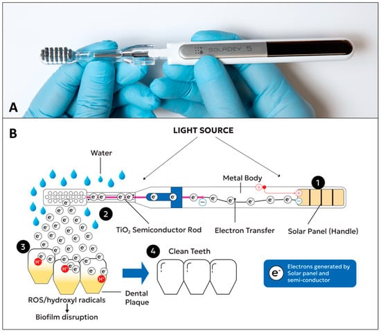
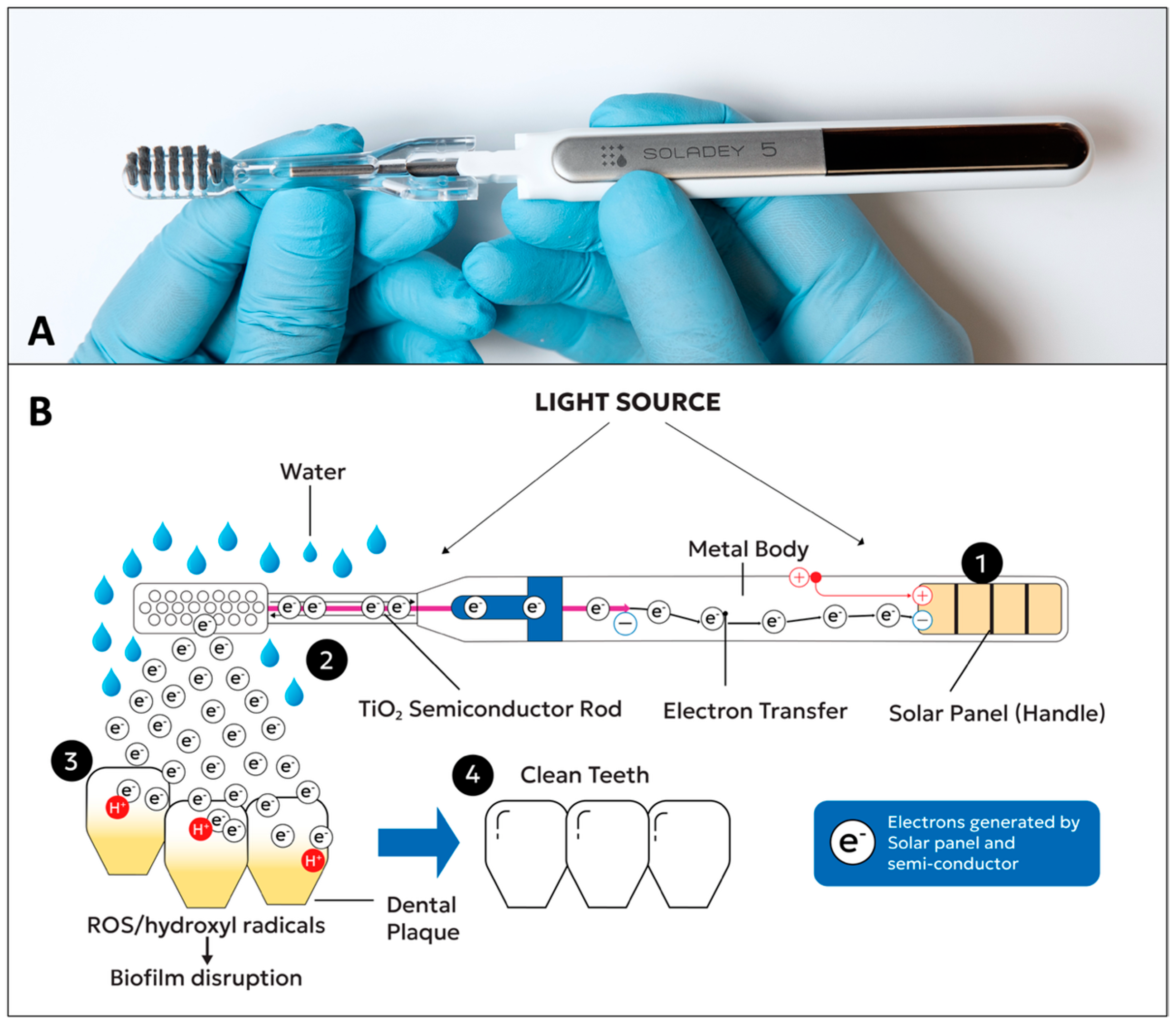
The application of solar energy has expanded remarkably across diverse technological domains. Cutting-edge advancements in photovoltaic engineering include the performance optimization of lead-free Cs2AgInBr6-based inorganic perovskite solar cells via advanced hole transport layer engineering through SCAPS-1D simulation studies, as well as the development of ultra-broadband absorbers based on titanium nitride concentric dual-ring arrays for high-efficiency solar energy harvesting [22,23]. These principles of light capture and conversion have increasingly inspired the integration of solar panels and semiconductor photovoltaic mechanisms into daily healthcare devices [24]. Within dentistry, this has led to the emergence of solar-powered photocatalytic toothbrushes, which incorporate a titanium dioxide (TiO2) photocatalytic semiconductor rod within the brush head, which gets activated by light and moisture [25] (Figure 1A,B). It generates electrons capable of disrupting microbial cell membranes and destabilizing the hydrogen ion matrix within dental plaque, thereby facilitating its removal [26,27]. The underlying photocatalytic mechanism function is followed by a solar panel on the handle that converts light into electrons, which are transferred to the TiO2 rod [28]. When the moistened bristles contact tooth surfaces, the semiconductor reacts with the acidic environment created by plaque bacteria, generating hydroxyl radicals and other oxidative species that attack the plaque biofilm without requiring any chemical dentifrice [29,30]. Laboratory and clinical studies have demonstrated that TiO2 photocatalysis effectively inactivates oral pathogenic bacteria, including Streptococcus mutans and Porphyromonas gingivalis, which are key etiological agents in dental caries and periodontal disease, respectively [31,32]. Previous studies reported significant plaque reduction on buccal surfaces of the teeth using a TiO2 semiconductor brush versus an identical brush without a semiconductor [25]. A double-blind crossover trial study found a significantly greater reduction in gingival index scores with the light-activated brush [33]. Another crossover trial of a TiO2 electrode and solar-cell brush receiving supportive periodontal therapy reported better plaque control, a lower gingival crevicular fluid volume and reduced oral bacterial counts across all sites [27]. An in vitro study by Sanders et al. showed that TiO2-containing brush surfaces disrupt established S. mutans biofilms and inhibit exopolysaccharide production [29].
Figure 1.
(A) Solar toothbrush (Soladay-5, Shiken Co Ltd., Osaka, Japan) with solar panel in handle and head design with semiconductor rod. (B). Diagram illustrating the photocatalytic mechanism of solar toothbrush. Step 1. Brushing begins with bristles moistened with water and ambient light falls on the solar panel in the handle. Step 2. Solar panel converts light into electrons which are transferred to the TiO2 semiconductor rod in the brush head. Activated TiO2 semiconductor rod generates more electrons. Step 3. Reaction with the acidic plaque environment generates hydroxyl radicals, and microbial cell membranes get disrupted; hydrogen ion matrix of plaque destabilized. Step 4. Clean tooth surface with reduced plaque accumulation.
Although prior works of research have documented the substantial challenges faced by visually impaired individuals in maintaining optimal oral hygiene, interventions targeting this population have focused primarily on education with little attention paid to the device design. Studies have reported that combining two or more educational approaches improved plaque, gingival and oral hygiene scores more than single-method instruction [34]. A Braille text instruction produced significantly lower plaque, calculus and oral hygiene indices than verbal instruction alone in Arabic-speaking visually impaired individuals [35]. Evidence on device selection in this population is inconsistent. Electric toothbrush users had lower scores at the final assessment [7]. Among visually impaired children, a three-arm trial found greater reductions in plaque and colony-forming units with a nano-bristle toothbrush than with electric or manual brushes [36]. A smaller randomized trial found that a chewable toothbrush improved oral hygiene more than a manual brush among institutionalized children [37]. Given this context, assessing the efficacy of alternative toothbrush disinfection methods including ultraviolet exposure, chemical immersion, and emerging photocatalytic technologies is essential for developing evidence-based hygiene protocols [38].
2. Materials and Methods
This was a single-blind, exploratory randomized parallel-group study with delayed introduction of the intervention. The research was conducted at multiple centers in Jazan City, Kingdom of Saudi Arabia. Although online public trial registration was not completed due to administrative and logistical constraints associated with recruiting vulnerable visually impaired cohorts within the region, complete ethical approval was secured from the Institutional Review Board of Jazan University (REC-47/06/1728) prior to study initiation, ensuring complete adherence to the CONSORT guidelines for reporting randomized trials [39]. The study was conducted in full accordance with the Declaration of Helsinki [40]. A single-blind methodology was adopted, whereby outcome assessors were masked to group allocation throughout the study. All participants, or their representatives were given the written informed consent prior to the study. The target population comprised visually impaired individuals aged 18–60 years with varying degrees of visual impairment, ranging from low vision to complete blindness, as classified by the World Health Organization criteria [41]. The inclusion criteria were as follows: presence of minimum of 20 natural teeth (excluding third molars); no prior experience with solar toothbrushes; and demonstrated willingness to comply with the study protocol. The exclusion criteria were as follows: presence of severe systemic diseases known to affect oral health; current orthodontic treatment with fixed appliances; any history of periodontal surgical intervention; or any systemic antibiotic or antimicrobial therapy.
2.1. Sample Size Calculation and Randomization
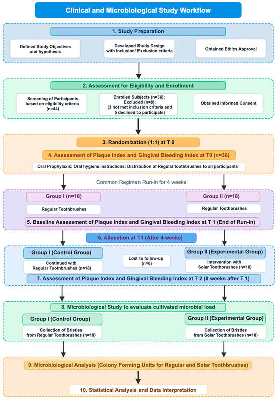
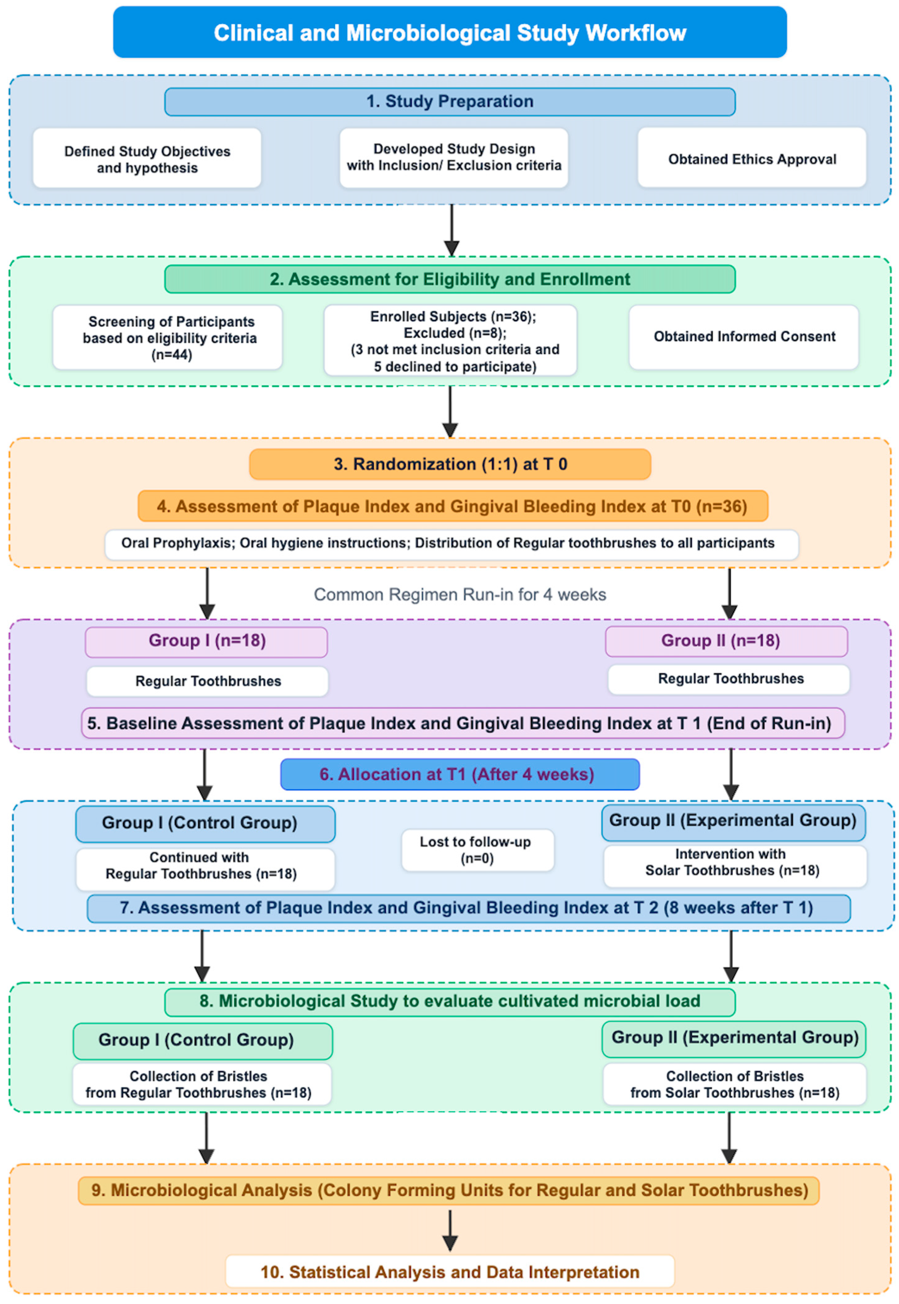
The co-primary outcome variables of this trial were the between-group differences in mean Plaque Index (PI) and mean Gingival Bleeding Index (GBI) change scores over the 8-week device-comparison interval. Secondary outcomes included bristle microbial contamination (CFU count) at T2 and within-group changes across all evaluated time intervals. Assuming a large standardized effect size (Cohen’s d = 0.97) derived from prior oral hygiene clinical trials in comparable visually impaired populations [42,43], a two-sided significance level of α = 0.05 and 80% statistical power (1 − β = 0.80), sample size of n = 18 per group (n = 36 total) was required. For continuous clinical outcomes with small sample sizes, Hedges’ “g” was computed alongside Cohen’s d to adjust for small-sample bias. Of 44 individuals screened for eligibility, 8 were excluded (3 did not meet the inclusion criteria; 5 declined to participate). (Figure 2) Among the thirty-six enrolled participants, 10 had low vision and 8 were total blind in Group I, while 8 had low vision and 10 were total blind in Group II; however, allocation was not stratified on this variable. All thirty-six participants were randomized at T0 in a 1:1 ratio (18 per group) using sequentially numbered, sealed, opaque envelopes prepared by an independent researcher. Both groups used identical conventional regular toothbrushes and toothpaste until four weeks (T1) [44]. The enrolled 36 participants entered the four-week run-in. Outcome examiners were masked to allocation throughout. Group identity was coded before analysis so that the analyst was unaware of the group-to-device correspondence. Inter- and intra-examiner agreement for the binary site-level scores was assessed using Cohen’s kappa (κ ≥ 0.80 required), and agreement for participant-level percentage scores using the intraclass correlation coefficient [45,46].
Figure 2.
Flow diagram showing enrolment, randomization at T0, common-regimen run-in (T0–T1), device introduction in Group II at T1, and follow-up to T2.
2.2. Intervention Groups
Run-in (T0–T1) phase: All Participants received ADA-approved, soft, medium-size head with nylon-bristled manual toothbrushes (Colgate Palmolive GmbH, Hamburg, Germany) and a standard 1000–1450 ppm fluoride toothpaste (Colgate Palmolive, Guangzhou, China) one-to-one tactile oral hygiene education (30–45 min), modified Bass instruction, an auditory timer, and oral prophylaxis after T0 assessment. The purpose of this phase was to allow all participants to learn the brushing routine and to reduce chance variation in baseline oral hygiene. Changes during this phase reflect prophylaxis, education, examination feedback, learning and participation effects, and cannot be attributed to any device. Participants were required to hold and manipulate a toothbrush independently or with the assistance.
Randomized device phase (T1–T2): Group I continued the new regular toothbrush and toothpaste to continue with same oral hygiene protocol. Participants in Group II (experimental) received the solar toothbrushes (Soladey-5, Shiken Co. Ltd., Osaka, Japan). The Group II participants and their caregivers were informed not to use any toothpaste and use only water to the bristles of the brush.
A standardized reinforcement session was conducted at week 4 for all participants to re-evaluate brushing technique, address queries, and reinforce protocol adherence. Any replacement toothbrushes were not provided during the 8-week device intervention phase (T1–T2). Participants were instructed to store their toothbrushes in a dry place after use throughout the intervention period. Brushing was performed in participants’ usual home environments under ambient daylight or standard indoor lighting. Brushing compliance was documented through participant or caregiver reports, with use of a timer and diary.
2.3. Study Timeline and Follow-Up Schedule
Study assessments were conducted at the following three time points (Figure 2):
T0 (Week 0) Initial assessment: Enrolled participants were randomized into two groups, Group I (n = 18) and Group II (n = 18). Plaque Index and Gingival Bleeding Index were recorded for all 36 participants. Participants then received oral prophylaxis to eliminate the confounding effect of pre-existing calculus and plaque deposits [47]. Both groups were then issued the conventional regular toothbrushes and fluoride toothpaste with identical oral hygiene instructions.
T1 (4 weeks after initial assessment): PI and GBI were re-recorded for all 36 participants (18 per group) after four weeks of conventional brushing. A standardized reinforcement session was delivered to all participants. Allocation for Group I participants issued new conventional toothbrush and fluoride toothpaste, while Group II participants were issued with solar toothbrushes. They were instructed to use water only, without dentifrice. T1 served as the baseline for the randomized comparison.
T2 (8 weeks after T1): PI and GBI were recorded after eight weeks of allocated device use, and used toothbrushes were collected for microbiological assessment.
Clinical assessments were performed by two calibrated examiners who were masked to group allocation throughout the trial. To minimize measurement bias, both examiners underwent a formal calibration procedure prior to the data collection, in accordance with established guidelines for examiner alignment and assessment in clinical periodontal research [46]. A training session of O’Leary Plaque Index and Ainamo and Bay Gingival Bleeding Index with clinical examinations of 10 non-study volunteers to assess intra- and inter-examiner agreement was conducted. Plaque was recorded using the O’Leary plaque index [48] after application of disclosing solution (Endekay Ceplac, Manx Healthcare Ltd., Warwick, UK). All teeth present (excluding third molars) were examined at four surfaces per tooth (mesial, distal, buccal and lingual/palatal). Each surface was scored dichotomously (0 = no plaque at the dento-gingival junction; 1 = plaque present). The PI for each participant was calculated as follows:
PI (%) = (number of surfaces with plaque ÷ total number of surfaces examined) × 100
Gingival inflammation was recorded using the Gingival Bleeding Index described by Ainamo and Bay [49]. A periodontal probe was gently inserted about 2 mm into the gingival sulcus at the same four sites per tooth, and bleeding within 10 s was recorded (0 = absent, 1 = present). The GBI for each participant was calculated as follows:
GBI (%) = (number of bleeding sites ÷ total number of sites examined) × 100
Both indices were expressed as percentages (0–100%), with higher values indicating poorer oral hygiene or greater gingival inflammation. Changes between time points were expressed in percentage points.
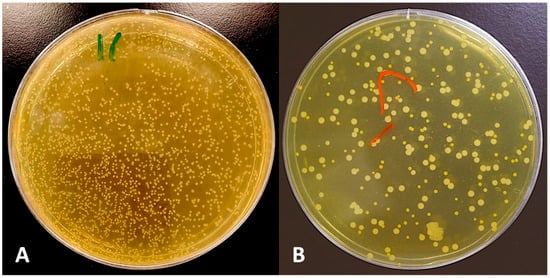
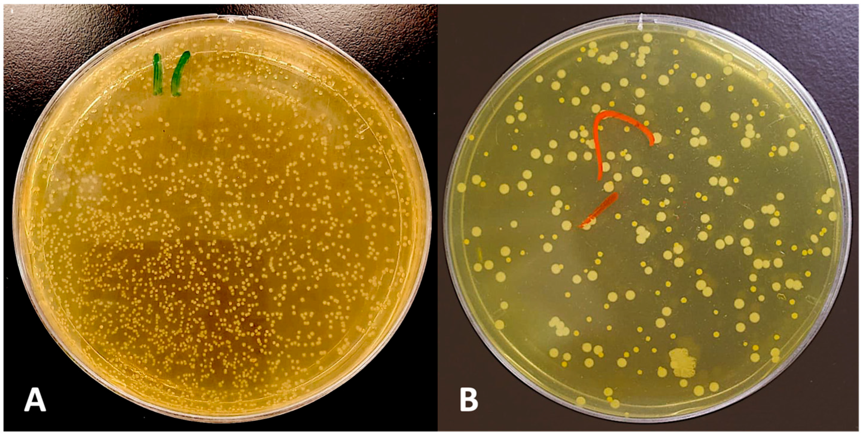
In the second part of the study, microbiological assessment was performed at T2 (Week 12 of the overall study), corresponding to eight weeks following the introduction of the allocated toothbrush interventions at T1 (Week 4). Used toothbrushes were collected from all participants in both groups. Approximately two-thirds of the bristle portion of each toothbrush was excised using a sterile scissor [10,13,49]. The excised bristles were transferred using sterile forceps into individual sterile test tubes containing 1 mL of Tris–EDTA (TE) buffer solution (Invitrogen, Life Technologies Corporation, Carlsbad, CA, USA). The tubes were then transferred into ice bags and transported to the Microbiology laboratory and processed immediately. Each tube was vortexed for 1 min to facilitate the detachment and dispersion of microorganisms adhering to the bristle surfaces into the suspension. The resulting microbial suspensions were used for culture-based quantification of viable microorganisms [50,51]. Then, 1 mL of the mixed sample were transported to Brucella agar media (supplemented with hemin (5 µg/mL), vitamin K1 (1 µg/mL), and 5% sheep blood for culture. The suspension was serially diluted 10-fold (10−1 to 10−3) and 100 µL of each dilution was spread on a plate. Plates were incubated anaerobically for 72 h at 37 °C, allowing time to grow for anaerobes. Plates were coded and colonies were counted by a microbiologist blinded to group allocation, using colony-counting software (ImageJ software, version 1.x, Bethesda, MD, USA) on standardized photographs. Following incubation, visible colonies were calculated and the microbial load recovered from each toothbrush specimen were converted to CFU/mL accounting for dilution and plating volume. Each toothbrush (n = 18 per group) was processed as one independent sample, and the mean was used for analysis (Figure 3A,B). This procedure provided an estimate of the total recoverable cultivable microbial load under the specified culture conditions.
Figure 3.
Microbiological assessment of toothbrushes. (A) Agar plates showing microbial colony-forming units (CFUs) for conventional regular toothbrush; (B) agar plates showing microbial colony-forming units (CFUs) for solar toothbrush.
2.4. Statistical Analysis
Statistical analyses were performed using SPSS Version 28.0 (IBM Corp., Armonk, NY, USA). All tests were two-sided, with statistical significance set at α = 0.05. Estimates of effect are reported with 95% confidence intervals. Two co-primary outcomes mean PI and mean Gingival Bleeding Index were defined. Preliminary normality testing using the Shapiro–Wilk test, normal probability plots, and skewness/kurtosis evaluations confirmed that the distribution of continuous scores and difference scores for both Plaque Index (PI) and Gingival Bleeding Index (GBI) approximated a normal distribution (p > 0.05), supporting the application of parametric tests. For paired analyses, normality was assessed on the within-participant difference scores. Since normality of the differences rather than of the marginal distributions is the assumption underlying the paired t-test; for two-sample comparisons, homogeneity of variance was assessed by Levene’s test. For within-group analysis, PI and GBI were analyzed as continuous percentage scores per participant. PI and GBI were compared across the three intervals (T0–T1, T1–T2, T0–T2) using paired-samples t-tests, reporting for each comparison the mean difference, SD of the difference, 95% CI of the difference, t statistic, degrees of freedom (df = n − 1) and exact p value. To account for the multiple testing of two co-primary endpoints across three-time intervals (T0–T1, T1–T2, T0–T2; total 6 testing combinations), statistical significance for within-group and between-group pairwise evaluations were interpreted against a Bonferroni-corrected threshold of α = 0.0083 (0.05/6). Both unadjusted and Bonferroni-adjusted p-values are reported to ensure full transparency. Between-group comparisons of PI and GBI change scores (T1–T2 and T0–T2) were performed using independent-samples t-tests, reporting the mean difference with its 95% confidence interval, the t statistic, degrees of freedom (df = n1 + n2 − 2) and the exact two-sided p value. Levene’s test was used to assess homogeneity of variance, and Welch’s correction was applied where variances were unequal.
For microbiological analysis, CFU counts were compared between groups by independent-samples t-test, with Levene’s test and Welch’s correction where variances were unequal, and the between-group mean difference is reported with its 95% CI. Because counts of this kind are frequently right-skewed, the Mann–Whitney U test was applied as a sensitivity analysis and medians with IQR are reported alongside means. Cohen’s d was calculated as the between-group mean difference divided by the pooled SD, with Hedges’ g as a small-sample correction.
3. Results
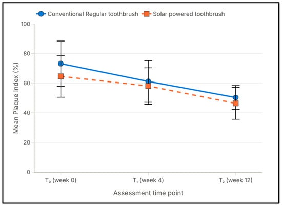
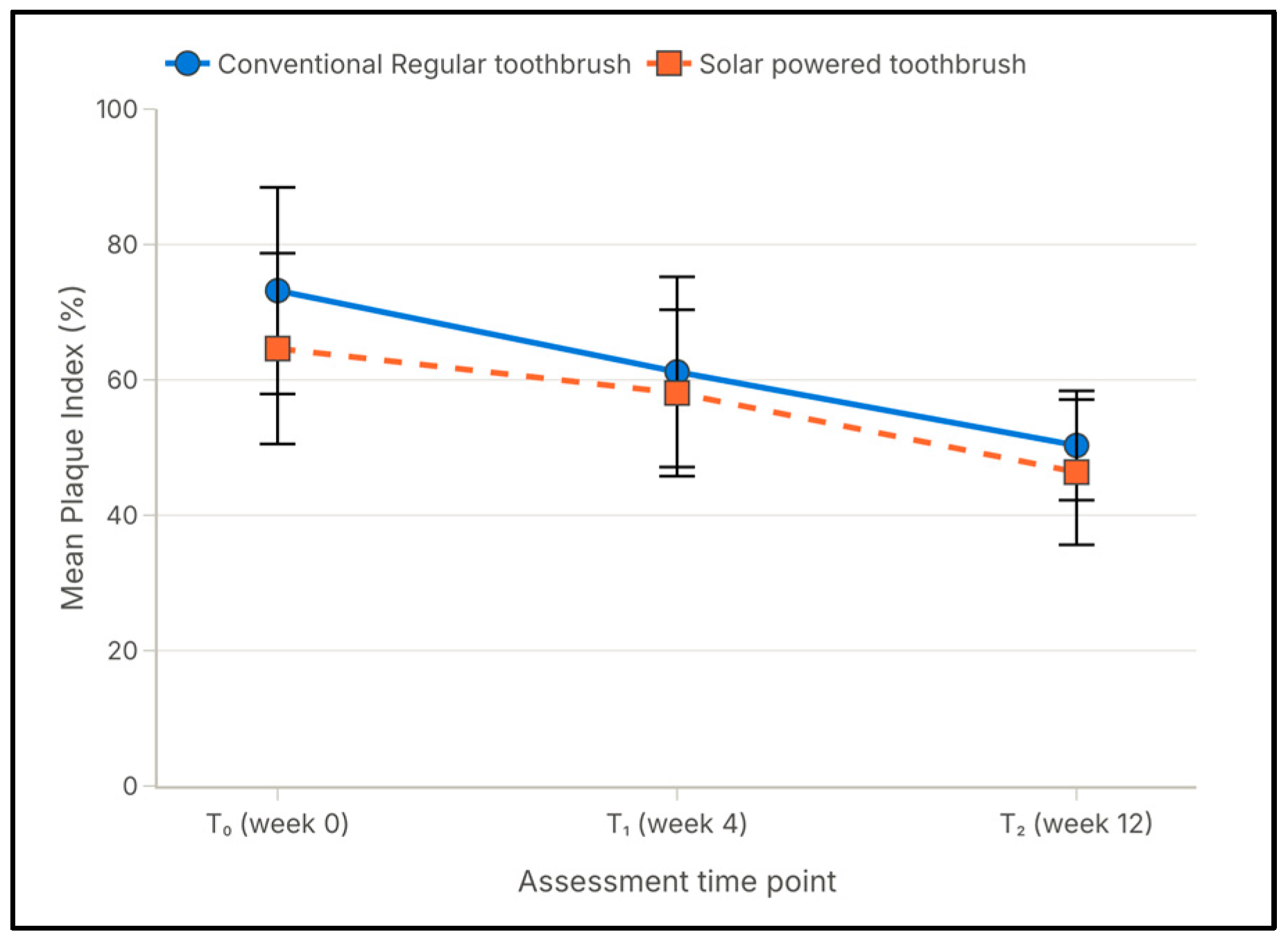
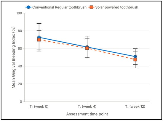
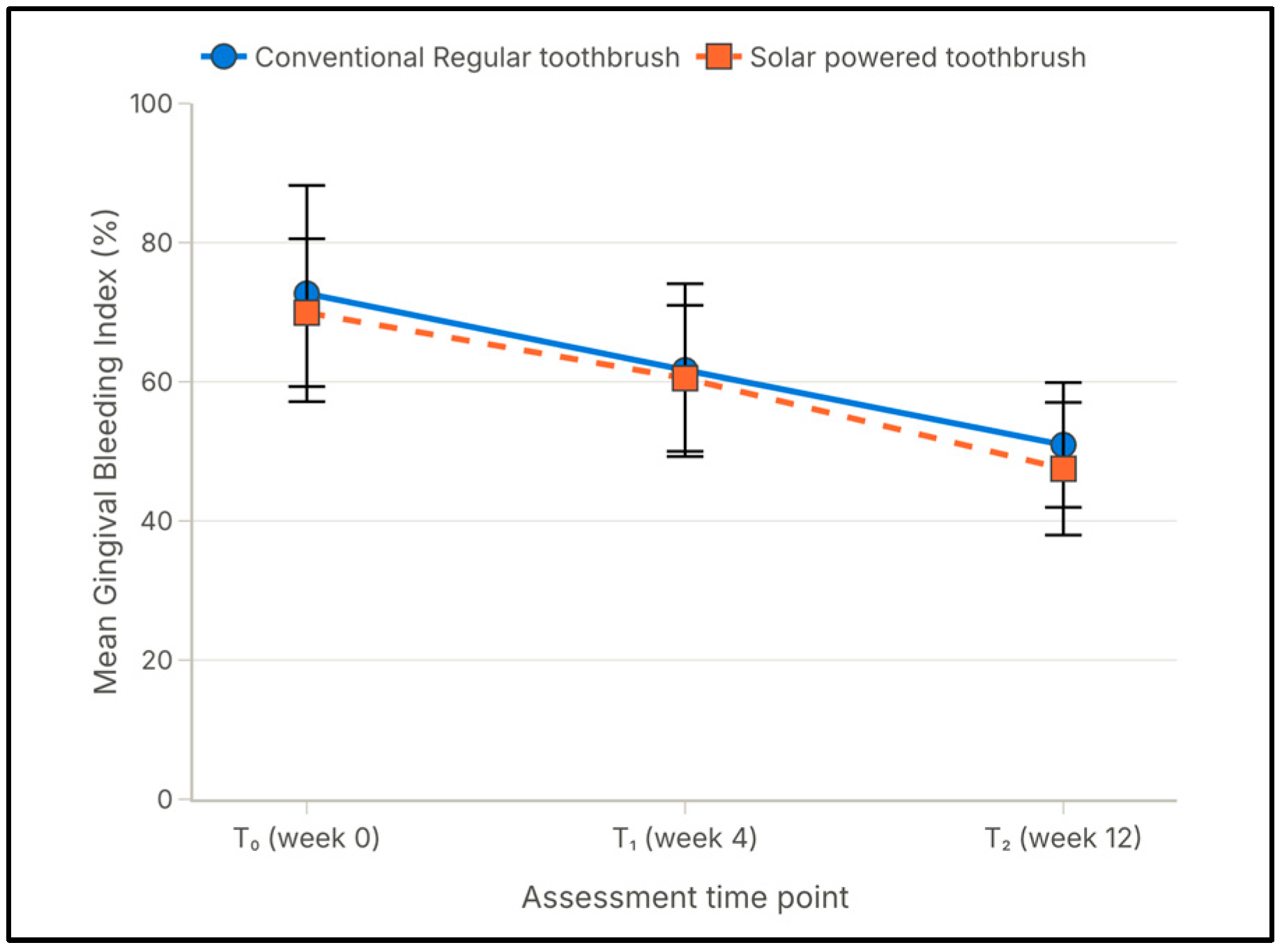
The clinical assessments were performed at three time points: initial assessment (T0), week 4 (T1) and week 12 (T2). All 36 participants (18 per group) attended every assessment and no participant was lost to follow-up, giving 100% retention in both groups. At initial assessment (T0), the mean Plaque Index (PI) score was 73.18 ± 15.267 in Group I and 64.62 ± 14.094 in Group II, and mean Gingival Bleeding Index (GBI) score was 72.68 ± 15.529 and 69.93 ± 10.611, respectively (Table 1; Figure 4 and Figure 5).
Table 1.
Mean O’Leary Plaque Index (% of surfaces with plaque) and Ainamo and Bay Gingival Bleeding Index (% of sites bleeding) scores (±SD) for Group I (conventional toothbrush) and Group II (solar toothbrush) at initial assessment (T0), week 4 (T1), and week 12 (T2).
Figure 4.
Mean Plaque Index scores of Group I (conventional toothbrush) and Group II (solar toothbrush) at initial (T0), week 4 (T1), and week 12 (T2), demonstrating progressive reductions across the 8-week intervention and 12-week study period.
Figure 5.
Mean Gingival Bleeding Index scores of Group I (conventional toothbrush) and Group II (solar toothbrush) at initial assessment (T0), week 4 (T1), and week 12 (T2), demonstrating progressive reductions across the 8-week intervention and 12-week study period.
3.1. Within-Group Change in Plaque and Gingival Bleeding Indices
Both indices PI and GBI declined progressively in both Group I and Group II across the three assessed time intervals. In Group I, the mean PI was reduced from 73.18 at T0 to 61.18 at T1 and 50.31 at T2, and the mean GBI was reduced from 72.68 to 61.68 and 50.93 at the same points. In Group II, the mean PI was reduced from 64.62 to 58.06 and 46.37, and the mean GBI from 69.93 to 60.50 and 47.50 (Table 1; Figure 4 and Figure 5). In Group I, the mean PI reduction was 12.00 for T0–T1, 10.87 for T1–T2 and 22.87 for T0–T2, and the mean GBI reduction was 11.00, 10.75 and 21.75 percentage points for the same intervals (p < 0.001; Table 2). In Group II, the corresponding PI reductions for T0–T1, T1–T2 and T0–T2 were 6.56, 11.68 and 18.25, and the GBI reductions were 9.43, 13.00 and 22.43 respectively (p < 0.001, Table 3). Every comparison was in the direction of improvement. The two groups differed in the distribution of that improvement across intervals: in Group I, the two intervals contributed almost equally (12.00 versus 10.87), whereas, in Group II, the reduction was concentrated in the intervention interval (6.56 versus 11.68).
Table 2.
Comparison of Plaque Index and Gingival Bleeding Index among Group I visually impaired individuals at different time intervals (Paired t test, p ≤ 0.05 Significant, CI = 95%).
Table 3.
Comparison of Plaque Index and Gingival Bleeding Index among Group II visually impaired individuals at different time intervals. (Paired t test, p ≤ 0.05 Significant, CI = 95%). Differences were calculated from unrounded values; small discrepancies are due to rounding.
3.2. Between-Group Comparisons
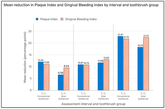
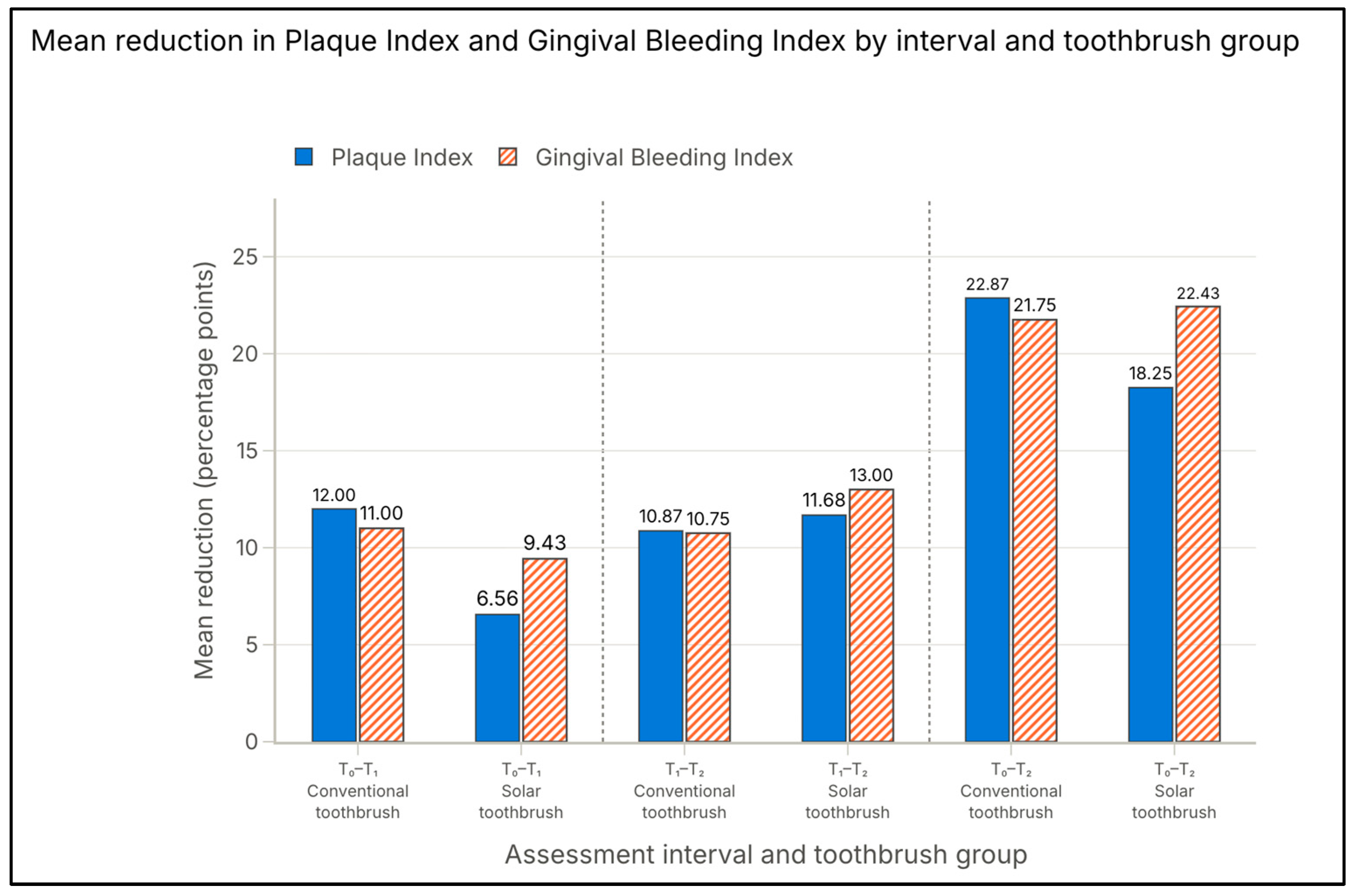
Between-group comparisons of the mean change in the Plaque Index and Gingival Bleeding Index are presented in Table 4 and Figure 6. For PI, the reduction during T0–T1 differed between the groups (12.00 vs. 6.56; t = 3.22; p = 0.003). Both groups improved markedly in T0–T1. Because T0 was recorded before prophylaxis, this includes the effect of professional cleaning, education, feedback and the Hawthorne effect. The continued improvement in T1-T2 may partly reflect the ongoing learning in both groups.
Table 4.
Intergroup comparison of mean differences in plaque index and gingival bleeding index scores between Group I and Group II across all time intervals (T0–T1, T1–T2, T0–T2), using independent-sample t-test (CI = 95%).
Figure 6.
Mean difference in Plaque Index and Gingival Bleeding Index scores between Group I (conventional toothbrush) and Group II (solar toothbrush) across three-time intervals (T0–T1, T1–T2, and T0–T2).
During the intervention interval (T1–T2), the PI reduction was 10.87 in Group I and 11.68 in Group II (t = −0.31; p = 0.76), and the cumulative T0–T2 reduction was 22.87 and 18.25, respectively (t = 1.42; p = 0.17). For GBI, no between-group difference was detected at any interval: T0–T1, 11.00 vs. 9.43 (t = 0.91; p = 0.37); T1–T2, 10.75 vs. 13.00 (t = −0.93; p = 0.36); T0–T2, 21.75 vs. 22.43 (t = −0.20; p = 0.85). No statistically significant between-group difference was detected for either index during the intervention period or over the study.
3.3. Microbial Contamination of Toothbrushes
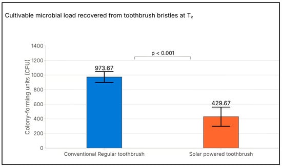
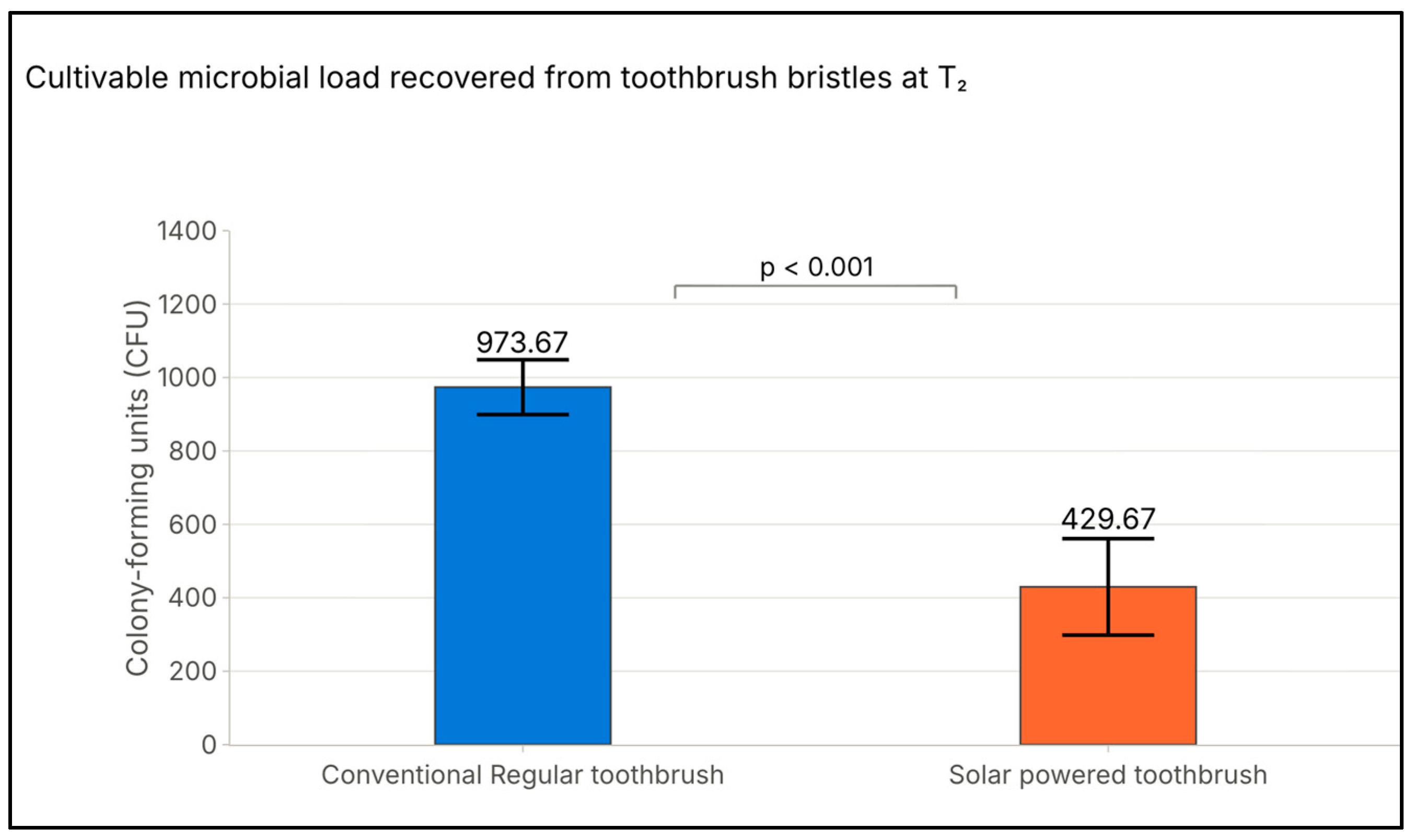
At T2 (Week 12 of the overall study, following eight weeks of allocated toothbrush use), the mean CFU count recovered from conventional toothbrush bristles was 973.67 ± 74.86 CFU/mL, compared with 429.67 ± 131.47 CFU/mL for solar toothbrushes (Table 5, Figure 7). The mean between-group difference was 544.00 CFU/mL (95% CI: 471.5–616.5), representing approximately 55.9% lower recoverable microbial counts in the solar toothbrush group (t = 15.26; p < 0.001). These findings demonstrate a statistically significant difference in total cultivable microbial load between the two toothbrush groups. However, the culture-based assessment did not identify individual bacterial species or establish the underlying mechanism responsible for the observed difference.
Table 5.
Comparison of microbial contamination between regular and solar toothbrushes at T2. A positive mean difference indicates greater microbial contamination of conventional toothbrushes.
Figure 7.
The mean colony-forming unit (CFU) counts for conventional regular toothbrushes demonstrated a higher mean microbial load (973.67 CFU) compared with solar toothbrushes (429.67 CFU). Statistical significance: p < 0.001.
4. Discussion
4.1. Principal Findings
This exploratory randomized trial study with delayed intervention compared the toothpaste-free solar-powered photocatalytic toothbrush with a conventional manual toothbrush used with fluoride toothpaste in 36 adults with a visual impairment over 12 weeks. Both groups demonstrated progressive and statistically significant reductions in plaque and gingival bleeding indices at three different time intervals. By week 12 (T2), the mean Plaque Index (PI) decreased by 31.25% (22.87) in Group I and 28.2% (18.25) in Group II relative to baseline, including the 4-week run-in. Concurrently, the mean Gingival Bleeding Index (GBI) declined by 29.9% (21.75) in Group I and 32.1% (22.43) in Group II. Between-group comparisons during the active intervention period (T1–T2) revealed no statistically significant differences for either PI (10.87 vs. 11.68, p = 0.76) or GBI (10.75 vs. 13.00, p = 0.36). Similarly, cumulative changes from the initial assessment (T0 to T2) showed no significant intergroup variations for PI (22.87 vs. 18.25, p = 0.17) or GBI (21.75 vs. 22.43, p = 0.85). The only significant difference observed between-group occurred during T0–T1, before allocation of the devices, and thus reflects a chance imbalance in response to the common run-in regimen rather than a device-specific effect. In contrast, the microbiological evaluation of toothbrush bristles at T2 revealed a statistically significant 55.9% reduction in mean microbial load on solar toothbrushes (429.67 ± 131.47 CFU for solar toothbrushes compared to 973.67 ± 74.86 CFU for conventional toothbrushes), yielding a mean difference of 544.00 CFU (p < 0.001). In the absence of a prespecified equivalence margin, these findings support comparability between the two regimens but do not demonstrate the superiority of either device.
4.2. Comparison with Previous Clinical Evaluations of Photocatalytic Toothbrushes
Clinical evaluations of titanium dioxide (TiO2) photocatalytic toothbrushes remain limited, with heterogeneous findings across the existing literature. In a three-week crossover trial, Hoover et al. reported significantly greater plaque reduction on buccal surfaces using a semiconductor brush compared to a control brush lacking the semiconductor [25]. However, no significant differences were observed on lingual surfaces. This finding was attributed to the limited ambient light exposure in posterior lingual regions. While the present study evaluated plaque across four surfaces per tooth without a separate surface-region analysis, no overall intergroup plaque differences were detected. The plaque control findings in the present study closely align with a double-blind cross-over study by Uswak et al., who observed significant within-group plaque reductions with both light-activated and conventional brushes without significant intergroup differences [33]. Similarly, Yoshimine et al. observed enhanced plaque suppression, reduced gingival crevicular fluid volume, and lower bacterial counts across all sites in periodontally maintained patients using TiO2 and solar-cell toothbrushes [27]. Niwa and Fukuda reported equivalent Plaque Index reductions between TiO2 semiconductor toothbrushes and conventional toothbrushes, yet observed significantly lower PMA Gingival Index scores in the semiconductor group by week 3 [52]. While two prior studies report enhanced plaque removal [25,27] and three report improved gingival outcomes with photocatalytic brushes, the present study observed comparable clinical indices across both groups [27,33,52]. Several methodological factors likely account for this divergence such as the fact that participants received individualized, model-based tactile instruction and auditory timer guidance, which likely optimized manual brushing efficiency and attenuated small device-driven differences. The control group utilized a manual toothbrush paired with high-concentration fluoride toothpaste (1000–1450 ppm) rather than an inactive/identical brush head.
4.3. Consistency with the Proposed Photocatalytic Mechanism
The clinical effects observed in this study were modest compared with those reported under in vitro conditions. When exposed to light, anatase TiO2 produces electron–hole pairs that interact with oxygen and water to generate reactive oxygen species. Its bactericidal activity has been associated with adsorption to the catalyst surface and the subsequent disruption of membrane integrity [26]. For a solar-powered brush, 90.1 ± 1.4% disrupted biofilm removal with released cytoplasmic contents has been reported, which is 1.3 times that of non-solar electric brushes [28]. TiO2-containing brush surfaces have been shown to disrupt established Streptococcus mutans biofilm and inhibit exopolysaccharide production [29]. The modest clinical effects observed in this study are mechanistically coherent, as indoor light exposure during a two-minute brushing session provides a much lower photon dose than experimental conditions.
4.4. Comparison with Other Powered and Light-Based Devices
Evidence from other powered and light-based oral-hygiene devices shows a similar pattern as found in this study. Both groups often improved, but additional benefits from the device were inconsistent. Bjurshammar et al. evaluated a 450 nm blue-light toothbrush and recorded marked reductions in plaque (57%), gingival (46%), and bleeding (15%) indices across all participants, without significant incremental benefits over control brushing [53]. In contrast, microcurrent-emitting and bioelectric toothbrushes have demonstrated incremental plaque reduction benefits in specific patient cohorts without altering caries rates [54]. In vitro studies by Lee and Kim suggest that bioelectric activation enhances biofilm detachment while minimizing abrasive tooth wear [55]. In vitro, bioelectric brushing improved plaque removal efficiency while minimizing tooth wear on typodonts [30], and dual-light antimicrobial photodynamic therapy has been proposed as an adjunct for patients with reduced manual dexterity [56]. A recurring pattern of substantial within-group improvement in both arms, but no incremental clinical benefit of the device, was detected in this study.
4.5. Within-Group Improvement and the Contribution of Oral Hygiene Education
The marked clinical improvements observed in both study arms underscore the significant impact of structured oral hygiene training in visually impaired populations. Khurana observed significant plaque and gingivitis reductions following the distribution of Braille-embossed oral health kits, while Fageeh et al. demonstrated that combining verbal and Braille instructions yielded superior 12-week oral hygiene indices compared to verbal advice alone [6,35]. Systematic reviews by Bhadauria et al. and Khan et al. confirm that multifaceted educational approaches incorporating tactile models and auditory feedback consistently outperform single-modality instruction [34,57]. Even the brushing technique appears not to matter much, since the horizontal scrub and modified Bass methods reduced the plaque and gingival indices to a similar degree in visually impaired students [58]. Because both groups in this study received identical, intensive baseline training and a 4-week reinforcement session, the clinical gains observed in the control group reflect the efficacy of structured instruction and regular prophylaxis. Consequently, the comparable performance of the solar toothbrush group indicates that the device provided effective mechanical and photocatalytic biofilm disruption, maintaining clinical gains without requiring dentifrice.
4.6. Manual Versus Powered Brushing in Visually Impaired Populations
Comparative evidence regarding toothbrush modalities in visually impaired individuals remains inconsistent. A 6-month randomized trial by Thanalakshme and Ramesh in blind children found manual toothbrushes superior to electric toothbrushes in reducing plaque scores and dental anxiety [59]. Conversely, Jaya and Choudhar reported that nano-bristle toothbrushes achieved greater plaque and bacterial reductions than both electric and manual brushes [36]. Meghana et al. observed that chewable toothbrushes significantly improved oral hygiene over manual brushes in institutionalized settings, whereas Křížová et al. noted lower long-term plaque scores among powered toothbrush users [7,37]. Given these conflicting reports, our finding of clinical parity between solar and conventional toothbrushes contributes valuable data to an evolving literature base.
4.7. The Toothpaste-Free Regimen and Procedural Implications
A key feature of the solar toothbrush intervention was the elimination of toothpaste, addressing a major practical barrier for visually impaired individuals. This instruction was specified in the protocol rather than left to participant preference. It followed the manufacturer’s instructions for the photocatalytic device, in which the semiconductor rod requires a continuous aqueous interface. The photocatalytic reaction is an aqueous surface phenomenon that a particulate dentifrice slurry would occlude at the rod–enamel interface. Case-control studies indicate that nearly one-third of visually impaired adults rely on caregivers for oral care, while studies in blind children highlight frequent difficulties in dispensing appropriate dentifrice volumes, leading to product wastage, ingestion risks, or caregiver dependency [4,21]. Dedicated toothpaste packaging has been developed to give visually impaired and mobility-impaired users control over the dispensed volume [20]. A toothpaste-free device removes that step entirely, and the absence of a detectable between-group difference in plaque and bleeding scores over eight weeks of intervention suggests that the solar toothbrush provides greater personal independence during daily hygiene routines. However, this practical benefit carries an important clinical caveat. Conventional fluoride toothpaste remains the gold standard for caries prevention [60]. Because plaque and bleeding indices measure periodontal status rather than cariogenesis, a toothpaste-free regimen should not replace systemic or topical fluoride delivery. To ensure long-term enamel protection, toothpaste-free devices should be complemented by alternative fluoride such as fluoridated mouthrinses or professional varnish applications.
4.8. Microbial Contamination of Bristles
The 55.9% lower bacterial colony count observed on solar toothbrush bristles after 8 weeks aligns with the literature on antimicrobial bristle technology. AlNaqa et al. previously demonstrated significantly lower microbial retention on charcoal-infused bristles compared to standard nylon bristles [10]. Standard toothbrushes rapidly collect oral and environmental pathogens, harboring bacterial loads up to 4 × 10⁸ CFU/mL, including Staphylococcus, Escherichia, and Pseudomonas species [61]. Contaminated bristles can act as microbial reservoirs, posing opportunistic infection risks to vulnerable patient groups [51]. While routine toothbrush replacement is recommended, studies show that new bristles accumulate S. mutans rapidly upon initial use [38]. The reduced bacterial counts observed on the solar toothbrushes indicate that photo-activated semiconductor activity may contribute to inhibiting microbial survival and retention on bristle surfaces. Nevertheless, several limitations should be considered. The extraction protocols differed across studies, making relative percentage reductions more informative than absolute CFU counts. The microbial burden was assessed at only one time point, so the patterns of long-term recolonization remain unknown. Colony counts reflect total viable anaerobic growth and do not identify species-specific changes. Future research should use genomic sequencing to assess changes in the bristle microbiome.
From a public health and clinical practice perspective, solar toothbrushes may improve oral care for visually impaired individuals. It eliminates the need to locate, uncap, and dispense toothpaste and simplify the brushing process. It also avoids the risk of ingesting excessive fluoride with dentifrice. The detachable, replaceable brush head is another advantage. Users can identify bristle wear by touch and replace the head without replacing the entire device. This may lower long-term costs, an important consideration for people with disabilities who face financial barriers to procure oral hygiene products. These features support the solar toothbrush’s practical suitability as a long-term oral hygiene option for visually impaired populations.
4.9. Strengths and Limitations
This study possesses several methodological strengths, including prospective randomization with concealed allocation, 100% participant retention, robust examiner masking and pre-calibrated inter-examiner reliability (κ ≥ 0.80), a 12-week longitudinal evaluation.
Few limitations of the study include device allocation at T1 following a 4-week run-in phase; thus, the randomized comparison is confined to the T1–T2 period. The follow-up of eight weeks after allocation is short relative to the chronic nature of periodontal disease, and no caries outcome was measured. Standardized prophylaxis and intensive instruction applied across both groups prevented isolating device-driven effects from educational reinforcement. The microbiological analysis was culture-based, without species identification. Because the oral cavity and toothbrush bristles harbor many obligate and facultative anaerobes, which are slow-growing under these conditions, the counts reported here represent total cultivable anaerobic microorganisms and probably underestimate the true microbial load. No species-level identification, Gram staining, separate aerobic culture, molecular identification, or quantitative biofilm analysis was performed. Moreover, specific storage parameters, including the storage container type, ventilation, temperature, relative humidity, and duration of drying between brushing sessions, were not standardized. Therefore, the potential differences in household storage environments and moisture retention could not be excluded as contributing factors to the observed microbial counts.
4.10. Implications and Future Research
For clinical practice, solar-powered photocatalytic toothbrushes represent an effective, dentifrice-free alternative for visually impaired individuals, particularly those who experience difficulty dispensing toothpaste. When adopting a toothpaste-free regimen, clinicians should ensure fluoride exposure through alternative topical measures. For research, three main priorities emerge: Light intensity, ambient spectrum, and exposure geometry should be standardized in future clinical trials. Multi-center trials evaluating long-term caries incidence and metagenomic microbial profiling are needed. Future studies should evaluate photocatalytic toothbrushes as part of comprehensive, technology-assisted oral health programs tailored for individuals with special needs.
5. Conclusions
The present study demonstrates that solar-powered toothbrushes represent a viable, user-friendly alternative for maintaining oral health in populations with visual impairments. Specifically, they offer clinical plaque reductions comparable to traditional methods while eliminating the requirement for dentifrice. In addition to these clinical and practical benefits, the significantly lower microbial burden observed on solar toothbrushes points to a lower cultivable antimicrobial burden that warrants further long-term evaluation.
Author Contributions
Conceptualization, M.A. and M.E.S.; methodology, M.A., M.E.S., H.N. and R.A.A.; software, M.A., M.E.S., A.Y.K., A.M.M. and R.A.A.; validation, M.A., M.E.S., A.Y.K., A.M.M. and R.A.A.; formal analysis, M.A., M.E.S., H.N., A.Y.K. and A.M.M.; investigation, M.A., H.N., A.Y.K. and A.M.M.; resources, M.A., M.E.S., A.Y.K., A.M.M. and R.A.A.; data curation, M.A., M.E.S., H.N., A.Y.K., A.M.M., H.E.H. and R.A.A.; writing—original draft preparation, M.A., M.E.S., H.N., H.E.H. and R.A.A.; writing—review and editing, M.A., M.E.S., H.N., A.Y.K., A.M.M. and R.A.A.; visualization, M.A., M.E.S., H.N., H.E.H., A.Y.K. and A.M.M.; supervision, M.A., M.E.S., H.N., H.E.H. and R.A.A.; project administration, M.A., M.E.S., H.N., H.E.H., A.Y.K. and A.M.M.; funding acquisition, M.A., M.E.S., A.Y.K. and A.M.M. All authors have read and agreed to the published version of the manuscript.
Funding
This research received no external funding.
Institutional Review Board Statement
The research protocol was approved by the standing committee for scientific research ethics, Jazan University (Reference number: REC-47/06/1728).
Informed Consent Statement
Informed consent was obtained from all subjects involved in the study. Written informed consent has been obtained from the patient(s) to publish this paper.
Data Availability Statement
Data are unavailable due to privacy or ethical restrictions. The data that support the findings of this study are available from the corresponding authors upon reasonable request.
Conflicts of Interest
The authors declare no conflicts of interest.
References
- Okesanya, O.J.; Adebayo, U.O.; Oso, T.A.; Ayelaagbe, O.B.; Obadeyi, K.B.; Lamem, M.F.H.; Othman, Z.K.; Ahmed, M.M.; Jamil, S.; Lucero-Prisno, D.E. Bridging Global Oral Health Gaps through Evidence Based Frameworks and Community Centered Interventions. Discov. Public Health 2026, 23, 679. [Google Scholar] [CrossRef] [Scilit]
- Botelho, J.; Mascarenhas, P.; Viana, J.; Proença, L.; Orlandi, M.; Leira, Y.; Chambrone, L.; Mendes, J.J.; Machado, V. An Umbrella Review of the Evidence Linking Oral Health and Systemic Noncommunicable Diseases. Nat. Commun. 2022, 13, 7614. [Google Scholar] [CrossRef] [Scilit] [PubMed]
- D’Addazio, G.; Santilli, M.; Sinjari, B.; Xhajanka, E.; Rexhepi, I.; Mangifesta, R.; Caputi, S. Access to Dental Care—A Survey from Dentists, People with Disabilities and Caregivers. Int. J. Environ. Res. Public Health 2021, 18, 1556. [Google Scholar] [CrossRef] [Scilit] [PubMed]
- Alshatrat, S.M.; Bakri, I.A.; Omari, W.A.; Tabnjh, A.K. Oral Health Knowledge, Behaviour, and Access to Dental Care in Visually Impaired Individuals in Jordan: A Case-Control Study. Open Dent. J. 2021, 15, 33–40. [Google Scholar] [CrossRef] [Scilit]
- AlSadhan, S.A.; Al-Jobair, A.; Bafaqeeh, M.; Abusharifa, H.; Alagla, M. Dental and Medical Health Status and Oral Health Knowledge among Visually Impaired and Sighted Female Schoolchildren in Riyadh: A Comparative Study. BMC Oral Health 2017, 17, 154. [Google Scholar] [CrossRef] [Scilit] [PubMed]
- Khurana, C. Empowering Visually Impaired Individuals for Good Oral Health. Int. Dent. J. 2024, 74, S44. [Google Scholar] [CrossRef] [Scilit]
- Křížová, P.; Himmlová, L.; Chramosta, P.; Vařejčko, D.; Vašáková, J.; Urbanová, W. Oral Health in Youth with Visual Impairment: A Longitudinal Survey. Sci. Rep. 2024, 14, 12273. [Google Scholar] [CrossRef] [Scilit] [PubMed]
- Hotic, M.; Ackermann, M.; Bopp, J.; Hofmann, N.; Karygianni, L.; Paqué, P.N. Critical Hydrodynamic Force Levels for Efficient Removal of Oral Biofilms in Simulated Interdental Spaces. Clin. Oral Investig. 2024, 28, 346. [Google Scholar] [CrossRef] [Scilit] [PubMed]
- Twetman, S.; Axelsson, S.; Dahlgren, H.; Holm, A.; Källestål, C.; Lagerlöf, F.; Lingström, P.; Mejàre, I.; Nordenram, G.; Norlund, A.; et al. Caries-preventive Effect of Fluoride Toothpaste: A Systematic Review. Acta Odontol. Scand. 2003, 61, 347–355. [Google Scholar] [CrossRef] [Scilit] [PubMed]
- AlNaqa, N.; AlDhawi, R.; Tashkandi, O.; Gamal, A.; AlShammery, H.; Eltom, S. Antimicrobial Efficacy of Charcoal vs. Non-Charcoal Toothbrushes: A Randomized Controlled Study. J. Int. Soc. Prev. Community Dent. 2020, 10, 719. [Google Scholar] [CrossRef] [Scilit] [PubMed]
- Efstratiou, M.A.; Papaioannou, W.; Nakou, M.; Ktenas, E.; Vrotsos, I.A.; Panis, V. Contamination of a Toothbrush with Antibacterial Properties by Oral Microorganisms. J. Dent. 2006, 35, 331–337. [Google Scholar] [CrossRef] [Scilit]
- Gautam, B.; Pokhrel, S.; Aryal, S.; Basnet, A. Efficacy of Toothpaste in Reducing Micro-Flora Isolated from Toothbrush. Kathmandu Univ. J. Sci. Eng. Technol. 2018, 13, 71–78. [Google Scholar] [CrossRef] [Scilit]
- Zinn, M.; Schages, L.; Bockmühl, D.P. The Toothbrush Microbiome: Impact of User Age, Period of Use and Bristle Material on the Microbial Communities of Toothbrushes. Microorganisms 2020, 8, 1379. [Google Scholar] [CrossRef] [Scilit] [PubMed]
- Mital, P.; Amit; Raisinghani, D. A Cross-Sectional Study to Compare Knowledge, Attitude and Practices of Dental and Medical Students About Toothbrush Maintenance and Use. Int. J. Health Sci. Res. 2023, 13, 175–183. [Google Scholar] [CrossRef] [Scilit]
- Kozai, K.; Iwai, T.; Miura, K. Residual Contamination of Toothbrushes by Microorganisms. ASDC J. Dent. Child. 1989, 56, 201–204. [Google Scholar] [PubMed]
- Frazelle, M.; Munro, C.L. Toothbrush Contamination: A Review of the Literature. Nurs. Res. Pract. 2012, 420630, 1–6. [Google Scholar] [CrossRef] [Scilit] [PubMed]
- Ralephenya, T.; Molepo, J.; Molaudzi, M.; Volchansky, A.; Shangase, S.L. Contamination of Used Toothbrushes and Their Decontamination with Disinfecting Agents. S. Afr. Dent. J. 2020, 75, 478–484. [Google Scholar] [CrossRef] [Scilit]
- Muralidharan, D.; Fareed, N.; Shanthi, M. Comprehensive Dental Care for Visually Impaired Children. J. Indian Soc. Pedod. Prev. Dent. 2012, 30, 63–67. [Google Scholar]
- Espinoza, L.K.R.; López-Romo, H.N.; Jumbo, A.; Castillo, M. Description of Oral Hygiene in Visually Impaired People in the City of Ibarra, Ecuador. Salud Cienc. Tecnol. 2024, 4, 928. [Google Scholar] [CrossRef] [Scilit]
- Cernic, B.G.; Kogawa, A.C.; Salgado, H.R.N. Development of Differentiated Pharmaceutical Packaging for Greater Autonomy and Quality of Life for Physically and Visually Impaired Patients. Saudi Pharm. J. 2018, 26, 921–924. [Google Scholar] [CrossRef] [Scilit] [PubMed]
- Adawiyyah, E.; Sutomo, B.; Wiyatini, T.; Sunarjo, L. Smart Audio Toothbrush With UV-Filtered Toothpaste Dispenser to Improve Skills Brushing Teeth and Reducing Debris Index Blind Children. Int. J. Innov. Sci. Res. Technol. 2024, 9, 2323–2328. [Google Scholar] [CrossRef] [Scilit]
- Hossain, M.K.; Samajdar, D.P.; Das, R.C.; Arnab, A.A.; Rahman, M.F.; Rubel, M.H.K.; Islam, M.R.; Bencherif, H.; Pandey, R.; Madan, J.; et al. Design and Simulation of Cs2BiAgI6 Double Perovskite Solar Cells with Different Electron Transport Layers for Efficiency Enhancement. Energy Fuels 2023, 37, 3957–3979. [Google Scholar] [CrossRef] [Scilit]
- Gopal, R.; Narayanan, V.L.; Dhaked, D.K.; Gupta, A. Performance Enhancement of Inorganic Cs2AgInBr6-Based Perovskite Solar Cell by Numerical Simulation. J. Phys. Chem. Solids 2024, 193, 112184. [Google Scholar] [CrossRef] [Scilit]
- Mahajan, U.M.; Dhonde, M.; Sahu, K.; Ghosh, P.; Shirage, P.M. Titanium Nitride (TiN) as a Promising Alternative to Plasmonic Metals: A Comprehensive Review of Synthesis and Applications. Mater. Adv. 2024, 5, 846–895. [Google Scholar] [CrossRef] [Scilit]
- Hoover, J.; Singer, D.; Pahwa, P.; Komiyama, K. Clinical Evaluation of a Light Energy Conversion Toothbrush. J. Clin. Periodontol. 1992, 19, 434–436. [Google Scholar] [CrossRef] [Scilit] [PubMed]
- Gogniat, G.; Thyssen, M.; Denis, M.; Pulgarín, C.; Dukan, S. The Bactericidal Effect of TiO2 Photocatalysis Involves Adsorption onto Catalyst and the Loss of Membrane Integrity. FEMS Microbiol. Lett. 2006, 258, 18–24. [Google Scholar] [CrossRef] [Scilit] [PubMed]
- Yoshimine, M.; Yamasaki, R.; Okazaki, K.; Ogiso, R.; Kamoi, H.; Asaki, E. Effect on Oral Hygiene of a Tooth Brush Equipped with a Built-in Titanium Oxide (TiO2) Electrode and Solar Cell. J. Jpn. Soc. Periodontol. 2020, 62, 58–73. [Google Scholar] [CrossRef] [Scilit]
- Sato, T.; Hirai, N.; Oishi, Y.; Uswak, G.; Komiyama, K.; Hamada, N. Efficacy of a Solar-Powered TiO2 Semiconductor Electric Toothbrush on P. gingivalis Biofilm. Am. J. Dent. 2015, 28, 81–84. [Google Scholar] [PubMed]
- Sanders, M.K.; Duarte, S.; Ayoub, H.M.; Scully, A.C.; Vinson, L.A.; Gregory, R.L. Effect of Titanium Dioxide on Streptococcus Mutans Biofilm. J. Appl. Biomater. Funct. Mater. 2023, 21, 22808000221131892. [Google Scholar] [CrossRef] [Scilit] [PubMed]
- Lee, J.; Park, H.M.; Kim, Y.W. Comparative Analysis of Plaque Removal and Wear between Electric–Mechanical and Bioelectric Toothbrushes. Bioengineering 2024, 11, 474. [Google Scholar] [CrossRef] [Scilit] [PubMed]
- Wang, D.; Zhu, L.; Zhang, L.; Zhang, G.-M.; Qian, Y.; Wu, L.; Ma, D.; Dai, T.; Shan, X.; Luo, D.; et al. Multi-Scale Composite Ag-TiO2 Nanoreactor for Long-Lasting Antibacterial and Immunomodulatory Therapy of Periodontitis. Nano Res. 2024, 18, 94907527. [Google Scholar] [CrossRef] [Scilit]
- Kang, S.; Lee, H.-N.; Kim, H.; Kim, B. Influence of TiO2 Concentrations and Irradiation Lights on the Photocatalytic Reaction for Inhibiting Growth of Streptococcus Mutans. J. Dent. Hyg. Sci. 2014, 14, 319–324. [Google Scholar] [CrossRef] [Scilit]
- Uswak, G.; Lee, J.; Li, J. A Double-Blind Cross-over Study Evaluating the Efficacy of a Light-Activated Toothbrush. Int. J. Exp. Dent. Sci. 2012, 1, 57–60. [Google Scholar] [CrossRef] [Scilit]
- Bhadauria, U.S.; Gupta, V.A.; Kaur, H. Interventions in Improving the Oral Hygiene of Visually Impaired Individuals: A Systematic Review. Health Soc. Care Community 2021, 30, e1092–e1100. [Google Scholar] [CrossRef] [Scilit] [PubMed]
- Fageeh, H.N.; Mansour, M.A.; Muyidi, H.M.; Ahmed, A.B.; Ogayshi, S.J.; Preethanath, R.S. Impact of Verbal and Braille Text Oral Hygiene Instructions on Visually Impaired Individuals: A Randomized Clinical Trial. World J. Dent. 2020, 11, 439–445. [Google Scholar] [CrossRef] [Scilit]
- Jaya, A.; Choudhar, K.M. Clinical and Microbiological Evaluation of Manual Toothbrush, Electric Toothbrush and Nano-b Toothbrush on Plaque Removing Efficacy among Visually Impaired Children: A Randomised Clinical Trial. J. Clin. Diagn. Res. 2022, 16, ZC06–ZC10. [Google Scholar] [CrossRef] [Scilit]
- Meghana, D.; Havaldar, K.S.; Sathyaprasad, S.; Antony, A. Efficiency of Chewable Toothbrush in Improving Oral Hygiene of Institutionalized Visually Impaired Children in DK District, Karnataka: A Randomized Controlled Trial. J. South Asian Assoc. Pediatr. Dent. 2025, 8, S43–S51. [Google Scholar] [CrossRef] [Scilit]
- Komiyama, E.Y.; Back-Brito, G.N.; Balducci, I.; Koga-Ito, C.Y. Evaluation of Alternative Methods for the Disinfection of Toothbrushes. Braz. Oral Res. 2010, 24, 28–33. [Google Scholar] [CrossRef] [Scilit] [PubMed]
- Schulz, K.F.; Altman, D.G.; Moher, D.; CONSORT Group. CONSORT 2010 Statement: Updated Guidelines for Reporting Parallel Group Randomised Trials. BMJ 2010, 340, c332. [Google Scholar] [CrossRef] [Scilit] [PubMed]
- World Medical Association. World Medical Association Declaration of Helsinki: Ethical principles for medical research involving human subjects. JAMA 2013, 310, 2191–2194. [Google Scholar] [PubMed]
- World Health Organization. Chapter 9: Diseases of the Visual System. In ICD-11: International Classification of Diseases, 11th Revision; WHO: Geneva, Switzerland, 2022. [Google Scholar]
- Min, K.; Głowacki, A.; Bosma, M.L.; McGuire, J.A.; Tian, S.; McAdoo, K.R.; DelSasso, A.; Fourre, T.; Gambogi, R.J.; Milleman, J.L.; et al. Quantitative Analysis of the Effects of Essential Oil Mouthrinses on Clinical Plaque Microbiome: A Parallel-Group, Randomized Trial. BMC Oral Health 2024, 24, 578. [Google Scholar] [CrossRef] [Scilit] [PubMed]
- Eminoğlu, D.Ö.; Sahin, B.; Biçer, D.; Dal, G.; Huseynlı, I.; Dadashov, A.; Doğan, D.B.; Kaşalı, K. Using Patients’ Oral Photographs for Oral Hygiene Motivation Promotes Periodontal Health: A Prospective Randomized Controlled Trial. BMC Oral Health 2024, 24, 786. [Google Scholar] [CrossRef] [Scilit] [PubMed]
- Schulz, K.F.; Grimes, D.A. Allocation Concealment in Randomised Trials: Defending against Deciphering. Lancet 2002, 359, 614–618. [Google Scholar] [CrossRef] [Scilit] [PubMed]
- Landis, J.R.; Koch, G.G. The Measurement of Observer Agreement for Categorical Data. Biometrics 1977, 33, 159. [Google Scholar] [CrossRef] [Scilit]
- Hefti, A.F.; Preshaw, P.M. Examiner Alignment and Assessment in Clinical Periodontal Research. Periodontol. 2000 2012, 59, 41–60. [Google Scholar] [CrossRef] [Scilit] [PubMed]
- Axe, A.; Patel, N.R.; Qaqish, J.; Ling, M.R.; Araga, M.; Parkinson, C.R.; Goyal, C.R. Efficacy of an Experimental Toothpaste Containing Sodium Bicarbonate, Sodium Hyaluronate and Sodium Fluoride on Gingivitis. BMC Oral Health 2024, 24, 209. [Google Scholar] [CrossRef] [Scilit] [PubMed]
- O’Leary, T.J.; Drake, R.B.; Naylor, J.E. The Plaque Control Record. J. Periodontol. 1972, 43, 38. [Google Scholar] [CrossRef] [Scilit] [PubMed]
- Ainamo, J.; Bay, I. Problems and Proposals for Recording Gingivitis and Plaque. Int. Dent. J. 1975, 25, 229–235. [Google Scholar] [PubMed]
- Fallahi, F.; Mohebian, M.; Zeighami, H.; Gholami, N.; Jordehi, A.Y. The Efficacy of Different Immersion Times in Chlorhexidine for Reducing the Lactobacillus acidophilus Contamination of the Toothbrush. J. Oral Health Oral Epidemiol. 2023, 12, 21–26. [Google Scholar] [CrossRef] [Scilit]
- Blaustein, R.A.; Michelitsch, L.-M.; Glawe, A.; Lee, H.; Huttelmaier, S.; Hellgeth, N.; Maamar, S.B.; Hartmann, E.M. Toothbrush Microbiomes Feature a Meeting Ground for Human Oral and Environmental Microbiota. Microbiome 2021, 9, 32. [Google Scholar] [CrossRef] [Scilit] [PubMed]
- Niwa, M.; Fukuda, M. Clinical Study on the Control of Dental Plaque Using a Photo Energy Conversion Toothbrush Equipped with a TiO2 Semiconductor. Shigaku 1989, 77, 598–606. [Google Scholar] [PubMed]
- Bjurshammar, N.; Malmqvist, S.; Johannsen, G.; Boström, E.A.; Fyrestam, J.; Östman, C.; Johannsen, A. Effects of Adjunctive Daily Blue Light Toothbrushing on Dental Plaque and Gingival Inflammation—A Randomized Controlled Study. Open J. Stomatol. 2018, 8, 287–303. [Google Scholar]
- Kim, J.-H.; Yu, J.; Mangal, U.; Liu, J.; Jung, H.; Cha, J. Evaluation of Dental Plaque Reduction Using Microcurrent-Emitting Toothbrushes in Orthodontic Patients: A Randomized, Double-Blind, Crossover Clinical Trial. Sci. Rep. 2024, 14, 12126, Correction in Sci. Rep. 2024, 14, 18398. [Google Scholar] [CrossRef] [Scilit] [PubMed]
- Lee, J.; Kim, Y.W. Bioelectric Device for Effective Biofilm Inflammation Management of Dental Implants. Sci. Rep. 2023, 13, 21372. [Google Scholar] [CrossRef] [Scilit] [PubMed]
- Gusseva, L.; Lill, L.L.; Pätilä, T.; Runnel, R. Plaque Control by Dual-Light Photodynamic Therapy in Healthy Adults—A Randomized Crossover Pilot Study. J. Dent. 2026, 172, 106641. [Google Scholar] [CrossRef] [Scilit] [PubMed]
- Khan, A.J.; Ahmad, M.S.; Sabri, S.M. The Implications of Oral Health Education Interventions in Providing Oral Hygiene Care for Individuals with Visual Impairment: A Systematic Review. Spec. Care Dent. 2023, 44, 659–675. [Google Scholar] [CrossRef] [Scilit] [PubMed]
- Smutkeeree, A.; Rojlakkanawong, N.; Yimcharoen, V. A 6-Month Comparison of Toothbrushing Efficacy between the Horizontal Scrub and Modified Bass Methods in Visually Impaired Students. Int. J. Paediatr. Dent. 2011, 21, 278–283. [Google Scholar] [CrossRef] [Scilit] [PubMed]
- Thanalakshme, P.; Ramesh, R. Comparative Evaluation of the Effectiveness of Manual and Electric Toothbrushes in Blind Children: A Randomised Controlled Trial. J. Clin. Diagn. Res. 2025, 19, ZC10–ZC14. [Google Scholar] [CrossRef] [Scilit]
- van der Weijden, F.; Slot, D.E. Oral Hygiene in the Prevention of Periodontal Diseases: The Evidence. Periodontol. 2000 2010, 55, 104–123. [Google Scholar] [CrossRef] [Scilit] [PubMed]
- Chandrika R, J.; Chavan, S.; Pandian R, P.; Kumar P, P.; Murugesan, S.; Gurusamy, R. Comparing Antibacterial Efficacy of 70% Isopropyl Alcohol and 0.2% Chlorhexidine on Toothbrushes: A Double-Blind Randomized Controlled Trial. World J. Dent. 2025, 16, 543–550. [Google Scholar] [CrossRef] [Scilit]

Disclaimer/Publisher’s Note: The statements, opinions and data contained in all publications are solely those of the individual author(s) and contributor(s) and not of MDPI and/or the editor(s). MDPI and/or the editor(s) disclaim responsibility for any injury to people or property resulting from any ideas, methods, instructions or products referred to in the content. |
© 2026 by the authors. Licensee MDPI, Basel, Switzerland. This article is an open access article distributed under the terms and conditions of the Creative Commons Attribution (CC BY) license.