


Abstract
Facial scars resulting from burns or trauma present substantial clinical and psychosocial challenges. This narrative review synthesizes current clinical and mechanistic evidence on ablative fractional lasers (AFLs), non-ablative fractional lasers (NAFLs), pulsed dye lasers (PDLs), and intense pulsed light (IPL), linking wavelength-dependent chromophore targeting, pulse duration relative to thermal relaxation time, and treatment density to therapeutic outcomes. Carbon dioxide (CO2) AFL demonstrates the strongest evidence base among these modalities, although evidence certainty is rated low to very low. NAFL offers reduced downtime with a favorable safety profile in darker skin types, while PDL targets vascular scar components. Pooled evidence supports starting laser treatment within the first year after burn injury, and facial traumatic scars have been treated within weeks, but no single window is optimal across outcome domains. Consensus recommendations favor combining modalities according to the dominant scar component, phase, and phototype. We separate face-specific evidence from findings extrapolated from other body sites, compare facial with non-facial skin and facial subsites with each other, and summarize these data as frameworks for parameter selection. Because most evidence derives from mixed anatomical sites and only one study reports outcomes for a single facial subsite, future trials should report outcomes by subsite.
1. Introduction
Laser therapy is a well-established approach to scar management, using targeted photothermal injury to drive dermal remodeling. Facial burn and traumatic scars present distinct challenges given the region’s visibility, functional complexity, and high psychosocial burden. Despite extensive clinical application, variability in treatment protocols and anatomical responses justifies a consolidated mechanistic review. Because most published evidence derives from burn and traumatic scars across mixed anatomical sites rather than facial subsites specifically, this review synthesizes that broader literature and interprets it for facial application and identifies where face-specific evidence is lacking (Limitations of Current Evidence Section). This review adopts an engineering-oriented approach, tracing device-level parameters, such as wavelength, energy density, and pulse duration, directly to the biological remodeling response, grounded in the underlying physics of laser–tissue interaction rather than treating device settings as empirically fixed. Unlike a purely clinical review, it also considers real-time tissue monitoring and biomechanical assessment, which may refine parameter selection. Defining these relationships can inform modality selection and protocol design across different types of facial scars.
Several systematic reviews and meta-analyses have assessed laser treatment of burn and hypertrophic scars [1,2,3,4,5,6,7,8], and a 2020 international consensus has issued treatment recommendations [9]. These reviews pooled outcomes by modality, timing or treatment interval, and several judged the evidence insufficient for firm recommendations [4,7,8]; none were limited to facial scars or related device parameters to the optical and thermal properties of skin and scars. The present review adds four elements. It relates wavelength, pulse duration, fluence and treatment density to chromophore absorption, thermal confinement and penetration depth; it separates face-specific clinical evidence from findings extrapolated from other body sites (Section 5.1); it compares facial with non-facial skin, and facial subsites with each other, in terms of the anatomical properties that govern laser dosing (Section 4.5 and Section 4.6); and it examines why reported optimal treatment windows differ (Section 5.2). The aim is a physics-based framework for parameter selection and future trial design rather than a treatment protocol, which the current evidence cannot support.
2. Methods
This narrative review synthesizes the current literature on laser modalities used in the treatment of facial burn and traumatic scars, with an emphasis on biomedical engineering principles and clinical outcomes. The literature search was conducted in PubMed and Google Scholar for articles published in English from January 2000 to July 2026. During revision, the search was extended to Embase, Europe PMC and OpenAlex, with additional facial and physics/engineering searches (January 2000 through October 2026; final searches run in October 2026). Exact search strings, dates and record counts are provided in Supplementary Table S1. Search terms included “burn scars,” “hypertrophic scar,” “facial scars,” “fractional CO2 laser,” “Nd:YAG laser,” “non-ablative laser,” “pulsed dye laser,” “intense pulsed light,” and “laser-tissue interaction.” Priority was given to randomized controlled trials, systematic reviews, meta-analyses, and consensus guidelines. Candidate studies were screened by title and abstract, followed by full-text review of potentially relevant articles, with additional sources identified through reference list screening. Inclusion criteria comprised human studies reporting clinical outcomes relevant to laser treatment of burn or traumatic scars, whether facial or at mixed anatomical sites, and studies of facial and regional skin anatomy relevant to laser dosing, along with a small number of preclinical animal studies providing direct mechanistic evidence supporting molecular findings observed in human tissue; non-English publications and studies without outcome data were excluded. Foundational anatomical and laser-physics references were cited regardless of publication date. The original PubMed search returned 176 records; these and the revision-stage records (Table S1) were narrowed through title and abstract screening and full-text review to the 72 references cited in this narrative synthesis. These span clinical outcome data, formal evidence-certainty appraisals, dose–response parameters, molecular and mechanistic data on transforming growth factor-β (TGF-β) and microRNA regulation, and foundational laser–tissue interaction physics and optics. As a narrative rather than systematic review, this manuscript does not include quantitative synthesis or a formal risk-of-bias assessment, so a PRISMA flow diagram was not generated. Pooled effect sizes, confidence intervals, and percentages appearing throughout this manuscript (including in the tables) are reported as published by the original meta-analyses and systematic reviews cited, rather than computed by this review. Study inclusion reflected the authors’ judgment of relevance to facial burn and traumatic scars, study design, methodological quality, and mechanistic contribution to understanding laser–tissue interactions.
During revision (October 2026), the authors used Claude Opus 5.5 (Anthropic, San Francisco, CA, USA) to assist with literature searching, data extraction for the summary tables, reference verification, generation of the chromophore absorption figure from published datasets, and language editing. The authors reviewed all outputs against the source articles and take full responsibility for the content.
3. Pathophysiology of Scar Formation
Burn injuries initiate a cascade of biological processes that drive wound healing and scar formation. The inflammatory phase, lasting several days to weeks, is marked by cytokine-mediated recruitment of immune cells aimed at restoring the skin barrier [10]. During the subsequent proliferative phase, fibroblasts synthesize Type III collagen, which provides initial structural support but is relatively disorganized and less durable than Type I collagen, contributing to scar stiffness [10]. In deep burn scars, fibroblasts secrete large amounts of Type III collagen and extracellular matrix (ECM) with reduced regulatory collagenase activity, producing dense, cross-linked scar tissue [10]. In normal wound healing, the remodeling phase gradually replaces Type III with Type I collagen, restoring scar strength and pliability [10]. In burn scars, however, persistent overexpression of disorganized Type III collagen and underexpression of matrix metalloproteinases (MMPs) impair this transition, sustaining a prolonged proliferative state that drives excessive ECM deposition and hypertrophic scar formation [10]. TGF-β is among the cytokines released during this process and is overexpressed in hypertrophic scars [10]. The isoform-specific evidence underlying this process (TGF-β1, TGF-β2, TGF-β3) and associated microRNA regulatory changes, including the miR-17-92 cluster reported after CO2 AFL treatment of mature burn scars, are discussed in Section 4.1.
4. Laser and Light Modalities: Mechanisms and Applications
Laser therapies are classified by tissue interaction (ablative versus non-ablative) and delivery pattern (fractionated versus continuous) [11,12]. Ablative systems vaporize tissue through epidermal removal, while non-ablative modalities deliver subsurface thermal energy with epidermal preservation. Fractionation creates discrete microthermal treatment zones (MTZs), enabling controlled dermal remodeling while maintaining tissue scaffolding for accelerated healing [11,13,14]. This spatial confinement of thermal injury depends on matching pulse duration to the thermal relaxation time of the targeted tissue volume: when pulse duration is shorter than this characteristic thermal diffusion time, energy deposition remains localized to the absorbing volume rather than dissipating into surrounding tissue [15]. Wavelength selection determines chromophore targeting, water (2940 nm, 10,600 nm), oxyhemoglobin (585–595 nm), or melanin (532–755 nm), allowing for selective dermal modulation of structural, vascular, or pigmentary scar components [11,13,14] (Figure 1). Beyond chromophore absorption, tissue light scattering also governs penetration depth: skin’s reduced scattering coefficient decreases with wavelength following a power-law relationship, so longer-wavelength devices are scattered less strongly and penetrate more deeply independent of their absorption profile [16].
Figure 1.
Absorption coefficients of the major skin chromophores (oxyhemoglobin, deoxyhemoglobin, melanin, and water) versus wavelength (300–13,000 nm, log-log axes), with key therapeutic laser wavelengths marked: 532 nm (KTP), 585–595 nm (PDL), 694 nm (Ruby), 755 nm (Alexandrite), 1064 nm (Nd:YAG), 1550 nm (Er:glass NAFL), 1927 nm (Thulium), 2940 nm (Er:YAG), and 10,600 nm (CO2). Hemoglobin curves use Prahl’s molar extinction coefficients [17], converted to whole-blood absorption coefficients (150 g hemoglobin/L, intravascular rather than solution-phase). Melanin follows the λ−3 power-law fit of Jacques [16]; water values are from Hale and Querry [18], both in vitro/ex vivo measurements. Hemoglobin is plotted from tabulated data, which end at 1000 nm [17]; melanin is plotted from a power-law fit over 300–1200 nm [16]. At 1064 nm, absorption by hemoglobin and melanin is lower than at visible wavelengths and water absorption is near a local minimum [16,17,18], which accounts for the deeper penetration and lower selectivity of Nd:YAG. Both hemoglobin species are shown to reflect their coexistence in scar vasculature, though PDL/IPL discussion focuses on oxyhemoglobin, the dominant species in well-perfused vessels.
Specific treatment parameters for each modality, including fluence, pulse duration, density, and wavelength settings relevant to burn scar management, are summarized in Table 1.
Table 1.
Laser and light modalities for burn and traumatic scars: target chromophore, main indication, phototype considerations, and settings reported in the cited sources [9,11,14,16,19,20,21,22,23,24,25,26,27]. Settings are examples from individual studies or consensus survey data, not recommended protocols.
4.1. Ablative Lasers
Ablative lasers vaporize the epidermis and outer dermal layers to treat scar tissue. The two primary ablative lasers used in burn scar management are CO2 (10,600 nm) and Er:YAG (2940 nm), both of which target water as their primary chromophore.
CO2 lasers are highly effective for deep scars and extensive skin resurfacing due to their deep tissue penetration, but for facial application this is delivered almost exclusively in fractional form; non-fractional (fully ablative) CO2 lasers cover larger areas in a continuous manner, while CO2 AFLs create MTZs in the epidermis and dermis, stimulating collagen production and remodeling to improve skin texture, elasticity, and appearance [13,14]. This fractional delivery method reduces recovery time and complications while maintaining therapeutic efficacy across a range of hypertrophic burn scar presentations [13,28]. Facial skin’s relatively high density of pilosebaceous units is thought to support faster re-epithelialization after ablative fractional resurfacing compared with nonfacial sites such as the neck and chest, which have fewer pilosebaceous units and are generally treated with greater caution using this technique [11] (Section 4.5).
At the molecular level, AFL-induced thermal injury triggers a wound-healing cascade within and around each MTZ. The controlled thermal damage upregulates heat shock proteins [19] and reduces TGF-β2 and TGF-β3 expression [29], broadly reducing the profibrotic drive that sustains hypertrophic scar formation and shifting the tissue environment toward organized collagen remodeling, though the specific isoforms affected and their directionality vary by scar maturity and treatment timing, as detailed below.
In mature human burn scars treated with CO2 AFL, Qu et al. found significant reductions in TGF-β2, TGF-β3, and basic fibroblast growth factor (bFGF) expression 48 h after the first treatment session. This was accompanied by increased expression of miR-18a and miR-19a, two microRNAs belonging to the miR-17-92 cluster with established roles in TGF-β pathway regulation, raising the possibility that these microRNA changes contribute to the antifibrotic transcriptional shift observed after AFL [29]. The TGF-β2 reduction is consistent with TGF-β2’s conventionally profibrotic role. The TGF-β3 reduction is more surprising: it complicates the conventional model in which TGF-β3 alone is considered uniformly anti-fibrotic, an association derived largely from fetal wound-healing studies. Qu et al. themselves noted that this result was contrary to expectation and proposed that in mature, laser-treated burn scar tissue, TGF-β3 may instead act in a context-dependent profibrotic capacity, implying that isoform-specific roles are less fixed than the fetal-derived model implies [29]; this reinterpretation is drawn from a single small cohort and remains speculative pending replication. By contrast, in an animal model of early intervention, CO2 AFL applied soon after injury suppressed TGF-β1/Smad3 signaling specifically and limited hypertrophic scar formation [30], suggesting that the dominant TGF-β isoform affected may vary with scar age and treatment timing rather than reflecting a single fixed pathway. Concurrently, MMP-1 is upregulated and type I and III procollagen expression falls after treatment [29].
CO2 AFL produces a larger coagulation zone around each ablated column because the water absorption coefficient at 10,600 nm is substantially lower than at 2940 nm (Er:YAG), where water absorption reaches its peak, cited in the range of approximately 12,000–13,000 cm−1 depending on measurement conditions (12,000 cm−1 is used in the calculation below) [15,18]. At 10,600 nm, tabulated water optical constants place the absorption coefficient over an order of magnitude lower, about 850 cm−1, interpolated from tabulated values [18]. These absorption coefficients set a characteristic thermal diffusion time for ablation confinement, td ≈ 1/(κμa2), where κ is the thermal diffusivity of tissue (≈1.3 × 10⁻³ cm2/s) [15,31]: this yields td ≈ 5 μs for Er:YAG versus td ≈ 1.1 ms for CO2. This two-to-three-order-of-magnitude difference explains why CO2’s deeper optical penetration produces a larger zone of residual thermal coagulation and deeper dermal remodeling than Er:YAG, albeit with increased thermal injury [14,19].
Er:YAG lasers operate at 2940 nm with high water absorption, producing precise ablation with a minimal surrounding coagulation zone. The reduced thermal spread means less residual thermal injury [19], and a consensus panel cited Er:YAG as carrying lower post-inflammatory hyperpigmentation (PIH) risk than CO2 in Fitzpatrick IV–VI [9]. The shallower penetration depth supports a favorable safety profile when conservative settings are used [9,28].
4.2. Non-Ablative Lasers
Non-ablative lasers deliver thermal energy to the dermis while preserving the epidermis, stimulating neo-collagenesis and improving texture with minimal downtime. Although they typically require more sessions than ablative lasers, they are preferred for patients with darker skin types (Fitzpatrick IV–VI) or superficial scars due to their reduced risk of PIH [9,20,21].
Nd:YAG lasers are versatile non-ablative devices that operate at 1064 nm with relatively low absorption by melanin and water. Melanin absorption follows an approximately inverse-cube relationship with wavelength (an exponent of roughly −3), so absorption at 1064 nm is substantially lower than at visible wavelengths [16], reducing epidermal competition and enabling deeper, safer penetration, including in darker skin types. Selective vascular targeting at this wavelength instead relies on hemoglobin, which continues to absorb more strongly than the surrounding non-vascular dermis even at 1064 nm, with oxyhemoglobin absorbing more strongly than deoxyhemoglobin near 1000 nm [17]. Consensus panelists considered millisecond 1064 nm Nd:YAG appropriate for scar erythema, particularly in deeper scars or darker skin types [9].
Erbium:glass (Er:glass) and thulium lasers are non-ablative fractional devices that target water; Er:glass reaches the mid-dermis, whereas thulium acts more superficially, creating columns of coagulative necrosis (the MTZs) surrounded by viable tissue bridges that enable rapid re-epithelialization without epidermal disruption. This subablative thermal injury induces heat shock proteins and increased collagen III production [9]. NAFL carries a lower PIH risk than AFL, particularly in skin of color [9,20]. The viable tissue bridges surrounding each MTZ also accelerate healing by providing an intact cellular reservoir for repopulation, with less downtime than AFL [9]. Among NAFL devices, Er:glass operates at 1540–1550 nm, penetrating up to approximately 1.5 mm, and has demonstrated histological evidence of collagen remodeling with normalization of scar architecture in mature burn scars [9,22]. Thulium lasers at 1927 nm penetrate more superficially, targeting the papillary dermis, and are well suited to superficial pigmentary changes and dyschromia [9].
4.3. Pulsed Dye Lasers (PDLs)
PDL operates via selective photothermolysis: a sufficiently brief, selectively absorbed pulse of light can thermally damage a target chromophore while sparing surrounding tissue [31]. In scar treatment, this principle is applied to target oxyhemoglobin in abnormal scar vasculature. Operating at 585–595 nm, PDL wavelengths align with the oxyhemoglobin absorption peak at 577 nm, concentrating energy in erythrocytes within abnormal vessels while sparing the surrounding dermis. Pulse durations of 0.45–10 ms are of the same order as the thermal relaxation time of dermal vessels, consistent with the principle that thermal relaxation time scales with the square of target diameter, causing thermocoagulation and vessel destruction without thermal spread to adjacent tissue [21,32,33]. For cylindrical targets such as vessels, thermal relaxation time is approximately d2/16κ, where d is vessel diameter and κ is the thermal diffusivity of tissue (about 1.3 × 10⁻³ cm2/s) [31]. For dermal vessels 10–100 µm in diameter this gives roughly 0.05–5 ms, so the 0.45–1.5 ms pulses most commonly used [9] are matched to vessels of about 30–55 µm, and longer pulses heat larger vessels. Fluence selection follows similar reasoning: international consensus survey data indicate that among practitioners using a 595 nm PDL with a 10 mm spot and 1.5 ms pulse, 41% select a fluence of 5–6 J/cm2, with 27% each favoring 4–5 J/cm2 and 6–7 J/cm2 [9]. More broadly, 68% of respondents treat erythematous traumatic scars at fluences of 6 J/cm2 or less, and 68% favor pulse widths of 1.5 ms or less [9], settings deliberately lower than those used for vascular-destructive indications such as port-wine stains. This reflects a mechanistic distinction: scar treatment targets graded thermal injury sufficient to trigger cytokine-mediated antifibrotic remodeling, rather than the bulk vessel ablation required for vascular-destructive indications. In hypertrophic burn scars, destroying this abnormal microvasculature removes the blood supply that sustains fibroblast activity and collagen production; histologic examination of PDL-treated hypertrophic burn scars has shown decreased fibroblast density alongside reduced erythema and pruritus and improved pliability [33]. In children with burn scars, PDL started 4–6 months after injury outperformed earlier or later initiation [26]. PDL demonstrates greater efficacy in lighter skin types (Fitzpatrick I–III) due to competing melanin absorption at shorter wavelengths, though longer pulse durations and lower fluences can extend its safe use to Fitzpatrick IV in experienced hands [21,26,32,33].
4.4. Intense Pulsed Light (IPL)
IPL uses a flashlamp to emit broad-spectrum light rather than a single wavelength, which distinguishes it from true lasers and gives it versatility across skin types and conditions. Filters narrow the output to target specific chromophores (hemoglobin for vascular scar components, melanin for pigmentary changes) by blocking shorter wavelengths while transmitting the desired range. The absorbed light is converted to heat within the target chromophore, causing selective thermal damage through the same photothermolysis principle that underlies PDL [23]. IPL improves scar color and texture and is effective in combination with CO2 AFL, particularly when sequenced to address vascular and pigmentary components alongside structural remodeling [23,24]. These modalities use selective photothermolysis to target distinct scar components (structural, vascular, and pigmentary) through wavelength-specific energy deposition, and are frequently combined in clinical practice with modality selection guided by scar phase, chromophore dominance, and skin phototype. Across all modalities, the same set of device parameters governs the biological remodeling response: wavelength selects the dominant chromophore; fluence sets the magnitude of thermal injury delivered to that target; pulse duration, matched to the target’s thermal relaxation time, determines whether energy stays confined to the chromophore or spreads into surrounding tissue; and treatment density (for fractional devices) or spot size (for non-fractional devices) sets what fraction of the treatment area is affected per session, trading faster remodeling against recovery time. These parameters are interdependent rather than freely adjustable: the wavelength chosen fixes the achievable absorption coefficient, which in turn constrains the fluence and pulse duration needed to reach a therapeutic thermal endpoint without excessive collateral injury.
4.5. Facial Versus Non-Facial Skin
Most clinical studies of laser treatment for burn and traumatic scars enrolled patients with scars at several body sites (Table 2), so findings from the trunk and limbs are often applied to the face. Normal skin differs across these regions, although not always in the direction assumed. In an ultrasound study of 189 adults, mean dermal thickness was 1722 μm on the forehead and 1802 μm on the cheek, greater than on the limbs (915–1342 μm) but similar to the chest (1822 μm) and less than the back (2839 μm) [34]. Neck dermis is thinner than cheek dermis and thicker than forearm dermis [35]. Thickness alone therefore does not separate facial from trunk skin.
Table 2.
Clinical studies of laser treatment for burn and traumatic scars. (A) Primary clinical studies, with the extent of facial data in each; (B) systematic reviews and meta-analyses. Studies are identified by first author, year and reference number [1,2,3,4,5,6,7,8,22,24,25,26,27,29,36,37,38,39,40,41,42,43,44,45,46,47,48,49,50,51,52].
Vascular supply and adnexal density differ more clearly. In a histologic study of 20 body sites, dermal capillary density was greater in the head, face and neck than in the lower body, in both the papillary and reticular dermis, and the lowest on the calf and shin [53]. The forehead had the highest hair follicle density of seven body sites studied [54]. Mechanical load also differs: neck skin was about three times as extensible as cheek skin [35], and hypertrophy in non-facial scars can be driven by tension from large muscles and joint movement and by pressure and shear over bony prominences [55].
Response to ablative fractional laser also differs by region. In five patients treated on the face and neck in the same session, hypertrophic scars developed only on the neck, at pulse energies of 20–30 mJ (630–859 μm depth) and 25–30% coverage [56]. On the neck, ablation depth increased with pulse energy, and dermal remodeling continued for 4 weeks after the surface had healed [57]. Fewer pilosebaceous units and a more limited vascular supply have been proposed to explain poorer healing off the face [56,57], consistent with the regional differences above, and ablative settings warrant caution where pilosebaceous units are sparse [11]. Both studies treated photodamaged rather than scarred skin. Taken together, settings tolerated on the face may be excessive on the neck and trunk, where lower energy, density and coverage and intervals of at least 4 weeks have been recommended [56,57], and outcomes from non-facial scars, which carry added mechanical drivers of hypertrophy, may not predict facial response. No study has compared laser outcomes for facial and non-facial burn or traumatic scars directly.
4.6. Facial Subsite Considerations
Facial skin is not uniform. High-frequency ultrasound mapping of 38 facial sites found that anatomical location explained more of the variance in skin thickness than age, with median values ranging from 574 μm on the upper eyelid to 1907 μm at the nasal tip [58]. Cadaveric histology confirms the eyelid as the thinnest facial skin and places the lower nasal sidewall and ala among the thickest [59], with the brow and forehead about 2.8 times thicker than the upper eyelid [60]. Absolute values differ by method: the nasal tip was the thickest site by ultrasound in living adults [58] and 3.3 times the upper eyelid in a three-cadaver series [60] but mid-range in a larger histologic series of older cadavers [59]. The cheek shows higher erythema and transepidermal water loss than the forehead or temple [61].
These differences bear directly on device settings. In ablative fractional treatment, penetration depth is set chiefly by pulse energy, and in burn scars the greatest thinning occurred when penetration reached 51–75% of ultrasound-measured scar thickness in immature scars, with no significant improvement at 75% or more and a tendency to worsen beyond full thickness [36]. Re-epithelialization after ablation depends on surviving adnexa [62]; sites with few pilosebaceous units warrant caution with ablative settings [11], and in facial burn scars slower re-epithelialization was associated with hypochromia, which persisted only in Fitzpatrick V–VI [37]. Higher energy was also associated with prolonged erythema in facial traumatic scars [38]. Table 3 summarizes these data by subsite. The thinnest skin, at the eyelids and canthi, calls for the lowest energy and fewer passes [11], and the functionally sensitive sites (canthi, oral commissure, nasal ala) call for close attention to contracture [62]. The ala, among the thickest sites in both ultrasound and histologic data, tolerates deeper penetration, although its contracture risk still applies [58,59].
Table 3.
Facial subsite anatomy relevant to laser treatment of burn and traumatic scars [11,36,37,38,39,58,59,60,62].
One series reports outcomes for a single facial subsite. In 154 patients with hypertrophic or keloid eyelid scars (39% traumatic, 21% burn), fractional CO2 with energy and spot spacing adjusted to the observed tissue response reduced incomplete eyelid closure from 2.14 to 1.03 mm and ectropion from 2.15 to 0.58 mm over 12 months, with recurrence in 10.4% and hypopigmentation and atrophy in 5.2% each; burn scars improved less than traumatic scars [39]. The series was uncontrolled and included surgical scars and keloids.
This framework is derived from normative anatomy and from studies that, apart from the eyelid series above, did not report outcomes by facial subsite. It is intended to guide parameter selection and future trial design, not to replace measurement of the individual scar, and it requires validation in studies that stratify outcomes by subsite.
5. The Current Literature
This section synthesizes clinical evidence on AFL, NAFL, PDL, and IPL for facial burn and traumatic scar treatment, examining therapeutic outcomes, intervention timing, and protocol considerations.
5.1. Clinical Evidence and Outcomes
In a recent systematic review and meta-analysis, Mahar et al. [1] evaluated the efficacy of CO2 AFL therapy for burn scars using the Vancouver Scar Scale (VSS), one of several systematic reviews to adopt this outcome measure for burn scar assessment [1,2]. In Choi et al.’s pooled cohort, head/neck scars accounted for roughly 17% of patients (48 of 298 with scar location reported); facial sites are thinly represented even in the broader burn-scar literature [2]. In Mahar et al.’s analysis, across 282 patients in eight included studies, a statistically significant 29% overall improvement in VSS scores was observed [1]. Treatment protocols varied in session number and energy parameters, but the consistent trend toward improvement supports its clinical utility. Heterogeneity across studies in session number and energy parameters reflects the need for more standardized protocols to optimize outcomes [1,9]. These findings are corroborated by a larger meta-analysis pooling 20 studies, which reported a weighted mean difference of −3.24 in absolute VSS score (95% CI: −4.30 to −2.18; p < 0.001) following CO2 AFL therapy [3].
Complementing these findings, Daoud et al. [24] evaluated the efficacy of combining IPL with CO2 AFL for treating large hypertrophic scars in a split-scar RCT in which each of 23 adults’ scars was divided into three segments randomized to combined IPL/CO2 AFL, CO2 AFL alone, or untreated control. This study demonstrated benefit when IPL was used concurrently with CO2 AFL in mature scars. Using the Manchester Scar Scale (MSS) as the primary outcome, both treatment arms improved significantly over baseline across most domains after four sessions, with no statistically significant difference in overall MSS score between the two treatment arms, consistent with CO2 AFL driving the majority of the improvement. Only the combination group improved significantly in the color domain, which the authors attributed to IPL’s targeting of dyspigmentation. POSAS, the secondary outcome, also improved from baseline in all treated subjects, most in the combination group. No adverse events were reported, supporting the safety of the combined approach [24] (Figure 2 and Figure 3). These findings support using IPL as a concurrent adjunct to AFL in mature scars to address residual dyschromia [24].
Figure 2.
Representative clinical photographs of a male patient treated with combined IPL and CO2 AFL for post-burn facial erythema and hypertrophic changes. Four IPL sessions were followed by three CO2 AFL sessions over six months, demonstrating reduction in erythema and textural improvement. Original clinical photographs from the authors’ institution.
Figure 3.
Representative clinical photographs of a female patient treated with combined IPL and CO2 AFL: six IPL sessions over six months were followed by five CO2 AFL sessions, for a total treatment course of eight months, demonstrating progressive improvement in pigmentation, skin texture, and cosmetic integration. Original clinical photographs from the authors’ institution.
NAFL has also been evaluated as an effective alternative to ablative approaches. Taudorf et al. conducted an RCT of 20 patients (17 completed) with normotrophic, hypertrophic, and atrophic burn scars, each receiving three monthly treatments with a 1540 nm Er:glass NAFL, using energy settings of 70 mJ/microbeam for the initial pass and 50 mJ/microbeam with 50% grid overlap for a second pass, both at 15 ms pulse durations, parameters that trade per-pulse injury magnitude against treated area fraction along the lines described in Section 4. The study reported a significant reduction in modified POSAS (mPOSAS) scores, dropping from a median of 7 at baseline to 4 at six months post-treatment (p < 0.001). Histological analysis confirmed collagen remodeling, and 82% of patients reported improved scar texture. Side effects were mild and transient, supporting NAFL as a safe and effective option for mature burn scars [22,28].
PDL therapy has demonstrated efficacy in improving the vascular components of burn scars, particularly erythema and telangiectasia. Clinical data indicate that after multiple treatment sessions, vascularity scores on the VSS fall significantly, with patients reporting improvements in scar color and vascularity [32]. Incorporating PDL into multimodal scar therapy, in sequence with CO2 AFL, may improve outcomes by addressing vascular and pigmentary components in tandem; however, standardized RCTs are needed to establish optimal protocols. While the largest randomized PDL trial to date (ELABS) found improvement in patient-rated scar quality, this benefit did not extend to quality of life or observer-rated measures [25] (see Limitations of Current Evidence Section for full discussion).
Face-specific evidence is smaller and mostly uncontrolled. Of the primary studies in Table 2A, thirteen were confined to the face [37,38,39,40,41,42,43,44,45,46,47,48,52] and one enrolled mostly facial scars [49]. Two were randomized trials, one of them face-only [43,49], and two used split-face or split-scar designs [40,45]; the rest were case series or cohort studies. Devices, settings and outcome measures varied, and apart from one eyelid series (Section 4.6) none reported outcomes by facial subsite.
5.2. Timing of Intervention
Studies disagree on when laser treatment should begin, and much of the disagreement reflects differences in scar type, outcome measure and comparison group. Karmisholt et al. reviewed laser treatment started within three months of injury and found benefit over untreated control scars in most studies, although not always statistically significant [4]; this supports early treatment but does not compare early with later treatment. Bai et al. pooled 39 studies of burn scars and found that treatment begun 6–12 months after injury produced greater improvement than treatment begun after one year (p = 0.02), whereas treatment within six months did not differ significantly from either group [5]. Ma et al. found the reverse for two domains: scar vascularity and height improved more when treatment began more than 12 months after injury [6].
During the early post-injury period, laser therapy can modulate active collagen turnover and vascular proliferation before the tissue becomes fibrotic and less responsive to photothermal remodeling. Specifically, early AFL can interrupt the dysregulated proliferative phase by targeting activated fibroblasts before excessive matrix accumulation occurs, while early PDL can selectively reduce the abnormal microvasculature supplying these fibroblasts, thereby attenuating fibrotic activity [4,32].
Four factors help reconcile these findings. The first is outcome domain. Early treatment tends to favor patient-reported symptoms, whereas clinician-scored and structural measures do not consistently favor it: in 69 patients with burn hypertrophic scars, starting AFL at 3–6 months improved five of six symptom scales, but the clinician-scored VSS improved significantly only in patients treated at 6–12 months [50]. A within-patient randomized trial of AFL begun about 3 months after burn injury found the same pattern: patient-rated thickness and irregularity and blinded photographic ratings favored treated sites, whereas clinician-scored modified VSS, erythema and pigmentation did not differ [51]. The second is modality. In pediatric burn scars treated with PDL, starting at 4–6 months produced greater improvement in VSS and perfusion than starting at 0–3 months or after 6 months [26], suggesting that vascular targets have their own window. The third is scar type and baseline severity. Traumatic facial lacerations are treated far earlier than burn scars: in a randomized trial of 45 patients with facial traumatic scars, AFL started at suture removal, 2 weeks or 4 weeks later improved VSS in all groups without between-group differences [43], and in 106 children with facial linear scars, starting at 1 month produced lower VSS than starting at 3 or 6 months, although the groups differed at baseline [47]. Neither meta-analysis stratified results by burn depth or baseline severity [5,6]. The fourth is anatomical location. None of the burn timing studies or meta-analyses reported results by body site, so whether facial burn scars follow the same pattern is unknown; the facial timing data available come mostly from traumatic, linear scars [43,47]. The one facial burn series did not compare windows directly: in 57 young patients with facial hypertrophic burn scars, PDL (combined with z-plasty in 60%) reduced scar proliferation when started within a year of injury and still treated persistent erythema up to 17 years later [41].
Based on this evidence, a reasonable approach is to begin AFL for facial traumatic scars within the first month after suture removal [43,47] and to begin treatment of burn scars within the first year: AFL at 3–6 months when symptoms predominate [50] and PDL at about 4–6 months for erythematous scars [26], consistent with expert consensus that most clinicians begin laser treatment of burns within 4 months of injury [9]. Scars older than one year still respond, including facial burn scars treated many years after injury [41], and their vascularity and height may improve as much or more [6], so scar age should not exclude treatment. Treatment intervals of 8 weeks or less produced greater improvement than longer intervals [6]. Because most pooled studies followed patients for 3 months or less after the final session [5], these windows should be regarded as provisional until trials report outcomes by domain, scar type and anatomical site with longer follow-up.
Treatment parameters also vary by facial subsite. Thin periorbital skin calls for lower fluence and fewer passes [11], and subsite-specific considerations are summarized in Section 4.6 and Table 3. Continued research is needed to standardize energy settings, pulse durations, and treatment intervals across facial subsites to ensure consistent results (Section 4.6).
5.3. Safety Profile
Although laser treatments are generally safe and effective, they carry a predictable profile of mild adverse effects. The most commonly reported side effects include transient erythema, edema, and discomfort, as well as post-inflammatory hyperpigmentation (PIH), which occurs more frequently in Fitzpatrick skin types IV–VI. Erythema and edema occur in most patients immediately after treatment [22] and usually resolve [63], although erythema persisted at 6 months in 12% of patients in one AFL cohort [27].
PIH risk varies by modality, with ablative lasers carrying higher risk in darker skin types compared to non-ablative alternatives such as Er:glass and thulium systems [9,13]. Preventive strategies include conservative parameter selection for darker phototypes, pretreatment skin preparation, aggressive photoprotection, dynamic cooling systems, and post-treatment monitoring protocols; together these measures have substantially improved the precision and tolerability of laser therapies [13,20]. When PIH occurs, treatment includes topical agents such as hydroquinone, tretinoin, and azelaic acid, often used in combination (e.g., triple combination cream: hydroquinone 4%, tretinoin 0.05%, fluocinolone acetonide 0.01%), along with chemical peels for recalcitrant cases [64,65].
Severe complications such as scarring, persistent pain, or secondary infection are rare and largely preventable with appropriate pre- and post-treatment care; infection rates in particular are low (<1%) [66]. For ablative facial resurfacing, antiviral prophylaxis is recommended in patients with a history of facial herpes simplex virus infection or undergoing full-face ablative treatment [63]. Reported reactivation rates vary by technique: one retrospective review found 7.4% reactivation after full-field ablative facial laser resurfacing, while another reported 1.8% with non-ablative fractional laser when antiviral prophylaxis was administered. A separate series found reactivation rates fell from 0.3% to 0% after adding five days of prophylactic antivirals in patients with a history of facial herpes simplex [9].
5.4. Quality of Life and Psychosocial Impact
Evidence on quality of life is mixed. In facial scars, AFL improved emotional wellbeing and social functioning on the SF-36 [48], whereas quality of life did not change in mature burn scars with high baseline scores [27] or after early PDL [25]. Miletta et al. [27] demonstrated patient- and observer-rated POSAS improvements in vascularity, pigmentation, thickness, and pliability sustained for at least six months following CO2 AFL, using a median treatment density of 10% and pulse energy of 35 mJ/microbeam, settings that reflect the density-versus-area-fraction trade-off described in Section 4. Pain and pruritus showed no significant change, an effect likely explained by already-minimal baseline symptom scores in this cohort of mature scars [27]. The psychosocial burden of facial burn scars is well documented [67,68]. Van Loey and Van Son found that a large proportion of burn survivors meet criteria for post-traumatic stress disorder, depression, or anxiety; objective characteristics of disfigurement, including facial involvement, were found to play only a minor role in these outcomes, with subjective factors such as body image, shame-proneness, and low self-esteem carrying more weight [67]. Blakeney et al. similarly reported that the extent and severity of burn scarring is not a strong determinant of psychosocial recovery; instead, enduring family and social support and a patient’s willingness to take social risks were the factors most consistently associated with good long-term adjustment [68]. Laser therapy may nonetheless contribute to these outcomes indirectly, primarily through relief of physical symptoms such as pain and pruritus that affect daily functioning and sleep, rather than through cosmetic scar improvement alone [66]. These findings suggest that burn rehabilitation should pair laser-based scar treatment with family and social support interventions, with quality-of-life improvement representing a clinically meaningful endpoint alongside objective scar measures [67,68].
6. Discussion
The clinical evidence reviewed here can be read through the device-parameter framework described above. Differences in outcomes across modalities largely reflect how well each device’s wavelength, fluence, pulse duration, and treatment density are matched to its target chromophore and the intended remodeling response, rather than factors unrelated to the underlying physics. Laser therapy is now a well-supported primary modality for managing facial burn and traumatic scars [9], and AFL is effective in remodeling dermal architecture through controlled thermal injury and stimulation of neocollagenesis. Patients also report improvements in scar appearance, pain, and pruritus that persist for at least six months after AFL treatment, alongside gains in elasticity [27]. Ma et al. [6], however, in a meta-analysis pooling VSS/POSAS outcomes across modalities, found that PDL produced a numerically greater reduction in composite scar scores than either non-ablative or ablative lasers [6], a finding that warrants caution, since the Cochrane review discussed below rated the underlying PDL evidence as only low-certainty, and the result may stem from the scar populations studied rather than a generalizable PDL advantage. Even so, vascular-targeted therapy may match or exceed structural remodeling on some composite measures, especially for scars with a prominent vascular component.
Of all the variables influencing treatment success, timing is the most clinically actionable. Evidence supports beginning treatment within the first year after burn injury, and within weeks for facial traumatic scars, but no single window is optimal across outcome domains (Section 5.2); early treatment coincides with active collagen turnover, angiogenesis, and fibroblast responsiveness, when laser therapy can modulate processes that would otherwise drive progressive fibrosis [4,5,50]. Early PDL reduces the abnormal microvasculature supporting activated fibroblasts, diminishing fibrotic signaling, while early AFL disrupts the dysregulated proliferative phase before excessive matrix accumulation occurs [4,9,32]. Meaningful improvement remains achievable beyond this window, and scars older than one year still respond [1,24], although Bai et al. found that treatment begun after one year underperformed treatment begun at 6–12 months [5]. This pattern does not hold uniformly across every outcome measure, however: Ma et al. found that scar vascularity and height specifically improved more when laser treatment began more than 12 months after injury than when initiated earlier [6], which runs counter to Bai et al.’s finding. This discrepancy may partly reflect differences between the two meta-analyses: Ma et al.’s comparison drew on a smaller pool of studies (11 studies, 491 patients) using a strict two-group early/latent split, whereas Bai et al.’s larger analysis (39 studies; 2372 patients across timing subgroups) assessed three timing groups across multiple composite measures. Factors that may explain these differences are discussed in Section 5.2.
Treatment depth is set in proportion to scar thickness, and treatment continues until function and appearance are satisfactory [14,28]. Density and pulse energy matter just as much as wavelength. In hypertrophic burn scars treated with CO2 AFL at densities of 5–10% and pulse energies of 30–50 mJ per microbeam (1.2–2.2 mm depth), even these comparatively low settings produced lasting gains in scar elasticity and thickness [27]. Density and energy can therefore be tuned within a platform rather than maximized.
When laser monotherapy is insufficient for scars with significant functional limitation, adjunctive approaches such as surgical release, silicone gel, and intralesional agents may be required [69]. Laser-assisted drug delivery (LAD) offers another route for hypertrophic scars: in a split-scar randomized trial of 19 patients with surgical hypertrophic scars, topical clobetasol applied immediately after fractional Er:YAG produced a borderline greater reduction in scar thickness at 3 months, but the difference was not significant at 6 months, and laser treatment alone accounted for most of the improvement [70]. Injectable fillers may be combined with AFL or NAFL for atrophic scars [9].
Combining modalities allows each scar component to be targeted; in the one randomized comparison, adding IPL to AFL improved color but not the overall scar score [24]. Within such combinations, PDL targets abnormal vascularity and erythema, AFL remodels collagen architecture, and IPL and NAFL further improve pigmentary irregularities and surface texture. Treatment selection can be conceptualized across three axes: scar phase, dominant chromophore, and patient phototype. Together, these factors guide modality selection and sequencing at each stage [14,28,32,33].
Overall safety profiles remain favorable. The most commonly reported adverse effects are transient erythema, edema, and PIH [13,22,27]. PIH occurs disproportionately in patients with Fitzpatrick skin types IV–VI but is generally manageable through conservative parameter selection, rigorous photoprotection, and topical therapies [64,65]. Severe complications are uncommon, with infection rates in particular reported at less than 1% [66]. The principal limitation across the literature is substantial protocol heterogeneity (Table 2). Differences in energy settings, session number, and outcome measures restrict direct comparison between studies and limit reproducibility.
Table 4 brings these findings together as a starting framework for choosing a first modality according to the dominant feature of a facial burn or traumatic scar. It draws on the 2020 international consensus [9] and on the facial and parameter studies reviewed above. It is not a treatment protocol: the consensus panel notes that optimal settings have not been fully established and must be adjusted to the device, the clinical findings at each visit and the patient [9], and depth should be set from the measured thickness of the individual scar (Section 4.6).
Table 4.
Framework for initial laser selection by dominant scar feature, with facial considerations [9,11,20,25,26,36,37,43,45,46,47,50,56].
Limitations of Current Evidence
This review has several limitations. Laser parameters, session intervals, patient populations, and outcome measures vary substantially across the available studies, which limits direct cross-study comparison. Many of the included trials are small, and most enrolled patients with scars at mixed anatomical sites. Of the primary studies in Table 2(A), thirteen were confined to the face and one enrolled mostly facial scars; only one, a series of eyelid scars, reported outcomes for a single facial subsite [39], and none compared facial with non-facial scars directly (Section 4.5 and Section 4.6). Follow-up was also short, with most studies in the largest timing meta-analysis following patients for 3 months or less after the final session [5]. The subsite framework in Table 3 and the treatment framework in Table 4 therefore rest on normative anatomy, expert consensus and indirect evidence, and they require validation in trials that report outcomes by facial subsite.
As a narrative rather than systematic review, this study did not apply a formal framework for article inclusion or quantitative synthesis, so selection bias in the literature reviewed cannot be excluded. Formal evidence appraisals raise the same concerns: a 2022 Cochrane systematic review rated the certainty of evidence for PDL’s effect on scar severity as low and for CO2 AFL’s effect on hypertrophic scars as very low, citing small sample sizes, inconsistent outcome measures, and risk of bias across included trials [8]. An earlier systematic review of laser therapy for hypertrophic burn scars reached a similar conclusion: although most included studies reported symptom or scar improvement across CO2, PDL, IPL, and low-level laser therapy, all were of low or unclear methodological quality, and the authors judged the evidence insufficient and called for RCTs with standardized, objective outcome measures [7].
The largest randomized trial of early PDL to date, ELABS, randomized 153 patients with early burn scars across seven UK centers (138 included in the complete-case analysis) to three PDL sessions plus standard care versus standard care alone, finding a statistically significant improvement in patient-rated scar quality at six months but no significant difference in quality of life, observer-rated POSAS scores, or objective color measurement. Early PDL was also not cost-effective at a willingness-to-pay threshold of £20,000 per quality-adjusted life year at six months, and the authors called for follow-up beyond two years given how prolonged and dynamic scar maturation can be [25]. Patient-reported and observer-rated outcomes do not always align, and composite scar scales may not fully capture clinically meaningful benefit. Multicenter, prospective trials with standardized outcome instruments are needed for facial burn and traumatic scars.
7. Future Directions
Building on the limitations above, multicenter RCTs using validated, facial scar-specific instruments are needed to establish subsite-specific dosing guidelines and optimal treatment intervals for each modality [2,14,50]. Adopting standardized core outcome sets for burn scar trials would also help: the recurring heterogeneity in outcome measures (VSS vs. POSAS vs. mPOSAS) identified throughout this review currently limits cross-study comparison and pooled analysis.
Objective instruments have already been used in burn cohorts: in a retrospective study of 165 Chinese children with hypertrophic burn scars treated with 595 nm PDL, 20 MHz high-frequency ultrasound and laser Doppler flowmetry quantified statistically significant reductions in scar height (3.5 to 2.0 mm) and blood perfusion (61.3 to 40.0 perfusion units) alongside conventional VSS improvement, providing objective measures to complement ordinal scar scales [26]. Broader adoption of such instrumentation, potentially paired with AI-guided real-time monitoring, could reduce operator variability in parameter selection [71]. Integrating skin biomechanical data may help refine patient selection and timing, given the known relationship between mechanical stress patterns and scar formation [72]. Platelet-rich plasma (PRP) as a laser adjunct has yielded discordant results: the largest randomized trial of laser ± PRP to date, in a cohort where the face was the most common scar site (accounting for 21 of 30 patients in each arm), found that adding PRP to CO2 AFL conferred no additional benefit over laser monotherapy in posttraumatic and postburn scars while increasing rates of pain and erythema, so its role warrants further controlled study before routine adoption [49]. Long-term outcomes data across diverse skin types also remain sparse; prospective registries tracking results beyond six months would help close this gap [9,27].
8. Conclusions
Facial burn and traumatic scars respond to a range of laser modalities, each targeting a distinct structural, vascular, or pigmentary component; among these, CO2 AFL demonstrates the broadest evidence base across multiple study designs, albeit with evidence certainty rated low to very low, while PDL, NAFL, and IPL each address complementary aspects of scar remodeling. Expert consensus favors early treatment; pooled evidence supports starting within the first year after burn injury, but no single window is optimal across outcome domains. Consensus recommendations also call for combining modalities sequenced by dominant scar component [9,14]. Face-specific evidence remains limited, and the facial subsite and treatment frameworks presented here (Table 3 and Table 4) require validation in trials that report outcomes by subsite. Standardized multicenter trials with validated facial scar outcome measures are needed to establish evidence-based dosing guidelines. Across modalities, matching wavelength to target chromophore and pulse duration to thermal relaxation time is the physical basis connecting device parameter selection to clinical outcome, and emerging tools for real-time tissue monitoring and biomechanical assessment may refine these choices as they mature.
Supplementary Materials
The following supporting information can be downloaded at: https://www.mdpi.com/article/10.3390/bioengineering13111297/s1, Table S1: Literature searches.
Author Contributions
Conceptualization, R.C. and S.A.; methodology, S.A. and J.Y.; validation, S.A., C.L., W.W., and R.C.; formal analysis, S.A.; investigation, S.A.; resources, S.A.; data curation, S.A.; writing—original draft preparation, S.A.; writing—review and editing, S.A., C.L., W.W., J.Y., and R.C.; visualization, S.A., C.L., W.W., and R.C.; supervision, R.C.; project administration, S.A. All authors have read and agreed to the published version of the manuscript.
Funding
This research received no external funding; no grants, awards, or sponsorship were received from any funding body for this work. The Acknowledgments Section includes a standard institutional-affiliation disclosure required of authors employed by HCA Healthcare-affiliated facilities, which is administrative in nature and does not reflect financial support for this specific project.
Institutional Review Board Statement
This research was determined not to require IRB oversight by HCA Healthcare’s IRB-exemption determination process (C.A.R.R.I.E. Submission ID #2025-180, determination dated 5 February 2025), consistent with 45 CFR 46.102(l) and/or 46.104. Publication of the clinical photographs in Figure 2 and Figure 3 is governed separately by the informed consent obtained from the patients, as detailed below.
Informed Consent Statement
Written informed consent was obtained from the patients to publish this paper, including the identifiable clinical images included in this manuscript. Consent explicitly covers the images in Figure 2 and Figure 3 as presented, with periorbital regions obscured to reduce identifiability, and permits distribution of the manuscript under the journal’s open-access (CC BY) license.
Data Availability Statement
Acknowledgments
This research was supported (in whole or in part) by HCA Healthcare and/or an HCA Healthcare-affiliated entity. The views expressed in this publication represent those of the authors and do not necessarily represent the official views of HCA Healthcare or any of its affiliated entities. The views expressed in this publication are those of the authors and do not necessarily reflect the official policy or position of Brooke Army Medical Center, the U.S. Army Institute of Surgical Research, the Department of the Army, the Department of Defense, or the U.S. Government. During the preparation of this revision, the authors used Claude Opus 5.5 (Anthropic, San Francisco, CA, USA) for the purposes described in the Methods. The authors have reviewed and edited the output and take full responsibility for the content of this publication.
Conflicts of Interest
The authors declare no conflicts of interest.
Abbreviations
The following abbreviations are used in this manuscript:
| AFL | Ablative Fractional Laser |
| NAFL | Non-Ablative Fractional Laser |
| PDL | Pulsed Dye Laser |
| IPL | Intense Pulsed Light |
| PIH | Post-inflammatory Hyperpigmentation |
| Er:YAG | Erbium: Yttrium-Aluminum-Garnet |
| CO2 | Carbon Dioxide |
| POSAS | Patient and Observer Scar Assessment Scale |
| PRP | Platelet-Rich Plasma |
| ECM | Extracellular Matrix |
| MTZs | Microthermal Treatment Zones |
| VSS | Vancouver Scar Scale |
| MMP | Matrix Metalloproteinase |
| RCT | Randomized Controlled Trial |
| mPOSAS | Modified Patient and Observer Scar Assessment Scale |
| LAD | Laser-Assisted Drug Delivery |
| TGF-β | Transforming Growth Factor Beta |
| bFGF | Basic Fibroblast Growth Factor |
| RF | Radiofrequency |
| MSS | Manchester Scar Scale |
| Nd:YAG | Neodymium-Doped Yttrium-Aluminum-Garnet |
| NIH | National Institutes of Health |
| SF-36 | 36-Item Short Form Health Survey |
| KTP | Potassium Titanyl Phosphate |
| Er:glass | Erbium-Doped Glass |
| AI | Artificial Intelligence |
References
- Mahar, P.D.; Spinks, A.B.; Cleland, H.; Bekhor, P.; Waibel, J.S.; Lo, C.; Goodman, G. Improvement of burn scars treated with fractional ablative CO2 lasers—A systematic review and meta-analysis using the Vancouver Scar Scale. J. Burn Care Res. 2021, 42, 200–206. [Google Scholar] [CrossRef] [Scilit]
- Choi, K.J.; Williams, E.A.; Pham, C.H.; Collier, Z.J.; Dang, J.; Yenikomshian, H.A.; Gillenwater, T.J. Fractional CO2 laser treatment for burn scar improvement: A systematic review and meta-analysis. Burns 2021, 47, 259–269. [Google Scholar] [CrossRef] [Scilit] [PubMed]
- Peng, W.; Zhang, X.; Kong, X.; Shi, K. The efficacy and safety of fractional CO2 laser therapy in the treatment of burn scars: A meta-analysis. Burns 2021, 47, 1469–1477. [Google Scholar] [CrossRef] [Scilit] [PubMed]
- Karmisholt, K.E.; Haerskjold, A.; Karlsmark, T.; Waibel, J.; Paasch, U.; Haedersdal, M. Early laser intervention to reduce scar formation—A systematic review. J. Eur. Acad. Dermatol. Venereol. 2018, 32, 1099–1110. [Google Scholar] [CrossRef] [Scilit] [PubMed]
- Bai, Y.; Zhang, Y.; Ni, W.; Yao, M. Laser, intense pulsed light, and radiofrequency for the treatment of burn scarring: A systematic review and meta-analysis. Eur. Burn J. 2023, 4, 142–162. [Google Scholar] [CrossRef] [Scilit] [PubMed]
- Ma, Y.; Barnes, S.P.; Chen, Y.-Y.; Moiemen, N.S.; Lord, J.M.; Sardeli, A.V. Influence of scar age, laser type and laser treatment intervals on adult burn scars: A systematic review and meta-analysis. PLoS ONE 2023, 18, e0292097. [Google Scholar] [CrossRef] [Scilit] [PubMed]
- Zuccaro, J.; Ziolkowski, N.; Fish, J. A systematic review of the effectiveness of laser therapy for hypertrophic burn scars. Clin. Plast. Surg. 2017, 44, 767–779. [Google Scholar] [CrossRef] [Scilit] [PubMed]
- Leszczynski, R.; da Silva, C.A.; Pinto, A.C.P.N.; Kuczynski, U.; da Silva, E.M. Laser therapy for treating hypertrophic and keloid scars. Cochrane Database Syst. Rev. 2022, 9, CD011642. [Google Scholar] [CrossRef] [Scilit] [PubMed]
- Seago, M.; Shumaker, P.R.; Spring, L.K.; Alam, M.; Al-Niaimi, F.; Anderson, R.R.; Artzi, O.; Bayat, A.; Cassuto, D.; Chan, H.H.; et al. Laser treatment of traumatic scars and contractures: 2020 International consensus recommendations. Lasers Surg. Med. 2020, 52, 96–116. [Google Scholar] [CrossRef] [Scilit] [PubMed]
- Finnerty, C.C.; Jeschke, M.G.; Branski, L.K.; Barret, J.P.; Dziewulski, P.; Herndon, D.N. Hypertrophic scarring: The greatest unmet challenge after burn injury. Lancet 2016, 388, 1427–1436. [Google Scholar] [CrossRef] [Scilit] [PubMed]
- Sobanko, J.F.; Alster, T.S. Laser treatment for improvement and minimization of facial scars. Facial Plast. Surg. Clin. N. Am. 2011, 19, 527–542. [Google Scholar] [CrossRef] [Scilit] [PubMed]
- Patil, U.A. Overview of lasers. Indian J. Plast. Surg. 2008, 41, 101–113. [Google Scholar] [CrossRef] [Scilit]
- Hultman, C.S.; Edkins, R.E.; Lee, C.N.; Calvert, C.T.; Cairns, B.A. Shine on: Review of laser- and light-based therapies for the treatment of burn scars. Dermatol. Res. Pract. 2012, 2012, 243651. [Google Scholar] [CrossRef] [Scilit] [PubMed]
- Anderson, R.R.; Donelan, M.B.; Hivnor, C.; Greeson, E.D.; Ross, E.V.; Shumaker, P.R.; Uebelhoer, N.S.; Waibel, J.S. Laser treatment of traumatic scars with an emphasis on ablative fractional laser resurfacing: Consensus report. JAMA Dermatol. 2014, 150, 187–193. [Google Scholar] [CrossRef] [Scilit] [PubMed]
- Vogel, A.; Venugopalan, V. Mechanisms of Pulsed Laser Ablation of Biological Tissues. Chem. Rev. 2003, 103, 577–644. [Google Scholar] [CrossRef] [Scilit] [PubMed]
- Jacques, S.L. Optical Properties of Biological Tissues: A Review. Phys. Med. Biol. 2013, 58, R37–R61. [Google Scholar] [CrossRef] [Scilit] [PubMed]
- Prahl, S. Tabulated Molar Extinction Coefficient for Hemoglobin in Water, compiled from data of W.B. Gratzer (Medical Research Council Labs, London) and N. Kollias (Wellman Laboratories, Harvard Medical School, Boston). Oregon Medical Laser Center. 1998. Available online: https://omlc.org/spectra/hemoglobin/summary.html (accessed on 19 September 2026).
- Hale, G.M.; Querry, M.R. Optical Constants of Water in the 200-nm to 200-μm Wavelength Region. Appl. Opt. 1973, 12, 555–563. [Google Scholar] [CrossRef] [Scilit] [PubMed]
- Klifto, K.M.; Asif, M.; Hultman, C.S. Laser management of hypertrophic burn scars: A comprehensive review. Burns Trauma 2020, 8, tkz002. [Google Scholar] [CrossRef] [Scilit] [PubMed]
- Kaushik, S.B.; Alexis, A.F. Nonablative fractional laser resurfacing in skin of color: Evidence-based review. J. Clin. Aesthet. Dermatol. 2017, 10, 51–67. [Google Scholar] [PubMed]
- Tao, J.; Champlain, A.; Weddington, C.; Moy, L.; Tung, R. Treatment of burn scars in Fitzpatrick phototype III patients with a combination of pulsed dye laser and non-ablative fractional resurfacing 1550 nm erbium:glass/1927 nm thulium laser devices. Scars Burn. Heal. 2018, 4, 2059513118758510. [Google Scholar] [CrossRef] [Scilit] [PubMed]
- Taudorf, E.H.; Danielsen, P.L.; Paulsen, I.F.; Togsverd-Bo, K.; Dierickx, C.; Paasch, U.; Haedersdal, M. Non-ablative fractional laser provides long-term improvement of mature burn scars—A randomized controlled trial with histological assessment. Lasers Surg. Med. 2015, 47, 141–147. [Google Scholar] [CrossRef] [Scilit] [PubMed]
- González-Rodríguez, A.J.; Lorente-Gual, R. Current Indications and New Applications of Intense Pulsed Light. Actas Dermosifiliogr. 2015, 106, 350–364. [Google Scholar] [CrossRef] [Scilit]
- Daoud, A.A.; Gianatasio, C.; Rudnick, A.; Michael, M.; Waibel, J. Efficacy of combined intense pulsed light (IPL) with fractional CO2-laser ablation in the treatment of large hypertrophic scars: A prospective, randomized control trial. Lasers Surg. Med. 2019, 51, 678–685. [Google Scholar] [CrossRef] [Scilit] [PubMed]
- Brewin, M.P.; Docherty, S.; Heaslip, V.; Rhodes, S.; Ukoumunne, O.C.; Burnett-Fry, N.C.; Breheny, K.; Pleat, J.M.; Attrill, K.M.; Shokrollahi, K.; et al. Early Laser for Burn Scars (ELABS): Randomised controlled trial of pulsed dye laser treatment and standard care versus standard care alone for the treatment of hypertrophic burn scars. Burns 2025, 51, 107500. [Google Scholar] [CrossRef] [Scilit] [PubMed]
- Li, N.; Yang, L.; Cheng, J.; Han, J.; Yang, X.; Zheng, Z.; Guan, H.; Hu, D. A retrospective study to identify the optimal parameters for pulsed dye laser in the treatment of hypertrophic burn scars in Chinese children with Fitzpatrick skin types III and IV. Lasers Med. Sci. 2021, 36, 1671–1679. [Google Scholar] [CrossRef] [Scilit] [PubMed]
- Miletta, N.; Siwy, K.; Hivnor, C.; Clark, J.; Shofner, J.; Zurakowski, D.; Anderson, R.R.; Lee, K.; Donelan, M. Fractional ablative laser therapy is an effective treatment for hypertrophic burn scars: A prospective study of objective and subjective outcomes. Ann. Surg. 2021, 274, e574–e580. [Google Scholar] [CrossRef] [Scilit] [PubMed]
- Uebelhoer, N.S.; Ross, E.V.; Shumaker, P.R. Ablative fractional resurfacing for the treatment of traumatic scars and contractures. Semin. Cutan. Med. Surg. 2012, 31, 110–120. [Google Scholar] [CrossRef] [Scilit] [PubMed]
- Qu, L.; Liu, A.; Zhou, L.; He, C.; Grossman, P.H.; Moy, R.L.; Mi, Q.S.; Ozog, D. Clinical and molecular effects on mature burn scars after treatment with a fractional CO2 laser. Lasers Surg. Med. 2012, 44, 517–524. [Google Scholar] [CrossRef] [Scilit] [PubMed]
- Yang, Z.; Yang, Z.; Zuo, Z. Early intervention of carbon dioxide fractional laser in hypertrophic scar through TGFβ-1/Smad3 signaling pathway. Lasers Med. Sci. 2024, 39, 78. [Google Scholar] [CrossRef] [Scilit] [PubMed]
- Anderson, R.R.; Parrish, J.A. Selective Photothermolysis: Precise Microsurgery by Selective Absorption of Pulsed Radiation. Science 1983, 220, 524–527. [Google Scholar] [CrossRef] [Scilit] [PubMed]
- Tian, Q.; Zhang, Y.X.; Wang, J.J.; Huang, G.B. Advances in photoelectric therapy for the early intervention and treatment of traumatic scars. Clin. Cosmet. Investig. Dermatol. 2023, 16, 869–877. [Google Scholar] [CrossRef] [Scilit] [PubMed]
- Richards, K.A.; Garden, J.M. The pulsed dye laser for cutaneous vascular and nonvascular lesions. Semin. Cutan. Med. Surg. 2000, 19, 276–286. [Google Scholar] [CrossRef] [Scilit] [PubMed]
- Wang, S.; Yu, R.X.; Fan, W.; Li, C.X.; Fei, W.M.; Li, S.; Zhou, J.; Hu, R.; Liu, M.; Xu, F.; et al. Detection of skin thickness and density in healthy Chinese people by using high-frequency ultrasound. Skin Res. Technol. 2023, 29, e13219. [Google Scholar] [CrossRef] [Scilit] [PubMed]
- Kim, E.; Cho, G.; Won, N.G.; Cho, J. Age-related changes in skin bio-mechanical properties: The neck skin compared with the cheek and forearm skin in Korean females. Skin Res. Technol. 2013, 19, 236–241. [Google Scholar] [CrossRef] [Scilit] [PubMed]
- Issler-Fisher, A.C.; Fisher, O.M.; Haertsch, P.; Li, Z.; Maitz, P.K.M. Ablative fractional resurfacing with laser-facilitated steroid delivery for burn scar management: Does the depth of laser penetration matter? Lasers Surg. Med. 2020, 52, 149–158. [Google Scholar] [CrossRef] [Scilit] [PubMed]
- Salles, A.G.; Luitgards, B.F.; Moraes, L.B.; Remigio, A.F.D.N.; Zampieri, L.A.; Gemperli, R. Fractional carbon dioxide laser in patients with skin phototypes III to VI and facial burn sequelae: 1-year follow-up. Plast. Reconstr. Surg. 2018, 142, 342e–350e. [Google Scholar] [CrossRef] [Scilit] [PubMed]
- Yao, B.; Chen, T.; Zhang, Y.; Zhang, L.; Cai, M.; Yu, X. Clinical characteristics of patients receiving fractional laser therapy for facial scars and their association with the duration of postoperative erythema. Am. J. Transl. Res. 2025, 17, 8665–8674. [Google Scholar] [CrossRef] [Scilit] [PubMed]
- Wang, D.; Mao, Z.; Gao, W.; Huang, Y.; Li, X.; Mao, J.; Chen, R.; Liang, X.; Lin, X. Clinically relevant manual one-shot learning technique (MOST) to personalize fractional carbon dioxide laser treatment for eyelid scars. Aesthet. Surg. J. 2025, 45, 1002–1011. [Google Scholar] [CrossRef] [Scilit] [PubMed]
- Weshahy, R.H.; Aly, D.G.; Shalaby, S.; Mohammed, F.N.; Sayed, K.S. Clinical and histological assessment of combined fractional CO2 laser and growth factors versus fractional CO2 laser alone in the treatment of facial mature burn scars: A pilot split-face study. Lasers Surg. Med. 2020, 52, 952–958. [Google Scholar] [CrossRef] [Scilit] [PubMed]
- Donelan, M.B.; Parrett, B.M.; Sheridan, R.L. Pulsed dye laser therapy and z-plasty for facial burn scars: The alternative to excision. Ann. Plast. Surg. 2008, 60, 480–486. [Google Scholar] [CrossRef] [Scilit] [PubMed]
- Yang, Q.; Yin, Y.; Dou, W.; Fan, X.; Xue, P.; Li, T. Effect of fractional carbon dioxide laser combined with recombinant bovine basic fibroblast growth factor gel in the treatment of mature scar after facial scald burns: A retrospective cohort study. Lasers Med. Sci. 2025, 40, 332. [Google Scholar] [CrossRef] [Scilit] [PubMed]
- Esmat, S.M.; Khafagy, M.M.; Mostafa, B.A.; Amer, M.A. Traumatic scars: Evaluation of the best timing for early intervention with fractional CO2 laser using optical coherence tomography. Photodermatol. Photoimmunol. Photomed. 2026, 42, e70071. [Google Scholar] [CrossRef] [Scilit] [PubMed]
- Liu, H.; Tang, X.; Yan, X. Clinical application of ultra-tension-reducing suturing technique combined with fractional CO2 laser in emergency facial debridement and suturing. Curr. Probl. Surg. 2026, 79, 102021. [Google Scholar] [CrossRef] [Scilit] [PubMed]
- Shim, H.S.; Jun, D.W.; Kim, S.W.; Jung, S.N.; Kwon, H. Low versus high fluence parameters in the treatment of facial laceration scars with a 1,550 nm fractional erbium-glass laser. BioMed Res. Int. 2015, 2015, 825309. [Google Scholar] [CrossRef] [Scilit] [PubMed]
- Rasheed, A.I.; Shawki, S.R.; Mostafa, R.G.; Sharara, M.A. A comparative clinical and histopathological pilot study of different fractional CO2 laser parameters in treatment of atrophic linear scars. J. Cosmet. Dermatol. 2021, 20, 116–123. [Google Scholar] [CrossRef] [Scilit] [PubMed]
- Xu, J.; Niu, B.; Zheng, J.; Jiang, W.; Zhu, L. Early intervention with an ultrapulse CO2 fractional laser for the treatment of traumatic facial scars in children: A retrospective study. J. Cosmet. Dermatol. 2025, 24, e70365. [Google Scholar] [CrossRef] [Scilit] [PubMed]
- Meynköhn, A.; Fischer, S.; Neuss, C.; Willkomm, L.M.; Kneser, U.; Kotsougiani-Fischer, D. Fractional ablative carbon dioxide laser treatment of facial scars: Improvement of patients’ quality of life, scar quality, and cosmesis. J. Cosmet. Dermatol. 2021, 20, 2132–2140. [Google Scholar] [CrossRef] [Scilit] [PubMed]
- Godara, S.; Arora, S.; Dabas, R.; Arora, G.; Renganathan, G.; Choudhary, R. A comparative study on the efficacy of fractional CO2 laser and fractional CO2 laser with autologous platelet-rich plasma in scars. Indian Dermatol. Online J. 2020, 11, 930–936. [Google Scholar] [CrossRef] [Scilit] [PubMed]
- Slavinsky, V.; Wong, J.H.; Carney, B.C.; Lee, D.T.; Allely, R.; Shupp, J.W.; Tejiram, S.; Travis, T.E. Addressing burn hypertrophic scar symptoms earlier: Laser scar revision may begin as early as 3–6 months after injury. Lasers Surg. Med. 2024, 56, 632–641. [Google Scholar] [CrossRef] [Scilit] [PubMed]
- Lewis, C.J.; Douglas, H.; Martin, L.; Deng, Z.; Melton, P.; Fear, M.W.; Wood, F.M.; Rea, S. Carbon dioxide laser treatment of burn-related scarring: Results of the ELIPSE (Early Laser Intervention Promotes Scar Evolution) prospective randomized controlled trial. J. Plast. Reconstr. Aesthet. Surg. 2023, 84, 368–376. [Google Scholar] [CrossRef] [Scilit] [PubMed]
- Lee, Y. Combination treatment of surgical, post-traumatic and post-herpetic scars with ablative lasers followed by fractional laser and non-ablative laser in Asians. Lasers Surg. Med. 2009, 41, 131–140. [Google Scholar] [CrossRef] [Scilit] [PubMed]
- Pasyk, K.A.; Thomas, S.V.; Hassett, C.A.; Cherry, G.W.; Faller, R. Regional differences in capillary density of the normal human dermis. Plast. Reconstr. Surg. 1989, 83, 939–945. [Google Scholar] [CrossRef] [Scilit]
- Otberg, N.; Richter, H.; Schaefer, H.; Blume-Peytavi, U.; Sterry, W.; Lademann, J. Variations of hair follicle size and distribution in different body sites. J. Investig. Dermatol. 2004, 122, 14–19. [Google Scholar] [CrossRef] [Scilit] [PubMed]
- Keaney, T.C. Prevention and treatment of nonfacial scars. Semin. Cutan. Med. Surg. 2015, 34, 153–157. [Google Scholar] [CrossRef] [Scilit] [PubMed]
- Avram, M.M.; Tope, W.D.; Yu, T.; Szachowicz, E.; Nelson, J.S. Hypertrophic scarring of the neck following ablative fractional carbon dioxide laser resurfacing. Lasers Surg. Med. 2009, 41, 185–188. [Google Scholar] [CrossRef] [Scilit] [PubMed]
- Grunewald, S.; Bodendorf, M.; Illes, M.; Kendler, M.; Simon, J.C.; Paasch, U. In vivo wound healing and dermal matrix remodelling in response to fractional CO2 laser intervention: Clinicopathological correlation in non-facial skin. Int. J. Hyperth. 2011, 27, 811–818. [Google Scholar] [CrossRef] [Scilit] [PubMed]
- Korzekwa, S.; Matusz, K.; Kukulski, M.; Wawrzaszek, P.; Górna, N.; Rosiński, W.; Włosiański, J.; Przystańska, A. Quantitative analysis of the human face skin thickness—A high-frequency ultrasound study. J. Clin. Med. 2025, 14, 8401. [Google Scholar] [CrossRef] [Scilit] [PubMed]
- Chopra, K.; Calva, D.; Sosin, M.; Tadisina, K.K.; Banda, A.; De La Cruz, C.; Chaudhry, M.R.; Legesse, T.; Drachenberg, C.B.; Manson, P.N.; et al. A comprehensive examination of topographic thickness of skin in the human face. Aesthet. Surg. J. 2015, 35, 1007–1013. [Google Scholar] [CrossRef] [Scilit] [PubMed]
- Ha, R.Y.; Nojima, K.; Adams, W.P., Jr.; Brown, S.A. Analysis of facial skin thickness: Defining the relative thickness index. Plast. Reconstr. Surg. 2005, 115, 1769–1773. [Google Scholar] [CrossRef] [Scilit] [PubMed]
- Machková, L.; Švadlák, D.; Dolečková, I. A comprehensive in vivo study of Caucasian facial skin parameters on 442 women. Arch. Dermatol. Res. 2018, 310, 691–699. [Google Scholar] [CrossRef] [Scilit] [PubMed]
- Tripathi, P.B.; Nelson, J.S.; Wong, B.J. Posttraumatic laser treatment of soft tissue injury. Facial Plast. Surg. Clin. 2017, 25, 617–628. [Google Scholar] [CrossRef] [Scilit] [PubMed]
- Metelitsa, A.I.; Alster, T.S. Fractionated laser skin resurfacing treatment complications: A review. Dermatol. Surg. 2010, 36, 299–306. [Google Scholar] [CrossRef] [Scilit] [PubMed]
- Kaufman, B.P.; Aman, T.; Alexis, A.F. Postinflammatory Hyperpigmentation: Epidemiology, Clinical Presentation, Pathogenesis and Treatment. Am. J. Clin. Dermatol. 2018, 19, 489–503. [Google Scholar] [CrossRef] [Scilit] [PubMed]
- Mar, K.; Khalid, B.; Maazi, M.; Ahmed, R.; Wang, O.J.E.; Khosravi-Hafshejani, T. Treatment of Post-Inflammatory Hyperpigmentation in Skin of Colour: A Systematic Review. J. Cutan. Med. Surg. 2024, 28, 473–480. [Google Scholar] [CrossRef] [Scilit] [PubMed]
- Issler-Fisher, A.C.; Waibel, J.S.; Donelan, M.B. Laser modulation of hypertrophic scars: Technique and practice. Clin. Plast. Surg. 2017, 44, 757–766. [Google Scholar] [CrossRef] [Scilit] [PubMed]
- Van Loey, N.E.; Van Son, M.J. Psychopathology and psychological problems in patients with burn scars: Epidemiology and management. Am. J. Clin. Dermatol. 2003, 4, 245–272. [Google Scholar] [CrossRef] [Scilit] [PubMed]
- Blakeney, P.E.; Rosenberg, L.; Rosenberg, M.; Faber, A.W. Psychosocial care of persons with severe burns. Burns 2008, 34, 433–440. [Google Scholar] [CrossRef] [Scilit] [PubMed]
- Monstrey, S.; Middelkoop, E.; Vranckx, J.J.; Bassetto, F.; Ziegler, U.E.; Meaume, S.; Téot, L. Updated scar management practical guidelines: Non-invasive and invasive measures. J. Plast. Reconstr. Aesthet. Surg. 2014, 67, 1017–1025. [Google Scholar] [CrossRef] [Scilit] [PubMed]
- Manuskiatti, W.; Kaewkes, A.; Yan, C.; Ng, J.N.; Glahn, J.Z.; Wanitphakdeedecha, R. Hypertrophic Scar Outcomes in Fractional Laser Monotherapy Versus Fractional Laser-Assisted Topical Corticosteroid Delivery: A Randomized Clinical Trial. Acta Derm. Venereol. 2021, 101, adv00416. [Google Scholar] [CrossRef] [Scilit] [PubMed]
- Gold, M.H.; Goldust, M. Synergy of Artificial Intelligence and Laser Tech in Cosmetic Dermatology. J. Cosmet. Dermatol. 2025, 24, e16799. [Google Scholar] [CrossRef] [Scilit] [PubMed]
- Hosseini, M.; Brown, J.; Khosrotehrani, K.; Bayat, A.; Shafiee, A. Skin biomechanics: A potential therapeutic intervention target to reduce scarring. Burns Trauma 2022, 10, tkac036. [Google Scholar] [CrossRef] [Scilit] [PubMed]
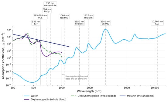
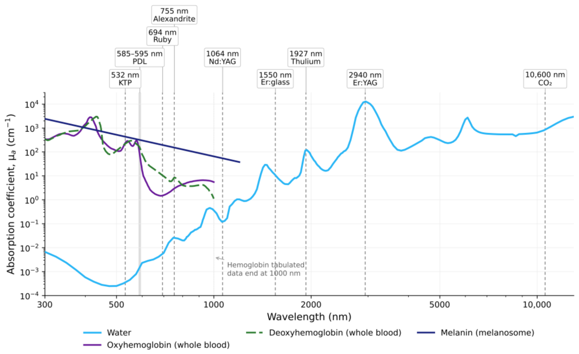
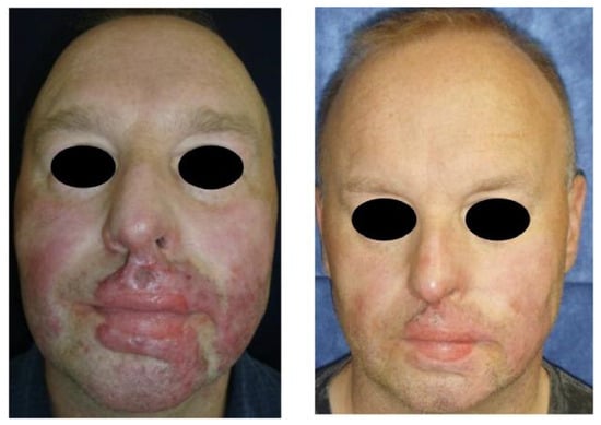
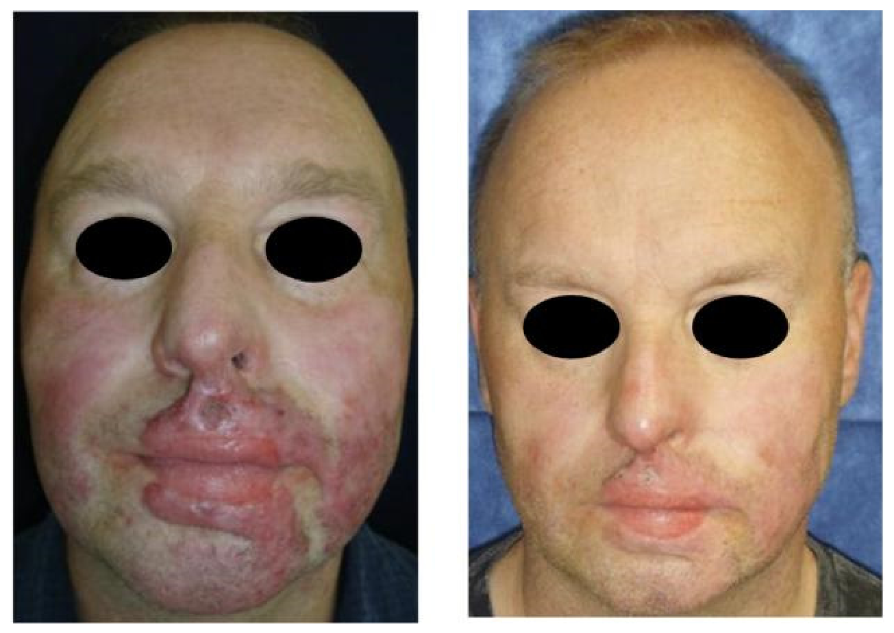
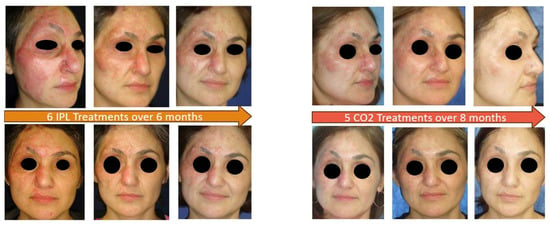
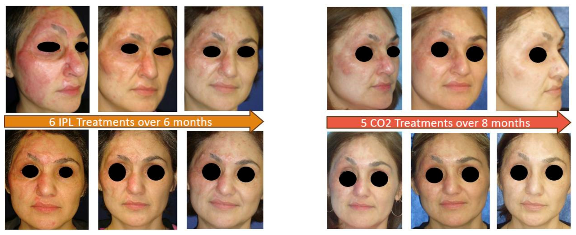

Disclaimer/Publisher’s Note: The statements, opinions and data contained in all publications are solely those of the individual author(s) and contributor(s) and not of MDPI and/or the editor(s). MDPI and/or the editor(s) disclaim responsibility for any injury to people or property resulting from any ideas, methods, instructions or products referred to in the content. |
© 2026 by the authors. Licensee MDPI, Basel, Switzerland. This article is an open access article distributed under the terms and conditions of the Creative Commons Attribution (CC BY) license.