


Abstract
This two-part review provides a comprehensive overview of autologous serum eye drops (ASEDs), addressing their composition, biological properties, and preparation protocols (Part I), as well as their clinical application in ocular surface diseases (Part II). Part II comprises a systematic review of the current evidence on ASEDs, including therapeutic indications, administration protocols, and treatment outcomes, particularly in patients with dry eye disease (DED) and persistent epithelial defect (PED). Clinical studies frequently report statistically significant improvements in ocular surface staining, tear film break-up time, epithelial healing, conjunctival impression cytology findings, and patient-reported symptoms following ASEDs therapy. Beneficial effects have been described particularly in DED associated with Sjögren syndrome, severe idiopathic DED, and ocular graft-versus-host disease, as well as in filamentary keratitis and postoperative ocular surface complications. ASEDs can support corneal nerve recovery and improve corneal sensitivity in neurotrophic keratopathy and can alleviate neuropathic corneal pain. Systematic searches of PubMed and Web of Science were performed on 15 September 2026 and identified 572 records, of which 502 were screened and 273 were assessed for eligibility. After inclusion of eligible human clinical studies from the personal literature database and removal of overlap, 191 studies, including randomized and non-randomized clinical trials, observational studies, case series, and case reports, were included. Formal risk-of-bias assessment and certainty-of-evidence grading were not performed. Despite considerable variability in preparation protocols, dosing regimens, and study design, available evidence supports the use of ASEDs as an effective supportive treatment and long-term therapeutic option in patients with moderate to severe ocular surface disease.
1. Introduction
Since the introduction of autologous serum eye drops (ASEDs) by Fox et al. in 1984 as a biological tear substitute for the treatment of keratoconjunctivitis sicca, increasing evidence has supported their use in the management of ocular surface disorders [1]. Over time, the clinical application of ASEDs has expanded from dry eye disease (DED) to a broad spectrum of ocular surface conditions associated with tear film instability, epithelial damage or dysfunction, impaired corneal healing, and chronic inflammation.
The indications for ASEDs are primarily related to severe idiopathic DED [2] and DED-associated conditions such as Sjögren syndrome [3], chronic ocular graft-versus-host disease (oGVHD) [4,5], neurotrophic keratopathy [6], filamentary keratitis [7], superior limbic keratoconjunctivitis (SLK) [8], Stevens–Johnson syndrome (SJS), ocular cicatricial pemphigoid [9,10], and DED associated with systemic or treatment-related conditions. ASEDs have also been used to treat DED following refractive surgery [11].
Another major indication for ASEDs is persistent epithelial defect (PED) of various etiologies (e.g., penetrating keratoplasty, refractive and vitreoretinal surgery, and other ocular procedures) [12,13,14]. ASEDs have also been used in the management of recurrent corneal erosion (RCE) [15,16,17].
According to the Tear Film & Ocular Surface Society Dry Eye Workshop II (TFOS DEWS II) staged management algorithm, ASEDs may be considered as third-step therapy when treatments recommended in the first and second steps are inadequate or fail to provide sufficient clinical control of DED [18]. Based on the available literature, ASEDs may be recommended particularly for patients with DED refractory to preservative-free artificial tears and characterized by reduced tear film break-up time (TBUT) (<5 s), Schirmer test (ST) (<5 mm) [19,20], positive corneal and conjunctival fluorescein staining (≥grade 1 according to the Oxford scale) [21], and severe symptoms reflected by a high Ocular Surface Disease Index (OSDI) score (>33) [22].
The objective of this systematic review (Part II) was to summarize the current evidence on the clinical application of ASEDs, including therapeutic indications, administration, monitoring of treatment response, efficacy across ocular surface conditions, and safety, with particular attention to ASEDs concentration, dosing frequency, and treatment duration. Complementing Part I, which addresses ASED characteristics and preparation, Part II also identifies limitations and gaps in the current clinical evidence.
2. Materials and Methods
2.1. Literature Search
The literature search and study selection process were conducted and reported in accordance with the PRISMA 2020 guidelines. To prepare this review, an extensive literature search was performed, and data from clinical studies were analyzed, while selected experimental studies were used as supporting background where relevant. Original studies were evaluated whenever available, while review articles and other secondary sources were used mainly for background information and to identify relevant original publications. When several publications included overlapping patient cohorts, they were considered separately only when they provided additional data relevant to the objectives of Part II. Articles concerning ASEDs and their application to the ocular surface were obtained from the PubMed and Web of Science databases. PubMed was selected as a major biomedical database covering the clinical literature relevant to ASEDs, while Web of Science was used as a complementary multidisciplinary source. No additional bibliographic databases were systematically searched for the present review. In addition, a personal literature database accumulated and continuously updated over approximately 25 years was used as a source of relevant publications. This database contains 650 records related to ASEDs and 123 records related to ocular surface diseases. Publications identified through the personal literature database were screened as an additional source of potentially eligible studies and were assessed according to the same eligibility criteria as records identified through the systematic database searches.
PubMed and Web of Science were systematically searched using predefined search strategies. The primary PubMed search strategy combined “autologous serum” with “eye” or “ocular” in the Title/Abstract fields and was restricted to human studies. Additional PubMed searches used predefined terms related to the main clinical indications addressed in Part II, including “autologous serum”, “autologous serum eye drops”, “eye”, “ocular”, “dry eye disease”, “keratoconjunctivitis sicca”, “ocular surface disease”, “Sjögren syndrome”, “graft-versus-host disease”, “filamentary keratitis”, “superior limbic keratoconjunctivitis”, “Stevens-Johnson syndrome”, “ocular cicatricial pemphigoid”, “Mooren’s ulcer”, “persistent epithelial defect”, “recurrent corneal erosion”, and “neurotrophic keratopathy”.
Additional terms related to postoperative ocular surface disease, LASIK, cataract surgery, corneal pain, adverse events, and complications were also included. The results of the individual searches were combined using the OR operator to identify unique records. Web of Science was searched using corresponding terms to identify additional relevant publications.
A separate targeted search was performed for reviews addressing other blood-derived ocular surface therapies, including allogeneic serum, umbilical cord serum, platelet-rich plasma, and plasma rich in growth factors. These publications were used to support the comparative section on alternative blood-derived products and were not included in the main clinical synthesis of ASEDs. The complete search strategies and search dates are provided in Supplementary Tables S1 and S2. Statistical significance was reported according to the criteria used in the original studies.
2.2. Eligibility Criteria
Publications were considered eligible for inclusion in the review if they addressed ASEDs and provided original data relevant to the objectives of Part II, including their clinical use, therapeutic effects, treatment protocols, concentration and dosing, treatment duration, clinical outcomes, patient-reported outcomes, or safety in ocular surface diseases. Studies evaluating ASEDs in dry eye disease, Sjögren syndrome, ocular graft-versus-host disease, postoperative ocular surface disease, persistent epithelial defects, recurrent corneal erosions, neurotrophic keratopathy, corneal pain, and other ocular surface disorders were considered eligible.
Studies were excluded if they did not address ASEDs or did not provide data relevant to the objectives of Part II; focused exclusively on ASED preparation, composition, storage, or stability without information relevant to their clinical application; investigated only other blood-derived or biological eye-drop preparations without separately reported ASED data; or were performed exclusively in animals. Review articles, guidelines, and other secondary sources were not included in the main evidence synthesis but could be used to provide background information and to identify relevant original publications. Non-English-language publications were included when a full-text translation was available and sufficient for assessment; otherwise, they were excluded. The included studies comprised randomized and non-randomized clinical studies, prospective and retrospective observational studies, case series, and case reports. Formal risk-of-bias assessment and certainty-of-evidence grading were not performed. The evidence was synthesized descriptively because of the clinical and methodological heterogeneity of the included studies.
2.3. Study Selection
The identified records were screened independently by KJ and LB, based on their titles and abstracts, for relevance to the objectives of Part II. Disagreements between the reviewers were resolved by discussion and consensus. Potentially relevant publications were subsequently assessed in full text by KJ and LB according to the predefined eligibility criteria. Overlap between the systematic database searches and the personal literature database was identified by comparison of the bibliographic information of individual publications, and duplicate records were removed before final inclusion.
2.4. Data Extraction and Synthesis
Relevant information was independently extracted from the included publications by KJ and KK. Individual studies were assigned to one or more thematic area according to the data relevant to the objectives of Part II, which they reported. The extracted information was subsequently cross-checked, and any discrepancies were resolved by discussion and consensus. Data of interest included study design, study population, underlying ocular diagnosis, number of treated patients or eyes, ASED concentration, dosing frequency, treatment duration, clinical outcome measures, therapeutic response, patient-reported outcomes, and adverse events. Clinical outcome measures included subjective and objective parameters such as the Ocular Surface Disease Index, tear break-up time, Schirmer test, ocular surface staining, impression cytology, in vivo confocal microscopy, best-corrected visual acuity, corneal sensitivity, pain assessment, epithelial healing, and other diagnosis-specific outcomes. The extracted information was organized according to the main topics addressed in Part II and synthesized descriptively, because of the methodological and clinical heterogeneity among the included publications. Where appropriate, results were summarized according to the underlying diagnosis, ASED concentration and dosing regimen, treatment duration, clinical outcomes, and safety. Publications cited in this review, including primary studies and relevant background sources, are summarized according to the main thematic areas in Supplementary Table S2. Study design, presence of a comparator, sample size, and major study limitations were considered when interpreting the clinical findings. For DED, the frequency of studies reporting statistically significant improvement from baseline was summarized for each clinical outcome, with each study counted once per diagnostic group and outcome. For PED, unweighted mean complete healing rates were calculated from studies reporting separate ASED-specific outcomes, without pooling patient or eye counts.
3. Results of the Literature Search
The database searches identified 572 records, including 414 records from PubMed and 158 from Web of Science. After removal of 70 duplicate and overlapping records, 502 records were screened by title and abstract, of which 148 were excluded. Of the 354 reports sought for retrieval, 81 could not be retrieved, leaving 273 reports for eligibility assessment. Following this assessment, 147 reports were excluded, resulting in 126 studies included from the database searches. In addition, 773 records were identified from the personal literature database; 141 reports could not be retrieved, 632 were assessed for eligibility, and 149 studies met the inclusion criteria. Across both sources, 275 studies were initially included. After removal of 84 overlapping studies, a total of 191 unique studies were included in the systematic review. The study selection process is presented in the PRISMA flow diagram in Figure 1.
Figure 1.
PRISMA 2020 flow diagram of study selection.
4. Application of ASEDs
4.1. Information for Patients
Successful ASED therapy depends not only on appropriate clinical indication and correct instillation technique but also on proper handling and storage of ASEDs by patients. Detailed written instructions covering transport, handling, storage conditions, and administration should therefore be provided. Particular attention should be paid to factors affecting ASEDs quality, especially storage temperature, protection from microbial contamination, and protection from light. ASEDs should ideally be transported home in an insulated container with frozen cooling packs and placed promptly in a refrigerator or freezer as advised [23].
As ASEDs are biologically active preparations, patients should also be informed that the drops are intended strictly for individual use and educated about the potential risk of disease transmission or microbial contamination during handling and administration [16,24,25]. In the event of ocular irritation or other adverse symptoms potentially related to therapy, patients should contact their ophthalmologist promptly [26,27]. For patients with reduced manual dexterity, specially designed eye drop applicators may improve safe handling and adherence [28].
ASEDs are commonly prescribed for long-term use, often for weeks to several months. The vial currently in use should be stored at approximately 4–8 °C, whereas the remaining supply should be maintained at −20 °C until required [2,23,29]. In households without temperature monitoring, placement of a thermometer in the refrigerator and freezer may be advisable to ensure appropriate storage conditions [2,26]. Patients should be advised that exposure of ASEDs to inappropriate temperatures may result in degradation or loss of bioactive components and consequently jeopardize their therapeutic efficacy. Therefore, exposure to room temperature should be limited strictly to the time required for administration [30].
Since the risk of contamination increases with prolonged repeated use of the same vial or applicator [31], strict hand hygiene during handling and administration is essential. Both patients and clinicians should remain vigilant for signs of clinically apparent infection [32]. ASEDs remaining in a storage vial should not be used beyond the recommended expiration period [2]. Patients should also be advised not to reduce the prescribed frequency of administration during the treatment course, as dose reduction following symptomatic improvement has been observed and may lead to misinterpretation of therapeutic outcomes [33].
4.2. Administration of ASEDs
ASEDs are used as part of the management of ocular surface diseases and are usually combined with other topical therapies [34], particularly ocular lubricants such as non-preserved artificial tears, to enhance ocular surface hydration and support epithelial healing [29]. In severe DED or ocular surface disease associated with systemic or autoimmune disorders, ASEDs may be combined with antibiotics, when indicated, or anti-inflammatory/immunosuppressive therapy [2,25,34,35]. These preparations may be administered alternately during the day [25,36]. When combined with artificial tears, ASEDs have been administered approximately 15 min after artificial tear instillation [33]. In contrast, ASEDs have also been used as topical monotherapy [35,36,37,38,39,40]. These approaches should be distinguished when interpreting outcomes, because improvement during add-on treatment cannot be attributed to ASEDs alone.
The frequency of ASEDs administration varies depending on disease severity and underlying diagnosis. Reported dosing regimens range from 4–12 applications per day, with individual studies describing specific regimens, such as four times daily [38,41], ten times daily [4], and up to twelve times daily in severe cases [33]. Systematic analyses indicate that most randomized controlled trials employ dosing frequencies of four to six applications per day [42]. In patients with severe DED, however, higher dosing frequencies of at least 6–10 applications daily are commonly used [29,43]. In contrast, in patients with PEDs or severe ocular surface damage, more intensive regimens are often required. In these cases, hourly application of ASEDs has been reported [5,44], to provide continuous epithelial support and promote corneal healing.
The duration of ASEDs therapy varies across studies and depends on the underlying diagnosis and disease severity, with most treatment periods ranging from four weeks to three months [45,46]. In patients with PEDs, treatment is usually continued until complete epithelial closure is achieved, which may require prolonged or intensive therapy. Long-term use extending from several months to years has also been reported in chronic ocular surface disorders, particularly severe DED and autoimmune-associated disease [23,27,41]. Symptomatic relief has been reported after a 3-month course of 20% ASEDs therapy; however, recurrence of symptoms following discontinuation has also been observed [33]. Similarly, a return of symptoms to baseline levels was reported approximately nine weeks after the end of treatment [36]. These findings have led to the proposal of intermittent treatment regimens, consisting of alternating periods of ASEDs use and treatment-free intervals, such as three months of therapy followed by three months without treatment [33].
5. Monitoring the Effectiveness of ASEDs
5.1. Clinical Examination
Clinical outcome measures commonly used to assess the effectiveness of ASEDs include visual acuity, ST, TBUT, and ocular surface staining with fluorescein, Rose Bengal (Figure 2), or the better-tolerated lissamine green, reflecting tear film instability and epithelial damage, including superficial punctate keratitis, associated with DED and PED [47,48]. It should be noted that combined fluorescein and lissamine green staining may provide additional information, as fluorescein stains living but damaged epithelial cells, whereas lissamine green also stains dead cells [49]. Staining is often graded using standardized scoring systems such as the Oxford grading scheme, as recommended by the TFOS DEWS III report [50]. These clinical parameters are routinely assessed in studies evaluating ASEDs therapy [3,38,51].
Figure 2.
Dry eye disease associated with Sjögren syndrome: (a) Slit-lamp biomicroscopy demonstrates widespread punctate corneal epithelial erosions revealed by fluorescein staining. (b) The ocular surface shows positive corneal and conjunctival Rose Bengal staining. Photographs from the authors’ archive (EH).
Additional insight into ocular surface status may be obtained using in vivo confocal microscopy (IVCM), which allows assessment of corneal nerve morphology and density, as well as epithelial cell density, morphology, reflectivity and inflammatory cell infiltration [52,53,54,55]. Among clinical parameters, corneal pain can be assessed using a patient-reported numerical rating scale, whereas corneal sensitivity can be measured using esthesiometry, particularly in patients with neuropathic corneal pain, toxic corneal epitheliopathy, and other conditions associated with corneal nerve dysfunction [55,56].
5.2. Laboratory Examination
For laboratory analyses, ocular surface cells are commonly collected using conjunctival impression cytology performed under topical anesthesia [57]. Nitrocellulose filter paper strips (e.g., MF Millipore GSWP04700, pore size 0.22 µm; Millipore, Billerica, MA, USA) are typically used to obtain samples intended for histological staining [57,58,59], whereas polyethersulfone membrane filters (Supor®-200, pore size 0.22 µm; PALL) or Millicell membranes (MILLICELL®-CM cell culture inserts, PICM 01250, pore size 0.4 µm; Millipore) are frequently employed for the collection of ocular surface material suitable for subsequent immunofluorescence analysis [60,61].
A combination of periodic acid-Schiff and Gill’s modified Papanicolaou staining [62] is used to assess epithelial and goblet cell density and morphology. The most characteristic ocular surface change associated with DED is squamous metaplasia, which is characterized by a reduced nuclear-to-cytoplasmic ratio of epithelial cells, epithelial keratinization, decreased epithelial cell density, and partial or complete goblet cell depletion [63,64]. Additional pathological features detectable by conjunctival impression cytology include epithelial cell apoptosis [33,65], chromatin alterations such as snake-like chromatin [62], and the presence of inflammatory cells on the ocular surface [33,65]; see Figure 3. Cells harvested by impression cytology can be recovered from the collection membrane and subsequently used for further analyses, such as flow cytometry [60].
Figure 3.
Conjunctival impression cytology samples, stained with a combination of periodic acid–Schiff and Gill’s modified Papanicolaou stains: (a) healthy individual showing epithelial and goblet cells and the presence of mucin (asterisks); (b) patient with dry eye disease showing no goblet cells, with snake-like chromatin (arrowheads) and marked nuclear and cytoplasmic enlargement; (c) patient with dry eye disease showing pronounced squamous metaplasia with cytoplasmic enlargement and keratinization, with the presence of inflammatory cell infiltration (arrow). Scale bar: 10 µm (shown in panel (c); the same scale applies to all panels). Photographs from the authors’ archive (KJ).
5.3. Subjective Assessment of Ocular Surface Symptoms
Subjective evaluation of ocular surface symptoms is an essential component of assessing treatment response [50]. Two main types of subjective scoring systems have been used in clinical studies: global assessment tools evaluating overall ocular surface status or global symptom scores [12,36,66], and symptom-specific questionnaires assessing individual DED-related symptoms, including dryness, discomfort, foreign body sensation, photophobia, and pain [2,3,43,66]. Among the most commonly used tools is the standardized 12-item OSDI questionnaire, designed to quantify the severity of subjective symptoms associated with DED. It generates a total score ranging from 0 (no symptoms) to 100 (maximum symptom severity), reflecting ocular discomfort and vision-related functional impairment. The questionnaire was originally developed by Walt, Rowe et al. (1997) [67], and its reliability and validity were subsequently confirmed in clinical studies [68], including the assessment of symptom severity before and after ASEDs therapy [37,69].
6. Clinical Outcomes of ASEDs Application
ASEDs are generally well tolerated and frequently perceived as beneficial by patients; however, the overall certainty of evidence supporting their clinical effectiveness remains limited. Several systematic reviews, meta-analyses and evidence reports have evaluated the efficacy of ASEDs [34,48,70,71,72]. An earlier Cochrane review found low- to very-low-certainty evidence, with possible short-term symptom improvement but insufficient evidence for objective or sustained benefits [48]. More recent analyses generally report symptom relief and improvement in at least one objective clinical parameter, most commonly TBUT and ocular surface staining. However, they continue to emphasize important methodological limitations, particularly the heterogeneity of serum preparation protocols, dosing regimens, and study designs, underscoring the need for larger, well-designed randomized controlled trials [34,70,71,72]. A recent meta-analysis of 12 randomized controlled trials in patients with DED found that ASEDs, predominantly at a 20% concentration, significantly improved ST and TBUT and reduced corneal fluorescein staining and OSDI scores (all p < 0.001) compared with artificial tears [42]. Based on the published literature, ASEDs are currently used mainly in specialized ophthalmic centers across Europe, North America, and Asia, with the highest number of publications originating from Japan, the USA, South Korea, Spain, China, and Italy.
7. ASEDs in the Treatment of DED: Clinical Outcomes
For descriptive synthesis, DED studies were grouped according to the proportion of patients with Sjögren syndrome into Sjögren syndrome-related or Sjögren syndrome-dominant DED (≥67% Sjögren syndrome) (Table 1), mixed DED (34–66% Sjögren syndrome) (Table 2), idiopathic and other non-Sjögren syndrome-dominant DED (0–33% Sjögren syndrome) or DED with unreported Sjögren syndrome status (Table 3), and oGVHD-associated DED (Table 4). The one-third/two-thirds thresholds were chosen pragmatically to provide a simple and symmetric classification in which ≥67% indicates clear Sjögren syndrome predominance, 34–66% represents mixed cohorts, and ≤33% indicates the predominance of idiopathic or other non-Sjögren syndrome DED etiologies. Studies with unreported Sjögren syndrome status were included in Table 3 as a separate subgroup and were not assumed to represent non-Sjögren syndrome DED.
7.1. ASEDs in DED Related to Sjögren Syndrome
This section includes studies specifically evaluating patients with Sjögren syndrome [29,35,43,66,73,74,75], as well as studies in which patients with Sjögren syndrome were analyzed separately within larger cohorts [46,76,77,78] or comprised at least 67% of the study population [3,33].
In patients with Sjögren syndrome-associated DED, ASEDs were most commonly used at 20% and less frequently at 50% or 100% (Table 1). Significant improvements in several clinical outcomes were reported in most studies using 20% ASEDs. The most consistent clinical findings were significant reductions in ocular surface staining [3,27,29,41,43,66,73,74,76,77,78] and improved tear film stability, reflected by significantly prolonged TBUT [3,27,35,43,66,73,74,76,77,78]. Significant increases in ST values were reported less frequently [33,35,73,74,77]. Evidence for an effect on visual acuity was limited: significant gains were reported in only a minority of studies following ASEDs treatment [73,77] while nonsignificant favorable trends were also observed [66,78]. Overall, ASEDs treatment in Sjögren syndrome-related DED showed consistent improvements in subjective symptoms, ocular surface staining, and TBUT, whereas significant increases in ST were less frequent (Table 1).
Table 1.
ASEDs in dry eye disease related to Sjögren syndrome (Sjögren syndrome-dominant dry eye disease; ≥67% SS).
In addition to the routinely assessed clinical parameters, IVCM was also performed, demonstrating beneficial changes in corneal nerve parameters [73,75]. In a comparative study, ASEDs showed significantly better outcomes than artificial tears in OSDI and selected IVCM-derived corneal nerve parameters, particularly subbasal nerve branching and nerve beadings [75].
Subjective symptoms, including discomfort, foreign-body sensation, dryness, and photophobia, were significantly alleviated in most studies compared with baseline, including several studies using 20% ASEDs [3,29,33,35,66,74]. Improvement was also reported with 50% ASEDs [43,74,78] and 100% ASEDs [77].
In the study comparing the efficacy of 20% and 50% ASEDs in patients with recalcitrant moderate or severe DED, both concentrations produced significant benefits in patients with moderate DED, although 50% ASEDs resulted in better ST, TBUT, and staining outcomes. In patients with severe DED, however, 20% ASEDs improved symptoms only, whereas 50% ASEDs improved all four outcomes [74]. In another comparative study, 50% ASEDs reduced corneal staining and symptoms, although bandage contact lenses resulted in lower staining and OSDI scores after six weeks [78].
In a randomized controlled study of 65 patients with primary Sjögren syndrome-related DED, 20% ASEDs were compared with 0.05% cyclosporine A (CsA) combined with preservative-free artificial tears over three months. ST, TBUT, and OSDI changed significantly from baseline in both groups; however, greater increases in ST and TBUT were observed with CsA plus artificial tears, whereas the reduction in OSDI was greater with ASEDs [35]. These findings are supported by a recent network meta-analysis, in which ASEDs showed favorable effects on ocular surface staining and tear film stability, whereas CsA showed a favorable effect on tear secretion assessed by ST [79]. An earlier study in patients with primary Sjögren syndrome also reported that anti-inflammatory therapy including topical ASEDs, CsA, and corticosteroids ameliorated subjective symptoms in 70% of patients and significantly reduced fluorescein staining [80].
ASEDs treatment also reduced conjunctival squamous metaplasia, with increased epithelial and goblet cell densities, normalization of the nuclear-to-cytoplasmic ratio, and reduced epithelial keratinization [29,33]. ASEDs therapy has also been associated with significant reductions in tear film debris and superficial punctate keratitis [33,81]. Conjunctival impression cytology has demonstrated a significant reduction in cells exhibiting snake-like chromatin [33], the prevalence of which correlates with the severity of DED [82], as well as a reduction of epithelial cell apoptosis [33].
Differences in the pathogenesis of primary and secondary Sjögren syndrome raise the question of whether ASEDs exert comparable therapeutic effects in both conditions, particularly given the stronger inflammatory background typically observed in secondary Sjögren syndrome. In line with this, differential responses to 50% ASEDs (diluted in sodium hyaluronate) were found after four weeks of treatment. Significant improvements in ocular surface staining, TBUT, and subjective symptoms were observed in patients with primary Sjögren syndrome, whereas none of these outcomes improved in secondary Sjögren syndrome. Patients with secondary Sjögren syndrome had significantly higher serum levels of pro-inflammatory cytokines, including TNF-α, IL-1β, IL-6, and IL-8, which may partly explain the attenuated ocular surface response in this group [43]. A statistically significant decrease in HLA-DR-positive epithelial cells was observed after 3 months of treatment with 20% ASEDs in a group of patients with DED, more than 70% of whom had primary or secondary Sjögren syndrome [33].
7.2. ASEDs in Mixed Dry Eye Disease
This section includes studies with mixed DED populations, in which Sjögren syndrome accounted for 34–66% of patients (Table 2). The remaining patients had heterogeneous non-Sjögren DED etiologies, including idiopathic DED or keratoconjunctivitis sicca, oGVHD, meibomian gland dysfunction, Stevens–Johnson syndrome, rheumatoid arthritis, systemic sclerosis, and other ocular surface or systemic autoimmune disorders [36,53,69,83,84].
Table 2.
ASEDs in mixed dry eye disease (34–66% Sjögren syndrome).
Subjective symptoms decreased significantly in several studies [36,45,53,69,84,85,86]. In a study involving patients with systemic autoimmune diseases, ASEDs treatment for a mean of 14 months produced complete or partial objective improvement in 85% of eyes and symptom relief in 85% of patients [83]. Ocular surface staining also improved in most studies [45,53,69,83,84]. A significant increase in TBUT was observed with 20% ASEDs [45]. ST generally showed no significant improvement with ASEDs [45,69,85]. Across mixed DED populations, symptomatic improvement was the most consistent finding, whereas significant changes in TBUT and ST were much less frequent (Table 2).
Changes at the cellular level were also demonstrated. IVCM showed a decrease in basal corneal epithelial cell density together with reduced fluorescein staining and OSDI after three months, although subbasal nerve parameters did not change significantly [53]. Conjunctival impression cytology showed reduced squamous metaplasia and increased goblet cell density after ASEDs treatment [36,45,69].
Comparative studies of ASEDs and other blood-derived products showed variable results. Twenty-percent umbilical cord serum produced greater improvement in symptoms and keratoepitheliopathy than 20% ASEDs and also resulted in greater goblet cell density in the Sjögren subgroup [45]. No significant differences in the main clinical outcomes were found between 100% ASEDs and 100% platelet rich plasma (PRP) in another randomized trial [86]. In addition, 100% ASEDs produced larger improvements in OSDI and ocular surface staining than 20% ASEDs, but direct superiority was not established [84].
7.3. ASEDs in Idiopathic and Other Non-Sjögren Syndrome Dry Eye Disease
This section includes studies specifically evaluating non-Sjögren DED [37,38,46,87], studies with non-Sjögren-dominant populations [41,88,89,90,91], and studies in which Sjögren syndrome status was not reported [92,93,94,95]; see Table 3. ASEDs were most commonly used at 20%, while concentrations of 30% [88], 50% [41,46,90,92] and 100% were used less frequently [95].
Table 3.
ASEDs in idiopathic and other non-Sjögren syndrome–dominant dry eye disease, or DED with unreported Sjögren syndrome status (0–33% SS or SS status not reported).
Ocular surface staining improved significantly with 20% and 50% ASEDs [41,87,89,92], while TBUT was significantly longer after treatment in several studies using 20% ASEDs [38,87,93]. Significant increases in ST were less frequently reported [87,90,93]. The occasional increases in ST values may reflect a reduction in ocular surface inflammation rather than a direct stimulatory effect on tear secretion [41], consistent with the finding that 20% ASEDs treatment was associated with decreased tear inflammatory markers in perimenopausal women with severe DED, with the greatest overall improvement observed when ASEDs were combined with estrogen replacement therapy [94].
Subjective symptoms decreased significantly in several studies using 20–30% ASEDs [37,38,87,88,89]. In the non-Sjögren subgroup comparing different formulations, 100% ASEDs reduced symptoms and corneal staining more than 50% ASEDs diluted with ceftazidime, whereas 50% ASEDs diluted with saline and 100% ASEDs showed similar outcomes [46]. In non-Sjögren DED, ASEDs frequently improved subjective symptoms, while objective responses were more variable. Significant increases in TBUT and ST were also reported in several studies with unreported Sjögren syndrome status (Table 3).
Conjunctival impression cytology was assessed less frequently. In a randomized study, 20% ASEDs resulted in greater improvement in conjunctival impression cytology, together with OSDI, TBUT, ST, and corneal fluorescein staining, than preservative-free artificial tears over 12 weeks [87]. Favorable changes in conjunctival impression cytology, together with significant increases in TBUT and ST, were also reported after one month of 20% ASED treatment [93].
Comparative studies with other blood-derived products showed variable results. In a paired-eye study of severe DED, both 50% ASEDs and lacrimal gland PRP injections improved symptoms, ST, non-invasive TBUT, and corneal staining, but symptom reduction was greater with ASEDs [92]. More recently, 100% ASEDs significantly improved OSDI, TBUT, ST, ocular surface staining, and best-corrected visual acuity (BCVA) after six weeks, although PRP produced greater improvements in OSDI, ST, and staining [95].
Other studies examined treatment response and long-term outcomes. In a retrospective study of 50 patients (87 eyes) with refractory DED of various etiologies, 20% ASEDs administered eight times daily significantly reduced OSDI and Oxford staining scores, with 68% of patients classified as responders. Interestingly, non-responders had significantly higher serum EGF concentrations, suggesting that clinical response may also be related to the biological composition of individual ASED preparations [89]. In another cohort, patients with multisystem disease had significantly lower EGF concentrations and a smaller, although not statistically significant, reduction in OSDI than those with localized ocular surface disease, suggesting that underlying disease may influence ASEDs composition and clinical response [96].
A prospective observational study of 100 patients receiving 30% ASEDs demonstrated a significant reduction in DED symptoms, high patient satisfaction, and reduced use of other adjunctive therapies, with no treatment-related adverse effects [88]. Similarly, a retrospective study of 43 patients (73 eyes) documented a mean 44% gain in quality of life, reduced daily use of eye drops in 65% of patients, and better daily functioning in 85% following ASEDs treatment [97]. A later long-term study also showed a sustained clinical response to 20% ASEDs in approximately two-thirds of patients through 36–60 months of follow-up [91].
Interestingly, during long-term treatment with 50% ASEDs, OSDI and fluorescein staining decreased significantly at 3–6 and 6–12 months, but not at 12–24 months, whereas ST increased significantly only at 12–24 months [41]. This time-dependent response is consistent with previous observations of diminishing symptom relief with over time, which led to the suggestion of a three-month treatment period followed by a three-month break [33].
In contrast to the predominantly positive findings reported in other DED studies, a pilot randomized crossover trial showed limited changes after 50% ASEDs treatment. ST increased significantly, whereas changes in OSDI, ocular staining, TBUT, and BCVA did not reach statistical significance. No significant differences were found between autologous and allogeneic serum eye drops [90].
7.4. ASEDs in DED Related to Ocular Graft-Versus-Host Disease
Ocular GVHD most commonly manifests as severe DED, reported in approximately 80% of patients with ocular involvement in one retrospective series, but may also be associated with cataract, blepharitis, ocular hypertension, and filamentary keratitis [98]. Clinical efficacy and good tolerability of ASEDs have been reported in several clinical studies involving patients with DED associated with chronic oGVHD [4,25,98,99]; see Table 4.
Table 4.
ASEDs in ocular graft-versus-host disease-associated dry eye disease.
Frequent administration of 20% [4,54], 50% [41] or 100% ASEDs [5,25,98] has been associated with improvements in ocular surface parameters, including ocular surface staining [4,5,25,99], TBUT [4,41], and subjective symptom scores [4,5,25,54]. In one study, this effect was observed after four weeks of treatment with frequent (up to ten times daily) application [4], whereas treatment outcomes were assessed after six months in another study [25].
Recent retrospective studies further describe the use of ASEDs in combined treatment. Significant reductions in corneal staining and symptom scores from baseline were demonstrated with 50% ASEDs plus topical corticosteroids, with or without human immunoglobulin artificial tears [100]. ASEDs were used in 29% of 140 patients in another tertiary referral cohort [101], while their use reached 47% by year five in a cohort of 189 patients, in which filamentary keratitis was also observed during follow-up [102]. Patients receiving systemic immunosuppressive therapy may have detectable levels of some of these agents in their ASEDs. CsA and mycophenolic acid were detected in undiluted serum from patients with severe oGVHD, but their contribution to the clinical response remains uncertain. Importantly, no significant differences in OSDI scores or fluorescein staining were observed between patients with detectable and non-detectable levels of these agents [103].
Increased apoptosis of conjunctival epithelial cells is a well-recognized feature of DED compared with healthy controls. A 3-month course of 20% ASEDs significantly reduced the proportion of active caspase-3-positive conjunctival epithelial cells in patients with oGVHD, suggesting attenuation of ocular surface apoptosis [104]. Although 3 months of treatment with 20% ASEDs significantly lowered OSDI scores, elevated densities of aberrantly HLA-DR-positive conjunctival epithelial and Langerhans cells were observed, suggesting the persistence of mild subclinical inflammation despite clinical improvement [19]. Increased corneal nerve density and improved corneal sensitivity were observed after 6 weeks of ASEDs therapy in patients with oGVHD-associated DED, suggesting neurotrophic effects that may occur independently of improvements in conventional clinical signs [54]. Certain clinical characteristics specific to patients with oGVHD may influence the composition and potential biological activity of ASEDs. Superior limbic keratoconjunctivitis-like inflammation has also been described in oGVHD, and clinical improvement following ASEDs therapy has been reported in such cases [105].
7.5. Comparison of Clinical Outcomes Across DED Populations
Across DED populations, the frequency of statistically significant improvement after ASEDs treatment varied according to the clinical parameter (Table 5). In Sjögren-syndrome-dominant DED, improvements in ocular surface staining and TBUT were most consistent. In mixed DED, all seven studies reported a significant reduction in subjective symptoms, whereas none of the five studies assessing ST showed a significant increase.
Table 5.
Frequency of statistically significant clinical improvement after ASEDs treatment across dry eye disease populations.
7.6. ASEDs in the Treatment of Filamentary Keratitis
Filamentary keratitis is characterized by recurrent formation of filaments composed of mucus and epithelial cells that adhere to the corneal surface, resulting in pain, foreign body sensation, photophobia, and blurred vision. ASEDs have been used mainly in patients with refractory filamentary keratitis associated with severe DED, Sjögren syndrome, oGVHD, or increased mechanical friction between the eyelids and cornea [2,7,29,98,106,107].
Evidence supporting ASEDs in filamentary keratitis remains limited and is derived primarily from case reports, small case series, and retrospective observational studies [2,108]. One representative case involved a 61-year-old man with refractory filamentary keratitis following laser-assisted in situ keratomileusis (LASIK). After four months of unsuccessful conventional treatment, complete resolution of the corneal filaments was achieved within one week of initiating 20% ASEDs administered four times daily, with no recurrence during follow-up [7]. ASEDs also reduced the need for repeated mechanical filament removal and frequent application of artificial tears, indicating improved ocular surface stability. Furthermore, in patients with systemic autoimmune diseases, the prevalence of filamentary keratopathy decreased by approximately 57% following ASEDs treatment [83].
7.7. ASEDs in Superior Limbic Keratoconjunctivitis
Superior limbic keratoconjunctivitis (SLK) is a chronic inflammatory disease affecting the superior limbal conjunctiva and adjacent corneal surface. The frequent association of SLK with tear film instability and DED provides a rationale for the use of ASEDs or other biological tear substitutes [109,110].
After four weeks of treatment with 20% ASEDs administered 10 times daily in combination with preservative-free artificial tears, a cohort of 11 patients with SLK (22 eyes) showed significant improvement in clinical SLK scores, ocular surface staining, TBUT, and impression cytology findings. Subjective symptoms improved in 82% of treated patients [8]. Improvement in SLK symptoms and clinical signs, including reduced bulbar conjunctival inflammation and absence of fluorescein staining, was observed after 3 months of treatment with 50% ASEDs in combination with therapeutic bandage contact lenses in a complicated SLK case [111].
7.8. ASEDs in Stevens–Johnson Syndrome, Ocular Cicatricial Pemphigoid, and Limbal Stem Cell Deficiency
Stevens–Johnson syndrome (SJS) and ocular cicatricial pemphigoid (OCP) are severe immune-mediated disorders characterized by chronic inflammation and progressive cicatrization of the ocular surface, which may progress to bilateral blindness despite immunosuppressive therapy. Both conditions also frequently lead to profound tear film instability and severe secondary DED, resulting in PEDs and visual impairment [9,10].
It has been demonstrated that the epitheliotrophic capacity of ASEDs derived from patients with chronic SJS is comparable to that observed in controls with non-autoimmune DED, supporting its clinical applicability in this population [112]. Similarly, adjunctive ASEDs were associated with corneal and conjunctival epithelial healing within 3–6 weeks in patients with toxic epidermal necrolysis and severe acute ocular involvement receiving systemic plasmapheresis and intravenous immunoglobulins [113].
In patients with OCP, who often require multiple therapeutic interventions, including surgery and immunosuppressive therapy, ASEDs are used as supportive treatment to stabilize the ocular surface and promote epithelial regeneration [10]. In a cohort of 11 patients with OCP or SJS, combined allograft limbal transplantation, amniotic membrane transplantation, and tarsorrhaphy, followed by 20% ASEDs administered every 15 min, resulted in successful ocular surface reconstruction in 86% of eyes during a mean follow-up of 143 days [114].
ASEDs have also shown beneficial effects in limbal stem cell deficiency (LSCD) of different etiologies. In aniridia-associated keratopathy, ASEDs significantly promoted corneal epithelialization, reduced squamous metaplasia, and increased tear film stability, particularly in patients with mild to moderate disease [115]. In a prospective study of 21 patients (42 eyes) with limbal stem cell deficiency, eight weeks of 20% ASEDs treatment significantly increased tear stability, reduced squamous metaplasia, and alleviated and reduced corneal MUC5AC expression, indicating reduced conjunctivalization of the corneal surface [116]. Favorable outcomes have also been reported in contact lens-induced LSCD [117,118], whereas in glaucoma surgery-induced LSCD ASEDs mainly stabilized disease progression [119].
7.9. ASEDs in Mooren’s Ulcer
Mooren’s ulcer is a rare autoimmune form of peripheral ulcerative keratitis characterized by painful, progressive corneal ulceration and stromal thinning with a risk of corneal perforation, often requiring immunosuppressive therapy. The use of ASEDs in Mooren’s ulcer is limited to isolated case reports. In a patient with Mooren’s ulcer showing insufficient response to topical antibiotics, corticosteroids, and cyclosporine, the addition of 20% ASEDs was associated with corneal re-epithelialization, increased corneal thickness, and regression of the corneal defect. Recurrence of ulceration after treatment discontinuation was followed by further improvement after reintroduction of ASEDs [120]. Similar findings were reported in another case unresponsive to previous medical treatment, managed with amniotic membrane transplantation combined with 20% ASEDs administered four times daily, resulting in stabilization of ulcer progression and reduced corneal vascularization. The ulcer subsequently healed with symptomatic improvement during nine months of follow-up despite a small corneal perforation requiring cyanoacrylate glue application followed by bandage contact lens placement [121].
7.10. ASEDs After Ocular Surgery
Postoperative use of ASEDs encompasses two closely related aspects of ocular surface recovery: treatment of postoperative DED and promotion of corneal epithelial healing. Impaired epithelial healing associated with severe DED may progress to PED; however, early postoperative re-epithelialization is addressed here, whereas established PED is discussed separately in Section 8.1.
In patients with DED after cataract surgery, ASEDs combined with sodium hyaluronate were more effective than sodium hyaluronate alone in improving OSDI, TBUT, ocular surface staining, and impression cytology findings [122]. Similarly, in a randomized study of 150 patients with mild-to-moderate DED after cataract surgery, ASEDs combined with artificial tears significantly improved corneal sensation, TBUT, and fluorescein staining and reduced tear inflammatory markers compared with artificial tears alone [123]. ASEDs have been shown to alleviate persistent DED symptoms after LASIK, particularly in cases refractory to standard lubrication, with significant increases in TBUT and reductions in Rose Bengal staining [11]. In patients with Sjögren syndrome undergoing bilateral LASIK, perioperative treatment with ASEDs was associated with favorable postoperative ocular surface outcomes, without exacerbation of DED or significant epithelial complications [124].
Evidence for a beneficial effect on corneal epithelial healing is particularly relevant after penetrating keratoplasty (PKP). Topical 20% ASEDs significantly shortened the time to complete corneal re-epithelialization following PKP, with the greatest benefit observed in diabetic recipients and in larger grafts, both recognized risk factors for delayed epithelial healing. In diabetic recipients, complete re-epithelialization within seven days was achieved in 59% of eyes receiving 20% ASEDs compared with 33% of eyes without serum, while the corresponding rates at 10 days were 82% and 45%, respectively [125]. ASEDs have also been used in high-risk PKP settings, such as aniridia-associated keratopathy and chronic oGVHD, where they may facilitate corneal epithelial regeneration and limit postoperative epithelial complications [126,127].
Similar benefits have been reported following other types of ocular surgery. ASEDs significantly shortened epithelial healing time compared with artificial tears after laser-assisted subepithelial keratectomy [128] and photorefractive keratectomy [129]. After epithelium-off corneal collagen cross-linking, 20% ASEDs accelerated epithelial healing and reduced postoperative pain compared with artificial tears [130]. Faster resolution of corneal epithelial defects has been reported in eyes treated with ASEDs compared with hyaluronic acid drops following pars plana vitrectomy, particularly in diabetic patients [131]. In a randomized clinical study following pterygium excision, 50% ASEDs significantly accelerated corneal epithelialization and alleviated postoperative pain compared with artificial tears [132]. In addition, 20% ASEDs have been used to alleviate neuropathic corneal pain, most commonly occurring after refractive surgery, with significant pain reduction reported after a mean follow-up of 3.8 months [55].
7.11. ASEDs in DED Associated with Other Systemic and Treatment-Related Conditions
DED has been reported in approximately 81% of patients with Hansen’s disease (leprosy) [133]. A randomized study showed that 20% ASEDs administered six times daily for 6 weeks significantly alleviated symptoms and favorably affected TBUT, fluorescein staining, and conjunctival impression cytology findings, whereas ST did not change significantly [134].
Drug-induced DED has also been described in patients receiving systemic isotretinoin therapy. In a prospective, crossover, double-blind study, 40% ASEDs prolonged TBUT and lowered OSDI scores significantly more than preservative-free artificial tears, while ST values increased similarly in both treatment groups [135]. ASEDs (30%) also improved bilateral corneal lesions associated with the anticancer treatment capecitabine [136].
In patients with ocular surface disease caused by long-term use of preserved topical antiglaucoma therapy, particularly containing benzalkonium chloride, 20% ASEDs administered eight times daily for one month significantly decreased OSDI scores and corneal/conjunctival staining and increased TBUT and corneal sensitivity, while ST results did not change significantly [56]. Similarly, treatment with 20% ASEDs for two months significantly improved TBUT, OSDI scores, and ocular surface staining, including in patients who continued their glaucoma medication throughout the study [137].
Taken together, the available evidence suggests that ASEDs may improve both subjective symptoms and objective ocular surface parameters in secondary forms of DED associated with systemic diseases or treatment-related ocular surface damage, although the current evidence remains limited by relatively small and heterogeneous study populations.
8. ASEDs in the Treatment of Epithelial Defects: Clinical Outcomes
8.1. ASEDs in Persistent Epithelial Defects
PEDs occur when the corneal epithelium fails to heal despite standard supportive therapy and may lead to serious complications [12,138]. They may result from severe DED, neurotrophic keratopathy, autoimmune disorders such as SJS and OCP, limbal stem cell deficiency, diabetes mellitus, as well as ocular surgery, trauma, infection, or exposure-related ocular surface damage [138,139]. Untreated PED may progress to corneal ulceration, stromal scarring, melting, or perforation, with potentially severe visual consequences [13,140].
Several clinical studies have evaluated the efficacy of ASEDs in patients with PEDs refractory to conventional therapy [12,13,138,141,142,143,144]. The duration of PED before ASEDs initiation varied considerably, from approximately two weeks in some cohorts to several months in others [12,139,144,145]. ASEDs were administered at varying concentrations and frequencies, most commonly 20% [12,138,139,141,145,146], but also at 50% or 100% [13,142,147] with administration frequencies ranging from several times daily to hourly, according to disease severity and clinical response. In a series of 70 eyes treated with 100% ASEDs, complete epithelial healing was reported in 57 (81%), including eyes with PED following PKP [143]. Complete healing was also reported in 60% of eyes receiving 50% or 100% ASEDs [13], 80% of patients receiving 100% ASEDs [142], and 87% of eyes receiving 100% serum (104 ASED-treated and five allogeneic-serum-treated eyes), compared with 70% in historical controls receiving conventional treatment [147].
Across 14 studies with ASED-specific healing outcomes (Table 6), the unweighted mean complete PED healing rate was approximately 74%, with corresponding rates of 72% for 20% ASEDs and 81% for 100% ASEDs (two studies). Differences in healing outcomes may reflect PED etiology, severity, pretreatment duration, and concomitant therapy. Although some studies suggested faster healing with a shorter pretreatment PED duration [12,138,139,142,147], Mirza, Ghani et al. (2008) found no significant association [144]. Impression cytology also demonstrated regression of squamous metaplasia in six of seven eyes with severe DED and OCP treated with ASEDs [146].
Table 6.
Persistent epithelial defects: clinical studies evaluating ASED treatment.
Continued ASEDs treatment after complete re-epithelialization may reduce PED recurrence [148]. Poon et al. (2001) reported recurrence in 6/9 successfully treated eyes after discontinuation of ASEDs [13]. In contrast, Lee et al. (2016) achieved complete healing in all 21 eyes treated with 20% ASEDs and silicone-hydrogel bandage contact lenses. During the subsequent three-month follow-up, recurrence occurred in 5/10 eyes (50%) after ASEDs discontinuation, compared with none of the 11 eyes in which treatment was continued for another two weeks after healing [148]. ASEDs were frequently combined with other treatments, including lubricants and therapeutic contact lenses [138,139,145], although some studies have reported favorable outcomes with serum therapy alone, particularly at higher concentrations [142]. Favorable healing was also reported with ASEDs combined with bandage contact lenses [138,149]; these outcomes cannot be attributed to ASEDs alone.
In a retrospective comparative study, complete epithelial healing was achieved in 48% (11/23 patients) treated with 20% ASEDs compared with 84% (51/61 patients) treated with topical insulin (1 IU/mL), with a significantly shorter mean healing time in the insulin group [150]. A protocol for a randomized controlled trial comparing these treatments was subsequently published [151].
8.2. ASEDs in Recurrent Corneal Erosion
Recurrent corneal erosion (RCE) is characterized by repeated episodes of corneal epithelial breakdown caused by impaired adhesion between the epithelium and the underlying basement membrane. Patients typically present with recurrent ocular pain, tearing, photophobia, and foreign body sensation, most often upon awakening [152,153]. RCE most commonly develops after previous corneal trauma or in association with epithelial basement membrane dystrophy, but may also occur after refractive surgery or with ocular surface disorders such as DED, meibomian gland dysfunction, and diabetes mellitus [152,153].
ASEDs have been assessed in patients with RCE unresponsive to conventional therapy. In 11 eyes with recurrent macroform corneal erosions, 20% ASEDs administered for three months reduced the recurrence rate from 2.2 to 0.03 recurrences per month during a mean follow-up of 9.4 months, with no reported side effects. Three patients experienced one recurrence each [15]. Long-term benefit was also observed in 33 eyes treated with 20% ASEDs for six months: no recurrences occurred during treatment, and 28/33 eyes (85%) remained relapse-free during a mean follow-up of 30 months; the other five eyes each experienced one recurrence after treatment cessation [17]. ASEDs were also evaluated following transepithelial phototherapeutic keratectomy. Among 25 eyes treated with keratectomy followed by ASEDs six times daily for six weeks, 20 (80%) remained recurrence-free during a median follow-up of 16 months. Of the five eyes with recurrence, four were subsequently treated with ASEDs and one underwent repeat keratectomy [16]. These studies suggest that ASEDs may help reduce recurrences in refractory RCE, but the independent contribution of ASEDs cannot be determined when combined with keratectomy [110,153].
9. ASEDs in the Treatment of Neurotrophic Keratopathy
ASEDs have been used in the treatment of neurotrophic keratopathy (NK) of various etiologies, including diabetes mellitus, herpes simplex or herpes zoster infection, and postoperative conditions following keratoplasty, LASIK, or photorefractive keratectomy [6,140,154,155]. A systematic review and meta-analysis identified ASEDs, nerve growth factor, and amniotic membrane transplantation among the effective treatments for neurotrophic keratopathy, with complete epithelial healing reported in 92% of ASEDs-treated eyes after a mean healing time of 28 days [156]. ASEDs were administered at concentrations of 20% [6,154,155,157] or 50% [140], with reported application frequencies ranging from 5 to 10 times daily to every two hours while awake. Individual clinical studies also reported favorable epithelial healing outcomes with ASEDs in NK of various etiologies [6,140,154]. A retrospective comparative study reported successful epithelial healing with both ASEDs and amniotic membrane transplantation [155].
In addition to promoting epithelial healing, ASEDs have been associated with enhanced corneal sensitivity [6], better visual acuity [6,154,155], and significantly higher ST values and longer TBUT [6,154]. More recently, a prospective study of patients with stage 1 NK without PED associated with paralytic lagophthalmos showed a decrease in OSDI and significant increases in corneal nerve fiber length and density assessed by IVCM after ASEDs treatment [157].
10. Autologous Serum in Vitreoretinal Surgery
Autologous serum has also been investigated as an intraoperative adjunct in vitreoretinal surgery, particularly in the treatment of full-thickness macular holes. During pars plana vitrectomy, a small volume of undiluted autologous serum is applied directly over the macular hole, with the aim of promoting tissue adhesion and hole closure. Early clinical series reported anatomical closure rates of approximately 67–100% together with improvements in visual acuity [158,159,160]. Similarly, a more recent study combining autologous serum with an autologous anterior lens capsule flap in 18 eyes with idiopathic or refractory full-thickness macular holes achieved anatomical closure in 94% of eyes [161]. However, subsequent comparative studies provided less consistent results. A multicenter study found no significant overall benefit after adjustment for baseline differences, although a possible advantage was observed in larger macular holes [162], while a randomized clinical trial found that the addition of autologous serum did not improve anatomical or visual outcomes compared with vitrectomy alone [163]. More recent evidence is similar; a meta-analysis of four comparative studies including 373 eyes found no significant improvement in macular hole closure or complication rates with autologous serum. Thus, its role in vitreoretinal surgery remains investigational rather than established [164].
11. Adverse Events After ASEDs Application
ASEDs are generally considered a safe and well-tolerated therapeutic option due to their autologous and largely non-immunogenic nature. In most clinical studies, either no significant adverse events [25,38,41,56,78,83,98,139] or only mild and transient complications have been observed [4,23,90].
The adverse events and safety concerns associated with ASEDs can be broadly grouped into three areas: (1) contamination-related complications, (2) ocular surface intolerance, and (3) systemic or procedural limitations. The principal potential risk is microbial contamination, which may occur during preparation or, more frequently, during repeated handling and application by the patient. Reported complications include conjunctivitis and, rarely, infectious keratitis, including fungal keratitis [4,13,36,69,99]. Although such complications are uncommon, patients with a severely compromised ocular surface, particularly those with PEDs, may be at increased risk [46]. Nevertheless, a five-year retrospective study of 752 patients found only three cases of infectious keratitis potentially associated with ASEDs therapy, with none considered likely treatment-related [165].
The second group of adverse events, involving ocular surface intolerance or immunological reactions, has been reported particularly with higher ASEDs concentrations (≥50%) [166]. However, some patients reported occasional eye dryness and photophobia during treatment with 20% ASEDs, although no complications were reported [23]. A rare case of bilateral limbitis was reported following treatment with 20% ASEDs in a patient with atopic keratoconjunctivitis; the inflammation resolved after ASEDs discontinuation in the affected eye and recurred in the fellow eye after treatment was resumed, resolving again after discontinuation. Although the exact cause remains unclear, immunologically active serum components in atopic patients may trigger ocular surface inflammation [167].
Case reports described immunoglobulin deposition in the cornea in a patient with PED and a prior history of multiple corneal grafts, who developed acute rejection after approximately one week of treatment with undiluted ASEDs [44]. Peripheral corneal infiltration and ulceration have also been reported shortly after initiation of ASEDs therapy in a patient with rheumatoid arthritis [13]. In addition, subepithelial corneal infiltration and a sticky ocular sensation were observed in a small proportion of patients treated with undiluted serum [14]. The underlying mechanism is not fully understood but may involve immune complex deposition or the presence of pro-inflammatory serum components [13]. A case report described bilateral corneal verticillata shortly after initiation of 20% ASEDs in a patient receiving long-term amiodarone, suggesting that medications present in serum may be transferred to the ocular surface through ASEDs and contribute to corneal deposition [168]. This possibility is supported by the detection of CsA and mycophenolic acid in serum used to prepare ASEDs in patients with oGVHD, although their presence was not associated with differences in treatment efficacy or ocular surface parameters [103]. The rapid development of verticillata may have reflected increased ocular exposure to amiodarone through both the tear film and ASEDs, promoting its deposition in regenerating epithelial cells [168].
In patients with oGVHD, a mild eczema-like reaction on the eyelids has also been reported in an isolated case following 20% ASEDs therapy [4].
Finally, some limitations are associated with the preparation and use of ASEDs, particularly when higher concentrations are required. These include repeated blood collection, which may be problematic in patients with anemia, poor venous access, or limited ability to tolerate repeated phlebotomy [14], and increased viscosity of undiluted serum, which may cause transient ocular discomfort [166].
12. Other Blood-Derived Biological Tears in the Treatment of Ocular Diseases
Several blood-derived products other than ASEDs have been investigated as biological tear substitutes, including allogeneic serum, umbilical cord serum (UCS), platelet-rich plasma (PRP), and plasma rich in growth factors (PRGF) [50,169,170].
Allogeneic serum and UCS have also been investigated as alternatives to ASEDs particularly in severe DED. Both 20% ASEDs and 20% UCS significantly improved ocular surface parameters, but UCS was more effective in reducing symptoms and keratoepitheliopathy [45]. Similarly, in patients with severe DED, corneal nerve parameters improved significantly more with UCS than with allogeneic serum [171]. More recently, in five women with Sjögren syndrome, allogeneic serum eye drops were associated with lower OSDI scores than previous ASEDs treatment [172]. Allogeneic blood-derived products have also been explored for epithelial healing in PED; in a case report, maternal finger-prick blood promoted complete closure of a refractory PED without an observed immunological reaction [173]. However, their heterologous origin involves additional concerns regarding donor screening, immunologic reactions, and infectious disease transmission.
PRP is an autologous alternative that has shown beneficial effects in DED. In a randomized paired-eye trial, 20% PRP was not inferior to 20% ASEDs in treating DED, with no significant differences in symptoms, ocular surface staining, or TBUT. However, improvement in ST was significantly greater with PRP [85]. Similarly, 100% PRP was non-inferior to 100% ASEDs in reducing symptoms and ocular surface staining, with no significant differences in remaining clinical parameters [86]. In contrast, a more recent randomized study showed significantly better results for ocular surface staining, ST, and OSDI with 100% PRP than with 100% ASEDs [95]. A subsequent study confirmed differences in the epitheliotrophic factor profiles of PRP and serum, although these biochemical differences do not establish the superior clinical efficacy of either product [174].
PRGF is another autologous platelet-derived preparation that has been associated with improvements in symptoms and ocular surface parameters in clinical studies [175,176]. In vitro studies suggest that PRGF may exert stronger anti-inflammatory and proliferative effects and have a more favorable proteomic profile than ASEDs [177,178].
Whole-blood autologous tears are another emerging DED treatment using a finger-prick protocol. Their addition to conventional therapy significantly reduced OSDI scores in a multicenter randomized trial, with no adverse effects, while a prospective case series reported improvements in corneal staining, TBUT, visual acuity, and ocular comfort [50].
13. Therapeutic Applications of Autologous Serum in Other Medical Disciplines
Beyond ophthalmology, autologous serum has also been investigated for epithelial wound healing in other medical fields.
In otorhinolaryngology, autologous serum eardrops have been used with non-surgical treatments for chronic tympanic membrane perforations, promoting epithelial healing and significantly increasing perforation closure rates [179,180].
Autologous serum may have regenerative effects beyond the ocular surface, reducing postoperative pain and accelerating mucosal healing after tonsillectomy, although systemic conditions such as chronic renal failure may lower serum growth factor levels and potentially reduce its regenerative capacity [181,182].
14. Discussion
ASEDs may be considered for temporary treatment after surgery or ocular injury; prolonged or intermittent treatment of chronic DED, including Sjögren-syndrome-related DED and oGVHD; and intensive treatment of PED aimed at epithelial closure [2,48]. Clinical response depends on disease severity, patient selection and adherence, and appropriate preparation, storage, and handling.
Overall, Table 5 suggests that statistically significant improvements from baseline were more consistently observed in ocular surface staining than in ST, with symptom scores and TBUT showing intermediate patterns across DED populations. Similarly, a meta-analysis of changes from baseline found significant improvements in symptoms, TBUT, and keratoepitheliopathy, but not in ST [183].
In a study of oGVHD-associated DED, 20% ASEDs were associated with significantly lower OSDI scores and greater corneal nerve density and corneal sensitivity at 12 weeks than at baseline, without significant changes in TBUT, ST, or ocular surface staining [54]. These findings suggest that ASEDs may provide neurotrophic benefits not fully reflected by conventional ocular surface tests. Corneal nerve regeneration and attenuation of ocular surface inflammation may also contribute to pain relief [54,184,185,186,187]. Additional endogenous serum mediators with anti-inflammatory, analgesic, and neuroprotective properties may help alleviate pain, although their role in ASEDs remains to be established [188,189].
The underlying disease and individual serum composition may further influence treatment response. In primary Sjögren syndrome, 50% ASEDs significantly improved symptoms, staining, and TBUT, whereas no significant improvement was observed in secondary Sjögren syndrome, which was associated with higher serum concentrations of inflammatory cytokines [43]. Levy et al. (2019) found significantly higher serum EGF concentrations in non-responders than in responders [89]. In a separate cohort, Ripa et al. (2020) reported lower EGF concentrations in patients with systemic disease than in those with localized ocular surface disease, without a significant between-group difference in symptom improvement [96]. These findings indicate that the concentration of individual bioactive components alone may not predict clinical efficacy.
A 10-year clinical experience combined with a review of more than 30 studies found that 20% ASEDs were used in 69% of patients and 30% in 21%, whereas 50% and 100% formulations were less common [190]. The frequency of use does not establish which concentration is most effective. A recent meta-analysis found no significant difference in subjective symptoms between 20%, 50%, and 100% ASEDs, although a statistically significant reduction in keratoepitheliopathy was observed only in the 50% subgroup; substantial heterogeneity remained among studies [183]. Direct clinical comparisons also suggest that the effect of concentration may depend on disease severity and the clinical parameter assessed. In severe DED, a significant increase in ST was observed with 50% but not 20% ASEDs [74]. In another randomized trial, 100% ASEDs produced numerically greater improvements in OSDI and ocular surface staining than 20% ASEDs, but statistical superiority was not established at the prespecified significance threshold [84]. Thus, the available evidence does not establish a consistently superior concentration across clinical indications.
Treatment regimens and outcome assessment should reflect the clinical indication: symptom control and ocular surface recovery in DED, epithelial closure and healing time in PED, and prevention of recurrence in RCE. Published regimens include ASEDs administered 6–10 times daily in chronic DED [29,43] and as frequently as hourly in PED [142,143]. Differences in PED etiology, severity, defect duration, concomitant treatment, and follow-up limit direct comparisons of healing rates across studies. In RCE, reduced recurrence rates have been reported, but the independent contribution of ASEDs is difficult to determine when treatment is combined with keratectomy [16,17].
Because Table 5 includes both ASEDs monotherapy and concomitant treatment, improvements from baseline cannot always be attributed to ASEDs alone. Direct comparisons with other biological tear substitutes have also produced variable results. Umbilical cord serum was more effective than ASEDs for selected clinical parameters in one study [45], whereas another randomized trial found no significant differences in the main clinical outcomes between ASEDs and platelet-rich plasma [86]. In PED, a retrospective comparison showed higher healing rates with topical insulin than with 20% ASEDs [150]. These findings emphasize the importance of evaluating ASEDs against appropriate alternative treatments rather than relying solely on improvements from baseline.
Safety is generally favorable, but autologous origin does not eliminate procedural risks. Microbial contamination during preparation, storage, and handling remains an important concern. Patient education on proper use and storage is essential for maintaining product quality and safety [2,25,191].
Methodological and clinical heterogeneity in study design, populations, treatment protocols, follow-up, and outcome measures precluded formal meta-analysis. The proportions of studies reporting statistically significant improvement in Table 5 are descriptive, unweighted by sample size or study quality, and do not represent pooled treatment effects or direct comparisons between DED populations. Statistical significance does not necessarily indicate clinically meaningful improvement.
The absence of a registered protocol, formal risk-of-bias assessment, and certainty-of-evidence grading limits confidence in conclusions regarding treatment efficacy. Although study design, comparator availability, sample size, and reported limitations were considered, this descriptive approach cannot replace a formal appraisal.
Future prospective trials should therefore evaluate standardized ASEDs protocols against established therapies and other blood-derived biological tear substitutes, including allogeneic serum, umbilical cord serum, platelet-rich plasma, and plasma rich in growth factors.
15. Conclusions
ASEDs represent a distinctive biological treatment for ocular surface disease, combining tear supplementation with epitheliotrophic, anti-inflammatory, and regenerative properties. Their clinical application encompasses a broad range of ocular surface disorders, particularly moderate-to-severe DED, Sjögren syndrome- or oGVHD-related DED, postoperative ocular surface disease, and neurotrophic or other persistent epithelial disorders.
Across DED populations, ASEDs most consistently relieve subjective symptoms and enhance ocular surface integrity, whereas increases in tear secretion are less predictable. Their benefits also include epithelial healing in PED and improvement in corneal nerve parameters, with treatment response potentially influenced by the underlying disease and individual serum composition. No optimal ASEDs concentration or dosing regimen has been established across clinical indications, and treatment should be individualized.
ASEDs are frequently combined with other treatments, and their independent contribution cannot always be determined. Despite encouraging clinical outcomes, the evidence remains limited by substantial clinical and methodological heterogeneity and the relatively small number of high-quality comparative trials. These limitations precluded a formal meta-analysis and restrict direct comparison across studies. Future research should therefore focus on standardized clinical protocols and direct comparisons with established therapies and other blood-derived biological tear substitutes. Such trials should distinguish monotherapy from adjunctive treatment and evaluate clinically meaningful outcomes.
Supplementary Materials
The following supporting information can be downloaded at: https://www.mdpi.com/article/10.3390/bioengineering13111292/s1, Supplementary Table S1: History and Search Details, PubMed, Web of Science; Supplementary Table S2: List of studies included in the systematic review.
Author Contributions
Conceptualization, K.J. and K.K.; writing—original draft preparation, K.J. and K.K.; visualization, K.K., E.H., L.B. and T.P.U.; editing, K.J., T.P.U. and P.S.; funding acquisition, K.J. All authors have read and agreed to the published version of the manuscript.
Funding
This study was supported by research project BBMRI_CZ LM2023033. Institutional support (Charles University, Prague) was provided by the Cooperation: Medical Diagnostics and Basic Medical Sciences.
Institutional Review Board Statement
Not applicable.
Informed Consent Statement
Not applicable.
Data Availability Statement
No new primary data were generated. The data used for the descriptive analyses were obtained from published studies and are summarized in the manuscript and Supplementary Materials.
Conflicts of Interest
The authors declare no conflicts of interest.
Abbreviations
The following abbreviations are used in this manuscript:
| ASEDs | Autologous serum eye drops |
| BCVA | Best-corrected visual acuity |
| CsA | Cyclosporine A |
| DED | Dry eye disease |
| IL | Interleukin |
| IVCM | In vivo confocal microscopy |
| LASIK | Laser-assisted in situ keratomileusis |
| NK | Neurotrophic keratopathy |
| OCP | Ocular cicatricial pemphigoid |
| OSDI | Ocular Surface Disease Index |
| oGVHD | Ocular graft-versus-host disease |
| PEDs | Persistent epithelial defects |
| PKP | Penetrating keratoplasty |
| PRGF | Plasma rich in growth factors |
| PRP | Platelet rich plasma |
| RCE | Recurrent corneal erosion |
| SJS | Stevens–Johnson syndrome |
| SLK | Superior limbic keratoconjunctivitis |
| ST | Schirmer test |
| TBUT | Tear break-up time |
| TNF-α | Tumor necrosis factor alpha |
| UCS | Umbilical cord serum |
References
- Fox, R.I.; Chan, R.; Michelson, J.B.; Belmont, J.B.; Michelson, P.E. Beneficial effect of artificial tears made with autologous serum in patients with keratoconjunctivitis sicca. Arthritis Rheum. 1984, 27, 459–461. [Google Scholar] [CrossRef] [Scilit] [PubMed]
- Geerling, G.; Maclennan, S.; Hartwig, D. Autologous serum eye drops for ocular surface disorders. Br. J. Ophthalmol. 2004, 88, 1467–1474. [Google Scholar] [CrossRef] [Scilit] [PubMed]
- Kojima, T.; Ishida, R.; Dogru, M.; Goto, E.; Matsumoto, Y.; Kaido, M.; Tsubota, K. The effect of autologous serum eyedrops in the treatment of severe dry eye disease: A prospective randomized case-control study. Am. J. Ophthalmol. 2005, 139, 242–246. [Google Scholar] [CrossRef] [Scilit] [PubMed]
- Ogawa, Y.; Okamoto, S.; Mori, T.; Yamada, M.; Mashima, Y.; Watanabe, R.; Kuwana, M.; Tsubota, K.; Ikeda, Y.; Oguchi, Y. Autologous serum eye drops for the treatment of severe dry eye in patients with chronic graft-versus-host disease. Bone Marrow Transplant. 2003, 31, 579–583. [Google Scholar] [CrossRef] [Scilit] [PubMed]
- Rocha, E.M.; Pelegrino, F.S.; de Paiva, C.S.; Vigorito, A.C.; de Souza, C.A. GVHD dry eyes treated with autologous serum tears. Bone Marrow Transplant. 2000, 25, 1101–1103. [Google Scholar] [CrossRef] [Scilit] [PubMed]
- Matsumoto, Y.; Dogru, M.; Goto, E.; Ohashi, Y.; Kojima, T.; Ishida, R.; Tsubota, K. Autologous serum application in the treatment of neurotrophic keratopathy. Ophthalmology 2004, 111, 1115–1120. [Google Scholar] [CrossRef] [Scilit] [PubMed]
- Read, S.P.; Rodriguez, M.; Dubovy, S.; Karp, C.L.; Galor, A. Treatment of Refractory Filamentary Keratitis With Autologous Serum Tears. Eye Contact Lens 2017, 43, e16–e18. [Google Scholar] [CrossRef] [Scilit] [PubMed]
- Goto, E.; Shimmura, S.; Shimazaki, J.; Tsubota, K. Treatment of superior limbic keratoconjunctivitis by application of autologous serum. Cornea 2001, 20, 807–810. [Google Scholar] [CrossRef] [Scilit] [PubMed]
- Vazirani, J.; Sridhar, U.; Gokhale, N.; Doddigarla, V.R.; Sharma, S.; Basu, S. Autologous serum eye drops in dry eye disease: Preferred practice pattern guidelines. Indian J. Ophthalmol. 2023, 71, 1357–1363. [Google Scholar] [CrossRef] [Scilit] [PubMed]
- Abbott, K.S.; Palestine, A.G.; Hauswirth, S.G.; Gregory, D.G.; Patnaik, J.L.; Reddy, A.K. Treatment of Ocular Surface Disease in Ocular Cicatricial Pemphigoid. Ocul. Immunol. Inflamm. 2024, 32, 2479–2485. [Google Scholar] [CrossRef] [Scilit] [PubMed]
- Noda-Tsuruya, T.; Asano-Kato, N.; Toda, I.; Tsubota, K. Autologous serum eye drops for dry eye after LASIK. J. Refract. Surg. 2006, 22, 61–66. [Google Scholar] [CrossRef] [Scilit] [PubMed]
- Tsubota, K.; Goto, E.; Shimmura, S.; Shimazaki, J. Treatment of persistent corneal epithelial defect by autologous serum application. Ophthalmology 1999, 106, 1984–1989. [Google Scholar] [CrossRef] [Scilit] [PubMed]
- Poon, A.C.; Geerling, G.; Dart, J.K.; Fraenkel, G.E.; Daniels, J.T. Autologous serum eyedrops for dry eyes and epithelial defects: Clinical and in vitro toxicity studies. Br. J. Ophthalmol. 2001, 85, 1188–1197. [Google Scholar] [CrossRef] [Scilit] [PubMed]
- Lekhanont, K.; Jongkhajornpong, P.; Choubtum, L.; Chuckpaiwong, V. Topical 100% serum eye drops for treating corneal epithelial defect after ocular surgery. Biomed. Res. Int. 2013, 2013, 521315. [Google Scholar] [CrossRef] [Scilit] [PubMed]
- del Castillo, J.M.; de la Casa, J.M.; Sardina, R.C.; Fernandez, R.M.; Feijoo, J.G.; Gomez, A.C.; Rodero, M.M.; Sanchez, J.G. Treatment of recurrent corneal erosions using autologous serum. Cornea 2002, 21, 781–783. [Google Scholar] [CrossRef] [Scilit] [PubMed]
- Holzer, M.P.; Auffarth, G.U.; Specht, H.; Kruse, F.E. Combination of transepithelial phototherapeutic keratectomy and autologous serum eyedrops for treatment of recurrent corneal erosions. J. Cataract Refract. Surg. 2005, 31, 1603–1606. [Google Scholar] [CrossRef] [Scilit] [PubMed]
- Ziakas, N.G.; Boboridis, K.G.; Terzidou, C.; Naoumidi, T.L.; Mikropoulos, D.; Georgiadou, E.N.; Georgiadis, N.S. Long-term follow up of autologous serum treatment for recurrent corneal erosions. Clin. Exp. Ophthalmol. 2010, 38, 683–687. [Google Scholar] [CrossRef] [Scilit] [PubMed]
- Jones, L.; Downie, L.E.; Korb, D.; Benitez-Del-Castillo, J.M.; Dana, R.; Deng, S.X.; Dong, P.N.; Geerling, G.; Hida, R.Y.; Liu, Y.; et al. TFOS DEWS II Management and Therapy Report. Ocul. Surf. 2017, 15, 575–628. [Google Scholar] [CrossRef] [Scilit] [PubMed]
- Jirsova, K.; Seidler Stangova, P.; Palos, M.; Mahelkova, G.; Kalasova, S.; Rybickova, I.; Utheim, T.P.; Vesela, V. Aberrant HLA-DR expression in the conjunctival epithelium after autologous serum treatment in patients with graft-versus-host disease or Sjogren’s syndrome. PLoS ONE 2020, 15, e0231473. [Google Scholar] [CrossRef] [Scilit] [PubMed]
- Danjo, Y. Diagnostic usefulness and cutoff value of Schirmer’s I test in the Japanese diagnostic criteria of dry eye. Graefes Arch. Clin. Exp. Ophthalmol. 1997, 235, 761–766. [Google Scholar] [CrossRef] [Scilit] [PubMed]
- Craig, J.P.; Nichols, K.K.; Akpek, E.K.; Caffery, B.; Dua, H.S.; Joo, C.K.; Liu, Z.; Nelson, J.D.; Nichols, J.J.; Tsubota, K.; et al. TFOS DEWS II Definition and Classification Report. Ocul. Surf. 2017, 15, 276–283. [Google Scholar] [CrossRef] [Scilit] [PubMed]
- Aljarousha, M.; Alghamdi, W.M.; Attaallah, S.; Alhoot, M.A. Ocular Surface Disease Index questionnaire in different languages. Med. Hypothesis Discov. Innov. Ophthalmol. 2024, 13, 190–200. [Google Scholar] [CrossRef] [Scilit] [PubMed]
- Lee, G.A.; Chen, S.X. Autologous serum in the management of recalcitrant dry eye syndrome. Clin. Exp. Ophthalmol. 2008, 36, 119–122. [Google Scholar] [CrossRef] [Scilit] [PubMed]
- Sauer, R.; Bluthner, K.; Seitz, B. Sterility of non-preserved autologous serum drops for treatment of persistent corneal epithelial defects. Ophthalmologe 2004, 101, 705–709. [Google Scholar] [CrossRef] [Scilit] [PubMed]
- Tahmaz, V.; Gehlsen, U.; Sauerbier, L.; Holtick, U.; Engel, L.; Radojska, S.; Petrescu-Jipa, V.M.; Scheid, C.; Hallek, M.; Gathof, B.; et al. Treatment of severe chronic ocular graft-versus-host disease using 100% autologous serum eye drops from a sealed manufacturing system: A retrospective cohort study. Br. J. Ophthalmol. 2017, 101, 322–326. [Google Scholar] [CrossRef] [Scilit] [PubMed]
- Leite, S.C.; de Castro, R.S.; Alves, M.; Cunha, D.A.; Correa, M.E.; da Silveira, L.A.; Vigorito, A.C.; de Souza, C.A.; Rocha, E.M. Risk factors and characteristics of ocular complications, and efficacy of autologous serum tears after haematopoietic progenitor cell transplantation. Bone Marrow Transplant. 2006, 38, 223–227. [Google Scholar] [CrossRef] [Scilit] [PubMed]
- Liu, Y.; Hirayama, M.; Cui, X.; Connell, S.; Kawakita, T.; Tsubota, K. Effectiveness of Autologous Serum Eye Drops Combined With Punctal Plugs for the Treatment of Sjogren Syndrome-Related Dry Eye. Cornea 2015, 34, 1214–1220. [Google Scholar] [CrossRef] [Scilit] [PubMed]
- Spaniol, K.; Koerschgen, L.; Sander, O.; Koegler, G.; Geerling, G. Comparison of application systems for autologous serum eye drops. Curr. Eye Res. 2014, 39, 571–579. [Google Scholar] [CrossRef] [Scilit] [PubMed]
- Tsubota, K.; Goto, E.; Fujita, H.; Ono, M.; Inoue, H.; Saito, I.; Shimmura, S. Treatment of dry eye by autologous serum application in Sjogren’s syndrome. Br. J. Ophthalmol. 1999, 83, 390–395. [Google Scholar] [CrossRef] [Scilit] [PubMed]
- Bradley, J.C.; Simoni, J.; Bradley, R.H.; McCartney, D.L.; Brown, S.M. Time- and temperature-dependent stability of growth factor peptides in human autologous serum eye drops. Cornea 2009, 28, 200–205. [Google Scholar] [CrossRef] [Scilit] [PubMed]
- Lagnado, R.; King, A.J.; Donald, F.; Dua, H.S. A protocol for low contamination risk of autologous serum drops in the management of ocular surface disorders. Br. J. Ophthalmol. 2004, 88, 464–465. [Google Scholar] [CrossRef] [Scilit] [PubMed]
- Thanathanee, O.; Phanphruk, W.; Anutarapongpan, O.; Romphruk, A.; Suwan-Apichon, O. Contamination risk of 100% autologous serum eye drops in management of ocular surface diseases. Cornea 2013, 32, 1116–1119. [Google Scholar] [CrossRef] [Scilit] [PubMed]
- Jirsova, K.; Brejchova, K.; Krabcova, I.; Filipec, M.; Al Fakih, A.; Palos, M.; Vesela, V. The application of autologous serum eye drops in severe dry eye patients; subjective and objective parameters before and after treatment. Curr. Eye Res. 2014, 39, 21–30. [Google Scholar] [CrossRef] [Scilit] [PubMed]
- Shtein, R.M.; Shen, J.F.; Kuo, A.N.; Hammersmith, K.M.; Li, J.Y.; Weikert, M.P. Autologous Serum-Based Eye Drops for Treatment of Ocular Surface Disease: A Report by the American Academy of Ophthalmology. Ophthalmology 2020, 127, 128–133. [Google Scholar] [CrossRef] [Scilit] [PubMed]
- Berhuni, M.; Istek, S.; Tiskaoglu, N.S. 20% Autologous serum vs. 0.05% cyclosporine and preservative-free artificial tears in the treatment of Sjogren related dry eye. Arq. Bras. Oftalmol. 2022, 87, e2022-0192. [Google Scholar] [CrossRef] [Scilit] [PubMed]
- Noble, B.A.; Loh, R.S.; MacLennan, S.; Pesudovs, K.; Reynolds, A.; Bridges, L.R.; Burr, J.; Stewart, O.; Quereshi, S. Comparison of autologous serum eye drops with conventional therapy in a randomised controlled crossover trial for ocular surface disease. Br. J. Ophthalmol. 2004, 88, 647–652. [Google Scholar] [CrossRef] [Scilit] [PubMed]
- Urzua, C.A.; Vasquez, D.H.; Huidobro, A.; Hernandez, H.; Alfaro, J. Randomized double-blind clinical trial of autologous serum cversus artificial tears in dry eye syndrome. Curr. Eye Res. 2012, 37, 684–688. [Google Scholar] [CrossRef] [Scilit] [PubMed]
- Celebi, A.R.; Ulusoy, C.; Mirza, G.E. The efficacy of autologous serum eye drops for severe dry eye syndrome: A randomized double-blind crossover study. Graefes Arch. Clin. Exp. Ophthalmol. 2014, 252, 619–626. [Google Scholar] [CrossRef] [Scilit] [PubMed]
- Kojima, T.; Higuchi, A.; Goto, E.; Matsumoto, Y.; Dogru, M.; Tsubota, K. Autologous serum eye drops for the treatment of dry eye diseases. Cornea 2008, 27, S25–S30. [Google Scholar] [CrossRef] [Scilit] [PubMed]
- Ma, I.H.; Chen, L.W.; Tu, W.H.; Lu, C.J.; Huang, C.J.; Chen, W.L. Serum components and clinical efficacies of autologous serum eye drops in dry eye patients with active and inactive Sjogren syndrome. Taiwan J. Ophthalmol. 2017, 7, 213–220. [Google Scholar] [CrossRef] [Scilit] [PubMed]
- Hussain, M.; Shtein, R.M.; Sugar, A.; Soong, H.K.; Woodward, M.A.; DeLoss, K.; Mian, S.I. Long-term use of autologous serum 50% eye drops for the treatment of dry eye disease. Cornea 2014, 33, 1245–1251. [Google Scholar] [CrossRef] [Scilit] [PubMed]
- He, C.Z.; Zeng, Z.J.; Liu, J.Q.; Qiu, Q.; He, Y. Autologous serum eye drops for patients with dry eye disease: A systematic review and meta-analysis of randomized controlled trials. Front. Med. 2024, 11, 1430785. [Google Scholar] [CrossRef] [Scilit] [PubMed]
- Hwang, J.; Chung, S.H.; Jeon, S.; Kwok, S.K.; Park, S.H.; Kim, M.S. Comparison of clinical efficacies of autologous serum eye drops in patients with primary and secondary Sjogren syndrome. Cornea 2014, 33, 663–667. [Google Scholar] [CrossRef] [Scilit] [PubMed]
- McDonnell, P.J.; Schanzlin, D.J.; Rao, N.A. Immunoglobulin deposition in the cornea after application of autologous serum. Arch. Ophthalmol. 1988, 106, 1423–1425. [Google Scholar] [CrossRef] [Scilit] [PubMed]
- Yoon, K.C.; Heo, H.; Im, S.K.; You, I.C.; Kim, Y.H.; Park, Y.G. Comparison of autologous serum and umbilical cord serum eye drops for dry eye syndrome. Am. J. Ophthalmol. 2007, 144, 86–92. [Google Scholar] [CrossRef] [Scilit] [PubMed]
- Cho, Y.K.; Huang, W.; Kim, G.Y.; Lim, B.S. Comparison of autologous serum eye drops with different diluents. Curr. Eye Res. 2013, 38, 9–17. [Google Scholar] [CrossRef] [Scilit] [PubMed]
- Rodriguez Calvo-de-Mora, M.; Dominguez-Ruiz, C.; Barrero-Sojo, F.; Rodriguez-Moreno, G.; Antunez Rodriguez, C.; Ponce Verdugo, L.; Hernandez Lamas, M.D.C.; Hernandez-Guijarro, L.; Villalvilla Castillo, J.; Fernandez-Baca Casares, I.; et al. Autologous versus allogeneic versus umbilical cord sera for the treatment of severe dry eye disease: A double-blind randomized clinical trial. Acta Ophthalmol. 2022, 100, e396–e408. [Google Scholar] [CrossRef] [Scilit] [PubMed]
- Pan, Q.; Angelina, A.; Marrone, M.; Stark, W.J.; Akpek, E.K. Autologous serum eye drops for dry eye. Cochrane Database Syst. Rev. 2017, 2, CD009327. [Google Scholar] [CrossRef] [Scilit] [PubMed]
- Begley, C.; Braun, R.; Caffery, B.; Nelson, D. Corneal staining: Beyond the grade. Ocul. Surf. 2026, 40, 178–194. [Google Scholar] [CrossRef] [Scilit] [PubMed]
- Jones, L.; Craig, J.P.; Markoulli, M.; Karpecki, P.; Akpek, E.K.; Basu, S.; Bitton, E.; Chen, W.; Dhaliwal, D.K.; Dogru, M.; et al. TFOS DEWS III: Management and Therapy. Am. J. Ophthalmol. 2025, 279, 289–386. [Google Scholar] [CrossRef] [Scilit] [PubMed]
- Lopez-Garcia, J.S.; Garcia-Lozano, I.; Rivas, L.; Gimenez, C.; Acera, A.; Suarez-Cortes, T. Effects of Autologous Serum Eye Drops on Conjunctival Expression of MUC5AC in Patients With Ocular Surface Disorders. Cornea 2016, 35, 336–341. [Google Scholar] [CrossRef] [Scilit] [PubMed]
- Villani, E.; Baudouin, C.; Efron, N.; Hamrah, P.; Kojima, T.; Patel, S.V.; Pflugfelder, S.C.; Zhivov, A.; Dogru, M. In vivo confocal microscopy of the ocular surface: From bench to bedside. Curr. Eye Res. 2014, 39, 213–231. [Google Scholar] [CrossRef] [Scilit] [PubMed]
- Mahelkova, G.; Jirsova, K.; Seidler Stangova, P.; Palos, M.; Vesela, V.; Fales, I.; Jiraskova, N.; Dotrelova, D. Using corneal confocal microscopy to track changes in the corneal layers of dry eye patients after autologous serum treatment. Clin. Exp. Optom. 2017, 100, 243–249. [Google Scholar] [CrossRef] [Scilit] [PubMed]
- Carreno-Galeano, J.T.; Johns, L.K.; Dana, R.; Yin, J. Autologous serum tears improve corneal nerve density and sensitivity in patients with ocular graft-versus-host disease-associated dry eye disease. Ocul. Surf. 2023, 28, 37–39. [Google Scholar] [CrossRef] [Scilit] [PubMed]
- Aggarwal, S.; Colon, C.; Kheirkhah, A.; Hamrah, P. Efficacy of autologous serum tears for treatment of neuropathic corneal pain. Ocul. Surf. 2019, 17, 532–539. [Google Scholar] [CrossRef] [Scilit] [PubMed]
- Yoon, C.H.; Lee, H.J.; Park, H.Y.; Kim, H.; Kim, M.K.; Jeoung, J.W.; Oh, J.Y. Effects of topical autologous serum on the ocular surface in patients with toxic corneal epitheliopathy induced by anti-glaucoma drugs. Int. Ophthalmol. 2020, 40, 547–552. [Google Scholar] [CrossRef] [Scilit] [PubMed]
- Tseng, S.H.; Chen, Y.T.; Cheng, H.C.; Huang, F.C.; Lee, S.C.; Chen, F.K. Impression cytology study of conjunctival epithelial phenotypes on the healing ocular surface after pterygium excision. Cornea 2001, 20, 244–250. [Google Scholar] [CrossRef] [Scilit] [PubMed]
- Jirsova, K.; Juklova, K.; Alfakih, A.; Filipec, M. Presence of snake-like chromatin in epithelial cells of keratoconjunctivitis sicca followed by a large number of micronuclei. Acta Cytol. 2007, 51, 541–546. [Google Scholar] [CrossRef] [Scilit] [PubMed]
- Jirsova, K.; Vesela, V.; Skalicka, P.; Ruzickova, E.; Glezgova, J.; Zima, T.; Dusinska, M.; Collins, A.; Bednar, J. The micronucleus cytome assay—A fast tool for DNA damage screening in human conjunctival epithelial cells. Ocul. Surf. 2021, 20, 195–198. [Google Scholar] [CrossRef] [Scilit] [PubMed]
- Brignole-Baudouin, F.; Ott, A.C.; Warnet, J.M.; Baudouin, C. Flow cytometry in conjunctival impression cytology: A new tool for exploring ocular surface pathologies. Exp. Eye Res. 2004, 78, 473–481. [Google Scholar] [CrossRef] [Scilit] [PubMed]
- Reim, M.; Becker, J.; Genser, C.; Salla, S. Assessment of conjunctival epithelium after severe burns and surgical reconstruction with Tenon plasty by means of a modified impression cytology procedure. Cornea 1998, 17, 365–370. [Google Scholar] [CrossRef] [Scilit] [PubMed]
- Jirsova, K.; Juklova, K.; Vesela, V.; Filipec, M. Morphological and immunocytochemical characterization of snake-like chromatin cells. Histol. Histopathol. 2006, 21, 355–360. [Google Scholar] [CrossRef] [Scilit] [PubMed]
- Tseng, S.C.G. Staging of Conjunctival Squamous Metaplasia by Impression Cytology. Ophthalmology 1985, 92, 728–733. [Google Scholar] [CrossRef] [Scilit] [PubMed]
- Nelson, J.D.; Havener, V.R.; Cameron, J.D. Cellulose acetate impressions of the ocular surface. Dry eye states. Arch. Ophthalmol. 1983, 101, 1869–1872. [Google Scholar] [CrossRef] [Scilit] [PubMed]
- Brignole, F.; Pisella, P.J.; Goldschild, M.; De Saint Jean, M.; Goguel, A.; Baudouin, C. Flow cytometric analysis of inflammatory markers in conjunctival epithelial cells of patients with dry eyes. Invest. Ophthalmol. Vis. Sci. 2000, 41, 1356–1363. [Google Scholar] [PubMed]
- Lopez-Garcia, J.S.; Garcia-Lozano, I.; Rivas, L.; Ramirez, N.; Raposo, R.; Mendez, M.T. Autologous serum eye drops diluted with sodium hyaluronate: Clinical and experimental comparative study. Acta Ophthalmol. 2014, 92, e22–e29. [Google Scholar] [CrossRef] [Scilit] [PubMed]
- Walt, J.G.; Rowe, M.M.; Stern, K.L. Evaluating the functional impact of dry eye: The ocular surface disease index. Drug Inf. J. 1997, 31, 1436. [Google Scholar]
- Schiffman, R.M.; Christianson, M.D.; Jacobsen, G.; Hirsch, J.D.; Reis, B.L. Reliability and validity of the Ocular Surface Disease Index. Arch. Ophthalmol. 2000, 118, 615–621. [Google Scholar] [CrossRef] [Scilit] [PubMed]
- Tananuvat, N.; Daniell, M.; Sullivan, L.J.; Yi, Q.; McKelvie, P.; McCarty, D.J.; Taylor, H.R. Controlled study of the use of autologous serum in dry eye patients. Cornea 2001, 20, 802–806. [Google Scholar] [CrossRef] [Scilit] [PubMed]
- Franchini, M.; Cruciani, M.; Mengoli, C.; Marano, G.; Capuzzo, E.; Pati, I.; Masiello, F.; Veropalumbo, E.; Pupella, S.; Vaglio, S.; et al. Serum eye drops for the treatment of ocular surface diseases: A systematic review and meta-analysis. Blood Transfus. 2019, 17, 200–209. [Google Scholar] [CrossRef] [Scilit] [PubMed]
- Wang, L.; Cao, K.; Wei, Z.; Baudouin, C.; Labbe, A.; Liang, Q. Autologous Serum Eye Drops versus Artificial Tear Drops for Dry Eye Disease: A Systematic Review and Meta-Analysis of Randomized Controlled Trials. Ophthalmic Res. 2020, 63, 443–451. [Google Scholar] [CrossRef] [Scilit] [PubMed]
- Quan, N.G.; Leslie, L.; Li, T. Autologous Serum Eye Drops for Dry Eye: Systematic Review. Optom. Vis. Sci. 2023, 100, 564–571. [Google Scholar] [CrossRef] [Scilit] [PubMed]
- Bachtalia, K.; Plakitsi, A.; Voudouri, A.; Terzidou, C.; Dalianis, G.; Kopsinis, G.; Palioura, S. The Effect of Autologous Serum Tears 50% on the Ocular Surface of Patients With Severe Dry Eye Disease due to Sjogren Syndrome: A Prospective, Double-Blind, Randomized, Controlled, Contralateral Eye Study. Cornea 2025, 44, 856–865. [Google Scholar] [CrossRef] [Scilit] [PubMed]
- Kumari, N.; Kusumesh, R.; Kumari, R.; Sinha, B.P.; Singh, V. Comparative evaluation of effectiveness of twenty versus fifty percent autologous serum eye drops in treatment of dry eye. Indian J. Ophthalmol. 2023, 71, 1603–1607. [Google Scholar] [CrossRef] [Scilit] [PubMed]
- Semeraro, F.; Forbice, E.; Nascimbeni, G.; Taglietti, M.; Romano, V.; Guerra, G.; Costagliola, C. Effect of Autologous Serum Eye Drops in Patients with Sjogren Syndrome-related Dry Eye: Clinical and In Vivo Confocal Microscopy Evaluation of the Ocular Surface. In Vivo 2016, 30, 931–938. [Google Scholar] [CrossRef] [Scilit] [PubMed]
- Kang, M.J.; Lee, J.H.; Hwang, J.; Chung, S.H. Efficacy and safety of platelet-rich plasma and autologous-serum eye drops for dry eye in primary Sjogren’s syndrome: A randomized trial. Sci. Rep. 2023, 13, 19279. [Google Scholar] [CrossRef] [Scilit] [PubMed]
- Wrobel-Dudzinska, D.; Przekora, A.; Kazimierczak, P.; Cwiklinska-Haszcz, A.; Kosior-Jarecka, E.; Zarnowski, T. The Comparison between the Composition of 100% Autologous Serum and 100% Platelet-Rich Plasma Eye Drops and Their Impact on the Treatment Effectiveness of Dry Eye Disease in Primary Sjogren Syndrome. J. Clin. Med. 2023, 12, 3126. [Google Scholar] [CrossRef] [Scilit] [PubMed]
- Li, J.; Zhang, X.; Zheng, Q.; Zhu, Y.; Wang, H.; Ma, H.; Jhanji, V.; Chen, W. Comparative Evaluation of Silicone Hydrogel Contact Lenses and Autologous Serum for Management of Sjogren Syndrome-Associated Dry Eye. Cornea 2015, 34, 1072–1078. [Google Scholar] [CrossRef] [Scilit] [PubMed]
- Gao, A.; Xie, T.; Huang, W.; Jiang, D.; Hu, Y. Comparative efficacy and safety of topical therapies for ocular involvement in Sjogren’s syndrome: A systematic review and network meta-analysis. Front. Immunol. 2026, 17, 1899484. [Google Scholar] [CrossRef] [Scilit] [PubMed]
- Hyon, J.Y.; Lee, Y.J.; Yun, P.Y. Management of ocular surface inflammation in Sjogren syndrome. Cornea 2007, 26, S13–S15. [Google Scholar] [CrossRef] [Scilit] [PubMed]
- Jirsova, K.; Hrdlickova, E.; Alfakih, A.; Juklova, K.; Filipec, M.; Faltus, V.; Vesela, V. The application of the autologous serum eye drops results in significant improvement of the conjunctival status in patients with the dry eye syndrome. Cesk Slov. Oftalmol. 2008, 64, 52–56. [Google Scholar] [PubMed]
- Marner, K. ‘Snake-like’ appearance of nuclear chromatin in conjunctival epithelial cells from patients with keratoconjunctivitis sicca. Acta Ophthalmol. 1980, 58, 849–853. [Google Scholar] [CrossRef] [Scilit] [PubMed]
- Ali, T.K.; Gibbons, A.; Cartes, C.; Zarei-Ghanavati, S.; Gomaa, M.; Gonzalez, I.; Gonzalez, A.E.; Ozturk, H.E.; Betancurt, C.; Perez, V.L. Use of Autologous Serum Tears for the Treatment of Ocular Surface Disease From Patients With Systemic Autoimmune Diseases. Am. J. Ophthalmol. 2018, 189, 65–70. [Google Scholar] [CrossRef] [Scilit] [PubMed]
- Roels, D.; van Gremberghe, I.; Huys, L.; Boelens, J.; Deprez, J.; Peene, I.; Elewaut, D.; Leroy, B.P.; Claerhout, I.; Kerre, T. Efficacy and safety of 20% and 100% autologous serum eye drops in patients with severe dry eye disease: A prospective, blinded, randomized, crossover study. Ocul. Surf. 2026, 42, 317–327. [Google Scholar] [CrossRef] [Scilit] [PubMed]
- Metheetrairut, C.; Ngowyutagon, P.; Tunganuntarat, A.; Khowawisetsut, L.; Kittisares, K.; Prabhasawat, P. Comparison of epitheliotrophic factors in platelet-rich plasma versus autologous serum and their treatment efficacy in dry eye disease. Sci. Rep. 2022, 12, 8906. [Google Scholar] [CrossRef] [Scilit] [PubMed]
- Jongkhajornpong, P.; Lekhanont, K.; Rattanasiri, S.; Numthavaj, P.; McKay, G.; Attia, J.; Thakkinstian, A. Efficacy of 100% autologous platelet-rich plasma and 100% autologous serum in dry eye disease: A randomised controlled trial. BMJ Open Ophthalmol. 2024, 9, e001857. [Google Scholar] [CrossRef] [Scilit] [PubMed]
- Zheng, N.; Zhu, S.Q. Randomized controlled trial on the efficacy and safety of autologous serum eye drops in dry eye syndrome. World J. Clin. Cases 2023, 11, 6774–6781. [Google Scholar] [CrossRef] [Scilit] [PubMed]
- Kreimei, M.; Sorkin, N.; Boutin, T.; Slomovic, A.R.; Rootman, D.S.; Chan, C.C. Patient-reported outcomes of autologous serum tears for the treatment of dry eye disease in a large cohort. Ocul. Surf. 2019, 17, 743–746. [Google Scholar] [CrossRef] [Scilit] [PubMed]
- Levy, N.; Wang Yin, G.H.; Noharet, R.; Ghazouane, R.; Grimaud, F.; Aboudou, H.; Darque, A.; Delmotte, N.; Veran, J.; Hoffart, L.; et al. A retrospective analysis of characteristic features of responder patients to autologous serum eye drops in routine care. Ocul. Surf. 2019, 17, 787–792. [Google Scholar] [CrossRef] [Scilit] [PubMed]
- van der Meer, P.F.; Verbakel, S.K.; Honohan, A.; Lorinser, J.; Thurlings, R.M.; Jacobs, J.F.M.; de Korte, D.; Eggink, C.A. Allogeneic and autologous serum eye drops: A pilot double-blind randomized crossover trial. Acta Ophthalmol. 2021, 99, 837–842. [Google Scholar] [CrossRef] [Scilit] [PubMed]
- Velier, M.; Champoussin, J.; Dambricourt, L.; Bec, C.; Darque, A.; Lento, C.; Veran, J.; Dumoulin, C.; Dignat-George, F.; David, T.; et al. Long-Term Evaluation of Autologous Serum Eye Drops in Dry Eye Disease. Clin. Ophthalmol. 2026, 20, 1–3. [Google Scholar] [CrossRef] [Scilit] [PubMed]
- Allam, I.Y. Autologous serum eye drops versus lacrimal gland injection of platelet-rich plasma for severe dry eye. Delta J. Ophthalmol. 2021, 22, 251–258. [Google Scholar] [CrossRef] [Scilit] [PubMed]
- Valencia Castillo, S.L.; Martin, E.S.; Garcia Frade, L.J.; Garcia-Miguel, F.J. Autologous serum eye drops improve tear production, both lachrymal flow and stability tests and conjunctival impression cytology with transfer in dry eye disease. Blood Transfus. 2021, 19, 45–53. [Google Scholar] [CrossRef] [Scilit] [PubMed]
- Li, T.; Tang, J.; Wang, X.; Lin, Z.; Li, C.; Liu, G.; Xiao, G. Evaluation of the therapeutic effect of estrogen replacement therapy combined with autologous serum therapy in controlling severe dry eye disease in perimenopausal women. Sci. Rep. 2025, 15, 26096. [Google Scholar] [CrossRef] [Scilit] [PubMed]
- Mandour, S.S.; Mariey, H.; Elgyoshy, A.; Fayed, A.S. Autologous platelet-rich plasma versus autologous serum in treatment of dry eye disease. Graefes Arch. Clin. Exp. Ophthalmol. 2026, 264, 2097–2104. [Google Scholar] [CrossRef] [Scilit] [PubMed]
- Ripa, M.; Jabbehdari, S.; Yazdanpanah, G.; Lukacs, E.; Karcher, B.; Iqbal, O.; Bouchard, C. The Role of Multisystem Disease in Composition of Autologous Serum tears and ocular surface symptom improvement. Ocul. Surf. 2020, 18, 499–504. [Google Scholar] [CrossRef] [Scilit] [PubMed]
- Rivoal, Y.; Miguel, A.; Bailleul, H.; Canino, M.; Bacon, S.; Saint-Lorant, G.; Deloison, E.; Quintyn, J.C. Study of quality of life in patients treated with autologous serum eye drops. J. Fr. Ophtalmol. 2025, 48, 104373. [Google Scholar] [CrossRef] [Scilit] [PubMed]
- Lin, X.; Cavanagh, H.D. Ocular manifestations of graft-versus-host disease: 10 years’ experience. Clin. Ophthalmol. 2015, 9, 1209–1213. [Google Scholar] [CrossRef] [Scilit] [PubMed]
- Azari, A.A.; Karadag, R.; Kanavi, M.R.; Nehls, S.; Barney, N.; Kim, K.; Longo, W.; Hematti, P.; Juckett, M. Safety and efficacy of autologous serum eye drop for treatment of dry eyes in graft-versus-host disease. Cutan. Ocul. Toxicol. 2017, 36, 152–156. [Google Scholar] [CrossRef] [Scilit] [PubMed]
- Dabre, Z.G.; Sheth, T.; Hernandez, E.; Mun, J.; Hernandez, D.; Ascencio, N.; Kim, C.; Sarwar, M.; Byrne, K.; Pradeep, A.; et al. Ocular graft-versus-host disease: Experience, incidence, and treatment outcomes at a university-based tertiary care ophthalmology center. Ocul. Surf. 2025, 37, 219–226. [Google Scholar] [CrossRef] [Scilit] [PubMed]
- Bourdin, A.; Gournay, V.; Doan, S.; Prata, P.H.; Kaphan, E.; Michonneau, D.; Socie, G.; Peffault de Latour, R.; Gabison, E.E. Severe corneal manifestations of graft-versus-host disease: Experience of a tertiary referral center. Ocul. Surf. 2025, 36, 19–24. [Google Scholar] [CrossRef] [Scilit] [PubMed]
- Gill, K.; Burgos-Blasco, B.; Verma, A.; Iovieno, A.; Yeung, S.N. Long-term clinical outcomes in chronic ocular graft-versus-host disease: A retrospective cohort study. Eye 2026, 40, 1477–1482. [Google Scholar] [CrossRef] [Scilit] [PubMed]
- Tahmaz, V.; Wiesen, M.H.J.; Gehlsen, U.; Sauerbier, L.; Stern, M.E.; Holtick, U.; Gathof, B.; Scheid, C.; Muller, C.; Steven, P. Detection of systemic immunosuppressants in autologous serum eye drops (ASED) in patients with severe chronic ocular graft versus host disease. Graefes Arch. Clin. Exp. Ophthalmol. 2021, 259, 121–128. [Google Scholar] [CrossRef] [Scilit] [PubMed]
- Rybickova, I.; Vesela, V.; Fales, I.; Skalicka, P.; Jirsova, K. Apoptosis of conjunctival epithelial cells before and after the application of autologous serum eye drops in severe dry eye disease. Biomed. Pap. Med. Fac. Univ. Palacky. Olomouc Czech. Repub. 2016, 160, 271–275. [Google Scholar] [CrossRef] [Scilit] [PubMed]
- Sivaraman, K.R.; Jivrajka, R.V.; Soin, K.; Bouchard, C.S.; Movahedan, A.; Shorter, E.; Jain, S.; Jacobs, D.S.; Djalilian, A.R. Superior Limbic Keratoconjunctivitis-like Inflammation in Patients with Chronic Graft-Versus-Host Disease. Ocul. Surf. 2016, 14, 393–400. [Google Scholar] [CrossRef] [Scilit] [PubMed]
- Weiss, M.; Molina, R.; Ofoegbuna, C.; Johnson, D.A.; Kheirkhah, A. A review of filamentary keratitis. Surv. Ophthalmol. 2022, 67, 52–59. [Google Scholar] [CrossRef] [Scilit] [PubMed]
- Ortiz-Morales, G.; Nordmann-Gomes, M.C.; Navarrete-Azuara, M.; Loya-Garcia, D.; Navas, A.; Ramirez-Miranda, A.; Graue-Hernandez, E.O. Filamentary Keratitis: A Persistent Challenge in Ocular Surface Disease. Semin. Ophthalmol. 2026, 41, 99–107. [Google Scholar] [CrossRef] [Scilit] [PubMed]
- Lee, S.M.; Jun, R.M.; Choi, K.R.; Han, K.E. Clinical Manifestation and Risk Factors Associated With Remission in Patients With Filamentary Keratitis. Am. J. Ophthalmol. 2020, 218, 78–83. [Google Scholar] [CrossRef] [Scilit] [PubMed]
- Lahoti, S.; Weiss, M.; Johnson, D.A.; Kheirkhah, A. Superior limbic keratoconjunctivitis: A comprehensive review. Surv. Ophthalmol. 2022, 67, 331–341. [Google Scholar] [CrossRef] [Scilit] [PubMed]
- Quinto, G.G.; Campos, M.; Behrens, A. Autologous serum for ocular surface diseases. Arq. Bras. Oftalmol. 2008, 71, 47–54. [Google Scholar] [CrossRef] [Scilit]
- Noor, N.A. Combination of Bandage Contact Lens and Autologous Serum Eye Drop for the Treatment of Superior Limbic Keratoconjunctivitis. Clin. Optom. 2023, 15, 75–80. [Google Scholar] [CrossRef] [Scilit] [PubMed]
- Phasukkijwatana, N.; Lertrit, P.; Liammongkolkul, S.; Prabhasawat, P. Stability of epitheliotrophic factors in autologous serum eye drops from chronic Stevens-Johnson syndrome dry eye compared to non-autoimmune dry eye. Curr. Eye Res. 2011, 36, 775–781. [Google Scholar] [CrossRef] [Scilit] [PubMed]
- Pinna, A.; Nuvoli, E.; Blasetti, F.; Posadinu, M.A.; Boscia, F. Plasmapheresis, Intravenous Immunoglobulins, and Autologous Serum Eyedrops in the Acute Eye Complications of Toxic Epidermal Necrolysis. Eur. J. Ophthalmol. 2017, 27, 658–663. [Google Scholar] [CrossRef] [Scilit] [PubMed]
- Tsubota, K.; Satake, Y.; Ohyama, M.; Toda, I.; Takano, Y.; Ono, M.; Shinozaki, N.; Shimazaki, J.U.N. Surgical Reconstruction of the Ocular Surface in Advanced Ocular Cicatricial Pemphigoid and Stevens-Johnson Syndrome. Am. J. Ophthalmol. 1996, 122, 38–52. [Google Scholar] [CrossRef] [Scilit] [PubMed]
- Lopez-Garcia, J.S.; Rivas, L.; Garcia-Lozano, I.; Murube, J. Autologous serum eyedrops in the treatment of aniridic keratopathy. Ophthalmology 2008, 115, 262–267. [Google Scholar] [CrossRef] [Scilit] [PubMed]
- Lopez-Garcia, J.S.; Garcia-Lozano, I.; Rivas, L.; Gimenez, C.; Suarez-Cortes, T.; Acera, A. Changes in Corneal Expression of MUC5AC after Autologous Serum Eyedrop Treatment in Patients with Limbal Stem Cell Deficiency. Curr. Eye Res. 2019, 44, 934–940. [Google Scholar] [CrossRef] [Scilit] [PubMed]
- Yeh, S.I.; Chu, T.W.; Cheng, H.C.; Wu, C.H.; Tsao, Y.P. The Use of Autologous Serum to Reverse Severe Contact Lens-induced Limbal Stem Cell Deficiency. Cornea 2020, 39, 736–741. [Google Scholar] [CrossRef] [Scilit] [PubMed]
- Kim, J.J.; Park, J.Y.; Song, J.Y.; Byun, Y.S.; Kim, H.S.; Chung, S.H. Clinical Outcomes of 50% Autologous Serum Eye Drops for Contact Lens-Induced Limbal Stem Cell Deficiency: A Staging-Based Evaluation. Korean J. Ophthalmol. 2026. [Google Scholar] [CrossRef] [Scilit] [PubMed]
- Niruthisard, D.; Bonnet, C.; Tanasugarn, L.; Le, B.; Deng, S.X. Autologous Serum Eye Drops in the Management of Limbal Stem Cell Deficiency Associated With Glaucoma Surgery. Eye Contact Lens 2023, 49, 19–24. [Google Scholar] [CrossRef] [Scilit] [PubMed]
- Mavrakanas, N.A.; Kiel, R.; Dosso, A.A. Autologous serum application in the treatment of Mooren’s ulcer. Klin. Monbl. Augenheilkd. 2007, 224, 300–302. [Google Scholar] [CrossRef] [Scilit] [PubMed]
- Lavaju, P.; Sharma, M.; Sharma, A.; Chettri, S. Use of amniotic membrane and autologous serum eye drops in Mooren’s ulcer. Nepal J. Ophthalmol. 2013, 5, 120–123. [Google Scholar] [CrossRef] [Scilit] [PubMed]
- Jiang, L.; Wu, Y.; Shu, X.; Fan, F.; Deng, Z. The effect of autologous serum eye drops combined with sodium hyaluronate eye drops in the treatment of dry eye syndrome after cataract surgery. Medicine 2025, 104, e43833. [Google Scholar] [CrossRef] [Scilit] [PubMed]
- Wang, Y.; Xiao, C.; Niu, H.; Li, J.; Rao, H.; Li, J. Effects of autologous serum and artificial tears on corneal sensation and tear film stability in patients with mild to moderate xerophthalmia after cataract surgery. Int. Ophthalmol. 2025, 45, 32. [Google Scholar] [CrossRef] [Scilit] [PubMed]
- Toda, I.; Asano-Kato, N.; Hori-Komai, Y.; Tsubota, K. Ocular surface treatment before laser in situ keratomileusis in patients with severe dry eye. J. Refract. Surg. 2004, 20, 270–275. [Google Scholar] [CrossRef] [Scilit] [PubMed]
- Chen, Y.M.; Hu, F.R.; Huang, J.Y.; Shen, E.P.; Tsai, T.Y.; Chen, W.L. The effect of topical autologous serum on graft re-epithelialization after penetrating keratoplasty. Am. J. Ophthalmol. 2010, 150, 352–359.e2. [Google Scholar] [CrossRef] [Scilit] [PubMed]
- Farah, C.J.; Fries, F.N.; Latta, L.; Kasmann-Kellner, B.; Seitz, B. An attempt to optimize the outcome of penetrating keratoplasty in congenital aniridia-associated keratopathy (AAK). Int. Ophthalmol. 2021, 41, 4091–4098. [Google Scholar] [CrossRef] [Scilit] [PubMed]
- Fernando, A.I.; Burton, B.J.; Smith, G.T.; Corbett, M.C. Autologous serum drop-dependent re-epithelialisation following penetrating keratoplasty in chronic graft vs. host disease. Eye 2005, 19, 823–825. [Google Scholar] [CrossRef] [Scilit] [PubMed][Green Version]
- Hondur, A.M.; Akcam, H.T.; Karaca, E.E.; Yazici Eroglu, H.; Aydin, B. Autologous Serum Eye Drops Accelerate Epithelial Healing After LASEK. Curr. Eye Res. 2016, 41, 873. [Google Scholar] [CrossRef] [Scilit] [PubMed]
- Akcam, H.T.; Unlu, M.; Karaca, E.E.; Yazici, H.; Aydin, B.; Hondur, A.M. Autologous serum eye-drops and enhanced epithelial healing time after photorefractive keratectomy. Clin. Exp. Optom. 2018, 101, 34–37. [Google Scholar] [CrossRef] [Scilit] [PubMed]
- Kirgiz, A.; Akdemir, M.O.; Yilmaz, A.; Kaldirim, H.; Atalay, K.; Asik Nacaroglu, S. The Use of Autologous Serum Eye Drops after Epithelium-off Corneal Collagen Crosslinking. Optom. Vis. Sci. 2020, 97, 300–304. [Google Scholar] [CrossRef] [Scilit] [PubMed]
- Schulze, S.D.; Sekundo, W.; Kroll, P. Autologous serum for the treatment of corneal epithelial abrasions in diabetic patients undergoing vitrectomy. Am. J. Ophthalmol. 2006, 142, 207–211. [Google Scholar] [CrossRef] [Scilit] [PubMed]
- Sul, S.; Korkmaz, S.; Alacamli, G.; Ozyol, P.; Ozyol, E. Application of autologous serum eye drops after pterygium surgery: A prospective study. Graefes Arch. Clin. Exp. Ophthalmol. 2018, 256, 1939–1943. [Google Scholar] [CrossRef] [Scilit] [PubMed]
- Pavezzi, P.D.; do Prado, R.B.; Boin Filho, P.A.; Gon, A.D.S.; Tuma, B.; Fornazieri, M.A.; Scalone, F.M.; Alves, L.R.M.; Montero, R.H.; Casella, A.M.B. Evaluation of ocular involvement in patients with Hansen’s disease. PLoS Negl. Trop. Dis. 2020, 14, e0008585. [Google Scholar] [CrossRef] [Scilit] [PubMed]
- Mukhopadhyay, S.; Sen, S.; Datta, H. Comparative role of 20% cord blood serum and 20% autologous serum in dry eye associated with Hansen’s disease: A tear proteomic study. Br. J. Ophthalmol. 2015, 99, 108–112. [Google Scholar] [CrossRef] [Scilit] [PubMed]
- Yilmaz, U.; Kucuk, E.; Koc, C.; Gokler, E. Comparison of Autologous Serum Versus Preservative Free Artificial Tear in Patients with Dry Eyes Due to Systemic Isotretinoin Therapy. Curr. Eye Res. 2017, 42, 827–831. [Google Scholar] [CrossRef] [Scilit] [PubMed]
- DI Staso, F.; Gattazzo, I.; Salimbeni, B.T.; Lambiase, A.; Scuderi, G.; DI Staso, S.; Ciancaglini, M. Treatment of Capecitabine Corneal Side Effects With Autologous Blood-derived Serum Eye Drops. In Vivo 2021, 35, 1881–1884. [Google Scholar] [CrossRef] [Scilit] [PubMed]
- So, H.R.; Park, H.Y.L.; Chung, S.H.; Kim, H.S.; Byun, Y.S. Effect of Autologous Serum Eyedrops on Ocular Surface Disease Caused by Preserved Glaucoma Eyedrops. J. Clin. Med. 2020, 9, 3904. [Google Scholar] [CrossRef] [Scilit] [PubMed]
- Schrader, S.; Wedel, T.; Moll, R.; Geerling, G. Combination of serum eye drops with hydrogel bandage contact lenses in the treatment of persistent epithelial defects. Graefes Arch. Clin. Exp. Ophthalmol. 2006, 244, 1345–1349. [Google Scholar] [CrossRef] [Scilit] [PubMed]
- Young, A.L.; Cheng, A.C.; Ng, H.K.; Cheng, L.L.; Leung, G.Y.; Lam, D.S. The use of autologous serum tears in persistent corneal epithelial defects. Eye 2004, 18, 609–614. [Google Scholar] [CrossRef] [Scilit] [PubMed]
- Jeng, B.H.; Dupps, W.J., Jr. Autologous serum 50% eyedrops in the treatment of persistent corneal epithelial defects. Cornea 2009, 28, 1104–1108. [Google Scholar] [CrossRef] [Scilit] [PubMed]
- Vajpayee, R.B.; Mukerji, N.; Tandon, R.; Sharma, N.; Pandey, R.M.; Biswas, N.R.; Malhotra, N.; Melki, S.A. Evaluation of umbilical cord serum therapy for persistent corneal epithelial defects. Br. J. Ophthalmol. 2003, 87, 1312–1316. [Google Scholar] [CrossRef] [Scilit] [PubMed]
- Arain, M.A.; Dar, A.J.; Adeeb, L. Autologous serum eye drops for the treatment of persistent corneal epithelial defects. J. Coll. Physicians Surg. Pak. 2013, 23, 737–739. [Google Scholar] [CrossRef] [Scilit] [PubMed]
- Ferreira de Souza, R.; Kruse, F.E.; Seitz, B. Autologous serum for otherwise therapy resistant corneal epithelial defects—Prospective report on the first 70 eyes. Klin. Monbl. Augenheilkd. 2001, 218, 720–726. [Google Scholar] [CrossRef] [Scilit] [PubMed]
- Mirza, A.U.B.; Ghani, N.; Khan, A.B. Epitheliotrophic Effect of Autologous Serum in Persistent Corneal Epithelial Defects. Pak. J. Ophthalmol. 2008, 24, 19–25. [Google Scholar] [CrossRef]
- von Hofsten, J.; Egardt, M.; Zetterberg, M. The use of autologous serum for the treatment of ocular surface disease at a Swedish tertiary referral center. Int. Med. Case Rep. J. 2016, 9, 47–54. [Google Scholar] [CrossRef] [Scilit] [PubMed]
- Alvarado Valero, M.C.; Martinez Toldos, J.J.; Borras Blasco, J.; Alminana Alminana, A.; Perez Ramos, J.M. Treatment of persistent epithelial defects using autologous serum application. Arch. Soc. Esp. Oftalmol. 2004, 79, 537–542. [Google Scholar] [CrossRef] [Scilit] [PubMed]
- Lekhanont, K.; Jongkhajornpong, P.; Anothaisintawee, T.; Chuckpaiwong, V. Undiluted Serum Eye Drops for the Treatment of Persistent Corneal Epitheilal Defects. Sci. Rep. 2016, 6, 38143. [Google Scholar] [CrossRef] [Scilit] [PubMed]
- Lee, Y.K.; Lin, Y.C.; Tsai, S.H.; Chen, W.L.; Chen, Y.M. Therapeutic outcomes of combined topical autologous serum eye drops with silicone-hydrogel soft contact lenses in the treatment of corneal persistent epithelial defects: A preliminary study. Cont. Lens Anterior Eye 2016, 39, 425–430. [Google Scholar] [CrossRef] [Scilit] [PubMed]
- Wang, W.Y.; Lee, Y.K.; Tsai, S.H.; Lin, Y.C.; Chen, Y.M. Autologous Serum Eye Drops Combined With Silicone Hydrogen Lenses for the Treatment of Postinfectious Corneal Persistent Epithelial Defects. Eye Contact Lens 2017, 43, 225–229. [Google Scholar] [CrossRef] [Scilit] [PubMed]
- Diaz-Valle, D.; Burgos-Blasco, B.; Rego-Lorca, D.; Puebla-Garcia, V.; Perez-Garcia, P.; Benitez-Del-Castillo, J.M.; Herrero-Vanrell, R.; Vicario-de-la-Torre, M.; Gegundez-Fernandez, J.A. Comparison of the efficacy of topical insulin with autologous serum eye drops in persistent epithelial defects of the cornea. Acta Ophthalmol. 2022, 100, e912–e919. [Google Scholar] [CrossRef] [Scilit] [PubMed]
- Arriola-Villalobos, P.; Burgos-Blasco, B.; Ascaso-Del-Rio, A.; Medina-Ramirez, L.; Rivas, A.B.; Perez-Macias, N.; Diaz-Valle, D.; Portoles-Perez, A. Comparison of treatment efficacy between topical insulin and autologous serum in persistent epithelial defects: A randomised controlled trial protocol in Spain. BMJ Open 2025, 15, e102423. [Google Scholar] [CrossRef] [Scilit] [PubMed]
- Diez-Feijoo, E.; Grau, A.E.; Abusleme, E.I.; Duran, J.A. Clinical presentation and causes of recurrent corneal erosion syndrome: Review of 100 patients. Cornea 2014, 33, 571–575. [Google Scholar] [CrossRef] [Scilit] [PubMed]
- Miller, D.D.; Hasan, S.A.; Simmons, N.L.; Stewart, M.W. Recurrent corneal erosion: A comprehensive review. Clin. Ophthalmol. 2019, 13, 325–335. [Google Scholar] [CrossRef] [Scilit] [PubMed]
- Guadilla, A.M.; Balado, P.; Baeza, A.; Merino, M. Effectiveness of topical autologous serum treatment in neurotrophic keratopathy. Arch. Soc. Esp. Oftalmol. 2013, 88, 302–306. [Google Scholar] [CrossRef] [Scilit]
- Turkoglu, E.; Celik, E.; Alagoz, G. A comparison of the efficacy of autologous serum eye drops with amniotic membrane transplantation in neurotrophic keratitis. Semin. Ophthalmol. 2014, 29, 119–126. [Google Scholar] [CrossRef] [Scilit] [PubMed]
- Roumeau, S.; Dutheil, F.; Sapin, V.; Baker, J.S.; Watson, S.L.; Pereira, B.; Chiambaretta, F.; Navel, V. Efficacy of treatments for neurotrophic keratopathy: A systematic review and meta-analysis. Graefes Arch. Clin. Exp. Ophthalmol. 2022, 260, 2623–2637. [Google Scholar] [CrossRef] [Scilit] [PubMed]
- Lucchino, L.; Mastrogiuseppe, E.; Giovannetti, F.; D’Andrea, M.; Mecarelli, G.; Bruscolini, A.; Lambiase, A.; Marenco, M. Corneal nerve regeneration and ocular surface restoration following treatment with autologous serum eye drops in patients with paralytic lagophthalmos and neurotrophic keratitis. Graefes Arch. Clin. Exp. Ophthalmol. 2026. [Google Scholar] [CrossRef] [Scilit] [PubMed]
- Liggett, P.E.; Skolik, D.S.A.; Horio, B.; Saito, Y.; Alfaro, V.; Mieler, W. Human Autologous Serum for the Treatment of Full-thickness Macular Holes. Ophthalmology 1995, 102, 1071–1076. [Google Scholar] [CrossRef] [Scilit] [PubMed]
- Wells, J.A.; Gregor, Z.J. Surgical treatment of full-thickness macular holes using autologous serum. Eye 1996, 10, 593–599. [Google Scholar] [CrossRef] [Scilit] [PubMed]
- Kusaka, S.; Sakagami, K.; Kutsuna, M.; Ohashi, Y. Treatment of full-thickness macular holes with autologous serum. Jpn. J. Ophthalmol. 1997, 41, 332–338. [Google Scholar] [CrossRef] [Scilit] [PubMed]
- Motta, L.; Ripa, M.; De Rosa, C.; De Rosa, P.; Goh, L.Y.; McHugh, D. Autologous anterior lens capsule flap and serum transplant in managing idiopathic and refractory full-thickness macular holes. Eur. J. Ophthalmol. 2024, 34, 1239–1246. [Google Scholar] [CrossRef] [Scilit] [PubMed]
- Banker, A.S.; Freeman, W.R.; Azen, S.P.; Lai, M.Y. A multicentered clinical study of serum as adjuvant therapy for surgical treatment of macular holes. Vitrectomy for Macular Hole Study Group. Arch. Ophthalmol. 1999, 117, 1499–1502. [Google Scholar] [CrossRef] [Scilit] [PubMed]
- Ezra, E.; Gregor, Z.J. Morfields Macular Hole Study Ggroup Report No. 1. Surgery for idiopathic full-thickness macular hole: Two-year results of a randomized clinical trial comparing natural history, vitrectomy, and vitrectomy plus autologous serum: Moorfields Macular Hole Study Group Report no. 1. Arch. Ophthalmol. 2004, 122, 224–236. [Google Scholar] [CrossRef] [Scilit] [PubMed]
- Amaral, D.C.; Rassi, T.N.O.; Jacometti, R.; Cunha, A.B.M.; Kwitko, L.C.; Caneca, K.O.; Tebicherane, L.S.; Alves, M.R.; Monteiro, M.L.R.; Maia, M.; et al. Autologous Serum for the Treatment of Macular Holes: A Systematic Review and Meta-Analysis. Cureus 2024, 16, e66174. [Google Scholar] [CrossRef] [Scilit] [PubMed]
- Sanak, F.; Iselin, K.; Job, O.; Kaufmann, C.; Hedinger, Y.M.; Thiel, M. Five-Year Risk and Safety Profile of Autologous Serum Eye Drop Therapy. Klin. Monbl. Augenheilkd. 2024, 241, 388–391. [Google Scholar] [CrossRef] [Scilit] [PubMed]
- Azari, A.A.; Rapuano, C.J. Autologous serum eye drops for the treatment of ocular surface disease. Eye Contact Lens 2015, 41, 133–140. [Google Scholar] [CrossRef] [Scilit] [PubMed]
- Welder, J.D.; Bakhtiari, P.; Djalilian, A.R. Limbitis secondary to autologous serum eye drops in a patient with atopic keratoconjunctivitis. Case Rep. Ophthalmol. Med. 2011, 2011, 576521. [Google Scholar] [CrossRef] [Scilit] [PubMed]
- Saigal, K.; Sussmane, S.; Tuli, S. Corneal Amiodarone Deposition and Its Implications for Autologous Serum Eye Drop Therapy: A Case Report. Cureus 2025, 17, e96670. [Google Scholar] [CrossRef] [Scilit] [PubMed]
- Giannaccare, G.; Versura, P.; Buzzi, M.; Primavera, L.; Pellegrini, M.; Campos, E.C. Blood derived eye drops for the treatment of cornea and ocular surface diseases. Transfus. Apher. Sci. 2017, 56, 595–604. [Google Scholar] [CrossRef] [Scilit] [PubMed]
- Stepien, P.; Zarnowski, T.; Wrobel-Dudzinska, D. Blood Derivatives in the Therapy of Ocular Surface Diseases. Int. J. Mol. Sci. 2025, 26, 11097. [Google Scholar] [CrossRef] [Scilit] [PubMed]
- Giannaccare, G.; Pellegrini, M.; Bernabei, F.; Moscardelli, F.; Buzzi, M.; Versura, P.; Campos, E.C. In Vivo Confocal Microscopy Automated Morphometric Analysis of Corneal Subbasal Nerve Plexus in Patients With Dry Eye Treated With Different Sources of Homologous Serum Eye Drops. Cornea 2019, 38, 1412–1417. [Google Scholar] [CrossRef] [Scilit] [PubMed]
- Janus, J.; Chmielewska, K.; Antoniewicz-Papis, J. Allogeneic serum-based eye drops may give better results than autologous drops in Sjogren’s syndrome dry eye. Transfus. Apher. Sci. 2024, 63, 103991. [Google Scholar] [CrossRef] [Scilit] [PubMed]
- Pujari, R.; Deshmukh, R.; Sheth, C.; Rajan, M.S. Maternal finger-prick allogenic blood for persistent corneal epithelial defects. BMJ Case Rep. 2021, 14, e241138. [Google Scholar] [CrossRef] [Scilit] [PubMed]
- Jongkhajornpong, P.; Lekhanont, K.; Rattanasiri, S.; Pisitkun, P.; Thakkinstian, A. Comparison of Corneal Epitheliotrophic Factors of Undiluted Autologous Platelet-Rich Plasma and Autologous Serum Eye Drops for Dry Eye Disease. Ophthalmol. Ther. 2025, 14, 363–377. [Google Scholar] [CrossRef] [Scilit] [PubMed]
- Merayo-Lloves, J.; Sanchez, R.M.; Riestra, A.C.; Anitua, E.; Begona, L.; Orive, G.; Fernandez-Vega, L. Autologous Plasma Rich in Growth Factors Eyedrops in Refractory Cases of Ocular Surface Disorders. Ophthalmic Res. 2015, 55, 53–61. [Google Scholar] [CrossRef] [Scilit] [PubMed]
- Sanchez-Avila, R.M.; Merayo-Lloves, J.; Riestra, A.C.; Fernandez-Vega Cueto, L.; Anitua, E.; Begona, L.; Muruzabal, F.; Orive, G. Treatment of patients with neurotrophic keratitis stages 2 and 3 with plasma rich in growth factors (PRGF-Endoret) eye-drops. Int. Ophthalmol. 2018, 38, 1193–1204. [Google Scholar] [CrossRef] [Scilit] [PubMed]
- Anitua, E.; Muruzabal, F.; de la Fuente, M.; Riestra, A.; Merayo-Lloves, J.; Orive, G. PRGF exerts more potent proliferative and anti-inflammatory effects than autologous serum on a cell culture inflammatory model. Exp. Eye Res. 2016, 151, 115–121. [Google Scholar] [CrossRef] [Scilit] [PubMed]
- Anitua, E.; Muruzabal, F.; Pino, A.; Prado, R.; Azkargorta, M.; Elortza, F.; Merayo-Lloves, J. Proteomic Characterization of Plasma Rich in Growth Factors and Undiluted Autologous Serum. Int. J. Mol. Sci. 2021, 22, 12176. [Google Scholar] [CrossRef] [Scilit] [PubMed]
- Kakehata, S.; Hirose, Y.; Kitani, R.; Futai, K.; Maruya, S.; Ishii, K.; Shinkawa, H. Autologous serum eardrops therapy with a chitin membrane for closing tympanic membrane perforations. Otol. Neurotol. 2008, 29, 791–795. [Google Scholar] [CrossRef] [Scilit] [PubMed]
- Yazdani, Y.; Lee, E.J.; Tawk, K.; Abouzari, M.; Djalilian, H.R. Adjunctive Serum Drops to Paper Patch for Tympanic Membrane Perforation. Ann. Otol. Rhinol. Laryngol. 2026, 135, 631–637. [Google Scholar] [CrossRef] [Scilit] [PubMed]
- Kang, N.H.; Lee, S.; Jun, R.M. Comparison of epitheliotrophic factors in autologous serum eyedrops from sera of chronic renal failure patients vs. normal controls. Graefes Arch. Clin. Exp. Ophthalmol. 2015, 253, 1705–1712. [Google Scholar] [CrossRef] [Scilit] [PubMed]
- Kara, M.; Erdogan, H.; Altinisik, H.B.; Aylanc, H.; Guclu, O.; Derekoy, F.S. Does topical use of autologous serum help to reduce post-tonsillectomy morbidity? A prospective, controlled preliminary study. J. Laryngol. Otol. 2016, 130, 662–668. [Google Scholar] [CrossRef] [Scilit] [PubMed]
- Pertiwi, A.N.; Susiyanti, M. Efficacy of Different Concentrations of Autologous Serum Eye Drops in the Treatment of Ocular Surface Diseases: A Systematic Review and Meta-Analysis. J. Curr. Ophthalmol. 2025, 37, 159–171. [Google Scholar] [CrossRef] [Scilit] [PubMed]
- Aggarwal, S.; Kheirkhah, A.; Cavalcanti, B.M.; Cruzat, A.; Colon, C.; Brown, E.; Borsook, D.; Pruss, H.; Hamrah, P. Autologous Serum Tears for Treatment of Photoallodynia in Patients with Corneal Neuropathy: Efficacy and Evaluation with In Vivo Confocal Microscopy. Ocul. Surf. 2015, 13, 250–262. [Google Scholar] [CrossRef] [Scilit] [PubMed]
- Siedlecki, A.N.; Smith, S.D.; Siedlecki, A.R.; Hayek, S.M.; Sayegh, R.R. Ocular pain response to treatment in dry eye patients. Ocul. Surf. 2020, 18, 305–311. [Google Scholar] [CrossRef] [Scilit] [PubMed]
- Anam, A.; Liu, C.; Tong, L.; Liu, Y.C. Blood-Derived Eye Drops for the Treatment of Corneal Neuropathic Pain. J. Ocul. Pharmacol. Ther. 2024, 40, 281–292. [Google Scholar] [CrossRef] [Scilit] [PubMed]
- Goyal, S.; Hamrah, P. Understanding Neuropathic Corneal Pain—Gaps and Current Therapeutic Approaches. Semin. Ophthalmol. 2016, 31, 59–70. [Google Scholar] [CrossRef] [Scilit] [PubMed]
- Svobodova, A.; Vrkoslav, V.; Smeringaiova, I.; Jirsova, K. Distribution of an analgesic palmitoylethanolamide and other N-acylethanolamines in human placental membranes. PLoS ONE 2023, 18, e0279863. [Google Scholar] [CrossRef] [Scilit] [PubMed]
- Schreiber, D.; Harlfinger, S.; Nolden, B.M.; Gerth, C.W.; Jaehde, U.; Schomig, E.; Klosterkotter, J.; Giuffrida, A.; Astarita, G.; Piomelli, D.; et al. Determination of anandamide and other fatty acyl ethanolamides in human serum by electrospray tandem mass spectrometry. Anal. Biochem. 2007, 361, 162–168. [Google Scholar] [CrossRef] [Scilit] [PubMed]
- Dominguez-Lopez, A.; Buentello-Volante, B.; Magana-Guerrero, N.; Magana-Guerrero, F.S.; Vivanco-Rojas, O.; Graue Hernandez, E.O.; Garfias, Y. Uses of Autologous Serum in Ocular Surface Diseases: A 10-Year Experience and Review of the Literature. Semin. Ophthalmol. 2026, 41, 471–478. [Google Scholar] [CrossRef] [Scilit] [PubMed]
- Dormann, F.; Hahnel, V.; Muller, V.; Burkhardt, R.; Offner, R. Allogenic and autologous nondiluted serum eye drops-validation strategy compliant with good manufacturing practice. Transfus. Med. 2024, 34, 319–325. [Google Scholar] [CrossRef] [Scilit] [PubMed]
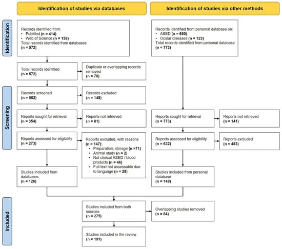
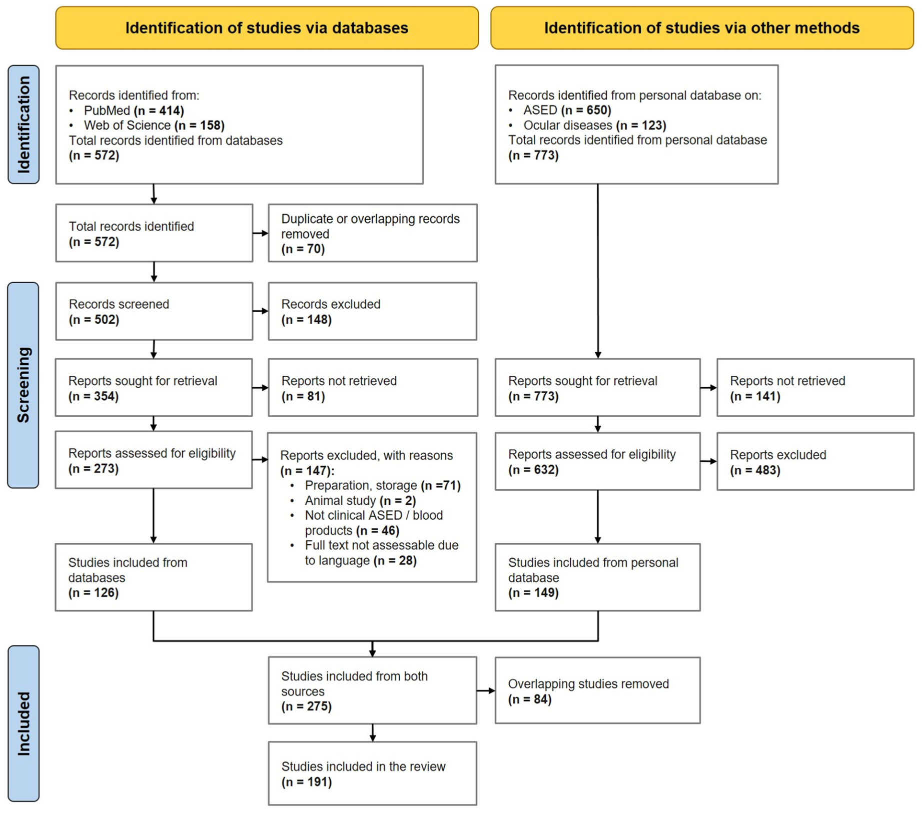
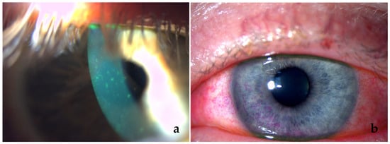
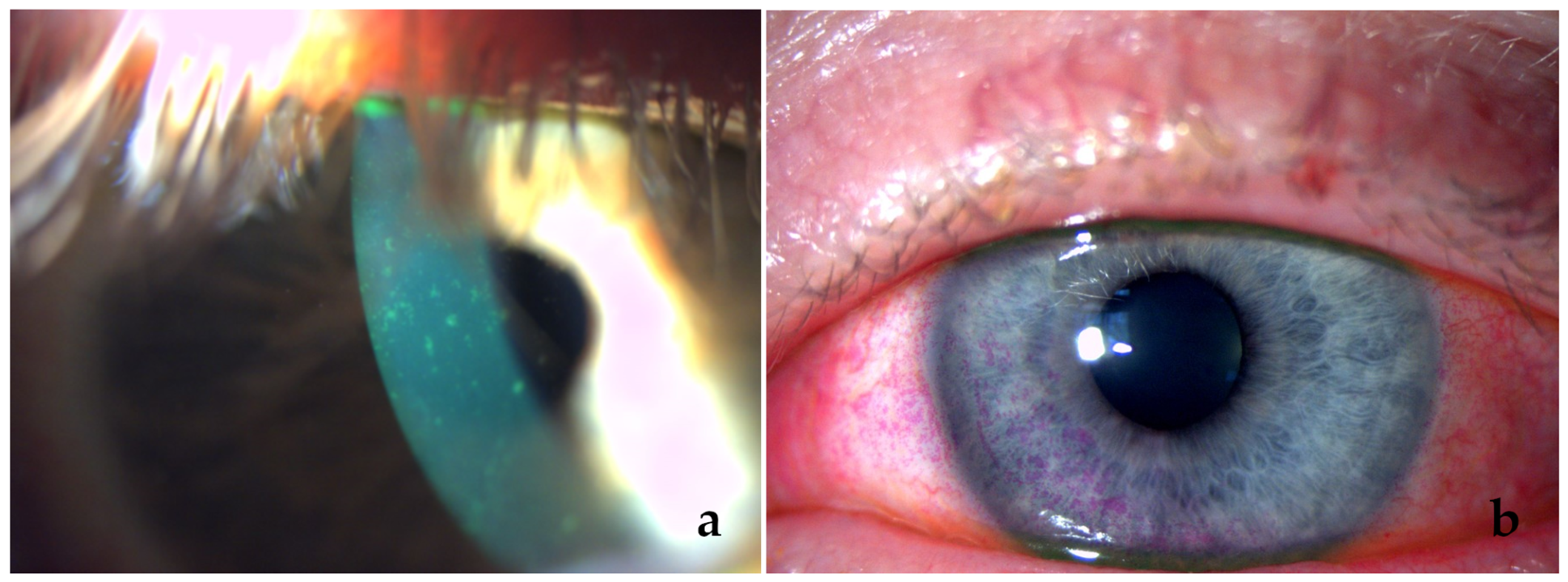
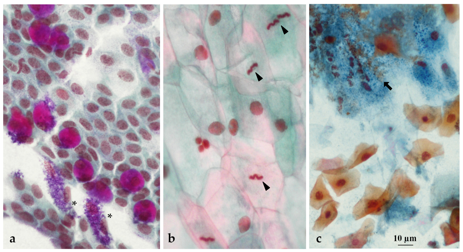

Disclaimer/Publisher’s Note: The statements, opinions and data contained in all publications are solely those of the individual author(s) and contributor(s) and not of MDPI and/or the editor(s). MDPI and/or the editor(s) disclaim responsibility for any injury to people or property resulting from any ideas, methods, instructions or products referred to in the content. |
© 2026 by the authors. Licensee MDPI, Basel, Switzerland. This article is an open access article distributed under the terms and conditions of the Creative Commons Attribution (CC BY) license.