


Abstract
Neurotransmitters are key factors for cell communication within the central and peripheral nervous systems. Recent evidence indicates that they also regulate the function of non-neuronal tissues, including cartilage, as chondrocytes express neurotransmitter receptors, transporters, and other signaling molecules, enabling them to respond to neurochemical cues affecting homeostasis and disease. This review synthesizes the current literature on the roles of major neurotransmitters in chondrocytes and cartilage biology, focusing on norepinephrine, glutamate, γ-aminobutyric acid (GABA), acetylcholine, and serotonin. Specifically, we conducted an extensive literature review within major databases such as PubMed and Scopus, which led to 2748 papers being screened based on various inclusion/exclusion criteria. A total of 42 relevant manuscripts were further selected and are presented to describe the role of neurotransmitters in chondrocytes/cartilage. The data demonstrate that neurotransmitter signaling regulates diverse aspects of chondrocyte biology, including proliferation, differentiation, endochondral ossification, mechanotransduction, extracellular matrix remodeling, inflammation, apoptosis, autophagy, and osteoarthritis (OA). Norepinephrine is the most extensively studied neurotransmitter and exhibits context-dependent effects on cartilage development, OA progression, and repair through adrenergic receptor signaling and neuroendocrine pathways. Glutamatergic signaling, particularly through N-methyl-D-aspartate (NMDA) receptors, is implicated in mechanotransduction, inflammatory activation, circadian regulation, and OA-associated phenotypic changes, whereas GABAergic signaling primarily regulates growth plate maturation and chondrogenesis. Finally, acetylcholine signaling appears to promote chondrocyte survival and anti-inflammatory responses, while serotonin is linked to altered calcium signaling, inflammatory mediator production, and cartilage catabolism. The biological effects of these neurotransmitters are highly dependent on receptor subtype, disease stage, inflammatory status, and mechanical environment, highlighting the complexity of neuro-cartilage communication. Although significant knowledge gaps remain regarding receptor-specific mechanisms and translational significance, current data support neurotransmitter signaling as an important regulator of cartilage and a promising avenue for the development of novel therapeutic strategies for OA.
Keywords:
neurotransmitters; serotonin; dopamine; norepinephrine; epinephrine; acetylcholine; glutamate; GABA; chondrocytes; cartilage; arthritis 1. Introduction
Articular cartilage is a vital component in providing a low-friction surface between articulating bone surfaces of diarthrodial joints and facilitating load transmission with a low frictional coefficient [1]. Articular cartilage is a specialized type of connective tissue composed of hyaline cartilage and is relatively thin (2–4 mm). Unlike most tissues, it is devoid of blood vessels, nerves, or lymphatics, but it is composed of a dense extracellular matrix (ECM) with a sparse distribution of chondrocytes [1]. The cartilaginous ECM is primarily composed of water, glycosaminoglycans (GAGs), and proteoglycans, and exists as a gel to provide frictional resistance to flow and pressurization of water, and to provide the joint with the ability to withstand significant mechanical loads [1]. Chondrocytes are the resident cells of articular cartilage that are metabolically active during development, maintenance, and repair of cartilage ECM through their ability to synthesize matrix components such as proteoglycans and GAGs [1]. However, the metabolic activity of chondrocytes can be altered through changes in their chemical and mechanical environment, as proinflammatory cytokines such as interleukin-1 and tumor necrosis factor-α have catabolic and anabolic effects on the degradation and synthesis of ECM macromolecules [1,2]. Essentially, chondrocytes are responsible for cartilage homeostasis through maintenance of the ECM, but without active blood supply and low proliferative activity, they render articular cartilage with a very limited capacity to regenerate [3]. As the articular cartilage ages and becomes stiffer, and in some cases with defects, chondrocytes are unable to meet the demands of cartilage regeneration to provide a stable ECM to withstand the imposed mechanical loads and provide a low-friction articular surface, thus leading to pathological states such as osteoarthritis (OA) [1,2,3,4,5].
Traditionally, neurotransmitters were studied in the context of the nervous system (NS), with different types demonstrating critical functions within the central and peripheral nervous system [6] (CNS, PNS). Catecholamines are composed of tyrosine-derived neurotransmitters such as dopamine, norepinephrine, and epinephrine. Dopamine was shown to be involved in neural reward pathways, emotional learning, and voluntary motor functions [7]. Norepinephrine (NE) modulates attention, arousal and is a critical component of the sympathetic nervous system, whereas epinephrine acts primarily as a hormone but has established roles in certain neural pathways [7]. Glutamate and γ-aminobutyric acid (GABA) serve as the CNS’s primary excitatory and inhibitory neurotransmitters, respectively [8]. Acetylcholine is a major neurotransmitter in both the CNS and PNS, involved at the neuromuscular junction and in muscle contraction, and regulates memory, attention, and learning in the brain [9]. Serotonin, a monoamine neurotransmitter, operates in both CNS and PNS to regulate mood, sleep, digestion, and cardiovascular functions [10]. Although these neurotransmitters were described extensively in the NS, growing evidence indicates that they are active in other tissues. For example, serotonin was shown to be a critical regulator of inflammation, as it signals to mast cells and macrophages to modulate the inflammatory response in either direction [11]. Acetylcholine has been shown to be anti-inflammatory by inhibiting macrophages from releasing inflammatory cytokines, and NE creates an immunosuppressive environment by upregulating regulatory T cells [12]. In addition, neurotransmitters also have a role in skeletal and connective tissues [13,14,15]. The evidence is growing and indicates that neurotransmitters operate in non-neural tissues and may act as local signaling molecules or neuroimmune mediators involved in cellular survival, inflammation, differentiation, and tissue remodeling.
In cartilage, the literature has identified neurotransmitter receptors, transporters, and signaling pathways associated within chondrocytes [16,17,18,19,20,21,22,23]. Further investigations into the neurotransmitter-associated machinery and pathways within chondrocytes have implicated them in chondrocyte differentiation, mechanotransduction, inflammation, ECM metabolism, apoptosis, autophagy, and OA progression [17,23,24,25,26,27,28,29,30,31,32,33,34,35,36,37,38,39,40,41,42,43,44,45,46,47,48,49,50,51,52,53,54,55,56]. With growing evidence that neurotransmitter signaling has a role in chondrocyte biology, this review aims to present evidence related to whether neurotransmitter signaling in chondrocytes primarily contributes to cartilage homeostasis, degeneration, inflammation, repair, or whether these effects are dependent on the receptor subtype expressed or the pathological state of cartilage. As such, this systematic review aims to synthesize the available literature on neurotransmitter roles in chondrocytes and cartilage with a focus on catecholamines, glutamate, GABA, acetylcholine, and serotonin. Specifically, the review evaluates the reported data on the effects of neurotransmitters in chondrocyte differentiation, maturation, mechanotransduction, inflammation, ECM metabolism, survival, and cartilage pathology.
2. Materials and Methods
2.1. Search Strategy
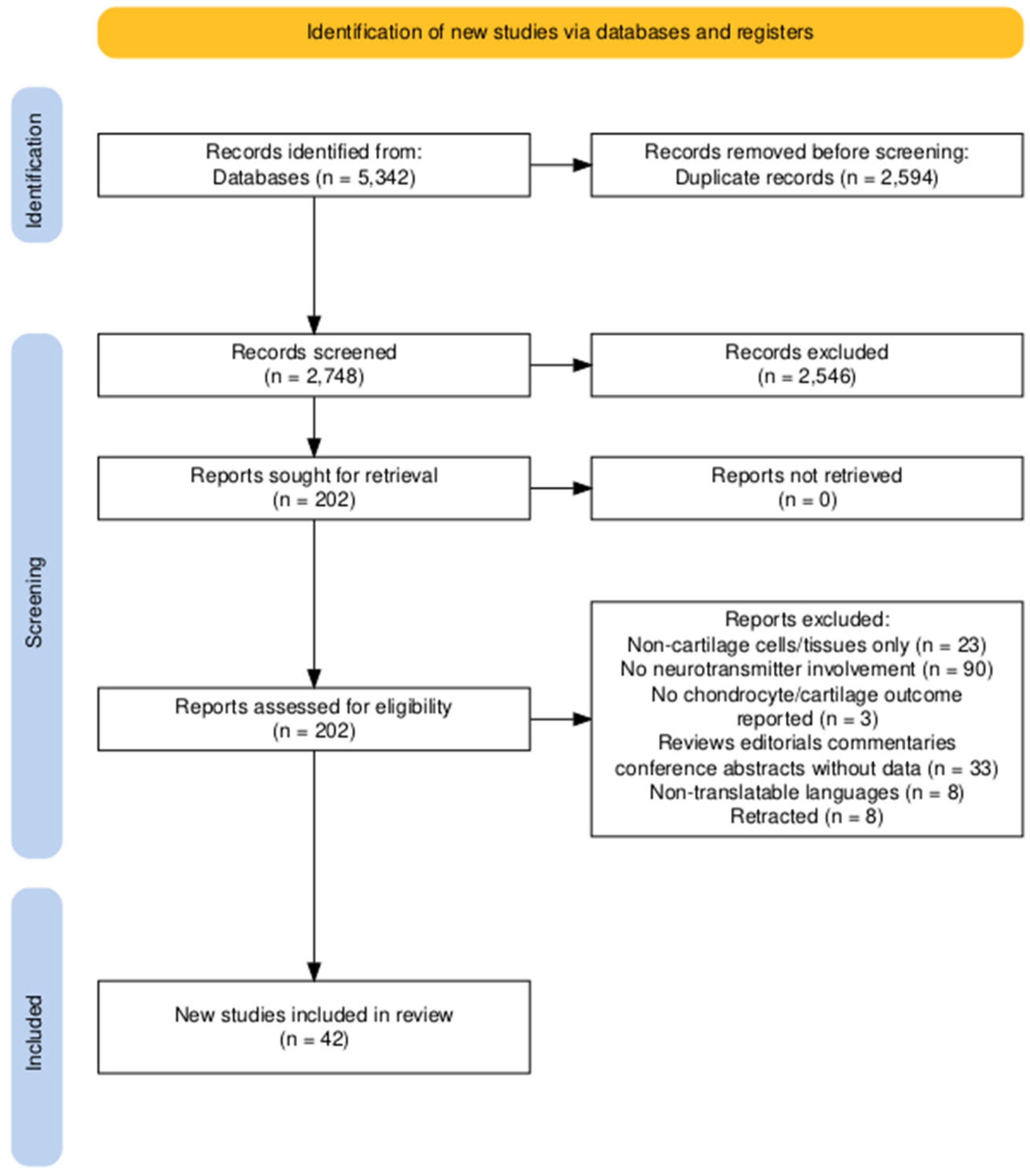
The literature search was conducted from 6 April 2026 to 6 August 2026 using specific terms and database search methods across all databases. Specific databases (i.e., PubMed, Scopus, Cochrane, CINAHL, and Epistemonikos) required explicit terms to ensure a wide yet relevant search (Figure 1, Table S1).
Terms such as ‘tiab’ were used in the PubMed search as search tags, indicating that the database should look for terms in the title and abstract, similar to the search in Scopus, where the search term “TITLE-ABS-KEY” served the same function. The main search terms consisted of the neurotransmitter (i.e., serotonin, dopamine, norepinephrine, epinephrine, acetylcholine, glutamate, and GABA), followed by a Boolean operator such as “AND” or “OR” (See Table S1 for exact search terms used). Neuropeptides were included in the search to ensure that any relevant papers investigating neurotransmitters were also included (See Table S1). Further, to ensure all aspects of neurotransmitters, including their receptors, were included, we used search terms for specific receptor types, such as “adrenergic receptors” (See Table S1). Lastly, chondrocyte and cartilage search terms were expanded to include other relevant investigations, such as “growth plate” and “articular cartilage,” as well as chondrocyte markers [57].
Figure 1.
PRISMA Flow Diagram. Number of articles identified, screened, and included in this review. PRISMA flow diagram generated using an R package and Shiny app for producing PRISMA 2020-compliant flow diagrams [58].
2.2. Study Selection
Following the completion of the initial search (which resulted in 5342 manuscripts) and removal of duplicates (2594), we used a two-step screening process. The first step consisted of title and abstract screening and focused primarily on reading only the title and abstract to determine if the study met the eligibility criteria. Following the title and abstract screen, a full-text screening was conducted to confirm eligibility criteria had been met. Rayyan.ai, a mobile and desktop software developed to increase efficiency and accuracy at title and abstract screening, was used to conduct the first stage of the screening process [59].
2.3. Data Extraction
Once the screening process was complete and all studies used in this investigation were compiled, specific data parameters were obtained and recorded (Figure 1). The species and cell source, neurotransmitter and its applicable receptor investigated, study methods and results, and the overall direction of effect were noted and recorded. The online dataset of unpublished raw data was recorded and stored in Google Sheets, where the data was subsequently used to determine the results of our investigation. Also, inclusion and exclusion criteria were used as shown in Table 1. Lastly, no new data was created or analyzed.
Table 1.
Inclusion and Exclusion Criteria for articles.
2.4. Risk of Bias
Following PRISMA 2020 guidelines, we assessed the risk of bias in the included manuscripts to evaluate the validity of the reported results. We systematically evaluated each study’s design, conduct, and analysis to assess the validity of the results. Despite the outcome of the assessment, we excluded no studies based on risk of bias; instead, we used the risk of bias assessment to inform the interpretation and synthesis of the results presented.
All studies that met our inclusion criteria were original preclinical research, and we appraised those using in vivo models with SYRCLE’s risk-of-bias tool for animal models to assess the methodological quality and internal validity of their experimental methods [60]. We selected SYRCLE because it is the only known risk-of-bias tool developed and validated for animal intervention studies. The tool evaluates ten domains spanning six bias types: sequence generation and baseline characteristics and allocation concealment (selection bias); random housing and blinding of caregivers/investigators (performance bias); random outcome assessment and blinding of outcome assessors (detection bias); incomplete outcome data (attrition bias); selective outcome reporting (reporting bias); and other sources of bias. Each entry was judged and rated as “low risk” (criterion met), “high risk” (criterion not met), or “unclear risk” (insufficient detail reported to permit a judgment) [60]. One benefit of the SYRCLE tool is its flexibility, which we used when judging studies that employed a fixed genotype as their “intervention,” which cannot be randomly allocated to animals. As such, we judged the selection and performance bias items based on the randomizable elements of the experimental design. Two reviewers independently assessed each study across all ten domains, with disagreements resolved by discussion. In keeping with the recommendation of the tool’s developers, no composite scores were given to each study; rather, the results are presented as a traffic light plot showing the distribution of low, high, and unclear judgments for each domain, generated using the robvis tool [61]. A high proportion of “unclear” judgments, particularly with sequence generation, allocation concealment, random housing, and blinding, were anticipated and are interpreted as reflecting incomplete methodological reporting rather than poor study conduct (Supplemental Figure S1).
In vitro and cell-line studies were assessed with a modified version of the Office of Health Assessment and Translation (OHAT) RoB tool. We selected the OHAT tool for the in vitro and cell-line studies because it was designed to evaluate the credibility of experimental and observational evidence-based judgments [62] and because OHAT has been recommended and previously adapted for reviews of in vitro studies [63,64]. OHAT items that apply to whole-animal or human control designs were either removed or re-specified for a cell-based context, and additional signaling questions relevant to in vitro validity were added. Two reviewers independently assessed the nine domains of our modified OHAT too, with disagreements resolved through discussion. Our modified OHAT evaluated internal validity across nine key domains: randomization and treatment allocation; concurrent and identical group handling; adequacy of controls; characterization of the exposure/intervention (identity, purity, concentration, and consistency); outcome assessment validity and blinding; data completeness; selective outcome reporting; appropriate statistical unit definitions (biological versus technical replicates); and overall threats to study validity. Consistent with criteria identified as frequently under-reported in in vitro reviews [63], we included signaling items pertaining to the authentication of the cell line or primary cell source and documentation of independent biological replicates. Each signaling item was rated on the standard four-level OHAT scale: ‘definitely low’ risk of bias (RoB), ‘probably low’ RoB, ‘probably high’ RoB, or ‘definitely high’ RoB [65]. Items that were not applicable to a study (for example, assessor blinding for an objective, instrument-read outcome) were recorded as ‘not applicable’ and excluded from the roll-up for that domain rather than penalized. Items for which a study provided insufficient information were rated as high risk. Overall study-level RoB was determined using a tiered approach adapted from OHAT guidance, where key domains carried greater weight [62,66]. Studies were classified into three tiers based on these ratings: low risk (Tier 1), intermediate risk (Tier 2), or high risk (Tier 3). Following OHAT principles, we did not exclude studies with high RoB; instead, we weighted the confidence ascribed to findings during evidence synthesis (Supplemental Figure S2).
3. Results
3.1. Catecholamines
NE is the primary neurotransmitter of the sympathetic nervous system, classically regulating cardiovascular and systemic physiological functions, including heart rate, myocardial contractility, and blood pressure [67]. The studies identified in this review indicate that NE signaling also extends to cartilage and chondrocyte biology. Specifically, NE was primarily shown to act through adrenergic receptors [17,31,32,34,35,37,39,42,45,47,48,51,53,55] and was implicated in chondrocyte differentiation [17,45,51], cartilage remodeling [32,37,48], inflammatory responses [42,51], and cellular metabolism [33,42]. Collectively, these findings suggest that adrenergic signaling represents an important neurochemical pathway through which sympathetic activity may influence cartilage development, maintenance, degeneration, and repair in a context-dependent manner.
Several studies demonstrated that NE regulates chondrocyte differentiation and skeletal development through β2-adrenergic receptor signaling [17,45,51]. In mouse models, β2-adrenergic receptor activation promoted embryonic growth plate chondrocyte proliferation while inhibiting differentiation [17]. β2-adrenergic receptor signaling also showed the ability to regulate the transition from proliferative to hypertrophic chondrocytes during long bone growth and fracture healing [39]. Mechanistically, NE-mediated β2-adrenergic receptor activation was linked to ERK1/2 and PKA signaling [45] (Figure 2). PKA activation was reported to stimulate the AP-1 transcription factor Jun-B, leading to reduced expression of chondrogenic markers, including Sox6 and type II collagen [45] (Figure 2). Related findings with epinephrine further suggest that β2-adrenergic receptor signaling interferes with chondrogenic differentiation through suppression of Sox6, which ultimately results in the downregulation of the Sox9 family of proteins, the master regulators of cartilage differentiation [53].
Figure 2.
NE-stimulated β2-adrenergic receptor chondrocyte signaling (adapted from Mitchell et al. [45]). ((Top Left) panel): (A–D), β2-AR inhibition of Col IIa1 is mediated by protein kinase A and ERK1/2 MAPK in primary chondrocytes. ((Top right) panel): (A–D), β2-AR stimulation of activator protein 1 (AP-1) proteins and activity in primary chondrocytes. ((Bottom left) panel): (A–D), β2-AR inhibition of Sox-6 is mediated by Jun-B in primary chondrocytes. ((Bottom right) schematic): The β2-adrenergic receptor signaling pathway in chondrocytes was created in BioRender.
NE also appears to have effects on cartilage remodeling and OA progression. Loss of peripheral NE release in osteoarthritic C57BL/6J mice was associated with exacerbated disease progression and increased cartilage calcification; however, there was no effect on cartilage surface degradation or synovial inflammation, suggesting that adrenergic signaling may contribute to cartilage homeostasis in a tissue-dependent manner [47]. Subsequent work in C57BL/6J mice further implicated loss of NE-mediated β2-adrenergic receptor signaling in cartilage, synergistically with increased leptin levels through promotion of osteoblast activity and suppression of osteoclast activity [48]. This was suggested as a key mechanism underlying cartilage calcification and subchondral bone changes during OA [48]. In contrast, sympathetic innervation of aging mouse joints was reported to promote cartilage degeneration through β1-adrenergic receptor signaling, which induced chondrocyte-derived exosomal miR-125 release and promoted osteoblast differentiation in subchondral bone [32]. These findings suggest that the effects of NE on cartilage are highly dependent on receptor subtype, tissue compartment, disease stage, and the local inflammatory or mechanical microenvironment.
In addition to its role in disease-related cartilage degeneration, NE signaling may participate in cartilage repair and regeneration through central-peripheral neural circuits [34,55]. Studies identified a hypothalamic paraventricular nucleus–mediated circuit that promotes mature articular cartilage formation, in part through inhibition of β2-adrenergic receptor signaling [34]. Additional experiments with the hypothalamic neural circuit suggested that mechanical stimulation of articular cartilage reduces hypothalamic-driven NE release, thereby supporting renewal of superficial zone chondrocytes [55]. The study further expanded on how NE promotes superficial zone (SFZ) chondrocyte anabolism by demonstrating that NE acts on the Adrb2 receptor on the chondrocytes to drive internalization of an Adrb2-TβRIII (TGF-β type II receptor) complex to decrease TGF-β signaling, which is responsible for lubricin and ECM protein synthesis [55]. This mechanism was further explored in the study by showing that physical activity creates joint friction that degrades the superficial membrane of articular cartilage, which is regenerated by SFZ chondrocytes under the influence of the hypothalamic neural circuit [55]. Specifically, during mechanical loading of the joint, there is an increase in PGE2 from osteoblasts activating the prostaglandin E2 receptor, EP4, on sensory nerves and sending an interoceptive signal from the skeleton to the paraventricular nucleus of the hypothalamus. Ultimately, this signal serves as negative feedback, reducing hypothalamic sympathetic output. This decrease in sympathetic NE release near articular cartilage reduces β2 signaling in SFZ chondrocytes, thus allowing for the restoration of anabolic cartilage ECM synthesis via TGF-β and lubricin signaling [55]. Interestingly, the same study performed an inducible Adrb2 deletion in SFZ chondrocytes, which damaged the superficial membrane. The authors suggested that since Adrb2 is exclusively expressed in SFZ, it serves as a defining feature of the cell population. Therefore, complete abolition of the receptor within the population causes the loss of maintenance and survival input necessary for SFZ chondrocytes to retain their identity [55]. The discrepancy between decreasing NE signaling and abolition of the Adrb2 receptor suggests that lowering NE helps establish optimal balance for SFZ anabolism, whereas deleting Adrb2 removes a constitutive maintenance signal that eliminates SFZs and the superficial membrane they produce [55]. Collectively, these findings indicate that NE signaling is not uniformly catabolic or anabolic but instead may regulate cartilage remodeling through both local receptor-mediated mechanisms and broader neuroendocrine feedback pathways.
NE has also been implicated in chondrocyte inflammatory responses, and its effects appear to be dose- and receptor-dependent. For example, at lower concentrations in human articular cartilage, NE was reported to promote chondrocyte apoptosis through α-adrenergic receptor signaling, whereas at higher concentrations it promoted chondrocyte proliferation through β-adrenergic receptor signaling by modulating cell-cycle progression [42]. Also, in monolayer cultures of human osteoarthritic articular chondrocytes under inflammatory conditions, NE did not demonstrate synergy with IL-1β to promote chondrocyte dedifferentiation toward a fibroblast-like phenotype, with increased pro-catabolic mediator release and downregulation of chondrogenic markers [51]. The dedifferentiation effects driven by IL-1β were independent of NE signaling. Co-treatment with NE did not further potentiate the IL-1β-induced downregulation of fibroblastic/chondrogenic markers (SOX9, type II collagen, COMP, aggrecan) or the upregulation of matrix metalloproteinases (MMP13, ADAMTS4) [51]. These findings suggest that the role of NE in inflammatory and catabolic changes in chondrocytes toward dedifferentiation, particularly in the presence of cytokine-mediated low-grade inflammation, remains poorly understood. Adrenergic receptor subtypes appear to be central to the direction of NE-mediated chondrocyte responses. Although many effects of NE in cartilage were attributed to β2-adrenergic receptor signaling, α-adrenergic receptors have also been linked to catabolic pathways [33,37]. Activation of α2-adrenergic receptors was associated with increased catabolic factor release, reduced anabolic factor production, and enhanced mitochondrogenesis in rat chondrocytes [33]. In a rat temporomandibular joint model, α2-adrenergic receptor activation promoted degenerative remodeling through chondrocyte catabolism [37]. Additional work in human articular cartilage and chondrocytes showed that IL-1β altered adrenergic receptor activity and contributed to catabolic signaling, including changes in glycosaminoglycan release [35]. Together, these findings support a model in which NE-mediated cartilage effects are shaped by inflammatory status, receptor subtype expression, and downstream metabolic signaling.
The literature also suggests a relationship between sympathetic stress signaling and impaired cartilage and skeletal growth. Seven-week-old C57BL/6J mice were placed in a chronic psychosocial stress environment through chronic subordinate colony housing, exhibiting reduced tibia and femur lengths as determined by microCT and histological analyses. The reduced limb lengths were due to altered endochondral ossification, as the growth plate thickness had increased along with decreased growth plate mineral deposition. Interestingly, bone marrow cells located at the growth plate had increased tyrosine hydroxylase expression, indicating that local catecholamine synthesis or sympathetic activity is altered in growth plate cartilage, thus interfering with physiological endochondral ossification [31]. Lastly, this was accompanied by a decreased hypertrophic chondrocyte population and reduced Runx2 expression, further providing evidence that chronic social stress alters local adrenergic signaling at the growth plate, impairing overall limb growth [31]. These findings suggest that psychosocial stress-associated adrenergic signaling may impair endochondral ossification by altering growth plate chondrocyte maturation.
Although NE was the primary catecholamine identified in this section, studies related to catecholaminergic mediators are also present in the literature. Dopamine was reported to reduce cartilage degradation and promote cartilage stability by inhibiting cartilage matrix-degrading factors while increasing ECM content in eight-week-old C57BL/6J mice [44]. Epinephrine was implicated in ATDC5 chondrocyte dedifferentiation through β2-adrenergic receptor signaling [53] and was reported to be chondrotoxic at high concentrations under low-pH conditions in human chondrocytes [30]. These findings suggest that catecholaminergic regulation of cartilage extends beyond NE alone, but the distinct roles of NE, epinephrine, and dopamine should be interpreted separately. Lastly, all of the aforementioned studies are summarized in Table 2.
Table 2.
Data from investigations into the effects of catecholamine neurotransmitters or their respective receptors in cartilage cell lines or animal models. Note: GPCs = growth plate chondrocytes, AR = adrenergic receptor, SFZ = superficial zone, AAV = adeno-associated virus, OA = osteoarthritis, Syx = sympathectomy, Col II = collagen type II, GAG = glycosaminoglycans, DA = dopamine, OARSI = Osteoarthritis Research Society International.
3.2. Glutamate/GABA
Glutamate is the primary excitatory neurotransmitter, whereas γ-aminobutyric acid (GABA) is the primary inhibitory neurotransmitter in the CNS [8]. Both neurotransmitters signal through ionotropic and metabotropic receptors and are implicated in multiple neurological and psychiatric disorders [68]. The studies identified in this review suggest that glutamatergic and GABAergic signaling are not restricted to the nervous system but are also present in cartilage and chondrocytes. Several studies demonstrated that glutamate and GABA may have roles in chondrocyte differentiation and maturation, mechanotransduction, endochondral ossification, inflammatory response, and chondrogenesis [18,19,20,21,22,26,28,29,36,38,46,50,52,54].
Specific studies demonstrated that chondrocytes express the molecular machinery required for glutamatergic signaling. In cultured rat costal chondrocytes, AMPA receptor subunits and vesicular glutamate transporter-1 (VGLUT1) were expressed, and endogenous glutamate release was prolonged following AMPA receptor activation [54]. Using a similar rat chondrocyte model, Hinoi et al. demonstrated expression of the excitatory amino acid transporters GLAST, GLT-1, and EAAC1, with glutamate uptake showing sodium- and membrane–potential–dependent transport characteristics [19]. Further, in embryonic mouse cartilage models, excitatory amino acid transporters were expressed in a differentiation stage–specific pattern, suggesting a potential role for glutamate transport in chondrocyte maturation and endochondral ossification [19]. Glutamate was also implicated in OA pathology, as rat knees with experimentally induced OA showed significantly elevated glutamate levels in joint dialysates compared with controls [36]. In addition, chondrocytes were shown to express machinery for autocrine glutamate signaling, and N-methyl-D-aspartate receptor (NMDAR) activity was linked to inflammatory processes associated with cartilage degradation [46]. Collectively, these studies support the presence of functional glutamatergic signaling in chondrocytes and suggest potential roles in cartilage maturation, inflammation, and OA pathology.
Studies also focused on NMDAR-mediated glutamate signaling. Human articular chondrocytes respond to mechanical stimulation through NMDAR-dependent mechanisms [21]. In normal chondrocytes, NMDAR antagonism inhibited the hyperpolarization response to mechanical stimulation, whereas OA chondrocytes showed an altered depolarization response that was not affected by NMDAR antagonism [21]. These findings suggest that NMDAR-mediated mechanotransduction is disrupted in OA. Additional studies demonstrated that normal and OA human chondrocytes differ in NMDAR subunit expression [20]. In OA chondrocytes, NMDAR signaling required extracellular calcium and was associated with PSD-95 and nNOS activity. Downstream signaling activated tetrodotoxin-sensitive sodium channels and small-conductance calcium-activated potassium channels, a response not observed in normal chondrocytes [20].
NMDAR signaling was also linked to inflammation, apoptosis, and catabolic phenotypic changes. In human and rat chondrocytes, NMDAR signaling contributed to inflammatory pathways associated with cartilage damage, and IL-1β altered glutamate metabolism while inducing pro-inflammatory gene expression [46]. In SW1353 chondrocytic cells, inhibition of both NMDAR signaling and complement C5 reduced apoptosis and inflammatory mediator expression, suggesting interaction between glutamatergic and complement-mediated inflammatory pathways [43]. NMDAR signaling was also shown to regulate chondrocyte circadian clock genes and phenotype [38]. In OA chondrocytes, NMDAR inhibition restored BMAL1 and PER2 expression toward control levels and reduced pro-catabolic factor expression, whereas GluN2B expression produced the opposite effect [38]. Further work demonstrated that IL-1β-mediated regulation of PER2 and BMAL1 involved downstream NMDAR/CREB and NF-κB signaling pathways [26]. Together, these findings suggest that NMDAR activity may couple inflammatory signaling, circadian dysregulation, apoptosis, and catabolic cartilage remodeling.
NMDAR signaling also appears to contribute to chondrogenesis. In chicken limb bud–derived chondroprogenitor cells, NMDAR activation induced transient intracellular calcium release, which was required for chondroprogenitor differentiation into matrix-producing chondroblasts [22]. Inhibition or knockdown of NMDAR signaling reduced calcium signaling and impaired chondrogenesis, suggesting that NMDAR-mediated calcium transients may support early cartilage differentiation [22].
Although NMDARs were the most frequently studied glutamate receptors, AMPA and metabotropic glutamate receptor pathways were also implicated in chondrocytes [23,54]. Rat costal chondrocytes expressed the GluR3 AMPA receptor subunit and VGLUT1, indicating that chondrocytes possess machinery similar to that used for vesicular glutamate release in neurons [54]. These cells also release endogenous glutamate, and AMPA receptor activation prolonged this release, supporting an autocrine or paracrine glutamatergic signaling mechanism [54]. Glutamate also promoted chondrocyte (mice and rats) differentiation toward mineralization through pathways involving group III metabotropic glutamate receptor activation, retrograde cystine/glutamate antiporter activity, intracellular glutathione depletion, and apoptosis-mediated mineralization [23]. The authors also suggested that at physiologic glutamate concentrations, glutamate activates group III mGluR, inhibiting cAMP synthesis and preventing chondrocyte differentiation toward mineralization. However, at supraphysiological extracellular glutamate concentrations, the cystine/Glu antiporter reverses direction, contributing to the pathway that induces chondrocyte differentiation toward mineralization. Thus, Wang et al. demonstrate a role for glutamate in chondrocyte differentiation and maturation through changes in the cystine/Glu antiporter [23] (Figure 3).
Figure 3.
Glutamate’s role in cartilage differentiation (adapted from Wang et al. [23]). ((Top left) panel (a–e)) shows the effect of Glu on cell viability in cultured chondrocytes. ((Top right) panel (a–c)) shows the effect of Glu on ROS production and reduced glutathione (GSH) levels in cultured chondrocytes. ((Bottom left) schematic) depicts the effects of glutamate on chondrocytes at physiologic concentrations based on results from Wang et al. [23]. ((Bottom right) schematic) represents the proposed mechanism by Alihali et al. [26] of how the NMDAR/CREB and NF-κB signaling pathways regulate the core circadian clock components PER2 and BMAL1 in chondrocytes (created in BioRender).
Glutamatergic signaling was repeatedly associated with inflammatory and OA-related chondrocyte responses. In human and rat chondrocytes, NMDAR activity interacted with IL-1β-mediated inflammatory signaling and altered glutamate metabolism in ways that promoted pro-inflammatory and cartilage-degrading pathways [46]. In SW1353 cells, inflammatory stimulation increased NMDAR subunits NR1 and NR2A and complement C5, while inhibition of these pathways reduced apoptosis and inflammatory mediators including TNF-α, IL-6, and NLRP343. In primary equine chondrocytes, inflammatory stimulation altered calcium signaling to glutamate and increased glutamate release [50]. Under inflammatory conditions, glutamate-induced calcium signaling shifted from single-peak transients toward oscillatory calcium responses, while glutamate transporters GLAST and GLT-1 were upregulated [50]. A subsequent equine chondrocyte study confirmed that inflammatory stimulation increased glutamate release in both healthy and osteoarthritic chondrocytes within 24 h, suggesting that glutamate release may represent an early response to acute inflammatory stimulation [49]. These findings are supported by a rat OA model in which glutamate levels were elevated in joint dialysates after OA induction [36].
Compared with glutamate, fewer studies examined GABAergic signaling in cartilage; however, the available evidence suggests roles in chondrocyte differentiation, growth plate physiology, calcium-sensing receptor signaling, and osmotic adaptation [18,28,29,52]. In rat growth plate chondrocytes and ATDC5 cells, GABAA and GABAB receptor subunits were expressed, and receptor-specific agonist and antagonist experiments suggested that GABA signaling may regulate chondrogenic differentiation and chondrocyte proliferation [18]. These findings support a potential autocrine or paracrine role for GABA in growth plate cartilage.
GABAB receptor signaling was particularly associated with chondrogenesis and endochondral ossification. Specifically, GABAB receptor subunit-1 (GABAB-R1) was expressed in chondrocytes, especially in prehypertrophic and hypertrophic cartilage regions and GABAB-R1 deficiency was associated with reduced body size, delayed skeletal calcification, and reduced Indian hedgehog (Ihh) expression [52]. Mechanistic experiments suggested that GABAB-R1 promotes chondrogenesis by enhancing ATF4 nuclear translocation and ATF4-dependent Ihh promoter activation [52]. GABAB-R1 was also shown to interact with the extracellular calcium-sensing receptor in mouse growth plate chondrocytes [28]. These receptors were coexpressed, colocalized, and physically associated, and loss of GABAB-R1 impaired calcium-mediated signaling, reduced proliferation, increased apoptosis, and shifted marker expression toward late differentiation [28], suggesting that GABAB-R1 regulates growth plate chondrocyte maturation partly through interaction with calcium-sensing receptor pathways.
GABA-related transport was also implicated in the osmotic regulation of articular chondrocytes. In primary porcine articular chondrocytes, hypertonic culture conditions induced a time-dependent shift in transport activity, with early activation of sodium-dependent amino acid transport system A followed by delayed induction of betaine–GABA transport [29]. This response required new RNA and protein synthesis, but BGT-1 mRNA was not detected, suggesting that chondrocytes may use an alternative or unidentified osmolyte transport mechanism [29].
Collectively, these studies suggest that glutamate and GABA signaling pathways are active in chondrocytes and cartilage but appear to regulate partially distinct biological processes. Glutamate signaling is most strongly associated with mechanotransduction, inflammatory activation, OA-related phenotypic changes, apoptosis, mineralization, and chondrogenesis, particularly through NMDAR-, AMPAR-, transporter-, and cystine/glutamate exchange–dependent mechanisms. In contrast, GABAergic signaling appears more closely linked to growth plate chondrocyte proliferation, chondrogenesis, endochondral ossification, calcium-sensing receptor interactions, and osmotic adaptation. Lastly, all of the aforementioned studies are summarized in Table 3.
Table 3.
Findings from studies investigating glutamate, GABA, or their respective receptors in cartilage animal models or cell lines. Note: NMDAR = N-methyl-D-aspartate receptor, PSD-95 = postsynaptic density 95, SK channels = small-conductance calcium-activated potassium channels, EAATs = excitatory amino acid transporters, LPS = lipopolysaccharide, GABA = ɣ-aminobutyric acid.
3.3. Acetylcholine
Acetylcholine is a major neurotransmitter of the CNS and PNS and is classically involved in processes such as voluntary muscle contraction, autonomic regulation of heart rate, memory consolidation, and attention [9]. Studies suggest that acetylcholine-related signaling also extends beyond the nervous system and may contribute to chondrocyte survival, inflammatory regulation, and OA pathology [16,27,40,41,56,69].
Evidence for cholinergic signaling in cartilage was demonstrated in studies of human hip OA and rheumatoid arthritis (RA) [16]. Beckmann et al. identified several proteins involved in choline import and acetylcholine release, including Organic Cation Transporter 1 (OCT1), Organic Cation Transporter 3 (OCT3), Organic Cation/Carnitine Transporter 1 (OCTN1), and members of the choline transporter-like family [16]. These findings suggest that cartilage contains the molecular machinery capable of supporting local cholinergic signaling, although transporter expression did not appear to differ substantially in pathological cartilage [16].
Several studies focused on the α7 nicotinic acetylcholine receptor (α7nAChR) and its role in OA-related cartilage degeneration. In human and murine chondrocytes, α7nAChR stimulation was reported to induce anti-inflammatory and anti-catabolic responses, suggesting a protective role in OA pathology [69]. Additional experiments with human articular cartilage and α7nAChR-deficient chondrocytes suggested that it regulates the balance between autophagy and apoptosis [41]. Specifically, human OA cartilage showed reduced α7nAChR expression compared with healthy cartilage, which was associated with dysregulated autophagy and apoptosis [41] (Figure 4). Further, α7nAChR-deficient chondrocytes had reduced LC3 (Microtubule-associated protein 1A/1B-light chain 3) levels and greater sensitivity to chemically induced arthritis, supporting a role of α7nAChR in maintaining chondrocyte homeostasis and survival [41] (Figure 4). However, the results from Liu et al. do not definitively demonstrate that α7-nAChR activation promotes autophagic flux because the LC3-II levels reflect a single time point of steady-state levels between autophagosome synthesis and lysosomal turnover. The steady-state data cannot be conclusively attributed to an increase in autophagic flux because no turnover assays were performed to definitively demonstrate this. In an α7nAChR-deficient mouse model of induced arthritis through type II collagen immunization, α7nAChR-deficient mice developed milder arthritis, reduced cartilage destruction, and had lower measures of adaptive immunity compared with controls [56]. Therefore, α7nAChR signaling may have disease-context–dependent effects, with potentially significant roles in degenerative OA and immune-mediated inflammatory arthritis. However, additional confirmatory assays and studies are required to determine the causative relationship of α7nAChR signaling in cartilage disease models.
Figure 4.
Effects of α7-nAChRs deficiency on apoptosis and autophagy in primary cultured chondrocytes (adapted from Liu et al. [41]) (A–E). Increased apoptosis in α7-nAChR knockout chondrocytes compared with wild-type controls, as demonstrated by nuclear staining, FACS analysis, and Western blotting. (F–J), Demonstrates autophagy as a measurement of various markers and their ratios. The right figure represents a working model proposed by Liu et al. [41] showing that α7-nAChRs play a role in switching from autophagy to apoptosis in cartilage degradation of OA. It also shows that activation of α7-nAChRs could inhibit phosphorylation of mTOR, promote autophagic flux, and prevent cartilage degradation.
Although the majority of studies focused on α7nAChR, muscarinic acetylcholine receptors were also implicated in cartilage and arthritis models [27,40]. M3 muscarinic receptor (M3R)-deficient mice subjected to collagen antibody-induced arthritis were not protected against disease and were associated with a greater inflammatory response and increased type II collagen degradation in articular cartilage [27]. These findings suggest that M3R signaling may contribute in a protective role in arthritis. In chick scleral chondrocytes, muscarinic receptor antagonism reduced sulfate incorporation, a proxy for glycosaminoglycan synthesis, suggesting a role for muscarinic receptor signaling, particularly M1 receptor activity, in regulating cartilage ECM metabolism [40]. However, because this study used scleral rather than articular chondrocytes, its relevance to joint cartilage should be interpreted cautiously.
Collectively, these studies suggest that cartilage contains components of a non-neuronal cholinergic signaling system and that acetylcholine receptors may regulate chondrocyte survival, autophagy, apoptosis, inflammation, and ECM metabolism. The strongest evidence centers on α7nAChR, which appears to support anti-inflammatory, anti-catabolic, and survival-related pathways in OA models and collagen antibody-induced arthritis. Muscarinic receptor studies further indicate that cholinergic signaling in cartilage is receptor subtype–specific and may vary according to tissue type, disease model, and inflammatory context. Lastly, all of the aforementioned studies are summarized in Table 4.
Table 4.
Findings from studies investigating acetylcholine or the respective receptors in cartilage animal models or cell lines. Note: nAChR = nicotinic acetylcholine receptor, CAIA = collagen-antibody-induced arthritis, RA = rheumatoid arthritis, mAChR = muscarinic acetylcholine receptor.
3.4. Serotonin
Serotonin is a major neurotransmitter and modulatory neurohormone involved in neurological functions such as mood, appetite, and sleep, as well as gastrointestinal motility [10]. Serotonin also functions as an immune-modulating mediator that can either suppress or promote inflammation depending on the tissue type and receptor subtype [70]. The studies identified in this review suggest that serotonin-mediated signaling in chondrocytes may similarly be tissue-, receptor-, and disease-dependent.
For example, in primary equine chondrocytes, inflammatory stimulation altered intracellular calcium signaling in response to serotonin [50]. Under inflammatory conditions, calcium transient events shifted from a single-peak response to an oscillatory pattern, a response previously described in astrocytes [50]. This finding suggests that inflammatory conditions may alter chondrocyte calcium signaling through overstimulation of Gq protein–coupled receptors (GqPCRs), including mGluR5 and 5-HT2A [50]. Additional work in primary equine chondrocytes showed that osteoarthritic chondrocytes exhibited stronger serotonin-induced intracellular calcium release than healthy chondrocytes under inflammatory conditions [49] (Figure 5). These findings suggest that serotonin signaling may contribute to altered calcium responses in osteoarthritic chondrocytes and may be relevant to OA-associated pain-related processing [49] (Figure 5).
Figure 5.
Ca2+ release in chondrocytes adapted from (Skiöldebrand et al. [49]). ((Left) panel) displays calcium responses evoked by serotonin in cultured chondrocytes from healthy and osteoarthritic joints stimulated with either IL-1β, lipopolysaccharide (LPS), or was untreated. CC BY-NC-ND 4.0 ((Right) panel) depicts the action of the 5HT3 receptor once activated in chondrocytes and how its promotion of inflammatory mediators influences pain processing in arthritis (based on Stratz et al. [24] and created in BioRender).
Other studies also implicate specific serotonin receptors in inflammatory and catabolic chondrocyte responses. For example, overstimulation of 5-HT2A was suggested to contribute to inflammation-associated chondrocyte pathology through altered calcium signaling [50]. In human chondrocytes, antagonism of the 5-HT3 receptor produced anti-inflammatory effects by reducing IL-6, prostaglandin E2, and free radical production, with indirect inhibition of COX-2 activity24, suggesting that endogenous 5-HT3 signaling may contribute to pro-inflammatory responses in these cells [24].
Serotonin was also linked to catabolic activity in human osteoarthritic articular chondrocytes as serotonergic receptors were identified on chondrocytes, and receptor activation was associated with phospholipase A2 activation, prostaglandin E2 synthesis, and increased collagenolytic activity [25]. These findings suggest that serotonin signaling may contribute to OA-related cartilage degradation through inflammatory prostaglandin pathways and collagenolytic enzyme (collagenase) activity [25].
Collectively, these aforementioned studies suggest that serotonin signaling may influence chondrocyte inflammatory responses, calcium signaling, pain-associated OA mechanisms, and cartilage matrix degradation. These effects appear to depend on receptor subtype and inflammatory context, with 5-HT2A implicated in altered calcium signaling and 5-HT3 signaling associated with pro-inflammatory mediator production. However, the current evidence remains limited and should be interpreted cautiously until receptor-specific mechanisms are more fully delineated in articular chondrocytes/cartilage and OA models. Lastly, all of the aforementioned studies are summarized in Table 5.
Table 5.
Findings from studies investigating serotonin or the respective receptors in cartilage animal models or cell lines.
4. Discussion
Although cartilage is an avascular and aneural tissue, it is hard to conceive that neurotransmitters would have a role or effects on chondrocytes and cartilage biology. But, published data clearly shows that they do. Specifically, this review demonstrates that classical neurotransmitters exert diverse and biological effects on cartilage and chondrocyte function, extending well beyond their established roles in the nervous system. Across the literature, norepinephrine, glutamate, GABA, acetylcholine, and serotonin were each shown to be involved with distinct yet overlapping aspects of cartilage biology, including chondrocyte differentiation, mechanotransduction, inflammatory signaling, extracellular matrix homeostasis, cartilage degeneration, and repair. Collectively, these findings support the concept that cartilage is an active neuroresponsive tissue capable of integrating neurotransmitter-mediated signals through locally expressed receptors, transporters, and intracellular signaling pathways.
4.1. Catecholamines
Among the neurotransmitters examined, NE has the most extensive evidence linking sympathetic nervous system activity to cartilage biology. While NE is classically recognized for regulating cardiovascular physiology [67], the reviewed studies demonstrate that adrenergic signaling also influences chondrocyte differentiation, cartilage remodeling, OA progression, inflammatory signaling, and cell metabolism. These effects appeared to be highly receptor- and tissue-dependent, with β-adrenergic and α-adrenergic pathways producing distinct and sometimes opposing effects. Predominantly, β2-adrenergic receptor signaling was strongly associated with chondrocyte differentiation and skeletal development. Several studies suggested that norepinephrine regulates chondrocyte maturation through β2-adrenergic receptor activation of ERK1/2 and PKA signaling, leading to induction of the AP-1 transcription factor Jun-B and reduced expression of chondrogenic markers such as Sox6 and type II collagen [45]. Similarly, β2-adrenergic effects were also observed with epinephrine [53], suggesting that catecholamine-mediated regulation of chondrocyte phenotype may extend beyond norepinephrine alone. However, these findings are largely derived from in vitro and in vivo animal models thus, further studies are needed to determine whether the same β2-adrenergic signaling mechanisms regulate chondrocyte differentiation in human cartilage.
NE was also implicated in cartilage remodeling and diseased states such as OA, although its effects differed by adrenergic receptor subtype. β2-adrenergic signaling appeared to support cartilage homeostasis in some models, whereas β1-adrenergic signaling [32] was associated with cartilage degeneration, including chondrocyte-derived exosomal signaling and subchondral bone remodeling. These findings suggest that adrenergic signaling may influence OA progression through both cartilage-intrinsic and osteochondral mechanisms. Additional studies should focus on human osteoarthritic cartilage in order to define the receptor-specific roles of β- and α-adrenergic signaling in disease progression.
A particularly notable finding was the identification of a hypothalamic–skeletal neural circuit regulating articular cartilage homeostasis [34,55]. In this model, mechanical loading promoted skeletal PGE2-mediated interoceptive signaling through EP4 receptors on sensory nerves, which relays information to the paraventricular nucleus of the hypothalamus [55]. This mechanical signaling reduced sympathetic norepinephrine output to the joint, thereby decreasing β2-adrenergic receptor activation in chondrocytes of the superficial zone and promoted anabolic matrix activity, lubricin expression, and superficial cartilage maintenance [55]. Although this circuit provides a compelling mechanism linking joint loading, neural regulation, and cartilage regeneration, the evidence remains limited to a small number of preclinical studies and further research is needed to determine whether this circuit is active in loaded human joints and whether it can be therapeutically modulated in OA.
Adrenergic signaling was also implicated in chondrocyte inflammatory responses And the reviewed studies suggest that α- and β-adrenergic receptors may exert different effects depending on receptor subtype, norepinephrine concentration, and inflammatory state. α-adrenergic signaling was associated with chondrocyte apoptosis and catabolic activity, whereas β-adrenergic signaling was linked to chondrocyte proliferation and dedifferentiation toward a fibroblast-like phenotype in inflammatory osteoarthritic conditions. As such, adrenergic signaling can be either protective or detrimental depending on the cell environment and disease state. α2-adrenergic signaling was associated with pro-catabolic, mitochondrogenic, and anti-anabolic effects in chondrocytes and this suggested that α2-adrenergic receptor activation may promote cartilage degeneration by altering matrix metabolism and mitochondrial activity [33]. Whether adrenergic signaling pathways play a major role in healthy, inflamed, and OA human chondrocytes, remains unexplored.
The reviewed literature also suggested a potential link between chronic psychosocial stress and impaired cartilage or growth plate function. Chronic stress was associated with altered growth plate chondrocyte metabolism, reduced chondrocyte proliferation, and disrupted endochondral ossification [31]. These findings suggest that stress-related catecholaminergic signaling may influence skeletal growth and cartilage physiology. However, the evidence remains limited to few animal models, and further mechanistic studies are needed to define the neural, endocrine, and molecular pathways connecting chronic stress to chondrocyte dysfunction. Finally, dopamine was less extensively studied than norepinephrine but was reported to reduce cartilage degradation and promote cartilage stability in one included study [44]. This finding suggests that catecholaminergic regulation of cartilage may involve multiple neurotransmitters, not only norepinephrine, but dopamine’s exact role in cartilage remodeling and OA also requires further investigation.
4.2. Glutamate/GABA
The studies examining glutamate and GABA also implicates them in several aspects of chondrocyte biology, including differentiation, maturation, mechanotransduction, chondrogenesis, endochondral ossification, inflammation, and osmotic adaptation [18,19,20,21,22,23,26,28,29,36,38,43,46,50,52,54]. Glutamatergic signaling was demonstrated in chondrocytes through their expression of glutamate receptors, transporters, and endogenous glutamate release [54]. In OA mouse models, increased glutamate levels were associated with disease induction, suggesting a potential role for glutamate in OA-associated cartilage pathology [20]. Glutamate transporters also showed differentiation stage–specific expression, supporting a possible role in chondrocyte maturation and endochondral ossification [19]. Further studies would potentially benefit from investigating the translation of glutamate’s role in developmental mineralization to articular cartilage repair.
Among glutamate receptors, N-methyl-D-aspartate receptors (NMDARs) have received the greatest attention and were implicated in chondrocyte mechanotransduction, with normal and OA chondrocytes showing different electrophysiologic responses to mechanical stimulation [21]. This suggests that NMDAR-dependent mechanotransduction may be altered in OA. NMDAR signaling was also associated with pro-inflammatory and pro-catabolic responses, including inflammatory gene expression and potential interaction with other signaling [43]. In addition, NMDAR activity was linked to chondrocyte circadian regulation through downstream CREB and NF-κB pathways, which influenced circadian clock genes, PER2 and BMAL1 expression and may contribute to altered chondrocyte phenotype in OA [26]. In developmental models, NMDAR-mediated calcium signaling was required for chondroprogenitor differentiation into chondroblasts, suggesting that NMDAR activity may have distinct roles in cartilage development versus inflammatory cartilage degeneration [22]. Overall, research supports a strong relationship between NMDAR signaling and chondrocyte biology, but further studies are needed to clarify its relevance in human clinical samples and in human chondrogenic differentiation.
Although NMDARs were the most frequently studied glutamate receptors, AMPA and metabotropic glutamate receptors were also implicated in chondrocyte function. Rat chondrocytes expressed AMPA-related glutamatergic machinery similar to neurons, and AMPA receptor activation appeared to prolong endogenous glutamate release, supporting a potential autocrine or paracrine signaling mechanism [54]. Metabotropic glutamate receptor pathways were also linked to chondrocyte differentiation and endochondral ossification in animal models [23]. Together, these findings suggest that glutamatergic regulation of chondrocytes extends beyond NMDAR signaling. However, the roles of AMPA and metabotropic glutamate receptors in human cartilage and disease pathology remain insufficiently defined and because the evidence supporting these glutamate-chondrocyte findings depends heavily on the type of experimental model used, future research must validate the data ideally in human cartilage models. Although considerably fewer studies have investigated GABA compared with glutamate, the available evidence suggests that GABA may also contribute to chondrocyte differentiation, growth plate physiology, calcium-dependent signaling, and osmotic adaptation [18,28,29,52]. GABAA and B receptor expression was reported in growth plate chondrocytes and chondrogenic cell models, supporting a possible autocrine or paracrine role for GABA in cartilage development [18]. GABAB-R1 expression in prehypertrophic and hypertrophic cartilage regions further suggests involvement in chondrogenesis and endochondral ossification. Mechanistically, GABAB-R1 signaling was linked to enhanced ATF4 nuclear translocation and activation of the ATF4-dependent Ihh promoter, a pathway associated with chondrocyte proliferation and differentiation [52]. Additional studies suggested that GABAB-R1 may regulate growth plate chondrocyte maturation through interactions with calcium-sensing receptor signaling [28].
GABA-related transport was also implicated in chondrocyte osmotic adaptation. Hypertonic conditions induced a time-dependent shift in transport activity, with delayed induction of betaine–GABA transport that required gene expression [29]. Because BGT-1 mRNA was not detected, these findings suggest that chondrocytes may rely on an unidentified osmolyte transport mechanism. Regardless, further studies are needed to define the broader relevance of GABAergic signaling as well as transport pathways and determine whether it is altered in cartilage disease states.
4.3. Acetylcholine
Acetylcholine represents another important neurotransmitter whose functions extend beyond neuronal communication. The identification of transport proteins required for acetylcholine synthesis and release within human cartilage supports the existence of a local non-neuronal cholinergic signaling system. Specifically, acetylcholine-related signaling was implicated in chondrocyte survival, inflammatory regulation, and OA pathology [16,27,40,41,56,69]. Evidence for a potential cholinergic role in cartilage was supported by studies of human hip OA and rheumatoid arthritis cartilage, which demonstrated the presence of acetylcholine receptors and transporters involved in choline import and acetylcholine release [16]. These components appeared to be present across healthy and pathological cartilage states, suggesting that cartilage possesses cholinergic signaling machinery, although its expression may not necessarily differ by disease status. But much of the acetylcholine literature focused on the α7 nicotinic acetylcholine receptor and its role in OA-related cartilage degeneration. In human and murine chondrocytes, α7-nAChR stimulation appeared to exert protective effects by promoting anti-inflammatory and anti-catabolic responses [69]. Moreover, human OA cartilage showed reduced α7-nAChR expression as compared with control cartilage, which was associated with dysregulated autophagy and apoptosis [41]. In α7-nAChR-deficient chondrocytes, reduced LC3 expression and increased sensitivity to chemically induced arthritis further suggested that α7-nAChR may help maintain chondrocyte homeostasis by regulating the balance between autophagy and apoptosis.
However, findings from inflammatory arthritis models suggested that α7-nAChR signaling may be disease-context dependent. In a model of induced arthritis, α7-nAChR-deficient mice demonstrated reduced arthritis severity and less cartilage degradation, in contrast with studies suggesting a protective role for α7-nAChR in OA [56]. These opposing findings suggest that α7-nAChR may have different roles in degenerative OA compared with immune-mediated inflammatory arthritis. Additional studies are needed to clarify whether these differences are due to disease mechanisms, arthritis induction models, immune-cell involvement, receptor-specific signaling, or differences within chondrocyte-intrinsic cholinergic effects.
Although α7-nAChR was the primary focus of the reviewed studies, muscarinic acetylcholine receptors were also implicated in cartilage biology and appeared to have a protective or anti-inflammatory role in collagen antibody-induced arthritis, as M3R deficiency was associated with greater disease sensitivity, increased inflammatory response, and enhanced type II collagen degradation in articular cartilage [27]. Further, M1 receptor signaling was also suggested to influence ECM metabolism in chick scleral chondrocytes, as muscarinic receptor antagonism reduced glycosaminoglycan synthesis [40]. However, the relevance of this finding to articular cartilage remains uncertain because the study used scleral rather than joint chondrocytes. Together, these findings suggest that acetylcholine signaling contributes to maintaining cartilage homeostasis while also modulating inflammatory responses under pathological conditions. But future studies using human articular cartilage, disease-specific models, and receptor-targeted approaches are needed to clearly define the roles of nicotinic and muscarinic acetylcholine receptors in cartilage pathology.
4.4. Serotonin
Compared with the aforementioned neurotransmitters, serotonin remains the least well characterized in cartilage biology. However, the available evidence suggests that serotonin may contribute to chondrocyte inflammatory responses, calcium signaling, catabolic activity, and OA-related pathology. In primary equine chondrocytes, inflammatory stimulation altered the cellular response to serotonin, shifting calcium transients from a single-peak pattern to an oscillatory pattern as a result of overstimulation of Gq-coupled receptors, including 5-HT2A, and suggesting that inflammatory conditions may modify serotonin-dependent calcium signaling in chondrocytes [50].
Additional studies with primary equine chondrocytes demonstrated that OA chondrocytes exhibited greater serotonin-induced intracellular calcium release compared with healthy controls, suggesting that serotonin signaling may be altered in OA chondrocytes and may contribute to calcium-dependent mechanisms relevant to OA-associated pain processing [49]. However, the results warrant further investigation to test whether this calcium response directly contributes to pain signaling or reflects broader inflammatory changes in OA chondrocytes through improved nociceptive outcome measurements.
Research also implicates serotonin receptors in inflammatory and catabolic chondrocyte responses. 5-HT3 receptor signaling may contribute to inflammation, as antagonism of this receptor was associated with reduced production of inflammatory and oxidative mediators, including IL-6, PGE2, and free radicals [24]. Serotonin signaling was also linked to catabolic activity in human OA chondrocytes, where serotonergic receptor activation was associated with phospholipase A2 activation, increased PGE2 synthesis, and enhanced collagenolytic activity, suggesting that serotonin may promote inflammatory and ECM-degrading pathways in chondrocytes under OA or inflammatory conditions [25]. However, further studies are needed to define serotonin receptor subtype-specific mechanisms, determine whether these pathways operate in vivo, and clarify how serotonin signaling contributes to OA progression, pain-related mechanisms, and other aspects of chondrocyte biology.
5. Conclusions
The available data demonstrate that classical neurotransmitters are involved in the regulation of cartilage biology despite the tissue being avascular and aneural. Norepinephrine, glutamate, GABA, acetylcholine, and serotonin each are demonstrated to have processes involved in chondrocyte differentiation, mechanotransduction, inflammation, ECM homeostasis, and cartilage degeneration and repair through distinct receptor-mediated pathways. Although many findings are supported by animal models or in vitro systems, translation to human cartilage biology remains limited. In addition, the manuscripts reviewed used a vast array of experimental models ranging from primary human articular chondrocytes to ATDC5 cells. Therefore, these results must be interpreted with caution: human cartilage findings carry greater clinical relevance, while other models suggest avenues for further investigation using higher-order experimental systems. We suggest that future studies using human chondrocytes and cartilage, receptor-specific approaches, and clinically relevant disease models will be essential to clarify neurotransmitter-specific mechanisms and determine whether these signaling pathways can be harnessed as novel therapeutic targets for OA and other cartilage-related disorders.
Supplementary Materials
The following supporting information can be downloaded at: https://www.mdpi.com/article/10.3390/bioengineering13111262/s1. Figure S1: The SYRCLE Traffic Plot shows the risk-of-bias results for in vivo animal studies.; Figure S2: The modified OHAT risk-of-bias results for the in vitro studies. Table S1: Presentation of specific search terms used in each database used in the literature review.
Author Contributions
Conceptualization, I.G. and M.H.; methodology, I.G.; formal analysis, I.G.; investigation, I.G.; data curation, I.G.; writing—original draft preparation, I.G. and M.H.; writing—review and editing, I.G. and M.H.; supervision, M.H.; project administration, M.H. All authors have read and agreed to the published version of the manuscript.
Funding
This research received no external funding.
Institutional Review Board Statement
Not applicable.
Informed Consent Statement
Not applicable.
Data Availability Statement
Supporting data and additional study details are available from the corresponding author upon request.
Conflicts of Interest
The authors declare no conflicts of interest.
References
- Sophia Fox, A.J.; Bedi, A.; Rodeo, S.A. The basic science of articular cartilage: Structure, composition, and function. Sports Health 2009, 1, 461–468. [Google Scholar] [CrossRef] [Scilit] [PubMed]
- Buckwalter, J.A.; Mankin, H.J. Articular cartilage: Part I. J. Bone Jt. Surg. 1997, 79, 600. [Google Scholar]
- Chawla, S.; Mainardi, A.; Majumder, N.; Dönges, L.; Kumar, B.; Occhetta, P.; Martin, I.; Egloff, C.; Ghosh, S.; Bandyopadhyay, A.; et al. Chondrocyte Hypertrophy in Osteoarthritis: Mechanistic Studies and Models for the Identification of New Therapeutic Strategies. Cells 2022, 11, 4034. [Google Scholar] [CrossRef] [Scilit] [PubMed]
- Dreier, R. Hypertrophic differentiation of chondrocytes in osteoarthritis: The developmental aspect of degenerative joint disorders. Arthritis Res. Ther. 2010, 12, 216. [Google Scholar] [CrossRef] [Scilit] [PubMed]
- Rim, Y.A.; Nam, Y.; Ju, J.H. The role of chondrocyte hypertrophy and senescence in osteoarthritis initiation and progression. Int. J. Mol. Sci. 2020, 21, 2358. [Google Scholar] [CrossRef] [Scilit] [PubMed]
- Teleanu, R.I.; Niculescu, A.-G.; Roza, E.; Vladâcenco, O.; Grumezescu, A.M.; Teleanu, D.M. Neurotransmitters—Key factors in neurological and neurodegenerative disorders of the central nervous system. Int. J. Mol. Sci. 2022, 23, 5954. [Google Scholar] [CrossRef] [Scilit] [PubMed]
- Kobayashi, K. Role of catecholamine signaling in brain and nervous system functions: New insights from mouse molecular genetic study. J. Investig. Dermatol. Symp. Proc. 2001, 6, 115–121. [Google Scholar] [CrossRef] [Scilit] [PubMed]
- Schousboe, A.; Bak, L.K.; Waagepetersen, H.S. Astrocytic control of biosynthesis and turnover of the neurotransmitters glutamate and GABA. Front. Endocrinol. 2013, 4, 102. [Google Scholar] [CrossRef] [Scilit] [PubMed]
- Sam, C.; Bordoni, B. Physiology, Acetylcholine. In StatPearls; StatPearls Publishing: Treasure Island, FL, USA, 2026. [Google Scholar]
- Berger, M.; Gray, J.A.; Roth, B.L. The expanded biology of serotonin. Annu. Rev. Med. 2009, 60, 355–366. [Google Scholar] [CrossRef] [Scilit] [PubMed]
- Wu, H.; Denna, T.H.; Storkersen, J.N.; Gerriets, V.A. Beyond a neurotransmitter: The role of serotonin in inflammation and immunity. Pharmacol. Res. 2019, 140, 100–114. [Google Scholar] [CrossRef] [Scilit] [PubMed]
- Yang, J.; Wu, Y.; Lv, X.; Liu, S.; Yuan, Z.; Chen, Y.; Ding, X.; Li, Z.; Wang, X. Neurotransmitters: An emerging target for therapeutic resistance to tumor immune checkpoint inhibitors. Mol. Cancer 2025, 24, 216. [Google Scholar] [CrossRef] [Scilit] [PubMed]
- Grässel, S. The role of peripheral nerve fibers and their neurotransmitters in cartilage and bone physiology and pathophysiology. Arthritis Res. Ther. 2014, 16, 485. [Google Scholar] [CrossRef] [Scilit] [PubMed]
- Hassan, M.G.; Horenberg, A.L.; Coler-Reilly, A.; Grayson, W.L.; Scheller, E.L. Role of the peripheral nervous system in skeletal development and regeneration: Controversies and clinical implications. Curr. Osteoporos. Rep. 2023, 21, 503–518. [Google Scholar] [CrossRef] [Scilit] [PubMed]
- Grässel, S.; Muschter, D. Peripheral nerve fibers and their neurotransmitters in osteoarthritis pathology. Int. J. Mol. Sci. 2017, 18, 931. Available online: https://www.mdpi.com/1422-0067/18/5/931 (accessed on 7 July 2026). [CrossRef] [Scilit] [PubMed]
- Beckmann, J.; Schubert, J.; Morhenn, H.-G.; Grau, V.; Schnettler, R.; Lips, K.S. Expression of choline and acetylcholine transporters in synovial tissue and cartilage of patients with rheumatoid arthritis and osteoarthritis. Cell Tissue Res. 2015, 359, 465–477. [Google Scholar] [CrossRef] [Scilit] [PubMed]
- Lai, L.P.; Mitchell, J. Beta2-adrenergic receptors expressed on murine chondrocytes stimulate cellular growth and inhibit the expression of Indian hedgehog and collagen type X. J. Cell. Biochem. 2008, 104, 545–553. [Google Scholar] [CrossRef] [Scilit] [PubMed]
- Tamayama, T.; Maemura, K.; Kanbara, K.; Hayasaki, H.; Yabumoto, Y.; Yuasa, M.; Watanabe, M. Expression of GABA(A) and GABA(B) receptors in rat growth plate chondrocytes: Activation of the GABA receptors promotes proliferation of mouse chondrogenic ATDC5 cells. Mol. Cell. Biochem. 2005, 273, 117–126. [Google Scholar] [CrossRef] [Scilit] [PubMed]
- Hinoi, E.; Wang, L.; Takemori, A.; Yoneda, Y. Functional expression of particular isoforms of excitatory amino acid transporters by rodent cartilage. Biochem. Pharmacol. 2005, 70, 70–81. [Google Scholar] [CrossRef] [Scilit] [PubMed]
- Ramage, L.; Martel, M.-A.; Hardingham, G.E.; Salter, D.M. NMDA receptor expression and activity in osteoarthritic human articular chondrocytes. Osteoarthr. Cartil. 2008, 16, 1576–1584. [Google Scholar] [CrossRef] [Scilit] [PubMed]
- Salter, D.M.; Wright, M.O.; Millward-Sadler, S.J. NMDA receptor expression and roles in human articular chondrocyte mechanotransduction. Biorheology 2004, 41, 273–281. [Google Scholar] [PubMed]
- Matta, C.; Juhász, T.; Fodor, J.; Hajdú, T.; Katona, É.; Szűcs-Somogyi, C.; Takács, R.; Vágó, J.; Oláh, T.; Bartók, Á.; et al. N-methyl-D-aspartate (NMDA) receptor expression and function is required for early chondrogenesis. Cell Commun. Signal. CCS 2019, 17, 166. [Google Scholar] [CrossRef] [Scilit] [PubMed]
- Wang, L.; Hinoi, E.; Takemori, A.; Nakamichi, N.; Yoneda, Y. Glutamate inhibits chondral mineralization through apoptotic cell death mediated by retrograde operation of the cystine/glutamate antiporter. J. Biol. Chem. 2006, 281, 24553–24565. [Google Scholar] [CrossRef] [Scilit] [PubMed]
- Stratz, C.; Anakwue, J.; Bhatia, H.; Pitz, S.; Fiebich, B.L. Anti-inflammatory effects of 5-HT3 receptor antagonists in interleukin-1beta stimulated primary human chondrocytes. Int. Immunopharmacol. 2014, 22, 160–166. [Google Scholar] [CrossRef] [Scilit] [PubMed]
- Richard, M.; Vignon, E.; Peschard, M.J.; Broquet, P.; Carret, J.P.; Louisot, P. Serotonin-stimulated phospholipase A2 and collagenase activation in chondrocytes from human osteoarthritic articular cartilage. FEBS Lett. 1991, 278, 38–40. [Google Scholar] [CrossRef] [Scilit] [PubMed]
- Alhilali, M.; Hearn, J.; Rong, J.; Jain, L.; Bolam, S.; Monk, A.; Munro, J.; Dalbeth, N.; Poulsen, R. IL-1β induces changes in expression of core circadian clock components PER2 and BMAL1 in primary human chondrocytes through the NMDA receptor/CREB and NF-κB signalling pathways. Cell. Signal. 2021, 87, 110143. [Google Scholar] [CrossRef] [Scilit] [PubMed]
- Beckmann, J.; Dittmann, N.; Schütz, I.; Klein, J.; Lips, K.S. Effect of M3 muscarinic acetylcholine receptor deficiency on collagen antibody-induced arthritis. Arthritis Res. Ther. 2016, 18, 17. [Google Scholar] [CrossRef] [Scilit] [PubMed]
- Cheng, Z.; Tu, C.; Rodriguez, L.; Chen, T.-H.; Dvorak, M.M.; Margeta, M.; Gassmann, M.; Bettler, B.; Shoback, D.; Chang, W. Type B gamma-aminobutyric acid receptors modulate the function of the extracellular Ca2+-sensing receptor and cell differentiation in murine growth plate chondrocytes. Endocrinology 2007, 148, 4984–4992. [Google Scholar] [CrossRef] [Scilit] [PubMed]
- De Angelis, E.; Petronini, P.G.; Borghetti, P.; Borghetti, A.F.; Wheeler, K.P. Induction of betaine-gamma-aminobutyric acid transport activity in porcine chondrocytes exposed to hypertonicity. J. Physiol. 1999, 518, 187–194. [Google Scholar] [CrossRef] [Scilit] [PubMed]
- Dragoo, J.L.; Korotkova, T.; Kim, H.J.; Jagadish, A. Chondrotoxicity of low pH, epinephrine, and preservatives found in local anesthetics containing epinephrine. Am. J. Sports Med. 2010, 38, 1154–1159. [Google Scholar] [CrossRef] [Scilit] [PubMed]
- Foertsch, S.; Haffner-Luntzer, M.; Kroner, J.; Gross, F.; Kaiser, K.; Erber, M.; Reber, S.O.; Ignatius, A. Chronic psychosocial stress disturbs long-bone growth in adolescent mice. Dis. Model. Mech. 2017, 10, 1399–1409. [Google Scholar] [CrossRef] [Scilit] [PubMed]
- Guan, Z.; Liu, Y.; Luo, L.; Jin, X.; Guan, Z.; Yang, J.; Liu, S.; Tao, K. Sympathetic innervation induces exosomal miR-125 transfer from osteoarthritic chondrocytes, disrupting subchondral bone homeostasis and aggravating cartilage damage in aging mice. J. Adv. Res. 2025, 69, 245–260. [Google Scholar] [CrossRef] [Scilit] [PubMed]
- He, J.; Qin, W.; Zhang, Y.; Yan, J.; Han, X.; Gao, J.; Li, Q.; Jiao, K. Upregulated Mitochondrial Dynamics Is Responsible for the Procatabolic Changes of Chondrocyte Induced by α2-Adrenergic Signal Activation. Cartilage 2024, 15, 440–452. [Google Scholar] [CrossRef] [Scilit] [PubMed]
- Huo, K.; Ma, X.; Xue, Y.; Zhao, L.; Wang, F.; Wang, G.; You, Y.; Jiang, W.; Wang, R.; Li, Y.; et al. A brain-driven neural circuit contributes to tissue regeneration in joint cartilage. Ann. Rheum. Dis. 2026, 85, 743–760. [Google Scholar] [CrossRef] [Scilit] [PubMed]
- Hwang, H.S.; Lee, M.H.; Go, D.J.; Kim, H.A. Norepinephrine modulates IL-1β-induced catabolic response of human chondrocytes. BMC Musculoskelet. Disord. 2021, 22, 724. [Google Scholar] [CrossRef] [Scilit] [PubMed]
- Jean, Y.-H.; Wen, Z.-H.; Chang, Y.-C.; Huang, G.-S.; Lee, H.-S.; Hsieh, S.-P.; Wong, C.-S. Increased concentrations of neuro-excitatory amino acids in rat anterior cruciate ligament-transected knee joint dialysates: A microdialysis study. J. Orthop. Res. 2005, 23, 569–575. [Google Scholar] [CrossRef] [Scilit] [PubMed]
- Jiao, K.; Zeng, G.; Niu, L.-N.; Yang, H.-X.; Ren, G.-T.; Xu, X.-Y.; Li, F.-F.; Tay, F.R.; Wang, M.-Q. Activation of α2A-adrenergic signal transduction in chondrocytes promotes degenerative remodelling of temporomandibular joint. Sci. Rep. 2016, 6, 30085. [Google Scholar] [CrossRef] [Scilit] [PubMed]
- Kalev-Zylinska, M.; Hearn, J.; Rong, J.; Zhu, M.; Munro, J.; Cornish, J.; Dalbeth, N.; Poulsen, R. Altered N-methyl D-aspartate receptor subunit expression causes changes to the circadian clock and cell phenotype in osteoarthritic chondrocytes. Osteoarthr. Cartil. 2018, 26, 1518–1530. [Google Scholar] [CrossRef] [Scilit] [PubMed]
- Kuhn, M.R.; Haffner-Luntzer, M.; Dieterich, S.; Sachs, M.; Rösch, G.; Jenei-Lanzl, Z.; Reber, S.O.; Ignatius, A.; Tschaffon-Müller, M.E.A. β2-adrenoceptor signaling in chondrocytes regulates long bone growth, bone homeostasis, and fracture healing in mice. JBMR Plus 2026, 10, ziag021. [Google Scholar] [CrossRef] [Scilit] [PubMed] [PubMed Central]
- Lind, G.J.; Chew, S.J.; Marzani, D.; Wallman, J. Muscarinic acetylcholine receptor antagonists inhibit chick scleral chondrocytes. Investig. Ophthalmol. Vis. Sci. 1998, 39, 2217–2231. [Google Scholar] [PubMed]
- Liu, Y.; Xu, S.; Zhang, H.; Qian, K.; Huang, J.; Gu, X.; Li, Y.; Fan, Y.; Hu, J. Stimulation of α7-nAChRs coordinates autophagy and apoptosis signaling in experimental knee osteoarthritis. Cell Death Dis. 2021, 12, 448. [Google Scholar] [CrossRef] [Scilit] [PubMed]
- Lorenz, J.; Schäfer, N.; Bauer, R.; Jenei-Lanzl, Z.; Springorum, R.; Grässel, S. Norepinephrine modulates osteoarthritic chondrocyte metabolism and inflammatory responses. Osteoarthr. Cartil. 2016, 24, 325–334. [Google Scholar] [CrossRef] [Scilit] [PubMed]
- Lu, W.; Yao, J.; Cheng, Y.; Mao, D.; Luo, D.; Wang, L. N-methyl-D-aspartate receptor together with complement C5 accelerated chondrocyte apoptosis and inflammation of osteoarthritis via mediating NLRP3. Trop. J. Pharm. Res. 2023, 22, 759–766. [Google Scholar] [CrossRef] [Scilit]
- Lu, W.; Ding, Z.; Liu, F.; Shan, W.; Cheng, C.; Xu, J.; He, W.; Huang, W.; Ma, J.; Yin, Z. Dopamine delays articular cartilage degradation in osteoarthritis by negative regulation of the NF-κB and JAK2/STAT3 signaling pathways. Biomed. Pharmacother. 2019, 119, 109419. [Google Scholar] [CrossRef] [Scilit] [PubMed]
- Mitchell, J.; Lai, L.P.; Peralta, F.; Xu, Y.; Sugamori, K. β2-adrenergic receptors inhibit the expression of collagen type II in growth plate chondrocytes by stimulating the AP-1 factor Jun-B. Am. J. Physiol. Endocrinol. Metab. 2011, 300, E623–E629. [Google Scholar] [CrossRef] [Scilit] [PubMed]
- Piepoli, T.; Mennuni, L.; Zerbi, S.; Lanza, M.; Rovati, L.; Caselli, G. Glutamate signaling in chondrocytes and the potential involvement of NMDA receptors in cell proliferation and inflammatory gene expression. Osteoarthr. Cartil. 2009, 17, 1076–1083. [Google Scholar] [CrossRef] [Scilit] [PubMed]
- Rösch, G.; El Bagdadi, K.; Muschter, D.; Taheri, S.; Dorn, C.; Meurer, A.; Straub, R.H.; Zaucke, F.; Schilling, A.F.; Grässel, S.; et al. Sympathectomy aggravates subchondral bone changes during osteoarthritis progression in mice without affecting cartilage degeneration or synovial inflammation. Osteoarthr. Cartil. 2022, 30, 461–474. [Google Scholar] [CrossRef] [Scilit] [PubMed]
- Rösch, G.; Muschter, D.; Taheri, S.; El Bagdadi, K.; Dorn, C.; Meurer, A.; Zaucke, F.; Schilling, A.F.; Grässel, S.; Straub, R.H.; et al. β2-Adrenoceptor Deficiency Results in Increased Calcified Cartilage Thickness and Subchondral Bone Remodeling in Murine Experimental Osteoarthritis. Front. Immunol. 2021, 12, 801505. [Google Scholar] [CrossRef] [Scilit] [PubMed]
- Skiöldebrand, E.; Ley, C.; Björklund, U.; Lindahl, A.; Hansson, E. Serotonin-evoked cytosolic Ca2+ release and opioid receptor expression are upregulated in articular cartilage chondrocytes from osteoarthritic joints in horses. Vet. Anim. Sci. 2019, 8, 100078. [Google Scholar] [CrossRef] [Scilit] [PubMed] [PubMed Central]
- Skiöldebrand, E.; Thorfve, A.; Björklund, U.; Johansson, P.; Wickelgren, R.; Lindahl, A.; Hansson, E. Biochemical alterations in inflammatory reactive chondrocytes: Evidence for intercellular network communication. Heliyon 2018, 4, e00525. [Google Scholar] [CrossRef] [Scilit] [PubMed] [PubMed Central]
- Speichert, S.; Molotkov, N.; El Bagdadi, K.; Meurer, A.; Zaucke, F.; Jenei-Lanzl, Z. Role of Norepinephrine in IL-1β-Induced Chondrocyte Dedifferentiation under Physioxia. Int. J. Mol. Sci. 2019, 20, 1212. [Google Scholar] [CrossRef] [Scilit] [PubMed]
- Takahata, Y.; Hinoi, E.; Takarada, T.; Nakamura, Y.; Ogawa, S.; Yoneda, Y. Positive regulation by γ-aminobutyric acid B receptor subunit-1 of chondrogenesis through acceleration of nuclear translocation of activating transcription factor-4. J. Biol. Chem. 2012, 287, 33293–33303. [Google Scholar] [CrossRef] [Scilit] [PubMed]
- Takarada, T.; Hojo, H.; Iemata, M.; Sahara, K.; Kodama, A.; Nakamura, N.; Hinoi, E.; Yoneda, Y. Interference by adrenaline with chondrogenic differentiation through suppression of gene transactivation mediated by Sox9 family members. Bone 2009, 45, 568–578. [Google Scholar] [CrossRef] [Scilit] [PubMed]
- Wang, L.; Hinoi, E.; Takemori, A.; Yoneda, Y. Release of endogenous glutamate by AMPA receptors expressed in cultured rat costal chondrocytes. Biol. Pharm. Bull. 2005, 28, 990–993. [Google Scholar] [CrossRef] [Scilit] [PubMed][Green Version]
- Wang, Z.; Han, X.; Xu, J.; Zhang, W.; Patel, K.; Zheng, J.; Wan, M.; Zheng, J.; Cao, X. Hypothalamus Regulates Anabolic Metabolism of Articular Cartilage Superficial Chondrocytes through PGE2 Skeletal Interoception. Adv. Sci. 2025, 12, e2501039. [Google Scholar] [CrossRef] [Scilit] [PubMed]
- Westman, M.; Saha, S.; Morshed, M.; Lampa, J. Lack of acetylcholine nicotine alpha 7 receptor suppresses development of collagen-induced arthritis and adaptive immunity. Clin. Exp. Immunol. 2010, 162, 62–67. [Google Scholar] [CrossRef] [Scilit] [PubMed]
- Page, M.J.; McKenzie, J.E.; Bossuyt, P.M.; Boutron, I.; Hoffmann, T.C.; Mulrow, C.D.; Shamseer, L.; Tetzlaff, J.M.; Akl, E.A.; Brennan, S.E.; et al. The PRISMA 2020 statement: An updated guideline for reporting systematic reviews. BMJ 2021, 372, n71. [Google Scholar] [CrossRef] [Scilit] [PubMed]
- Haddaway, N.R.; Page, M.J.; Pritchard, C.C.; McGuinness, L.A. PRISMA2020: An R package and Shiny app for producing PRISMA 2020-compliant flow diagrams, with interactivity for optimised digital transparency and Open Synthesis. Campbell Syst. Rev. 2022, 18, e1230. [Google Scholar] [CrossRef] [Scilit] [PubMed]
- Ouzzani, M.; Hammady, H.; Fedorowicz, Z.; Elmagarmid, A. Rayyan-a web and mobile app for systematic reviews. Syst. Rev. 2016, 5, 210. [Google Scholar] [CrossRef] [Scilit] [PubMed]
- Hooijmans, C.R.; Rovers, M.M.; de Vries, R.B.; Leenaars, M.; Ritskes-Hoitinga, M.; Langendam, M.W. SYRCLE’s risk of bias tool for animal studies. BMC Med. Res. Methodol. 2014, 14, 43. [Google Scholar] [CrossRef] [Scilit] [PubMed] [PubMed Central]
- McGuinness, L.A.; Higgins, J.P.T. Risk-of-bias VISualization (robvis): An R package and Shiny web app for visualizing risk-of-bias assessments. Res. Synth. Methods 2021, 12, 55–61. [Google Scholar] [CrossRef] [Scilit] [PubMed]
- Rooney, A.A.; Boyles, A.L.; Wolfe, M.S.; Bucher, J.R.; Thayer, K.A. Systematic review and evidence integration for literature-based environmental health science assessments. Environ. Health Perspect. 2014, 122, 711–718. [Google Scholar] [CrossRef] [Scilit] [PubMed] [PubMed Central]
- Tran, Q.D.; Dolgun, A.; Demirhan, H. The impact of grey zones on the accuracy of agreement measures for ordinal tables. BMC Med. Res. Methodol. 2021, 21, 70. [Google Scholar] [CrossRef] [Scilit] [PubMed] [PubMed Central]
- Tippett, V.L.; Tattersall, L.; Ab Latif, N.B.; Shah, K.M.; Lawson, M.A.; Gartland, A. The strategy and clinical relevance of in vitro models of MAP resistance in osteosarcoma: A systematic review. Oncogene 2023, 42, 259–277. [Google Scholar] [CrossRef] [Scilit] [PubMed] [PubMed Central]
- National Academies of Sciences, Engineering, and Medicine; Health and Medicine Division; Division on Earth and Life Studies; Board on Population Health and Public Health Practice; Board on Environmental Studies and Toxicology; Committee on the Guidance on PFAS Testing and Health Outcomes. Guidance on PFAS Exposure, Testing, and Clinical Follow-Up. Appendix D, Evidence Review: Methods and Approach; National Academies Press: Washington, DC, USA, 2022. Available online: https://www.ncbi.nlm.nih.gov/books/NBK584706/ (accessed on 28 July 2022).
- Wikoff, D.; Urban, J.D.; Harvey, S.; Haws, L.C. Role of Risk of Bias in Systematic Review for Chemical Risk Assessment: A Case Study in Understanding the Relationship Between Congenital Heart Defects and Exposures to Trichloroethylene. Int. J. Toxicol. 2018, 37, 125–143. [Google Scholar] [CrossRef] [Scilit] [PubMed]
- Foulon, P.; de Backer, D. The hemodynamic effects of norepinephrine: Far more than an increase in blood pressure! Ann. Transl. Med. 2018, 6, S25. [Google Scholar] [CrossRef] [Scilit] [PubMed]
- Sarawagi, A.; Soni, N.D.; Patel, A.B. Glutamate and GABA homeostasis and neurometabolism in major depressive disorder. Front. Psychiatry 2021, 12, 627862. [Google Scholar] [CrossRef] [Scilit] [PubMed]
- Courties, A.; Do, A.; Leite, S.; Legris, M.; Sudre, L.; Pigenet, A.; Petit, J.; Nourissat, G.; Cambon-Binder, A.; Maskos, U.; et al. The Role of the Non-neuronal Cholinergic System in Inflammation and Degradation Processes in Osteoarthritis. Arthritis Rheumatol. 2020, 72, 2072–2082. [Google Scholar] [CrossRef] [Scilit] [PubMed]
- Shajib, M.S.; Khan, W.I. The role of serotonin and its receptors in activation of immune responses and inflammation. Acta Physiol. 2015, 213, 561–574. [Google Scholar] [CrossRef] [Scilit] [PubMed]
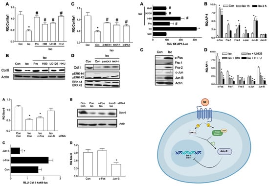
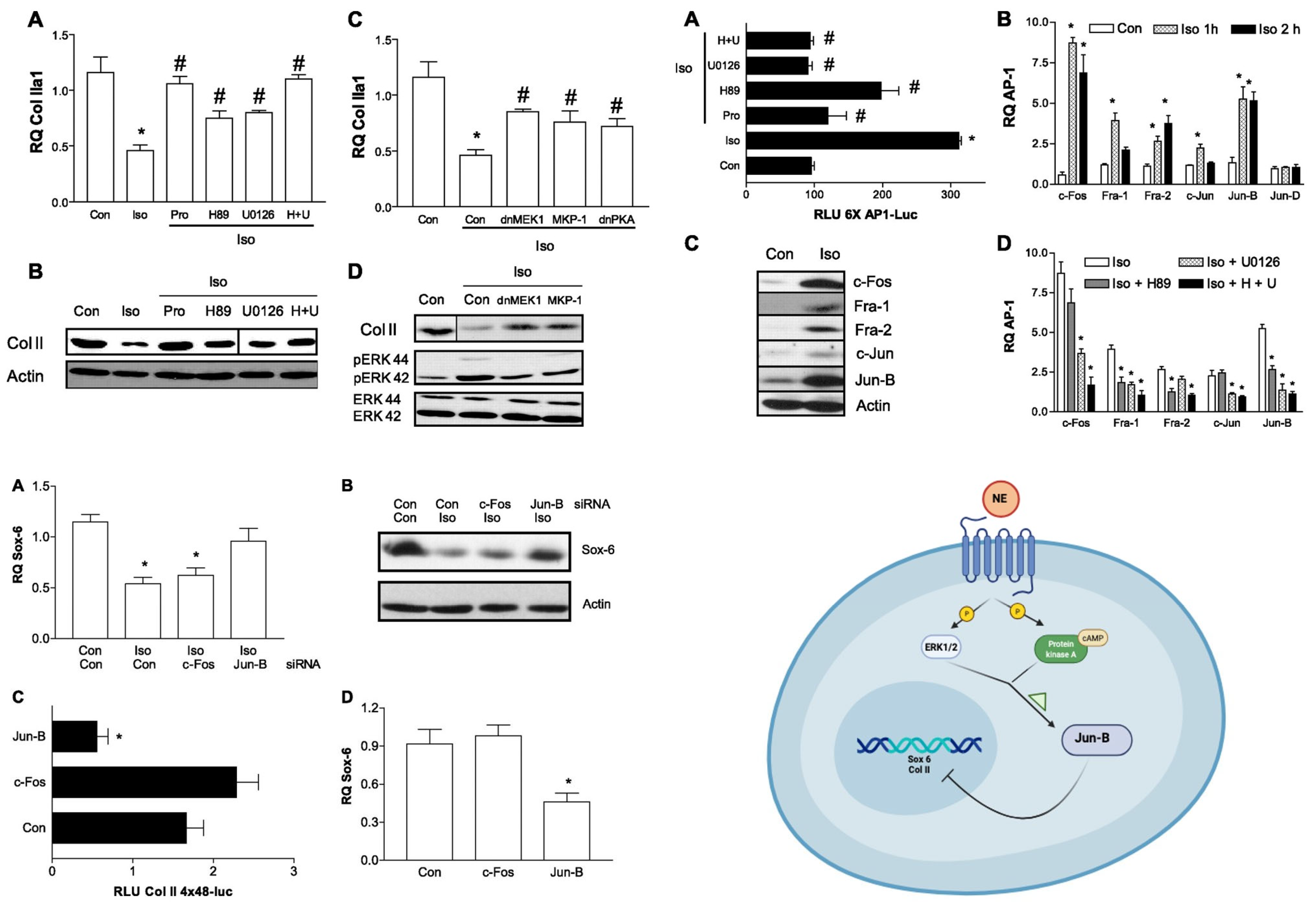
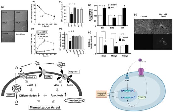
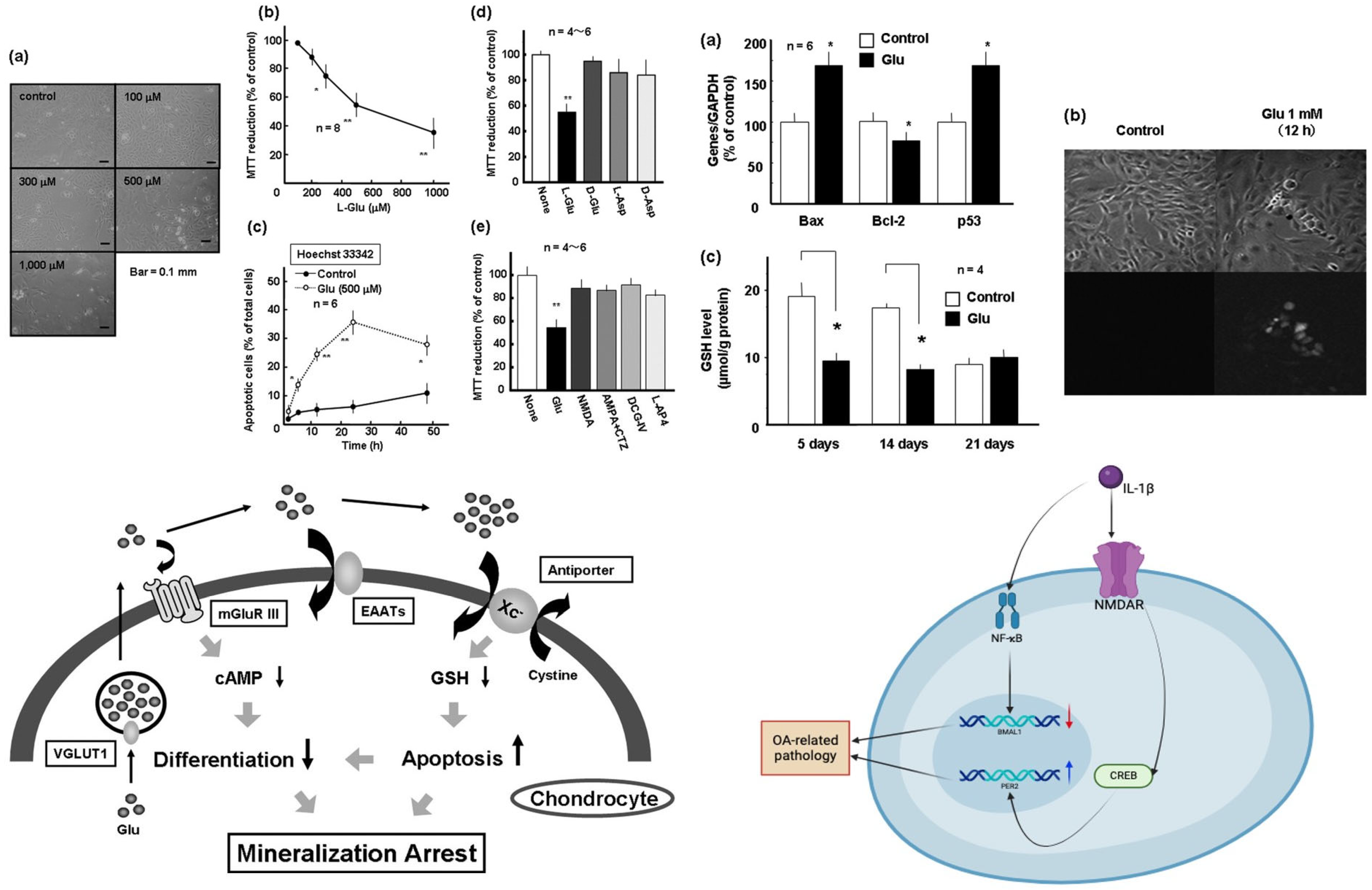
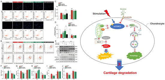
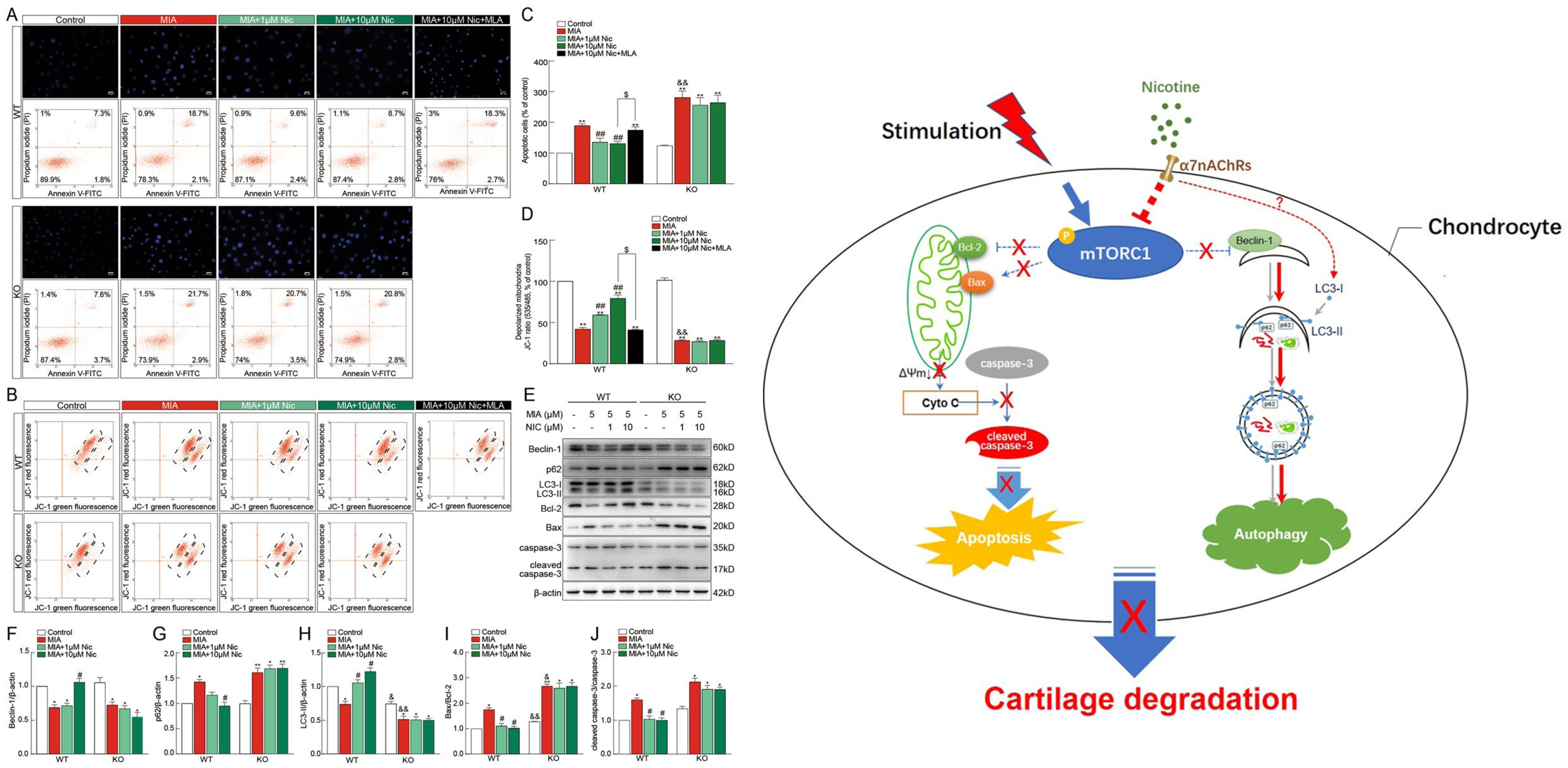
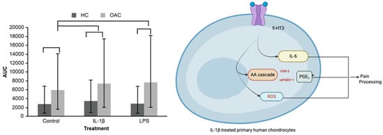
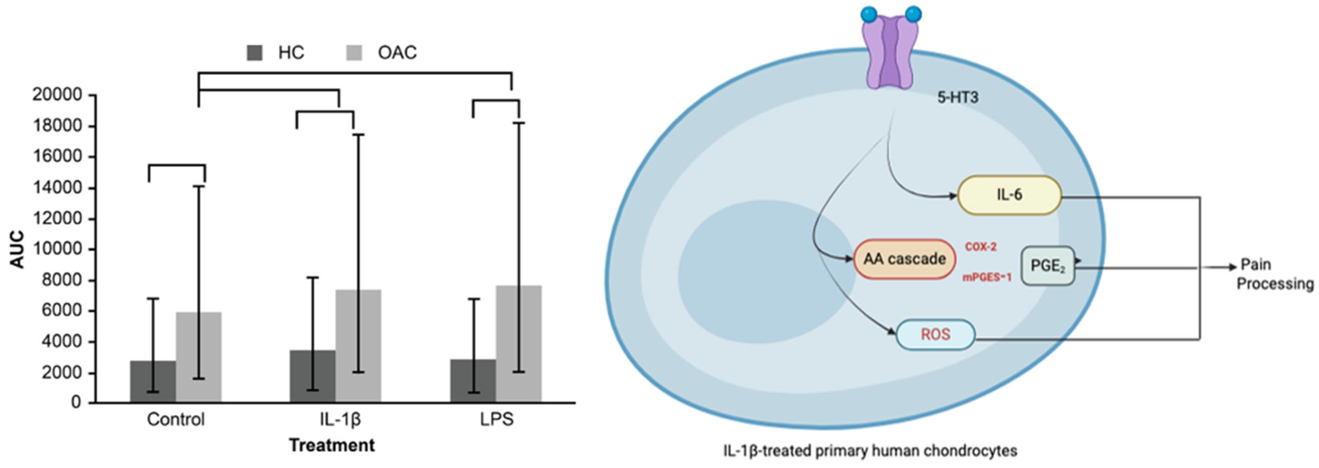

Disclaimer/Publisher’s Note: The statements, opinions and data contained in all publications are solely those of the individual author(s) and contributor(s) and not of MDPI and/or the editor(s). MDPI and/or the editor(s) disclaim responsibility for any injury to people or property resulting from any ideas, methods, instructions or products referred to in the content. |
© 2026 by the authors. Licensee MDPI, Basel, Switzerland. This article is an open access article distributed under the terms and conditions of the Creative Commons Attribution (CC BY) license.