


Abstract
Objectives: Non-pharmacological interventions for Alzheimer’s disease (AD) are typically studied as single modalities over months; long-term, multi-modal outcome data in one individual, of clear relevance to the psychiatric care of older adults with dementia over time, are rare. We describe the cognitive trajectory of one participant who, over 10 years, took part sequentially in five non-invasive AD intervention studies, and place his cognitive change in the context of decline rates reported in published AD cohorts. Methods: Retrospective, single-participant analysis integrating data from a university research laboratory (University of Manitoba, Canada; 2013–2025) across five sequential protocols: virtual-reality (VR) navigational neurorehabilitation; a randomized trial of repetitive transcranial magnetic stimulation (rTMS); cognitive training with/without 40 Hz transcranial alternating current stimulation (tACS); a crossover sham-controlled tACS study; and an ongoing personalized-frequency tACS/tDCS study. The participant was one male, healthy at 2013 enrollment, later diagnosed with mild cognitive impairment (2016) and AD (2018), who received no pharmacological AD treatment. Interventions included VR spatial navigation training; bilateral DLPFC rTMS; 40 Hz and personalized gamma/theta tACS/tDCS over left DLPFC paired with cognitive exercises; and intermittent cognitive-training-only blocks. Outcomes were assessed with the MoCA, ADAS-Cog, Neuropsychiatric Inventory Questionnaire, and VR navigation error. Long-term ADAS-Cog change was quantified with linear regression (slope, 95% CI) for the intervention and post-intervention periods, with sensitivity and segmented (interrupted time-series) analyses. Results: Over the ~5.4-year intervention period (November 2018–April 2024), which comprised 12 four-week intervention blocks separated by treatment-free intervals, ADAS-Cog fluctuated between 12.3 and 23.0 but showed no significant long-term trend (slope +0.32 points/year; 95% CI −0.25 to 0.89). Published group-level data suggest a decline of approximately 10–12 points over a comparable interval. ADAS-Cog was higher at the two latest assessments after treatment ended (22.0 and 22.3), but these values were within the range observed during intervention, and post-intervention data were too sparse to estimate a reliable trend. MoCA was 21 in 2018 and 2021, 19 in October 2022, and 13 in March 2025; when the latter decline occurred within that interval cannot be determined. Conclusions: In this single participant, a period of repeated, intermittent non-invasive brain stimulation combined with cognitive training was temporally associated with a flat long-term ADAS-Cog trajectory, slower than published group-level decline rates. Because the design is retrospective and uncontrolled, these observations cannot establish the efficacy of any intervention or attribute later changes to treatment cessation. They are hypothesis-generating, relevant to clinicians managing long-term cognitive care in dementia, and support controlled studies of long-term individualized non-invasive brain stimulation with cognitive training.
1. Introduction
Alzheimer’s Disease (AD) leads to accelerated destruction of neurons and neuronal connections in the brain, severely compromising memory and other cognitive functions. Clinical studies report an average annual cognitive decline of approximately 5.8 points per year on the Alzheimer’s Disease Assessment Scale–Cognitive Subscale (ADAS-Cog) among untreated patients [1]. Given this substantial deterioration, effective interventions to slow or stabilize cognitive decline are critically needed.
Despite extensive research and considerable investment, pharmacological treatments have had limited success in improving cognitive function. A large multisite clinical trial involving 7954 AD patients, including 2795 on placebo, showed that cholinesterase inhibitors (donepezil, rivastigmine, galantamine) offered only 2.4% significant improvement over placebo after three months, with a modest stabilization effect of about 5.3% [2]. The primary obstacle to pharmaceutical effectiveness is the blood–brain barrier, which, while protecting the brain from harmful substances, also restricts therapeutic molecules [3].
Over the last 20 years, in response to these pharmacological limitations, various non-pharmacological interventions aimed at promoting brain plasticity and preserving synaptic connections—cognitive exercises tailored for dementia patients [4], transcranial direct current stimulation (tDCS) [5], transcranial alternating current stimulation (tACS) [6], and repetitive transcranial magnetic stimulation (rTMS) [7,8,9]—have been extensively studied, generally as single-modality interventions delivered over weeks to months. Other less-studied therapies include hyperbaric oxygen therapy [10] and microwave therapy [11], typically in much smaller samples.
The mechanisms underlying these interventions’ efficacy are not fully understood, and patient response is known to be heterogeneous. This variability reflects the complex, multifactorial nature of AD, which affects patients differently depending on age [12] and concurrent conditions such as cerebrovascular disease (cvd) [13]; AD is frequently mixed with cvd, particularly at older ages [14]. This heterogeneity makes it difficult to interpret treatment outcomes clearly in small samples, and it also means that most of what is known about non-pharmacological interventions comes from short, single-modality trials rather than from how an individual responds when multiple modalities are used sequentially over many years.
One of the earliest reported symptoms of AD is deterioration of egocentric spatial orientation, sometimes preceding overt memory impairment [15]. Our group was the first to apply virtual reality (VR) navigation (VRN) [16,17] to strengthen egocentric spatial orientation as a form of neurorehabilitation in a case study with promising outcomes [18]. Because VR-based interventions are resource-intensive and depend on sustained participant adherence, long-term longitudinal follow-up is uncommon. However, a small number of highly adherent individuals—including the participant described here—have continued to take part in our sequential intervention studies over many years, offering a rare window into the cumulative, long-term trajectory of an AD patient receiving varied, individualized non-pharmacological care.
This report addresses that gap. Rather than presenting a single clinical encounter, we integrate cognitive outcome data collected across five sequential, independently IRB-approved research protocols in which one participant took part from 2013 to 2025, and describe his observed cognitive trajectory in the context of published group-level decline rates. We use this to generate hypotheses—not to demonstrate efficacy—about the potential of sustained, individualized non-invasive brain stimulation and cognitive training in patients with AD. Because neuropsychiatric symptoms and long-term care burden are central concerns in the psychiatric management of dementia, we also report caregiver-observed neuropsychiatric symptom data (NPI-Q) alongside the cognitive outcomes, where available.
2. Methods
2.1. Participant and Diagnosis
The participant was a male, healthy and without depressive symptoms, who volunteered in July 2013 (age 72) for our VRN egocentric spatial assessment study, designed to detect early AD-related change [16,17]. He was diagnosed with mild cognitive impairment (MCI) in 2016 and with AD in 2018 by his treating neuropsychiatrist, based on clinical assessment and MRI brain scan. In May 2021, his diagnosis was revised to mixed AD (AD with a cerebrovascular component) by his treating neuropsychiatrist, based on clinical examination and MRI evidence indicating moderate diffuse brain volume loss with more pronounced mesial temporal involvement along with patchy periventricular and deep cerebral hemispheric white matter signal abnormalities; this revised diagnosis is reflected in Table 1 from May 2021 onward. The authors of this paper have had no role in clinical diagnoses of this individual; they served only as study investigators. The participant’s assessments were conducted by trained research team members independent of the authors of this paper. The participant received no pharmacological treatment for AD (personal choice of the individual and his caregiver) at any point during the 10-year follow-up. Written informed consent was obtained from the participant and his caregiver for each of the five studies described below, under approvals from the University of Manitoba Biomedical and Health Research Ethics Boards (HS11296/H2009:033; HS19970/B2016:073; HS21624/B2018:029; HS25171/B2021:089). The participant and his spouse additionally provided signed consent for publication of this report.
Table 1.
Date, participant’s age, diagnosis, MoCA, ADAS-Cog and NPI-Q severity scores and the treatments received between any two shown dates. Note: a decrease in ADAS-Cog indicates improvement and vice versa. The recorded diagnosis was revised from AD to mixed AD in May 2021 (Section 2.1).
2.2. Overview of Sequential Study Protocols
Between 2016 and 2024, the participant took part in five sequential, non-invasive intervention studies in our laboratory, summarized chronologically below.
VR-based neurorehabilitation (2016). The VRN assessment, developed and validated in 319 individuals as a diagnostic aid for early AD [16,17], was adapted as an individualized training protocol for this participant and delivered over seven consecutive weeks (3 days/week, 45 min/day) with step-by-step instructor guidance, to enhance egocentric spatial navigation skills [18]. The VRN task involves navigating a three-story building, identical on all sides except the entrance, to locate a target room shown briefly before entry; the software records rooms visited, distance traveled, and completion time. The participant’s navigation error fell from over 66% at baseline to zero after training [18].
rTMS randomized controlled trial (enrolled November 2018). This was a large (n = 156), multisite, double-blind, randomized, placebo-controlled trial evaluating rTMS in mild-to-moderate AD [7,19]. Inclusion criteria were AD diagnosis confirmed by a neurologist/psychiatrist, age > 55, MoCA between 7 and 25, absence of major depression, stable AD medication (if any) for >3 months, and English/French fluency; exclusions included seizure history, other neurodegenerative or psychotic disorders, intellectual disability, and current substance abuse. Participants received 2–4 weeks of active high-frequency rTMS (20 Hz; 30 pulses/train; 25 trains/session; 10 s inter-train interval; figure-of-eight Magstim coil) sequentially to left and right DLPFC via MRI-guided neuronavigation, or 4 weeks of sham stimulation using a visually and sensorially matched sham coil; only the treatment administrator and study coordinator were unblinded. Assessments occurred at baseline, post-treatment, and every 2 months for 6 months thereafter. This participant was randomized to the 4-week active arm initially and received one additional unblinded 4-week active block approximately 7 months later.
Cognitive training with/without 40 Hz tACS (enrolled December 2019). This open-label study (2019–2021) compared cognitive training alone versus combined with 1.5 mA 40 Hz tACS (Soterix Medical tES-Model-2001; active electrode over left DLPFC, reference over contralateral supraorbital area) in individuals with AD (n = 28) [4], in two 30 min sessions/day with a 30 min break, 5 days/week for 4 weeks, followed by optional maintenance blocks. Cognitive training used the MindTriggers(Version 3) app [20], a set of seven serious games targeting associative and spatial cognitive skills, designed by the first author on neuroplasticity principles. This participant received one 4-week block in December 2019, followed by two additional 4-week cognitive-training-only blocks delivered online in 2020 during COVID-19 restrictions (no tACS was available online).
Crossover sham-controlled 40 Hz tACS study (enrolled January 2021). This double-blind, crossover study (2021–2024) compared real versus sham 40 Hz tACS with the same stimulation and cognitive-training protocol as above (n = 42) [21], with participants randomized to real-sham or sham-real order and a 2-month washout between blocks; blinding was maintained via a sham mode that delivered only a brief onset ramp. This participant completed the crossover blocks (sham, February 2022; real, May 2022) and received two additional open-label real 40 Hz tACS blocks (June 2021 and November 2022; Table 1).
Longitudinal personalized tACS/tDCS study (enrolled 2023). This ongoing double-blind study compares personalized-frequency gamma-tACS, personalized theta-tACS, tDCS (2 mA), and sham, each delivered as a 4-week block (5 days/week, two 30 min sessions/day, 20–30 min break) paired with tutored cognitive exercises, with 8–20-week washout between blocks and assessments at pre-, immediately post-, and one-month post-treatment. Personalized gamma/theta frequencies are individually determined via EEG spectral analysis during rest and MindTriggers gameplay. This participant received tDCS and 30 Hz and 49 Hz tACS blocks, each paired with cognitive exercises, from 2023 to 2024. No treatment was received after 5 April 2024, when his caregiver was no longer able to arrange daily transportation for treatment; after the protocol’s one-month follow-up assessment (May 2024), two further assessment visits (without treatment) occurred in July 2024 and March 2025.
Definition of study periods. For clarity, the following terms are used consistently throughout. The intervention period spans the participant’s first ADAS-Cog baseline (21 November 2018) to the assessment immediately after his last treatment block (9 April 2024; last session 5 April 2024), approximately 5.4 years. It was not a period of continuous stimulation: it comprised 12 four-week blocks (two rTMS; 4 real 40 Hz-tACS; one sham tACS; one tDCS; one each of 30 Hz and 49 Hz tACS; and two online cognitive-training-only blocks), about 48 weeks of treatment in total, separated by treatment-free follow-up and washout intervals of variable length. All tACS, tDCS, and sham blocks were delivered simultaneously with cognitive training, whereas rTMS was not. The post-intervention period spans 9 April 2024 to the final assessment on 5 March 2025 (approximately 11 months, no treatment). The full ADAS-Cog observation period (Figure 1) is November 2018–March 2025, approximately 6.3 years.
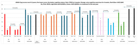
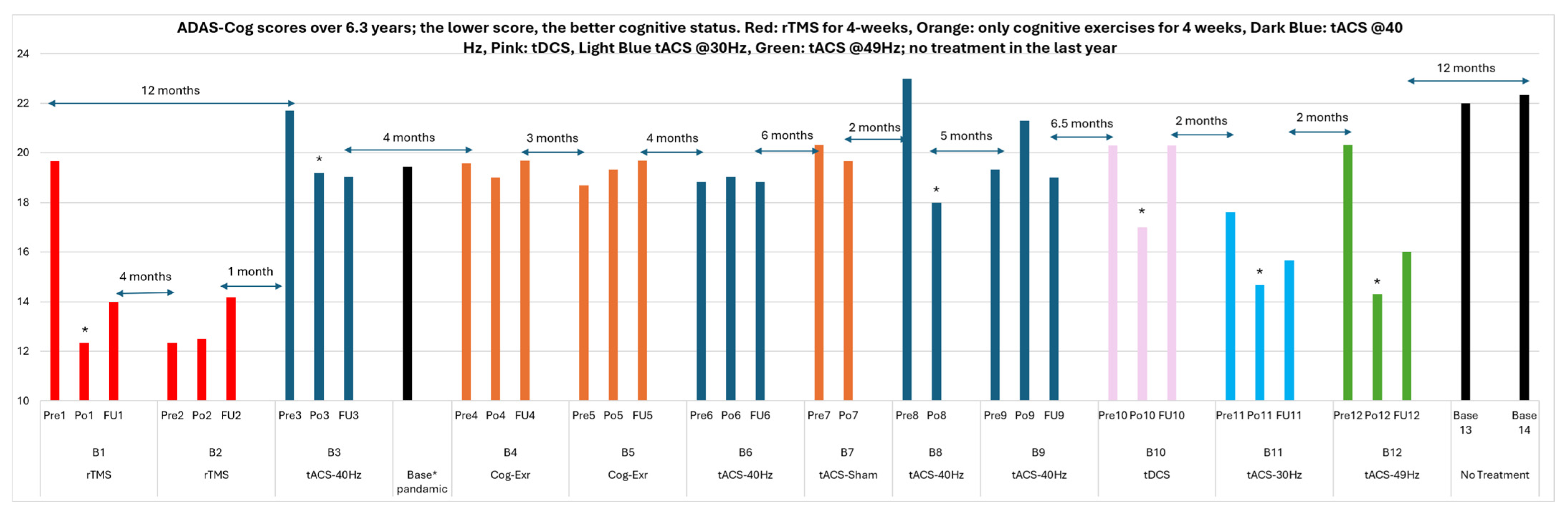
Figure 1.
ADAS-Cog scores of the patient over the ~6.3-year observation period from November 2018 to March 2025 (intervention period November 2018–April 2024; post-intervention period April 2024–March 2025). For the ADAS-Cog score, a decrease indicates improvement; a decrease of 3 points or more is considered a clinically meaningful improvement [22]. Every block of treatment was 1 month (5 days/week); thus, the time between any baseline (Pre#) and post-treatment (Po#) in Figure 1 is 1 month. All the follow-up (FU) assessments except FU1 and FU2 were at 1-month post-treatment. The FU1 and FU2 were conducted at 2 and 3 months post-treatment, respectively. The treatment blocks of B7 and B8 were part of a cross-over design study (he was enrolled in sham first and then real tACS); thus, there were no follow-up assessments for those blocks. The gap time between different blocks of treatment (shown on the graph) was variable due to patient’s schedule and our studies’ availability. All the tACS and tDCS treatments were paired simultaneously with tutored cognitive exercises using the MindTriggers app. The rTMS treatment did not have any simultaneous cognitive exercises. * denotes statically significant change from its baseline (p < 0.05).
2.3. Outcome Measures
Cognitive status was assessed using the MoCA [23], ADAS-Cog, and, for the initial VRN period, egocentric spatial navigation error. Caregiver-reported neuropsychiatric symptom severity was assessed with the NPI-Q [24] when collected (Table 1). ADAS-Cog was selected as the primary longitudinal outcome across studies because it was developed specifically to track cognitive change in mild-to-moderate AD [22], with minimal floor effects in this population [25], whereas MoCA is a brief screening tool optimized for detecting early, global cognitive change [23]. Different ADAS-Cog word lists were used across repeated administrations to reduce practice effects. Table 1 lists all available assessment dates, participant age, diagnosis, MoCA/ADAS-Cog/NPI-Q scores, and the treatment received between consecutive assessments. Figure 1 plots ADAS-Cog scores across the full ~6.3-year observation period (November 2018–March 2025), which comprises the ~5.4-year intervention period and the ~11-month post-intervention period defined in Section 2.2.
2.4. Expected Decline Comparators
To contextualize this participant’s observed trajectory, we reviewed published studies reporting ADAS-Cog change over comparable follow-up intervals (6–24 months) in individuals with mild-to-moderate AD who were untreated, on placebo, or on standard pharmacological therapy [1,26,27,28,29]. These comparators are population-based group-level estimates and are not intended as individually predictive; they are used here only to provide approximate context for interpreting a single participant’s trajectory, not as a matched control or an individualized counterfactual. The comparator cohorts differ from this participant in age, disease severity, diagnostic criteria, concomitant treatment, follow-up duration, and ADAS-Cog version (Section 4), and none followed participants for as long as the present intervention period; any extrapolation beyond their follow-up duration is therefore approximate.
2.5. Statistical Analysis
To distinguish long-term trend from short-term within-person variability, we fitted ordinary least-squares (OLS) linear regressions of ADAS-Cog score on time (years) separately for the intervention period (n = 34 assessments) and the post-intervention period (n = 4, starting with the 9 April 2024 assessment), and report slopes with 95% confidence intervals (CIs). Because repeated measures in one individual may be serially correlated (Durbin–Watson statistic for the intervention-period residuals, 1.13), we also report Newey–West heteroscedasticity- and autocorrelation-consistent (HAC; lag 2) CIs and the non-parametric Theil–Sen slope. In a sensitivity analysis, we excluded the 12 assessments obtained immediately after a treatment block, retaining only pre-treatment baseline and follow-up assessments, to limit the influence of short-term post-treatment fluctuations. Finally, a segmented (interrupted time-series) regression over the full observation period (n = 37) estimated changes in level and slope after the last treatment block. We defined the long-term ADAS-Cog trajectory as stable if the intervention-period slope did not differ significantly from zero and the upper bound of its 95% CI was below 2 points/year, a conservative threshold below most published annual decline rates in mild-to-moderate AD (approximately 2–6 points/year; Section 4). With a single participant and few post-intervention assessments, these analyses are descriptive and do not support causal inference.
2.6. Rationale for an Integrative Single-Participant Design
We elected to report this participant’s data across all five protocols, rather than describe only one study in isolation, because no single protocol captured more than 1–2 years of follow-up, while this participant’s continuous engagement across studies provided a uniquely long observation window (10 years overall, including a ~5.4-year period of repeated intervention blocks) that is otherwise unavailable in the AD non-pharmacological intervention literature. We report this as a retrospective, hypothesis-generating single-participant analysis, not as an interventional trial: assessment schedules, washout intervals, and stimulation parameters varied across protocols per each study’s own design, which limits the ability to isolate the effect of any one modality and is addressed further in Limitations.
3. Results
At his initial healthy-volunteer assessment (July 2013, age 72), the participant’s MoCA score was 28, though his VRN navigation error exceeded 66%. On reassessment in July 2015 (age 74), his MoCA score was 24 and VRN error 71%; given the practice effect expected on repeated VRN testing, this higher error strongly suggested emerging AD-related change. Following the 7-week VRN training program in 2016, his navigation error fell to zero [18]; around this time he was diagnosed with MCI by his treating neuropsychiatrist.
In November 2018, following his AD diagnosis, the participant enrolled in the rTMS trial [7,19]. The initial 4-week active rTMS block was followed by a 7.3-point ADAS-Cog improvement (19.67 to 12.33), which was maintained at the 2- and 6-month follow-ups. A second, unblinded rTMS block approximately 7 months later was not followed by further improvement—possibly reflecting a ceiling effect—and ADAS-Cog was 2.2 points higher at the 3-month follow-up. One month later (November 2019), without intervening treatment, ADAS-Cog was 21.7, close to the pre-rTMS baseline.
From 2019 onward, he participated in the cognitive-training/tACS studies described in Section 2.2, receiving cognitive training alone during COVID-19-related restrictions and combined tACS/cognitive training before and after that period, followed by the personalized tACS/tDCS protocol in 2023–2024. Full longitudinal MoCA, ADAS-Cog, and NPI-Q values, and treatments received between assessments, are shown in Table 1; ADAS-Cog scores across the full observation period are plotted in Figure 1. No treatment was received after 5 April, 2024; three follow-up assessments (without treatment) occurred, in May 2024, July 2024, and March 2025.
During the ~5.4-year intervention period (November 2018–April 2024), ADAS-Cog scores showed considerable short-term within-person variability (range 12.33–23.00; residual SD around the fitted trend 2.7 points), with values often lower immediately after a treatment block than at the preceding baseline or subsequent follow-up (Figure 1, Table 1). The long-term trend, however, was essentially flat (Table 2; Supplementary Figure S1): the ordinary least-squares slope was +0.32 points/year (95% CI −0.25 to 0.89; HAC 95% CI −0.49 to 1.13) and the Theil–Sen slope was +0.17 points/year (95% CI −0.35 to 0.63), corresponding to a fitted change of about +1.7 points (95% CI −1.3 to +4.8) over 5.4 years. Excluding the 12 assessments obtained immediately after a treatment block gave a similar slope (+0.42 points/year; 95% CI −0.24 to 1.07). By the criterion defined in Section 2.5, the long-term ADAS-Cog trajectory over the intervention period was therefore stable, despite marked short-term fluctuation. No treatment-related side effects were reported by this participant at any point, consistent with the generally mild side-effect profiles previously reported for rTMS and tACS/tDCS in our other trials [7,19].
Table 2.
Linear-trend analyses of ADAS-Cog scores. Positive slopes indicate worsening. OLS: ordinary least squares; HAC: heteroscedasticity- and autocorrelation-consistent (lag 2); CI: confidence interval. Immediate post-treatment assessments are those obtained at the end of a treatment block (n = 12). The segmented model estimates the change in level and slope after the last treatment block relative to the intervention-period trend.
After the last treatment block, ADAS-Cog was 14.3 immediately post-treatment (April 2024), 16.0 one month later (May 2024), and 22.0 and 22.33 in July 2024 and March 2025. These two latest values were about four points above the intervention-period mean (18.2) but within the range observed during intervention (e.g., 21.7 in November 2019 and 23.0 in April 2022). With only four post-intervention assessments, the post-intervention slope was imprecise (+8.0 points/year; 95% CI −10.5 to 26.4), and the segmented model did not detect a statistically significant change in level (−1.4 points; 95% CI −6.7 to 3.9) or slope (+5.6 points/year; 95% CI −3.6 to 14.8) after April 2024 (Table 2). These later values therefore cannot be distinguished from within-person variability or ongoing disease progression, and do not by themselves indicate a decline attributable to treatment cessation.
MoCA, assessed less frequently, was 21 in Nov. 2018 and May 2021, 19 in Oct. 2022, and 13 in Mar. 2025. Because MoCA was not assessed between Oct. 2022 and Mar. 2025—an interval that included about 18 months of continued intervention and 11 months without treatment—the timing of the 6-point decline within that interval cannot be determined.
4. Discussion
To our knowledge, this is among the longest documented single-participant records of sequential non-pharmacological intervention in AD. Over approximately 5.4 years of repeated, intermittent non-invasive brain stimulation and cognitive training blocks, this participant’s long-term ADAS-Cog trend was essentially flat, despite considerable short-term variability and a gradual decline in MoCA. Higher ADAS-Cog values were observed after treatment ended, but these could not be statistically distinguished from within-person variability. Because this is an uncontrolled, retrospective observation in one individual, these findings describe a temporal association and do not establish that any intervention caused the observed trajectory.
As background for interpreting this trajectory, we reviewed published ADAS-Cog change over 6–24-month intervals in untreated or placebo-treated AD populations. Reported decline rates vary considerably: one study of 90 individuals with mild AD reported increases in ADAS-Cog of 2 ± 4.8, 4.6 ± 6, and 9.8 ± 9.4 points at 6, 12, and 24 months respectively, though with a shrinking sample (37 by 24 months) and large variance [26]. A 477-patient multisite trial of the memory supplement idebenone showed 4–6 points of decline over just 6 months with no cognitive benefit from treatment [27]. A pooled analysis of 733 placebo-group patients across multiple trials found 24-week ADAS-Cog changes ranging from –0.7 ± 7 to 1.7 ± 6.4 [28]. Age-stratified data suggest younger patients with AD decline faster, but even older patients showed 4–9 points of decline over 18 months [29]. A meta-analysis of 140 pooled trials reported an average annual decline of approximately 5.8 ADAS-Cog points [1].
These comparators differ from this participant in several important respects. Most were drawn from clinical-trial cohorts with mild-to-moderate AD defined by trial-specific criteria; many participants were receiving cholinesterase inhibitors; follow-up rarely exceeded 24 months; ADAS-Cog versions and administration procedures differed; and baseline age and severity varied. In contrast, this participant was older at the start of intervention (77 years), carried a mixed AD diagnosis from 2021, received no pharmacological AD treatment, and was a highly adherent, self-selected research volunteer with strong caregiver support—characteristics that may themselves be associated with a slower course. The comparison below is therefore descriptive only.
This participant was 77 years old with a MoCA of 21 when brain stimulation began in November 2018. Based on age-related trajectories reported by Bernick et al. [29], roughly 4 points of ADAS-Cog decline would be expected over the first 18 months and a further 2.5 points over the subsequent 18 months—i.e., about 6.5 points over 3 years (≈2.2 points/year). Extending these rates linearly over the 5.4-year intervention period gives an expected change of approximately 10.5 points (assuming the slower second-period rate of ≈1.7 points/year for the remaining 2.4 years) to 12 points (assuming the 3-year average rate); the lower rates reported in some of the placebo cohorts described above would imply less. This figure is a descriptive, group-level benchmark derived by extrapolating beyond the duration of the source data, not an individualized counterfactual trajectory, and the true expected course for this participant is unknown. With that caveat, the observed fitted ADAS-Cog change over the intervention period (+1.7 points; 95% CI −1.3 to +4.8) was smaller than this benchmark.
The apparent dissociation between gradually declining MoCA and stable ADAS-Cog scores over the same period is consistent with the different design intents of the two instruments: MoCA is a brief screening tool most sensitive to early, global, and executive/attentional change [23], while ADAS-Cog was specifically developed to track longitudinal change in AD, with greater emphasis on memory, language, and praxis domains most relevant to disease progression [1]; the two measures are nonetheless known to correlate longitudinally [30]. In this participant, MoCA was unchanged between November 2018 and May 2021 and was 2 points lower by October 2022, while the ADAS-Cog trend was flat. MoCA then declined by 6 points between October 2022 and March 2025, while ADAS-Cog was 3 points higher at the final assessment than in October 2022 (19.33 to 22.33). Because MoCA was not assessed between October 2022 and March 2025, and that interval included about 18 months of continued intervention as well as the 11-month untreated period, we cannot determine when the MoCA decline occurred or attribute it to treatment cessation.
Across intervention blocks, short-term ADAS-Cog changes were descriptively larger after some rTMS and tACS blocks than after sham stimulation or cognitive-training-only blocks (three such blocks: two during COVID-19, one within the placebo-controlled crossover study—two showed slight improvement, one a slight decline). Response to 40 Hz-tACS was inconsistent across four separate blocks (two were followed by apparent improvement, two were not), and the single block at his personalized gamma frequency (49 Hz) was followed by a 6-point improvement. Because this was a single block, preceded by a relatively high pre-treatment score (20.33 versus 15.67 two months earlier) that makes regression to the mean plausible, and the improvement was not maintained at the 3-month follow-up (22.0), it does not support a frequency-specific effect. Controlled testing is needed to determine whether personalized frequencies confer any advantage. More generally, pre–post differences within single blocks were of similar magnitude to changes observed without treatment (e.g., 14.7 to 21.7 within one month in late 2019), so individual block responses should be interpreted with caution. Given AD’s well-documented non-linear progression, this inconsistency across nominally identical protocol repetitions is not unexpected and may reflect measurement variability, disease non-linearity, and possible evolving cerebrovascular contribution over the 6-year period, which we could not confirm without follow-up neuroimaging; surface stimulation modalities such as tACS/tDCS have been hypothesized to be relatively more effective than deeper-penetrating rTMS in AD with a cerebrovascular component [13].
For context (not as a matched comparison), 2-year donepezil trials have reported mean ADAS-Cog improvements over placebo of only 1.2–1.4 points at 6, 12, and 24 weeks [31]. Because this participant’s trajectory is an uncontrolled within-subject observation, it cannot be expressed as a treatment effect and is not directly comparable to such placebo-controlled estimates. These findings are broadly consistent with our recently published analysis of ADAS-Cog trajectories in 73 individuals who underwent similar non-invasive brain stimulation over 3 years [32], though that larger cohort does not by itself establish causality for this individual’s outcome.
We relied primarily on ADAS-Cog because it was the only measure collected with reasonable consistency across all five protocols; other measures (caregiver burden, activities of daily living, EEG) were collected inconsistently and are not reported here.
Limitations
This is a retrospective, single-participant, uncontrolled observational analysis; findings cannot be generalized to broader AD populations. There was no concurrent control, and the expected-decline benchmark is an extrapolated group-level estimate from cohorts that differ from this participant (Section 4), so it cannot serve as a counterfactual. All tACS, tDCS, and sham blocks were delivered simultaneously with cognitive training, and cognitive-training-only blocks were also administered; the separate contributions of stimulation and cognitive training therefore cannot be determined, and the observations relate to the combined intervention program. The interval between intervention blocks varied across studies for reasons unrelated to this participant’s individual response (study availability, washout requirements, pandemic disruption), introducing uncontrolled variability that may have influenced ADAS-Cog scores independent of treatment. Repeated ADAS-Cog administration carries a potential practice effect, which we mitigated by using different word lists across assessments. Some interventions (the second rTMS block and initial tACS applications) were unblinded, introducing possible expectancy bias; however, the unblinded rTMS block produced no additional improvement compared to the blinded block, suggesting limited contribution from expectancy effects in this case. To further minimize expectancy bias, the participant was not informed of stimulation type during blinded blocks, assessments were conducted by staff not involved in delivering stimulation, and procedures were kept identical between blinded and unblinded sessions. Because assessment intervals and washout periods differed by protocol, and because this participant was self-selected through years of continued voluntary participation, the specific contribution of any single modality cannot be isolated from this design, and the associations reported here should be read as observations in one individual rather than evidence of efficacy or of comparative efficacy between modalities. Serial correlation between repeated assessments, regression to the mean, and the small number of post-intervention assessments further limit statistical inference; the regression analyses are descriptive. Finally, the participant was highly selected—a healthy volunteer who developed AD while enrolled, with sustained adherence and caregiver support over a decade—and these characteristics may be associated with a more favorable course independent of any intervention.
5. Conclusions
In this single, highly adherent participant followed for a decade, the long-term ADAS-Cog trend was essentially flat across approximately 5.4 years of repeated, intermittent non-invasive brain stimulation combined with cognitive training, despite substantial short-term variability, whereas published group-level data would suggest a decline of approximately 10–12 points over a comparable interval. Higher ADAS-Cog scores were observed after treatment stopped, but post-intervention data were too sparse to distinguish this from within-person variability or ongoing disease progression. Short-term responses to specific modalities were inconsistent across repeated blocks, and the effects of stimulation and cognitive training cannot be separated. As a retrospective, uncontrolled, single-case observation, these findings cannot establish the efficacy of any intervention or be generalized beyond this individual. They are hypothesis-generating; together with our recently reported 3-year trajectory data in 73 individuals receiving similar interventions [32], they motivate prospective, adequately powered, controlled trials of long-term individualized non-invasive neuromodulation and cognitive training in AD, which are needed before the effectiveness of these interventions can be established. For clinicians involved in the long-term care of patients with dementia, this case illustrates that such long-term non-pharmacological programs are feasible and were well tolerated by this participant.
Supplementary Materials
The following supporting information can be downloaded at: https://www.mdpi.com/article/10.3390/bioengineering13111239/s1. Figure S1: ADAS-Cog scores over the full observation period with the ordinary least-squares trend (and 95% confidence band) fitted to the intervention period (November 2018–April 2024). Filled circles mark assessments obtained immediately after a treatment block; open circles mark pre-treatment baseline and follow-up assessments. The shaded area marks the post-intervention period (no treatment). Higher ADAS-Cog scores indicate worse cognition.
Author Contributions
Conceptualization: Z.M.; Methodology: Z.M.; Formal Analysis: M.G.-C.; Investigation: Z.M., M.A.U. and M.G.-C.; Resources: Z.M.; Data Curation: Z.M., M.A.U. and M.G.-C.; Writing—Original Draft: Z.M.; Writing—Review and Editing: Z.M., M.A.U. and M.G.-C.; Visualization: Z.M., M.G.-C.; Supervision: Z.M. and M.G.-C.; Project Administration: Z.M. and M.G.-C.; Funding Acquisition: Z.M. All authors have read and agreed to the published version of the manuscript.
Funding
This research was funded by NSERC (RPGIN-2017-05484, RPGIN-2023-04308, CRC-2021-2), the Weston Brain Institute (CT1400), and MITACS in partnership with Riverview Health Center Foundation (IT26869, IT13597).
Institutional Review Board Statement
All studies presented in this paper were conducted in accordance with the Declaration of Helsinki and approved by the University of Manitoba Biomedical and Health Research Ethics Boards (HS11296/H2009:033; HS19970/B2016:073; HS21624/B2018:029; HS25171/B2021:089).
Informed Consent Statement
Informed consent was obtained from the participant for all studies described. The participant and his spouse signed consent for publication of this report.
Data Availability Statement
Data are available from the corresponding author upon reasonable request.
Acknowledgments
We thank Brian Lithgow for his review and thoughtful recommendations on this manuscript. The authors reviewed and edited all content and take full responsibility for it.
Conflicts of Interest
The authors declare no conflicts of interest.
Abbreviations
| AD | Alzheimer’s Disease |
| ADAS-Cog | Alzheimer’s Disease Assessment Scale—Cognitive Subscale |
| DLPFC | Dorsolateral Prefrontal Cortex |
| MCI | Mild Cognitive Impairment |
| MoCA | Montreal Cognitive Assessment |
| tACS | Transcranial Alternating Current Stimulation |
| tDCS | Transcranial Direct Current Stimulation |
| rTMS | Repetitive Transcranial Magnetic Stimulation |
| NPI-Q | Neuropsychiatric Inventory Questionnaire |
| VRN | Virtual Reality Navigation |
| CI | Confidence Interval |
| HAC | Heteroscedasticity- and Autocorrelation-Consistent |
| OLS | Ordinary Least Squares |
References
- Zhang, N.; Zheng, X.; Liu, H.; Zheng, Q.; Li, L. Testing whether the progression of Alzheimer’s disease changes with the year of publication, additional design, and geographical area: A modeling analysis of literature aggregate data. Alzheimer’s Res. Ther. 2020, 12, 64. [Google Scholar] [CrossRef] [Scilit] [PubMed]
- Lanctot, K.L.; Herrmann, N.; Yau, K.K.; Khan, L.R.; Liu, B.A.; LouLou, M.M.; Einarson, T.R. Efficacy and safety of cholinesterase inhibitors in Alzheimer’s disease: A meta-analysis. Can. Med. Assoc. J. 2003, 169, 557–564. [Google Scholar]
- Pardridge, W.M. Drug transport across the blood-brain barrier. J. Cereb. Blood Flow Metab. 2012, 32, 1959–1972. [Google Scholar] [CrossRef] [Scilit] [PubMed]
- Moussavi, Z.; Kimura, K.; Kehler, L.; Francisco, C.O.; Lithgow, B. A Novel Program to Improve Cognitive Function in Individuals With Dementia Using Transcranial Alternating Current Stimulation (tACS) and Tutored Cognitive Exercises. Front. Aging 2021, 2, 632545. [Google Scholar] [CrossRef] [Scilit] [PubMed]
- Prathum, T.; Chantanachai, T.; Vimolratana, O.; Laksanaphuk, C.; Apiworajirawit, I.; Aneksan, B.; Latthirun, K.; Yang, C.T.; Klomjai, W. A systematic review and meta-analysis of the impact of transcranial direct current stimulation on cognitive function in older adults with cognitive impairments: The influence of dosage parameters. Alzheimer’s Res. Ther. 2025, 17, 37. [Google Scholar] [CrossRef] [Scilit] [PubMed]
- Benussi, A.; Cantoni, V.; Cotelli, M.S.; Cotelli, M.; Brattini, C.; Datta, A.; Thomas, C.; Santarnecchi, E.; Pascual-Leone, A.; Borroni, B. Exposure to gamma tACS in Alzheimer’s disease: A randomized, double-blind, sham-controlled, crossover, pilot study. Brain Stimul. 2021, 14, 531–540. [Google Scholar] [CrossRef] [Scilit] [PubMed]
- Moussavi, Z.; Uehara, M.; Rutherford, G.; Lithgow, B.; Millikin, C.; Wang, X.; Saha, C.; Mansouri, B.; Omelan, C.; Fellows, L.; et al. Repetitive transcranial magnetic stimulation as a treatment for Alzheimer’s disease: A randomized placebo-controlled double-blind clinical trial. Neurotherapeutics 2024, 21, e00331. [Google Scholar] [CrossRef] [Scilit] [PubMed]
- Koch, G.; Bonnì, S.; Pellicciari, M.C.; Casula, E.P.; Mancini, M.; Esposito, R.; Ponzo, V.; Picazio, S.; Di Lorenzo, F.; Serra, L.; et al. Transcranial magnetic stimulation of the precuneus enhances memory and neural activity in prodromal Alzheimer’s disease. Neuroimage 2018, 169, 302–311. [Google Scholar] [CrossRef] [Scilit] [PubMed]
- Nardone, R.; Tezzon, F.; Höller, Y.; Golaszewski, S.; Trinka, E.; Brigo, F. Transcranial Magnetic Stimulation (TMS)/repetitive TMS in mild cognitive impairment and Alzheimer’s disease. Acta Neurol. Scand. 2014, 129, 351–366. [Google Scholar] [CrossRef] [Scilit] [PubMed]
- Chen, J.; Zhang, F.; Zhao, L.; Cheng, C.; Zhong, R.; Dong, C.; Le, W. Hyperbaric oxygen ameliorates cognitive impairment in patients with Alzheimer’s disease and amnestic mild cognitive impairment. Alzheimers Dement. Transl. Res. Clin. Interv. 2020, 6, e12030. [Google Scholar] [CrossRef] [Scilit] [PubMed]
- Zhang, X.; Huang, W.J.; Chen, W.W. Microwaves and Alzheimer’s disease. Exp. Ther. Med. 2016, 12, 1969–1972. [Google Scholar] [PubMed]
- Park, D.C.; Bischof, G.N. The aging mind: Neuroplasticity in response to cognitive training. Dialogues Clin. Neurosci. 2013, 15, 109–119. [Google Scholar] [CrossRef] [Scilit]
- Lithgow, B.J.; Saha, C.; Dastgheib, Z.; Moussavi, Z. Surface Versus Penetrative rTMS Stimulation May Be More Effective for AD Patients with Cerebrovascular Disease. Neurosci. Insights 2025, 20, 26331055251328355. [Google Scholar] [CrossRef] [Scilit] [PubMed]
- Attems, J.; Jellinger, K.A. The overlap between vascular disease and Alzheimer’s disease—lessons from pathology. BMC Med. 2014, 12, 206. [Google Scholar] [CrossRef] [Scilit] [PubMed]
- Moussavi, Z.; Kimura, K.; Lithgow, B. Egocentric spatial orientation differences between Alzheimer’s disease at early stages and mild cognitive impairment: A diagnostic aid. Med. Biol. Eng. Comput. 2022, 60, 501–509. [Google Scholar] [CrossRef] [Scilit] [PubMed]
- Byagowi, A.; Mohaddes, D.; Moussavi, Z. Design and application of a novel virtual reality navigational technology (VRNchair). J. Exp. Neurosci. 2014, 8, 7–14. [Google Scholar] [CrossRef] [Scilit] [PubMed]
- Ranjbar-Pouya, O.; Byagowi, A.; Kelly, D.M.; Moussavi, Z. Introducing a New Age-and-Cognition-sensitive Measurement for Assessing Spatial Orientation using a Landmark-less Virtual Reality Navigational Task. Q. J. Exp. Psychol. 2017, 70, 1406–1419. [Google Scholar] [CrossRef] [Scilit] [PubMed]
- White, P.J.F.; Moussavi, Z. Neurocognitive Treatment for a Patient with Alzheimer’s Disease Using a Virtual Reality Navigational Environment. J. Exp. Neurosci. 2016, 10, 129–135. [Google Scholar] [CrossRef] [Scilit] [PubMed]
- Moussavi, Z.; Rutherford, G.; Lithgow, B.; Millikin, C.; Modirrousta, M.; Mansouri, B.; Wang, X.; Omelan, C.; Fellows, L.; Fitzgerald, P.; et al. Repeated Transcranial Magnetic Stimulation for Improving Cognition in Patients With Alzheimer Disease: Protocol for a Randomized, Double-Blind, Placebo-Controlled Trial. JMIR Res. Protoc. 2021, 10, e25144. [Google Scholar] [CrossRef] [Scilit] [PubMed]
- Moussavi, Z. MindTriggers. 2020. Available online: https://apps.apple.com/us/app/mind-triggers/id1508008769 (accessed on 1 January 2020).
- Uehara, M.A.; Kalia, S.; Garcia-Campuzano, M.; Jafari-Jozani, M.; Lithgow, B.; Moussavi, Z. Cognitive and Neuropsychiatric Effects of 40 Hz tACS Simultaneously with Cognitive Exercises for Dementia: A Randomized, Crossover, Double-Blind, Sham-Controlled Study. Medicina 2025, 61, 757. [Google Scholar] [CrossRef] [Scilit] [PubMed]
- Rockwood, K.; Fay, S.; Gorman, M.; Carver, D.; Graham, J.E. The clinical meaningfulness of ADAS-Cog changes in Alzheimer’s disease patients treated with donepezil in an open-label trial. BMC Neurol. 2007, 7, 26. [Google Scholar] [CrossRef] [Scilit] [PubMed]
- Nasreddine, Z.S.; Bédirian, P.N.; Charbonneau, V.; Whitehead, S.; Collin, V.; Cummings, I.; Chertkow, J.L. The Montreal Cognitive Assessment (MoCA): A Brief Screening Tool For Mild Cognitive Impairment. J. Am. Geriatr. Soc. 2005, 53, 695–699. [Google Scholar] [CrossRef] [Scilit] [PubMed]
- Kaufer, D.I.; Cummings, J.L.; Ketchel, P.; Smith, V.; MacMillan, A.; Shelley, T.; Lopez, O.L.; DeKosky, S.T. Validation of the NPI-Q, a brief clinical form of the Neuropsychiatric Inventory. J. Neuropsychiatry Clin. Neurosci. 2000, 12, 233–239. [Google Scholar] [CrossRef] [Scilit]
- Connor, D.J.; Sabbagh, M.N. Administration and scoring variance on the ADAS-Cog. J. Alzheimers Dis. 2008, 15, 461–464. [Google Scholar] [CrossRef] [Scilit]
- Podhorna, J.; Krahnke, T.; Shear, M.; Harrison, J.E.; Alzheimer’s Disease Neuroimaging Initiative. Alzheimer’s Disease Assessment Scale–Cognitive subscale variants in mild cognitive impairment and mild Alzheimer’s disease: Change over time and the effect of enrichment strategies. Alzheimer’s Res. Ther. 2016, 8, 8. [Google Scholar] [CrossRef] [Scilit] [PubMed]
- Thal, L.J.; Grundman, M.; Berg, J.; Ernstrom, K.; Margolin, R.; Pfeiffer, E.; Weiner, M.F.; Zamrini, E.; Thomas, R.G. Idebenone treatment fails to slow cognitive decline in Alzheimer’s disease. Neurology 2003, 61, 1498–1502. [Google Scholar] [CrossRef] [Scilit]
- Irizarry, M.C.; Webb, D.J.; Bains, C.; Barrett, S.J.; Lai, R.Y.; Laroche, J.P.; Hosford, D.; Maher-Edwards, G.; Weil, J.G. Predictors of placebo group decline in the Alzheimer’s disease Assessment Scale-cognitive subscale (ADAS-Cog) in 24 week clinical trials of Alzheimer’s disease. J. Alzheimers Dis. 2008, 14, 301–311. [Google Scholar] [CrossRef] [Scilit] [PubMed]
- Bernick, C.; Cummings, J.; Raman, R.; Sun, X.; Aisen, P. Age and Rate of Cognitive Decline in Alzheimer Disease: Implications for Clinical Trials. Arch. Neurol. 2012, 69, 901–905. [Google Scholar] [PubMed]
- Benoit, J.S.; Chan, W.; Piller, L.; Doody, R. Longitudinal Sensitivity of Alzheimer’s Disease Severity Staging. Am. J. Alzheimer’s Dis. Other Dement. 2020, 35, 1533317520918719. [Google Scholar] [CrossRef] [Scilit] [PubMed]
- Seltzer, B.; Zolnouni, P.; Nunez, M.; Goldman, R.; Kumar, D.; Ieni, J.; Richardson, S. Efficacy of Donepezil in Early-Stage Alzheimer Disease: A Randomized Placebo-Controlled Trial. Arch. Neurol. 2004, 61, 1852–1856. [Google Scholar] [CrossRef] [Scilit]
- Uehara, M.A.; Kalia, S.; Garcia Campuzano, M.; Moussavi, Z. ADAS-Cog Trajectories Differ from Expected Decline in Dementia Following Repeated Non-Invasive Interventions over 3 Years. Medicina 2025, 61, 1994. [Google Scholar] [CrossRef] [Scilit] [PubMed]

Disclaimer/Publisher’s Note: The statements, opinions and data contained in all publications are solely those of the individual author(s) and contributor(s) and not of MDPI and/or the editor(s). MDPI and/or the editor(s) disclaim responsibility for any injury to people or property resulting from any ideas, methods, instructions or products referred to in the content. |
© 2026 by the authors. Licensee MDPI, Basel, Switzerland. This article is an open access article distributed under the terms and conditions of the Creative Commons Attribution (CC BY) license.